The Integument Pathology - MCCCbehrensb/documents/documents/2011Skinw4.pdf · Skin Cells become...
57
The Integument Pathology
-
Upload
phungnguyet -
Category
Documents
-
view
224 -
download
0
Transcript of The Integument Pathology - MCCCbehrensb/documents/documents/2011Skinw4.pdf · Skin Cells become...

The Integument
Pathology
Normal Skin
Derma-” = Skin
3 layers
Epidermis = primarily composed of keratinocytes and
scattered melanocytes
Dermis = consists of connective tissue
Supplies nutrients and fluid that diffuse into the epidermis
from the blood vessels
Also contains nerves and glands
Subcutaneous tissue= adipose (fat)

Skin
Epidermis contains melanocytes
Pigment producing cells
Melanin- dark pigment
Dependent upon genetic and environmental factors
Protects the skin from ultraviolet light

Skin
Cells become flatter as they progress upward away from the dermis
Contain Keratin
Protein found in skin, hair and nails
Prevents loss of body fluids
Eventually die from a lack of nutrients
Constantly sloughed off from the surface a few weeks after being formed in the basal layer
Skin
Dermis
Thick layer of connective tissue that includes elastic and
collagen fibers
Strength
Support for nerves and blood vessels
Main Functions of Skin
First line of defense against micro-organisms
Prevents excessive fluid loss
Controls body temperature
Protection from UV radiation
Sensation
Synthesis and activation of vitamin D on exposure to
small amounts of UV light

Facts to know
Best barrier against infection is intact dry skin
Moist skin facilitates entry of bacteria
Our skin is not sterile - covered with bacteria
May be affected by systemic metabolic and
immune diseases

Integument Pathology

Signs and Symptoms of Disease
Pruritus
Itching
Most common symptom
Urticaria
Hives
Smooth, elevated patches
Rash
Generalized term for eruption of the skin
Blisters
Fluid-containing elevated lesion of the skin
Clear watery or bloody contents
Xeroderma
Excessive dryness
Things to Note About Skin Lesions
Location
Length of time the lesion has been present
Any changes that have occurred over time
Physical appearance
Color
Elevation
Texture
Type of exudate
Presence of pain
Pruritis (itiching)

Pigmentation of the Skin
Albinism
Lack of melanin production
Vitiligo
Small areas of
hypopigmentation
Malasma
Patches of darker skin that
may develop during
pregnancyhttp://www.knowlton.clara.net/family/Albinism/Bianca_hd.jpg
http://www.bagofnothing.com/wordpress/wp-content/uploads/2007/12/bwthomasap.jpg
http://pics.drugstore.com/catimg/132148/somme_science_C_A.jpg

Mechanical Trauma
Blunt trauma
Caused by objects (ex. club, hammer)
Contusion
“bruise”
Bleeding into the skin and soft tissue, because of
disrupted blood vessels
Laceration
A cut
Thermal Injury
Heat injuries (Burns)
Extent of injury depends on mode of exposure, duration
of exposure, the temperature, and site of injury
Clinical outcomes depends on
Depth of skin injury
Extent of body surface affected
Classified by
Superficial burn, superficial partial thickness burn, deep
partial thickness burn, full thickness burn, subdermal burn

Burns
Superficial burns
Affect epidermis
Erythema with minimal edema
Resolves in several days
No scarring
Superficial partial thickness burn
Destroys epidermis and damage to part of dermis
Blood vessels intact
Blisters form
Heals in about 10 days
Minimal scarring
Danger of infection
Burns
Deep partial thickness burn
Destroys epidermis and damage to more of dermis
Damage to nerve endings, blood vessels, hair follicles, sweat glands
Severe edema
Heals in 3-5 weeks
Danger of infection
Possible keloid scarring
Full thickness burn
Destroys epidermis, dermis, and often subcutaneous fat
Eschar tissue
No sensation
Hair follicles destroyed
Do not heal well without surgical intervention

Burns
Subdermal burns
Destroy epidermis, dermis, subcutaneous fat, muscle,
bone
Require surgical intervention… possibly amputation
Skin grafting
May be lethal

Thermal Injury – Full thickness Burn
http://www.booneville.k12.ms.us/science/A&P/burn3rd.jpg
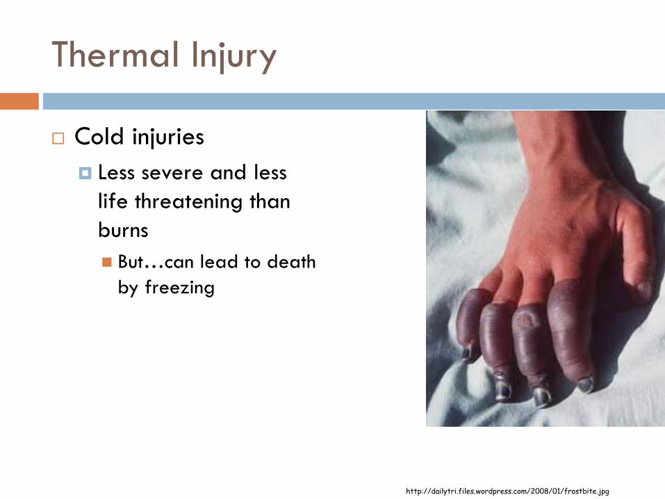
Thermal Injury
Cold injuries
Less severe and less
life threatening than
burns
But…can lead to death
by freezing
http://dailytri.files.wordpress.com/2008/01/frostbite.jpg

Electrical Injury
Contact with unprotected or inadequately isolated
electrical wires or lightening
Skin conducts electricity that creates heat in tissues
Essentially “electrical burn”
But…can pass beyond site of contact, into deeper tissues
and interfere with internal organs
Electrical conduction system of the heart
Spinal cord
Blood vessels
Kidneys

Electrical Burn
http://www.pritzkerlaw.com/images_08/ItemImages/fires-burns-explosions/electric-burn-hand.jpg
Contact Dermatitis
Exposure to an allergen or by direct chemical or
mechanical irritation of the skin
Causes acute or chronic inflammation
Symptoms
Pruritis
Erythema
Edema
May progression to vesiculation, oozing, crusting,
scaling

Contact Dermatitis
http://georgiahealthinfo.gov/cms/files/global/images/image_popup/r7_contactdermatitis.jpg

Allergic Dermatitis
Exposure to a multitude of substances, including:
Metals, cosmetics, soaps, chemicals and plants
Sensitization occurs on the first exposure and on subsequent exposures
Erythematous and pruritic area often covered with small vessicles
Manifestations of pruritic rash develop at the site a few hours after exposure
Eczema
Characterized by edema in the epidermis, and
inflammatory cell infiltrates in the dermis
Chronic erythematous lesions covered with crusts,
usually symmetrically located on the face, neck,
extensor surfaces of the UE and LE and buttocks
Characterized by pruritis (itching)

Eczema
Atopic dermatitis
Most common type of eczema
Affects primarily children
Atopic = inherited tendency toward allergic conditions
Symptoms
Red, oozing, crusting rash that becomes a dry, brownish
color with time
Pruritis
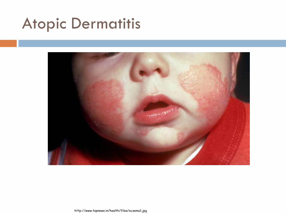
Atopic Dermatitis
http://www.topnews.in/health/files/eczema1.jpg

Urticaria
Hives
Commonly caused by ingested substances such as
shellfish, or certain fruits or drugs
Subsequent release of histamine causes the eruption of
hard, raised erythematous lesions on the skin, often
scattered all over the body
Highly pruritic

Psoriasis
Chronic inflammatory disorder of unknown origin with remissions and exacerbations
Exacerbations related to trauma and emotional stress
Cellular proliferation increases leading to thickening of the dermis and epidermis
Epidermal shedding in 3-4 days rather than several weeks
Found on face, scalp, elbows and knees Red papules that enlarge, silvery white plaque forms, bleeding points
become apparent
Now considered to be autoimmune

Psoriasis
http://www.slipperylittlesuckers.com.my/cutenews/data/upimages/450px-Psoriasis_on_back.jpg

Furuncles
Boils
An infection which begins in the hair follicle and spreads into the surrounding dermis
Initially, the lesion is firm, red, painful
Develops into large, painful mass that frequently drains large amounts of pus
Squeezing boils can result in the spread of infection by AUTOINOCULATION
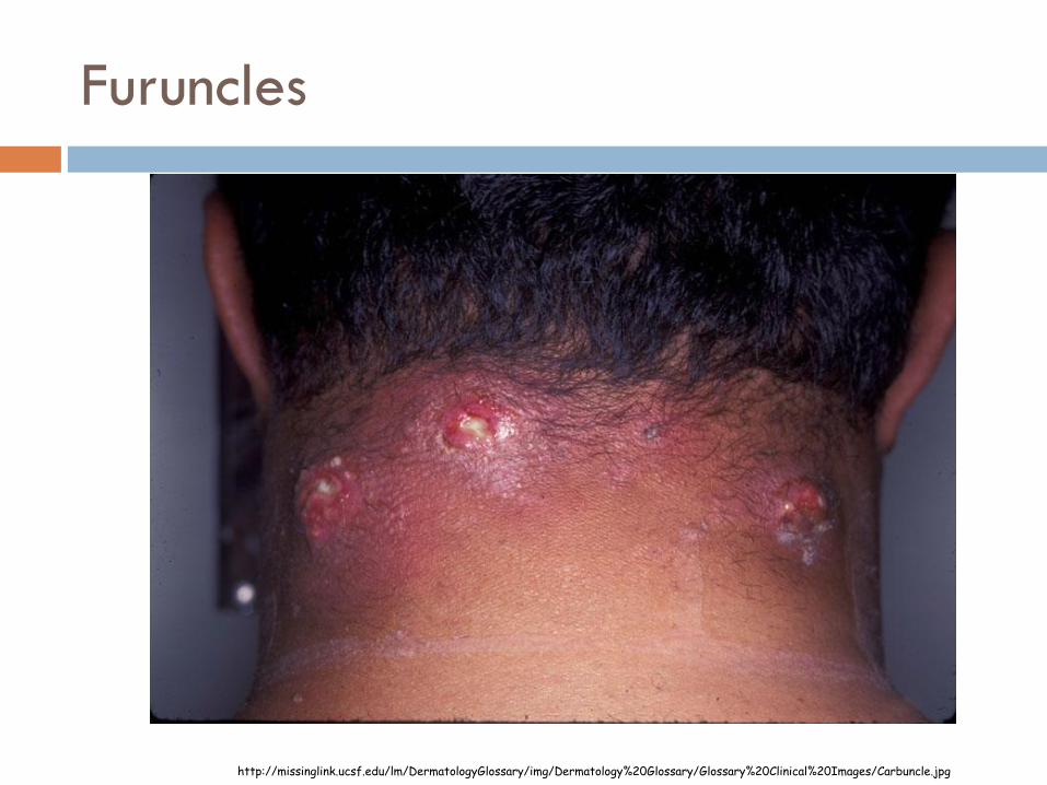
Furuncles
http://missinglink.ucsf.edu/lm/DermatologyGlossary/img/Dermatology%20Glossary/Glossary%20Clinical%20Images/Carbuncle.jpg

Impetigo
Common superficial infection
Characterized by small, pus filled pimples that rupture, to form yellowish-brown crusty masses with large red moist areas underneath
Highly contagious infection
Spread by direct contact with hands, eating utensils, towels
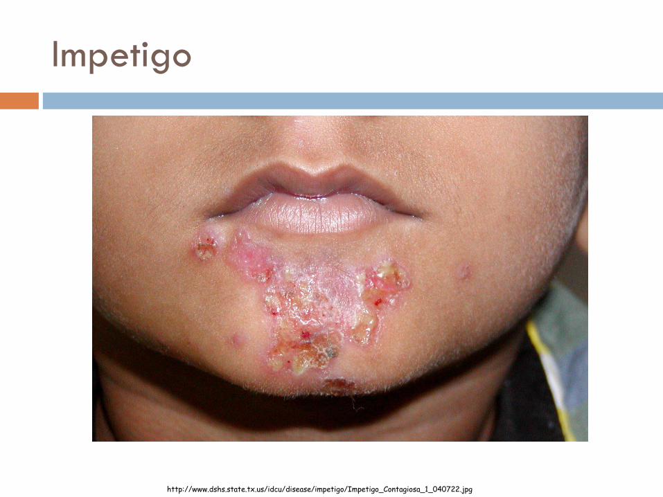
Impetigo
http://www.dshs.state.tx.us/idcu/disease/impetigo/Impetigo_Contagiosa_1_040722.jpg
Herpes Simplex (cold sores)
Viral infection
Primary infection may be asymptomatic but the virus remains in a latent stage in the sensory nerve ganglion of the trigeminal nerve
Triggers for recurrence
Stress, infection from common cold, and sun exposure
Herpes Zoster (Shingles)
Occurs years after the primary infection of chickenpox
Usually affects one cranial nerve or one dermatome on
one side of the body
Pain, paresthesia and vesicular rash develop in a line
unilaterally
Treatment: supportive treatment to relieve itching and
neuralgic pain
Medicines to slow/stop progression of symptoms
More common in older individuals, people who are
immunosuppressed
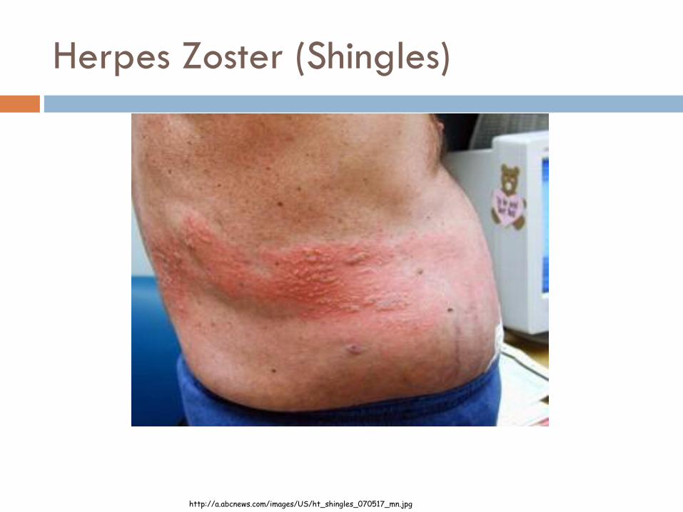
Herpes Zoster (Shingles)
http://a.abcnews.com/images/US/ht_shingles_070517_mn.jpg

Verrucae (Warts)
Benign viral infections of the skin
Caused by human papillomaviruses
Plantar warts
On the sole of the foot
Appear as firm raise papule, develop a rough surface
White or tan in color and are often multiple
May be painful if pressure is applied
Cellulitis
Rapidly spreading, inflammation of the dermis and
subcutaneous tissue
Risk increases when other disease/injury occurs
Impaired lymphatic drainage
Lymph node dissection
Saphenous vein harvesting after CABG
Diabetes, malnutrition
Edema, eczema, burns trauma

Cellulitis
Symptoms
Erythema, edema,
tender, warm
Most often seen in foot
and lower leg
http://battlegames.files.wordpress.com/2008/12/cellulitis_leg-front.jpg

Malignant Melanoma
Develops from melanocytes
Dependent upon
Genetic factors
Exposure to sunlight
Hormonal influences
Pigmentation
Presence of multiple moles

Malignant Melanoma
Often appears as a multicolored lesion with an
irregular border
Tumors are neither painful nor pruretic and therefore
may not be noticed.
Treatment: surgical excision
Chemo, radiation prn
Prognosis:
Extremely malignant and invasive
Curable if detected early
Does spread quickly and can become life threatening at
early stage
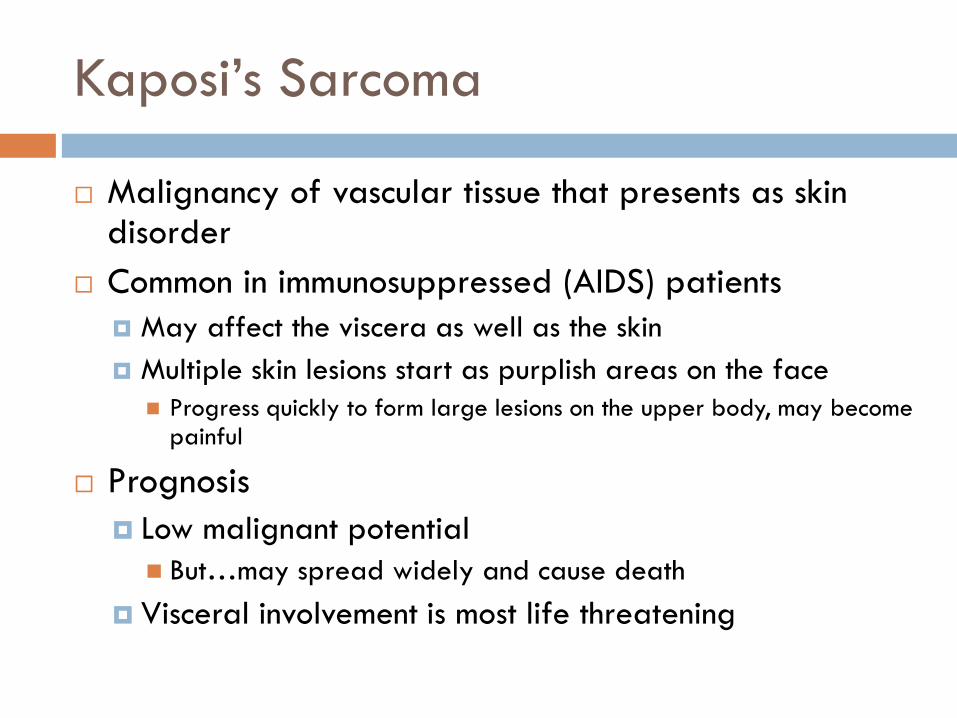
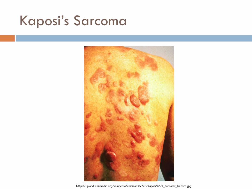
Kaposi’s Sarcoma
Malignancy of vascular tissue that presents as skin disorder
Common in immunosuppressed (AIDS) patients
May affect the viscera as well as the skin
Multiple skin lesions start as purplish areas on the face
Progress quickly to form large lesions on the upper body, may become painful
Prognosis
Low malignant potential
But…may spread widely and cause death
Visceral involvement is most life threatening
Kaposi’s Sarcoma
http://upload.wikimedia.org/wikipedia/commons/c/c2/Kaposi%27s_sarcoma_before.jpg

Skin Ulcerations
Caused by:
Arterial insufficiency
Venous insufficiency
Pressure areas

Arterial Insufficiency Ulcers
Etiology:
Reduction in oxygen perfusion of tissue
Risk factors
Heredity
Smoking
HTN
Diabetes
High cholesterol
Atherosclerosis
Usually occur in LE

Venous Stasis Ulcers
Etiology:
Impaired venous circulation
Usually have history of trauma to area
Risk factors:
Heart disease
DVT
Obesity
Defects in valves of veins
Usually occur in LE
Pressure (Decubitus) Ulcers
Etiology
Prolonged pressure on tissues over underlying bony prominences
resulting in ischemia
Risk factors
Immobility
Poor nutritional status
Most susceptible to decubitus ulcers
Sacrum
Ischial tuberosity
Lateral malleoli
Calcaneal area
Ribs
Scapulae
Greater trochanter

Scleroderma
Diffuse connective tissue disease
Fibrosis of skin, joints, blood vessels, internal organs
Etiology: unknown
Symptoms: thickening and loss of elasticity of skin with
progressive fibrosis of body organ systems

Diagnostic Tests
Diagnostic Tests
A-B-C-D of diagnosis
Asymmetry
Borders
Color
Diameter
Biopsy
Removal of a small area of skin
Used for the detection of malignant changes in tissue

Diagnostic Tests
Culture staining
Skin scrapings or other procedures
Used to detect bacterial or fungal infections
Blood Tests
To determine allergic or abnormal immune reactions
Patch or Scratch Tests
To determine allergic or abnormal immune reactions

Physical Therapy

Interventions
Stress management
To manage exacerbations
Thermal and electrical injuries
Management of contractures
Position of comfort = position of contracture
Splinting
Exercise
Functional activities
Precautions
Precautions with some treatment modalities
US gel
Mobilization creams
Topical agents that include alcohol
Which are contagious?
Impetigo
standard Precautions
Shingles
standard precautions
Isolation (contact) if you have not had chicken pox

Standard Precautions
Treat all blood and body fluids as if they were
contagious/infected
Gloves
Cover open wounds