The infection dynamics of PaV1 in the Caribbean spiny ...
151
W&M ScholarWorks W&M ScholarWorks Dissertations, Theses, and Masters Projects Theses, Dissertations, & Master Projects 2007 The infection dynamics of PaV1 in the Caribbean spiny lobster The infection dynamics of PaV1 in the Caribbean spiny lobster Panulirus argus Panulirus argus Caiwen Li College of William and Mary - Virginia Institute of Marine Science Follow this and additional works at: https://scholarworks.wm.edu/etd Part of the Environmental Sciences Commons, Fresh Water Studies Commons, Oceanography Commons, and the Virology Commons Recommended Citation Recommended Citation Li, Caiwen, "The infection dynamics of PaV1 in the Caribbean spiny lobster Panulirus argus" (2007). Dissertations, Theses, and Masters Projects. Paper 1539616740. https://dx.doi.org/doi:10.25773/v5-m7ca-dj49 This Dissertation is brought to you for free and open access by the Theses, Dissertations, & Master Projects at W&M ScholarWorks. It has been accepted for inclusion in Dissertations, Theses, and Masters Projects by an authorized administrator of W&M ScholarWorks. For more information, please contact [email protected].
Transcript of The infection dynamics of PaV1 in the Caribbean spiny ...

W&M ScholarWorks W&M ScholarWorks
Dissertations, Theses, and Masters Projects Theses, Dissertations, & Master Projects
2007
The infection dynamics of PaV1 in the Caribbean spiny lobster The infection dynamics of PaV1 in the Caribbean spiny lobster
Panulirus argus Panulirus argus
Caiwen Li College of William and Mary - Virginia Institute of Marine Science
Follow this and additional works at: https://scholarworks.wm.edu/etd
Part of the Environmental Sciences Commons, Fresh Water Studies Commons, Oceanography
Commons, and the Virology Commons
Recommended Citation Recommended Citation Li, Caiwen, "The infection dynamics of PaV1 in the Caribbean spiny lobster Panulirus argus" (2007). Dissertations, Theses, and Masters Projects. Paper 1539616740. https://dx.doi.org/doi:10.25773/v5-m7ca-dj49
This Dissertation is brought to you for free and open access by the Theses, Dissertations, & Master Projects at W&M ScholarWorks. It has been accepted for inclusion in Dissertations, Theses, and Masters Projects by an authorized administrator of W&M ScholarWorks. For more information, please contact [email protected].

THE INFECTION DYNAMICS OF PAV1 IN THE CARIBBEAN SPINY LOBSTER
PANULIRUS ARGUS
A Dissertation
Presented to
The Faculty of the School of Marine Science
The College o f William and Mary
In Partial Fulfillment
O f the Requirements for the Degree of
Doctor of Philosophy
By
Caiwen Li
2007
Reproduced with permission of the copyright owner. Further reproduction prohibited without permission.

This dissertation is submitted in partial fulfillment of
The requirements for the degree of
Approved July, 2007
Doctor of Philosophy
Caiwen Li
Stephen L. Kaattari, Ph.D.
Wolfgang K.Wbgelbein, Ph.D.
Robert E. Ratzlaff, Ph.D. Old Dominion University Norfolk, VA
ii
Reproduced with permission of the copyright owner. Further reproduction prohibited without permission.
TABLE OF CONTENTS
Page
DEDICATION.................................................................................... v
ACKNOWLEDGEMENTS........................................................................................................ vi
LIST OF TABLES...................................................................................................................... vii
LIST OF FIGURES...................................................................................................................viii
ABSTRACT.................................................................................................................................xi
GENERAL INTRODUCTION....................................................................................................2
GOALS AND OBJECTIVES........................................................................... 19
CHAPTER1
MANUSCRIPT ABSTRACT...................................................................................... 22
INTRODUCTION......................................................................................................... 23
MATERIALS AND METHODS................................................................................. 25
RESULTS....................................................................................................................... 31
DISCUSSION.................................................................................................................33
CHAPTER2
MANUSCRIPT ABSTRACT...................................................................................... 42
INTRODUCTION.........................................................................................................44
MATERIALS AND METHODS.................................................................................46
iii
Reproduced with permission of the copyright owner. Further reproduction prohibited without permission.

RESULTS....................................................................................................................... 51
DISCUSSION.................................................................................................................55
CHAPTER3
MANUSCRIPT ABSTRACT.......................................................................................6 8
INTRODUCTION......................................................................................................... 69
MATERIALS AND METHODS........................................................ 71
RESULTS....................................................................................................................... 76
DISCUSSION.................................................................................................................82
GENERAL CONCLUSIONS....................................................................................................96
LITERATURE CITED.................................................................. 101
VITA........................................................................................................................................... 116
iv
Reproduced with permission of the copyright owner. Further reproduction prohibited without permission.

DEDICATION
This work is dedicated to my parents, my wife and my son, who have supported me
through everything.
V
Reproduced with permission of the copyright owner. Further reproduction prohibited without permission.

ACKNOWLEGMENTS
I have received the guidance and assistance of many people from both inside and outside the VIMS community through the course o f this work. First of all, I would like to thank my advisor Dr. Jeffrey D. Shields, who works as my mentor throughout my research at VIMS and help me in various ways in US. I would also like to thank the other members o f my advisory committee, Dr. Stephen L. Kaattari, Wolfgang K. Vogelbein, Kimberly S. Reece, and Robert E. Ratzlaff; I am deeply grateful for their guidance, patience and assistance on my research.
I would like to thank Kersten Wheeler, who helps me processing large amounts of histological samples and taking care o f lobsters. I would also like to thank Hamish Small on the expert assistance on molecular works. I own special thanks to Pat Blake, who taught me the histological techniques and processed samples for my early studies, and to Patrice Mason for the assistance on preparing samples for electron microscopy. I would also like to thank all the members in Kim’s laboratory, they provide convenient and agreeableness circumstances for my molecular work.
All lobsters for this work were collected and provided by Dr. Mark J. Butler and his ODU crews at the Florida Keys, I own deeply thanks for their hard work especially during hurricane seasons; I am hoping that I could get some experiences from Don Behringer on collecting lobsters from the Keys. I would also like to thank all the members of Bob’s laboratory for their help on inoculation trials and generous assistance on molecular diagnosis of PaVl. I would also like to thank all other peoples involved in this project directly or indirectly.
This work was funded by NOAA Saltonstall-Kennedy Program (Grant No. NA17FD2366) and NSF Biological Oceanography Program (Grants # OCE-0136894 and OCE-0452805). I would also like to thank the Office of Graduate Studies of VIMS and the Reves Center of the College of William and Mary for supporting me to attend national and international conferences.
Finally, I would like to thank all the friends in the VIMS Chinese community who have kept me active, positive and progressive in my research, especially my wife Jie Xiao, whose love and support has carried me through.
vi
Reproduced with permission of the copyright owner. Further reproduction prohibited without permission.
LIST OF TABLES
Table
Page
CHAPTER 1
1. Sequence of the 110-bp DNA probe from PaVl and location o f the PaVl 110 F/R
primer set............................................................................................................................... 36
CHAPTER 3
1. Categorization of the severity of PaVl disease in the Caribbean spiny lobster
Panulirus argus..................................................................................................................... 8 8
2. Sequential progression of PaVl in the tissues of spiny lobsters over the time course of
experimental infection in Trial II........................................................................................ 89
3. Biochemical analysis of lobster serum compared with disease category...................... 90
vii
Reproduced with permission of the copyright owner. Further reproduction prohibited without permission.

LIST OF FIGURES
Figure
Page
GENERAL INTRODUCTION
1. (A) Caribbean spiny lobster (Panulirus argus) collected from the Florida Keys.
(B) Life cycle o f the Caribbean spiny lobster Panulirus argus.
................................................................................................................................................... 17
2. (A) Panulirus argus Virus 1 (PaVl) in the hepatopancreas o f a heavily infected spiny
lobster. (B) Internal anatomy of lobster............................................................................. 18
CHAPTER 1
1. Dot blot hybridization with the 110-bp PaV 1 probe......................................................37
2. FISH using the PaVl 110-bp probe on histological sections of spiny lobster infected
with P aV l..............................................................................................................................38
3. (A) FISH image of the hepatopancreas of a lobster infected with PaVl. (B) H&E
staining of the hepatopancreas of an infected lobster......................................................39
4. A, B. TEM of hepatopancreas from an infected lobster................................................40
Reproduced with permission of the copyright owner. Further reproduction prohibited without permission.
CHAPTER 2
1. Light microscopy o f hemocytes from the spiny lobster..................................................59
2. (A) Viability of unseparated hemocytes of P. argus cultured in L-15, ML-15, Grace’s
Insect medium, and RPMI-1640 medium. (B) Viability of unseparated hemocytes o f P.
argus cultured in ML-15 medium supplemented with 0%, 5%, 10%, and 15%
FBS......................................................................................................................................... 60
3. Light microscopy of unseparated hemocytes of the spiny lobster P. argus at 1st (A)
and 5th (B) day in culture....................................................................................................61
4. Light microscopy of P. argus hemocytes in fractions from Percoll discontinuous
gradient separation ................................................................................................ 62
5. Cultures of separated hemocytes grown in ML-15 media. H: hyalinocytes; SG:
semigranulocytes.................................................................................................................. 63
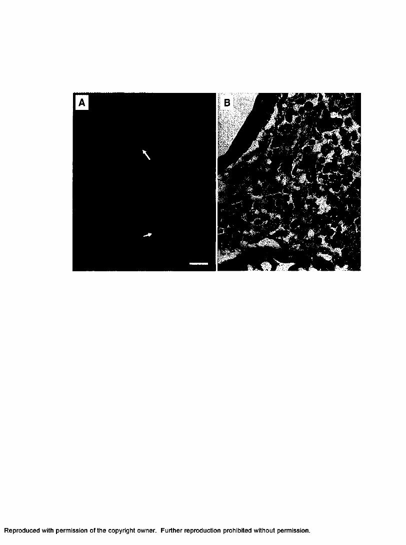
6 . Cytopathic effects of cultured hemocytes infected with PaVl inoculum...................... 64
7. Detection of PaVl in primary cultures of separated hemocytes (hyalinocytes and
semigranulocytes) of P. argus by in situ hybridization...................................................65
8 . Survival of cultured hemocytes o f P. argus inoculated with serially diluted viral
inoculum................................................................................................................................ 6 6
CHAPTER 3
1. Infection status of spiny lobsters experimentally inoculated with PaV 1........... 91
ix
Reproduced with permission of the copyright owner. Further reproduction prohibited without permission.

2. Pathological changes in the tissues of infected lobsters from Trial 1..................92
3. Total hemocyte counts (THC) of lobsters from control and inoculation group over
early time course of infection (Trial I)..............................................................................93
4. Pathological changes in the hepatopancreas of P. argus from Trial II............ 94
5. Pathological changes in the hindgut (A), gill (B), spongy connective tissues around
nerve tissues (C) and heart (D, E, F)................................................................................ 95
x
Reproduced with permission of the copyright owner. Further reproduction prohibited without permission.
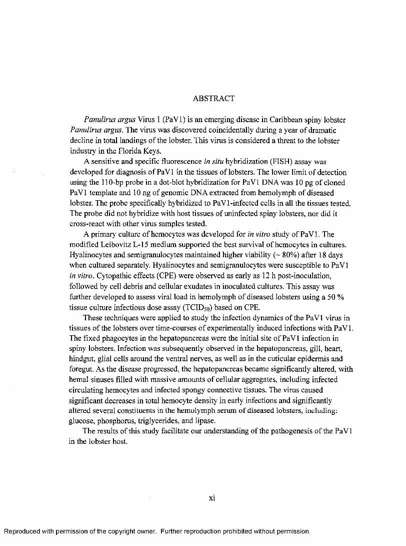
ABSTRACT
Panulirus argus Virus 1 (PaVl) is an emerging disease in Caribbean spiny lobster Panulirus argus. The virus was discovered coincidentally during a year o f dramatic decline in total landings of the lobster. This virus is considered a threat to the lobster industry in the Florida Keys.
A sensitive and specific fluorescence in situ hybridization (FISH) assay was developed for diagnosis of PaV 1 in the tissues o f lobsters. The lower limit o f detection using the 110-bp probe in a dot-blot hybridization for PaVl DNA was 10 pg of cloned PaVl template and 10 ng of genomic DNA extracted from hemolymph of diseased lobster. The probe specifically hybridized to PaVl-infected cells in all the tissues tested. The probe did not hybridize with host tissues of uninfected spiny lobsters, nor did it cross-react with other virus samples tested.
A primary culture of hemocytes was developed for in vitro study of PaVl. The modified Leibovitz L-15 medium supported the best survival of hemocytes in cultures. Hyalinocytes and semigranulocytes maintained higher viability (~ 80%) after 18 days when cultured separately. Hyalinocytes and semigranulocytes were susceptible to PaV 1 in vitro. Cytopathic effects (CPE) were observed as early as 12 h post-inoculation, followed by cell debris and cellular exudates in inoculated cultures. This assay was further developed to assess viral load in hemolymph of diseased lobsters using a 50 % tissue culture infectious dose assay (TCID50) based on CPE.
These techniques were applied to study the infection dynamics o f the PaV 1 virus in tissues of the lobsters over time-courses of experimentally induced infections with PaVl. The fixed phagocytes in the hepatopancreas were the initial site o f PaV 1 infection in spiny lobsters. Infection was subsequently observed in the hepatopancreas, gill, heart, hindgut, glial cells around the ventral nerves, as well as in the cuticular epidermis and foregut. As the disease progressed, the hepatopancreas became significantly altered, with hemal sinuses filled with massive amounts of cellular aggregates, including infected circulating hemocytes and infected spongy connective tissues. The virus caused significant decreases in total hemocyte density in early infections and significantly altered several constituents in the hemolymph serum of diseased lobsters, including: glucose, phosphorus, triglycerides, and lipase.
The results of this study facilitate our understanding of the pathogenesis of the PaV 1 in the lobster host.
xi
Reproduced with permission of the copyright owner. Further reproduction prohibited without permission.

THE INFECTION DYNAMICS OF PAV1 IN THE CARIBBEAN SPINY LOBSTER
PANULIRUS ARGUS
Reproduced with permission of the copyright owner. Further reproduction prohibited without permission.

GENERAL INTRODUCTION
Viral disease in the Caribbean spiny lobster
The Caribbean spiny lobster, Panulirus argus (Crustacea: Decapoda: Palinuridae)
(Fig. 1 A) is widely distributed throughout the Caribbean basin and along the Atlantic
Coast ranging from Brazil to Georgia, USA (Field and Butler, 1994). Spiny lobsters are
important links in marine food webs, serving as major predators of various benthic
species (e.g. snails, clams, and urchins) and important prey o f large predators (e.g. sharks
and finfish) (Lipcius and Eggleston, 2000).
The spiny lobster has a complex life cycle (Fig. IB). It has five major life history
stages, with adult, egg, phyllosoma larval stages, puerulus (or the post-larval stage) and
juvenile stages (Phillips et al., 1980; Lipcius and Eggleston, 2000). The females bear
eggs that hatch into phyllosoma in the spring and summer (Phillips and McWilliam,
1986). The planktonic larvae change into postlarvae after molting 11 times over 6-12
months. The postlarvae move onshore year around, settle in vegetation on shallow reef
flats and metamorphose into the asocial early benthic juvenile stage (Lipcius and
Eggleston, 2000). Once the juveniles reach approximately 15 mm in carapace length,
they become social and take up refuge in crevices (Marx, 1986). Approximately two
2
Reproduced with permission of the copyright owner. Further reproduction prohibited without permission.

years after settlement, lobsters mature and migrate seaward to reefs where mating and
spawning occur (Forcucci et al., 1994).
The Caribbean spiny lobster supports a valuable commercial fishery in Florida
estimated at >$30 million/year ($500 million Caribbean-wide - Cochrane and Chakalall,
2001). It also supports an important recreational fishery, which now accounts for 22% of
the total catch (Flarper, 1995; Butler, 2001). In Florida, for example, the commercial
landings o f Caribbean spiny lobster have varied between 4.3 million pounds and 7.9
million pounds per year from 1970 to 1999. In 1999, the total landings o f the spiny
lobster decreased and by 2001 they had dropped to 3.4 million pounds, the lowest
reported landings since 1982, approximately 45% less than the historical average
landings (FMRI, 2005; Muller et al., 1997).
In 1999 and 2000, a pathogenic virus Panulirus argus Virus 1 (PaVl) (Fig. 2A)
was discovered in juvenile Caribbean spiny lobsters (Shields and Behringer, 2004). PaVl
is a large, non-enveloped, icosahedral, presumptive DNA virus with nucleocapsids
ranging from 173 to 191 nm in diameter, and nucleoids approximately 118 ± 4 nm in
diameter (Shields and Behringer, 2004). The virus primarily infects the small benthic
juveniles (20 to 55 mm carapace length), with prevalence decreasing rapidly in larger
sizes. The virus was prevalent throughout the Florida Keys with overall prevalences
(among juveniles) ranging from 6% to 8%, with certain loci reaching as high as 37%.
Because PaV 1 is widespread in the Keys and highly pathogenic to juvenile spiny lobsters,
Shields and Behringer (2004) speculated that it might be responsible for the recent
declines in lobster populations since 1999.
Reproduced with permission of the copyright owner. Further reproduction prohibited without permission.

4
PaV 1 infects certain hemocytes (hyalinocytes and semi-granulocytes), and soft
connective tissues in the hepatopancreas (digestive gland), hindgut (intestine), foregut
(pyloric stomach), heart and elsewhere (Fig. 2B) (Shields and Behringer, 2004). However,
the sites o f early infection and the progression of PaV 1 infection in the spiny lobster
remain unknown. Heavily infected animals have characteristically milky hemolymph that
does not clot (Shields and Behringer, 2004). This implies that there are pathological
changes in the hemolymph such as an alteration in total hemocyte count (THC),
differential hemocyte count (DHC), and serum constituents (total carbohydrate, total
protein, hemocyanin, etc.) that are associated with viral load. Considering the
catastrophic impact of shrimp viruses (see below) and their global spread, and the
potential effect o f PaVl on the fishery for spiny lobsters, the development o f efficient
diagnostic tools and the assessment of the infection dynamics of the virus are keys to
determining if the virus is a significant threat to the industry.
Viral diseases in other crustaceans
Prior to 2004, no naturally occurring viruses had been reported from lobsters
(Shields and Behringer, 2004). However, over 30 viruses have been reported to infect
crustaceans, primarily shrimp. Since Couch (1974a, b) described the first recognized
crustacean virus, Baculovirus penaei, in Penaeus duorarum from the Gulf o f Mexico,
more than 20 viruses have been reported from penaeid shrimps (Brock and Lightner,
1990; Flegel, 1997; Lightner and Redman, 1998). At least 4 of these viruses are highly
pathogenic and have severely damaged aquaculture stocks and, in some cases, fishery
stocks o f shrimps (Brock and Lightner, 1990; Evans et al., 2000; Flegel, 1997). Viruses
Reproduced with permission of the copyright owner. Further reproduction prohibited without permission.

5
such as infectious hypodermal and hematopoietic necrosis virus (IHHNV) (Lightner et al.,
1983; Lu et al., 1989; Mari et al., 1993), Taura syndrome virus (TSV) (Lightner, 1995;
Bonami et al., 1997; Mari et al, 1998), yellow head virus (YHV) (Wongteerasupaya et al.,
1995, 1997; Lightner, 1999), and white spot syndrome virus (WSSV) (Inouye et al., 1994;
Cai et al., 1995; Wongteerasupaya et al., 1996; Lo et al., 1996; Wang et al., 1998;
Lightner, 1999), have severely impacted aquaculture production, causing catastrophic
losses to the shrimp farming industries in Asia and America (Lightner, 1999). Further,
WSSY was accidentally introduced into the Americas where it has caused widespread
damage to native and cultured shrimp stocks (Lightner, 1999). These introductions were
thought to have occurred from infected brood stock and possibly from infected frozen
carcasses.
Viruses have been reported from other crustacean species; however, they have not
received as much attention as the shrimp viruses, primarily because o f the huge economic
importance of the shrimp aquaculture industry. At least eight viruses have been reported
from blue crab, Callinectes sapidus (see Shields and Overstreet, 2004 for review), of
which four are known to be moderately or severely pathogenic. However, little is known
about the effect o f these viruses on blue crab populations other than their implication in
occasional mortalities in short-term holding pens or shedding facilities (Johnson, 1983).
Viruses have also been identified in several other crab species; five viruses have been
identified in European shore crabs, Carcinus maenas and Carcinus mediterraneus, three
from the crab, Macropipus depur at or, one from the blue king crab Paralithodes
plathypus, and one from the mud crab, Rhithropanopeus harrisi (see Brock and Lightner,
1990; Bonami and Lightner, 1991 for review). Several other viruses are also known from
Reproduced with permission of the copyright owner. Further reproduction prohibited without permission.

diverse crustaceans including iridovirus infections in the ivory barnacle, Balanus
eburneus (Leibovitz and Koulish, 1989), the pillbug, Armadillidium vulgare and the sow
bug, Porcellio dilatatus (Federici, 1980).
PaVl shares some properties with the herpes-like virus (bi-facies Virus, BFV)
from the blue crab, Callinectes sapidus (Johnson 1978, 1988), the herpes-like virus in the
mud crab, Rhithropanopeus harrisi (Payen and Bonami 1979), and the herpes-like virus
from the blue king crab, Paralithodes platypus (Sparks and Morado, 1986). All o f these
virions are roughly similar in size, are icosahedral in shape, and are presumptive DNA
viruses. PaVl even caused similar pathologic changes as the BFV. Both viruses infect
hemocytes and connective tissue cells in various tissues; cause reduction in the number of
hemocytes, and a milky appearance o f the hemolymph together with an abnormal clotting
activity (Johnson, 1978, 1988; Shields and Behringer, 2004). However, PaVl is
unenveloped, large and does not form inclusion bodies in the nuclei o f the infected cells
(Shields and Behringer, 2004), distinguishing it from the Herpesviridae (Minson et al.,
2000).
PaV 1 is also similar to the iridoviruses such as irido-like virus (M dlLV) in the
crab, Macropipus depurator (Montanie and Bonami, 1993) and the ivory barnacle,
Balanus eburneus (Leibovitz and Koulish 1989), with respect to its size, shape,
presumptive dsDNA, and lack o f envelope. However, PaVl virions are assembled
entirely within the nucleus, whereas iridoviruses are assembled within the cytoplasm of
host cells (Williams et al., 2000). As with most crustacean viruses, fundamental data (e.g.
ultrastructure, DNA sequence, and capsid structure) necessary for the classification of
PaV 1 are lacking, thus its family assignment remains to be determined.
Reproduced with permission of the copyright owner. Further reproduction prohibited without permission.

7
Application of in situ hybridization in study of viral diseases in crustaceans
In the past, diagnosis o f viral infections in crustaceans relied upon clinical signs
of disease, histological examination and electron microscopy (Bell and Lightner, 1988;
Brock and Lightner, 1990; Johnson, 1995). However, these methods are laborious or
time-consuming, or have other limitations, such as the difficulty o f diagnosing disease
from a large number o f samples using electron microscopy. Sometimes similar
pathological signs can be caused by a number o f factors such as hypoxia, crowding, a
sudden change in environmental factors, or even other pathogens, thus, reducing the
capacity of certain diagnostic techniques to obtain a specific diagnosis (Lightner, 1988).
In the past two decades, several molecular diagnostic methods have been developed as
important diagnostic tools for viral pathogens of crustaceans. One such method is in situ
hybridization (ISH), which detects specific types o f pathogens in cells and tissues by
hybridization of a labeled gene probe to a unique nucleic acid sequence (Singer et al.,
1989).
ISH was initially developed to identify the genotype o f human embryos and
genomic constitution of human pre-implantation embryos (Sart and Choo, 1998;
Andreeff and Pinkel, 1999; Darby, 2000). Because of the problems associated with
radioactive probes and the time required for autoradiography, nonradioactive in situ
hybridization is now the preferred method (Singer et al., 1989; Sart & Choo, 1998). The
improved nonradioactive technique is essentially a 2-3 day procedure that involves the
stable labeling of the nucleic acid probe, an overnight hybridization of probe onto target,
post-hybridization washes followed by fluorescent or enzyme-immunochemistry for
Reproduced with permission of the copyright owner. Further reproduction prohibited without permission.

8
hybrid molecule detection, and visualization of localized probes by fluorescent or light
microscopy (Singer et al., 1989; Sart & Choo, 1998; Andreeff and Pinkel, 1999; Darby,
2000).
Lately, ISH has been applied to the diagnosis of various viral diseases in penaeid
shrimp (Lightner and Redman, 1998). The first gene probe to be used to diagnose a viral
disease in a crustacean was developed to diagnose IHHNV (Mari et al., 1993). Small
DNA fragments (dsDNA) were selected from libraries of cloned fragments o f IHHNV
DNA, labeled with digoxigenin-11-dUTP (DIG) and applied to diagnosis o f IHHNV in
histological sections. This technique led to the development of the first commercial
diagnostic kit for crustacean viruses named ShrimProbes™ by DiagXotics (Wilton, CT,
USA). Using specific gene probes, ISH has been subsequently applied to the diagnoses of
several other crustacean viruses, such as Baculovirus penaei (BP) (Bruce et al, 1993,
1994), WSSV (Lo et al., 1997; Nunan and Lightner, 1997; Chang et al., 1998), HPV
(Pantoja and Lightner, 2001; Phromjai et a l , 2002) and gill associated virus (GAV)
(Spann et al., 2003).
ISH is a sensitive and specific method to confirm infections associated with
specific pathogens. A DIG-labeled DNA probe used in the diagnosis of Baculovirus
penaei was capable o f detecting the baculovirus well before the typical tetrahedral
occlusion bodies (TOBs) were observable through routine tissue smears or histological
examination (Bruce et al., 1993, 1994). The probes detected viral infections at 12-h post
infection, whereas H&E histology required a minimum of 24 h for detection. Similarly,
Chang et al. (1996) detected WSSV-positive cells at 16-h post infection in the stomach,
gill, cuticular epidermis and hepatopancreas the shrimp, Penaeus monodon using a
Reproduced with permission of the copyright owner. Further reproduction prohibited without permission.
specific DIG-labeled DNA probe. More importantly, various mesodermally- and
ectodermally-derived tissues, such as connective tissue, epithelia, nervous tissues and
muscle, were also shown to be infected by the virus.
ISH has also been applied to diagnose pathogens in other marine organisms. A
sensitive and specific DNA probe was developed and applied to diagnose the protozoan
oyster pathogen Ha.plosporid.ium nelsoni (commonly called MSX) in the eastern oyster,
Crassostrea virginica. The probe could detect 100 pg of cloned H. nelsoni rDNA and the
presence of H. nelsoni in 1 pg o f genomic DNA from an infected oyster (Stokes and
Burreson, 1995). Lipart and Renault (2002) developed two DNA probes that were
specific to oyster herpes virus in Pacific oysters, Crassostrea gigas; the probes were able
to detect 50 pg of viral DNA in Southern blot hybridizations. Carnegie et al. (2003)
designed a fluorescent in situ hybridization (FISH) assay to detect the parasite Bonamia
ostreae in the flat oyster Ostrea edulis. The characteristic green, ring-shaped fluorescence
was observed inside infected hemocytes, reflecting specific binding of the parasites
distinguished from the host tissue background.
In this study, I developed a FISH assay to detect PaV 1 infection in tissues o f the
Caribbean spiny lobster. With this technique, I can identify the major tissues or sites of
initial viral infection (early tissue tropisms), and the infection dynamics o f PaVl in early
stage o f the disease.
Application of cell culture in the study of viral diseases in crustaceans
Crustacean cell lines are currently not available. However, in the past two decades,
primary cell cultures have been obtained from various tissues and organs o f crustaceans,
Reproduced with permission of the copyright owner. Further reproduction prohibited without permission.

such as the lymphoid (oka) tissues (Tong and Miao, 1995; Hsu et al., 1995; Tapay et al.,
1997; Chen and Wang, 1999; Kasomchandra et al., 1999; Owens and Smith, 1999; West
et al., 1999; Itami et al., 1999; Wang et al., 2000; Lang et al., 2002; Assavalapsakul et al.,
2003), embryonic tissues (Frerichs, 1996; Toullec et al., 1996; Fan and Wang, 2002),
gonads (Luedeman and Lightner, 1992; Chen and Wang, 1999; Fraser and Hall, 1999;
Owens and Smith, 1999; Lang et al., 2002; Maeda et al., 2004), heart (Tong and Miao,
1996; Chen and Wang, 1999; Owens and Smith, 1999), nerve tissues (Nadala et al., 1993;
Owens and Smith, 1999; Gao et al., 2003), gut (Nadala et al., 1993), hepatopancreas
(Owens and Smith, 1999) and hemolymph (Sano, 1998; Walton & Smith, 1999; Itami et
al., 1999).
O f the tissues tested in primary culture, embryonic or larval tissues show promise
for developing cell lines, as they contain undifferentiated and mitotically active cells.
Therefore, the use of embryonic cells to establish long-term cultures and to obtain cell
lines has been attempted in several crustacean species including freshwater prawn,
Macrobrachium rosenbergii (Frerichs, 1996), and various penaeid shrimps (Toullec et al.,
1996; Fan and Wang, 2002). Frerichs (1996) established subcultures of cells from the
eggs o f M. rosenbergii at 7-13 days post-fertilization. Cells were observed to proliferate
in primary culture, but their passage into fresh medium resulted in the loss o f adherence,
cessation o f cell multiplication and consequent failure to establish. Toullec (1996) also
failed to obtain cultures from cells from the embryos of Penaeus vannamei and P. indicus.
The cultures stopped at the 16-cell stage, and differentiated into three cells types,
fibroblast-like cells, nerve-like cells and contractile cells. Fan and Wang (2002) tested
two growth factors, insulin-like growth factor (IGF-II) and basic fibroblast growth fact
Reproduced with permission of the copyright owner. Further reproduction prohibited without permission.

11
(bFGF) in primary cultures o f embryonic tissue o f Penaeus chinensis. They found that
passage of primary cultures resulted in rapid proliferation and good adherence in the
presence of IGF-II at 80 ng/ml and bFGF at 20 ng/ml. Cells maintained in subculture for
up to 10 passages still had good cellular morphology and division rates. However, despite
their efforts, only long-term primary cultures could be obtained.
Tissue culture is a standard tool employed in the diagnosis o f viral pathogens of
vertebrates, but it has not been fully developed for assessment of viral infection in
invertebrates (Rinkevich, 1999; Toullec, 1999; Villena, 2003). Currently, only primary
culture techniques have been developed for propagation and analysis o f crustacean
viruses. Lymphoid tissues are frequently applied in in vitro viral pathogenic studies, as
these cells are often the targets for pathogenic viruses in shrimp (Lu et al., 1995; Tapay et
al., 1997; Chen and Wang, 1999; Wang et al., 2000; Maeda et al. 2003; Assavalapsakul et
al., 2003). Lu et al. (1995) developed a quantal assay for yellow head baculovirus (YBV)
using primary cultures of shrimp lymphoid organ cells from two species of penaeid
shrimp, P. stylirostris and P. vannamei. Visible cytopathic effects (CPE) appeared at 3
days post-inoculation. A gill suspension from YHV-infected shrimp was determined to
have an infectious virus titer of 5 x 10⁵ 75 TCID50 unit /ml. Tapay et al. (1997) used
primary cultures of lymphoid organ to quantify a baculo-like virus isolated from P.
japonicus and P. stylirostris using a TCID50 assay. The virus caused cytopathic effects at
2 days post-infection; initially, the cells rounded up and finally detached from the culture
vessels as the infection progressed.
Chen and Wang (1999) developed primary cultures o f ovary, heart, lymphoid
tissue and peripheral hemocytes from three species o f penaeid shrimps P. monodon, P.
Reproduced with permission of the copyright owner. Further reproduction prohibited without permission.

12
japonicus, and P. penicillatus. They found that lymphoid tissues were better for the
formation of confluent cell monolayers. Lymphoid tissues and ovary were subcultured up
to three times and were maintained for at least 20 days. At 5-7 days after inoculation with
WSSV or YHV, significant CPE was observed in cell monolayers derived from the
lymphoid organ. Virions o f WSSV and YHV were observed in the nuclei and cytoplasm
of cultured cells when examined by electron microscopy. Similar studies have also been
carried out by Wang et al. (2000). CPE was first observed 2 days post-inoculation with
WSSV filtrate. Hypertrophy of the nucleus, margination and diminution o f nuclear
chromatin was associated with WSSV infections.
The propagation profile o f YHV was described using a primary culture of
lymphoid organ and an in vitro quantal assay (TCID50) (50% tissue culture infectious
dose) (Assavalapsakul et al., 2003). Virus was detectable by PCR as early as 24 h post
inoculation. Maximal viral yields were reached by 4 days post-infection, approximately
24 h after the onset o f the detectable cytopathic effects. The in vitro propagation of
WSSV was studied in primary ovarian cultures from the kuruma shrimp Marsupenaeus
japonicus (Maeda et al., 2004). WSSV caused marked cytopathic effect after 72 h post
inoculation, followed by a rounding and detachment of most cells; the levels of WSSV in
culture supernatant gradually increased during the period between 24 h and 120 h.
The PaVl virus infects certain hemocytes and soft connective tissues (Shields and
Behringer, 2004), and causes a characteristically milky hemolymph. This implies that
hemocytes and soft connective tissues are potential target tissues for the in vitro study of
the virus. Connective tissues have not been successfully obtained in culture. However,
primary cultures o f hemocytes have been obtained from the penaeid shrimp, Penaeus
Reproduced with permission of the copyright owner. Further reproduction prohibited without permission.
japonicus (Sano, 1998; Itami et al., 1999) and two species o f crab, Liocarcinus depurator
and Carcinus maeuas (Walton and Smith, 1999). Sano (1998) cultured hemocytes from
the kuruma shrimp P. japonicus and observed the unusual growth of pleomorphic cells in
vitro. Itami et al. (1999) cultured granular hemocytes from P. japonicus for up to 10 days.
Curiously, these hemocytes could not be infected by the penaeid rod-shaped DNA virus
(RADY). Walton and Smith (1999) separated and collected hyaline hemocytes from the
crabs, Liocarcinus depurator and Carcinus maenas. They were able to maintain these
cells for up to 14 days with more than 70% viability in an optimized media.
In this study, I developed a primary culture o f the hemocytes from the spiny
lobster, Panulirus argus for studies on the in vitro propagation of PaVl. I assessed the
utility of an in vitro quantal assay (Reed and Muench, 1938; Dee and Shuler, 1997;
LaBarre and Lowy, 2001) based on induced cytopathic effects (CPE). A quantal assay
can be used to quantify the viral load in hemolymph and other host tissues.
Hematological responses of crustaceans to viral infections
Although crustaceans do not possess an inducible immune system with a high
degree of specificity and memory as in vertebrates, crustaceans do have efficient means
to protect themselves against potential pathogens (Soderhall and Cerenius, 1992; Roch,
1999). The external cuticle is an effective barrier that impedes the entry o f infectious
agents as well as protecting internal soft tissues from mechanical damage (Sugumaran,
2000). Once pathogens gain entry into the host, subsequent innate host responses are
activated, including non-self recognition, phagocytosis, coagulation and encapsulation.
This latter response is mediated by the prophenoloxidase (proPO) system (Johansson and
Reproduced with permission of the copyright owner. Further reproduction prohibited without permission.

Soderhall, 1989; Soderhall and Cerenius, 1992; Kopacek et al., 1993; Vargas-Albores et
al., 1996; Bachere, 2000; Lee and Soderhal, 2001; Theopold et al., 2004; Jiravanichpaisal
et al., 2006). Most host innate responses against pathogens involve a combination of
cellular defenses (e.g. phagocytosis and encapsulation) as well as constitutive humoral
molecules (e.g. lectins and antimicrobial peptides) (Soderhall and Cerenius, 1992;
Johansson and Soderhall, 1989; Relf et al., 1999; Bachere, 2000; Marques and Barracco,
2000; Acharya et al., 2004; Alpuche et al., 2005; Kurtz, 2005; Jiravanichpaisal et al.,
2006).
Hemocytes of crustaceans play a key role in host innate responses against foreign
invasion (Soderhall and Cerenius, 1992; Bachere, 2000; Jiravanichpaisal et al., 2006).
Based on morphology, three types of circulating hemocytes are generally described in
crustaceans: granulocytes, semi-granulocytes and hyalinocytes (Johnson, 1980; Bauchau,
1981; Johansson et al., 2000; Jiravanichpaisal et al., 2006). Semi-granulocytes are
responsible for encapsulation and have a limited function in the storage of proPO system
(Soderhall and Cerenius, 1992; Johansson et al. 2000). This cell type is also capable of
phagocytosis in several crustacean species (Hose et al., 1990). Granulocytes are the major
storage cell in the proPO system and have a limited role in encapsulation (Hose and
Martin, 1989; Hose et al., 1990; Soderhall and Cerenius, 1992; Johnsson et al. 2000).
There is no consensus about the function o f hyalinocytes (or hyaline cells); hyalinocytes
are capable o f phagocytosis in freshwater crayfish (Soderhall and Smith, 1983). However,
hyalinocytes are not phagocytic in three other crustacean species; they play a significant
role in clotting (Hose et al., 1990).
Reproduced with permission of the copyright owner. Further reproduction prohibited without permission.

Hemocytes can be infected by several types o f viruses in crustaceans.
Granulocytes and semi-granulocytes are the targets for white spot syndrome virus
(WSSV) infection in the shrimp P. merguiensis. Infection o f these cell types was thought
to seriously damage the immune system of the shrimp due to destruction of immune
mediation from those hemocytes (Wang et al., 2002). Significant reductions in total
hemocyte counts (THC) were observed in the shrimp Penaeus indicus infected with
WSSV (Yoganandhan et al., 2003). WSSV can infect granulocytes and semigranulocytes
of the crayfish Pacifastacus leniusculus, and semigranulocytes are more susceptible to
the virus; while the proportion of granulocytes was significantly elevated from days 3 to
8 post-inoculation (Jiravanichpaisal et al., 2001). It is not clear whether the changes in
hemocyte counts upon pathogen challenge can hamper the defense system of crustaceans
(Jiravanichpaisal et al., 2006). In some cases, severe viral infection causes poor
coagulation in the hemolymph of the blue crab Callinectes sapidus (Johnson 1976), the
Caribbean spiny lobster P. argus (Shields and Behringer, 2004) and the shrimp P.
vannamei (Song et al., 2003). However, it is not known how this lack o f clotting ability
otherwise affects the defensive responses o f affected animals.
There have been a few studies on the biochemical changes that occur in the
crustacean hosts with viral infection. Viral infections cause significant changes in
biochemical and physiological parameters in hemolymph of crustaceans; and these may
be associated with the host defense responses or they may result from pathological
changes from infection. A significant decrease in hemocyanin content, and a significant
increase in glucose and total carbohydrate levels occurs in the hemolymph of shrimp P.
indicus infected with WSSV (Yoganandhan et al., 2003). Marked elevation in the
Reproduced with permission of the copyright owner. Further reproduction prohibited without permission.

activities of transaminases, alanine transaminase (ALT) and aspartate transaminase (AST)
has also been observed in hemolymph of the shrimp P. indicus infected with WSSV
(Mohankumar and Ramasamy, 2006 a). When challenged with Taura syndrome virus
(TSV), hemocyanin and clottable proteins decreased significantly in hemolymph of the
shrimp P. vanamei, and the generation of intra-hemocytic superoxide anion, O2" and
plasma proPO activity increased significantly (Song et al., 2003). A significant decrease
in the activities of the antioxidant enzymes in the hemolymph of the shrimp P. indicus
was observed with the progression of WSSV infection (Mohanhumar and Ramasamy,
2006 b). The study of these parameters in the hemolymph of crustaceans complements
histopathological studies on the health status o f crustacean host when challenged with
viral pathogens.
Reproduced with permission of the copyright owner. Further reproduction prohibited without permission.
17
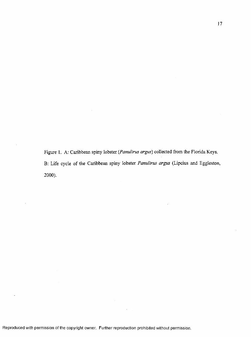
Figure 1. A: Caribbean spiny lobster (Panulirus argus) collected from the Florida Keys.
B: Life cycle o f the Caribbean spiny lobster Panulirus argus (Lipcius and Eggleston,
2000).
Reproduced with permission of the copyright owner. Further reproduction prohibited without permission.

POSTLARVAL & LARVAL TRANSPORT
SETTLEMENT
MATING
GROWTH
Reproduced with permission of the copyright owner. Further reproduction prohibited without permission.

18
Figure 2. A: Panulirus argus Virus 1 (PaVl) in the hepatopancreas of a heavily infected
spiny lobster. Scale bar =100 pm. B: Internal anatomy of lobster.
(http ://www.maine .gov/dmr/rm/aquarium/teachers_guide/lobster_intemal_anatomy .j pg)
Reproduced with permission of the copyright owner. Further reproduction prohibited without permission.

B
Internal Anatomy
Extensor Muscle
PericardiaPyloric Stomach
Heart
lutes tineFlexorMuscle
Ganglion
Gastric MillCardiac Stomach
Brain
Compound Eye
Green Gland
Mandibular Muscle
Mouth
Digestive Gland (toraali)
Ventral Nerve Cord
Reproduced with permission of the copyright owner. Further reproduction prohibited without permission.

GOALS AND OBJECTIVES
The overall goal of these studies was to determine the infection dynamics of
Panulirus argus Virus 1 (PaVl) in the Caribbean spiny lobster, Panulirus argus. This
was accomplished by examining the pathology and hematology of spiny lobsters
experimentally infected with PaV 1 using molecular, cell culture and histological
techniques. Individual portions o f this thesis were designed to address the following
objectives:
Chapter 1.
To develop a fluorescence in situ hybridization (FISH) assay for diagnosis of PaV 1
infections in tissues o f lobsters.
Hypothesis: PaVl has preferred target cells and specific tissue tropisms, which can
be determined by histology and FISH.
Chapter 2.
1. To develop a primary culture o f the hemocytes from the Caribbean spiny
lobster Panulirus argus.
19
Reproduced with permission of the copyright owner. Further reproduction prohibited without permission.

20
2. To assess the susceptibility o f the primary culture o f hemocytes from the spiny
lobster to PaV 1 infection.
3. To quantify infectious dose o f PaV 1 in hemolymph of spiny lobsters infected
with PaV 1.
Hypothesis: PaVl is infectious in vitro, causing infection and detectable cytopathic
effects (CPE) to cultured hemocytes.
Chapter 3.
To study the pathology and hematology of the Caribbean spiny lobsters over a time
course o f experimental infection by PaV 1.
Hypothesis
1) PaVl causes significant pathological changes in the spiny lobsters infected
by PaV 1 that can be determined by histology and FISH.
2) PaV 1 infects the hemocytes causing demonstrable alterations to hemocyte
subpopulations and hemolymph constituents that are related to the progression
and severity of infection.
Reproduced with permission of the copyright owner. Further reproduction prohibited without permission.

CHAPTER 1
Detection of Panulirus argus virus 1 (PaV l) in the Caribbean spiny lobster using fluorescence in situ hybridization (FISH)
Published:
Li, C., Shields, J.D., Small, H.J., Reece, K. S., Hartwig, C.L., Cooper, R.A., Ratzlaff, R.E., 2006. Diagnosis of Panulirus argus virus 1 (PaVl) in the Caribbean spiny lobster using fluorescence in situ hybridization. Diseases of Aquatic Organisms 72, 185-192.
21
Reproduced with permission of the copyright owner. Further reproduction prohibited without permission.

22
MANUSCRIPT ABSTRACT
Panulirus argus virus 1 (PaVl) is the first virus known to be pathogenic to a wild
lobster. It infects the Caribbean spiny lobster, Panulirus argus from the Florida Keys,
and has a predilection for juveniles. The monitoring of the virus in wild populations and
study of its behavior in the laboratory require the development o f reliable diagnostic tools.
A sensitive and specific fluorescence in situ hybridization (FISH) assay was developed
for detection of PaVl. The lower detection limit using a 110-bp DNA probe in a dot-blot
hybridization for PaVl DNA was 10 pg o f cloned template PaVl DNA and 10 ng of
genomic DNA extracted from hemolymph of diseased spiny lobster. The fluorescein
(FITC)-labeled probe specifically hybridized to PaVl-infected cells in hepatopancreas,
hindgut, gills, heart, foregut, and nerve tissues. FITC staining was observed around the
inner periphery o f the nuclear membrane, with lighter staining in a more dispersed pattern
within the nucleus. The probe did not hybridize with host tissues of uninfected spiny
lobsters, nor did it cross-react with the four other virus samples tested. This assay will
facilitate our understanding of the pathogenesis o f the viral disease and help in
monitoring efforts directed at determining the prevalence of PaVl in juvenile nurseries
for the lobster.
Reproduced with permission of the copyright owner. Further reproduction prohibited without permission.

23
INTRODUCTION
Panulirus argus Vims 1 (PaVl) causes disease in juvenile Caribbean spiny
lobsters from the Florida Keys (Shields & Behringer 2004). It is a large, non-enveloped,
icosahedral, presumptive DNA vims with nucleocapsids ranging from 173 to 191 nm in
diameter, and nucleoids approximately 118 ± 4 nm in diameter. The vims infects
certain hemocytes (hyalinocytes and semi-granulocytes) and spongy connective tissues
(Shields & Behringer 2004). Infected cells have a characteristic appearance with
emarginated condensed chromatin, hypertrophied nuclei and faint eosinophilic inclusions.
Because PaVl is widespread in the Florida Keys and is highly pathogenic to juvenile
spiny lobsters, Shields and Behringer (2004) speculated that it may be responsible for
recent declines in lobster populations since 1999. However, their study relied on
histology and visual diagnosis, which may fail to identify low grade, latent or subclinical
infections. Until now, there have been no molecular tools for diagnosis of PaVl
infections.
Diagnosis of viral infections in crustaceans has traditionally relied on clinical signs
of disease, histological examination and electron microscopy (Bell & Lightner 1988,
Brock & Lightner 1990, Johnson & Cassout 1995). Lately, more sensitive, specific and
Reproduced with permission of the copyright owner. Further reproduction prohibited without permission.

24
rapid molecular techniques have been developed as important diagnostic tools for viral
pathogens o f crustaceans (e.g., Lightner & Redman 1998). One such method is in situ
hybridization (ISH), which detects specific nucleic acid sequences in cells and tissues by
hybridization of a labeled gene probe to a specific target nucleic acid sequence (Singer et
al. 1989). ISH has been subsequently applied to diagnosis o f several crustacean viruses,
such as Baculoviruspenaei (BP) (Bruce et al. 1993, 1994), white spot syndrome virus
(WSSV) (Durand et al. 1996, Lo et al. 1997, Nunan & Lightner 1997, Chang et al. 1998),
hepatopancreatic parvovirus (HPV) (Pantoja & Lightner 2001, Phromjai et al. 2002) and
gill-associated virus (GAV) (Spann et al. 2003). ISH has also been applied to the
diagnosis o f several other pathogens of marine organisms (Stokes & Burreson 1995,
Chang et al. 1996, Lo et al. 1997, Pantoja & Lightner 2001, Carnegie et al. 2003, Small et
al. 2006). It is a useful tool to detect the presence of virions in infected tissues and
determine tissue tropism of viral infections in hosts. Therefore, the objective of this study
was to develop a fluorescence in situ hybridization (FISH) assay for the diagnosis of
PaV 1 infections in lobsters.
Reproduced with permission of the copyright owner. Further reproduction prohibited without permission.

25
MATERIALS AND METHODS
Sample collection
Juvenile spiny lobsters, Panulirus argus, were collected from several sites
located throughout the Florida Keys, USA. Tissue samples of hepatopancreas,
hindgut, foregut, gill, heart, skin, nerve and in some cases ovary were dissected and
fixed in 10 % neutral buffered formalin for approximately 48 h and then held in 70 %
EtOH until further processing. Fixed tissues were dehydrated, embedded in paraffin
and sectioned at 5 pm thickness on a rotary microtome. To verify the presence of the
virus, sections were stained with hematoxylin and eosin (H&E) for histology
(Humason 1979); infections were further confirmed by transmission electron
microscopy (TEM) (Shields & Behringer 2004). Sections from the same tissue blocks
were placed onto positively charged slides (Fisher Scientific) for fluorescence in situ
hybridization (see below).
Fluorescent DNA probe synthesis
A 110-bp DNA probe was derived from a 177-bp DNA fragment (GenBank
accession No. DQ465025) that putatively codes for a portion of the DNA polymerase
from PaVl (Robert Ratzlaff, unpublished data). The 110-bp DNA probe (PaVl 110)
containing fluorescein-12-dUTP (fluorescein isothiocyanate, FITC) was synthesized
Reproduced with permission of the copyright owner. Further reproduction prohibited without permission.

26
using a PCR Fluorescein Labeling Mix (Roche Applied Science). A plasmid vector
(pCR 4-TOPO) containing the 177-bp DNA fragment was used as a template for probe
synthesis. A specific primer set (PaVl 110F/R, generated with Invitrogen
OligoPerfect™ Designer) was used to amplify and label a 110-bp fragment from the
plasmid DNA containing the 177-bp insert. (See Table 1 for sequence of the 110-bp
DNA probe and location of the PaV 1110 F/R primer set.) The polymerase chain
reaction (PCR)-labeling reaction was performed according to the manufacturer’s
instructions (Roche Applied Science). Briefly, each PCR reaction contained the
following: PCR buffer at a 1 x concentration, 4 mM MgCfe, 200 pM PCR Fluorescein
Labeling Mix dNTP, 0.5 pM of each primer, 1 unit Taq DNA polymerase, 100 pg
plasmid template, and distilled water (dFLO) to a final volume of 100 pi.
Thermocycling conditions were as follows: an initial denaturation at 94°C for 4 min; 35
cycles of denaturation at 94°C for 30 seconds, annealing at 57.2°C for 30 seconds, and
extension at 72°C for 90 seconds; followed by final extension at 72°C for 5 min. PCR
products were purified using a QIAquick spin purification kit (Qiagen), and were
visualized by agarose gel electrophoresis (2 %) with ethidium bromide staining. The
amount of DNA was quantified using a Hoefer DyNA Quant200 Fluorometer.
To ensure that the PaVl 110F/R primer set was amplifying the correct
domain of the viral 177-bp insert for synthesis of the 110-bp DNA probe, the PCR
(above) was repeated with the Fluorescein labeling mix replaced with a standard
dNTP mix (125 pM). The 110-bp product was visualized by agarose gel
electrophoresis and excised from the gel using a sterile scalpel and purified using a
Reproduced with permission of the copyright owner. Further reproduction prohibited without permission.

QIA-quick gel extraction kit (Qiagen). The amplicon was cloned using a TOPO TA
Cloning Kit for Sequencing (Invitrogen) following the manufacturers protocols. Six
clones were sequenced bidirectionally and analyzed using an ABI 3130 Prism Genetic
Analyzer (Applied Biosystems) as in Dungan & Reece (2006). Sequences were
compared to the original 177-bp fragment using the Clustal-W algorithm in the
MacVector DNA sequence analysis package (Accelrys).
DNA probe sensitivity
The sensitivity of the probe was determined by dot-blot hybridization against a
10-fold serial dilution from 10 ng to 1 pg of plasmid DNA containing the PaVl
177-bp fragment. Additional controls consisted of 10 ng genomic DNA extracted
from hemolymph of a healthy spiny lobster and 10 ng genomic DNA extracted from
the hemolymph of a spiny lobster heavily infected with PaV 1 (Infection was
determined histologically). Genomic DNA was extracted using the DNeasy® Tissue
kit according to the manufacturer’s instructions (Animal blood protocol - Qiagen).
Briefly, DNA solutions were denatured at 100°C for 10 min and transferred to ice for
5 min. The solution of denatured DNA was loaded onto a positively charged
membrane (BrightStar®-Plus, Ambion) using a Bio-Rad Microfiltration Apparatus
(Bio-Rad laboratories), and rinsed with 100 pi of 0.4 M NaOH. DNA was
immobilized by UV crosslinking with a Stratalinker 1800 UV crosslinker (Stratagene).
The membrane was placed in a sealed plastic bag containing pre-warmed (42°C)
pre-hybridization solution (Sigma-Aldrich) and incubated for 30 min with gentle
Reproduced with permission of the copyright owner. Further reproduction prohibited without permission.

28
agitation at room temperature (RT, 25°C). FITC-labeled probe was denatured as
described above, diluted in hybridization buffer (Sigma-Aldrich) to a final
concentration of 10 ng ml'1, and incubated with membranes in a sealed plastic bag
overnight at 42°C with gentle agitation. A series of stringency washes followed: 2x
SSC (0.3 M NaCl, 30 mM Sodium Citrate; pH 7.0), 10 min, RT; lx SSC, 10 min, RT
and 0.1 x SSC, 10 min, RT. The membrane was blocked for 30 min at RT with
blocking buffer (Sigma-Aldrich), then incubated in anti-fluorescein alkaline
phosphatase antibody (1:1000 diluted in blocking buffer) (Sigma-Aldrich) for 2 h
with gentle agitation at RT. This was followed by removal of unbound antibody with
two 15 min washes with TN buffer (0.1M Tris, 0.15 M NaCl, pH 7.5) and a 5 min
wash with TNM buffer (0.1 M Tris, 0.1 M NaCl, 0.05 M MgCl2, pH 9.5). The
membrane was then incubated with BCIP/NBT liquid substrate solution
(Sigma-Aldrich) for 2 h in a sealed plastic bag covered with foil. Color development
was stopped with a 5-min TE buffer wash (10 mM Tris, ImM EDTA, pH 7.5) and
dH20 for 5 min. The wet membrane was scanned with a Hewlett Packard Scanjet
3570c scanner for documentation.
Fluorescence in situ hybridization (FISH)
The FISH methodology was derived from published ISH protocols (Singer et al.
1989, Stokes & Burreson 1995, Darby 2000, Beatty et al. 2002). Sections were
deparaffmized in xylene (5 min, 2x), rehydrated through a descending ethanol series:
100 % (5 min, 2x), 95 % (1 min, 2x), 70 % (1 min, 2x), and equilibrated in
Reproduced with permission of the copyright owner. Further reproduction prohibited without permission.

29
phosphate-buffered saline (PBS; once for 5 min, once for 3 min). The sections were
then digested with Proteinase K (100 pg ml'1 in PBS) for 15 min at 37°C, followed by a
5-min wash in 0.2 % glycine PBS solution to stop proteolysis, and incubated in 2x SSC
for 10 min at room temperature. Slides were incubated in pre-hybridization buffer (4*
SSC, 50 % formamide, 0.5 mg ml'1 Salmon sperm DNA, and 1 % fetal bovine serum) at
42°C for 45 min. After incubation, excess pre-hybridization buffer was carefully
drained off, the area with tissue was outlined with a Frame-seal incubation chamber
(MJ Research), aliquots of 50 pi of hybridization solution (50 % deionized formamide,
4* SSC, 0.5 % SDS, and 25 pg ml'1 DNA fluorescein probe) were added, and the slides
sealed with a plastic cover slip. The slides were then placed in a thermal cycler for 3
min at 72°C and cooled on ice for 2 min. Slides were incubated in a humid chamber
saturated with prehybridization buffer overnight at 42°C. The slides were then washed
in 2x SSC (5 min), 1* SSC (5 min), PBS (10 min), air dried, mounted with anti-fading
mounting medium (90 % glycerol, 0.1 m Tris-HCl, pH 8.0 and 2.3 % DABCO) and
covered with glass coverslips. Clear fingernail polish was applied to the edges of the
cover slips to prevent evaporation. Slides were examined using an Olympus BX51
microscope equipped with a FITC-Texas Red filter (U-MF2, Olympus), and images
were captured with a Nikon DXM 1200 digital camera for comparison between
matching sections stained with H&E.
To test the specificity of the probe, tissues with other viral infections were
assessed. These included tissues with a herpes-like virus (HLV) from a blue king crab
Paralithodes platypus obtained from Frank Morado (NOAA) (see Sparks & Morado
Reproduced with permission of the copyright owner. Further reproduction prohibited without permission.

30
1986); lymphocystis disease virus (LDV) from a striped bass Morone saxatilis,
obtained from Wolfgang Vogelbein (VIMS) (see Smail & Munro 2001 for review);
Ostreid Herpesvirus 1 (OsHV-1) from an infected Pacific oyster Crassostrea gigas,
obtained from Carolyn Friedman (Univ. Washington) (see Le Deuff & Renault 1999,
Lipart & Renault 2002) and Intranuclear bacilliform virus (IBV) from an infected
brown shrimp Crangon crangon from Grant Stentiford (CEFAS, UK) (Stentiford et al.
2004).
TEM
The hepatopancreas from an infected lobster was fixed for transmission
electron microscopy (TEM) using 3 % glutaraldehyde (containing 0.2 M sodium
cacodylate, 30 mg ml'1 NaCl, 20 ug ml'1 CaCl2, pH 7.0) (Factor & Naar 1985). After
fixation, tissues were washed 3 times in buffer and postfixed in 1 % osmium tetroxide
in buffer. Samples were processed through an ethanol dehydration, en bloc stained with
uranyl acetate, dehydrated further with propylene oxide, infiltrated through several
changes of propylene oxide in various ratios with Spurr’s resin, and finally embedded
in Spurr’s resin. Sections were cut on a Reichert-Jung ultramicrotome E, processed
through a routine lead citrate stain, and observed with a Zeiss CEM-902 TEM.
Reproduced with permission of the copyright owner. Further reproduction prohibited without permission.

31
RESULTS
DNA probe synthesis and sensitivity
The primer pair PaVl 110 F/R specifically amplified a single 110-bp
fragment (Table 1) when using the plasmid containing the 177-bp DNA fragment as
a template in the PCR labeling reaction. The 110-bp DNA probe sequence from 6
clones sequenced was 100 % identical to the corresponding region in the original
177-bp plasmid.
In dot-blot hybridizations (Fig. 1), the probe had a minimum sensitivity of 10
pg of the cloned plasmid DNA with the 177-bp insert. Additionally, the probe
detected the presence of viral DNA from 10 ng of genomic DNA extracted from
hemolymph of a PaVl infected spiny lobster. A negative result was obtained when
the probe was tested with genomic DNA extracted from the hemolymph of a healthy
spiny lobster (Fig. 1, g).
Fluorescence in situ hybridization
The FITC-labeled probe hybridized to PaVl-infected cells in all tissues
tested. The probe bound to those infected hemocytes and spongy connective tissue
cells in or around the hepatopancreas, hindgut, foregut, gill, heart, skin, nerve and
even ovary tissues (Fig. 2 A, B, C). The distribution of FITC-stained structures
Reproduced with permission of the copyright owner. Further reproduction prohibited without permission.

32
inside infected cells matched the pathological changes caused by the viral infection
when diagnosed by H & E staining (Fig. 3) and TEM (Fig. 4). Most FITC-stained foci
were located around the inner periphery of the hypertrophied nuclear membrane, with
a few dispersed throughout the inside of the nucleus.
The probe did not bind to the tissues of healthy spiny lobsters. No
FITC-stained particles were present in tissues from healthy spiny lobsters. Only a
weak brown/red background was observed (Fig. 2 D). The probe did not hybridize
with HLV, OsHV-1, LDV, nor with IBV.
Reproduced with permission of the copyright owner. Further reproduction prohibited without permission.

33
DISCUSSION
We have developed a FISH assay for the detection of the recently identified
PaVl virus from the Caribbean spiny lobster, Panulirus argus, using a sensitive and
specific DNA probe. The probe detected 10 pg of plasmid DNA containing a 177-bp
DNA fragment from PaV 1 in a dot-blot hybridization. It could detect the presence of
viral DNA in 10 ng genomic DNA extracted from the hemolymph of a diseased
spiny lobster. The probe hybridized to PaVl-infected cells in all tissues tested by
FISH. The specific binding of the 110-bp probe for PaVl was visualized as ring-like
green staining of infected cells, whereas only a brown or red background was
observed in healthy tissues from uninfected spiny lobsters. This unique distribution
pattern of the green staining fits the pattern observed in infected tissue with TEM.
Most virions were diffusely distributed within the inner periphery of the
hypertrophied nuclei of infected cells, and the probe specifically bound to
complementary sequence of viral DNA in infected cells during in situ hybridization.
Traditional diagnostic tools such as histology or electron microscopy can not
differentiate among certain etiologies. Occasionally, similar pathological signs can
be caused by several factors including hypoxia, crowding, a sudden change in
environmental factors, or even other pathogens, thus, reducing the capacity of
certain diagnostic techniques to obtain a specific diagnosis (Lightner 1988). When
Reproduced with permission of the copyright owner. Further reproduction prohibited without permission.

34
examined by TEM, PaV 1 had properties similar to the Herpesviridae and the
Iridoviridae (Shields & Behringer 2004). It even induces pathological changes similar
to those caused by the herpes-like virus (Bi-Facies virus, BFV) from the blue crab,
Callinectes sapidus (Johnson 1976, 1988; Shields & Behringer 2004). However, the
110-bp probe did not bind with the other viruses: OsHV (Le Deuff & Renault 1999,
Lipart & Renault 2002), HLV (Sparks & Morado 1986), LDV (Smail & Munro 2001)
and a virus outside these families, the bacilliform virus (Stentiford et al. 2004).
Therefore, the specificity of the probe will facilitate its use in properly diagnosing
PaV 1 infections in lobsters.
In situ hybridization (ISH) has been applied to diagnose viral diseases in
several crustaceans (Lightner & Redman 1998). A digoxigenin (DIG)-labeled DNA
probe used in the diagnosis of Baculoviruspenaei detected the baculovirus well
before the typical tetrahedral occlusion bodies (TOBs) were observable in routine
tissue smears or histological examinations (Bruce et al. 1993, 1994). The probe
detected viral infections at 12-h post-infection, whereas H&E histology required a
minimum of 24 h for detection. Similarly, Chang et al. (1996) detected
WSSV-positive cells at 16-h post infection in the stomach, gill, cuticular epidermis
and hepatopancreas of the shrimp Penaeus monodon using a specific DIG-labeled
DNA probe. While we have not examined the infection dynamics of PaVl over such
short time periods, the specific binding of the 110-bp probe, coupled with the
excitation sensitivity of FITC to fluorescence, should facilitate examining viral
tropism over periods of a few days post inoculation.
Reproduced with permission of the copyright owner. Further reproduction prohibited without permission.

Using H&E and FISH, we found infected cells in the ovaries of an infected
lobster. Most of these cells were circulating hemocytes or spongy connective tissue
cells; oocytes did not appear to be infected by the virus. Lo et al. (1997) reported that
WSSV can infect oocytes in the ovary of the shrimp Penaeus monodon. However,
infected oocytes were unable to develop into mature ova; therefore, WSSV was
unlikely be transmitted to offspring. In our case, infected juvenile lobsters are not
likely to survive to reproduce as they typically die within 30-80 d after infection
(Shields & Behringer 2004). Further, whereas adults can become infected by PaVl,
the prevalence in adults is extremely low (Shields & Behringer 2004); therefore,
transovarial transmission is unlikely to play a major role in the spread of the virus.
Given the sensitivity of the 110-bp PaVl probe and its apparent specificity, this FISH
assay is a powerful tool for detecting the presence of PaVl virions in host tissues.
With this technique we can identify the major tissues involved in infections and the
initial sites of viral infection, investigate other hosts as reservoirs for the virus, and
monitor disease prevalence in nursery populations of P. argus in the Caribbean Sea.
Reproduced with permission of the copyright owner. Further reproduction prohibited without permission.

36
Table 1. Sequence of the 110-bp DNA probe from PaVl and location of the PaVl 110
F/R primer set (bold).
Reproduced with permission of the copyright owner. Further reproduction prohibited without permission.

1 CTCGGTGTAT GGGTTTACGG GGGTGACGAA
41 GGCTTCGAAC CCGTCGCGGC GAGCATCACC
81 GACAGTCCGT GCTGAAGGCG AAGAAACACT
AAAGGCCATC
GCCGTGGGGC
Reproduced with permission of the copyright owner. Further reproduction prohibited without permission.

37
Figure 1. Dot blot hybridization with the 110-bp PaVl probe. Left row of each dot
blot a, b, c, d, e, f is 10 ng, lng, 100 pg, 10 pg, 1 pg, 0.1 pg of plasmid DNA
containing the 177-bp fragment, respectively. Right row of dot blot, g is 10 ng
genomic DNA from hemolymph of healthy lobster; h is 10 ng genomic DNA from
hemolymph from lobster that was infected with PaVl.
Reproduced with permission of the copyright owner. Further reproduction prohibited without permission.

Reproduced with permission of the copyright owner. Further reproduction prohibited without permission.
a
b
c
d
e g
f h
Reproduced with permission of the copyright owner. Further reproduction prohibited without permission.
38
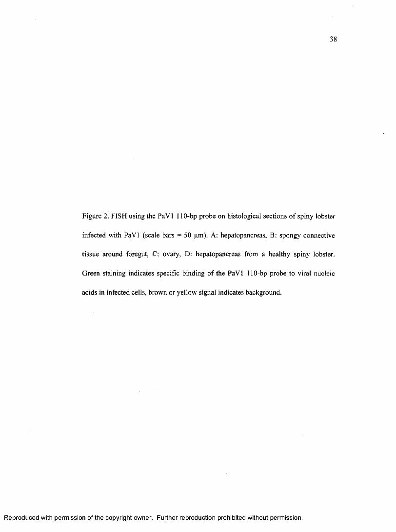
Figure 2. FISH using the PaVl 110-bp probe on histological sections of spiny lobster
infected with PaVl (scale bars = 50 pm). A: hepatopancreas, B: spongy connective
tissue around foregut, C: ovary, D: hepatopancreas from a healthy spiny lobster.
Green staining indicates specific binding of the PaVl 110-bp probe to viral nucleic
acids in infected cells, brown or yellow signal indicates background.
Reproduced with permission of the copyright owner. Further reproduction prohibited without permission.

Reproduced with permission of the copyright owner. Further reproduction prohibited without permission.Reproduced with permission of the copyright owner. Further reproduction prohibited without permission.

39
Figure 3. (A) FISH image of the hepatopancreas of a lobster infected with PaV l. Note
the green staining of the virally infected hemocytes by the PaV 1 110-bp probe (white
arrows), scale bar = 20 pm. (B) H&E staining of the hepatopancreas of an infected
lobster. Infected cells exhibit hypertrophied nuclei, and faint eosinophilic inclusions.
Black arrows indicate infected hemocytes, scale bar = 20 pm.
Reproduced with permission of the copyright owner. Further reproduction prohibited without permission.
Reproduced with permission of the copyright owner. Further reproduction prohibited without permission.Reproduced with permission of the copyright owner. Further reproduction prohibited without permission.

40
Figure 4. A, B. TEM of hepatopancreas from an infected lobster; virions (V)
aggregated at inner periphery of the nuclear membrane, with a few dispersed inside
the nucleus. Notice the condensed and emarginated chromatin (E), scale bars = 2 pm.
Reproduced with permission of the copyright owner. Further reproduction prohibited without permission.

Reproduced with permission of the copyright owner. Further reproduction prohibited without permission.Reproduced with permission of the copyright owner. Further reproduction prohibited without permission.

CHAPTER 2
Primary culture of hemocytes from the Caribbean spiny lobster, Panulirus argus, and their susceptibility to Panulirus argus Virus 1 (PaVl)
Published:
Li, C., Shields, J. D., 2007. Primary culture of hemocytes from the Caribbean spiny lobster, Panulirus argus, and their susceptibility to Panulirus argus Virus 1 (PaVl). Journal of Invertebrate Pathology 94,48-55.
41
Reproduced with permission of the copyright owner. Further reproduction prohibited without permission.

42
MANUSCRIPT ABSTRACT
Primary cultures of hemocytes from the Caribbean spiny lobster Panulirus argus
were developed for studies on the in vitro propagation of Panulirus argus Virus 1 (PaVl).
A modified Leibovitz L-15 medium supported the best survival o f hemocytes in in vitro
primary cultures. However, degradation o f the cultures occurred rapidly in the presence
of granulocytes. A Percoll step gradient was used to separate hemocytes into three
subpopulations enriched in hyalinocytes, semigranulocytes, and granulocytes,
respectively. When cultured separately, hyalinocytes and semigranulocytes maintained
higher viability (~ 80%) after 18 days incubation compared with granulocytes, which
degraded over 2-3 days. Susceptibility of the cell types was investigated in challenge
studies with PaVl. Hyalinocytes and semigranulocytes were susceptible to PaVl.
Cytopathic effects (CPE) were observed as early as 12 h post-inoculation, and as the
infection progressed, CPE became more apparent, with cell debris and cellular exudates
present in inoculated cultures. Cell lysis was noticeable within 24 hrs of infection. The
presence of virus within cells was further confirmed by in situ hybridization using a
specific DNA probe. The probe gave a unique staining pattern to cells infected with
PaV 1 24-h post inoculation. Cells in the control treatment were intact and negative to
hybridization. This assay was further applied to the quantification of infectious virus in
hemolymph using a modified 50% tissue culture infectious dose assay (TCID50) based on
Reproduced with permission of the copyright owner. Further reproduction prohibited without permission.
43
CPE. These tools will now allow the quantification of PaVl using established culture-
based methods.
Reproduced with permission of the copyright owner. Further reproduction prohibited without permission.

44
INTRODUCTION
The Caribbean spiny lobster, Panulirus argus, is widely distributed throughout
the Caribbean basin and along the Atlantic Coast ranging from Brazil to Georgia, USA. It
supports one of the most valuable fisheries in the Caribbean. Recently, a pathogenic virus,
Panulirus argus Virus l(PaV l), was identified during field surveys o f juvenile lobsters
from the Florida Keys (Shields & Behringer, 2004). The virus infects the soft connective
tissues, and two classes of hemocytes: hyalinocytes and semigranulocytes. The virus is
highly pathogenic to juvenile spiny lobsters, which die within 30-80 days in
experimentally induced infections (Shields and Behringer, 2004). Healthy lobsters are,
however, able to detect diseased animals and avoid them (Behringer et al., 2006). Given
its distribution throughout the Florida Keys and its relatively high prevalence in juvenile
lobsters, PaVl is thought to have significant potential to damage the fishery. Thus, it is
critical to develop specific and sensitive diagnostic methods to better understand the
pathogenesis of this viral pathogen.
Tissue culture is an important tool employed in the studies o f viral pathogens o f
vertebrates, but it has not been fully developed for assessment o f viral infection in
invertebrates (Rinkevich, 1999; Toullec, 1999; Villena, 2003). A t present there is no
continuous culture o f crustacean cell lines, however, primary culture o f crustacean tissue
has previously been developed for the diagnosis and in vitro proliferation o f shrimp
viruses (Chen and Wang, 1999; Fraser and Hall, 1999; Frerichs, 1996; Hsu et al., 1995;
Reproduced with permission of the copyright owner. Further reproduction prohibited without permission.

Nadala et al., 1993; Rinkevich, 1999; Tapay et al., 1997; Toullec, 1996). Because PaVl
infects hyalinocytes, semigranulocytes, and soft connective tissues (Shields and
Behringer, 2004), these hemocytes and soft connective tissues represent targets for the in
vitro study of the virus. Connective tissues have not been successfully obtained in culture.
However, primary cultures of hemocytes have been obtained from the penaeid shrimp,
Penaeus japonicus (Itami et al., 1999; Sano, 1998) and two species of crab, Liocarcinus
depurator and Carcinus maenas (Walton and Smith, 1999). The present study aimed to
develop a primary culture o f the hemocytes from the spiny lobster, Panulirus argus, and
to assess the susceptibility of hemocytes to PaVl in these cultures. The cell culture
system with the virus was further developed into an in vitro assay for the quantification
of virus in the hemolymph of infected lobsters.
Reproduced with permission of the copyright owner. Further reproduction prohibited without permission.

46
MATERIALS AND METHODS
Experimental animals
Juvenile spiny lobsters, Panulirus argus, were collected from the Florida Keys, and
housed in clean aquaria (salinity = 35 ± l% o , temperature = 24 ± 1 °C) equipped with
biological filters (Whisper) filled with crushed coral. Lobsters were fed with squid three
times per week. Water quality was monitored weekly and water changes were made to
ensure that various water quality parameters remained within acceptable limits: ammonia
(0-0.3 ppm), nitrite (0-0.6 ppm), pH (7.4-8.4).
Analysis of Panulirus argus hemocytes
Hemolymph was drawn with a 27-ga syringe from the juncture between the base and
ischium of the fifth walking leg. Prior to bleeding, the sample area was wiped with 70%
ethanol. In most cases, hemolymph was collected into a syringe containing an equal
volume of anticoagulant (0.45M NaCl, 0.1M glucose, 30mM sodium citrate, 26mM citric
acid, lOmM EDTA; pH = 5.4; Soderhall and Smith, 1983). Freshly collected hemocytes
were examined with an Olympus BX51 microscope equipped with a U-UCD8 Universal
condenser and Nomarski Differential Interference Contrast Filter. Hemocytes were
categorized based on cell size, cell shape, and granularity (Soderhall and Cerenius, 1992).
Total hemocyte counts (THC) and differential hemocyte counts (DHC) were performed
using a hemacytometer (Neubauer improved, Bright Line; two counts per lobster).
Reproduced with permission of the copyright owner. Further reproduction prohibited without permission.

47
Optimization of culture media
Samples o f collected hemolymph were centrifuged at 250 x g for 10 min at 4 °C
(IEC Thermo Centra, with swinging bucket rotor), then resuspended in appropriate
medium. Aliquots of 0.5 ml of the hemocyte suspension at densities of ~1 x 10⁶m r'
were seeded into 24-well culture plates containing an additional 1.0 ml of culture medium
per well. Plates were incubated in a Chamber (Lab-Line®) at 22 - 24 °C. Media were
refreshed on day 2 and thereafter at 2-day intervals. Cultured cells were observed with an
Olympus 1X50 inverted microscope equipped with a Hoffman Modulation Contrast
condenser. Images were taken with a Nikon DXM 1200 digital camera at days 1, 3, 5, 7,
9, and 11, prior to media refreshment. Cell viability was confirmed by the Trypan Blue
exclusion method (Mascotti et al., 2000). Several commercial media were assessed for
hemocyte viability: Leibovitz L-15 medium (L-15), Modified Leibovitz L-15 medium
(ML-15: double strength components, supplemented with 0.6 g L '1 L-glutamine and 0.7
gL'1 glucose), Grace’s insect medium, and RPMI-1640 medium. Each medium was
adjusted to match the osmolarity o f the hemolymph of the spiny lobster (1025 ± 6 mOsM
kg'1) by addition o f NaCl. Penicillin (100 IU m l'1) and streptomycin (100 pg m l'1) were
added to each culture medium to minimize potential bacterial contamination. In addition,
some media were supplemented with 0%, 5%, 10%, or 15% charcoal-dextran-treated
fetal bovine serum (FBS) for putative growth assessment. All media were sterilized by
filtering through Nalgene® Disposable Filters (Pore size = 0.20 pm).
Primary culture of separated hemocytes
Reproduced with permission of the copyright owner. Further reproduction prohibited without permission.

Hemocytes were separated by centrifugation using Percoll (Amersham Biosciences)
in fine step-density gradients (1.110, 1.096, 1.091, 1.086, 1.082, 1.077, 1.072, 1.067, and
1.062 g m l'1 respectively) made with lobster physiological buffer (0.4M NaCl, 0.01M
KC1, 0.01M Na2H P04, 0.01M KH2P 0 4, NaHC03; pH - 7.8). Briefly, 1 ml o f each
density solution was carefully layered into the samel5-ml centrifuge tube. Aliquots of 3
ml of the hemolymph-anticoagulant suspensions were gently layered on the top o f the
stacked gradients, then centrifuged at 400 x g for 30 min at 4 °C. Cell fractions were
carefully aspirated into anticoagulant using Pasteur pipettes, and washed twice with
culture media. Cell fractions were centrifuged at 250 x g for 10 min at 4 °C to remove
residual Percoll prior to resuspension in fresh culture media. Separated hemocytes were
cultured in the optimized media and assessed for cell viability as above.
Virus inoculation of cell cultures
The virus inoculum was extracted from the hemolymph of a heavily diseased lobster.
Briefly, diseased hemolymph was mixed with 4-fold volume of ML-15 medium and
homogenized with a homogenizer (Pyrex®, Coming Inc.) at 4 °C for 10 min, the
homogenized mixture was centrifuged at 3000 * g for 10 min at 4° C, and the
supernatant was filtered through 0.45-pm filter. Viral filtrates were serially diluted with
ML-15 culture medium to 1:1, 1:10, 1:100 and 1:1000 and used as inoculum.
Hemolymph from a healthy lobster was processed similarly and used as a control.
Aliquots of 100 pi of inoculum were inoculated into 1 day old established hemocyte
cultures in 24-well tissue culture plates (Falcon®, Becton Dickinson Labware), the plates
were incubated at 22 - 24 °C for 4 days, and cultures were supplemented with new media
Reproduced with permission of the copyright owner. Further reproduction prohibited without permission.

49
at 2-day intervals. The inoculated primary cultures were examined daily with an inverted
microscope, and images were taken daily for assessment of cytopathic effects (CPE) and
calculation of 50% tissue culture infectious dose (TCID50) as in Reed and Muench (1938).
Diagnosis of in vitro infection of PaVl by in situ hybridization (ISH)
For diagnosis o f in vitro infections of PaVl, hemocytes were cultured and inoculated
with 1:10 diluted inoculum in chamber slides (Lab-Tek®). On days 1, 2, 3 and 4 post
inoculation, hemocytes cultured in chambers were fixed in 10% neutral buffered formalin
for 10 min at room temperature, and then gently rinsed briefly with phosphate buffered
saline (PBS) (8.0 g L '1 NaCl, 0.2 g L '1 KC1,1.44 g L '1 Na2H P04, 0.24 g L '1 KH2P 0 4; pH
7.4). Infections were then prepared for in situ hybridization (ISH). Briefly, slides were
permeabilized with 0.5% (v/v) Triton X-100 (in PBS) for 10 min, rinsed with PBS (5 min
x 3), then digested with Proteinase K (100 pg m l'1 in PBS) for 15 min at 37 °C, followed
by a 5-min wash in 0.2% (w/v) glycine PBS solution to stop proteolysis, and incubated in
2 x SSC (17.53 g L '1 NaCl, 8.82 g L '1 citric acid; pH 7.0) for 10 min at room temperature.
Slides were incubated in pre-hybridization buffer (4 x SSC, 50% (v/v) formamide, 0.5
mg m l'1 Salmon sperm DNA, and 1% (v/v) fetal bovine serum) at 42° C for 45 min. After
incubation, excess pre-hybridization buffer was carefully drained off, the area with tissue
was outlined with a Frame-seal incubation chamber (MJ Research INC.), then aliquots of
50-pl of hybridization solution (50% de-ionised formamide; 4 x SSC; 0.5% (w/v) SDS;
25 pg m l'1 FITC-labeled DNA probe) (probe synthesis as in Li et al. 2006) were added
within the area, sealed with plastic cover slip. Each slide was then placed into a thermal
cycler for 3 min at 72° C, and cooled on ice for 2 min. Slides were incubated in a humid
Reproduced with permission of the copyright owner. Further reproduction prohibited without permission.
50
chamber overnight at 42° C. The slides were then washed in 2 x SSC ( 2 x 5 min), 1 x
SSC (2><5 min), PBS (10 min) and Maleic Acid Buffer (MAB: 100 mM maleic acid, 150
mM NaCl; pH 7.5) for 10 min. The slides were then incubated in blocking solution (1%
(w/v) Blocking reagents in MAB, Roche). Anti-FITC alkaline phosphatase conjugate
antibody (Sigma-Aldrich) was diluted 1:2000 in blocking solution and sections were
incubated with the diluted antibody for 3 h at room temperature with gentle agitation.
Unbound antibody was removed with two 5 min washes in buffer I (100 mM Tris, 150
mM NaCl, pH 7.5) and two 5 min washes in Buffer II (100 mM Tris, 100 mM NaCl, 50
mM MgC^, pH 9.5). The slides were then incubated with BCIP/NBT liquid substrate
solution (Sigma-Aldrich) in dark for 2 h. The color reaction was stopped by washing in
TE buffer (10 mM Tris, ImM EDTA, pH 7.5) and dH20 for 5 min, respectively. The
slides were mounted with aqueous mounting medium (90 % glycerol, 0.1 m Tris-HCl, pH
8.0 and 2.3 % DABCO) and glass coverslips applied. Clear fingernail polish was applied
to the edges of the cover slips to prevent evaporation. Slides were examined using an
Olympus BX51 microscope, and images were captured with a Nikon DXM 1200 digital
camera.
Reproduced with permission of the copyright owner. Further reproduction prohibited without permission.

51
RESULTS
Hemocytes of the spiny lobster Panulirus argus
Three major hemocyte types were identified in the Caribbean spiny lobster:
hyalinocytes, semigranulocytes, and granulocytes (Fig. 1). Hyalinocytes contained no or
a few small granules, and were often found as a spindle or round shapes, ranging in size
from 12 to 18 pm. Semigranulocytes contained many small and a few larger (>1 pm)
granules, were morphologically variable from spindle, ovoid or round in shape, and
ranged in size from 14 to 23 pm. Granulocytes contained many large (>1 pm) and a few
small granules, were also variable in shape, and ranged from 17 to 28 pm.
The number of circulating hemocytes (total hemocyte count, THC) in the spiny
lobster was around 8.0 xlO6cells m r'(range 6.68 xlO6 - 9.58 x io 6, n =16). Hyalinocytes
(H) represented approximately 15% (15.2 ± 2.0%, n =16) of the total circulating
hemocytes, semigranulocytes accounted for 58% (57.9 ± 3.7%, n =16), and granulocytes,
27% (26.9 ± 4.2%, n = 16).
Optimization of culture media
The lobster hem ocytes survived but did not proliferate in each o f the four culture m edia
tested, and no mitosis was observed in any of the hemocyte cultures. After 1 day,
hemocytes cultured in ML-15, L-15, Grace’s media and RPMI-1640 maintained high
viability, with >90% of the cells remaining alive (Fig. 2 A). However, over the next three
Reproduced with permission of the copyright owner. Further reproduction prohibited without permission.

52
days, the viability of cells cultured in RPMI-1640 decreased to 25%, which was
significantly lower than cells in the other media (Fig. 2 A). By day 8, most cells cultured
in RPMI-1640 had died, whereas mean cell viability was 79.5 ± 4.7 % in ML-15 medium,
68.4 ± 7.2 % in Grace’s medium, 45.8 ± 3.8 % in L-15 medium. By day 12, cell viability
decreased to 43.0 ± 2.2 % in ML-15 medium, 32.4 ±4.1 % in Grace’s medium, and
26.4.0 ± 2.9 % in L-15 medium (Fig. 2 A).
Fetal bovine serum, a standard supplement in many cell culture systems, was not
an effective additive and hampered cell viability in this study. Hemocytes cultured in the
ML-15 medium supplemented with 5%, 10%, and 15% FBS, respectively, were
consistently degraded, with viabilities of only 3 ~ 4% after 11 days in culture (Fig. 2 B).
Hemocytes cultured in media without supplementation with FBS retained much higher
viability (Fig 2 A, B).
Primary cultures of separated hemocytes
In previous mixed hemocytes cultures, within 2-7 days, most of the granulocytes
dehisced, releasing their contents into the culture media, potentially impacting the
survival of other the cell types (Fig.3). In order to improve cell viabilities o f cultured
hemocytes, lobster hemocytes were separated into three distinct fractions using Percoll
discontinuous gradients and cultured separately.
In the colum n o f the Percoll step gradients, the fraction enriched w ith
hyalinocytes was located between density gradients of 1.062 and 1.067 g m l'1; in this
fraction (H), hyalinocytes comprised more than 80% of the cells with semigranulocytes
making up the rest. The fraction enriched with semigranulocytes was located between
Reproduced with permission of the copyright owner. Further reproduction prohibited without permission.
53
density gradients o f 1.067 and 1.072 g m l'1. This semigranulocytes (SG) fraction was
comprised of approximately 90% semigranulocytes, with 7% of hyalinocytes and 3% of
granulocytes. The granulocytes (G) fraction accumulated in the interface between 1.082
and 1.086 g m l'1 gradients; over 94 % of the cells in this fraction were granulocytes, with
only a small proportion (< 6 %) of semigranulocytes and no hyalinocytes (Fig. 4).
From the culture of unseparated hemocytes, cells maintained in ML-15 medium
survived better than those in other media; therefore, ML-15 medium was selected as the
medium for maintaining the separated cell types. Viability improved dramatically in
hyalinocytes and semigranulocytes grown in the absence of granulocytes. Hyalinocytes
and semigranulocytes survived up to 18 days with viabilities o f 77.6 % (± 6.4%) and 79.1
(± 7.1%) respectively. Separated granulocytes died quickly, within 4 or 5 days (Fig. 5).
In vitro propagation of PaVl
Hemocytes from cultures inoculated with the virus showed cytopathic effects
(CPE) after 12-h post-inoculation with 1:1 diluted PaVl inoculum. Initially, the affected
cells changed from round or oval shapes to an irregular shape (Fig. 6A). As the infection
progressed, the infected cells gradually shrank and became surrounded by exudates or
cell debris (Fig. 6B). After 72 hrs, cell death was obvious, and inoculated cultures were
comprised of few live cells and much cell debris (Fig. 6C).
The infection of PaVl in cultured hemocytes was confirmed by in situ
hybridization using a specific 110-bp DNA probe of Li et al. (2006). The unambiguous
dark staining of PaVl infected cells was observed after 24 h post-inoculation. No PaVl
Reproduced with permission of the copyright owner. Further reproduction prohibited without permission.

54
positive signals were detected in cell cultures inoculated with hemolymph filtrates from a
healthy lobster (Fig. 7).
By 48 h post inoculation, 23.45 % ( + 10 ) of the cells exposed to the l:l-diluted
inoculum had survived; over 50 % of cells exposed to greater dilutions of the inocula had
survived, whereas more than 93 % of cells in the control groups had survived. By 72 h
post inoculation, most cells that had been exposed to the virus had lysed in the 1 :1 and
1:10 dilution groups; whereas most of the cells in control groups remained alive (~ 90 %)
(Fig. 8 ). The amount of infectious virus in the undiluted inoculum was 4 x 10³ TCID50
ml' 1 when calculated with the end point dilution assay (Reed and Muench, 1938) based
on percentage of cells surviving at the end of the assay.
Reproduced with permission of the copyright owner. Further reproduction prohibited without permission.

55
DISCUSSION
We have developed the primary culture of hemocytes from the Caribbean spiny
lobster Panulirus argus for in vitro studies of Panulirus argus Virus l(PaV l). Primary
cultures of hyalinocytes and semigranulocytes were susceptible to PaVl, showing
obvious cytopathic effects (CPE) within hours of exposure to a high dose o f PaV 1. Even
though the cultured hyalinocytes and semigranulocytes were susceptible to PaVl, we
could not develop a plaque assay for quantitative study of the virus, because lobster
hemocytes do not undergo mitosis and no confluent cell layer could be formed; i.e., the
cells formed dispersed monolayers. However, a CPE assay using an estimate of the 50%
tissue culture infectious dose (TCID50) method provided an alternative to determine viral
titer (see Darling et al. 1998). Such assays have been successfully applied to quantify the
infectious titer of several other crustacean viruses including yellow head baculovirus
(Assavalapsakul et al., 2003; Lu et al., 1995) and non-occluded baculo-like virus (Tapay
et al., 1997). The in vitro quantal assay based on CPE will facilitate better understanding
of infection dynamics o f PaVl in the spiny lobster. However, because hemocytes do not
multiply in vitro, the application is limited in its ability to grow large quantities of the
virus for in vitro studies.
We demonstrated that the PaVl virus infected in vitro cultured hyalinocytes and
semigranulocytes using a specific DNA probe. The rapid infection and mortality of the
hemocytes is interesting given that infections in juvenile lobsters last from 30-80 d
Reproduced with permission of the copyright owner. Further reproduction prohibited without permission.

56
(Shields and Behringer, 2004). However, the dynamics within the host will no doubt be
different due to immune defenses, stimulation o f hematopoietic tissues, and other factors.
In the lobster, the virus also infects the cells o f the spongy connective tissues, and may
cause different effects in these, however this tissue cannot as yet be successfully cultured.
Lymphoid and ovary tissues are often the targets for pathogenic viruses in
shrimps; therefore, these cells are frequently used in in vitro studies of viral pathogenesis
(Assavalapsakul et al., 2003; Chen and Wang, 1999; Lu et al., 1995; Maeda et al. 2004;
Tapay et al., 1997; Wang et al., 2000). Comparatively limited efforts have been made to
develop in vitro cultures of hemocytes from other crustacean species. Itami et al. (1999)
cultured large granular hemocytes from P. japonicus for up to 10 days and inoculated
them with penaeid rod-shaped DNA virus, however, no CPE was observed over the 10
day period of incubation. Walton and Smith (1999) separated and collected hyalinocytes
from the crabs, Liocarcinus depurator and Carcinus maenas. They were able to maintain
these cells for up to 14 days with more than 70% viability in an optimized L-15 medium.
In our cultures, separated hyalinocytes and semigranulocytes survived up to 18 days with
high viability (~ 80%), and maintained an even higher viability (> 90%) in the earlier
period of culture.
A variety of culture media have been tested for the primary culture o f crustacean
tissues, these include Leibovitz L-15, Grace’s insect medium, RPMI-1640, Medium 199,
and several specifically formulated media (Luedeman and Lightner 1992; Nadala et al.,
1993; Tong et al., 1996; Walton and Smith, 1999). Leibovitz L-15 and Grace’s insect
medium have been considered the best commercial medium for tissue culture of
crustaceans (Luedeman and Lightner, 1992; Nadala et al., 1993; Walton and Smith, 1999).
Reproduced with permission of the copyright owner. Further reproduction prohibited without permission.

57
In our study, Grace’s insect medium and double strength Leibovitz L-15 medium (2 x L-
15) supported cell survival in the first 6 days of culture for unseparated lobster hemocytes;
however, the Modified L-15 medium yielded better results for longer term viability (>
80% by day 15). The higher concentration of glutamine and glucose in Grace’s media
and L-15 medium apparently benefited the survival of in vitro cultured hemocytes o f the
spiny lobster.
Fetal bovine serum (FBS) is often used as a supplement in crustacean and mollusk
cell culture (Chen and Wang, 1999; Luedeman and Lightner 1992; Sano, 1998; Walton
and Smith, 1999). In this study, fetal bovine serum (even treated with Charcoal-dextran
absorption) degraded the viability in whole hemocyte cultures even at a low
concentration of 5%. Semigranulocytes and granulocytes are sensitive to foreign particles,
particularly glucans, lipopolysaccharides, and bacteria, and often lyse to release
prophenoloxidase and other components involved in cytotoxicity and melanization
pathways (Soderhall and Cerenius, 1992). FBS presumably caused the granulocytes to
dehisce and lyse, causing further deterioration of remaining cells. Other supplements,
such as cell-free plasma or filtrates from homogenized tissues, that have been used to
support microbial pathogens of crustaceans (Toullec, 1999), were not tested in this study
due to their potential to induce cross reactions among individual lobsters.
Traditionally, crustacean hemocytes have been classified into three types of cells:
hyalinocytes, semigranulocytes, and granulocytes, according to the number and size of
granules they contain (Bauchau, 1980) and their biological function (Soderhall and Smith,
1983). Hose et al. (1990) proposed a different classification based on morphology,
cytochemistry, and studies of cell function, and suggested that hyalinocytes and
Reproduced with permission of the copyright owner. Further reproduction prohibited without permission.

58
granulocytes represent two distinct cell lineages, with granulocytes representing a
continuum of differentiation from the less mature small-granule hemocytes to the large-
granule hemocytes. We found that granulocytes were distinctly different from the other
two cell types in morphology, and that they were particularly sensitivity to the in vitro
environment, surviving only a short period when compared with the other two
subpopulations. In the initial period (2 to 3 days) of separated hemocyte cultures, some
hyalinocytes became morphologically like semigranulocytes, whereas no transformation
was observed between semigranulocytes and granulocytes. Although we retain the
traditional classification of hemocyte types in the Caribbean spiny lobster, more work is
needed to fully appreciate the functional and biochemical differences among cell types.
Additional experiments with culturing techniques will facilitate further in vitro study of
crustacean hemocytes.
Reproduced with permission of the copyright owner. Further reproduction prohibited without permission.
59
Figure 1. Light microscopy of hemocytes from the spiny lobster, Panulirus argus:
Hyalinocytes (H), semigranulocytes (SG), and granulocytes (G). Scale bar = 10 pm
Reproduced with permission of the copyright owner. Further reproduction prohibited without permission.

Reproduced with permission of the copyright owner. Further reproduction prohibited without permission.
•• • ~' t
• I'
Reproduced with permission of the copyright owner. Further reproduction prohibited without permission.

60
Figure 2. (A) Viability o f unseparated hemocytes o f P. argus cultured in L-15, ML-15,
Grace’s Insect medium, and RPMI-1640 medium. (B) Viability o f unseparated
hemocytes of P. argus cultured in ML-15 medium supplemented with 0%, 5%, 10%, and
15% FBS. Values are means ± STD (n=12).
Reproduced with permission of the copyright owner. Further reproduction prohibited without permission.

A*C ulture M edia T est
100 L-15ML-15Grace'sRMPI-164080
60
40
20
02 4 6 8 10 12
Days
D .ML-15 M edium & FBS
0% FBS 5% FBS 10% FBS 15% FBS
100
80 -
.oeg>
60 -
<1)O
20 -
2 4 6 8 10 12
T im e /d a y
Reproduced with permission of the copyright owner. Further reproduction prohibited without permission.

61
Figure 3. Light microscopy of unseparated hemocytes o f the spiny lobster P. argus at 1st
(A) and 5th (B) day in culture. Note those dehisced granulocyte (arrowheads). H:
hyalinocytes; SG: semigranulocytes; G: granulocytes. Scale bars = 10 pm.
Reproduced with permission of the copyright owner. Further reproduction prohibited without permission.
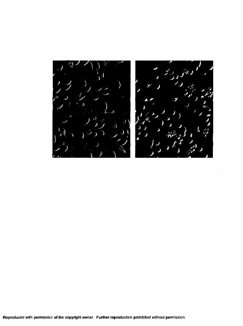
Reproduced with permission of the copyright owner. Further reproduction prohibited without permissionReproduced with permission of the copyright owner. Further reproduction prohibited without permission.

62
Figure 4. Light microscopy of P. argus hemocytes in fractions from Percoll
discontinuous gradient separation: H: hyalinocytes, SG: semi-granulocytes, and G:
granulocytes. Scale bars = 10 pm.
Reproduced with permission of the copyright owner. Further reproduction prohibited without permission.

Reproduced with permission of the copyright owner. Further reproduction prohibited without permission.Reproduced with permission of the copyright owner. Further reproduction prohibited without permission.

63
Figure 5. Cultures o f separated hemocytes grown in ML-15 media. H: hyalinocytes; SG:
semigranulocytes; G: granulocytes. Values are means ± STD (n=5).
Reproduced with permission of the copyright owner. Further reproduction prohibited without permission.
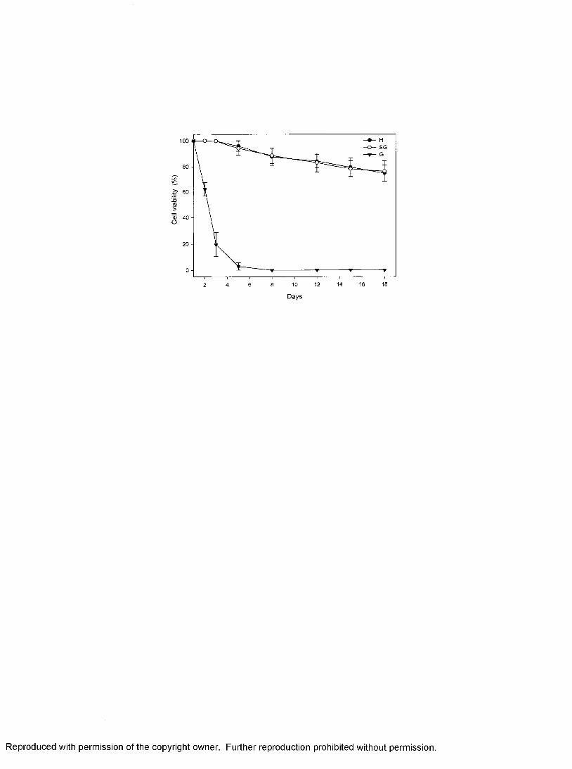
100SG
80 -
<9>© 40 - O
20 -
1612 14 18108
Days
Reproduced with permission of the copyright owner. Further reproduction prohibited without permission.

64
Figure 6. Cytopathic effects o f cultured hemocytes infected with PaVl inoculum (1:10
diluted). (A) 12 h post-inoculation with PaVl filtrates, (B) 48 h post-inoculation, (C) 72 h
post-inoculation. Note the morphological change (black arrow) and degradation (white
arrow) of hemocytes when infected with PaVl. (D) Control culture at 72 h post
inoculated with hemolymph filtrates from a healthy lobster. Scale bars = 20 pm.
Reproduced with permission of the copyright owner. Further reproduction prohibited without permission.

B■\ %
Reproduced with permission of the copyright owner. Further reproduction prohibited without permission.

65
Figure 7. Detection of PaVl in primary cultures o f separated hemocytes (hyalinocytes
and semigranulocytes) of P. argus by in situ hybridization. A: 24 h post-inoculation with
hemolymph fitrates from a healthy lobster, B: 24 h post-inoculation with PaVl filtrates;
note the dark staining of infected cells (black arrows), and the debris of lysed cells (arrow
heads). Transmitted light microscopy. Scale bars = 20 pm.
Reproduced with permission of the copyright owner. Further reproduction prohibited without permission.

Reproduced with permission of the copyright owner. Further reproduction prohibited without permission.Reproduced with permission of the copyright owner. Further reproduction prohibited without permission.

66
Figure 8. Survival of cultured hemocytes of P. argus inoculated with serially diluted viral
inoculum. Survival was defined as (numbers of survived cells / initial amount of cells) x
100 % of each well. Values are means ± STD (n = 12).
Reproduced with permission of the copyright owner. Further reproduction prohibited without permission.

100
80 -
4 0 -
1:101:1001:1000ControlBlank
20 -
72 84 960 12 24 36 48 60
Tim e post-inoculation (h)
Reproduced with permission of the copyright owner. Further reproduction prohibited without permission.
CHAPTER 3
Pathology and hematology of the Caribbean spiny lobster experimentally infected with Panulirus argus virus 1 (PaVl)
Manuscript submitted to the journal of Virus Research:
Li, C., Shields, J.D., Ratzlaff, R.E., Butler, M. J., 2007. Pathology and hematology of the Caribbean spiny lobster experimentally infected with Panulirus argus virus 1 (PaVl). Under review.
67
Reproduced with permission of the copyright owner. Further reproduction prohibited without permission.
68
MANUSCRIPT ABSTRACT
We conducted a study of the histopathological and hematological response of
the Caribbean spiny lobster to experimentally induced infections with PaV 1. The fixed
phagocytes in the hepatopancreas were the initial site of PaVl infection in spiny lobsters.
Fixed phagocytes were activated in early infections; however, as the disease progressed,
the fixed phagocytes became infected and eventually lysed. Infected cells were
subsequently observed in the hepatopancreas, gill, heart, hindgut, glial cells around the
ventral nerves, as well as in the cuticular epidermis and foregut. In advanced infections,
all o f the spongy connective tissues were infected as were the glial cells around the optic
nerves. The structure of the hepatopancreas was also significantly altered as the disease
progressed. The hemal sinuses among the hepatopancreatic tubules filled with massive
amounts of cellular aggregates, including infected circulating hemocytes and infected
spongy connective tissues. Atrophy o f the hepatopancreatic tubules occurred in the late
stage o f viral infection. The virus caused significant decreases in total hemocyte density
in later stages of infection and significantly altered several constituents in the hemolymph
serum of diseased lobsters, including: glucose, phosphorus, triglycerides, and lipase.
Reproduced with permission of the copyright owner. Further reproduction prohibited without permission.

69
INTRODUCTION
The Caribbean spiny lobster Panulirus argus occurs throughout the Caribbean
basin and Western Atlantic from Brazil to Bermuda (Holithius, 1991) and supports
valuable commercial and recreational fisheries throughout its range (FAO, 2001, 2004;
Harper, 1995). In Florida, for example, the commercial landings of Caribbean spiny
lobster have varied between 4.3 million pounds and 7.9 million pounds per year from
1970 to 1999. In 1999, the total landings o f the spiny lobster decreased and by 2001 they
had dropped to 3.4 million pounds, the lowest reported landings since 1982,
approximately 45% less than the historical average landings (FMRI, 2005; Muller et al.,
1997). The recently identified pathogenic virus, Panulirus argus Virus 1 (PaVl), is
thought to have contributed to the decline (Shields and Behringer, 2004).
PaVl primarily infects benthic juvenile lobsters (20 to 55 mm carapace length,
CL), with prevalence decreasing rapidly in relation to size (Shields and Behringer, 2004).
The virus occurs throughout the Florida Keys, with the prevalences of visibly infected
juveniles ranging from 6 % to 8 %, and reaching 37 % in some areas (Shields and
Behringer, 2004). The virus is transmitted by contact, through food, and over short
distances (< lm) through the water (Behringer 2003; Butler et al. in review), but healthy
lobsters can sense and avoid diseased lobsters and this may limit the spread of the virus in
the lobster population (Behringer et al., 2006). PaVl infects certain hemocytes and
Reproduced with permission of the copyright owner. Further reproduction prohibited without permission.

70
spongy connective tissues in several tissues and organs (Shields and Behringer, 2004).
However, the sites of early infection, the progression of PaVl infection in the spiny
lobster and the hematological response o f the host to viral infection have not been
examined. Therefore, we report the pathology and hematology of spiny lobster over a
time course of experimental infection by PaV 1.
Reproduced with permission of the copyright owner. Further reproduction prohibited without permission.

71
MATERIALS AND METHODS
Caribbean spiny lobster Panulirus argus
Juvenile spiny lobsters, 25 to 50 mm carapace length (CL), were collected from
the Florida Keys by hand using SCUBA. Lobsters with apparent signs o f disease (Shields
and Behringer, 2004) were held separately in 38 L glass aquaria with flow-through
ambient seawater (salinity = 35 ± 1 ppt, temperature = 24 ± 1 ° C) equipped with
biological filters (Whisper) filled with pre-conditioned crushed coral, and used as donors
for inoculation trials. All other lobsters were randomly divided into groups (four to five
animals) and housed separately in 76-L glass aquaria. Lobsters were acclimated for at
least two weeks prior to inoculation trials. During experiments, lobsters were fed squid
three times per week, and water changes were made as needed to ensure that water
quality parameters remained within acceptable limits: ammonia (0-0.3 ppm), nitrite
(0-0.6 ppm), pH (7.4-8.4), salinity (35 ± 1 ppt) and temperature (23 ± 1 ° C).
Viral inocula
Viral inocula were extracted from hemolymph of heavily diseased lobsters,
which were histologically confirmed to have heavy infections (Shields and Behringer,
2004). The infectious titer of the inocula was quantified later on frozen pooled sera
Reproduced with permission of the copyright owner. Further reproduction prohibited without permission.

72
using a 50 % tissue culture infectious dose assay (TCID50) from Li et al. (2007). Briefly,
diseased hemolymph was mixed with an equal volume of citrate/EDTA anticoagulant
(0.45M NaCl, 0.1M glucose, 30mM sodium citrate, 26mM citric acid, lOmM EDTA; pH
= 5.4; Soderhall and Smith, 1983) and homogenized with a glass homogenizer (Pyrex®,
Coming Inc.) at 4 0 C for five minutes. The homogenate was then centrifuged at 3000 x g
for 10 min at 4° C, and the supernatant filtered through a 0.45-pm filter. The viral
filtrates were serially diluted with modified Leibovitz L-15 medium (ML-15) (Li et al.,
2007) to 1:1, 1:10, 1:100 and 1:1000 and used as inocula.
Aliquots of 100 pi of inocula were inoculated separately into one-day old
hemocyte cultures (semigranulocytes and hyalinocytes) in 48-well tissue culture plates
(Costar®, Costar Corp.). The plates were incubated at 22 - 24 0 C for 96 h, and
supplemented with fresh media at two-day intervals. The inoculated primary cultures
were examined daily with an inverted microscope, and images were taken for assessment
of cytopathic effects (CPE). TCID50 was calculated at 96 h based on the end-point
dilution assay of Reed and Muench (1938).
Experimental infection
Lobsters were inoculated through the arthrodial membrane at the juncture o f the
basis and ischium of the fifth walking leg. Two separate inoculation trials were conducted
to examine the pathological response of the hosts over different time scales and viral
dosages. Trial I was a short term infection study, where lobsters were dosed with the
Reproduced with permission of the copyright owner. Further reproduction prohibited without permission.

73
virus, then necropsied over the course o f fifteen days. Trial II was a long term infection
study, where lobsters were inoculated with the virus, then necropsied at 1 0 -day intervals
over 77 days. Trials I and II were undertaken at different times with different viral doses
(see below).
In Trial I, hemolymph samples from heavily infected donors were pooled,
diluted with Citrate-EDTA anticoagulant (1:9) and used as inoculum (virus titer = 5.8 x
2 * • • • .10 TCID50 /ml). Aliquots o f 1 0 0 -pl o f inocula were injected separately into 30 healthy
lobsters using sterile 27-gauge needles. Inoculated animals were held together in groups
o f three to four animals per 76-L aquarium. Controls consisted of 18 animals injected
with an equivalent volume of the mixture o f the anticoagulant and hemolymph collected
from healthy lobsters. Animals serving as controls were held together in groups o f three
to four animals per 76-L aquarium. At days 1, 3, 5, 7, 10, andl5 post-inoculation (p.i.)
five animals from the virus inoculated group and three animals from control group were
randomly selected and processed for diseases assessment.
In Trial II, hemolymph from heavily infected donors (different donors than in
Trial I) was pooled, then diluted with Citrate-EDTA anticoagulant (1:2) and used as
inocula (virus titer = 1.2 x 10³ TCID50 /ml). Aliquots of 100-pl of inocula were injected
separately into 50 healthy lobsters using sterile 27-gauge needles. Controls consisted of
30 lobsters injected with an equivalent volume of the anticoagulant. Inoculated and
control animals were held separately in groups as detailed above. At roughly 10-day
intervals until 80 days p.i., five lobsters from the experimental group and three lobsters
from the control group were randomly selected, dissected and processed for
histopathological assessment o f PaVl infection and blood chemistry analysis.
Reproduced with permission of the copyright owner. Further reproduction prohibited without permission.
74
Hemolymph and other tissues were collected from each lobster and processed for
histological and hematological analysis as described below.
Diagnosis of PaVI infection
Several tissues were collected for histology from each dissected lobster,
including hepatopancreas, hindgut, gill, heart, cuticle epidermis, nerve tissue, and in
some cases, compound eyes and antennal gland. Tissues were fixed in Bourn’s solution
(Fisher) or Z-fix (Anatech Ltd.) for approximately 24 hours, rinsed with tap water for 45
min, then held in 70 % EtOH, processed through paraffin histology and stained with
Harris hematoxylin and eosin Y (H&E) (Humason, 1979). Eyes were decalcified
overnight in citrate-EDTA, cut in two and processed as above. All tissues were examined
using an Olympus BX51 microscope and photographs were taken using a Nikon
DXM1200 digital camera. When infections were ambiguous via normal histopathology,
tissue samples were processed for diagnosis o f viral infection using fluorescence in situ
hybridization (FISH) as detailed in Li et al. (2006).
Biochemical and hematological analyses
Hemolymph was drawn with a 27-ga. syringe from the juncture between the
basis and ischium of the fifth walking leg. Aliguots o f 1 ml hemolymph of each animal
were stored in an ultracold freezer (-80 °C) for biochemical analysis. Frozen hemolymph
samples were thawed on ice. Serum was collected using a pipette, and then centrifuged at
3,000 xg for 10 min at 4 °C (IEC Thermo Centra) to remove cell debris. Sera were then
processed through an Olympus AU400 clinical chemistry analyzer (Olympus Americas,
Reproduced with permission of the copyright owner. Further reproduction prohibited without permission.

Inc.) for blood chemistry analysis. Glucose, total protein, phosphorus, triglycerides,
lipase, creatinine, alanine aminotransferase (ALT), aspartate aminotransferase (AST),
alkaline phosphatase (ALKP), gamma-glutamyl transpeptidase (GGT), calcium, sodium,
potassium, and chloride were measured according to the manufacturer’s manuals.
Hemolymph collected from Trial II was immediately processed for total hemocyte counts
(THC) and differential hemocyte counts (DHC). Briefly, THC and DHC were processed
with a Neubauer hemacytometer using the Olympus BX51 microscope equipped with a
Nomarski Differential Interference Contrast Filter. Hemocytes were categorized as
hyalinocytes, semigranulocytes or granulocytes based on cell size, cell shape, and
granularity as in Li and Shields (2007). Differences in biochemical and hematological
constituents between control and experimental groups were examined statistically with
SYSTAT (SYSTAT Software Inc.). One-way ANOVA was used to examine differences
in measurement o f biochemical and hematological constituents in lobsters with different
disease categories (healthy, light-medium infection, and heavily infected) as determined
by histological examination. The data were log transformed to meet parametric
assumptions.
Reproduced with permission of the copyright owner. Further reproduction prohibited without permission.

76
RESULTS
Disease status of experimental lobsters
The severity of viral disease in lobsters was rated as Category 0, 1, 2 or 3 based
on pathological changes in all tissues examined (Table 1). Infected cells were
characterized by hypertrophied nuclei, margination and condensation o f peripheral
chromatin and eosinophilic Cowdry-like inclusions. For each tissue, the level of infection
was rated as 0, 1, 2, or 3 based on the number o f infected cells per section examined
microscopically at 400 x; 0, no cells with observable pathology; 1, <10 infected
cells/section; 2, 10 ~ 100 infected cells/section; 3, >100 infected cells/section.
The disease status o f lobsters is summarized in Figure 1. In Trial I, only one
lobster was lightly infected and another moderately infected by 15 day p.i. There was no
observable pathology in any of the lobsters before that time. In Trial II, the virus was
highly infectious, presumably because of the higher dose, and by day 10 p.i., 80 % of the
lobsters were infected, including one with a moderate infection. By day 20 p.i., all
lobsters inoculated with virus were infected; most (80 %) were moderately infected, and
one was heavily infected. By day 30 p.i., 60 % of the inoculated lobsters were heavily
infected and 40 % moderately infected. After 40 days p.i., all of the lobsters that were
inoculated were heavily infected. In both trials, all but one o f the control lobsters were
healthy, the single infected control was consistent with the background level of the virus
present in wild.
Reproduced with permission of the copyright owner. Further reproduction prohibited without permission.

77
Progression of disease in experimentally inoculated animals from Trial I
In Trial I, infected cells were initially observed in the hepatopancreas of the two
infected lobsters at day 15 p.i. Fixed phagocytes in the hepatopancreas appeared to be the
primary cell associated with PaVl infection in spiny lobsters. Significant alterations of
the fixed phagocytes were observed in the hepatopancreas in relation to the progression
of the infection (Fig. 2). Activated fixed phagocytes were significantly enlarged, with
highly vacuolated cytoplasm and sparse granules (Fig. 2 B). As the infections progressed,
fixed phagocytes were obviously infected by the virus (Fig. 2 C). In the hepatopancreas
of the moderately infected lobster, the typical rosette structure of fixed phagocytes
around the arterioles was no longer discernible. (Fig. 2 D). There were no overt
pathological changes in other tissues during this early period of infection, except that
granulomas were present in the antennal gland of the lightly infected lobster (Fig. 2 E).
As no infected cells were observed in the antennal gland, this pathological changes may
not be associated with PaV 1 infection.
Total hemocyte counts (THCs) varied over time and between treatments.
THCs in inoculated lobsters increased due to the stimulation of inoculation, and then
decreased significantly thereafter (Fig. 3). By day 15 p.i., THCs of PaV 1-inoculated
lobsters were significantly lower than those in controls (ANOVA, p<0.01). THCs of
lobsters from the control group varied in the first 3 to 4 days p.i. and then gradually
recovered to the initial density. Relative changes among types of hemocytes were not
significantly different between treatments (data not shown). Therefore, the absolute
changes in THC were consistent between cell types with treatments. However,
Reproduced with permission of the copyright owner. Further reproduction prohibited without permission.

78
granulocytes appeared to accumulate in the enlarged hemal spaces o f the hepatopancreas
of a lobster inoculated with PaVl by day 15 p.i. (Fig. 2 F).
Progression of disease in tissues of experimental infected animals from Trial II
As expected, the progression of infection and disease varied between Trials
(Fig. 1). The pathological changes in the tissues o f lobsters with light or moderate
infections were similar in both trials, but more animals were infected in Trial II.
Day 10 p.i.: Infected cells were observed in hepatopancreas, gill, heart, central
nerve tissue and hindgut, but they were not observed in the cuticular epidermis and
compound eyes (Table 2). The hepatopancreas o f lightly infected animals maintained
its normal architecture (Fig. 4 B). Many o f the fixed phagocytes were infected, but the
architecture o f the supporting spongy connective tissue was unchanged (Fig. 2 C).
Reserve inclusion (RI) cells were abundant in lobsters in the intermolt stage. No overt
pathological changes were observed in other tissues in the initial stage o f exposure,
except that a few infected circulated hemocytes (<10 / section) were occasionally present.
Day 20 p.i.: The hepatopancreas, gill, heart and central nerve tissues were
moderately infected by PaVl, with many infected cells (10 ~ 100 /section) present in
these tissues (Table 2). A few infected cells were observed in the cuticular epidermis
around the foregut, but no infected cells were observed in the tissues of the compound
eye. Infected cells were easily identified in tissue sections stained with H&E. As
infection progressed, the hemal sinuses within the hepatopancreas became filled with
infected cells, including fixed phagocytes, hyalinocytes and semigranulocytes (Fig. 4 C).
Reproduced with permission of the copyright owner. Further reproduction prohibited without permission.

79
Most fixed phagocytes were infected by the virus, and the rosette-like structure of the
fixed phagocytes was apparently altered, with cross sections showing few normal cells in
the rosette. There was no clear separation o f the connective tissue cells supporting the
arterioles and there was an apparent increase in spongy connective tissue cells (Fig. 4D).
Day 30 p.i.: The hepatopancreas, gill and central nerve tissue of diseased
lobsters were heavily infected; and the spongy connective tissue in the myocardium of
heart, hindgut and cuticular epidermis tissues was moderately infected. Spongy
connective tissues appeared to proliferate around the tubules of the hepatopancreas in one
heavily diseased lobster (Fig. 4 D). There were no obvious changes in the tissues of
diseased animals by day 40 p.i. compared with those of day 30 p.i., except that infected
cells occurred in the hemal sinuses of the optic nerve region in the eyes o f diseased
animals.
Day 50 p.i. and thereafter: There seems to be an abnormal proliferation of
spongy connective tissues within the hepatopancreas and hindgut (Fig. 4 E, Fig. 5 A).
However, the fibrous connective tissues showed no histological signs o f viral infection
(Fig. 5 B). The spongy connective tissues around the nerves were heavily infected (Fig. 5
C). In the hearts o f heavily diseased animals, there were slight proliferations in the
spongy connective tissues comprising the supportive, outer portion of the arterioles (Fig.
5 D). In lobsters chronically (> 60 days) infected by PaVl, the hemal spaces among the
tubules of the hepatopancreas were markedly dilated; the tubules of the hepatopancreas
were greatly atrophied and filled with large numbers of infected cells (Fig. 4 E, F). As the
Reproduced with permission of the copyright owner. Further reproduction prohibited without permission.

80
disease progressed, RI cells were gradually depleted in the hepatopancreas o f chronically
infected lobsters.
Hemolymph serum chemistry of experimental animals
Values o f the specific serum constituents were analyzed separately by trial using
time course and by severity. Severity o f the disease was significantly associated with
changes in a few constituents, but the time course was not significantly associated with
changes in serum constituents. That is, the sera from animals exhibiting light, moderate
or heavy infections showed significant differences in the study, but there was no
association with the time or length o f infection. Only four constituents showed
significant changes with severity of disease in Trial II (Table 3); whereas, there were no
significant alterations in the constituents in Trial I because o f the short span of time for
the disease to develop (data not shown). Separate controls were used for each of the
trials.
The changes in tissue constituents showed three patterns: (1) Absolute
decreases in relation to severity: i.e., glucose levels in the hemolymph of infected lobsters
decreased significantly in relation to severity, with healthy animals having the highest
glucose levels, and infected animals having depleted glucose levels. (2) Fluctuations in
relation to severity: i.e., phosphorus and triglyceride levels were significantly lower in
lobsters with light or moderate infection, and were significantly higher in lobsters with
heavy infections when compared to uninfected animals. (3) Dips in relation in severity:
i.e., lipase was significantly lower in lobsters with light or moderate infections, and but
not in lobsters with heavy infections when compared to controls. AST, ALKP, and serum
Reproduced with permission of the copyright owner. Further reproduction prohibited without permission.
protein had similar trends as phosphorus with severity; however, no significant changes
were observed due to the high variances. No significant differences were observed among
other biochemical constituents (Table 3). These changes likely reflect tissue degradation
and catabolism of the hepatopancreas in relation to the severity o f disease.
Reproduced with permission of the copyright owner. Further reproduction prohibited without permission.

82
DISCUSSION
PaVl initially infects fixed phagocytes in the hepatopancreas. Along with
circulating hemocytes, the fixed phagocytes in the hepatopancreas o f decapod
crustaceans play an important role in the cellular defenses by filtering foreign materials
from the hemolymph (Factor, 1995; Factor et al., 2005; Johnson, 1980; 1987). Fixed
phagocytes are the only cell type other than circulating hemocytes that phagocytize
foreign particles in the hemolymph (Johnson, 1987). For example, fixed phagocytes
apparently play a role in the phagocytosis and infection of BFV in the blue crab (Johnson,
1980), where virions aggregate around the degenerated cytoplasm of infected fixed
phagocytes, and enclosed by the interrupted layer, a basal lamina surrounding fixed
phagocytes (Johnson, 1980). Larger viruses are apparently recognized by the fixed
phagocytes and removed from the hemolymph, sometimes accumulating within the
interrupted layer. PaVl is a relatively large virus at 187 nm in diameter (Shields and
Behringer, 2004). Perhaps its large size facilitates its uptake by fixed phagocytes, which
then, inadvertently, become infected. Podocytes in the gills are also known to be involved
in the removal of small foreign particles from the hemolymph of decapod crustaceans
(Johnson 1980, Hejkal and Gerba, 1981). H ow ever, there w as no obvious infection o f the
podocytes by PaVl (unpublished data).
Reproduced with permission of the copyright owner. Further reproduction prohibited without permission.

83
The hepatopancreas was associated with the progression of the disease. In
light infections, the fixed phagocytes in the hepatopancreas were initially infected,
followed by adjacent spongy connective tissue cells and hemocytes. As infection
progressed, the hemal sinuses within the hepatopancreas became filled with infected cells;
and spongy connective tissues appeared proliferated within these sinuses. In heavy
infections, the hepatopancreatic tubules were significantly altered, atrophying, and the
hemal sinuses became filled with cellular aggregates. The atrophy of the hepatopancreas
was apparent at both the gross and microscopic levels of observation. The spongy
connective tissues and hemocytes in the other organs also became infected with PaV 1,
but the organs did not show gross alterations. These cellular aggregates associated with
the infection appear to be comprised of spongy connective tissues and not infiltrates of
hemocytes. Farley et al. (1972, 1978) reported the first invertebrate herpes-like virus
from the oyster Crassostrea virginica and found massive cellular aggregates derived
from hemocytes in the hemal sinuses and vascular tissues in advanced cases o f infection.
They speculated that the herpes-type viruses may have a proliferative component
manifesting as cellular aggregates in diseased oysters. Similarly, lymphoproliferative
disease is associated with herpes viruses in mammals, such as Epstein-Barr virus (EBV),
which plays a primary role in the development of several types o f B-lymphocyte
malignancies in humans (Theate et al., 2002; Snow and Martinez, 2007).
PaV 1 infects spongy connective tissue cells, fixed phagocytes, and circulating
hyalinocytes and semigranulocytes, but not granulocytes (Shields and Behringer, 2004),
nor fibrous connective tissue. These tissues are all developmentally derived
Reproduced with permission of the copyright owner. Further reproduction prohibited without permission.
84
embryologically from mesoderm, and it is not unusual for viruses to have specific
tropisms to particular developmental germ layers. At least six viruses primarily infect the
hemocytes of crustaceans and other viruses can infect hemocytes as well as other tissues
(Johnson, 1983; Brock and Lightner, 1990). For example, WSSV infects hemocytes and
other tissues originating from both mesodermal and ectodermal germ layers
(Wongteerasupaya et al., 1996; Chang et al., 1996; Lo et al., 1997). The bi-facies virus
(BFV) from the blue crab Callinectes sa.pid.us (Johnson, 1976, 1988) causes similar
pathological changes as PaV 1. However, no proliferation of tissues occurs in BFV
infections.
In the later stages of exposure to PaV 1 in Trial I, total hemocyte density
decreased; however, the proportion of each type of cell in the hemolymph did not change,
which is surprising because the granulocytes showed no histological signs o f infection.
Thus, one would expect to see a relative increase in granulocyte number relative to other
cell types. We speculate that this is caused by a commensurate decline in granulocytes
due to the presence of cellular aggregates interacting with the granulocytes in the tissues,
which we observed in several virally inoculated animals. The circulating hemocytes of
crustaceans play a key role in the host defense system against invasion of non-self
particles (Bachere et al., 1995; Jiravanichpaisal et al., 2006; Ratcliffe et al., 1985; Roch,
1999; Smith and Soderhall, 1983; Soderhall and Cerenius, 1992; Soderhall et al., 1986).
However, the densities of circulating hemocytes vary upon challenge by different
microorganisms. Taura syndrome virus (TSV) causes a significant decrease in THCs,
with relative decreases in the hyalinocytes and granulocytes o f infected Pacific white
shrimp Litopenaeus vannamei (Song et al., 2003). White spot syndrome virus (WSSV)
Reproduced with permission of the copyright owner. Further reproduction prohibited without permission.

85
infects the semigranulocytes and granulocytes of Penaeus indicus and causes significant
decreases in THCs (Yoganandhan et al., 2003). However, WSSV did not cause a decline
in THCs in infected freshwater crayfish Pacifastacus leniusculus, even though the
semigranulocytes and granulocytes were susceptible to the virus (Jiravanichpaisal et al.,
2001, 2006). The oomycete Aphanomyces astaci causes a decrease in THCs in the
crayfish P. leniusculus; and the decline in hemocytes is thought to lower resistance o f the
crayfish to the pathogen (Persson et al., 1987). Interestingly, we have found no
correlation between PaV 1 and other diseases in the field or the laboratory (Shields and
Behringer 2004; Shields unpublished data); therefore, it is not clear whether the loss of
hemocytes negatively affects the innate defenses o f the lobster host.
The analysis o f biochemical constituents in hemolymph is an important
assessment of tissue injury, overall health status, and immune function in crustaceans
(Battison, 2006; Mohankumar and Ramasamy, 2006 a, b; Song et al., 2003; Wu et al.,
2002; Yoganandhan et al., 2003). Glucose and total carbonhydrates in the hemolymph of
penaeid shrimp Penaeus indicus increase significantly in WSSV infections
(Yoganandhan et al., 2003). Similarily, activities o f transaminases (ALT and AST)
increased in the hemolymph, hepatopancreas, gills and muscles o f P. indicus infected
with WSSV (Mohankumar and Ramasamy, 2006 a). In lobsters infected with PaVl,
concentrations of glucose, phosphorus, triglycerides, and lipase in the hemolymph
differed from that of control lobsters. These constituents are involved in the short- and
intermediate-term energy reserves o f the lobster and their decline indicates the depletion
of reserve inclusions (RIs) in RI cells. RI cells contain granules that are composed of
polysaccharides, such as glycogen, and proteins such as hemocyanin (Johnson, 1980),
Reproduced with permission of the copyright owner. Further reproduction prohibited without permission.

86
and their depletion is often associated with disease agents (Shields and Behringer, 2004,
Stentiford and Shields, 2005), and is indicative of metabolic wasting or exhaustion
(Shields and Behringer 2004). However, serum proteins and other constituents (e.g., AST,
or ALKP) did not increase in response to infection. These enzymes are important
markers of liver function in vertebrates, and they likely indicate a similar function in
invertebrates. The changes in glucose, phosphorus, triglycerides and lipase likely reflect
tissue degradation and catabolism of the hepatopancreas in relation to severity of the
disease and support the hypothesis that metabolic exhaustion is the primary cause of
death for infected lobsters.
We have presented the first study of the histopathological and hematological
response o f the spiny lobster to PaVl over the time course of experimental viral infection.
The results of this study facilitate our understanding of the pathogenesis of the PaV 1 in
the lobster host. PaVl is widespread and highly pathogenic to spiny lobsters in Florida
Keys (Shields and Behringer, 2004). Considering the catastrophic impact o f viral diseases
in penaeid shrimp (Cai et al., 1995; Inouye et al., 1994; Lightner, 1999; Lo et al., 1996;
Wang et al., 1998; Wongteerasupaya et al., 1996), future emphasis should be placed on
development o f efficient diagnostic tools, effective control methods for applications in
aquaculture, and understanding the transmission of PaVl in nature.
Reproduced with permission of the copyright owner. Further reproduction prohibited without permission.

87
Table 1. Categorization of the severity o f PaVl disease in the Caribbean spiny lobster
Panulirus argus.
Reproduced with permission of the copyright owner. Further reproduction prohibited without permission.

Category 0 Healthy
Category 1 L ightly infected
Category 2 M oderately infected
Category 3 Heavily infected
No aberrant cells with hypertrophied nuclei, no peripheral chrom atin nor eosinphilic inclusions Hepatopancreas and other tissues appear normal Fixed phagocytes appear normal, not activated
A few infected cells (<10 per section) present in hepatopancreas or other organs Hepatopancreas and other tissues appear normal Fixed phagocytes in hepatopancreas activated or a few infected
M ore infected cells (10 to 100 per section) present inhepatopancreas or other organsInfected cells present in spongy connective tissuearound midgut, heart or gillsM ost fixed phagocytes activated or infected
Interstitial spaces in hepatopancreas filled with num erous infected cells (> 100 per section) H epatopancreatic tubules atrophied M any infected cells present in heart, and spongy connective tissue around midgut and other organs Focal necrosis in heart M orbid behavior
Reproduced with permission of the copyright owner. Further reproduction prohibited without permission.

88
Table 2. Sequential progression of PaV 1 in the tissues Of spiny lobsters over the time
course of experimental infection in Trial II. (Category o f infection in each tissue = mean
± std., n = 5 per time period)
Reproduced with permission of the copyright owner. Further reproduction prohibited without permission.

Days P.I. Hepatopancreas Gill H eartCentralnervetissue
H indgutCuticularepiderm is
Com poundeyes
Day 10 1.4 ± 0 .6 1.4 ± 0 .9 1.2 ± 0 .5 1.0 ± 0 .7 0.4 ± 0 .5 0.0 ± 0 .0 0.0 ± 0 .0
Day 20 2.0 ± 0.7 1.8 ± 0 .4 1.6 ± 0 .5 1.6 ± 0 .5 1.0 ± 0 .0 0.6 ± 0 .5 0.0 ± 0.0
Day 30 2.6 ± 0 .5 2.4 ± 0 .9 2.0 ± 0 .0 2.2 ± 0 .8 1.8 ± 0 .4 1.8 ± 0 .8 0.0 ± 0.0
D ay 40 2.8 ± 0.4 2.6 ± 0 .5 3.0 ± 0 .0 2.6 ± 0 .5 2.0 ± 0 .0 1.8 ± 0 .5 1.0 ± 1.0
Day 50 3.0 ± 0 .0 3.0 ± 0 .0 3.0 ± 0 .0 3.0 ± 0 .0 3.0 ± 0 .0 2.4 ± 0 .9 1.2 ± 1.3
D ay 60 3.0 ± 0 .0 3.0 ± 0.0 3.0 ± 0.0 3.0 ± 0.0 3.0 ± 0 .0 2.6 ± 0 .5 1.6 ± 0 .5
Day 77 3.0 ± 0 .0 3.0 ± 0 .0 3.0 ± 0 .0 3.0 ± 0 .0 3.0 ± 0 .0 3.0 ± 0.0 2.0 ± 0.7
Reproduced with permission of the copyright owner. Further reproduction prohibited without permission.

89
Table 3. Biochemical analysis of lobster serum compared with disease category (Each
value is a mean ± SE).
Reproduced with permission of the copyright owner. Further reproduction prohibited without permission.

V ariab le
H ealthy
(n = 13)
L igh t an d M o d e ra te
(n = 11)
H eavily infected
(n = 24)
Glucose (mg/dL) 30.00 ± 6.30“**’ 15.18 ± 2 .4 6 b 13.00 ± 1.86b
Creatinine (mg/dL) 0.23 ± 0.03 0.19 ± 0 .0 4 0.22 ± 0.02
Phosphorus (mg/dL) 1.66 ± 0 .1 6 “ 1.61 ± 0 .2 2 “ 2.25 ± 0.18b*’*
Calcium (mg/dL) 46.85 ± 1.33 47.31 ± 1.76 48.08 ± 0.99
ALT (U/L) 1.54 ± 0 .3 7 1.09 ± 0 .3 7 1.46 ± 0 .2 0
A ST (U/L) 26.85 ± 4 .7 3 25.00 ± 6 .2 3 38.00 ± 4 .9 8
ALKP (U/L) 383.23 ± 3 6 .6 9 434.64 ± 66.07 479.08 ± 49.23
G GT (U/L) 0.31 ± 0 .2 4 0.36 ± 0.15 0.79 ± 0 .3 3
Triglycerides (mg/dL) 2.92 ± 0.64“** 1.55 ± 0.62b** 4.54 ± 0.78°’*
Sodium (mEg/L) 474.62 ± 10.26 479.09 ± 8 .3 9 473.13 ± 6 .65
Potassium (mEg/L) 12.08 ± 0 .61 10.82 ± 0 .6 0 11.58 ± 0 .4 0
Chloride (mEg/L) 459.23 ± 9.72 471.82 ± 8 .3 5 463.33 ± 7 .4 8
Lipase (U/L) 25.73 ± 16.55“ 7.75 ± 0.76b* 27.63 ± 7.84“
Protein (g/dL) 4.04 ± 0.47 3.39 ± 0 .4 9 4.03 ±0.31
Reff actom eter (g/dL) 6.15 ± 0 .6 4 5.15 ± 0 .5 9 5.99 ± 0.42
D ata were analysed using One-way ANOVA with significance limits o f 0.01(***), 0.05 (**) or 0.08 (*). Values followed by different letter (a, b or c) were significantly different. Triglycerides were significant (p<0 .05 ) when not transform ed, but ju s t over (P<0.0603) when transform ed.
Reproduced with permission of the copyright owner. Further reproduction prohibited without permission.

90
Figure 1. Infection status of spiny lobsters experimentally inoculated with PaVl (Trial I:
viral dose = 5.80 x 10² TCID50 /ml; Trial I: viral dose = 1.20 x 10³ TCID50 /ml) (n = 5
per time period). Category 0 (C. 0): healthy; Category 1 (C. 1): lightly infected; Category
2 (C. 2): moderately infected; Category 3 (C. 3): heavily infected.
Reproduced with permission of the copyright owner. Further reproduction prohibited without permission.
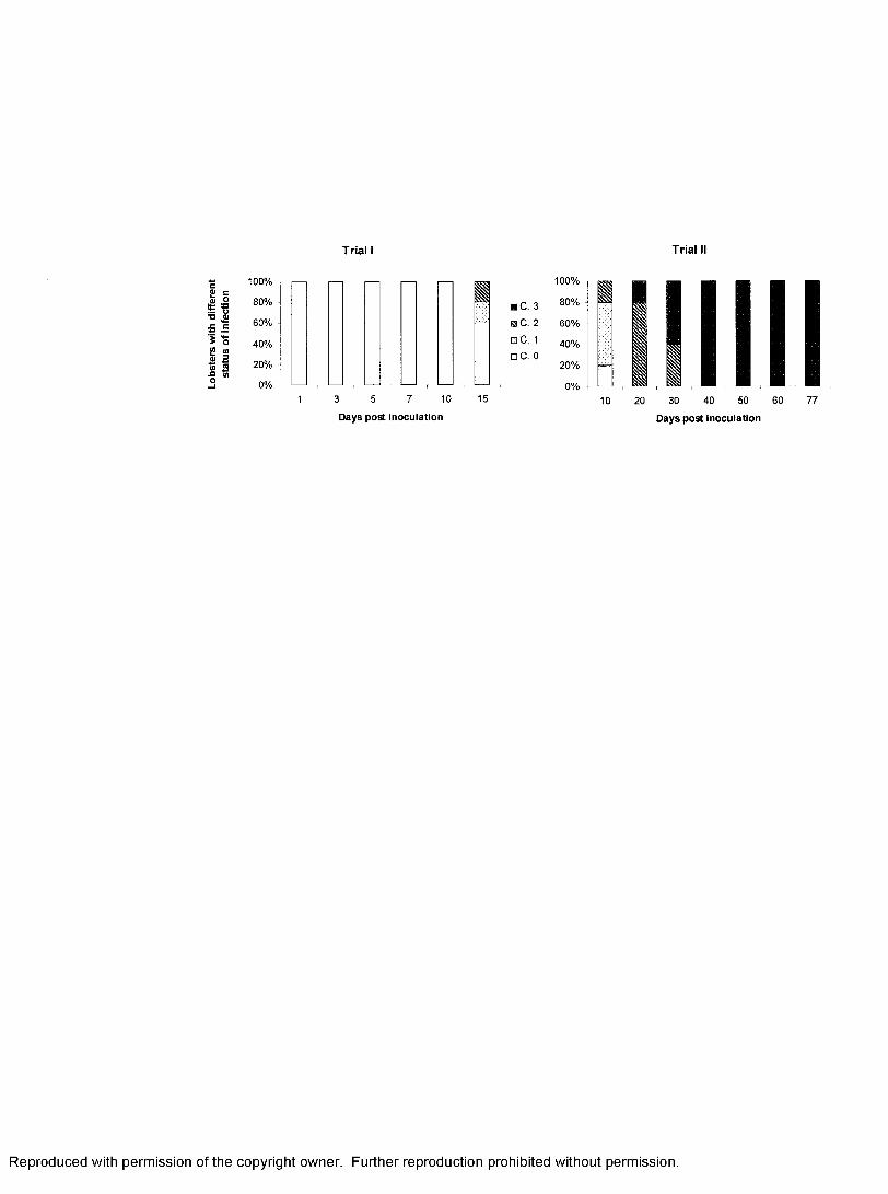
Lobs
ters
wi
th di
ffer
ent
T r ia l I T r ia l II
100%
5 80%01 60%
S<o
40%
20%
0%3 5 7 10
Days post inoculation
15
■ C. 3
a C . 2
O C . 1
□ C. 0
100%
80%
60%
40%
20%
0%10 20 30 40 50
Days post inoculation
Reproduced with permission of the copyright owner. Further reproduction prohibited without permission.

91
Figure 2. Pathological changes in the tissues o f infected lobsters from Trial I (low dose,
short term). A: resting fixed phagocytes surrounding an arteriole in the hepatopancreas of
a healthy lobster, B: activated fixed phagocytes in a lightly infected lobster, C: infected
fixed phagocytes (arrows) in the lightly infected lobster, D: infected fixed phagocytes
(arrows) in a moderately infected lobster, E: granulomas in the antennal gland of the
lightly infected lobster, F : accumulation o f granulocytes in hemal sinuses o f the
hepatopancreas o f the lightly infected lobster. Notice the alteration o f fixed phagocytes
in hemal sinuses o f the hepatopancreas over a time course o f PaV 1 infection, and the
change in cell densities surrounding the arterioles. FP: fixed phagocyte, A: arteriole, L:
lumen of tubule, Gr: granuloma, G: granulocyte.
Reproduced with permission of the copyright owner. Further reproduction prohibited without permission.
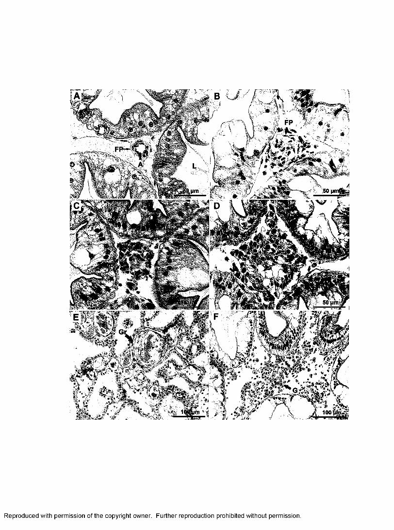
»»*.
Reproduced with permission of the copyright owner. Further reproduction prohibited without permission.

92
Figure 3. Total hemocyte counts (THC) o f lobsters from control and inoculation group
over early time course o f infection (Trial I). The number of cells was log transformed.
Sample size was three lobsters in control and five in experimental groups, bars = standard
deviation.
Reproduced with permission of the copyright owner. Further reproduction prohibited without permission.
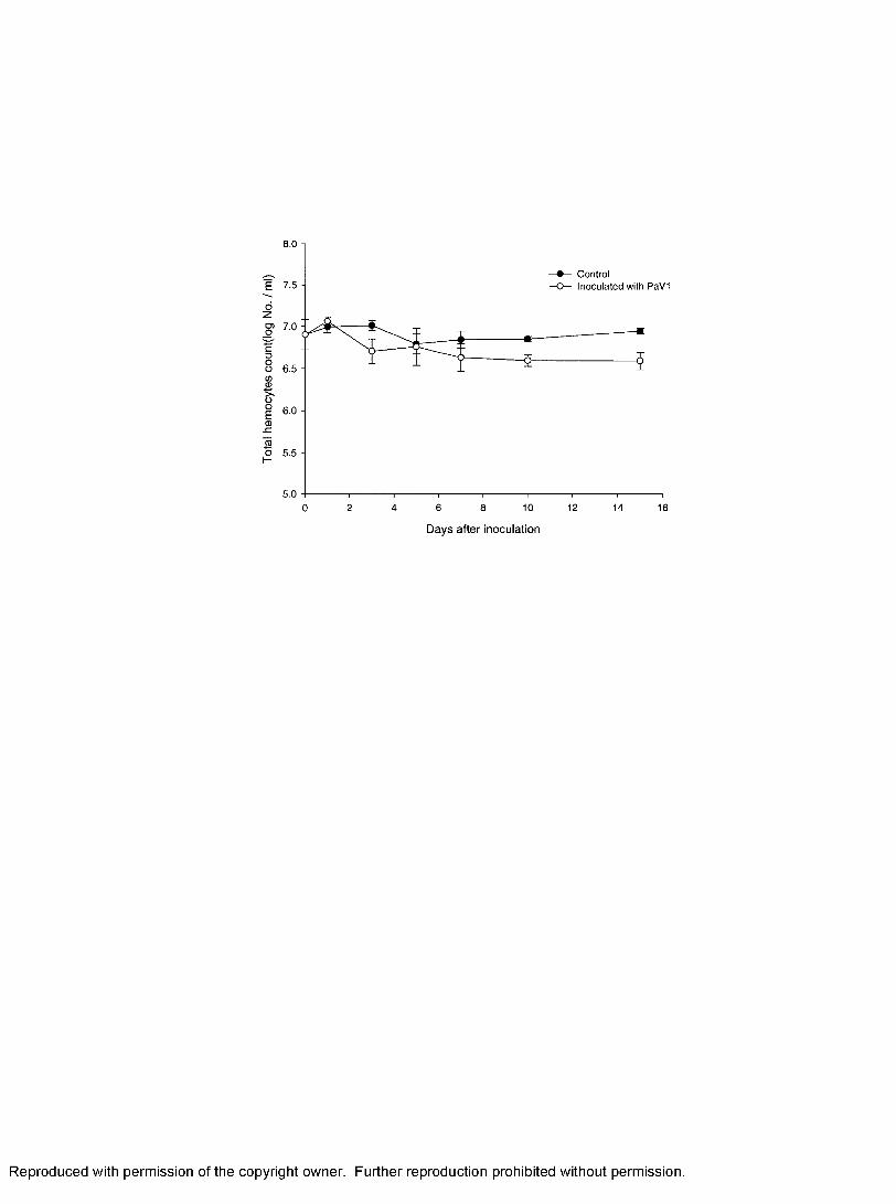
ControlInocu lated with PaV1E
ozo>oc3ootoCD
ooEoSI
ego 5 .5 -H
5.06 8 10 12 14 160 2 4
D ay s a fte r inoculation
Reproduced with permission of the copyright owner. Further reproduction prohibited without permission.

93
Figure 4. Pathological changes in the hepatopancreas of P. argus from Trial II (high
dose, long term). A: healthy, B: light infection, C: moderate infection, D: heavy infection,
E: chronic heavy infection, F: hemal space among the tubules of the hepatopancreas o f a
chronically infected lobster. Notice the accumulation of massive amount of cells in the
hemal sinus (HS) among the tubules o f the hepatopancreas in moderately and heavily
diseased animals. Arrows indicated infected cells. L: lumen of tubule, RI: reserved
inclusion cell, A: arteriole, SCT: spongy connective tissues.
Reproduced with permission of the copyright owner. Further reproduction prohibited without permission.

Reproduced with permission of the copyright owner. Further reproduction prohibited without permission.Reproduced with permission of the copyright owner. Further reproduction prohibited without permission.

94
Figure 5. Pathological changes in the hindgut (A), gill (B), spongy connective tissues
around nerve tissues (C) and heart (D, E, F). Infected cells (arrows) were present in the
hemal sinuses o f the heart, arteriole in the gill, and spongy connective tissues. Notice the
significant changes in hemal sinuses (HS) in the heart tissues of heavily infected lobster.
FCT: fibrous connective tissue, RI: reserve inclusion cell, M: myocardium, HS: hemal
sinus.
Reproduced with permission of the copyright owner. Further reproduction prohibited without permission.

Reproduced with permission of the copyright owner. Further reproduction prohibited without permission.Reproduced with permission of the copyright owner. Further reproduction prohibited without permission.

GENERAL CONCLUSIONS
Chapter 1
1) A specific 110-bp probe was generated from a plasmid containing a 177-bp fragment
o f the virus using the primer pair PaVl 110 F/R. The probe was applied to diagnose
PaVl in tissues of the Caribbean spiny lobsters Panulirus argus using fluorescence in
situ hybridization (FISH) assay.
2) In dot-blot hybridizations, the probe had a minimum sensitivity of 10 pg of cloned
177-bp plasmid DNA. The probe detected the presence of viral DNA from 10 ng of
genomic DNA extracted from hemolymph of a PaVl infected spiny lobster.
3) The fluorescein (FITC)-labeled probe specifically hybridized to PaV 1-infected
hemocytes and spongy connective tissue cells in or around the hepatopancreas,
hindgut, foregut, gill, heart, skin, nerve and even ovary tissues. Most FITC-stained
foci were located around the inner periphery of the hypertrophied nuclear membrane,
with a few dispersed throughout the inside of the nucleus.
95
Reproduced with permission of the copyright owner. Further reproduction prohibited without permission.

96
4) The probe did not hybridize with host tissues of uninfected spiny lobsters, nor
did it cross-react with a herpes-like virus (HLV) from a blue king crab Paralithodes
platypus, lymphocystis disease virus (LDV) from a striped bass Morone saxatilis,
Ostreid Herpesvirus 1 (OsHV-1) from an infected Pacific oyster Crassostrea gigas,
and Intranuclear bacilliform virus (IBV) from an infected brown shrimp Crangon
crangon.
This specific and sensitive FISH assay provides a useful tool to investigate infections
in tissues of lightly infected lobsters in our experimental trials. This probe can be used to
detect the presence of virus in hemolymph o f lobsters using dot-blot hybridization and to
monitor the prevalence o f PaVl in nurseries o f juvenile lobster.
Chapter 2
1) Three major hemocyte types - hyalinocytes, semi granulocytes, and granulocytes -
were identified in the Caribbean spiny lobster based on cell size, cell shape, and
granularity.
2) Lobster hemocytes were physically separated into three distinct subpopulations
enriched in hyalinocytes, semigranulocytes, and granulocytes, respectively, using
Reproduced with permission of the copyright owner. Further reproduction prohibited without permission.

97
Percoll discontinuous gradients centrifugation.
3) A modified Leibovitz L-15 medium supported the best survival of hemocytes in in
vitro primary cultures. Fetal bovine serum was not an effective additive for survival
o f hemocytes in vitro in this study.
4) When hemocytes were cultured together, most of the granulocytes dehisced, releasing
their contents into the culture media, negatively influencing the survival o f other the
cell types. When cultured separately, hyalinocytes and semigranulocytes maintained
higher viability (~ 80%) over 18 days incubation compared with granulocytes, which
degraded over 2-3 days.
5) Hyalinocytes and semigranulocytes cultured in vitro were susceptible to P aV l.
Cytopathic effects (CPE) were observed as early as 12 h post-inoculation, and cell
lysis was noticeable within 24 hrs of infection.
6) The presence o f virus within cells was confirmed by in situ hybridization using the
specific PaV 1 110-bp DNA probe derived from Chapter 1. The unique staining of
PaV 1 infected cells was observed after 24 h post-inoculation.
The in vitro assay can be applied to quantify the dose of infectious virus in hemolymph
using a 50% tissue culture infectious dose assay (TCID50) based on CPE. It may also be
used in propagation of PaV 1 in vitro, which is useful for isolation and purification of the
virus. The in vitro culture of hemocytes will facilitate better characterization o f different
Reproduced with permission of the copyright owner. Further reproduction prohibited without permission.

98
hemocyte types of lobster, as well as cell lineage of hemocyte types in crustacean.
Chapter 3
1) The fixed phagocytes in the hepatopancreas were the initial site of PaVl infection in
spiny lobsters. Fixed phagocytes were activated in early infections and then became
obviously infected as the disease progressed.
2) Infected cells, including hyalinocytes, semigranulocytes and spongy connective
tissue cells, were subsequently observed in the hepatopancreas, gill, heart, hindgut,
glial cells around the ventral nerves, as well as in the cuticular epidermis and foregut.
In advanced infections, all of the spongy connective tissues were infected as were,
the glial cells around the optic nerves.
3) The hepatopancreas of infected lobsters was associated with progression of the
disease. As the infection progressed, the hemal sinuses within the hepatopancreas
became filled with massive amounts o f cellular aggregates, including infected
circulating hemocytes and infected spongy connective tissues.
4) In lobsters chronically infected (> 60 days) by PaV l, the hemal spaces among the
tubules of the hepatopancreas were markedly dilated. The tubules of the
hepatopancreas were greatly atrophied.
Reproduced with permission of the copyright owner. Further reproduction prohibited without permission.

99
5) Reserved inclusions (RI) cells were gradually depleted in the hepatopancreas
o f chronically infected lobsters. This depletion supports the contention that the
cause of death in infected lobsters is metabolic exhaustion (Shields and Behringer,
2004).
6) The virus caused significant decreases in total hemocyte density in early infection;
however, there was no significant change in the proportion of each type of cell in the
hemolymph.
7) There were significant changes in concentrations of glucose, phosphorus,
triglycerides, and lipase in the hemolymph of lobsters infected with P aV l. The
changes in these parameters likely reflect tissue degradation and catabolism of the
hepatopancreas in relation to severity, and support the hypothesis that metabolic
exhaustion is the primary cause of death for infected lobsters.
The results of this study indicated that fixed phagocytes in the hepatopancreas of
experimentally injected lobsters were the initial sites of PaVl infection. I speculate that
once PaV 1 virions enter the open circulating hemolymph of the lobster, fixed phagocytes
in the hepatopancreas and elsewhere phagocytize the viruses and remove them from the
hemolymph. Because fixed phagocytes are susceptible to the virus or perhaps because of
phagocytosis, the fixed phagocytes become infected by the virus and possibly trigger a
series o f host defense responses (e.g. accumulation o f hemocytes) against the activated
Reproduced with permission of the copyright owner. Further reproduction prohibited without permission.

100
and infected fixed phagocytes. However, hyalinocytes and semigranulocytes are
also susceptible to the virus. Therefore, the virus may be spread via circulation of the
hemolymph, and cause infection of those tissues originated from mesoderm (e.g. spongy
connective tissues in hindgut and foregut, glial cells in nerve tissues). However, how the
virus gains entry into the lobster host naturally and how the virus is spread in the lobster
host with natural infections requires further study.
Reproduced with permission of the copyright owner. Further reproduction prohibited without permission.

LITERATURE CITED
Acharya S., Mohanty J., Sahoo P.K., 2004. Humoral defence factors in Indian river prawn Macrobrachinum malcolmsonii. Fish and Shellfish Immunology 17, 137-147.
Alpuche J., Pereyra A., Agundis C., Rosas C., Pascual C., Slomianny M.C.,Vaquez L., Zenteno E., 2005. Purification and characterization of a lectin from the white shrimp Litopenaeus setiferus (Crustacea decapoda) hemolymph. Biochimica et Biophysica Acta 1724, 86-93.
Andreeff, M., Pinkel, D., 1999. Introduction to fluorescence in situ hybridization- principles and clinical applications, 1st ed. Wiley-Liss, New York.
Assavalapsakul, W., Smith, D.R., Panyin, S., 2003. Propagation of infectiousyellow head virus particles prior to cytopathic effect in primary lymphoid cell cultures of Penaeus monodon. Diseases of Aquatic Organisms 55, 253-258.
Bachere E., 2000. Shrimp immunity and disease control. Aquaculture 191, 3-33.
Bachere, E., Mialhe, E., Rodriguez, J., 1995. Identification of defense effectors in hemolymph of crustaceans with particular reference to the shrimp Penaeus japonicus (Bate): prospects and applications. Fish and Shellfish Immunology 5, 597-612.
Battison, A., 2006. Tissue distribution and hemolymph activity of six enzymes in the American lobster (Homarus americanus): potential markers of tissue injury. Journal of Shellfish Research 25, 553-560.
Bauchau, A.G., 1980. Crustaceans. In: Ratcliffe, N.A. Rowley, A.F. (eds.), Invertebrate Blood Cells. Academic Press, New York, pp. 385-420.
Beatty B, Mai S, Squire J., 2002. FISH: a practical approach. Oxford University Press Oxford, New York.
101
Reproduced with permission of the copyright owner. Further reproduction prohibited without permission.

102
Behringer, D.C. Jr., 2003. The ecological ramifications o f disease and density on juvenile spiny lobster, Panulirus argus. Ph.D. Dissertation, Old Dominion University.
Behringer, D.C., Butler, M.J., Shields, J.D., 2006. Avoidance o f disease in social lobsters. Nature 441, 421.
Bell T.A., Lightner D.V., 1988. A handbook o f normal shrimp histology. Special Publication. World Aquaculture Society, Baton Rouge, LA
Bonami, J.R., Hasson, K.W., Mari, J., Poulos B.T., Lightner D.V., 1997. Taura syndrome of marine penaeid shrimp: characterization o f the viral agent. Journal o f General Virology 78,313-319.
Brock, J.A., Lightner, D.V., 1990. Diseases o f Crustacea: Diseases caused bymicroorganisms. In: Kinne, O. (eds), Diseases of Marine Animals (Vol. III). Biologische Anstalt Helgoland, Hamburg, pp.245-325.
Bruce L.D., Lightner, D.V., Redman, R.M., 1994. Comparison of traditional molecular detection methods for Baculovirus penaei infection in larval Penaeus vannamei. Journal of Aquatic Animal Health 6, 335-359.
Bruce L.D., Redman, R.M., Lightner, D.V., Bonami, J.R., 1993. Application o f gene probes to detect a penaeid shrimp baculovirus in fixed tissue using in situ hybridization. Diseases of Aquatic Organisms 17,215-221.
Butler, M. J., Behringer, D.C., Shields, J.D., 2007. Transmission of Panulirus argus virus 1 (PaVl) and its effect on the survival of juvenile Caribbean spiny lobster. Diseases o f Aquatic Organisms (in review).
Butler, M.J., 2001. The 6th International Conference and Workshop on Lobster Biology and Management: An Introduction. Marine & Freshwater Research 52,1033-1035.
Cai, S., J. Huang, C. Wang, X. Song, X. Sun, J. Yu, Y. Zhang, C. Yang., 1995.Epidemiological studies on the explosive epidemic disease of prawn in 1993 - 94. Journal of Fishery China 19, 112-117.
Carnegie, R.B., Barber, B.J., Distel, D.L., 2003. Detection of the oyster parasite Bonamia ostreae by fluorescent in situ hybridization. Diseases of Aquatic Organisms 55,247-252.
Reproduced with permission of the copyright owner. Further reproduction prohibited without permission.

103
Chang, P.S., Chen, H.C., Wang, Y.C., 1998. Detection of white spot syndrome associated baculovirus in experimentally infected wild shrimp, crab and lobsters by in situ hybridization. Aquaculture 164, 233- 242.
Chang, P. S., Lo, C.F., Wang, Y.C., Kou, G.H., 1996. Identification o f white spotsyndrome associated baculovirus (WSBV) target organs in the shrimp Penaeus monodon by in situ hybridization. Diseases o f Aquatic Organism 27, 131-139.
Chen, S.N., Wang, C.S., 1999. Establishment o f cell culture systems from penaeid shrimp and their susceptibility to white spot disease and yellow head viruses. Methods in Cell Sciences 21,199-206.
Cochrane, K.L., Chakalall B., 2001. The spiny lobster fishery in the WECAFC region - an approach to responsible fisheries management. Marine and Freshwater Research. 52, 1623-1631.
Couch J.A., 1974a. An enzootic nuclear polyhedrosis virus o f pink shrimp: ultrastructure, prevalence, and enhancement. Journal of Invertebrate Pathology 24, 311-331.
Couch J.A., 1974b. Free and occluded virus, similar to Baculovirus, in hepatopancreas of pink shrimp. Nature 247, 229-231.
Darby I.A., 2000. In situ hybridization protocols, 2nd ed. Humana Press, Totowa, N.J.
Darling, A.J., Boose, J.A., Spaltro, J., 1998. Virus assay methods: accuracy and validation. Biologicals 26, 105-110.
Dee, K.U., Shuler, M.L., 1997. Optimization of an assay for baculovirus titer and design of regimens for the synchronous infection of insect cells. Biotechnology Progress 13,14-24.
Dungan C.F., Reece, K.S., 2006. In vitro propagation of two Perkinsus spp. parasites from Japanese m anila clam s Venerupis philippinarum and description of Perkinsus honshuensis n. sp. Journal of Eukaryotic Microbiology 53:1-11.
Durand, S., Lightner, D.V., Nunan, L.M., Redman, R.M., Mari, J., Bonami, J.R., 1996. Application of gene probes as diagnostic tools for White Spot Baculovirus (WSBV) of penaeid shrimp. Diseases o f Aquatic Organisms 27, 59-66.
Reproduced with permission of the copyright owner. Further reproduction prohibited without permission.

104
Evans, L.H., Hones, J.B., Brock, J.A., 2000. Diseases of spiny lobster. In: Philips, B.F., Kittaka, J. (eds), Spiny lobster: fisheries and culture. Blackwell, Oxford, pp. 461-471.
Factor, J.R., Naar, M., 1985. The digestive system of the lobster, Homarus americanus: I. Connective tissue of the digestive gland. Journal of Morphology 184, 311-321.
Factor, J.R., 1995. Biology of the lobster Homarus americanus. Academic Press Inc., San Diego, CA, USA.
Factor, J.R., Orban, K., Szarowski, D.H., Lin, G., Larocca, T., Becker, A., Kapusta, K. J., 2005. A method for assessing removal o f foreign particles from the blood by fixed phagocytes o f the American lobster, Homarus americanus. Journal o f Shellfish Research 24, 713-717.
Fan, T.J., Wang, X.F., 2002. In vitro culture o f embryonic cells from the shrimp, Penaeus chinensis. Journal of Experimental Marine Biology and Ecology 267, 175-184.
FAO, 2001. Report o f the Workshop on Management of the Caribbean Spiny Lobster {Panulirus argus) Fisheries in the Area of the Western Central Atlantic Fishery Commission. Merida, Mexico, 4-8 September 2000. FAO Fisheries Report. No. 643. Rome, pp. 66.
FAO, 2004. Fisheries Global Information System, Factsheet for Panulirus argus. Website: http://www.fao.org/figis/servlet/species‘?fid=3445
Farley, C.A., 1978. Viruses and viruslike lesions in marine mollusks. Mar. Fish. Rev. 40, 18-20.
Farley, C.A., Banfield, W.G., Kasnic, G., Foster, W.S., 1972. Oyster herpes-type virus. Science 178, 759-760.
Federici, B.A., 1980. Isolation of an iridovirus from two terrestrial Isopods, the pill bug, Armadillidium vulgare, and the sow bug, Porcellio dilatatus. Journal of Invertebrate Pathology 36, 373-381.
Field, J.M., Butler IV, M.J., 1994. The influence o f temperature, salinity, and postlarval transport on the distribution of juvenile spiny lobsters, Panulirus argus (Lateille, 1804) in Florida Bay. Crustaceana 67, 26-45.
Reproduced with permission of the copyright owner. Further reproduction prohibited without permission.

105
Flegel, T.W., 1997. Major viral diseases o f the black tiger prawn {Peanuts monodon) in Thailand. World Journal o f Microbiology and Biotechnology 13, 433-442.
FMRI, 2003. Caribbean spiny lobster, Panulirus argus.http://research.mvfwc.com/features/category sub.asp?id=2326.
FMRI, 2005. A Summary of Recreational Spiny Lobster Landings and Effort in Florida. http://marineeconomics.noaa.gov/SocmonFK/FMRI Lobster Fact.ndf
Forcucci, D.F., M.J. Butler, Hunt, J.H., 1994. Growth and population dynamics of juvenile spiny lobsters, Panulirus argus, in Florida Bay. Bulletin of Marine Science 54, 805-818.
Fraser, C.A., Hall, M.R., 1999. Studies on primary cell cultures derived from ovarian tissue of Penaeus monodon. Methods in Cell Sciences 21, 213-218.
Frerichs, G.N., 1996. In vitro culture of embryonic cells from the freshwater prawn Macrobrchium rosenbergii. Aquaculture 143, 227-232.
Gao, C.L., Sun, J.S., Xiang, J.H., 2003. Primary culture and characteristic morphologies of medulla terminalis neurons in the eyestalks of Chinese shrimp,Fenneropenaeus chinensis. Journal of Experimental Marine Biology and Ecology 290, 71-80.
Harper, D. E., 1995. The 1995 spiny lobster update o f trends in landings, CPUE, and size o f harvested lobster. National Marine Fishery Service Report No. MIA-94/95-47, Miami, Florida.
Hejkal, T.W., Gerba, C.P., 1981. Uptake and survival of enteric viruses in blue crab, Callinectes sapidus. Applied and Environmental Microbiology 41, 207-211.
Holthuis, L.B., 1991. FAO species catalogue. Vol. 13. Marine lobsters o f the world. Anannotated and illustrated catalogue of species of interest to fisheries known to date. FAO Fisheries Synopsis, vol. 125 (13). FAO , R om e, pp. 292.
Hose J. E., Martin G. G., Gerard A.S., 1990. A decapod hemocyte classification scheme integrating morphology, cytochemistry and function. Biology Bulletin 178, 33-45.
Reproduced with permission of the copyright owner. Further reproduction prohibited without permission.

106
Hose, J.E., Martin, G.G., 1989. Defense functions o f granulocytes in the ridgeback prawn Sicyonia ingentis Burkenroad 1938. Journal o f Invertebrate Pathology 53,335-346.
Hsu, Y.L., Yang, Y. H., Chen, Y.C., Tung, M.C., Wu, J.L., Engelking, M. H., Leong, J.C., 1995. Development o f an in vitro subculture system for the oka organ of Penaeus monondon. Aquaculture 136, 43-55.
Humason, G.L., 1979. Animal tissue techniques, 4th ed. W.H. Freeman and company, San Francisco.
Inouye, K., Miwa, S., Oseko, N., Nakano, H., Kimura, T., 1994. Mass mortalities of cultured kuruma shrimp Penaeus Japonicus in Japan in 1993: electron microscopic evidence o f the causative virus. Fish Pathology 29, 149-158.
Itami, T., Maeda, M., Kondo, M., Takahashi, Y., 1999. Primary culture o f lymphoid organ cells and haemocytes of kuruma shrimp, Penaeus japonicus. Methods in Cell Sciences 21, 237-244.
Jiravanichpaisal, P. Bangyeekhun, E., Soderhall, K., Soderhall, I., 2001. Experimental infection o f white spot syndrome virus in freshwater crayfish Pacifastacus leniusculus. Diseases o f Aquatic Organisms 47, 151-157.
Jiravanichpaisal, P. Sricharoen, S., Soderhall, I., Soderhall, K., 2006. White spotsyndrome virus (WSSV) interaction with crayfish haemocytes. Fish and Shellfish Immunology 20, 718-727.
Johansson M.W., Keyser P., Sirtunyalucksana K., Soderhall K., 2000. Crustacean haemocytes and haematopoiesis. Aquaculture 191, 45-52.
Johansson M.W., Soderhall K., 1989. Cellular immunity in crustaceans and the proPO system. Parasitology Today 5, 171-177.
Johnson P.T., 1976. A H erpes-like virus from the blue crab, Callinectes sapidus. Journal o f Invertebrate Pathology 27, 419-420.
Johnson P.T., 1978. Viral diseases of blue crab, Callinectes sapidus. Marine Fisheries Review 40 (10), 13-15.
Reproduced with permission of the copyright owner. Further reproduction prohibited without permission.

107
Johnson, P.T., 1980. Histology of the blue crab, Callinectes sapidus. Praeger Publishers, New York, USA.
Johnson, P.T., 1983. Diseases caused by viruses, rickttsiae, bacteria, and fungi, in: Provenzano, A.J. (eds), The biology of crustacean: pathology (Vol. 6). Academic Press, New York, pp.1-78.
Johnson, P.T., 1987. A review of fixed phagocytic and pinocytotic cells of decapod crustaceans, with remarks on hemocytes. Developmental and Comparative Immunology 11, 679-704.
Johnson P.T., 1988. Development and morphology of an unusual nuclear virus of the blue crab, Callinectes sapidus. Diseases of Aquatic Organisms 4, 67-75.
Johnson, S.K., Cassout, D., 1995. Electron microscopic examination o f negatively stained samples for diagnosis of virus diseases o f shrimp. World Aquaculture 26, 37.
Kasomchandra, J., Khongpradit, R., Ekpanithanpong, U., Boonyarapalin, S., 1999.Progress in the development o f shrimp cell cultures in Thailand. Methods in Cell Sciences 21, 231-235.
Kopacek P., Hall M., Soderhal K., 1993. Characterization o f a clotting protein isolated from plasma of the freshwater crayfish Pacifastacus leniusculus. European Journal of Biochemistry 213, 591-597.
Kurtz J., 2005. Specific memory within innate immune systems. Trends in Immunology 26, 186-192.
LaBarre, D.D., Lowy, R.J., 2001. Improvement in methods for calculating virus titer estimates from TCID50 and plaque assays. Journal o f Virological Methods 96, 107-126.
Lang, G.H., Nomura, N., Matsumura, M., 2002. Growth by cell division in shrimp {Penaeusjaponicus) ce ll culture. Aquaculture 213, 73-83 .
Le Deuff, R.M., Renault, T., 1999. Purification and partial genome characterization o f a herpes-like virus infecting the Japanese oyster, Crassostrea gigas. Journal of General Virology 80,1317-1322.
Reproduced with permission of the copyright owner. Further reproduction prohibited without permission.

108
Lee, S.Y., Soderhall, K., 2001. Characterization of a pattern recognition protein a masquerade-like protein in the freshwater crayfish Pacifastacus leniusculus. Journal of Immunology 166, 7319-7326.
Leibovitz, L., Koulish, S., 1989. A viral disease of the ivory barnacle, Balanus eburneus, Gould (Crustacea, Cirripedia). Biology Bulletin 176, 301-307.
Li, C., Shields, J. D., 2007. Primary culture of hemocytes from the Caribbean spiny lobster, Panulirus argus, and their susceptibility to Panulirus argus Virus 1 (PaVl). Journal o f Invertebrate Pathology 94, 48-55.
Li, C., Shields, J.D., Small, H.J., Reece, K. S., Hartwig, C.L., Cooper, R.A., Ratzlaff, R.E., 2006. Diagnosis o f Panulirus argus virus 1 (PaVl) in the Caribbean spiny lobster using fluorescence in situ hybridization. Diseases of Aquatic Organisms 72, 185-192.
Lightner, D. V., Redman, R.M., Bell, T.A., 1983. Observations on the geographic distribution, pathogenesis and morphology of the baculovirus form Peaneus monodon. Aquaculture 32, 209-233.
Lightner, D.V., 1988. Muscle necrosis o f penaeid shrimp. In: Sindermann, C.J., Lightner,D.V. (eds) Disease diagnosis and control in North America marine aquaculture. Development in aquaculture and fisheries science (Vol. 17). Elsevier Press, New York, pp. 122-124.
Lightner, D.V., 1999. The penaeid shrimp viruses TSV, IHHNV, WSSV and YHV:Current status in the Americas, available diagnostic methods, and management strategies. Journal of Applied Aquaculture 9(2), 27-52.
Lightner, D.V., Redman, R.M., 1998. Shrimp diseases and current diagnostic methods. Aquaculture 164, 201-220.
Lightner, D.V., Redman, R.M., Hasson, K.W., Pantoja, C.R., 1995. Taura syndrome in Penaeus vannamei Crustacea: Decapoda: gross signs, h istopathology and ultrastructure. Diseases o f Aquatic Organisms 1, 53-59.
Lipart, C., Renault, T., 2002. Herpes-like virus diction in infected Crassostrea gigas spat using DIG-labeled probes. Journal of Virological Methods 101, 1-10.
Reproduced with permission of the copyright owner. Further reproduction prohibited without permission.
109
Lipcius, R.N., Eggleston, D.B., 2000. Ecology and fishery biology of spiny lobsters. In: Philips, B.F., Kittaka, J. (eds) Spiny lobsters: fisheries and culture, 2nd edn. Blackwell, Oxford, pp. 1-41.
Lo, C.F., Ho, C.H., Chen, C.H., Liu, K.F., Chiu, Y.L., Yeh, P.Y., Peng, S.E., Hsu, H.C., Liu, J.C., Chang, C.F., Su, M.S„ Wang, C.H., Kou, G.H., 1997. Detection and tissue tropism of white spot syndrome baculovirus (WSBV) in captured brooders of Penaeus monodon with a special emphasis on reproductive organs. Diseases of Aquatic Organisms 30, 53-72.
Lo, C.F., Ho, C.H., Peng, S.E., 1996. White spot syndrome baculovirus (WSBV) detected in cultured and captured shrimp, crabs and other arthropods. Diseases o f Aquatic Organisms 27, 215-225.
Lu,Y., Lo, P.C., Brock, J.A., 1989. Isolation, purification and characterization ofinfectious hypodermal and haematopoietic necrosis virus o f penaeid shrimps. Journal of Virological Methods 26, 339-344.
Lu,Y., Tapay, L.M., Loh, P.C., Brock, J.A., Gose, R., 1995. Development o f a quantal assay in primary shrimp cell culture for yellow head baculovirus (YBV) of penaeid shrimp. Journal o f Virological Methods 52, 231-236.
Luedeman, R., Lightner, D.V., 1992. Development of an in vitro primary cell culture from the penaeid shrimp, Penaeus stylirostris and Penaeus vannamei. Aquaculture 101, 205-211.
Maeda, M., Saitoh, H., Mizuki, E., Itami, T., Ohba, M., 2004. Replication o f white spot syndrome virus in ovarian primary cultures from the kuruma shrimp, Marsupenaeus japonicus. Journal o f Virological Methods 116(1), 89-94.
Mari, J., Bonami, J.R., Lightner, D.V., 1993. Partial cloning of the genome of infectious hypodermal and hematopoietic necrosis virus, an unusual parvovirus pathogenic for penaeid shrimps; diagnosis of the disease using a specific probe. Journal of General V irology 74, 2 6 3 7 —2643.
Mari, J., Bonami, J.R., Lightner, D.V., 1998. Taura syndrome of penaeid shrimp: cloning of viral genome fragments and development of specific gene proves. Diseases of Aquatic Organisms 33, 11-17.
Reproduced with permission of the copyright owner. Further reproduction prohibited without permission.

110
Marques M. R. F., Barracco M. A., 2000. Lectins, as non-self-recognition factors, in crustaceans. Aquaculture 191, 23-44.
Marx, J.M., 1986. Recruitment and settlement of spiny lobster pueruli in south Florida. Canadian Journal of Fisheries and Aquatic Sciences 43, 2221-2227.
Mascotti, K., McCullough, J., Burger, S.R., 2000. HPC viability measurement: trypan blue versus acridine orange and propidium iodide. Transfusion 40(6), 693-696.
Minson, A.C., Davison, A., Eberle, R., Desrosiers, R.C. and five others, 2000. Family Herpesviridae. In: van Regenmortel, M.H.V., Fauqet, C.M., Bishop, D.H.L., Carstens, E.B. and 7 others (eds), Virus taxonomy: classification and nomenclature o f viruses. Seventh Report of the International Committee on Taxonomy of Viruses. Academic Press, San Diego, pp. 203-225.
Mohankumar K., Ramasamy P., 2006 a. Activities o f membrane bound phosphates,transaminases and mitochondrial enzymes in white spot syndrome virus infected tissues of Fenneropenaeus indicus. Virus Research 118, 130-135.
Mohankumar, K., Ramasamy P., 2006 b. White spot syndrome virus infections decrease the activity of antioxidant enzymes in Fenneropenaeus indicus. Virus Research 115, 69-75.
Montanie, H., Bonami, J.R., 1993. Irido-like virus infection in the crab Macropipus depurator L. (crustacean, Decapoda). Journal of Invertebrate Pathology 61, 320-322.
Muller, R. G., Hunt, J. H., Matthews, T. R., and Sharp, W. C., 1997. Evaluation o f effort reduction in the Florida Keys spiny lobster, Panulirus argus, fishery using an age-structured population analysis. Marine and Freshwater Research 48, 1045-1058.
Nadala, E.C., Lu, Y., Loh, P.C., 1993. Primary culture of lymphoid, nerve, and ovary cells from Penaeus stylirostris and Penaeus vannamei. In Vitro Cellular and Developmental Biology 29A, 620-622.
Nunan, L.M., Lightner, D.V., 1997. Development o f a non-radioactive gene probe by PCR for detection of white spot syndrome virus (WSSV). Journal of Virological Methods 63, 193-201.
Reproduced with permission of the copyright owner. Further reproduction prohibited without permission.

I l l
Owens, L., Smith, J., 1999. Early attempts at production o f prawn cell lines. Methods in Cell Sciences 21, 207-211.
Pantoja, C.R., Lightner, D.V., 2001. Detection of hepatopancreatic parvovirus (HPV) of penaeid shrimp by in situ hybridization at the electron microscope level. Diseases of Aquatic Organisms 44, 87-96.
Payen, G.G., Bonami, J.R., 1979. Mise en evidence de particules d’allure virale associees aux noyaux des cellules mesodermiques de la zone germinative testiculaire du crabe Rhithropanopeus harrisii (Gould) (Brachyoure, Xanthide). Revue Trav Inst Pech Marit 43,361-365.
Persson, M., Cerenius, L., Soderhall, K., 1987. The influence o f the hemocyte number on the resistance of freshwater crayfish, Pacifastacus leniusculus to parasitic fungus Aphanomyces astaci. Journal o f Fish Diseases 10, 471-477.
Phillips, B.F., Cobb, J.S., George, R.W., 1980. General Biology. In: Cobb, J.S., Phillips, B.F. (eds), The biology and management o f lobsters (Vol. I). Academic Press, New York, USA, pp. 1-82.
Phillips, B.F., McWilliam, D.L., 1987. The pelagic phase of spiny lobster development. Canadian Journal o f Fisheries and Aquatic Sciences 43, 2153-2163.
Phromjai, J., Boonsaeng, V., Withyachumnamkul, B., Flegel, T.W., 2002. Detection of hepatopancreatic parvovirus in Thai shrimp Penaeus monodon by in situ hybridization, dot blot hybridization and PCR amplification. Diseases o f Aquatic Organisms 51, 227-232.
Ratcliffe, N.A., Rowley, A.F., Fitzgeral, S.W., 1985. Invertebrate immunity: basicconcepts and recent advances. International Review of Cytology 97, 183-349.
Reed, L.J., Muench, H., 1938. A simple method of estimating fifty percent end points. American Journal o f Hygiene 27, 493- 497.
Relf, J. M., Chisholm, J. R. S., Kemp, G.D., Smith, V.J., 1999. Purification and characterization of a cysteine-rich 11.5-kDa antibacterial protein from the granular haemocytes o f the shore crab, Carcinus maenas. European Journal of Biochemistry 264, 350-357.
Reproduced with permission of the copyright owner. Further reproduction prohibited without permission.

112
Rinkevich, B., 1999. Cell cultures from marine invertebrates: obstacles, new approaches and recent improvements. Journal o f Biotechnology 70, 133-153.
Roch, P., 1999. Defense mechanisms and disease prevention in farmed marine invertebrates. Aquaculture, 172, 125-145.
Sano, T., 1998. A novel tissue organized in the primary hemolymph culture o f Penaeus japonicus Bate. Aquaculture 164, 289-296.
Sart, D. D., Choo, K.H.A, 1998. The technique of in situ hybridization: principles and application. In: Rapley, R., Walker, J.M. (eds), Molecular biomethods handbook. Humana Press Inc., Totowa, NJ.
Shields, J. D., Behringer, D.C., 2004. A pathogenic herpes-like virus in the Caribbean spiny lobster, Panulirus argus, from the Florida Key. Diseases of Aquatic Organisms 59, 109-118.
Shields, J.D., Overstreet, R.M., 2004. Parasites, symbionts, and diseases. In: Kennedy, V. (ed), The biology and management of the blue crab. University o f Maryland Sea Grant Press, MD.
Singer, R.H., Mass, S., Lawrence, J.B., Maplevill, R.I., 1989. In situ hybridization todetect nucleic acid sequences in morphologically intact cells. United States Patent, No. 4888279.
Smail, D.A., Munro, A.L.S., 2001. The virology of teleosts. In: Roberts, R.J. (ed), Fish Pathology, 3rd edn. W B Saunders Co., London.
Small, H.J., Neil, D.M., Taylor, A.C., Atkinson, R.J.A., Coombs, G.H., 2006. Molecular detection of Hematodinium spp. in the Norway lobster Nephrops norvegicus (L.), and other crustaceans. Diseases o f Aquatic organisms 69, 185-195.
Snow, A.L., Martinez, O.M., 2007. Epstein-Barr Virus: Evasive maneuvers in the developm ent o f PTLD. Am erican Journal o f Transplantation 7, 271-277 .
Soderhall, K., Cerenius, L., 1992. Crustacean immunity. Annual Review of Fish Diseases, p p .3-23.
Reproduced with permission of the copyright owner. Further reproduction prohibited without permission.

113
Soderhall, K., Smith, V. J., 1983. Separation o f the haemocyte populations of Carcinus meanas and other marine decapods, and prophenoloxidase distribution. Developmental and Comparative Immunology 7, 229-239.
Soderhall, K., Smith, V.J., Johansson, M.W., 1986. Exocytosis and uptake of bacteria by isolated haemocyte populations o f crustaceans: evidence fro cellular co-operation in the defence reactions o f arthropods. Cell and Tissue Research 245, 43-49.
Song, Y.L., Yu, C.I., Lien, T.W., Huang, C. C., Lin, M.N., 2003. Haemolymphparameters of Pacific white shrimp (litopenaeus vannamei) infected with Taura syndrome virus. Fish and Shellfish Immunology 14, 317-331.
Spann, K.M., McCulloch, R.J., Cowley, J.A., East, I.J., Walker, P.J., 2003. Detection of gill-associated virus (GAV) by in situ hybridization during acute and chronic infections o f Penaeus monodon and P. esculentus. Diseases o f Aquatic Organisms 56, 1-10.
Sparks, A.K., Morado, J.F., 1986. A herpes-like virus disease in the blue king crab, Paralithodes platypus. Diseases o f Aquatic Organisms 1, 115-122.
Stentiford, G.D., Bateman, K., Feist, S.W., 2004. Pathology and ultrastructure of an intranuclear bacilliform virus (IBV) infecting brown shrimp Crangon crangon (Decapoda: Crangonidae). Diseases o f Aquatic Organisms 58, 89-97.
Stentiford, G.D., Shields, J.D., 2005. A review of the parasitic dinoflagellatesHematodinium species and Hematodinium-like infections in marine crustaceans. Diseases o f Aquatic Organisms 66, 47-70.
Stokes, N.A., Burreson, E.M., 1995. A sensitive and specific DNA probe for the oyster pathogen Haplosporidium nelsoni. Journal of Eukaryotic Microbiology 42(4), 350-357.
Sugumaran, M., Nellaiappan, K., 2000. Characterization o f new phenoloxidase inhibitor from the cuticle o f Manduca sexta. B iochem ical and B iophysical Research Communications 268, 379-383.
Tapay, L.M., Lu, Y., Gose, R.B., Nadala, E.C., Brock, J.A., Loh, P.C., 1997.Development of an in vitro quantal assay in primary cell cultures for a non-occluded baculo-like virus o f penaeid shrimp. Journal of Virological Methods 64,37-41.
Reproduced with permission of the copyright owner. Further reproduction prohibited without permission.

114
Theate, I., Michaux, L., Dardenne, S., Guiot, Y., Briere, J., Emile, F.J., Fabiani, B.,Detry, R ., Gaulard, P., 2002. Epstein-Barr virus-associated lymphoproliferative disease occurring in a patient with sarcoidosis treated by methotrexate and methylprednisolone. European Journal of Haematology Supplementum 69 (4),248-253.
Tong, S.L., Miao, H.Z., 1996. Attempts to initiate cell cultures from Penaeus chinensis tissues. Aquaculture 147,151- 157.
Toullec, J.Y., 1999. Crustacean primary cell culture: a technical approach. Methods in Cell Sciences 21, 193-198.
Toullec, J.Y., Crozat, Y., Patroid, J., Porcheron, P., 1996. Development of primary cultures from the penaeid shrimps Penaeus vannamei and P. indicus. Journal o f Crustacean Biology 16, 643-649.
Theopold, U., Schmidt, O., Soderhall, K., Dushay, M. S., 2004. Coagulation inarthropods: defence, wound closure and healing. Trends in Immunology 25, 289-294.
Vargas-Albores, F., Jimenez-Vega, F., Soderhall, K., 1996. A plasma protein isolated from brown shrimp (Penaeus californiensis) which enhances the activation o f prophenoloxidase system by /?-l, 3-glucan. Developmental and Comparative Immunology 20, 299-306.
Villena, A.J., 2003. Application and needs o f fish and shellfish cell culture for disease control in aquaculture. Reviews in Fish Biology and Fisheries 13, 111-140.
Walton, A., Smith, V.J., 1999. Primary culture of the hyaline haemocytes from marine decapods. Fish and Shellfish Immunology 9, 181-194.
Wang, Y.T., Liu, W., Seah, J.N., Lam, C.S., Xiang, J.H., Korzh, V., Kwang, J., 2002. W hite spot syndrom e virus (W S S V ) infects specific hem ocytes o f the shrimp Penaeus merguiensis. Diseases of Aquatic Organisms 52, 249-259.
Wang, C.S., Yang, H.N., Tang, C.Y., Lu, C.H., Kou, G.H., Lo, C.F., 2000. Ultrastructure o f white spot syndrome virus development in primary lymphoid organ cell cultures. Diseases of Aquatic Organisms 41, 91-104.
Reproduced with permission of the copyright owner. Further reproduction prohibited without permission.

115
Wang, Y.C., Lo, C.F., Chang, P.S., Kou, G.H., 1998. Experimental infection of whitespot baculovirus in some cultured and wild decapods in Taiwan. Aquaculture 164, 221-231.
West, L., Mahony, T., McCarthy, F., Watahabe, J., Hewitt, D., Hansford, S., 1999.Primary cell cultures isolated from Penaeus monodon prawns. Methods in Cell Sciences 21, 219-223.
Williams, T., Chinchar, G., Drari, G., Kalmakoff, J., Seligy, V., 2000. FamilyIridoviridae. In: van Regenmortel, M.H.V., Fauqet, C.M., Bishop, D.H.L., Carsten,E.B. and 7 others (eds) Virus taxonomy: classification and nomenclature o f viruses. Seventh Report o f the International Committee on Taxonomy of Viruses. Academic Press, San Diego, pp. 167-182.
Wongteerasupaya, C., Sriurairatane, S., Vickers, J. E., Akrajamom, A., Boonsaeng, V., Panyim, S., Tassanakajon, A., Withyachumnamkul, B., Flegel, T. W., 1995. Yellow-head virus of Penaeus monodon is an RNA virus. Diseases o f Aquatic Organisms 22, 45-50.
Wongteerasupaya, C., Wongwisansri, S., Boonsaeng, V., Payim, P., Nash, G.L.,Withyachumnamkul, B., Flegel, T.W., 1996. A non-occluded, system baculovirus that occurs in cells of ectodermal and mesodermal origin and causes high mortality in the black tiger prawn, Penaeus monodon. Diseases o f Aquatic Organisms 21, 69-77.
Wu, J.L., Nishioka, T., Mori, K., Nishizawa, T., Muroga, K., 2002. A time-course study on the resistance of Peaneus japonicus induced by artificial infection with white spot syndrome virus. Fish and Shellfish Immunology 13, 391-403.
Yoganandhan, K., Thirupathi, S., Sahul Hameed, A.S., 2003. Biochemical, physiological and hematological changes in white spot syndrome virus-infected shrimp,Penaeus indicus. Aquaculture 221,1-11.
Reproduced with permission of the copyright owner. Further reproduction prohibited without permission.

VITA
CAIWEN LI
Bom in Anqiu, Shangdong Province, People’s Republic of China on 24 November 1977. Graduated from the High School of Anqiu. Earned B.S. in Chemistry from Qingdao University in 1997, and M.S. in Marine Science from Institute of Oceanology, Chinese Academy of Science. Entered doctoral program in College of William and Mary, Virginia Institute of Marine Science in August, 2002.
116
Reproduced with permission of the copyright owner. Further reproduction prohibited without permission.