GEORGIOS MAVROUDIS Research on Consciousness via Psychedelics
Upload
baldwin-mcgeeCategory
view
216download
1
The impact of spore aggregation on viable and total counts
of Bacillus subtilis
Nikos Mavroudis and Catherine Bowe
Food Engineering and Separation of Actives, FoESA, laboratory
Department of Applied Sciences, Faculty of Health and Life Sciences
Northumbria University at Newcastle, UK

Introduction• In a number of occasions
controlling the number of microorganisms on surfaces or in complex liquid environments, from fruit juices and drinks to GIT fluids, is of importance. However in order to do so, it is important that an accurate bacterial enumeration method is in place to avoid over or under-estimating the remaining bacterial counts.
• Traditionally bacterial counts are obtained through plating techniques under the assumption of one colony generated from one cell.

Bacterial aggregation• Aggregation often occurs in bacterial
populations as a result of the physicochemical and biological parameters within the system.
• in the dormant spore form the physicochemical aspects will be the dominating factor
• Attachment of dormant spores, either between themselves or to surfaces, is considered to be a surface-driven phenomenon and can be interpreted using the Derjaguin-Landau-Verwey-Overbeek (DLVO) theory.

Bacterial aggregation and intermolecular forces
• Based on the DLVO theory the main forces at work causing aggregation are:– electrostatic forces– permanent dipole -
permanent dipole forces
– Lifshitz-van der Waals forces
a) Diagram showing different layers of the dormant spore, and b) Thin section transmission electron micrograph of a B. subtilis spore stained with ruthenium red.

Bacterial aggregation and intermolecular forces
McKenney et al. 2013 Nature Reviews Microbiology 11: 33-44
B. Subtilis spore coat and crust proteins• Based on the DLVO
theory the main forces at work causing aggregation are:– electrostatic forces– permanent dipole -
permanent dipole forces
– Lifshitz-van der Waals forces

AimsIn this communication spores of B. subtilis are exposed to PBS (100mM) at pH 1, 3 5 and 7 either with or without 1% tween 20 and we aim to investigate the impact of aggregation in the viable and total counts.
• Total counts as well as viable counts were assessed by plating and flow cytometry (FCM).
• Aggregation was assessed by particle size analysis using a Malvern masterziser.
• The electrostatic charge of spores was assessed by Zeta potential measurements using a Malvern zetasizer

Total counts from FCM showing spores immersed in PBS
1 3 5 77
7.2
7.4
7.6
7.8
8
8.2
8.4
8.6
8.8
9
PBS only
PBS with 1% tween
pH
log
cfu/
ml
Comparison of total spore FCM counts in PBS over a range of pHs. White bars indicate spores in PBS only and red bars show spores in PBS supplemented with 1% tween. Values are based on the total number of cells and spores according to FCM. Error bars represent the 95% CI.
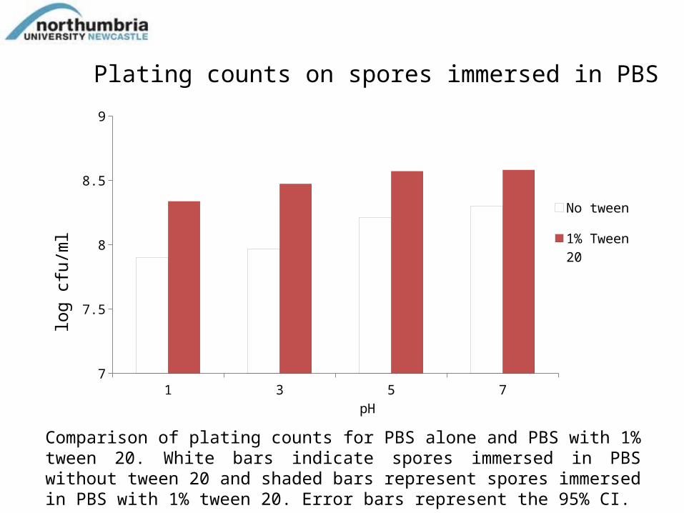
Plating counts on spores immersed in PBS
1 3 5 77
7.5
8
8.5
9
No tween
1% Tween 20
pH
log
cfu/
ml
Comparison of plating counts for PBS alone and PBS with 1% tween 20. White bars indicate spores immersed in PBS without tween 20 and shaded bars represent spores immersed in PBS with 1% tween 20. Error bars represent the 95% CI.

Spore particle size analysis
Particle size distribution of spores in PBS (100mM) at different pH levels. Lines represent the average of three samples. pH 7 - Red line pH 5 - Green line pH 3 - Blue line pH 1 - Black line
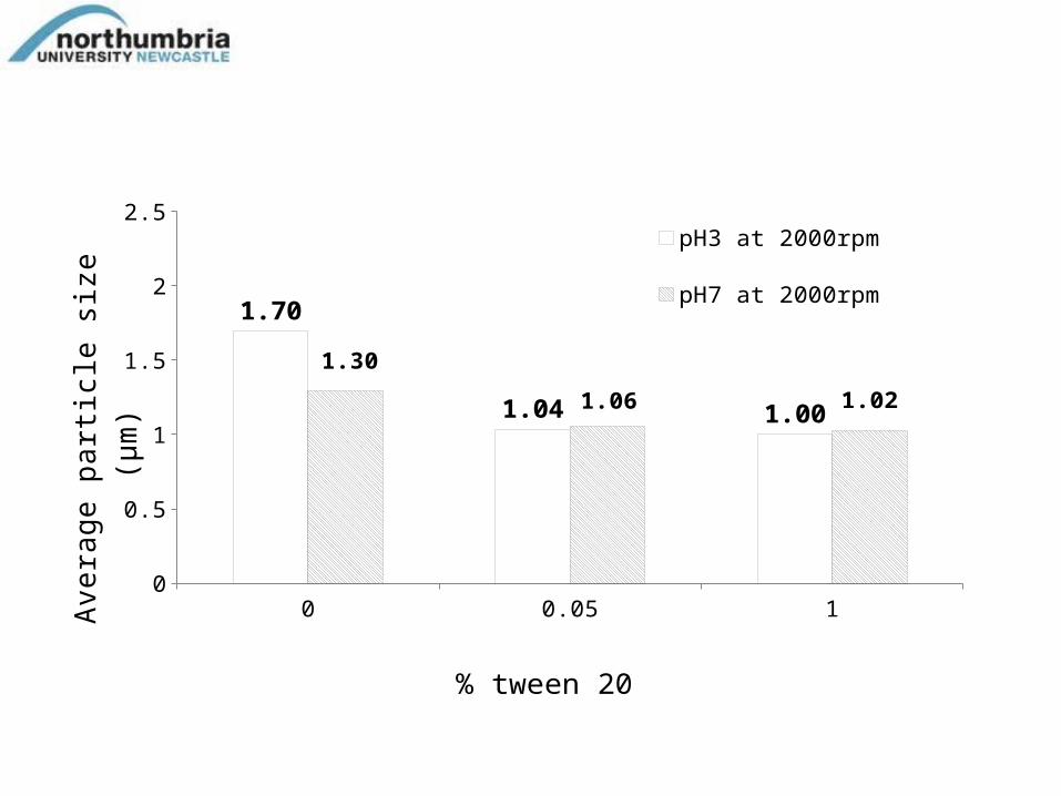
0 0.05 10
0.5
1
1.5
2
2.5
1.70
1.04 1.00
1.30
1.06 1.02
pH3 at 2000rpm pH7 at 2000rpm
% tween 20
Ave
rage
par
ticl
e si
ze (
µm
)

Zeta-potential of B Subtilis spores in PBS (100mM)
1 3 5 7
-8
-7
-6
-5
-4
-3
-2
-1
0pH
Zeta
pot
entia
l (m
V)
Zeta potential for spores immersed in PBS at pH 1, 3, 5 and 7. Data represent the average of 9 measurements and error bars represent the 95% CI

Conclusions • Across the investigated pH range 1-7 in PBS, tween
increased plate counts between 50-75%. This highlights the level of error in count one could expect as a result of the aggregation from standard enumeration techniques
• Data from the Malvern mastersizer strongly support the theory that the average particle size increases as the pH lowers, denoting a higher number of aggregate in the system.
• Data from the Malvern Zetasizer strongly support the theory that the aggregation is enhanced as the pH lowers as a result of the reduction of spore zeta potential.

Acknowledgements• Dr Catherine Bowe acknowledges the funding of her PhD
studies from NUDF.• Professor Stephen Harding and Dr. Richard Gillis at the
National Centre for Macromolecular Hydrodynamics at Nottingham University for their help and advice on the zeta potential measurements.
• Dr. Gerhard Nebe-von-Caron for his help and advice on the flow cytometry.
THANK YOU!

Back-up slides

Total FCM counts on spores immersed in LB
1 3 5 77
7.5
8
8.5
9
LB only
LB with 1% tween 20
White bars indicate spores in LB broth only and red bars show spores in LB broth supplemented with 1% tween. Values are based on the number of events in region B. Error bars represent the 95% CI .

Plate counts from spores immersed in LB broth
1 3 5 77
7.5
8
8.5
9
%Tween 20
Coun
ts/m
l
Plate counts of spores immersed in acidified LB broth. Error bars represent the 95% CI.

Methods
• Bacterial flow cytometry (FCM) was implemented to measure total counts of spores. Viability was also analysed by plating.
• FCM event were converted to counts/ml using the following equation
010 110 210 310 410
100
101
102
103
104
FL1-Height
FL
3-H
eig
ht
[Ungated]
A: 0.99%
010 110 210 310 410
100
101
102
103
104
Forward Scatter
Sid
e S
catt
er
[No beads]
B: 70.84%

Enumeration• FCM samples were stained with 48µM PI and 1.5µM Syto 16
to obtain viable counts. A microtube was filled with 540µl of this staining mixture and following the experimental 10µl of the relevant sample was added to this. Before FCM analysis, 490µl of this mixture was pipette into an FCM tube and 10µl of Spherotech beads were added to enable conversion of events to counts/ml.
• Plate counts were undertaken according to the Miles and Misra method (Miles et al. 1938). Briefly, serial dilutions (10-1 to 10-6) were plated out on an LB agar plate by pipetting 20µl of the respective dilution onto the labelled segment of the plate. The dilutions were performed in triplicate. After allowing time to be absorbed, the plates were inverted and incubated at 37°C overnight. The drop method reduces the number of bacteria lost by spreading (Pope et al. 2008).

• Particle size analysis: To gain a better understanding of the level of aggregation, the spores were also subject to particle size analysis using a Mastersizer 2000. Spores were immersed in PBS (100mM) at pH 1, 3, 5 and 7 or in the PBS (100mM) at pH 1, 3, 5 and 7 with 1 % tween 20.
• Zeta Potential: Measurement were conducted in the National Centre for Macromolecular Hydrodynamics at Nottingham University led by Prof Harding. A Malvern zetasizer was used to assess the zeta potential of spores at 7x10⁷/ml (OD580 = 0.45) in accordance with the previous study by Harding and Johnson (1984) in 100mM PBS at pH 1, 3 5 and 7 with and without 1% tween 20.

FCM settings• The FCM model used in these studies is the BD FACSCalibur, equipped with a 15 mW,
488 nm air-cooled argon ion laser. BD CellQuest Pro software was used for setting and acquiring data.
• All sample analysis was collected using the cytometer ‘low’ setting, with data acquisition set to 20,000 relevant events or 300 seconds. Two data acquisitions were performed, once using the FSC trigger, with threshold set to 190 and a second acquisition using the Side Scatter (SSC 390). The E01 voltage setting was selected for the Forward Scatter (FSC) channel. The Photomultiplier detector (PMT) voltage settings for each of the three fluorescent detector channels used in the studies were: a.) Green Fluorescent channel 1 (FL1) 595, b.) Red Fluorescent channel 2 (FL2) 634 and c.) Far Red Fluorescent Channel 3 (FL3) 683. Under these settings, unstained cells were kept in the first decade of the FL1 versus FL3 density plots. Compensation settings were FL2 to FL 1 30.6%, FL3-FL2 12.7%. These were checked monthly, using single stained cells and spores. The ‘low’ flow rate setting was selected corresponding with a flow of 12μL/min (Picot et al. 2012)
• To prevent the build-up of non-specific noise, both a daily and a monthly cleaning was carried out on the sheath lines and sample tubes. The monthly cleaning consisted of a 10% sodium hypochlorite solution followed by a ddH2O cleaning of the system (Cronin 2012).

Spore ProductionSpores of Bacillus subtilis DSM 23778 (German Collection of Microorganisms, Germany) were prepared on AK sporulating agar (BD, UK). Each plate was supplemented with 200µl of 3.4mM MnCl2 spread onto the surface with a sterile glass rod. This was then allowed to be absorbed and the plates were inoculated with 200µl of living cells, (grown overnight in LB broth). Plates were left an additional 30 minutes, then inverted and incubated for seven days at 37°C. After this, the spores were harvested by flooding the plate with cold (4°C) sterile deionised water and poured into sterile (15ml) falcon tubes. Based on the method of Zhao et al. (2008) these suspensions were washed twice at 10,000rpm at 4°C (Sorvall RC 5B plus centrifuge, Thermo Scientific, UK), and then re-suspended in 0.2µm filtered 50% EtOH (with H2O). This mixture was left for 18 hours at 6°C, and then washed three times more in ddH2O This method was modified slightly by leaving spores for two more days in pure water to burst any remaining vegetative cells, and washed again before being stored either at 6°C or -20°C (Kong et al. 2010).

References• Ampatzoglou, A., Schurr, B., Deepika, G., Baipong, S. and
Charalampopoulos, D. (2010). "Influence of fermentation on the acid tolerance and freeze drying survival of Lactobacillus rhamnosus GG." Biochemical Engineering Journal 52(1): 65-70.
• McKenney, P. T., Driks, A. and Eichenberger, P. (2013). "The Bacillus subtilis endospore: assembly and functions of the multilayered coat." Nature Reviews Microbiology 11(1): 33-44. doi: 10.1038/nrmicro2921. Epub 2012 Dec 1033.
• Shapiro, H. M. (2005). Practical Flow Cytometry, Wiley.
• Wilson, W. W., Wade, M. M., Holman, S. C. and Champlin, F. R. (2001). "Status of methods for assessing bacterial cell surface charge properties based on zeta potential measurements." Journal of Microbiological Methods 43(3): 153-164.