The Impact of Heparin Compounds on Cellular Inflammatory Responses: A Construct for Future...
8
Journal of Thrombosis and Thrombolysis 15(1), 11–18, 2003. C 2003 Kluwer Academic Publishers, Manufactured in The Netherlands. The Impact of Heparin Compounds on Cellular Inflammatory Responses: A Construct for Future Investigation and Pharmaceutical Development Essam Elsayed MD, Richard C. Becker MD Department of Medicine, UMass-Memorial Medical Center, Cardiovascular Thrombosis Research Center, University of Massachusetts Medical School, Worcester, MA, USA Abstract. Atherosclerotic disease is recognized as a chronic inflammatory disorder with intermittent and widely variable phases of cellular proliferation and heightened thrombotic activity. The multi-tiered links between inflammation, atherogenesis and thrombogen- esis provide a unique opportunity for research and de- velopment of pharmaceuticals which target one or more critical pathobiologic steps (Fig. 1). The purpose of the following review on heparin compounds is to comprehensively examine the multi- cellular, pleuripotential effects of a commonly used anticoagulant drug in the context of normal and disease-altered vascular responses and illustrate possi- ble constructs for avenues of subsequent investigation in the field of atherothrombosis. The overview is divided into five integrated parts; antiinflammatory properties of the normal vessel wall, the relationship between glycosaminoglycans and inflammation, heparin-mediated effects on cellular in- flammatory responses, association between molecular weight and antiinflammatory capabilities, and oral hep- arin compounds for achieving prolonged cell-based inhibition. Key Words. heparin compounds, inflammation anticoagulants, atherothrombosis Anti-Inflammatory Properties of the Normal Vessel Wall T he anti-inflammatory properties that characterize the normal vessel wall involve a variety of exter- nal signals and intracellular mediators [1] (Fig. 2). The external signals include anti-inflammatory cy- tokines, transforming growth factor-β , interleukin (IL)-10, IL-1 receptor agonist, and high density lipoprotein (HDL) cholesterol, as well as several angiogenic growth factors. Laminar shear stress, through the production of nitric oxide, is of particu- lar importance in protecting endothelial cells against inflammation [2]. A second line of defense is achieved by vascu- lar endothelial cells and smooth muscle cells which regulate chemokine-induced immunoinflammatory responses. Several antiapoptotic genes (cytoprotec- tive genes) have also been shown to possess anti- inflammatory properties. The importance of NO as an inhibitor of neutrophil-endothelial cell interactions has been demonstrated [3]. Transforming growth factor β -1 significantly decreases monocyte chemotactic pro- tein (MCP)-1 expression in human umbilical vein endothelial cells stimulated with TNFα [4]. It also inhibits the synthesis of IL-8 by TNF-activated en- dothelial cells [5]. IL-10 exerts its protective affect by attenuating NF (Kappa) B activity through both suppression of I (Kappa) B kinase activity and sup- pression of NF (Kappa) B DNA binding activity [6], inhibiting leukocyte-endothelial cell interactions [7], and suppressing proinflammatory cytokine synthe- sis by macrophages and lymphocytes [8–10]. IL-4 and IL-13 decrease monocyte-related chemokine pro- duction. High-density lipoprotein cholesterol inhibits cytokine-induced expression of E-selectin and inter- cellular adhesion molecule-1 (ICAM-1) on endothe- lial cells [11–14]. It also inhibits TNF-induced sphin- gosine kinase activity [15]. Physiological levels of shear stress play a pivotal role in maintaining a non-inflammatory state. Pro- longed exposure of endothelial cells to laminar flow causes down regulation of vascular cellular adhesion molecule-1 (VCAM-1) and ICAM-1 [16], while low or oscillatory shear-stress conditions enhance monocyte adhesion and expression of E-selectin, ICAM-1 and VCAM-1 [17]. Laminar shear stress induces Cu/Zn superoxide dismutase [18,19], minimizing the pro- duction of superoxide radicals. Antiinflammatory Properties of Surface Glycosaminoglycans The glycosaminoglycans (GAGs) are a physiolog- ically reactive class of highly acidic, negatively charged, structurally and functionally similar poly- saccharides expressed by many cells and tissues including endothelial surfaces. Endothelial cells Address for correspondence: Richard C. Becker, MD, Direc- tor, Duke Cardiovascular Thrombosis Center, Duke University Medical Center, Durham, NC 27710, USA. 11
-
Upload
essam-elsayed -
Category
Documents
-
view
214 -
download
1
Transcript of The Impact of Heparin Compounds on Cellular Inflammatory Responses: A Construct for Future...
Journal of Thrombosis and Thrombolysis 15(1), 11–18, 2003.C© 2003 Kluwer Academic Publishers, Manufactured in The Netherlands.
The Impact of Heparin Compounds on CellularInflammatory Responses: A Construct for FutureInvestigation and Pharmaceutical Development
Essam Elsayed MD, Richard C. Becker MDDepartment of Medicine, UMass-Memorial Medical Center,Cardiovascular Thrombosis Research Center, Universityof Massachusetts Medical School, Worcester, MA, USA
Abstract. Atherosclerotic disease is recognized as achronic inflammatory disorder with intermittent andwidely variable phases of cellular proliferation andheightened thrombotic activity. The multi-tiered linksbetween inflammation, atherogenesis and thrombogen-esis provide a unique opportunity for research and de-velopment of pharmaceuticals which target one or morecritical pathobiologic steps (Fig. 1).
The purpose of the following review on heparincompounds is to comprehensively examine the multi-cellular, pleuripotential effects of a commonly usedanticoagulant drug in the context of normal anddisease-altered vascular responses and illustrate possi-ble constructs for avenues of subsequent investigationin the field of atherothrombosis.
The overview is divided into five integrated parts;antiinflammatory properties of the normal vesselwall, the relationship between glycosaminoglycans andinflammation, heparin-mediated effects on cellular in-flammatory responses, association between molecularweight and antiinflammatory capabilities, and oral hep-arin compounds for achieving prolonged cell-basedinhibition.
Key Words. heparin compounds, inflammationanticoagulants, atherothrombosis
Anti-Inflammatory Propertiesof the Normal Vessel Wall
T he anti-inflammatory properties that characterizethe normal vessel wall involve a variety of exter-nal signals and intracellular mediators [1] (Fig. 2).The external signals include anti-inflammatory cy-tokines, transforming growth factor-β, interleukin(IL)-10, IL-1 receptor agonist, and high densitylipoprotein (HDL) cholesterol, as well as severalangiogenic growth factors. Laminar shear stress,through the production of nitric oxide, is of particu-lar importance in protecting endothelial cells againstinflammation [2].
A second line of defense is achieved by vascu-lar endothelial cells and smooth muscle cells whichregulate chemokine-induced immunoinflammatoryresponses. Several antiapoptotic genes (cytoprotec-tive genes) have also been shown to possess anti-inflammatory properties.
The importance of NO as an inhibitor ofneutrophil-endothelial cell interactions has beendemonstrated [3]. Transforming growth factor β-1significantly decreases monocyte chemotactic pro-tein (MCP)-1 expression in human umbilical veinendothelial cells stimulated with TNFα [4]. It alsoinhibits the synthesis of IL-8 by TNF-activated en-dothelial cells [5]. IL-10 exerts its protective affectby attenuating NF (Kappa) B activity through bothsuppression of I (Kappa) B kinase activity and sup-pression of NF (Kappa) B DNA binding activity [6],inhibiting leukocyte-endothelial cell interactions [7],and suppressing proinflammatory cytokine synthe-sis by macrophages and lymphocytes [8–10]. IL-4and IL-13 decrease monocyte-related chemokine pro-duction. High-density lipoprotein cholesterol inhibitscytokine-induced expression of E-selectin and inter-cellular adhesion molecule-1 (ICAM-1) on endothe-lial cells [11–14]. It also inhibits TNF-induced sphin-gosine kinase activity [15].
Physiological levels of shear stress play a pivotalrole in maintaining a non-inflammatory state. Pro-longed exposure of endothelial cells to laminar flowcauses down regulation of vascular cellular adhesionmolecule-1 (VCAM-1) and ICAM-1 [16], while low oroscillatory shear-stress conditions enhance monocyteadhesion and expression of E-selectin, ICAM-1 andVCAM-1 [17]. Laminar shear stress induces Cu/Znsuperoxide dismutase [18,19], minimizing the pro-duction of superoxide radicals.
Antiinflammatory Propertiesof Surface Glycosaminoglycans
The glycosaminoglycans (GAGs) are a physiolog-ically reactive class of highly acidic, negativelycharged, structurally and functionally similar poly-saccharides expressed by many cells and tissuesincluding endothelial surfaces. Endothelial cells
Address for correspondence: Richard C. Becker, MD, Direc-tor, Duke Cardiovascular Thrombosis Center, Duke UniversityMedical Center, Durham, NC 27710, USA.
11

12 Elsayed and Becker
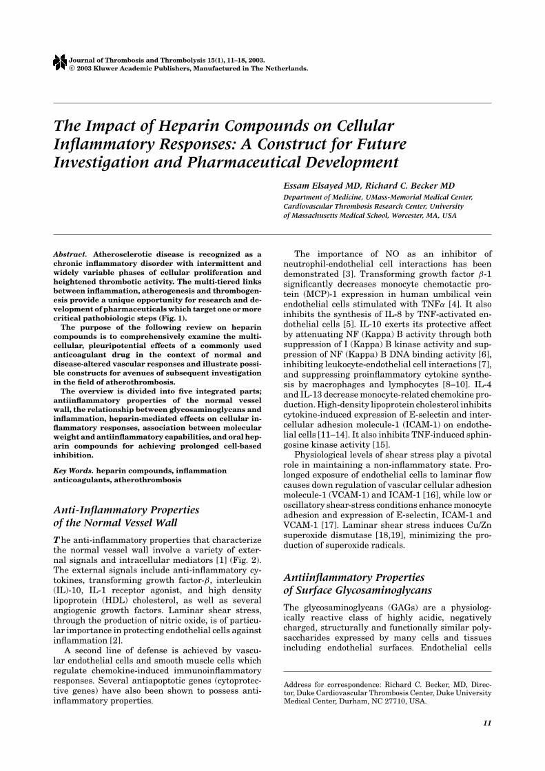
Fig. 1. The contribution of inflammatory cells and their mediators in atherothrombosis is recognized widely. Macrophage and Tlymphocyte production of tumor necrosis factor (TNF-∝) and interleukin-1 (IL-1) stimulate endothelial cells, prothromboticadhesion molecules and coagulation factors and proteolytic enzymes which can provoke plaque destabilization and localizedthrombosis. MMP, matrix metalloproteinase; IFNγ , interferon gamma; endothelial cell, EC, smooth muscle cell, SMC.
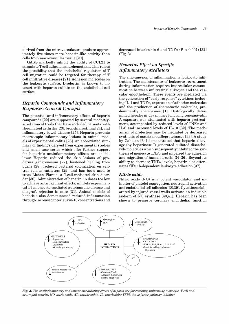
Fig. 2. Antiinflammatory properties, inherent to a normal vessel wall, are a vital defense against the proliferative, atherogenic andprothrombotic effects of injurious agents. NO, nitric oxide; IL, interleukin; VCAM, vascular cell adhesion molecule; ICAM,intercellular adhesion molecule; TGFβ, transforming growth factor beta.
Impact of Heparin Compounds 13
derived from the microvasculature produce approx-imately five times more heparin-like activity thancells from macrovascular tissue [20].
GAGS markedly inhibit the ability of CCL21 tostimulate T cell adhesion and chemotaxis. This raisesthe possibility that the endothelial regulation of Tcell migration could be targeted for therapy of Tcell infiltrative diseases [21]. Adhesion molecules onthe leukocyte surface, L-selectin, is known to in-teract with heparan sulfate on the endothelial cellsurface.
Heparin Compounds and InflammatoryResponses: General Concepts
The potential anti-inflammatory effects of heparincompounds [22] are supported by several modestly-sized clinical trials that have included patients withrheumatoid arthritis [23], bronchial asthma [24], andinflammatory bowel disease [25]. Heparin preventsmacroscopic inflammatory lesions in animal mod-els of experimental colitis [26]. An abbreviated sum-mary of findings derived from experimental studiesand small case series which offer further supportfor heparin’s antinflammatory effects are as fol-lows: Heparin reduced the skin lesions of pyo-derma gangrenosum [27], hastened healing fromburns [28], reduced bacterial colonization on cen-tral venous catheters [29] and has been used totreat Lichen Planus- a T-cell-mediated skin disor-der [30]. Administration of heparin, in doses too lowto achieve anticoagulant effects, inhibits experimen-tal T lymphocyte-mediated autoimmune disease andallograft rejection in mice [31]. Animal models ofhepatitis also demonstrated reduced inflammationthrough increased interleukin-10 concentrations and
HEPARIN
INTERACTIONS
Monocyte TFPI
AT
NEUTOPHILS -Superoxide -Myeloperoxidase -Lactoferrin - Granulocyte Activity
Smooth Muscle cell Proliferation
LYMPHOCYTES -Cytotoxic T cell Adhesion & migration -Natural killer cells
Complement activity
CHEMOKINES CYTOKINES -TNF-∝, IL-1, IL-6-1, IL-8, IL-12 -Laminin, collagen, elastase fibronectin
NO production
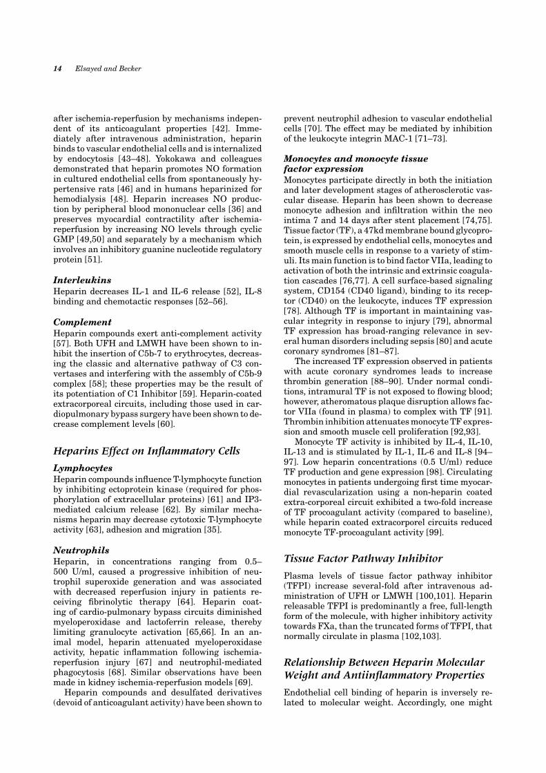
Fig. 3. The antiinflammatory and immunomodulating effects of heparin are far-reaching, influencing monocyte, T cell andneutrophil activity. NO, nitric oxide; AT, antithrombin; IL, interleukin; TFPI, tissue factor pathway inhibitor.
decreased interleukin-6 and TNFα (P < 0.001) [32](Fig. 3).
Heparins Effect on SpecificInflammatory Mediators
The sine-que-non of inflammation is leukocyte infil-tration. The maintenance of leukocyte recruitmentduring inflammation requires intercellular commu-nication between infiltrating leukocyte and the vas-cular endothelium. These events are mediated viathe generation of “early response” cytokines includ-ing IL-1 and TNFα, expression of adhesion moleculesand the production of chemotactic molecules, pre-dominantly chemokines [1]. Histologically deter-mined hepatic injury in mice following concanavalinA exposure was attenuated with heparin pretreat-ment, accompanied by reduced levels of TNFα andIL-6 and increased levels of IL-10 [32]. The mech-anism of protection may be mediated by decreasedsynthesis of matrix metalloproteinases [33]. A studyby Cahalon [34] demonstrated that heparin cleav-age (by heparinase I) generated sulfated disaccha-ride molecules which subsequently inhibited the syn-thesis of monocyte TNFα and impaired the adhesionand migration of human T-cells [34–36]. Beyond itsability to decrease TNFα levels, heparin also atten-uates CD11b-dependent leukocyte adhesion [37].
Nitric oxideNitric oxide (NO) is a potent vasodilator and in-hibitor of platelet aggregation, neutrophil activationand endothelial cell adhesion [38,39]. Cytokines elab-orated by injured vessel walls activate an inducibleisoform of NO synthase [40,41]. Heparin has beenshown to preserve coronary endothelial function
14 Elsayed and Becker
after ischemia-reperfusion by mechanisms indepen-dent of its anticoagulant properties [42]. Imme-diately after intravenous administration, heparinbinds to vascular endothelial cells and is internalizedby endocytosis [43–48]. Yokokawa and colleaguesdemonstrated that heparin promotes NO formationin cultured endothelial cells from spontaneously hy-pertensive rats [46] and in humans heparinized forhemodialysis [48]. Heparin increases NO produc-tion by peripheral blood mononuclear cells [36] andpreserves myocardial contractility after ischemia-reperfusion by increasing NO levels through cyclicGMP [49,50] and separately by a mechanism whichinvolves an inhibitory guanine nucleotide regulatoryprotein [51].
InterleukinsHeparin decreases IL-1 and IL-6 release [52], IL-8binding and chemotactic responses [52–56].
ComplementHeparin compounds exert anti-complement activity[57]. Both UFH and LMWH have been shown to in-hibit the insertion of C5b-7 to erythrocytes, decreas-ing the classic and alternative pathway of C3 con-vertases and interfering with the assembly of C5b-9complex [58]; these properties may be the result ofits potentiation of C1 Inhibitor [59]. Heparin-coatedextracorporeal circuits, including those used in car-diopulmonary bypass surgery have been shown to de-crease complement levels [60].
Heparins Effect on Inflammatory Cells
LymphocytesHeparin compounds influence T-lymphocyte functionby inhibiting ectoprotein kinase (required for phos-phorylation of extracellular proteins) [61] and IP3-mediated calcium release [62]. By similar mecha-nisms heparin may decrease cytotoxic T-lymphocyteactivity [63], adhesion and migration [35].
NeutrophilsHeparin, in concentrations ranging from 0.5–500 U/ml, caused a progressive inhibition of neu-trophil superoxide generation and was associatedwith decreased reperfusion injury in patients re-ceiving fibrinolytic therapy [64]. Heparin coat-ing of cardio-pulmonary bypass circuits diminishedmyeloperoxidase and lactoferrin release, therebylimiting granulocyte activation [65,66]. In an an-imal model, heparin attenuated myeloperoxidaseactivity, hepatic inflammation following ischemia-reperfusion injury [67] and neutrophil-mediatedphagocytosis [68]. Similar observations have beenmade in kidney ischemia-reperfusion models [69].
Heparin compounds and desulfated derivatives(devoid of anticoagulant activity) have been shown to
prevent neutrophil adhesion to vascular endothelialcells [70]. The effect may be mediated by inhibitionof the leukocyte integrin MAC-1 [71–73].
Monocytes and monocyte tissuefactor expressionMonocytes participate directly in both the initiationand later development stages of atherosclerotic vas-cular disease. Heparin has been shown to decreasemonocyte adhesion and infiltration within the neointima 7 and 14 days after stent placement [74,75].Tissue factor (TF), a 47kd membrane bound glycopro-tein, is expressed by endothelial cells, monocytes andsmooth muscle cells in response to a variety of stim-uli. Its main function is to bind factor VIIa, leading toactivation of both the intrinsic and extrinsic coagula-tion cascades [76,77]. A cell surface-based signalingsystem, CD154 (CD40 ligand), binding to its recep-tor (CD40) on the leukocyte, induces TF expression[78]. Although TF is important in maintaining vas-cular integrity in response to injury [79], abnormalTF expression has broad-ranging relevance in sev-eral human disorders including sepsis [80] and acutecoronary syndromes [81–87].
The increased TF expression observed in patientswith acute coronary syndromes leads to increasethrombin generation [88–90]. Under normal condi-tions, intramural TF is not exposed to flowing blood;however, atheromatous plaque disruption allows fac-tor VIIa (found in plasma) to complex with TF [91].Thrombin inhibition attenuates monocyte TF expres-sion and smooth muscle cell proliferation [92,93].
Monocyte TF activity is inhibited by IL-4, IL-10,IL-13 and is stimulated by IL-1, IL-6 and IL-8 [94–97]. Low heparin concentrations (0.5 U/ml) reduceTF production and gene expression [98]. Circulatingmonocytes in patients undergoing first time myocar-dial revascularization using a non-heparin coatedextra-corporeal circuit exhibited a two-fold increaseof TF procoagulant activity (compared to baseline),while heparin coated extracorporel circuits reducedmonocyte TF-procoagulant activity [99].
Tissue Factor Pathway Inhibitor
Plasma levels of tissue factor pathway inhibitor(TFPI) increase several-fold after intravenous ad-ministration of UFH or LMWH [100,101]. Heparinreleasable TFPI is predominantly a free, full-lengthform of the molecule, with higher inhibitory activitytowards FXa, than the truncated forms of TFPI, thatnormally circulate in plasma [102,103].
Relationship Between Heparin MolecularWeight and Antiinflammatory Properties
Endothelial cell binding of heparin is inversely re-lated to molecular weight. Accordingly, one might

Impact of Heparin Compounds 15
Table 1. Heparin Compounds and their Potential Impact on Inflammatory Responses
Cellular target Physiologic effect
Cytokines/Interleukins Decrease TNF production and effectDecrease IL-1 , IL-6 and IL-8 releaseDecrease TNF and IL-6Increase IL-10
Complement Inhibit both the classic and alternative pathwaysReduce with assembly of C5b-C9 ( MAC)Decrease complement levels
Lymphocytes Decrease cytotoxic T-cell activityImpair T-cell adhesion and migrationSuppress natural killer cell activity
Neutrophils, PMN Inhibit neutrophil superoxide productionReduce granulocyte activity and inhibit production of lactoferrin and myeloperoxidaseAttenuate neutrophil infiltrationDecrease neutrophil phagocytosisAttenuate CD-11b dependent adherent mechanism
Monocytes, TF, and TFPI Increase TFPI; prolonged infusions decrease TFPIBind to monocytes, inhibits TF production and gene expressionMay indirectly inhibit MTF by increasing IL-10 and decreasing IL-6 and IL-8 production.
anticipate that UFH, compared to LMWH or veryLMWH (pentasaccharide), would more effectivelylimit inflammatory responses on the endothelial sur-face. The same appears to be true for inhibition ofP-selectin-and L-selectin-associated adhesive events[104] and proliferation of smooth muscle cells [105].In contrast, lower molecular weight fragments in-hibit platelet binding to the subendothelium [106],allergic airway response [107], and leukocyte migra-tion more effectively than larger fragments.
Oral Heparin Compounds
The first generation of heparin was UFH. The sec-ond generation included LMWH preparations whilethe third generation encompasses chemically mod-ified heparins like oral heparin formulations andbiotechnologically derived heparinomimetics. Hep-arin is poorly absorbed from and rapidly degradedwithin the intestinal tract. In the last few years sev-eral delivery agents have been investigated. SNAC(sodium hydroxybenzoyl amino caprylat) allows gas-tric absorption of heparin [108]. Gastrointestinal ab-sorption of heparin occurs in a passive transcellularprocess without causing apparent damage to intesti-nal epithelium. SNAC does not have pharmacologicactivity [109]. The potential long-term antiinflam-matory effects achievable with oral heparin admin-istration has yet to be investigated.
Summary
The available information suggests that heparincompounds decrease several components of the in-flammatory process (Table 1). In some cases this ef-
fect may offer clinical benefit (ex. cardiopulmonarybypass); however, in most instances the overall rel-evance for patients with atherothrombotic disor-ders is unclear, particularly with very brief expo-sures and low vascular surface concentrations as arecommonly encountered in clinical practice. Ongoinginvestigations are designed to determine poten-tial differences between unfractionated heparin, lowmolecular weight heparin and very low molecularweight heparin (pentasaccharide). Future scientificefforts must also consider the capacity for desul-fated heparin molecules given orally to attenuatevascular inflammation and thrombotic potential overprolonged periods of time in patients with multi-bed atherosclerosis. Lastly, the development of phar-maceuticals for use in cardiovascular disease musttake into consideration the integrated paradigm ofinflammation, cellular proliferation and cell-basedcoagulation.
References
1. Keane Mm, Strieter R. Chemokine signaling in inflam-mation. Crit Care Med 2000;28:N13–N26.
2. Tedgui, Alain, Mallat, Ziad. Anti-inflammatory Mecha-nisms in the Vascular Wall. Circulation AHA 2001;88:877–887.
3. Kubes P, Suzuki M, Granger DN. Nitric oxide: an endoge-nous modulator of leukocyte adhesion. Proc Natl Acad SciUSA 1991;88:4651–4655.
4. Honda HM, Leitinger N, Frankel M, et al. Induction ofmonocyte binding to endothelial cells by MM-LDL: role oflipoxygenase metabolites. Arterioscler Thromb Vasc Biol1999;19:680–686.
5. Smith WB, Noack L, Khew-Goodall Y, Isenmann S, VadasMA, Gamble JR. Transforming growth factor—[beta]1 in-hibits the production of IL-8 and the transmigration of
16 Elsayed and Becker
neutrophils through activated endothelium. J Immunol1996;157:360–368.
6. Schottelius AJ, Mayo MW, Sartor RB, Baldwin AS, Jr.Interleukin-10 signaling blocks inhibitor of [kappa]B ki-nase activity and nuclear [kappa]B DNA binding. J BiolChem 1999;274:31868–31874.
7. Mulligan MS, Jones ML, Vaporciyan AA, HowardMC, Ward PA. Protective effects of IL-4 and IL-10against immune complex-induced lung injury. J Immunol1993;151:5666–5674.
8. Pugin J, Ulevitch RJ, Tobias PS. A critical role for mon-cytes and CD14 in endotoxin-induced endothelial cell ac-tivation. J Exp Med 1993;178:2193–2200.
9. Mallat Z, Heymes C, Ohan J, Faggin E, Leseche G, TedguiA. Expression of interleukin-10 in human atheroscleroticplaques: relation to inducible nitric oxide synthase ex-pression and cell death. Arterioscler Thromb Vasc Biol1999;19:611–616.
10. Mallat Z, Besnard S, Duriez M, et al. Protective role ofinterleukin-10 in atherosclerosis. Circ Res 1999;85:e17–e24.
11. Ashby DT, Rye KA, Clay MA, Vadas MA, Gamble JR,Barter PJ. Factors influencing the ability of HDL toinhibit expression of vascular cell adhesion molecule-1 in endothelial cells. Arterioscler Thromb Vasc Biol1998;18:1450–1455.
12. Cockerill GW, Saklatvala J, Ridley SH, et al. High-densitylipoproteins differentially modulate cytokine-induced ex-pression of E-selectin and cyclooxygenase-2. ArteriosclerThromb Vasc Biol 1999;19:910–917.
13. Baker PW, Rye KA, Gamble JR, Vadas MA, Barter PJ.Ability of reconstituted high density lipoproteins to in-hibit cytokine-induced expression of vascular cell adhe-sion molecule-1 in human umbilical vein endothelial cells.J Lipid Res 1999;40:345–353.
14. Cockerill GW, Huehns TY, Weerasinghe A, Stocker C,Lerch PG, Miller NE, Haskard DO. Elevation ofplasma high-density lipoprotein concentration reducesinterleukin-1-induced expression of E-selectin in anin vivo model of acute inflammation. Circulation 2001;103:108–112.
15. Xia P, Vadas MA, Rye KA, Barter PJ, Gamble JR. Highdensity lipoproteins (HDL) interrupt the sphingosine ki-nase signaling pathway: a possible mechanism for pro-tection against atherosclerosis by HDL. J Biol Chem1999;274:33143–33147.
16. Sampath R, Kukielka GL, Smith CW, Eskin SG, McintireLV. Shear stress-mediated changes in the expression ofleukocyte adhesion receptors on human umbilical veinendothelial cells in vitro. Ann Biomed Eng 1995;23:247–256.
17. Mohan S, Mohan N, Valente AJ, Sprague EA. Regula-tion of low shear flow-induced HAEC VCAM-1 expressionand monocyte adhesion. Am J Physiol 1999;276:C1100–C1107.
18. Inoue N, Ramasamy S, Fukai T, Nerem RM, HarrisonDG. Shear stress modulates expression of Cu/Zn super-oxide dismutase in human aortic endothelial gels. CircRes 1996;79:32–37.
19. Dimmeler S, Hermann C, Galle J, Zeiher AM. Upreg-ulation of superoxide dismutase and nitric oxide syn-thase mediates the apoptosis-suppressive effects of shearstress on endothelial cells. Arterioscler Thromb Vasc Biol1999;19:656–664.
20. Bombeli T, Mueller M, Haeberli A. Anticoagulant prop-erties of the vascular endoehtlium. Throm Haemost1997;77:408–423.
21. Kent W Christopherson, JJ Campbell. LMWH inhibitCCL21 induced T cell adhesion. J Pharm and Exp Therap2002;302:290–295.
22. Tyrell DJ. Heparin beyond its traditional role as an anti-coagulant. Therapeutic Trends Pharmacol 1995;16:198–204.
23. Gaffney A, Gaffney P. Rheumatoid arthritis and heparin.BR J Rheumatol 1996;35:808.
24. Bowler SD, Smith SM, Lavercombe PS. Heparin inhibitsthe immediate response to antigen in the skin and lungsof allergic subjects. Am Rev Respir Dis 1993;147:160–163.
25. Ang YS, Mahmud N. Randomized comparison of unfrac-tionated heparin in severe active inflammatory bowel dis-ease. Ailment Pharmacol Ther 2000;14:1015–1022.
26. Folwaczny C, Frick H, Hartman G. Anti-inflammatoryproperties of unfractionated heparin in patients withhighly active ulcerative colitis. AJ Gastroenterology 1997;92:5–10.
27. Fries W, et al. Aliment Parmacal Ther 1998;12:229–236.28. Saliba MJ, Jr. Heparin in the treatment of burns. Burns
2001;27:349–358.29. Appelgren P, Ransjo U, Bindslev L, Espersen F, Larm
O. Surface heparinization of central venous catheters re-duces microbial colonization in vitro and in vivo. CriticalCare Medicine 1996;24(9):1482–1489.
30. Hodak E, Yosipovitch G, David M, et al. Low-dose low-molecular weight heparin is beneficial in lichen planus.JA Acad Dermatology 1998;38:564–568.
31. Lider O, Baharoav E, Mekori YA, et al. Suppression ofexperimental autoimmune diseases and prolongation ofallograft survival by treatment of animals with low dosesof LMWH. J Clin Invest 1989;83:752–756.
32. Hershkoviz R, Bruck R, Aeed H, Shirin H, Halpren Z.Treatment of concanavalin A-induced hepatitis in micewith LMWH. J Hepatology 1999;31:834–840.
33. Dwarakanath AD, Yu LG, Brookes C, Pryce D, Rhodes JM.“Sticky” neutrophils, pathergic arthritis, and response toheparin in pyoderma gangrenosum complicating ulcera-tive colitis. Gut 1995;37:585–588.
34. Calahon L, Lider O, Schor H, et al. Heparin disaccharidesinhibit TNFα production by macrophages and arrest im-mune inflammation in rodents. International Immunol1997;9:1517–1522.
35. Hershkoviz R, Schor H, Ariel A, et al. Disaccharides gen-erated from heparin modulate chemokine-induced T-celladhesion. Immunol 2000;99:87–93.
36. Chelmonska A, Zimecki M, Nowachi W, Nikolajczuk M.Differential effects of heparin on NO and TNF. Comp Im-munol Micro Inf Diseases 2001;24:151–164.
37. Salas A, Sans M, Soriano A, et al. Heparin attenuatesTNFα induced inflammatory response through CD11b de-pendent mechanism. Gut 2000;47:88–96.
38. Radomski MW, Palmer RMJ, Moncada S. ComparativePharmacology of endothelium-derived relaxing factor, ni-tric oxide and prostacyclin in platelets. BR J Pharmacol1987;92:181–187.
39. Stamler JS, Mendelsohn ME, Amarante P, et al.N-Acetylcysteine potentiates platelet inhibition by endo-thelium-derived relaxing factor. Circ Res 1989;65:789–795.

Impact of Heparin Compounds 17
40. Nunokawa Y, Ishida N, Tanaka S. Cloning of induciblenitric oxide synthase in rat vascular smooth muscle cells.Biochem Biophys Res Commun 1993;191:89–94.
41. Joly GA, Schini VB, Vanhoutte PM. Balloon injury andinterleukin-1 sub beta induce nitric oxide synthase activ-ity in rat carotid arteries. Circ Res 1992;71:331–338.
42. Kouretas PC, Kim YD, Cahill PA, et al. Nonanticoagulantheparin prevents coronary endothelial dysfunction afterbrief ischemia-reperfusion injury in the dog. Circulation1999;99:1062–1068.
43. Hiebert LM, Jaques LB. The observation of heparin onendothelium after injection. Thromb Res 1976;8:195–204.
44. Yokokawa K, Kohno M, Tahara H, et al. Effect of heparinon endothelin-1 production by cultured human endothe-lial cells. J Cardiovasc Pharmacol 1993;22(Suppl 8):S46–S48.
45. Piatti PM, Monti LD, Valsecchi G, et al. Effects of low-dose heparin on arterial endothelin-1 release in humans.Circulation 1996;94:2703–2707.
46. Yokokawa K, Tahara H, Kohno M, Mandal AK, YasunariK, Takeda T. Heparin endothelin production throughendothelium-derived NO in human endothelial cells.J Clin Invest 1993;92:2080–2085.
47. Minami Mm, Yokokawa K, Kohno M, et al. Promotionof NO formation by heparin in cultured aortic endothe-lial cells from spontaneously hypertensive rats. Clin ExpPharmacol Physiol 1995;22(Suppl 1):S146–S147.
48. Yokokawa K, Mankus R, Saklayen MG, et al. IncreasedNO production in patients with hypotension duringhemodialysis. Ann Intern Med 1995;123:35–37.
49. Kouretas PC, Kim YD, Cahill PA, et al. Heparin preservesNO activity in coronary endothelium during ischemia-reperfusion injury. Ann Thorac Surg 1998;66:1210–1215.
50. Kouretas PC, Myers AK, Kim YD, et al. Heparin andnonanticoagulant heparin preserve regional myocardialcontractility after ischemia-reperfusion injury. J ThoracCardiovasc Surg 1998;115:440–448.
51. Kouretas PC, Hannan RL, Kapur NK, et al. Non-anticoagulant heparin increases endothelial nitric oxidesynthase activity. J Mol Cell Cardiol 1998;30:2669–2682.
52. Solberg R, Scholz T, Videm V, Okkenhaug C, Aasen AO.Heparin coating reduces cell activation and mediator re-lease. Transpl Int 1998;11:252–258.
53. Ramdin L, Perks B, Sheron N, Shute JK. Regulationof interleukin-8 binding and function by heparin andα2-macroglobulin. Clin Experim Allergy 1998;28:616–624.
54. Wrenshall LE, Stevens RB, Cerra FB, Platt JL. Modula-tion of macrophages and B cell function by glycosamino-glycans. J Leuko Biol 1999;66:391–400.
55. Fosse E, Mollnes TE, Ingvaldsen B. Complement activa-tion during major operations with or without cardiopul-monary bypass. J Thorac Cardiovasc Surg 1987;93:860–866.
56. Jansen PGM, Velthuis H, Huybregts RAJM, et al.Reduced complement activation and improved post-operative performance after cardiopulmonary bypasswith heparin-coated circuits. J Thorac Cardiovasc Surg1995;110:829–834.
57. Ninomiya H, Kawashima Y, Nagasawa T. Inhibition ofcomplement-mediated haemolysis in paroxysmal noctur-nal haemoglobinuria by heparin or LMWH. Br J Haema-tology 2000;109(4-II):875–881.
58. Baker PJ, Lint TF, McLeod BC, Behrends CL, Gewurz H.Studies on the inhibition of C56-induced lysis. J Immunol1975;114:554–558.
59. Rent R, Myhrman R, Fiedel A, Gewurz H. Potentiation ofC1 esterase inhibitory activity by heparin. Clin ExperimImmunol 1976;23:264–271.
60. Lazar HL, Bao Y, Ggaudiani J, Rivers S, Marsh H. To-tal Complement Inhibition: an effective strategy to limitischemic injury during coronary revascularization on car-diopulmonary bypass. Circ 1999;100:1438–1442.
61. Redegeld F, Smith P, Apasov S, Sitkovsky M. Phospho-rylation of T-lymphocyte plasma membrane—associatedproteins by ectoprotein kinase. BBA 1997;1328:151–165.
62. Zynska J, Stelmach I, Kuna P. The role of Heparin inallergic inflammation. Pol Merkuriusz Lek 2000;8:341–346.
63. Felzen B, Berke G, Gardner P, Binah O. Involvement ofthe IP3 cascade in the damage to guinea-pig ventricu-lar myocytes induced by cytotoxic T-lymphocytes. Pfluger-sArchiv Euro J Physiol 1997;433:721–726.
64. Riesenberg K, Schlaeffer F, Katz A, Levy R. Inhibition ofsuperoxide production in neutrophils by combinations ofheparin and thrombolytic agents. Br Heart J 1995;73:14–19.
65. Borowiec J, Jaramillo A, Venge P, Nilsson L, Thelin S.Effects of heparin-coating of CPB circuits on leukocytesduring simulated extracorporeal circulation. CardiovascSurg 1997;5:568–573.
66. Tamim M, Demircin M, Guvener M, Peker O, Yilmaz M.Heparin-coated circuits reduce complement activationand inflammatory response to CPB. Panminerva Med1999;41:193–198.
67. Hisama N, Yamaguchi Y, Okajima K, et al. Anticoag-ulant pretreatment attenuates production of cytokine-induced neutrophil chemoattractant following ischemia-reperfusion of rat liver. Dig Dis Sci 1996;41:1481–1486.
68. Salih H, Husfeld L, Adam D. Inhibitory effect of heparinon neutrophil phagocytosis. Eur J Med Res 1997;2:507–513.
69. Shin CS, Han JU, Kim JL, Schenarts PJ, Traber LD,Hawkins H. Heparin attenuated neutrophil infiltrationbut did not affect renal injury induced by ischemia reper-fusion. Yonsei Med J 1997;38:133–141.
70. Lever R, Hoult JR, Page CP. The effects of heparin andrelated molecules upon the adhesion of human polymor-phonuclear leucocytes to vascular endothelium in vitro.Br J Phamacol 2000;129:533–540.
71. Peter K, Schwarz M, Conradt C, et al. Heparin in-hibits Ligand binding to the leukocyte integrin Mac-1(CD11b/CD18). Circ 1999;100:1533–1539.
72. Carter AJ, Laird JR, Farb A, Kufs W, Wortham DC,Virmani R. Morphologic characteristics of lesion forma-tion and time course of smooth muscle cell proliferationin a porcine proliferative restenosis model. J Am Coll Car-diol 1994;24:1392–1405.
73. Pietersma A, Kofflard M, et al. Late lumen loss aftercoronary angioplasty is associated with the activationstatus of circulating phagocytes before treatment. Circ1995;91:1320–1325.
74. Rogers C, Welt F, Karnovsky M, Edelman E. Monocyterecruitment and neointimal hyperplasia in rabbits. Arte-riosclerosis Thomb Vasc Biol 1996;16:1312–1318.

18 Elsayed and Becker
75. van Beusekom HMM, van der Giessen WJ, van SuylenRJ, et al. Histology after stenting of human saphenousvein bypass grafts. J Am Coll Cardiol 1993;21:45–54.
76. Wilcox JN, Smith KM, Schwartz SM, Gordon D.Localization of tissue factor in normal vessel wall andin the atherosclerotic plaque. Proc Natl Acad Sci USA1989;86:2839–2843.
77. Toschi V, Gallo R, Lettino M, et al. Tissue factor mod-ulates the thrombogenicity of human atheroscleroticplaques. Circ 1997;95:594–599.
78. Mach F, Schonbeck U, Bonnefoy J-Y, et al. Acti-vation of monocyte/macrophage functions related toacute atheroma complication by ligation of CD40. Circ1997;96:396–399.
79. Leatham EW, Bath PMW, Tooze JA, Camm AJ. Increasedmonocyte TF expression in coronary disease. Br HeartJournal 1995;73:10–13.
80. Levi M, ten Cate H, van der Poll T, van Deventer SJ.Pathogenesis of disseminated intravascular coagulationin sepsis. JAMA 1993;270:975–979.
81. Annex BH, Denning SM, Channon KM, et al. Differentialexpression of TF protein in directional atherectomy spec-imens from patients with stable and unstable coronarysyndromes. Circ 1995;91:619–622.
82. Moreno PR, Falk E, Palcios IF, Newell JB, Fuster V, FallonJT. Macrophage infiltration in acute coronary syndromes.Circ 1994;90:775–778.
83. Soejima H, Ogawa H, Yasue H, et al. Heightened TFassociated with TF pathway inhibitor and prognosisin patients with unstable angina. Circ 1999;99:2908–2913.
84. Neri Serneri G, Abbate R, Gori A, et al. Transient in-termittent lymphocyte activation is responsible for theinstability of unstable angina. Circ 1992;86:790–797.
85. Singh R, Pan S, Mueske CS, et al. Role for TF path-way in murine model of vascular remodeling. Circ Res2001;89:71–76.
86. Ardissino D, Merlini PA, Ariens R, Coppola R, BramucciE, Mannucci PM. TF antigen and activity in human coro-nary atherosclerotic plaques. Lancet 1997;349:769–771.
87. Mallat Z, Benamer H, Hugel B, et al. Elevated levels ofshed membrane microparticles with procoagulant poten-tial in the peripheral circulating blood of patients withacute coronary syndromes. Circ 2000;101:841–843.
88. Penn MS, Topol EJ. TF, the emerging link between in-flammation, thrombosis, and vascular remodeling. CircRes 2001;89:1–2.
89. Siegbahn A, Johnell M, Rorsman C, Ezban M, Heldin CH.Binding of factor VIIa to TF on human fibroblasts leads toactivation of phospholipase C. Blood 2000;96:3452–3458.
90. Sato Y, Kataoka H, Asada Y, Marutsuka K, Kamikubo Y,Koono M. Overexpression of TFPI in aortic smooth mus-cle cells inhibit migration induced by TF/Factor VIIa com-plex. Thromb Res 1999;94:401–406.
91. Banai S, Gertz SD. TF as a therapeutic target in coronarysyndromes. Am J Cardiol 2001;87(6):763–765.
92. Gertz SD, Fallon JT, Gallo R, et al. Hirudin reduces TFexpression in the neointima after balloon injury in rabbitfemoral and porcine coronary arteries. Circ 1998;98:580–587.
93. Hasenstab D, Lea H, Hart CE, Lok S, Clowes AW. TFoverexpression in rat arterial neointima models throm-bosis and progression of advanced atherosclerosis. Circ2000;101:2651–2657.
94. Schuager I, Jungi TW. Effect of human recombinant cy-tokines on the induction of macrophage procoagulant ac-tivity. Blood 1994;83:152–160.
95. Neumann FJ, Ott I, Marx N, et al. Effect of humanrecombinant interleukin-6 and interleukin-8 on mono-cyte procoagulant activity. Arterioscler Thromb Vasc Biol1997;17:3399–3405.
96. Ernofsson M, Tenno T, Siegbahn A. Inhibition of TF sur-face expression in human perpheral blood monocytes ex-posed to cytokines. Br J Haematology 1996;95:249–257.
97. Lindmark E, Tenno T, Chen J, Siegbahn A. IL-10 inhibitsLPS-induced human monocyte TF expression in wholeblood. Br J Haematology 1998;102:597–604.
98. Pepe G, Giusti B, Attanasio M, et al. TF and plasminogenactivator inhibitor type 2 expression in human stimulatedmonocytes is inhibited by heparin. Semin Thromb Hemost1997;23(2):135–141.
99. Barstad M, Ovrum E, Ringdal ML, et al. Inductionof monocyte TF procoagulant activity during coronaryartery bypass surgery is reduced with heparin-coated ex-tracorporeal circuit. Br J Haematology 1996;94(3-I):517–525.
100. Novotny WF, Brown SG, Miletich JP, Rader DJ, BrozeGJ, Jr. Plasma antigen levels of the lipoprotein-associated coagulation inhibitor in patient samples.Blood 1991;78:387–393.
101. Sandset PM, Abildgaard U, Larsen ML. Heparin inducesrelease of extrinsic coagulation pathway inhibitor (EPI).Thromb Res 1988;50:803–813.
102. Novotny WF, Palmier M, Wun TC, Broze GJ, Jr,Miletich JP. Purification and properties of heparin-releasable lipoprotein-associated coagulation inhibitor.Blood 1991;78:394–400.
103. Lindahl AK, Jacobsen PB, Sandset PM, Abildgaard U.TF pathway inhibitor with high anticoagulant activityis increased in post-heparin plasma and in plasma fromcancer patients. Blood Coagul Fibrinolysis 1991;2:713–721.
104. Koening A, Norgard-Sumnicht K, Linhardt R, Varki A.Differential interactions of heparin and heparan sulfateglycosaminoglycans with the selectins. J Clinical Inves-tigation 1998;101:877–889.
105. Chan P, Mill S, et al. Heparin inhibition of humanvascular smooth muscle cell hyperplasia. Int Angiol1992;11:261–267.
106. Serra A, Esteve J, Reverter JC, et al. Differential effectof a LMWH (dalteparin) and UFH on platelet interactionwith the subendothelium under flow conditions. ThromRes 1997;87:405–410.
107. Campo C, Molinari J, Ungo J, Ahmed T. Molecular weightdependent effects of nonanticoagulant heparins on aller-gic airway responses. J Appl Physiol 1999;86:549–557.
108. Rivera TM, Leone-Bay A, et al. Oral delivery of heparin incombination with SNAC. Pharm Res 1997;14:1830–1834.
109. Brayden D, Creed E, et al. Heparin absorption across theintestine. Pharm Res 1997;14:1722–1779.


![In patients ≥75 years of age, Effient is generally not€¦ · , warfarin, heparin, fibrinolytic therapy, chronic use of non-steroidal anti-inflammatory drugs [NSAIDs]) [see Warnings](https://static.fdocuments.in/doc/165x107/605287db82a4b5425c5f10d6/in-patients-a75-years-of-age-effient-is-generally-not-warfarin-heparin-fibrinolytic.jpg)