The immunologic and antioxidant effects of L-phenylalanine ...
8
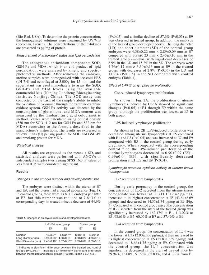
Summary. L-phenylalanine (L-PHE) is a synthetic precursor of catecholamines. Because it cannot be synthesised by an organism, it must be absorbed from the environment. Despite the wide use of L-PHE, whether L-PHE has a negative impact on embryo implantation and development is poorly understood. This study attempted to determine the roles of L-PHE in embryo implantation and development and in the immune response and antioxidant status of the uterus in early pregnancy mice injected intraperitoneally with 320 mg/kg L-PHE. The embryo number of treated mice decreased by 57.6%, and the size of their embryos was reduced by 2.8% (P>0.05) along the long diameter and 11.9% (P<0.05) along the short diameter at E9 compared with control mice. In addition, L-PHE significantly suppressed B lymphocyte proliferation. L-PHE increased IL-2 secretion but decreased the IL-4 concentration, thereby up-regulating the ratio of IL-2/IL-4 to 1.37-8.45. An analysis of the oxidant and antioxidant status showed that, compared with the control mice, the level of superoxide dismutase activity decreased by 21.54%- 39.94% and the glutathione peroxidase activity decreased by 15.27%-18.96% among the L-PHE-treated mice at E1-E9. However, the malonaldehyde content increased by 14.29%-90.11% among the L-PHE-treated mice. Therefore, L-PHE impaired embryo implantation by disrupting cytokine-based immunity and oxidative stress in the uterus. Key words: L-phenylalanine, Lymphocyte proliferation, Cytokines, Antioxidant, Pregnant mice Introduction L-phenylalanine (L-PHE) is the synthetic precursor of catecholamines (Grando et al., 2006; Fernstrom and Fernstrom, 2007) and can be transformed into norepinephrine and epinephrine via dopa decarboxylase (DDC). DDC is found in the central nervous system and in peripheral tissues and organs, such as the vascular wall, stomach, kidney and liver. Because L-PHE cannot be synthesised by organisms and must be absorbed from the environment, it is widely used as a component of aspartame, a medical intermediate, and as an additive in human food and animal forage. However, we have no specific knowledge as to whether side effects emerge when an organism continually absorbs L-PHE, particularly among embryos exposed to L-PHE. The fate of an embryo is determined by specific factors, including its mother’s immune responses (Bansal, 2010). The immune responses of the maternal uterus may play an important role in determining whether a pregnancy will be successful (Wegmann et al., 1993; Ho et al., 2001; Piccinni, 2003). Thus, studies of the mechanisms underlying the immunological modulation of the uterus during pregnancy are important for identifying the causes of embryonic loss. During early pregnancy, oxidative stress aids in the development of the placenta and chorion, and the expression levels of antioxidant enzymes increase. These enzymes include catalase (CAT), superoxide dismutase (SOD) and glutathione The immunologic and antioxidant effects of L-phenylalanine on the uterine implantation of mice embryos during early pregnancy Yulan Dong 1 , Yongping Bai 1 , Guanhui Liu 1 , Zixu Wang 1 , Jing Cao 1 , Yaoxing Chen 1 and Hongliang Yang 2 1 Laboratory of Veterinary Anatomy, College of Animal Medicine, China Agricultural University, Beijing, China and 2 Department of Microbiology, Immunology and Pathology, Colorado State University, USA Histol Histopathol (2014) 29: 1335-1342 DOI: 10.14670/HH-29.1335 http://www.hh.um.es Histology and Histopathology Cellular and Molecular Biology Offprint requests to: Yaoxing Chen, Prof of Laboratory of Veterinary Anatomy, College of Animal Medicine, China Agricultural University, Haidian, Beijing, 100193, P.R. China. e-mail: [email protected]
Transcript of The immunologic and antioxidant effects of L-phenylalanine ...
Summary. L-phenylalanine (L-PHE) is a syntheticprecursor of catecholamines. Because it cannot besynthesised by an organism, it must be absorbed fromthe environment. Despite the wide use of L-PHE,whether L-PHE has a negative impact on embryoimplantation and development is poorly understood.This study attempted to determine the roles of L-PHE inembryo implantation and development and in theimmune response and antioxidant status of the uterus inearly pregnancy mice injected intraperitoneally with 320mg/kg L-PHE. The embryo number of treated micedecreased by 57.6%, and the size of their embryos wasreduced by 2.8% (P>0.05) along the long diameter and11.9% (P<0.05) along the short diameter at E9 comparedwith control mice. In addition, L-PHE significantlysuppressed B lymphocyte proliferation. L-PHE increasedIL-2 secretion but decreased the IL-4 concentration,thereby up-regulating the ratio of IL-2/IL-4 to 1.37-8.45.An analysis of the oxidant and antioxidant status showedthat, compared with the control mice, the level ofsuperoxide dismutase activity decreased by 21.54%-39.94% and the glutathione peroxidase activitydecreased by 15.27%-18.96% among the L-PHE-treatedmice at E1-E9. However, the malonaldehyde contentincreased by 14.29%-90.11% among the L-PHE-treatedmice. Therefore, L-PHE impaired embryo implantationby disrupting cytokine-based immunity and oxidativestress in the uterus.
Key words: L-phenylalanine, Lymphocyte proliferation,Cytokines, Antioxidant, Pregnant mice
Introduction
L-phenylalanine (L-PHE) is the synthetic precursorof catecholamines (Grando et al., 2006; Fernstrom andFernstrom, 2007) and can be transformed intonorepinephrine and epinephrine via dopa decarboxylase(DDC). DDC is found in the central nervous system andin peripheral tissues and organs, such as the vascularwall, stomach, kidney and liver. Because L-PHE cannotbe synthesised by organisms and must be absorbed fromthe environment, it is widely used as a component ofaspartame, a medical intermediate, and as an additive inhuman food and animal forage. However, we have nospecific knowledge as to whether side effects emergewhen an organism continually absorbs L-PHE,particularly among embryos exposed to L-PHE. The fateof an embryo is determined by specific factors, includingits mother’s immune responses (Bansal, 2010). Theimmune responses of the maternal uterus may play animportant role in determining whether a pregnancy willbe successful (Wegmann et al., 1993; Ho et al., 2001;Piccinni, 2003). Thus, studies of the mechanismsunderlying the immunological modulation of the uterusduring pregnancy are important for identifying thecauses of embryonic loss. During early pregnancy,oxidative stress aids in the development of the placentaand chorion, and the expression levels of antioxidantenzymes increase. These enzymes include catalase(CAT), superoxide dismutase (SOD) and glutathione
The immunologic and antioxidant effects of L-phenylalanine on the uterine implantation of mice embryos during early pregnancyYulan Dong1, Yongping Bai1, Guanhui Liu1, Zixu Wang1, Jing Cao1, Yaoxing Chen1 and Hongliang Yang21Laboratory of Veterinary Anatomy, College of Animal Medicine, China Agricultural University, Beijing, China and 2Department of Microbiology, Immunology and Pathology, Colorado State University, USA
Histol Histopathol (2014) 29: 1335-1342DOI: 10.14670/HH-29.1335
http://www.hh.um.es
Histology andHistopathologyCellular and Molecular Biology
Offprint requests to: Yaoxing Chen, Prof of Laboratory of VeterinaryAnatomy, College of Animal Medicine, China Agricultural University,Haidian, Beijing, 100193, P.R. China. e-mail: [email protected]

peroxidase (GSH-Px), each of which can protect cellsfrom damage due to oxidative stress. Under pathologicalcircumstances, excessive oxygen radicals may occursuddenly, resulting in their accumulation and, ultimately,toxicity (Rao et al., 2003). To date, whether L-PHEplays a role in the regulation of uterine immunity andantioxidant ability remains poorly understood. In thecurrent paper, we investigated the role of L-PHE in theuteri of mice during early pregnancy on embryonicimplantation and the particular development of uterinelymphocyte proliferation, cytokine production,antioxidant status and lipid peroxidation.Materials and methods
Animal treatment
Eighty female (25-32 g) and 20 male (35-40 g)Institute for Cancer Research (ICR) mice werepurchased at 8 weeks of age from Vital River LaboratoryAnimal Technology (Beijing, China). After anadaptation period of 1 week, 40 female mice wereinjected intraperitoneally for 7 consecutive days with 0.3mL of a solution containing 320 mg/kg body weight(BW) of L-PHE (Beijing BioDee BioTech CorporationLtd, Beijing, China), diluted in sterile saline. Theremaining control females were treated with vehicle (0.3mL). After completion of the treatments, the femaleswere paired daily with males of proven fertilityfollowing the examination of vaginal smears (Wright’sstaining). On the following day, the females wereconfirmed to be pregnant by the presence of a vaginalplug or by vaginal smear, and the day was designated asembryonic day (E1). The mice were sacrificed underdeep anaesthesia with Nembutal (50 mg/kg BW) at E1,E3, E5, E7 or E9 on a sterile bench. Uterine sampleswere immediately removed from the abdominal cavity.The embryo number was counted, and the embryos wereremoved. All experimental protocols were performed inaccordance with the Guidelines for AnimalExperimentation of China Agricultural University.Lymphocyte preparation from isolated uteri
All left uterine horns without embryos were mincedinto small pieces and passed through a tissue sieve (200mesh) to prepare single-cell suspensions in 3 mL ofHank’s solution. The suspension was layered onto 3 mLof lymphocyte separation medium (Tianjin BloodResearch Centre, Tianjin, China) and separated bydensity-gradient centrifugation at 1,500g for 30 min atroom temperature. The lymphocyte precipitates wereresuspended in 3 mL of RPMI 1640 base medium(Gibco BRL, Grand Island, NY, USA) and centrifugedtwice at 1,000g at 4°C for 10 min. Lymphocytes wereresuspended in 0.5 mL of RPMI 1640 complete medium(Gibco BRL), and the cell concentrations were adjustedto 2×10⁵ cells per mL. Cell viability was determined bythe 0.1% trypan blue dye exclusion test, and the cells
were counted.Lymphocyte proliferation assay
Mitogen-induced lymphocyte proliferation wasselected as an in vitro index of immune function.Concanavalin A (ConA)- or lipopolysaccharide (LPS)-induced lymphocyte proliferation was assayed using themethylthiazolyldiphenyl-tetrazolium bromide (MTT)assay with some modifications. Each cell suspensionsample was assigned to ConA (Sigma-Aldrich Co., St.Louis, MO, USA) or LPS (Sigma-Aldrich Co. , St.Louis, MO, USA) treatment and control wells, withthree sequential triplicate wells for each combination.Briefly, the 100 µL cell suspension was added to theindividual wells of a 96-well microtitre plate (Corning,NY, USA). ConA (or LPS) and RPMI 1640 completemedium were added to the wells containing plated cellsto yield the ConA (or LPS) treatment and control wells.The final ConA concentration for both and LPS was 15µg/mL, and each well was filled with RPMI 1640complete medium to a final volume of 120 µL. Theplates were then incubated at 37°C in 5% CO2 for 72 h.Subsequently, 10 µL of MTT solution was added to eachwell to achieve a final concentration of 5 mg/mL in PBS(pH 7.4). The plates were incubated at 37°C for another4 h. Following incubation, 100 µL of 10% sodiumdodecyl sulphate (Shanghai Chemical Factory, Shanghai,China) in 0.01 M HCl was added to lyse the cells andsolubilise the MTT crystals. The plates were read at 570nm using an automated microplate reader (Bio-Rad,USA). The stimulation index was calculated for eachsample as the absorbance value for the cells, with theabsorbance values for the mitogens divided by the valuesfor the cells without mitogens.Cytokine measurement by ELISA
All uterine tissues without embryos werehomogenised in 10% wt/vol 0.01 M PBS (pH 7.4, 4°C)and then centrifuged at 1,500 rpm for 20 min at 4°C. Thesupernatants were collected and used to measure theconcentrations of interleukin 2 (IL-2) and IL-4 withELISA kits (Boster Biological Technology, China). Thekit sensitivity was 15.6 pg/mL for IL-2 and 7.8 pg/mLfor IL-4. Briefly, 100 µL of the samples or standards wasapplied to polystyrene microplates coated with a specificanti-mouse IL-2 or IL-4 monoclonal antibody. Afterincubation for 90 min at 37°C, the plates were washedtwice with PBS containing 0.05% Tween-20. Next, 100µL of biotinylated anti-cytokine antibody (anti-IL-2 oranti-IL-4, 1:100) was added for a 60 min incubation at37°C. After six washes, the plates were treated with 100μL of avidin-biotin-peroxidase complex (ABC, 1:100)for 30 min at 37°C and incubated with 0.01% 3,3’,5,5’-tetramethylbenzidine (TMB, Sigma) substrate plushydrogen peroxide (0.003%) for 10-15 min at 37°C.After adding 100 μL of 2 N sulphuric acid (H2SO4), areading was taken at 450 nm using an ELISA reader
1336L-phenyalamine in uterine implantation
(Bio-Rad, USA). To determine the protein concentration,the homogenised solutions were measured by UV/VIS(Secoman, French). The concentrations of the cytokinesare presented as pg/mg of protein.Measurement of antioxidant status and lipid peroxidation
The endogenous antioxidant components SOD,GSH-Px and MDA, which is an end product of lipidperoxidation, were analysed by kinetic and spectro-photometric methods. After removing the embryos,uterine samples were homogenised with ice-cold PBS(pH 7.4) and centrifuged at 3,000g for 15 min, and thesupernatant was used immediately to assay the SOD,GSH-Px and MDA levels using the availablecommercial kits (Nanjing Jiancheng BioengineeringInstitute, Nanjing, China). The SOD assay wasconducted on the basis of the sample’s ability to inhibitthe oxidation of oxyamine through the xanthine-xanthineoxidase system. GSH-Px activity was detected by theconsumption of glutathione, and MDA content wasmeasured by the thiobarbituric acid colourimetricmethod. Values were calculated using optical density(550 nm for SOD, 412 nm for GSH-Px and 532 nm forMDA) according to the formula described in themanufacturer’s instructions. The results are expressed asfollows: units (U) per mg protein for SOD and GSH-Pxand nmol/mg protein for MDA.Statistical analysis
All results are expressed as the means ± SD, andstatistical analyses were performed with ANOVA orindependent samples t-tests using SPSS 16.0. P values ofless than 0.05 were considered significant.Results
Changes in the embryo number and developmental size
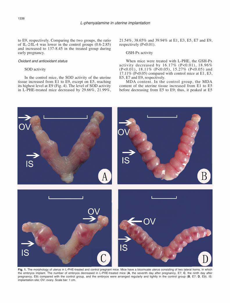
The embryos were distinct within the uterus at E7and E9, and the uterus had a beaded appearance (Fig. 1).In control groups, there were 13.8±1.8 embryos per litterat E7, but this number was reduced to 7.6±3.0 atcorresponding days in treated mice, a decrease of 44.9%
(P<0.05), and a similar decline of 57.6% (P<0.05) at E9was observed in treated group. In addition, the embryosof the treated group developed poorly. The long diameter(LD) and short diameter (SD) of the control groupembryos were 4.38±0.22 mm × 2.89±0.09 mm at E7compared with 3.99±0.23 mm × 2.45±0.10 mm in thetreated group embryos, with significant decreases of8.9% in the LD and 15.2% in the SD. The embryos were4.76±0.12 mm × 3.30±0.13 mm at E9 in the treatedgroup, with decreases of 2.8% (P>0.05) in the LD and11.9% (P<0.05) in the SD compared with controlembryos (Table 1).Effect of L-PHE on lymphocyte proliferation
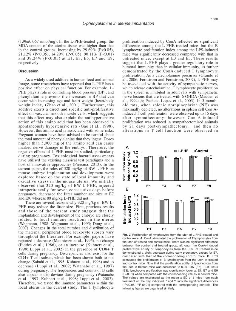
ConA-induced lymphocyte proliferationAs shown in Fig. 2A, the proliferation of uterine
lymphocytes induced by ConA showed no significantchanges (P>0.05) at E1 through E9 within the samegroup, although the proliferation was lowest at E5 inboth group.
LPS-induced lymphocyte proliferationAs shown in Fig. 2B, LPS-induced proliferation was
decreased among uterine lymphocytes at E5 comparedwith E1 and E3 (P<0.05) and was increased at E7 and E9compared with E5 in the control group during earlypregnancy. When compared with the correspondingcontrol mice, the LPS-induced proliferation of theuterine lymphocytes decreased to 0.90±0.07 (E5) -0.99±0.04 (E3), with significantly decreasedproliferation at E1, E7 and E9 (P<0.01).Lymphocyte-secreted cytokine activity in uterine tissuehomogenates
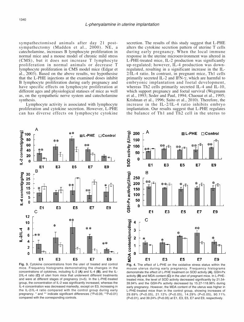
IL-2 secretion from lymphocytesDuring early pregnancy in the control group, the
concentration of IL-2 secreted from the uterine tissuehomogenate was lowest at E1 (11.13±2.42 pg/mg),increased to its highest concentration at E5 (43.64±8.69pg/mg) and decreased to 14.37±1.74 pg/mg at E9 (Fig.3). Compared with control group mice, the concentrationof IL-2 secreted from the uteri of the treated group wassignificantly increased by 162.17% at E1, 113.02% atE3, 98.61% at E5, 60.06% at E7 and 37.46% at E9.
IL-4 secretion from lymphocytesIn the control group, the concentration of IL-4 was
the lowest at E3 (12.90±3.06 pg/mg), it then increased toits highest concentration at E5 (25.83±0.69 pg/mg) anddecreased to 18.66±1.75 pg/mg at E9. Compared withthe control group, the IL-4 concentration wassignificantly decreased in the uteri of treated mice by39.94%, 16.08%, 51.66%, 65.80%, and 41.72% from E1
1337L-phenyalamine in uterine implantation
Table 1. Changes in embryo numbers and developmental sizes.
L-PHE-treated group Control groupE7 E9 E7 E9
Number 7.6±3.0** 5.6±2.7** 13.8±1.8 13.2±1.2Long Diameter (mm) 3.99±0.23* 4.63±0.10 4.38±0.22 4.76±0.12Short Diameter (mm) 2.45±0.10* 2.67±0.10** 2.89±0.09 3.03±0.13
*: indicates a significant difference between the treated and controlgroups (P<0.05); **: indicates an extremely significant differencebetween the treated and control groups (P<0.01). (mean ± SD, n=5).
to E9, respectively. Comparing the two groups, the ratioof IL-2/IL-4 was lower in the control groups (0.6-2.85)and increased to 137-8.45 in the treated group duringearly pregnancy.Oxidant and antioxidant status
SOD activityIn the control mice, the SOD activity of the uterine
tissue increased from E1 to E9, except on E5, reachingits highest level at E9 (Fig. 4). The level of SOD activityin L-PHE-treated mice decreased by 29.66%, 21.99%,
21.54%, 38.65% and 39.94% at E1, E3, E5, E7 and E9,respectively (P<0.01).
GSH-Px activityWhen mice were treated with L-PHE, the GSH-Px
activity decreased by 16.17% (P<0.01), 18.96%(P<0.01), 18.11% (P<0.05), 15.27% (P<0.05) and17.11% (P<0.05) compared with control mice at E1, E3,E5, E7 and E9, respectively.
MDA content. In the control group, the MDAcontent of the uterine tissue increased from E1 to E5before decreasing from E5 to E9; thus, it peaked at E5
1338L-phenyalamine in uterine implantation
Fig. 1. The morphology of uterus in L-PHE-treated and control pregnant mice. Mice have a bicornuate uterus consisting of two lateral horns, in whichthe embryos implant. The number of embryos decreased in L-PHE-treated mice (A, the seventh day after pregnancy, E7; C, the ninth day afterpregnancy, E9) compared with the control group, and the embryos were arranged regularly and tightly in the control group (B, E7; D, E9). IS:implantation site; OV: ovary. Scale bar: 1 cm.
(1.96±0.067 nmol/mg). In the L-PHE-treated group, theMDA content of the uterine tissue was higher than thatin the control groups, increasing by 29.69% (P<0.05),21.12% (P<0.05), 14.29% (P<0.05), 90.11% (P<0.01)and 39.24% (P<0.05) at E1, E3, E5, E7 and E9,respectively.Discussion
As a widely used additive in human food and animalforage, some researchers have reported that L-PHE has apositive effect on physical function. For example, L-PHE plays a role in controlling blood pressure (BP), andphenylalanine prevents the increases in BP that canoccur with increasing age and heart weight (heart/bodyweight index) (Zhao et al., 2001). Furthermore, thisadditive exerts a direct and specific anti-proliferativeeffect on vascular smooth muscle cells, which suggeststhat this effect may also explain the antihypertensiveaction of this amino acid that has been observed inspontaneously hypertensive rats (Gao et al., 1998).However, this amino acid is associated with some risks.Pregnant women have been advised to be careful aboutthe total amount of phenylalanine that they ingest. Doseshigher than 5,000 mg of the amino acid can causemarked nerve damage in the embryo. Therefore, thenegative effects of L-PHE must be studied, particularlyduring pregnancy. Toxicological hazard assessmentshave utilised the existing classical test paradigms and ahost of innovative approaches (Piersma, 2013). In thecurrent paper, the roles of 320 mg/kg of BW L-PHE onmouse embryo implantation and development wereexplored based on the state of local immunity andoxidative stress in the mouse uterus. We initiallyobserved that 320 mg/kg of BW L-PHE, injectedintraperitoneally for seven consecutive days beforepregnancy, decreased the litter number and size at E7and E9, whereas 80 mg/kg L-PHE did not.
There are several reasons why 320 mg/kg of BW L-PHE may reduce the litter size. First, previous resultsand those of the present study suggest that theimplantation and development of the embryo are closelyrelated to local immune reactions in the uterus(Wegmann, 1988; Wegmann et al., 1993; Dong et al.,2007). Changes in the total number and distribution ofthe maternal peripheral blood leukocyte subsets varythroughout the literature. For example, papers havereported a decrease (Matthiesen et al., 1995), no change(Fiddes et al., 1986), or an increase (Kuhnert et al.,1998; Luppi et al., 2002) in the presence of CD8+ Tcells during pregnancy. Discrepancies also exist for theCD4+ T-cell subset, which has been shown both to notchange (Sabahi et al., 1995; Kuhnert et al., 1998) and todecrease (Luppi et al., 2002; Watanabe et al., 1997)during pregnancy. The frequencies and counts of B cellsalso appear not to deviate during pregnancy (Watanabeet al., 1997; Kuhnert et al., 1998; Luppi et al., 2002).Therefore, we tested the immune parameters within thelocal uterus in the current study. The T lymphocyte
proliferation induced by ConA reflected no significantdifference among the L-PHE-treated mice, but the Blymphocyte proliferation index among the LPS-inducedmice was significantly decreased compared with that inuntreated mice, except at E3 and E5. These resultssuggest that L-PHE plays a greater regulatory role inhumoral immunity than in cellular immunity, as furtherdemonstrated by the ConA-induced T lymphocyteproliferation. As a catecholamine precursor (Grando etal., 2006; Fernstrom and Fernstrom, 2007), L-PHE maybe associated with the activity of sympathetic nerves,which release catecholamine. T lymphocyte proliferationin the spleen is inhibited in adult rats with sympatheticnerve lesions that are treated with 6-OHDA (Madden etal., 1994a,b; Pacheco-Lopez et al., 2003). In 3-month-old rats, when splenic norepinephrine (NE) wasmaximally depleted, no alterations in spleen cell Con A-induced T cell proliferation were observed up to 15 daysafter sympathectomy; however, Con A-inducedproliferation was reduced in sympathectomised animalsby 21 days post-sympathectomy, and then noalterations in T cell function were observed in
1339L-phenyalamine in uterine implantation
Fig. 2. Proliferation of lymphocytes from the uteri of L-PHE-treated andcontrol mice. A. ConA stimulated the proliferation of T lymphocytes fromthe uteri of treated and control mice. There was no significant differencebetween the control and treated group, although the ConA-inducedproliferative ability of lymphocytes from the uteri of treated micedemonstrated a slight decrease during early pregnancy, except for E7,compared with that of the corresponding control mice. B. LPSstimulated the proliferation of B lymphocytes from the uteri of treatedand control mice. Note that the proliferation ability of lymphocytes fromthe uteri in treated mice was decreased to 0.90±0.07 (E5) - 0.99±0.04(E3); lymphocyte proliferation was significantly lower at E1, E7 and E9(P<0.01) when compared with the corresponding values in control mice.The values are expressed as the mean ± SD of 5 mice from eachtreatment on the day indicated. * and ** indicate significant differences(*P<0.05, **P<0.01) compared with the corresponding controls. Thefollowing figures are organised similarly.
sympathectomised animals after day 21 post-sympathectomy (Madden et al., 2000). NE, acatecholamine, increases B lymphocyte proliferation innormal mice and a mouse model of chronic mild stress(CMS), but it does not increase T lymphocyteproliferation in normal animals or decrease Tlymphocyte proliferation in CMS model mice (Edgar etal., 2003). Based on the above results, we hypothesisethat the L-PHE injections at the examined doses inhibitB lymphocyte proliferation during early pregnancy andhave specific effects on lymphocyte proliferation atdifferent ages and physiological statuses of mice as wellas, on the sympathetic nerve system and catecholaminesynthesis.
Lymphocyte activity is associated with lymphocyteproliferation and cytokine secretion. However, L-PHEcan has diverse effects on lymphocyte cytokine
secretion. The results of this study suggest that L-PHEalters the cytokine secretion pattern of uterine T cellsduring early pregnancy. When the local immuneresponse in the uterine microenvironment was altered inL-PHE-treated mice, IL-2 production was significantlyup-regulated; however, IL-4 production was down-regulated, resulting in a significant increase in the IL-2/IL-4 ratio. In contrast, in pregnant mice, Th1 cellsprimarily secreted IL-2 and IFN-γ, which are harmful toembryonic implantation and foetal development,whereas Th2 cells primarily secreted IL-4 and IL-10,which support pregnancy and foetal survival (Wegmannet al., 1993; Seder and Paul, 1994; Chaouat et al., 1995;Krishnan et al., 1996; Saito et al., 2010). Therefore, theincrease in the IL-2/IL-4 ratio inhibits embryoimplantation. Our results suggest that L-PHE regulatesthe balance of Th1 and Th2 cell in the uterus to
1340L-phenyalamine in uterine implantation
Fig. 4. The effect of L-PHE on the oxidative stress status within themouse uterus during early pregnancy. Frequency histogramsdemonstrate the effect of L-PHE treatment on SOD activity (A), GSH-Pxactivity (B) and MDA content (C) in the uteri of pregnant mice. In L-PHE-treated mice, the level of SOD activity decreased significantly by 21.54-39.94% and the GSH-Px activity decreased by 15.27-118.96% duringearly pregnancy. However, the MDA content of the uterus was higher inL-PHE-treated mice than in the control group, showing increases of29.69% (P<0.05), 21.12% (P<0.05), 14.29% (P<0.05), 90.11%(P<0.01), and 39.24% (P<0.05) at E1, E3, E5, E7 and E9, respectively.
Fig. 3. Cytokine concentrations from the uteri of treated and controlmice. Frequency histograms demonstrating the changes in theconcentrations of cytokines, including IL-2 (A) and IL-4 (B), and the IL-2/IL-4 ratio (C) of uteri from mice that underwent different treatmentsand were at different stages of pregnancy (n=5). In the L-PHE-treatedgroup, the concentration of IL-2 was significantly increased, whereas theIL-4 concentration was decreased markedly, except on E3, increasing inthe IL-2/IL-4 ratio compared with the control group during earlypregnancy. * and ** indicate significant differences (*P<0.05; **P<0.01)compared with the corresponding controls.

influence embryo implantation and development duringearly pregnancy, similar to previous findings showingdecreases in norepinephrine secretion due to chemicalsympathectomy (Dong et al., 2007). L-PHE is aneurotransmitter and is thought to have marked medicalbenefits, which should be transferrable to norepinephrineand epinephrine. L-PHE might activate sympatheticnerves, stimulating norepinephrine and epinephrinesecretion. We deduced that the 320 mg/kg dose of L-PHE played a role in the local immune response in theuterus that was similar to that of chemicalsympathectomy.
Lymphocyte activity has a close relationship withoxidative stress, and pregnancy is a physiological statethat is marked primarily by oxidative stress (Casanuevaand Viteri, 2003). Low levels of reactive oxygen species(ROS) are required for normal cell functions. However,elevated ROS production can cause oxidative stress andcellular damage, generating both physiologic andpathologic effects within the placenta (Burton, 2009),embryo (Symonds et al., 2007), and foetus (Herrera etal., 2012). Increased maternal oxidative stress in earlyhuman pregnancy is associated with preeclampsia, ashortened gestation duration and lower infant birthweight (Stein et al., 2008). Hansen reviewed a number ofteratogens that elicit their deleterious effects on embryosthrough mechanisms involving the generation of ROSand oxidative stress (Hansen and Harris, 2013).Therefore, identifying the impact of L-PHE onintrauterine oxidative stress will be important forunderstanding cell-specific responses to L-PHE and forexploring the mechanisms of embryo implantation afterexposure to L-PHE. In our study, the activities of SODand GSH-Px were decreased (P<0.05) in the maternaluterus during early pregnancy after L-PHE treatment.Thus, the antioxidant function of the pregnant mouseuterus was impaired by L-PHE treatment. In contrast, theMDA content of L-PHE-treated uteri was increasedcompared with that found in the control group duringearly pregnancy (P<0.05). These results suggest thatmore free radicals were produced by L-PHE treatment,which may cause significant damage to cells. Our resultsdemonstrated that L-PHE had a modulatory effect onmaternal lipid peroxidation by adjusting the SOD andGSH-Px activities and MDA content during earlypregnancy. Chemical sympathectomy has a similar effecton the SOD, GSH-Px and MDA activities in the spleenduring early pregnancy (Bai et al., 2011). Macarthur etal. confirmed that oxidative stress is indeed involved inthe changes that occur in sympathetic neurotransmissionin hypertension (Macarthur et al., 2008).
Based on the current study, we speculate that L-PHEmay regulate embryo implantation through the followingroutes. (1) L-PHE may regulate the proliferation of localB lymphocytes, instead of T lymphocytes, in the uterus.(2) L-PHE may modulate cytokine secretion,predominantly through the up-regulation of IL-2, whichis harmful for embryo implantation, and through thedown-regulation of IL-4, which is beneficial for the
physiological state of pregnancy. (3) L-PHE may alsoattenuate oxidant stress states and antioxidant functionsin the pregnant mouse localised uterus. A dose of 320mg/kg L-PHE can decrease the SOD and GSH-Pxactivities, even while increasing the MDA content in theuterus during early pregnancy.Acknowledgments. This work was supported by National High-techResearch and Development Projects (863) (No. 2013AA10230603), theNational Natural Science Foundation of China (No. 31272483,30671514) and the Specialised Research Fund for the DoctoralProgram of Higher Education (No. 200800191004).Conflict of interest statement. The authors disclose that no conflicts offinancial interest that could inappropriately influence the work.
References
Bai Y., Wang T., Wang Z., Cao J., Dong Y. and Chen Y. (2011). Effectof sympathetic nerves on proliferation of splenic lymphocytes andantioxidant function of maternal spleen in early pregnant mice. Anat.Rec. 294, 875-882.
Bansal A. (2010). Joining the immunological dots in recurrentmiscarriage. Am. J. Reprod. Immunol. 64, 307-315.
Burton G. (2009). Oxygen, the Janus gas; its e�ects on human placentaldevelopment and function. J. Anat. 215: 27-35.
Casanueva E. and Viteri F. (2003). Iron and oxidative stress inpregnancy. J. Nutr. 133, 1700S-1708S.
Chaouat G., Meliani A., Martal J., Raghupathy R., Elliot J., Mosmann T.and Wegmann T. (1995). IL-10 prevents naturally occurring fetalloss in the CBA×DBA/2 mating combination, and local defect in IL-10 production in this abortion-prone combination is corrected by invivo injection of IFN-tau. J. Immunol. 154, 4261-4268.
Dong Y., Chen Y., Wang Z., Naito J. and Chen J. (2007). Role ofsympathetic nerves on early embryonic development and immunemodulation of uterus in pregnant mice. Auton. Neurosci. 131, 87-93.
Edgar V., Silberman D., Cremaschi G., Zieher L. and Genaro A. (2003).Altered lymphocyte catecholamine reactivity in mice subjected tochronic mild stress. Biochem. Pharmacol. 65, 15-23.
Fernstrom J. and Fernstrom M. (2007). Tyrosine, phenylalanine, andcatecholamine synthesis and function in the brain. J. Nutr. 137,1539S-1548S.
Fiddes T., O’Reilly D., Cetrulo C., Miller W., Rudders R., Osband M. andRocklin R. (1986). Phenotypic and functional evaluation ofsuppressor cells in normal pregnancy and in chronic aborters. CellImmunol. 97, 407-418.
Gao P., Zhu D., Zhan Y., Stepien O., Marche P. and Zhao G. (1998). L-phenylalanine and smooth muscle cell proliferation from SHR andWKY rats. Sheng Li Xue Bao. 50, 401-408.
Grando S., Pittelkow M. and Schallreuter K. (2006). Adrenergic andcholinergic control in the biology of epidermis: physiological andclinical significance. J. Invest. Dermatol. 126, 1948-1965.
Hansen J. and Harris C. (2013). Redox control of teratogenesis.Reprod. Toxicol. 35, 165-179.
Herrera E., Kane A., Hansell J., Thakor A., Allison B., Niu Y. andGiussani D. (2012). A role for xanthine oxidase in the control of fetalcardiovascular function in late gestation sheep. J. Physiol. 590:1825-1837.
Ho H., Chao K., Chen H., Chen S., Wu M. and Yang Y. (2001).
1341L-phenyalamine in uterine implantation
Distribution of Th1 and Th2 cell populations in human peripheral anddecidual T cells from normal and anembryonic pregnancies. Fertil.Steril. 76, 797-803.
Krishnan L., Guilbert L., Wegmann T., Belosevic M. and Mosmann T.(1996). T helper 1 response against Leishmania major in pregnantC57BL/6 mice increases implantation failure and fetal resorptions. J.Immunol. 156, 653-662.
Kuhnert M., Strohmeier R., Stegmuller M. and Halberstadt E. (1998).Changes in lymphocyte subsets during normal pregnancy. Eur. J.Obstet. Gynecol. Reprod. Biol. 76, 147-151.
Luppi P., Haluszczak C., Trucco M. and DeLoia J. (2002). Normalpregnancy is associated with leukocyte activation. Am. J. Reprod.Immunol. 47, 72-81.
Macarthur H., Westfall T. and Wilken G. (2008). Oxidative stressattenuates NO-induced modulation of sympatheticneurotransmission in the mesenteric arterial bed of spontaneouslyhypertensive rats. Am. J. Physiol. Heart Circ. Physiol. 294, H183-H189.
Madden K., Felten S., Felten D., Hardy C. and Livnat S. (1994a).Sympathetic nervous system modulation of the immune system. II.Induction of lymphocyte proliferation and migration in vivo bychemical sympathectomy. J. Neuroimmunol. 49, 67-75.
Madden K., Moynihan J., Brenner G., Felten S., Felten D. and Livnat S.(1994b). Sympathetic nervous system modulation of the immunesystem. III. Alterations in T and B cell proliferation and differentiationin vitro following chemical sympathectomy. J. Neuroimmunol. 49, 77-87.
Madden K., Stevens S., Felten D. and Bellinger D. (2000). Alterations inT lymphocyte activity following chemical sympathectomy in youngand old Fischer 344 rats. J. Neuroimmunol. 103, 131-145.
Matthiesen L., Berg G., Ernerudh J. and Skogh T. (1995). Lymphocytesubsets and autoantibodies in pregnancies complicated by placentaldisorders. Am. J. Reprod. Immunol. 33, 31-39.
Pacheco-López G., Niemi M., Kou W., Bildhäuser A., Gross C., GoebelM., Del Rey A. Besedovsky H. and Schedlowski M. (2003). Centralcatecholamine depletion inhibits peripheral lymphocyteresponsiveness in spleen and blood. J. Neurochem. 86: 1024-1031.
Piccinni M. (2003). Role of immune cells in pregnancy. Autoimmunity36, 1-4.
Piersma A. (2013). Innovations in testing strategies in reproductivetoxicology. Methods Mol. Biol. 947, 327-341.
Rao G., Kamath U., Raghothama C., Pradeep K. and Rao P. (2003).Maternal and fetal indicators of oxidative stress in various obstetriccomplications. Indian J. Clin. Biochem. 18, 80-86.
Sabahi F., Rola-Plesczcynski M., O’Connell S. and Frenkel L. (1995).Qualitative and quantitative analysis of T lymphocytes during normalhuman pregnancy. Am. J. Reprod. Immunol. 33, 381-393.
Saito S., Nakashima A., Shima T. and Ito M. (2010). Th1/Th2/Th17 andregulatory T-cell paradigm in pregnancy. Am. J. Reprod. Immunol.63, 601-610.
Seder R. and Paul W. (1994). Acquisition of lymphokine-producingphenotype by CD4+ T cells. Annu. Rev. Immunol. 12, 635-673.
Stein T., Scholl T. and Schluter M. (2008). Oxidative stress early inpregnancy and pregnancy outcome. Free Radical. Res. 42, 841-848.
Symonds M., Stephenson T., Gardner D. and Budge H. (2007). Long-term effects of nutritional programming of the embryo and fetus:mechanisms and critical windows. Reprod. Fertil. Dev. 19, 53-63.
Watanabe M., Iwatani Y., Kaneda T., Hidaka Y., Mitsuda N., MorimotoY. and Amino N. (1997). Changes in T, B, and NK lymphocytesubsets during and after normal pregnancy. Am. J. Reprod.Immunol. 37, 368-377.
Wegmann T. (1988). Maternal T cells promote placental growth andprevent spontaneous abortion. Immunol. Lett. 17, 297-302.
Wegmann T., Lin H., Guilbert L. and Mosmann T. (1993). Bidirectionalcytokine interactions in the maternal-fetal relationship: is successfulpregnancy a TH2 phenomenon? Immunol. Today 14, 353-356.
Zhao G., Li Z. and Gu T. (2001). Antihypertension and anti-cardiovascular remodeling by phenylalanine in spontaneouslyhypertensive rats: effectiveness and mechanisms. Chin. Med. J.(Engl) 114, 270-274.
Accepted May 6, 2014
1342L-phenyalamine in uterine implantation