The host-pathogen interaction of ecologically diverse ... · The host-pathogen interaction of...
188
The host-pathogen interaction of ecologically diverse coagulase-negative staphylococci in bovine mastitis, with a focus on prolactin Kristine Piccart Merelbeke, 2016
Transcript of The host-pathogen interaction of ecologically diverse ... · The host-pathogen interaction of...

The host-pathogen interaction of ecologically diverse
coagulase-negative staphylococci in bovine mastitis,
with a focus on prolactin
Kristine Piccart
Merelbeke, 2016


2
“You must look at the facts, because they look at you.”
Winston Churchill, 1925

3

4
The host-pathogen interaction of ecologically diverse coagulase-negative staphylococci in bovine
mastitis, with a focus on prolactin
Kristine Piccart
Cover: Hans De Clercq (design), Kristine Piccart (photos)
Printing: University Press, Zelzate
This research was financed by the Special Research Fund (BOF) of Ghent University.
Project number BOF10/STA/054.

5

6
The host-pathogen interaction of ecologically diverse coagulase-negative staphylococci in bovine
mastitis, with a focus on prolactin
Kristine Piccart
Department of Reproduction, Obstetrics, and Herd Health Faculty of Veterinary Medicine
Ghent University
Dissertation submitted in the fulfillment of the requirements for the degree of Doctor in Veterinary
Sciences, Faculty of Veterinary Medicine, Ghent University, May 17, 2017
−
Gastheer-pathogeen interactie van ecologisch diverse coagulase-negatieve staphylokokken in
boviene mastitis, met een focus op prolactine
Kristine Piccart
Vakgroep Voortplanting, Verloskunde en Bedrijfsdiergeneeskunde, Faculteit Diergeneeskunde,
Universiteit Gent
Proefschrift voorgedragen tot het behalen van de graad van Doctor in de Diergeneeskundige
Wetenschappen aan de Faculteit Diergeneeskunde, Universiteit Gent, 17 mei 2017

7

8
Promoters
Prof. dr. Sarne De Vliegher
Faculty of Veterinary Medicine, Ghent University, Belgium
Prof. dr. Freddy Haesebrouck
Faculty of Veterinary Medicine, Ghent University, Belgium
Dr. Sofie Piepers
Faculty of Veterinary Medicine, Ghent University, Belgium
Members of the examination committee
Prof. dr. Luc Duchateau
Ghent University, Belgium
Prof. dr. Catherine Delesalle
Ghent University, Belgium
Dr. Bert Devriendt
Ghent University, Belgium
Prof. dr. Evelyne Meyer
Ghent University, Belgium
Prof. dr. Geert Opsomer
Ghent University, Belgium
Dr. ing. Otlis Sampimon
Zoetis, The Netherlands
Dr. Suvi Taponen
University of Helsinki, Finland

9

10
Table of contents
Chapter 1 General introduction 14
Chapter 2 Aims of the thesis 52
Chapter 3 Local host response following an intramammary challenge with
Staphylococcus fleurettii and different strains of Staphylococcus
chromogenes in dairy heifers
58
Chapter 4 The role of milk prolactin in intramammary infections with coagulase-
negative staphylococci
88
4.1 Milk prolactin response and quarter milk yield after experimental infection
with coagulase-negative staphylococci in dairy heifers
90
4.2 Prolactin gene expression in bovine mammary epithelial cells after a
challenge with coagulase-negative staphylococci
114
Chapter 5 General discussion 130
Summary 160
Samenvatting 165
Curriculum vitae – Publications 172
Dankwoord - Acknowledgements 178

11

12
List of abbreviations
BSA Bovine serum albumin
C5a Complement component 5a
CFU Colony forming units
CNS Coagulase-negative staphylococci
cpm Counts per minute
cDNA Complementary DNA
gDNA Genomic DNA
DNA Deoxyribonucleic acid
FAO Food and Agricultural Organization of the United Nations
GH Growth hormone
GM-CSF Granulocyte macrophage colony stimulating factor
H2A Histone H2A
IgG1 Immunoglobin G1
IL Interleukin
IQR Interquartile range
IMI Intramammary infection
LPS Lipopolysaccharide
LSM Least square means
MAMP Microorganism-associated molecular patterns
MDL Minimal detection limit
MEC Mammary epithelial cells
MHC Major histocompatibility complex
mRNA Messenger RNA
NF-κB Nuclear factor kappa B
PBS Phosphate-buffered saline
PCR Polymerase chain reaction
PFGE Pulsed-field gel electrophoresis
PI Post-inoculation
PL Placental lactogen
PMN Polymorphonuclear neutrophil leukocyte
PRL Prolactin
PRR Pattern recognition receptor

13
QMY Quarter milk yield
qPCR Quantitative PCR
RIA Radioimmunoassay
RNA Ribonucleic acid
RPS15A Ribosomal Protein S15a
RT-PCR Reverse transcription PCR
SCC Somatic cell count
SD Standard deviation
SDHA Succinate dehydrogenase subunit A
SEM Standard error of the mean
SV40 Simian virus 40
tDNA-PCR Transfer RNA intergenic spacer PCR
TLR Toll-like receptor
TNF-α Tumor necrosis factor alpha
TRIR Total RNA isolation agent
UBC Ubiquitine C

14
Chapter 1
General introduction

15

Chapter 1. Introduction
16
1.1 Bovine mastitis, a burden on the dairy industry
Cow’s milk and dairy products make up a substantial part of the human diet. The Food and Agricultural
Organization of the United Nations (FAO) estimates that the worldwide milk demand will increase with
1.3% over the next years due to changing global consumption patterns (FAO, 2013). The European
Union currently produces around 160 million tons of milk on an annual basis (Eurostat, 2015). In
contrast to the past decennia, the dairy cattle population has expanded over the past three years in
Western Europe (Eurostat, 2016). Belgium counted approximately 508.000 lactating dairy cows in 2015
(FPSE, 2016). The Belgian dairy farms may be dwindling in numbers, but the herds are getting larger:
the average number of dairy cows per farm has increased with almost 52% since 2000 (from 33 to 50
cows in 2012 [FPSE, 2012]). This trend towards intensification is also seen at the individual cow level.
Nowadays, the average Flemish dairy cow produces around 8.500 kg of milk each year, which is an
increase of more than 7% compared to 10 years ago (CRV, 2015). The higher level of milk yield is
generally associated with negative health effects, such as an increased incidence of mastitis, i.e. an
inflammation of the mammary gland (Simianer et al., 1991; Van Dorp et al., 1998; Koeck et al., 2014).
Despite the implementation of well-known control and treatment strategies, mastitis remains to be a
burden on the dairy sector. Since the abolition of the European milk quota in 2015, the average annual
costs of mastitis are estimated at €240 per cow on Dutch dairy farms (van Soest et al., 2016). These
high costs are not surprising, since mastitis results in milk losses, high treatment costs, higher
probability of culling, etc. (Lescourret and Coulon, 1994; Halasa et al., 2007). Mastitis also affects the
milk quality. Milk from inflamed udder quarters, for instance, affects curd formation and produces off-
flavors during the processing of cheese (Le Marechal et al., 2011a). Locally administered
intramammary antimicrobials account for the majority of antimicrobial consumption in dairy herds,
either as dry-cow treatment or mastitis therapy (Stevens et al., 2016). Moreover, the use of
antimicrobials in food-producing animals has been linked to the development of antimicrobial
resistance (Chantziaras et al., 2014). Furthermore, there is also the issue of animal welfare, since
clinical mastitis is considered to be a painful condition (Leslie and Petersson-Wolfe, 2012). In Flanders,

Chapter 1. Introduction
17
the mean incidence rate of clinical mastitis is estimated at 7.4 quarters cases per 10,000 cow-days at
risk (Verbeke et al., 2014).
1.2 Etiology, clinical presentation and immune response
1.2.1. Etiology of bovine mastitis
Although several microorganisms can infect the mammary gland, the vast majority of bovine mastitis
cases are caused by bacteria (Watts, 1988). Nearly 80% of the mastitis cases can be attributed to either
staphylococci (which are routinely classified into coagulase-negative and -positive species),
streptococci or Escherichia coli (Bradley, 2002). In general, the bacteria enter through the teat canal of
the udder quarter, resulting in an intramammary infection (IMI) and subsequent inflammation (Blowey
and Edmondson, 2010).
In the past, it was generally assumed that the uninfected, healthy bovine mammary gland was a sterile
environment (Sheather, 1924; Perkins et al., 2009). This view can no longer be supported though, due
to the growing insight into the milk microbiota. Even healthy, non-mastitic milk samples display great
bacterial diversity after DNA sequencing (Kuehn et al., 2013; Oikonomou et al., 2014). The bacteria
found in healthy, uninfected quarters include Pseudomonas spp., Ralstonia spp., Psychrobacter spp.,
Faecalibacterium spp., Propionibacterium spp., Aeribacillus spp. and unclassified Lachnospiraceae
(Kuehn et al., 2013; Oikonomou et al., 2014). One study also found DNA of Streptococcus spp. and
Staphylococcus spp. in milk samples from healthy quarters (Oikonomou et al., 2012). Some of these
bacteria cannot be readily cultured during routine (aerobic) microbiological analyses, leading to
culture-negative results (Kuehn et al., 2013). It has even been suggested that mastitis could be the
result of a mammary dysbacteriosis, as opposed to a primary infection (Fernandez et al., 2013). The
importance of the mammary microbiota is also supported by incidental reports of clinical mastitis
outbreaks following a ‘blitz’ antimicrobial therapy (i.e. treating the entire herd or multiple infected
animals with antibiotics, in this case to eradicate Streptococcus agalactiae IMI [Edmondson, 2010]).
Chapter 1. Introduction
18
2.2 Clinical presentation
Based on the clinical presentation, mastitis can be classified into two types, namely clinical and
subclinical mastitis. Clinical mastitis is characterized by visual abnormalities in the milk (e.g. clots,
discoloration), the udder (e.g. swelling and redness) or the animal itself (sickness). Subclinical mastitis,
on the other hand, occurs when the gland is infected without any observable symptoms of
inflammation. It can be detected by, for instance, measuring the somatic cell count (SCC) in milk, which
increases through the influx of polymorphonuclear neutrophil leukocytes (PMN) in response to
invading pathogens (Harmon, 1994). Though different cut-off values are used in practice to distinguish
healthy from inflamed quarters, a threshold between 200,000 and 250,000 cells/mL at quarter level is
ideal to minimize diagnostic error (Schukken et al., 2003). In practice, cow level cell counts of 250,000
cells/mL and 150,000 cells/mL are often used as a threshold for multiparous cows and heifers,
respectively. The bulk milk SCC is considered an important indicator of the milk quality. As stated by
the Council Directive 92/46/EEC, the geometric average bulk milk SCC over a period of three months
should not exceed 400,000 cells/mL in the European Union.
Photo 1.1 shows a dairy cow with clinical mastitis in her left hind quarter, recognizable by the swelling
of that particular quarter. Photo 1.2 shows the difference between milk from a cow with subclinical
(i.e. no observable signs of inflammation) and clinical mastitis (in this case watery, yellowish milk with
few clots).
Photo 1.1 Cow with clinical mastitis in her left hind quarter.

Chapter 1. Introduction
19
The severity of mastitis cases depends on both the involved pathogen and the immune response
capacity of the cow. Host factors, such as the lactation stage, parity and genetic predisposition, have
an effect on the clinical course of an IMI (Burvenich et al., 2003). During early lactation, cows suffer
from an increased incidence of severe clinical mastitis due to the decreased function of leukocytes
around parturition (Mehrzad et al., 2001; Vangroenweghe et al., 2005). The clinical outcome of mastitis
is generally more severe in multiparous cows than in heifers (VanWerven et al., 1997; Mehrzad et al.,
2002; Vangroenweghe et al., 2004a), likely due to an age-associated impairment of the immune system
(Wojdak-Maksymiec et al., 2013). Managerial factors that affect the health status of the cow (e.g.
nutrition, housing, …) can also have an impact on the development risk of IMI (Smith et al., 1984;
Barkema et al., 1999a; Janosi et al., 2003).
In addition to the inherent and acquired host immune factors, the initial infection dose
(Vangroenweghe et al., 2004b; Günther et al., 2010) and the virulence of the involved bacterial strain
(Haveri et al., 2005) also affect the disease progression. Various mastitis pathogens, such as
Photo 1.2 Milk samples from udder quarters
with subclinical (left) and clinical mastitis (right).
Chapter 1. Introduction
20
Staphylococcus aureus, Streptococcus uberis or Streptococcus dysgalactiae, display strain-specific
differences in their ability to cause disease (Higgs et al., 1980; Haveri et al., 2007; Le Marechal et al.,
2011b; Tassi et al., 2013). For semantic clarity, a bacterial strain is defined in this thesis as “an isolate
or a group of isolates exhibiting characteristics that set it apart from other isolates belonging to the
same species” (Zadoks and Schukken, 2006).
1.2.3. Mammary gland immunity
A variety of defense mechanisms protect the mammary gland against invading pathogens (Figure 1.1).
First, pathogens need to overcome the teat canal, which constitutes an anatomical barrier with keratin
lining and sphincter muscles at the teat end (Sordillo et al., 1997). When bacteria successfully invade
the mammary gland, the innate branch of the immune system comes into play. The innate immune
response is activated within the first few hours, regardless of any previous exposure to the pathogen,
and is carried out by professional phagocytes (e.g. macrophages, PMN, dendritic cells, etc.) and
mammary epithelial cells (MEC), which are also able to recognize and respond to pathogens by
producing pro-inflammatory cytokines (Lahouassa et al., 2007). Foreign, microbial components are
recognized by immune cells with pattern recognition receptors (PRR), such as the transmembrane toll-
like receptor proteins (TLR). So far, ten different TLR have been described in cattle (Fisher et al., 2011).
The bacteria are recognized by the PRR due to highly conserved structures, referred to as
microorganism-associated molecular patterns (MAMP). Lipopolysaccharides (LPS) of Gram-negative
bacteria, for instance, are recognized by TLR4, whereas lipoteichoic acid of Gram-positive bacteria
binds to TLR2 (Rainard and Riollet, 2006). Following the recognition of the pathogen, various
inflammatory pathways are initiated, resulting in the upregulation of different cytokines and
chemokines (Table 1.1). Virtually every cell type is capable of producing and reacting to cytokines
(Dinarello, 2000). Though pro-inflammatory cytokines play a vital role in the host response, they can
also be harmful to the host, depending on the quantity and the extent of their expression (Bannerman,
2009). Tumor necrosis factor alpha (TNF-α) and interleukin 1 beta (IL-1β) induce a systemic host

Chapter 1. Introduction
21
response, resulting in fever and the production of acute phase proteins (Dinarello, 2000). Especially
IMI caused by Gram-negative bacteria (e.g. E. coli) induce the production of TNF-α, which correlates
with the clinical severity of mastitis cases (Burvenich et al., 2003; Bannerman et al., 2004). A well-
described chemokine in bovine mastitis is interleukin 8 (IL-8), which binds to receptors CXC-receptor 1
(CXCR1) and CXCR2 (Lahouassa et al., 2008). Polymorphonuclear neutrophil leukocytes are drawn to
the site of infection in massive numbers in response to IL-8 and other inflammatory mediators, such
as complement component 5a (C5a) (Stevens et al., 2012) or leukotriene B4 (Boutet et al., 2003).
The SCC can increase dramatically within 12 hours after exposure to bacteria, reaching more than 106
cells/mL (Rainard and Riollet, 2006). Macrophages and lymphocytes account for the majority of the
SCC in milk from uninfected quarters, but the cell type distribution shifts during inflammation, making
PMN the predominant cell population (Pilla et al., 2012; Damm et al., 2017). The accumulation of PMN
in the mammary gland cuts both ways though. While PMN are essential for the elimination of
pathogens, they may also damage the surrounding mammary gland tissue through the release of
extracellular proteolytic enzymes (such as elastase) and reactive oxygen species (Zhao and Lacasse,
2008). Consequently, their life span is limited: the PMN eventually undergo apoptosis, or programmed
cell death (Paape et al., 2003). Apoptotic PMN are quickly eliminated by macrophages, while their
membrane is still intact, to prevent the release of cytotoxic molecules and subsequent tissue damage
(Kennedy and DeLeo, 2009). The viability of PMN is linked to their activity (Mehrzad et al., 2004).
When the innate immune system is unable to eliminate the pathogen, the acquired or adaptive
immune system kicks in. The adaptive immunity, carried out by B- and T-lymphocytes, establishes an
antigen-specific immunological “memory”, enabling a faster, more efficient response in case of
repeated exposure to the pathogen (Sordillo et al., 2002; Iwasaki and Medzhitov, 2010). A previous
encounter with a particular pathogen results, for instance, in a larger influx of milk leukocytes in case
of repeated exposure (Rainard et al., 2016). The adaptive immune response can be classified into a
cell-mediated and antibody component, driven by the T-lymphocytes and B-lymphocytes, respectively.
Before the receptors on T-lymphocytes can recognize an antigen, it must be internalized, processed

Chapter 1. Introduction
22
and bound to the major histocompatibility complex (MHC) on the surface of antigen-presenting cells
(such as dendritic cells, macrophages and B-lymphocytes) or non-professional antigen-presenting cells
(such as epithelial cells) (Fitzpatrick et al. 1992; Quinn et al., 2015). Activated B-lymphocytes will
proliferate and differentiate into antibody-producing plasma-cells, or dormant memory cells (Sordillo
et al., 2002). However, Schukken et al. (2009a) propose that the mammary gland’s immunological
memory is limited, since the clinical response of dairy cows suffering from recurring infections is not
necessarily attenuated. The mastitis pathogens in that particular study were not identified at the strain
level though.
The dairy cow’s immune system reacts differently to IMI caused by epidemiologically and ecologically
varying species (Schukken et al., 2011; Günther et al., 2016). Typically, Gram-negative mastitis
pathogens (such as E. coli or Klebsiella spp.) evoke a more drastic host response than Gram-positive
bacteria, given the LPS in their cell walls (Schukken et al., 2011). However, the host response does not
only depend on the involved pathogen, but also on various cow factors (Burvenich et al., 2003), there
has been much debate on the “most desirable” type of response in dairy cattle following IMI (Benjamin
et al., 2015). In case of experimental E. coli mastitis, an early mobilization of PMN leads to lower
bacterial growth and faster clearance of the infection (Vandeputte-Van Messom et al.; 1993, Mehrzad
et al., 2005). Some authors even consider very low pre-challenge quarter milk SCC levels (< 20.000
cells/mL) a risk factor for establishing IMI (Schukken et al., 1999, Wellnitz et al., 2010). Another study
investigated the potential of the bacterial molecules LPS for enhancing leukocyte recruitment during
an experimental S. aureus challenge (Kauf et al., 2007). Although LPS increased the SCC levels in the S.
aureus-infected quarters, it did not facilitate the bacterial clearance.

Chapter 1. Introduction
23
Table 1.1. A non-exhaustive summary of cytokines involved in bovine mastitis (1/2).
Cytokine Primarily produced by Functions Pathogen-specific response
IL-1 Macrophages, lymphocytes, epithelial cells, etc.
The IL-1 family consists of 11 cytokines, including IL-1α and IL-1β. IL-1 induces local and systemic effects, such as fever and the synthesis of acute phase proteins.
Mainly Gram-negative mastitis pathogens induce a temporal IL-1β response (ranging between 0.3 – 8 ng/mL). The response varies greatly between individual cows.
IL-2 T-helper cells (Th1)
IL-2 stimulates the clonal expansion of T-lymphocytes, activates cytotoxic T-cells and natural killer (NK) cells, and the proliferation of B-lymphocytes.
IL-2 occurs in both healthy and infected mammary glands. The transcription of IL-2 decreases during S. aureus infection.
IL-4 T-helper cells (Th2)
IL-4 is a so-called anti-inflammatory cytokine that promotes the differentiation of T-cells into Th2 cells. It is the primary cytokine expressed by mononuclear cells in the post-partum period.
Little is currently known about the involvement of IL-4 in bovine mastitis.
IL-6 PMN, macrophages, lymphocytes, epithelial cells, etc.
IL-6 has both pro- and anti-inflammatory properties. It promotes the synthesis of hepatic APP. It has been suggested that IL-6 enables the shift from PMN to monocytes during inflammation.
Although IL-6 is found in milk from mammary quarters infected with Gram-negative and -positive bacteria, the transcription is minimal in case of S. aureus infections.
IL-8 MEC, PMN, lymphocytes, etc. IL-8 is a chemotactic cytokine (chemokine) that attracts PMN to the site of infection, and enhances their activity.
IL-8 increases within 20h during Gram-negative IMI (ranging between 100 – 1000 pg/mL). IL-8 transcription is diminished or absent in S. aureus IMI.

Chapter 1. Introduction
24
Table 1.1. A non-exhaustive summary of cytokines involved in bovine mastitis (2/2).
Cytokine Primarily produced by Functions Pathogen-specific response
IL-10 Th2 cells, B cells, eosinophils, mast cells, etc.
IL-10 is an anti-inflammatory that inhibits the production of pro-inflammatory cytokines in PMN, and impairs the Th1 response.
Various mastitis pathogens evoke an IL-10 response, although the expression is not always seen in S. aureus IMI. The production of IL-10 is preceded by an increase in TNF-α.
IL-12 Monocytes, macrophages, dendritic cells
IL-12 links the innate to the adaptive immune system: it promotes the differentiation of T-cells into Th1-cells, and stimulates the production of IFN- γ.
Similar elevations in IL-12 are found in E. coli and S. aureus infections. The increase in IL-12 coincides with the elevated IFN- γ levels.
IL-17 T-helper cells (Th17)
The IL-17 cytokine family contains 6 members, including IL-17A. IL-17A stimulates the inflammatory response of MEC, and attracts PMN and macrophages to the site of infection.
IL-17A is expressed in mammary tissue infected with E. coli, S. aureus or S. uberis. Intramammary infusion of IL-17A in E. coli IMI is associated with lower bacterial numbers, increased PMN recruitment and lower IL-10 levels.
IFN-γ Monocytes, lymphocytes
IFN-γ stimulates the activity of PMN and macrophages, and upregulates the expression of the major histocompatibility complex (MHC) class I molecules, enabling pathogen recognition by T-cells.
IFN-γ transcription occurs in both Gram-negative and –positive mastitis cases, but the highest concentrations are found in persistent IMI.
TNF-α Macrophages, lymphocytes, PMN, epithelial cells, etc.
TNF-α induces systemic effects, such as fever and the production of APP, and weakens the blood-milk barrier. TNF-α is also associated with shock, tissue damage and organ failure.
The TNF-α level increases in the blood and milk in a dose-responsive manner during Gram-negative IMI. The TNF-α response is absent in S. aureus IMI.
References: Sordillo and Streicher, 2002; Waller, 2002; Alluwaimi et al., 2003; Alluwaimi, 2004; Rainard and Riollet, 2006; Dinarello, 2007; Bannerman, 2009; Günther et al.,
2011; Porcherie et al., 2016

Chapter 1. Introduction
25
Figure 1.1. Simplified representation of an intramammary infection. Pathogens enter the mammary
gland through the teat canal and colonize the mammary epithelial cells (arrow). Once the pathogen is
recognized by macrophages and other immune cells, the host response is triggered. Neutrophils, or
PMN, travel in massive numbers from the blood stream to the site of infection to kill the invading
pathogens. Apoptotic PMN are then eliminated by macrophages to minimize tissue damage
Blood vessel Myoepithelial cell
Epithelial cell
Polymorphonuclear neutrophil leukocyte (PMN)
Macrophage
ALVEOLUS
UDDER
Pathogen

Chapter 1. Introduction
26
1.3 Coagulase-negative staphylococci
1.3.1. A complex, heterogeneous group
Coagulase-negative staphylococci (CNS) are named for their inability to coagulate rabbit plasma in
vitro. In bovine medicine, CNS is often used as a collective term for non-aureus staphylococci, although
this is not entirely accurate. For example, Staphylococcus intermedius, a staphylococcal species found
in bovine milk, is in fact coagulase-positive (Roberson et al., 1996), whereas Staphylococcus hyicus,
Staphylococcus agnetis and Staphylococcus chromogenes have a variable coagulase activity (Taponen
et al., 2012; dos Santos et al., 2016). Previously, CNS were often considered to be one large
homogeneous group of bacteria (Reneau, 1986; Hogan et al., 1987). Novel molecular identification and
typing techniques, however, have made it easier to study CNS on species level (Da Silva Santos et al.,
2008; Park et al., 2011; Supré et al., 2011; Piessens et al., 2011; Braem et al., 2012). So far, more than
50 staphylococcal species have been identified and described (Parte, 2014). Over 20 CNS species have
been found in bovine milk, but only 5 species are isolated on a routine basis: S. chromogenes,
Staphylococcus epidermidis, Staphylococcus xylosus, Staphylococcus haemolyticus and Staphylococcus
simulans (Vanderhaeghen et al., 2014).
1.3.2. Epidemiology and ecology
Coagulase-negative staphylococci are frequently isolated from cows with subclinical mastitis (Barkema
et al., 1999b; Pitkälä et al., 2004; Sampimon et al., 2009; Piepers et al., 2011; Rall et al., 2014), especially
in dairy heifers (Tenhagen et al., 2006; Fox, 2009; Sampimon et al., 2009). According to Vanderhaeghen
et al. (2015), CNS can be divided into two categories based on their epidemiology, namely “contagious”
and “opportunistic CNS”. Whereas contagious mastitis pathogens are typically transmitted from cow
to cow through a vector (such as the milking equipment or the milker’s hands) (Zadoks et al., 2011),
opportunistic mastitis bacteria originate from different sources, do not spread between cows and only
Chapter 1. Introduction
27
cause an IMI “under conditions favouring colonisation of the udder” (Vanderhaeghen et al., 2015). In
terms of their ecology (i.e. habitat), CNS are usually divided into “host-adapted” and “environmental”
species (Piessens et al., 2011; De Visscher et al., 2014; Fry et al., 2014). Across multiple studies, S.
fleurettii is regularly found in the environment of the dairy farm or milking parlor (Piessens et al., 2011;
De Visscher et al., 2014). The species rarely occurs in the milk of cows (De Visscher et al., 2016). In the
(occasional) occurrences that S. fleurettii is associated with an IMI, the infection is of a transient nature
(Supré et al., 2011). At the other end of the ecological spectrum are the host-adapted CNS species and
strains, which are characterized by their ability to colonize or invade the host. Staphylococcus
chromogenes is by all means a prime example of host-adapted species. For one, S. chromogenes is the
most frequently isolated CNS species in milk, especially in heifers (Aarestrup and Jensen, 1997;
Taponen et al., 2006, Adkins and Middleton, 2016). It has been suggested by some authors that S.
chromogenes is part of the normal skin microbiome of cattle (White et al., 1989; Taponen et al., 2008;
Taponen and Pyörälä, 2009), even though the species is not always present on the skin of the teats
(Braem et al., 2013; De Visscher et al., 2014). Although S. chromogenes can be effectively and swiftly
neutralized by macrophages (Åvall-Jääskeläinen et al., 2013), the species is known to invade and
replicate in bovine MEC (Hyvönen et al., 2009; Souza et al., 2016). It is not uncommon for S.
chromogenes to cause persistent IMI lasting an entire lactation (Taponen et al., 2007; Piessens et al.,
2011; Mørk et al., 2012). Ultimately though, the question remains: why do some CNS species thrive in
the mammary gland, as opposed to other body sites or the surrounding environment?
1.3.3. Intramammary infections with CNS
Since the clinical impact of CNS IMI is relatively limited, they are referred to as “minor pathogens”
(Djabri et al., 2002; Taponen et al., 2006). An IMI with CNS usually manifests itself as subclinical or mild
clinical mastitis (Waller et al., 2011). In general, the SCC increase caused by CNS is small compared to
the response seen in IMI with major mastitis pathogens (Sampimon et al., 2010). Some CNS species,
such as S. chromogenes and S. simulans, provoke a larger SCC increase than others though (Fry et al.,
Chapter 1. Introduction
28
2014, De Visscher et al., 2016a). Staphylococcus chromogenes can even evoke an increase in SCC
similar to the response seen in S. aureus IMI (Supré et al., 2011).
All in all, not much is known about the interaction between bovine-associated CNS and their host.
Gram-positive bacteria generally induce a weaker pro-inflammatory cytokine response than Gram-
negative mastitis pathogens (Riollet et al., 2000; Bannerman et al., 2004; Günther et al., 2016).
Previous challenge studies show that S. epidermidis is able to induce the production of IL-1β, IL-8 and
TNF-α in cows and ewes (Winter et al., 2003; Simojoki et al., 2011). This is in stark contrast to
experimental infections with S. aureus, where IL-8 and TNF-α are generally not detected in milk (Riollet
et al., 2000; Bannerman et al., 2004). The IL-1β response seen in an S. epidermidis or S. simulans
challenge is similar to S. aureus IMI (Simojoki et al., 2011), but can vary greatly between individual
animals (Winter et al., 2003; Bannerman, 2009).
Although it is assumed that most CNS infections resolve without antibiotic treatment (Taponen and
Pyörälä, 2009), certain species (i.e. S. chromogenes, S. simulans, S. epidermidis and others) can cause
persistent IMI lasting over several months (Taponen et al., 2007, Thorberg et al., 2009; Piessens et al.,
2011; Fry et al., 2014). A number of virulence factors have been identified in bovine-associated CNS
species, but the pathogenic potential of CNS is still not fully elucidated. Some CNS species may have
virulence factors similar to S. aureus, such as the ability to form a biofilm (Simojoki et al., 2012),
whereas other do not (Taponen and Pyörälä, 2009). In order to evade the host response and cause a
long-lasting IMI, S. aureus attaches itself to the surface of MEC and invades them (Cifrian et al., 1994;
Almeida et al., 1996). This strategy of invasion is also seen in mastitis-causing CNS, although to a lesser
extent (Hyvönen et al., 2009). Similar to S. aureus, certain CNS species and strains are also able to
adhere to and replicate in MEC (Hyvönen et al., 2009), and resist phagocytosis by macrophages (Åvall-
Jääskeläinen et al., 2013). However, the potential for adhesion and internalization varies between
species (Almeida and Oliver, 2001) and even within species (Souza et al., 2016). A S. chromogenes
strain recovered from a chronically infected quarter showed higher adhesion and internalization values
than another S. chromogenes strain originating from the teat skin (more specifically, the teat apex

Chapter 1. Introduction
29
[Souza et al., 2016]). The indication that some strains of S. chromogenes might be better suited to
invade and colonize the mammary gland than others sheds new light on the heterogeneity of CNS,
even within species.
1.3.4. Effect of CNS IMI on milk yield: a paradox?
Milk production losses associated with mastitis are either due to the bacterial infection itself, or to
inflammatory response following the infection (Detilleux et al., 2015). Evidently, not all mastitis
pathogens elicit the same degree of milk loss in dairy cattle (Gröhn et al., 2004). Over the past years,
the effect of CNS IMI on milk production has received a great deal of attention (Table 1.2). Yet, there
is no definitive consensus on the impact of CNS IMI on milk yield. Some research indicates that the
milk yield decreases in response to a CNS IMI (Timms and Schultz, 1987; Gröhn et al., 2004), while
others have observed barely any effect at all (Pearson et al., 2013; Tomazi et al., 2015). Interestingly
though, some studies even mention a positive effect on milk yield (Schukken et al., 2009b; Piepers et
al., 2010). There are multiple possible explanations for this counterintuitive finding. For one, high-
yielding dairy cows might inherently be more prone to CNS infections. Gröhn et al. (2004), for instance,
remarked that multiparous cows with clinical CNS mastitis produced significantly more milk (between
2.3 and 2.7 kg/day) one month before diagnosis than their non-infected herd mates. This effect was
not seen in primiparous cows. According to Piepers et al. (2013), the association between milk yield
and CNS IMI is only partially confounded by the genetic potential for milk production, as it did not fully
account for the observed difference in milk yield. Another hypothesis for the higher milk yield in CNS-
infected dairy cattle relies on the lactation hormone prolactin (PRL), which can also act as an
immunomodulatory factor (see below). Other research suggests that pre-existing CNS IMI or even CNS
teat apex colonization can have a protective effect against new infection with other (major) mastitis
pathogens, effectively lowering the risk of developing clinical mastitis and subsequent milk loss
(Rainard and Poutrel, 1988, Matthews et al., 1991, Nickerson and Boddie, 1994). The protective

Chapter 1. Introduction
30
mechanism of CNS infections might be attributed to bacterial competition between CNS and other
pathogens in the same niche (Hibbing et al., 2010), the stimulation of the innate immune system (i.e.
an increased SCC [Schukken et al., 1999]), or the production of bacteriocins and other antibacterial
substances (Braem et al., 2014). Only a handful of studies have focused on the species-specific effect
of CNS regarding their protecting outcome. De Vliegher et al. (2003) demonstrated that certain S.
chromogenes isolates, originating from the teat apex of primiparous cows, are able to inhibit the in
vitro growth of Gram-positive mastitis pathogens. In mice, the colonization of the mammary gland by
S. epidermidis attenuates the clinical response to a later challenge with S. aureus or E. coli (Anderson,
1978).
Still, the majority of the aforementioned studies do not account for potential differences between or
within CNS species, which might contribute to the overall conflicting results. Also, the protective effect
of CNS is more pronounced in experimental challenge studies where major pathogens are infused
directly into the mammary gland, compared to natural infections with mastitis pathogens (Reyher et
al., 2012).

Chapter 1. Introduction
31
Table 1.2. An overview of longitudinal field studies on the effect of intramammary infections with
coagulase-negative staphylococci (CNS) on the milk yield.
1 Identification of CNS at group level with bacterial culture, or at species level using restriction fragment length polymorphism (PCR-RFLP) or transfer RNA intergenic spacer PCR (tDNA-PCR). 2 Standard deviation. 3 Standard error of the mean.
Authors Animals CNS ID1 Effect on milk yield
Effect size compared to uninfected animals
Timm & Schutlz (1987) Dairy cows Bacterial culture
↓ - 2.9 kg/d
Gröhn et al. (2004) Dairy heifers Bacterial culture
↓ - 3.2 to - 1.0 kg/d
Leitner et al. (2004) Sheep & goats / ↓ - 0.64 to 0 kg/d
Gröhn et al. (2004) Dairy cows Bacterial culture
→ -
Pearson et al. (2013) Dairy cows Bacterial culture
→ -
Tomazi et al. (2015) Dairy cows PCR-RFLP → -
Koop et al. (2010) Goats Bacterial culture
→ -
Koop et al. (2012) Goats tDNA-PCR Staphylococcus
caprae: ↑
/
Schukken et al. (2009) Dairy cows Bacterial culture
↑ + 0.45 kg/d (SD2 ± 0.12)
Piepers et al. (2010) Dairy cows Bacterial culture
↑ + 2.95 kg/d
(SEM3 ± 0.98)
Piepers et al. (2013) Dairy cows Bacterial culture
↑ + 2.0 kg/d

Chapter 1. Introduction
32
1.4 Prolactin and udder health
1.4.1. Lactation hormone
Prolactin, thought to be present in all vertebrates (Malven, 1993), is involved in a plethora of biological
mechanisms. Over 300 distinct actions have been attributed to this versatile hormone (Goffin et al.,
2002). Riddle et al. (1933) were the first to extract and identify the hormone from the anterior lobe of
the pituitary gland. They proposed the name ‘prolactin’, seeing that the compound was able to
promote mammary growth and milk secretion in mammals. Even though PRL is historically well-known
for its role in lactation and mammary gland development, it is also involved in various behavioral
mechanisms, reproduction and immunoregulation (Freeman et al., 2000). The broad function of PRL is
evidenced by the vast number of tissues equipped with PRL receptors (Bole-Feysot et al., 1998). Bovine
pituitary-derived PRL is a polypeptide consisting of 199 amino acids weighing approximately 23 kDa
(Wallis, 1974). The hormone is primarily – though not exclusively- produced by the lactotrophs in the
anterior pituitary gland (Freeman et al., 2000). Other production sites include (among others) the
ovaries, the uterus, various regions of the brain, the skin, the spleen, the thymus, the tonsils and the
lymph nodes (Figure 1.2). Mammary tissue is also capable of synthetizing PRL (Leprovost et al., 1994).
Mammary-produced PRL controls the proliferation and differentiation of MEC through an autocrine or
paracrine mechanism (Naylor et al., 2003; Chen et al., 2012). Bovine PRL also limits the transfer of
immunoglobulins of the maternal circulation into colostrum, by reducing the expression of the IgG1-
receptor on mammary tissue (Barrington et al., 2001).

Chapter 1. Introduction
33
Figure 1.2. The anterior pituitary gland (1) is the primary site of prolactin synthesis. Extra-pituitary sites
of prolactin production in mammals include (but are not limited to) various regions of the brain (2),
the tonsils (3), the thymus (4), the myometrium (5), the ovaries (6), the spleen, (7) the mammary gland
(8) and the skin. (Adapted from Marano and Ben-Jonathan, 2014)
Prolactin
1
2
3
4 7
5
6
8
Hypothalamus
Posterior pituitary gland
Anterior pituitary gland

Chapter 1. Introduction
34
Nowadays, it is generally accepted that PRL is pivotal for the initiation and maintenance of lactation in
ruminants (Lacasse et al., 2015), although this was debated for many years. Early experiments failed
to demonstrate an effect on milk yield when suppressing the PRL release with the dopamine agonist
bromocriptine (Karg et al., 1972; Hart, 1973; Smith et al., 1974). Therefore, it was assumed that PRL
was not a galactopoetic hormone in cattle. However, newer studies show that the milk production
significantly drops when dairy cows receive a long-term quinagolide treatment (Lacasse et al., 2011),
which is a more selective and effective dopamine receptor agonist (Barlier and Jaquet, 2006). The milk
yield can also be increased by administering domperidone, a dopamine antagonist which increases the
circulating PRL level (Lacasse and Ollier, 2015). The contradictory findings between the older and
newer studies might be explained by the use of different compounds (bromocriptine versus
quinagolide) or duration of treatments (varying between 2 days and nine weeks).
In general, serum PRL lies between 10 and 60 ng/mL in adult dairy cows (Koprowski et al., 1972;
Malven, 1977; Fulkerson et al., 1980; Marcek and Swanson, 1984), but it can be affected by many
factors, such as the ambient temperature (Wetteman and Tucker, 1974), the exposure to light (Dahl
et al., 2000), or the milking process (Johke, 1970). Stress also has a high impact on circulating PRL levels
(Karg and Schams, 1974). GarcÍa-Ispierto et al. (2009) observed that dairy cows with intermediate to
high blood cortisol levels (> 3ng/mL) had higher levels of circulating PRL.
Approximately one week before calving, the serum PRL concentration starts to rise (80 – 110 ng/mL),
peaking one day before parturition (230 – 285 ng/mL) (Edgerton and Hafs, 1973; Ingalls et al., 1973).
The milk PRL level is at its highest (369 ± 56 ng/mL) immediately after parturition (Malven, 1977).
Besides PRL, there are many other hormones involved in the development of the mammary gland and
the secretion of milk, such as oxytocin, estrogen, progesterone and various metabolic hormones
(including glucocorticoids, growth hormone, etc.) (Neville et al., 2002). The mammary gland can
function as a self-regulating endocrine organ, largely independent from systemic influences (Wilde and
Peaker, 1990; Weaver and Hernandez, 2016). Nevertheless, the biological significance of autocrine PRL
has not been studied extensively in cattle.

Chapter 1. Introduction
35
1.4.2. Immunological aspects
In addition to its role in lactation, PRL also acts as an immunomodulatory factor. Hypophysectomized
rats, for instance, display signs of immunosuppression (more specifically normochromic normocytic
anemia, leucopenia and thrombocytopenia), which can be reversed by administering PRL (Berczi and
Nagy, 1981; Nagy and Berczi, 1991). Other research shows that PRL is able to inhibit the glucocorticoid-
induced apoptosis of T-lymphocytes (Krishnan et al., 2003), stimulate the production of reactive
oxygen species in macrophages (Edwards et al., 1987) and induces TLR2 expression on the membrane
of bovine MEC (Medina-Estrada et al., 2015). When challenging peripheral immune cells in the
presence of PRL, the production of pro-inflammatory cytokines (such as TNF-α and interleukin 12 [IL-
12]] increases while the production of the anti-inflammatory cytokine IL-10 decreases (Brand et al.,
2004). The opposite occurs as well: the expression of the PRL-receptor is upregulated in rat fibroblasts
in the presence of pro-inflammatory cytokines, making the cells more responsive to PRL (Corbacho et
al., 2003).
Previous research indicates that PRL might also play a role in the udder health of dairy cattle. A PRL
surge occurs around the time of calving (Convey, 1974), coinciding with the phenomenon of
periparturient immunosuppression (Drackley, 1999). The risk for developing an IMI is very high after
parturition (Burton and Erskine, 2003), combined with an increased severity of clinical mastitis cases
(Burvenich et al., 2003). It has previously been hypothesized that PRL counteracts the
immunosuppression caused by glucocorticoid hormones (Dorshkind and Horseman, 2000; Matalka,
2003; Fomicheva et al., 2004). Although the blood PRL concentration does not differ between healthy
cows and cows experiencing a clinical (Hockett et al., 2000; Vanselow et al., 2006) or chronic subclinical
mastitis (Boutet et al., 2007), a positive correlation exists between the SCC of infected udder quarters
and the milk PRL concentration (Boutet et al., 2007). Nuclear factor κB (NF-κB), the transcription factor
for the genes encoding numerous cytokines involved in the pathogenesis of mastitis (Boulanger et al.,
2003), is activated by PRL in a dose-dependent manner, resulting in the up-regulation of IL-1β, IL-6, IL-
8, TNF-α and macrophage colony stimulating factor (GM-CSF) in MEC (Boutet et al., 2007). However,

Chapter 1. Introduction
36
PRL can also elicit anti-inflammatory responses in MEC. Adding PRL to a mammary cell line infected
with S. aureus even promotes the internalization of the mastitis pathogen (Gutierrez-Barroso et al.,
2008), hereby enabling a persistent IMI (Hebert et al., 2000). Furthermore, S. aureus has the ability to
inhibit the PRL-driven activation of NF-κB (Lara-Zarate et al., 2011), which suppresses the host’s innate
immune response. All in all, the role of bovine PRL as a potential cytokine in the mammary gland
defense system is not yet well defined.
Chapter 1. Introduction
37
1.5 References
Aarestrup, F. M. and N. E. Jensen. 1997. Prevalence and duration of intramammary infection in Danish heifers during the peripartum period. J Dairy Sci 80(2):307-312. Adkins, P. R. F. and J. R. Middleton. 2016. Potential body site reservoirs for coagulase-negative staphylococcal intramammary infection in heifers. J Anim Sci 94:33-33. Alluwaimi, A. M. 2004. The cytokines of bovine mammary gland: prospects for diagnosis and therapy. Res Vet Sci 77(3):211-222. Alluwaimi, A. M., C. M. Leutenegger, T. B. Farver, P. V. Rossitto, W. L. Smith, and J. S. Cullor. 2003. The cytokine markers in Staphylococcus aureus mastitis of bovine mammary gland. J Vet Med B 50(3):105-111. Almeida, R. A., K. R. Matthews, E. Cifrian, A. J. Guidry, and S. P. Oliver. 1996. Staphylococcus aureus invasion of bovine mammary epithelial cells. J Dairy Sci 79(6):1021-1026. Almeida, R. A. and S. P. Oliver. 2001. Interaction of coagulase-negative Staphylococcus species with bovine mammary epithelial cells. Microb Pathog 31(5):205-212. Anderson, J. C. 1978. Absence of bacterial adherence in the establishment of experimental mastitis in mice. Vet Pathol 15(6):770-775. Åvall-Jääskeläinen, S., J. Koort, H. Simojoki, and S. Taponen. 2013. Bovine-associated CNS species resist phagocytosis differently. BMC Vet Res 9(1): 227. Bannerman, D. D., M. J. Paape, J. W. Lee, X. Zhao, J. C. Hope, and P. Rainard. 2004. Escherichia coli and Staphylococcus aureus elicit differential innate immune responses following intramammary infection. Clin Diagn Lab Immunol 11(3):463-472. Bannerman, D. D. 2009. Pathogen-dependent induction of cytokines and other soluble inflammatory mediators during intramammary infection of dairy cows. J of Anim Sci 87(13):10-25. Barkema, H. W., Y. H. Schukken, T. J. Lam, M. L. Beiboer, G. Benedictus, and A. Brand. 1999a. Management practices associated with the incidence rate of clinical mastitis. J Dairy Sci 82(8):1643-1654. Barkema, H. W., H.A. Deluyker, Y.H. Schukken, and T.J.G.M. Lam. 1999b. Quarter-milk somatic cell count at calving and at the first six milkings after calving. Prevent Vet Med 38(1), 1-9. Barlier, A. and P. Jaquet. 2006. Quinagolide - a valuable treatment option for hyperprolactinaemia. Eur J Endocrinol 154(2):187-195. Barrington, G. M., T. B. McFadden, M. T. Huyler, and T. E. Besser. 2001. Regulation of colostrogenesis in cattle. Livest Prod Sci 70(1-2):95-104. Benjamin, A. L., Green, B. B., Hayden, L. R., Barlow, J. W., and Kerr, D. E. 2015. Cow-to-cow variation in fibroblast response to a toll-like receptor 2/6 agonist and its relation to mastitis caused by intramammary challenge with Staphylococcus aureus. J Dairy Sci 98(3), 1836-1850.

Chapter 1. Introduction
38
Berczi, I. and E. Nagy. 1981. Immunodeficiency in Hypophysectomized Rats - Restoration by Prolactin. Fed Proc 40(3):1031-1031. Blowey, P. and P. Edmondson. 2010. Mastitis Control in Dairy Herds. 2nd Ed. ed. Cabi, Oxfordshire, UK. Bole-Feysot, C., V. Goffin, M. Edery, N. Binart, and P. A. Kelly. 1998. Prolactin (PRL) and its receptor: Actions, signal transduction pathways and phenotypes observed in PRL receptor knockout mice. Endocr Rev 19(3):225-268. Boulanger, D., F. Bureau, D. Melotte, J. Mainil, and P. Lekeux. 2003. Increased nuclear Factor kappa B activity in milk cells of mastitis-affected cows. J Dairy Sci 86(4):1259-1267. Boutet, P., F. Bureau, G. Degand, and P. Lekeux. 2003. Imbalance between lipoxin A(4) and leukotriene B-4 in chronic mastitis-affected cows. J Dairy Sci 86(11):3430-3439. Boutet, P., J. Sulon, R. Closset, J. Detilleux, J. F. Beckers, F. Bureau, and P. Lekeux. 2007. Prolactin-induced activation of nuclear factor kappa B in bovine mammary epithelial cells: Role in chronic mastitis. J Dairy Sci 90(1):155-164. Bradley, A. J. 2002. Bovine mastitis: An evolving disease. Vet J 164(2):116-128. Braem, G., S. De Vliegher, B. Verbist, M. Heyndrickx, F. Leroy, and L. De Vuyst. 2012. Culture-independent exploration of the teat apex microbiota of dairy cows reveals a wide bacterial species diversity. Vet Microbiol 157(3), 383-390. Braem, G., S. De Vliegher, B. Verbist, V. Piessens, E. Van Coillie, L. De Vuyst, and F. Leroy. 2013. Unraveling the microbiota of teat apices of clinically healthy lactating dairy cows, with special emphasis on coagulase-negative staphylococci. J Dairy Sci 96(3):1499-1510. Braem, G., B. Stijlemans, W. Van Haken, S. De Vliegher, L. De Vuyst, and F. Leroy. 2014. Antibacterial activities of coagulase-negative staphylococci from bovine teat apex skin and their inhibitory effect on mastitis-related pathogens. J Appl Microbiol 116(5):1084-1093. Brand, J. M., C. Frohn, K. Cziupka, C. Brockmann, H. Kirchner, and J. Luhm. 2004. Prolactin triggers pro-inflammatory immune responses in peripheral immune cells. Eur Cytokine Netw 15(2):99-104. Burton, J. L. and R. J. Erskine. 2003. Immunity and mastitis. Some new ideas for an old disease. Vet Clin North Am Food Anim Pract 19(1):1-45. Burvenich, C., V. Van Merris, J. Mehrzad, A. Diez-Fraile, and L. Duchateau. 2003. Severity of E. coli mastitis is mainly determined by cow factors. Vet Res 34(5):521-564. Chantziaras, I., F. Boyen, B. Callens, and J. Dewulf. 2014. Correlation between veterinary antimicrobial use and antimicrobial resistance in food-producing animals: a report on seven countries. J antimic chemother 69(3):827-834. Chen, C. C., D. B. Stairs, R. B. Boxer, G. K. Belka, N. D. Horseman, J. V. Alvarez, and L. A. Chodosh. 2012. Autocrine prolactin induced by the Pten-Akt pathway is required for lactation initiation and provides a direct link between the Akt and Stat5 pathways. Gene Dev 26(19):2154-2168. Cifrian, E., A. J. Guidry, C. N. O'Brien, S. C. Nickerson, and W. W. Marquardt. 1994. Adherence of Staphylococcus aureus to cultured bovine mammary epithelial cells. J Dairy Sci 77(4):970-983.

Chapter 1. Introduction
39
Convey, E. M. 1974. Serum hormone concentrations in ruminants during mammary growth, lactogenesis, and lactation: a review. J Dairy Sci 57(8):905-917. Corbacho, A. M., Y. Macotela, G. Nava, J. P. Eiserich, C. E. Cross, G. M. de la Escalera, and C. Clapp. 2003. Cytokine induction of prolactin receptors mediates prolactin inhibition of nitric oxide synthesis in pulmonary fibroblasts. Febs Lett 544(1-3):171-175. CRV. 2015. CRV Jaarstatistieken voor Vlaanderen. Accessed January 5, 2017. https://www.crv4all.be/wp-content/uploads/2016/04/Jaarstatistieken-2015-Vlaanderen.pdf Dahl, G. E., B. A. Buchanan, and H. A. Tucker. 2000. Photoperiodic effects on dairy cattle: A review. J Dairy Sci 83(4):885-893. Damm, M., C. Holm, M. Blaabjerg, M.N. Bro, and D. Schwarz. 2017. Differential somatic cell count—A novel method for routine mastitis screening in the frame of Dairy Herd Improvement testing programs. J Dairy Sci. In Press. Da Silva Santos O. C., E. M. Barros, M.A.V.P. Brito, M.D.C. de Freire Bastos, K.R.N. dos Santos and M. Giambiagi-deMarval 2008. Identification of coagulase-negative staphylococci from bovine mastitis using RFLP-PCR of the groEL gene. Vet Microbiol 130(1); 134-140. De Visscher, A., K. Supré, F. Haesebrouck, R. N. Zadoks, V. Piessens, E. Van Coillie, S. Piepers, and S. De Vliegher. 2014. Further evidence for the existence of environmental and host-associated species of coagulase-negative staphylococci in dairy cattle. Vet Microbiol 172(3-4):466-474. De Visscher, A., S. Piepers, F. Haesebrouck, and S. De Vliegher. 2016. Intramammary infection with coagulase-negative staphylococci at parturition: Species-specific prevalence, risk factors, and effect on udder health. J Dairy Sci 99(8):6457-6469. De Vliegher, S., H. Laevens, L. A. Devriese, G. Opsomer, J. L. Leroy, H. W. Barkema, and A. de Kruif. 2003. Prepartum teat apex colonization with Staphylococcus chromogenes in dairy heifers is associated with low somatic cell count in early lactation. Vet Microbiol 92(3):245-252. Detilleux, J., J. P. Kastelic, and H. W. Barkema. 2015. Mediation analysis to estimate direct and indirect milk losses due to clinical mastitis in dairy cattle. Prev Vet Med 118(4):449-456. Dinarello, C. A. 2000. Proinflammatory cytokines. Chest 118(2):503-508. Dinarello, C. A. 2007. Historical insights into cytokines. Eur J Immunol 37:S34-S45. Djabri, B., N. Bareille, F. Beaudeau, and H. Seegers. 2002. Quarter milk somatic cell count in infected dairy cows: a meta-analysis. Vet Res 33(4):335-357. Dorshkind, K. and N. D. Horseman. 2000. The roles of prolactin, growth hormone, insulin-like growth factor-I, and thyroid hormones in lymphocyte development and function: Insights from genetic models of hormone and hormone receptor deficiency. Endocr Rev 21(3):292-312. dos Santos, D. C., C. C. Lange, P. Avellar-Costa, K. R. N. dos Santos, M. A. V. P. Brito, and M. Giambiagi-deMarval. 2016. Staphylococcus chromogenes, a Coagulase-Negative Staphylococcus Species That Can Clot Plasma. J Clin Microbiol 54(5):1372-1375.

Chapter 1. Introduction
40
Drackley, J. K. 1999. ADSA Foundation Scholar Award. Biology of dairy cows during the transition period: the final frontier? J Dairy Sci 82(11):2259-2273. Edgerton, L. A. and H. D. Hafs. 1973. Serum Luteinizing-Hormone, Prolactin, Glucocorticoid, and Progestin in Dairy-Cows from Calving to Gestation. J Dairy Sci 56(4):451-458. Edmondson, P. 2010. Blitz therapy and Streptococcus agalactiae. Vet Rec 166(11):342. Edwards, C. K., J. M. Schepper, L. M. Yunger, and K. W. Kelley. 1987. Somatotropin and Prolactin Enhance Respiratory Burst Activity of Macrophages. J Neuroimmuno 16(1):49-49. Eurostat. 2015. Milk and milk product statistics. Accessed on March 9, 2017. http://ec.europa.eu/eurostat/statistics-explained/index.php/Milk_and_milk_product_statistics Eurostat. 2016. Agriculture, forestry and fishery statistics. Accessed on March 9, 2017. http://ec.europa.eu/eurostat/documents/3217494/7777899/KS-FK-16-001-EN-N.pdf/cae3c56f-53e2-404a-9e9e-fb5f57ab49e3 Fernandez, L., S. Langa, V. Martin, A. Maldonado, E. Jimenez, R. Martin, and J. M. Rodriguez. 2013. The human milk microbiota: origin and potential roles in health and disease. Pharmacol Res 69(1):1-10. Fisher, C. A., E. K. Bhattarai, J. B. Osterstock, S. E. Dowd, P. M. Seabury, M. Vikram, R. H. Whitlock, Y. H. Schukken, R. D. Schnabel, J. F. Taylor, J. E. Womack, and C. M. Seabury. 2011. Evolution of the bovine TLR gene family and member associations with Mycobacterium avium subspecies paratuberculosis infection. PLoS One 6(11):e27744. Fitzpatrick, J. L., P.J. Cripps, A.W. Hill, P.W. Bland, and C.R. Stokes. 1992. MHC class II expression in the bovine mammary gland. Vet Immunol Immunopathol, 32(1-2), 13-23. Fomicheva, E. E., E. A. Nemirovich-Danchenko, and E. A. Korneva. 2004. Immunoprotective effects of prolactin during stress-induced immune dysfunction. B Exp Biol Med+ 137(6):544-547. Food and Agriculture Organization of the United Nations. 2013. Milk and Dairy Products in Human Nutrition. Fox, L. K. 2009. Prevalence, incidence and risk factors of heifer mastitis. Vet Microbiol 134(1-2):82-88. FPSE. Federal Public Service Economy, Small and Medium Enterprises, Self-Employed, and Energy. 2012. Actualisering van de studie over de zuivelkolom. Accessed on January 3, 2017. http://economie.fgov.be/nl/binaries/Actualisering_Zuivelstudie_tcm325-253253.pdf FPSE. Federal Public Service Economy, Small and Medium Enterprises, Self-Employed, and Energy. 2016. Statistics Belgium. Accessed on January 3, 2017. http://statbel.fgov.be/nl/binaries/PERSBERICHT%20Landbouwcijfers%202015b_tcm325-277894.pdf Freeman, M. E., B. Kanyicska, A. Lerant, and G. Nagy. 2000. Prolactin: structure, function, and regulation of secretion. Physiol Rev 80(4):1523-1631. Fry, P. R., J. R. Middleton, S. Dufour, J. Perry, D. Scholl, and I. Dohoo. 2014. Association of coagulase-negative staphylococcal species, mammary quarter milk somatic cell count, and persistence of intramammary infection in dairy cattle. J Dairy Sci 97(8):4876-4885.

Chapter 1. Introduction
41
Fulkerson, W. J., G. J. Sawyer, and C. B. Gow. 1980. Investigations of Ultradian and Circadian-Rhythms in the Concentration of Cortisol and Prolactin in the Plasma of Dairy-Cattle. Aust J Biol Sci 33(5):557-561. García-Ispierto, I., F. Lopez-Gatius, S. Almeria, J. Yaniz, P. Santolaria, B. Serrano, G. Bech-Sabat, C. Nogareda, J. Sulon, N. M. de Sousa, and J. F. Beckers. 2009. Factors affecting plasma prolactin concentrations throughout gestation in high producing dairy cows. Domest Anim Endocrin 36(2):57-66. Goffin, V., N. Binart, P. Touraine, and P. A. Kelly. 2002. Prolactin: The new biology of an old hormone. Annu Rev Physiol 64:47-67. Gröhn, Y. T., D. J. Wilson, R. N. Gonzalez, J. A. Hertl, H. Schulte, G. Bennett, and Y. H. Schukken. 2004. Effect of pathogen-specific clinical mastitis on milk yield in dairy cows. J Dairy Sci 87(10):3358-3374. Günther, J., S. Z. Liu, K. Esch, H. J. Schuberth, and H. M. Seyfert. 2010. Stimulated expression of TNF-alpha and IL-8, but not of lingual antimicrobial peptide reflects the concentration of pathogens contacting bovine mammary epithelial cells. Veterinary immunology and immunopathology 135(1-2):152-157. Günther, J., K. Esch, N. Poschadel, W. Petzl, H. Zerbe, S. Mitterhuemer, H. Blum, and H. M. Seyfert. 2011. Comparative Kinetics of Escherichia coli- and Staphylococcus aureus-Specific Activation of Key Immune Pathways in Mammary Epithelial Cells Demonstrates That S. aureus Elicits a Delayed Response Dominated by Interleukin-6 (IL-6) but Not by IL-1A or Tumor Necrosis Factor Alpha. Infect Immun 79(2):695-707. Günther, J., M. Koy, A. Berthold, H. J. Schuberth, and H. M. Seyfert. 2016. Comparison of the pathogen species-specific immune response in udder derived cell types and their models. Vet Res 47. Gutierrez-Barroso, A., J. L. Anaya-Lopez, L. Lara-Zarate, P. D. Loeza-Lara, J. E. Lopez-Meza, and A. Ochoa-Zarzosa. 2008. Prolactin stimulates the internalization of Staphylococcus aureus and modulates the expression of inflammatory response genes in bovine mammary epithelial cells. Vet Immunol Immunop 121(1-2):113-122. Halasa, T., K. Huijps, O. Osteras, and H. Hogeveen. 2007. Economic effects of bovine mastitis and mastitis management: a review. Vet Q 29(1):18-31. Hart, I. C. 1973. Effect of 2-Bromo-Alpha-Ergocryptine on Milk Yield and Level of Prolactin and Growth-Hormone in Blood of Goat at Milking. J Endocrinol 57(1):179-180. Haveri, M., A. Roslo, L. Rantala, and S. Pyörälä. 2007. Virulence genes of bovine Staphylococcus aureus from persistent and nonpersistent intramammary infections with different clinical characteristics. J Appl Microbiol 103(4):993-1000. Haveri, M., S. Taponen, J. Vuopio-Varkila, S. Salmenlinna, and S. Pyörälä. 2005. Bacterial genotype affects the manifestation and persistence of bovine Staphylococcus aureus intramammary infection. J Clin Microbiol 43(2):959-961. Hebert, A., K. Sayasith, S. Senechal, P. Dubreuil, and J. Lagace. 2000. Demonstration of intracellular Staphylococcus aureus in bovine mastitis alveolar cells and macrophages isolated from naturally infected cow milk. Fems Microbiol Lett 193(1):57-62.

Chapter 1. Introduction
42
Hibbing, M. E., C. Fuqua, M. R. Parsek, and S. B. Peterson. 2010. Bacterial competition: surviving and thriving in the microbial jungle. Nat Rev Microbiol 8(1):15-25. Higgs, T. M., F. K. Neave, and A. J. Bramley. 1980. Differences in Intra-Mammary Pathogenicity of 4 Strains of Streptococcus Dysgalactiae. J Med Microbiol 13(3):393-399. Hockett, M. E., F. M. Hopkins, M. J. Lewis, A. M. Saxton, H. H. Dowlen, S. P. Oliver, and F. N. Schrick. 2000. Endocrine profiles of dairy cows following experimentally induced clinical mastitis during early lactation. Anim Reprod Sci 58(3-4):241-251. Hogan, J. S., D. G. White, and J. W. Pankey. 1987. Effects of Teat Dipping on Intramammary Infections by Staphylococci Other Than Staphylococcus Aureus. J Dairy Sci 70(4):873-879. Hyvönen, P., S. Kayhko, S. Taponen, A. von Wright, and S. Pyörälä. 2009. Effect of bovine lactoferrin on the internalization of coagulase-negative staphylococci into bovine mammary epithelial cells under in-vitro conditions. J Dairy Res 76(2):144-151. Ingalls, W. G., E. M. Convey, and H. D. Hafs. 1973. Bovine Serum LH, GH, and Prolactin during Late Pregnancy, Parturition and Early Lactation. P Soc Exp Biol Med 143(1):161-164. Janosi, S., M. Kulcsar, P. Korodi, L. Katai, J. Reiczigel, S. J. Dielemann, J. A. Nilolic, G. Salyi, P. Ribiczey-Szabo, and G. Huszenicza. 2003. Energy imbalance related predisposition to mastitis in group-fed high-producing postpartum dairy cows. Acta Vet Hung 51(3):409-424. Johke, T. 1970. Factors Affecting Plasma Prolactin Level in Cow and Goat as Determined by Radioimmunoassay. Endocrinol Japon 17(5):393-401. Karg, H. and D. Schams. 1974. Prolactin-Release in Cattle. J Reprod Fertil (4):463-472. Karg, H., D. Schams, and Reinhard.V. 1972. Effects of 2-Br-Alpha-Ergocryptine on Plasma Prolactin Level and Milk Yield in Cows. Experientia 28(5):574-576. Kauf, A. C., Vinyard, B. T., & Bannerman, D. D. (2007). Effect of intramammary infusion of bacterial lipopolysaccharide on experimentally induced Staphylococcus aureus intramammary infection. Res Vet Sci 82(1), 39-46. Kennedy, A. D. and F. R. DeLeo. 2009. Neutrophil apoptosis and the resolution of infection. Immunol Res 43(1-3):25-61. Koeck, A., S. Loker, F. Miglior, D. F. Kelton, J. Jamrozik, and F. S. Schenkel. 2014. Genetic relationships of clinical mastitis, cystic ovaries, and lameness with milk yield and somatic cell score in first-lactation Canadian Holsteins. J Dairy Sci 97(9):5806-5813. Koop, G., T. Van Werven, H.J. Schuiling, and M. Nielen. 2010. The effect of subclinical mastitis on milk yield in dairy goats. J Dairy Sci 93(12): 5809-5817. Koop, G., S. De Vliegher, A. De Visscher, K. Supré, F. Haesebrouck, M. Nielen, and T. Van Werven. 2012. Differences between coagulase-negative Staphylococcus species in persistence and in effect on somatic cell count and milk yield in dairy goats. J Dairy Sci 95(9): 5075-5084. Koprowski, J. A., H. A. Tucker, and E. M. Convey. 1972. Prolactin and Growth-Hormone Circadian Periodicity in Lactating Cows. P Soc Exp Biol Med 140(3):1012-1014.
Chapter 1. Introduction
43
Krishnan, N., O. Thellin, D. J. Buckley, N. D. Horseman, and A. R. Buckley. 2003. Prolactin suppresses glucocorticoid-induced thymocyte apoptosis in vivo. Endocrinol 144(5):2102-2110. Kuehn, J. S., P. J. Gorden, D. Munro, R. Rong, Q. Dong, P. J. Plummer, C. Wang, and G. J. Phillips. 2013. Bacterial community profiling of milk samples as a means to understand culture-negative bovine clinical mastitis. PloS one 8(4):e61959. Lacasse, P., V. Lollivier, R. M. Bruckmaier, Y. R. Boisclair, G. F. Wagner, and M. Boutinaud. 2011. Effect of the prolactin-release inhibitor quinagolide on lactating dairy cows. J Dairy Sci 94(3):1302-1309. Lacasse, P., S. Ollier, V. Lollivier, and M. Boutinaud. 2015. New insights into the importance of prolactin in dairy ruminants. J Dairy Sci 99(1):864–874. Lacasse, R. and S. Ollier. 2015. The dopamine antagonist domperidone increases prolactin concentration and enhances milk production in dairy cows. J Dairy Sci 98(11):7856-7864. Lahouassa, H., E. Moussay, P. Rainard, and C. Riollet. 2007. Differential cytokine and chemokine responses of bovine mammary epithelial cells to Staphylococcus aureus and Escherichia coli. Cytokine 38(1):12-21. Lahouassa, H., P. Rainard, A. Caraty, and C. Riollet. 2008. Identification and characterization of a new interleukin-8 receptor in bovine species. Mol Immunol 45(4): 1153-1164. Lara-Zarate, L., J. E. Lopez-Meza, and A. Ochoa-Zarzosa. 2011. Staphylococcus aureus inhibits nuclear factor kappa B activation mediated by prolactin in bovine mammary epithelial cells. Microb Pathogenesis 51(5):313-318. Leitner, G., U. Merin, A. Glickman, L. Weisblit, O. Krifucks, A. Shwimmer, and A. Saran. 2004. Factors influencing milk quantity and quality in Assaf sheep and goat crossbreds. S Afr J Anim Sci 34(5): 162-164. Le Marechal, C., R. Thiery, E. Vautor, and Y. Le Loir. 2011a. Mastitis impact on technological properties of milk and quality of milk products-a review. Dairy Sci Technol 91(3):247-282. Le Marechal, C., J. Jardin, G. Jan, S. Even, C. Pulido, J. M. Guibert, D. Hernandez, P. Francois, J. Schrenzel, D. Demon, E. Meyer, N. Berkova, R. Thiery, E. Vautor, and Y. Le Loir. 2011b. Staphylococcus aureus seroproteomes discriminate ruminant isolates causing mild or severe mastitis. Vet Res 42. Leprovost, F., C. Leroux, P. Martin, P. Gaye, and J. Djiane. 1994. Prolactin Gene-Expression in Ovine and Caprine Mammary-Gland. NeuroEndocrinol 60(3):305-313. Lescourret, F. and J. B. Coulon. 1994. Modeling the Impact of Mastitis on Milk-Production by Dairy-Cows. J Dairy Sci 77(8):2289-2301. Leslie, K. E. and C. S. Petersson-Wolfe. 2012. Assessment and Management of Pain in Dairy Cows with Clinical Mastitis. Vet Clin N Am-Food A 28(2):289-305. Malven, P. V. 1977. Prolactin and Other Protein Hormones in Milk. J Anim Sci 45(3):609-616. Malven, P. V. 1993. Prolactin. Mammalian Neuroendocrinology. P. V. Malven, ed. CRC Press, Boca Raton, Florida.
Chapter 1. Introduction
44
Marano, R. J., and N. Ben-Jonathan. 2014. Minireview: extrapituitary prolactin: an update on the distribution, regulation, and functions. Mol Endocrinol 28(5): 622-633. Marcek, J. M. and L. V. Swanson. 1984. Effect of Photoperiod on Milk-Production and Prolactin of Holstein Dairy-Cows. J Dairy Sci 67(10):2380-2388. Matalka, K. Z. 2003. Prolactin enhances production of interferon-gamma, interleukin-12, and interleukin-10, but not of tumor necrosis factor-alpha, in a stimulus-specific manner. Cytokine 21(4):187-194. Matthews, K. R., R. J. Harmon, and B. E. Langlois. 1991. Effect of Naturally-Occurring Coagulase-Negative Staphylococci Infections on New Infections by Mastitis Pathogens in the Bovine. J Dairy Sci 74(6):1855-1859. Medina-Estrada, I., N. Alva-Murillo, J. E. Lopez-Meza, and A. Ochoa-Zarzosa. 2015. Non-classical effects of prolactin on the innate immune response of bovine mammary epithelial cells: Implications during Staphylococcus aureus internalization. Microb Pathogenesis 89:43-53. Mehrzad, J., H. Dosogne, E. Meyer, R. Heyneman, and C. Burvenich. 2001. Respiratory burst activity of blood and milk neutrophils in dairy cows during different stages of lactation. J Dairy Res 68(3):399-415. Mehrzad, J., L. Duchateau, S. Pyörälä, and C. Burvenich. 2002. Blood and milk neutrophil chemiluminescence and viability in primiparous and pluriparous dairy cows during late pregnancy, around parturition and early lactation. J Dairy Sci 85(12):3268-3276. Mehrzad, J., Duchateau, L., & Burvenich, C. (2005). High milk neutrophil chemiluminescence limits the severity of bovine coliform mastitis. Vet Res 36(1), 101-116. Mørk, T., H. J. Jorgensen, M. Sunde, B. Kvitle, S. Sviland, S. Waage, and T. Tollersrud. 2012. Persistence of staphylococcal species and genotypes in the bovine udder. Vet Microbiol 159(1-2):171-180. Nagy, E. and I. Berczi. 1991. Hypophysectomized Rats Depend on Residual Prolactin for Survival. Endocrinol 128(6):2776-2784. Naylor, M. J., J. A. Lockefeer, N. D. Horseman, and C. J. Ormandy. 2003. Prolactin regulates mammary epithelial cell proliferation via autocrine/paracrine mechanism. Endocrine 20(1-2):111-114. Neville, M. C., T. B. McFadden, and I. Forsyth. 2002. Hormonal regulation of mammary differentiation and milk secretion. J Mammary Gland Biol 7(1):49-66. Nickerson, S. C. and R. L. Boddie. 1994. Effect of Naturally-Occurring Coagulase-Negative Staphylococcal Infections on Experimental Challenge with Major Mastitis Pathogens. J Dairy Sci 77(9):2526-2536. Oikonomou, G., M. L. Bicalho, E. Meira, R. E. Rossi, C. Foditsch, V. S. Machado, A. G. Teixeira, C. Santisteban, Y. H. Schukken, and R. C. Bicalho. 2014. Microbiota of cow's milk; distinguishing healthy, sub-clinically and clinically diseased quarters. PloS one 9(1):e85904. Oikonomou, G., V. S. Machado, C. Santisteban, Y. H. Schukken, and R. C. Bicalho. 2012. Microbial diversity of bovine mastitic milk as described by pyrosequencing of metagenomic 16s rDNA. PloS one 7(10):e47671.

Chapter 1. Introduction
45
Paape, M. J., D. D. Bannerman, X. Zhao, and J. W. Lee. 2003. The bovine neutrophil: Structure and function in blood and milk. Vet Res 34(5):597-627. Park Y., L. K. Fox, K.S. Seo, M.A. McGuire, Y.H. Park, F. R. Rurangirwa, W.M. Sischo, and G.A. Bohach. 2011. Comparison of phenotypic and genotypic methods for the species identification of coagulase-negative staphylococcal isolates from bovine intramammary infections. Vet Microbiol 147(1-2): 142-148. Parte, A. C. 2014. LPSN-list of prokaryotic names with standing in nomenclature. Nucleic Acids Res 42(D1):D613-D616. Pearson, L. J., J. H. Williamson, S. A. Turner, S. J. Lacy-Hulbert, and J. E. Hillerton. 2013. Peripartum infection with Streptococcus uberis but not coagulase-negative staphylococci reduced milk production in primiparous cows. J Dairy Sci 96(1):158-164. Perkins, N. R., D. F. Kelton, K. J. Hand, G. MacNaughton, O. Berke, and K. E. Leslie. 2009. An analysis of the relationship between bulk tank milk quality and wash water quality on dairy farms in Ontario, Canada. J Dairy Sci 92(8):3714-3722. Waller, K. P. 2002. Mammary gland immunology around parturition. In Biology of the Mammary Gland (p 231-245). Springer US. Piepers, S., G. Opsomer, H. W. Barkema, A. de Kruif, and S. De Vliegher. 2010. Heifers infected with coagulase-negative staphylococci in early lactation have fewer cases of clinical mastitis and higher milk production in their first lactation than noninfected heifers. J Dairy Sci 93(5):2014-2024. Piepers, S., K. Peeters, G. Opsomer, H.W. Barkema, K. Frankena, and S. De Vliegher. 2011. Pathogen group specific risk factors at herd, heifer and quarter levels for intramammary infections in early lactating dairy heifers. Prevent Vet Med 99(2): 91-101. Piepers, S., Y. H. Schukken, P. Passchyn, and S. De Vliegher. 2013. The effect of intramammary infection with coagulase-negative staphylococci in early lactating heifers on milk yield throughout first lactation revisited. J Dairy Sci 96(8):5095-5105. Piessens, V., E. Van Coillie, B. Verbist, K. Supré, G. Braem, A. Van Nuffel, L. De Vuyst, M. Heyndrickx, and S. De Vliegher. 2011. Distribution of coagulase-negative Staphylococcus species from milk and environment of dairy cows differs between herds. J Dairy Sci 94(6):2933-2944. Pilla, R., D. Schwarz, S. Konig, and R. Piccinini. 2012. Microscopic differential cell counting to identify inflammatory reactions in dairy cow quarter milk samples. J Dairy Sci 95(8):4410-4420. Pitkälä, A., M. Haveri, S. Pyörälä, V. Myllys, and T. Honkanen-Buzalski. 2004. Bovine mastitis in Finland 2001 - prevalence, distribution of bacteria, and antimicrobial resistance. J Dairy Sci 87(8): 2433-2441. Porcherie A., F.B. Gilbert, P. Germon, P. Cunha, A.Trotereau, C. Rossignol, N. Winter, P. Berthon and P. Rainard. 2016. L-17A Is an Important Effector of the Immune Response of the Mammary Gland to Escherichia coli Infection. J Immunol 196 (2): 803-812. Pyörälä, S. and S. Taponen. 2009. Coagulase-negative staphylococci-Emerging mastitis pathogens. Vet Microbiol 134(1-2):3-8.

Chapter 1. Introduction
46
Quinn, P. J., B.K. Markey, F.C. Leonard, E.S. FitzPatrick, and S. Fanning. 2015. Concise review of veterinary microbiology. John Wiley & Sons. Rainard, P. and B. Poutrel. 1988. Effect of Naturally-Occurring Intramammary Infections by Minor Pathogens on New Infections by Major Pathogens in Cattle. Am J Vet Res 49(3):327-329. Rainard, P. and C. Riollet. 2006. Innate immunity of the bovine mammary gland. Vet Res 37(3):369-400. Rall V.L.M., E.S. Miranda, I.G. Castilho, C.H. Camargo, H. Langoni, F.F. Guimarães, J.P. Araújo Júnior, and A. Fernandes Júnior. 2014. Diversity of Staphylococcus species and prevalence of enterotoxin genes isolated from milk of healthy cows and cows with subclinical mastitis. J Dairy Sci 97(2): 829-837. Reneau, J. K. 1986. Effective Use of Dairy-Herd Improvement Somatic-Cell Counts in Mastitis Control. J Dairy Sci 69(6):1708-1720. Reyher, K. K., D. Haine, I. R. Dohoo, and C. W. Revie. 2012. Examining the effect of intramammary infections with minor mastitis pathogens on the acquisition of new intramammary infections with major mastitis pathogens-A systematic review and meta-analysis. J Dairy Sci 95(11):6483-6502. Riddle, O., R. W. Bates, and D. S.W. 1933. The preparation, identification and assay of prolactin—a hormone of the anterior pituitary. Am J Physiol 105(1):191-216. Riollet, C., P. Rainard, and B. Poutrel. 2000. Differential induction of complement fragment C5a and inflammatory cytokines during intramammary infections with Escherichia coli and Staphylococcus aureus. Clin Diagn Lab Immunol 7(2):161-167. Roberson, J. R., L. K. Fox, D. D. Hancock, J. M. Gay, and T. E. Besser. 1996. Prevalence of coagulase-positive staphylococci, other than Staphylococcus aureus, in bovine mastitis. Am J Vet Res 57(1):54-58. Sampimon, O., B. H. P. van den Borne, I. Santman-Berends, H. W. Barkema, and T. Lam. 2010. Effect of coagulase-negative staphylococci on somatic cell count in Dutch dairy herds. J Dairy Res 77(3):318-324. Sampimon, O. C., H. W. Barkema, I. M. G. A. Berends, J. Sol, and T. J. G. M. Lam. 2009. Prevalence and herd-level risk factors for intramammary infection with coagulase-negative staphylococci in Dutch dairy herds. Vet Microbiol 134(1-2):37-44. Schukken, Y. H., K. E. Leslie, D. A. Barnum, B. A. Mallard, J. H. Lumsden, P. C. Dick, G. H. Vessie, and M. E. Kehrli. 1999. Experimental Staphylococcus aureus intramammary challenge in late lactation dairy cows: Quarter and cow effects determining the probability of infection. J Dairy Sci 82(11):2393-2401. Schukken, Y. H., D. J. Wilson, F. Welcome, L. Garrison-Tikofsky, and R. N. Gonzalez. 2003. Monitoring udder health and milk quality using somatic cell counts. Vet Res 34(5):579-596. Schukken, Y.H., J. Hertl, D. Bar, G.J. Bennett, R.N. González, B.J. Rauch, C. Santisteban, H.F. Schulte, L. Tauer, F.L. Welcome, Y.T. Gröhn. 2009a. Effects of repeated gram-positive and gram-negative clinical mastitis episodes on milk yield loss in Holstein dairy cows. J Dairy Sci 92(7): 3091-3105. Schukken, Y. H., R. N. Gonzalez, L. L. Tikofsky, H. F. Schulte, C. G. Santisteban, F. L. Welcome, G. J. Bennett, M. J. Zurakowski, and R. N. Zadoks. 2009b. CNS mastitis: Nothing to worry about? Veterinary Microbiol 134(1-2):9-14.
Chapter 1. Introduction
47
Schukken, Y. H., J. Günther, J. Fitzpatrick, M. C. Fontaine, L. Goetze, O. Holst, J. Leigh, W. Petzl, H. J. Schuberth, A. Sipka, D. G. Smith, R. Quesnell, J. Watts, R. Yancey, H. Zerbe, A. Gurjar, R. N. Zadoks, H. M. Seyfert, and c. members of the Pfizer mastitis research. 2011. Host-response patterns of intramammary infections in dairy cows. Veterinary immunology and immunopathology 144(3-4):270-289. Sheather, A. 1924. The diagnosis of bovine mastitis by milk examination. J. of Comp. Path. and Ther. 37(4):227-242. Simianer, H., H. Solbu, and L. R. Schaeffer. 1991. Estimated Genetic Correlations between Disease and Yield Traits in Dairy-Cattle. J Dairy Sci 74(12):4358-4365. Simojoki, H., P. Hyvönen, C. P. Ferrer, S. Taponen, and S. Pyörälä. 2012. Is the biofilm formation and slime producing ability of coagulase-negative staphylococci associated with the persistence and severity of intramammary infection? Vet Microbiol 158(3-4):344-352. Simojoki, H., T. Salomaki, S. Taponen, A. Iivanainen, and S. Pyörälä. 2011. Innate immune response in experimentally induced bovine intramammary infection with Staphylococcus simulans and S. epidermidis. Vet Res 42. Smith, K. L., J. H. Harrison, D. D. Hancock, D. A. Todhunter, and H. R. Conrad. 1984. Effect of Vitamin-E and Selenium Supplementation on Incidence of Clinical Mastitis and Duration of Clinical Symptoms. J Dairy Sci 67(6):1293-1300. Smith, V. G., T. W. Beck, E. M. Convey, and H. A. Tucker. 1974. Bovine Serum Prolactin, Growth-Hormone, Cortisol and Milk-Yield after Ergocryptine. Neuroendocrinol 15(3-4):172-181. Sordillo, L. M., K. Shafer-Weaver, and D. DeRosa. 1997. Immunobiology of the mammary gland. J Dairy Sci 80(8):1851-1865. Sordillo, L. M. and K. L. Streicher. 2002. Mammary gland immunity and mastitis susceptibility. J Mammary Gland Biol 7(2):135-146. Souza, F. N., S. Piepers, A. M. Della Libera, M. B. Heinemann, M. M. Cerqueira, and S. De Vliegher. 2016. Interaction between bovine-associated coagulase-negative staphylococci species and strains and bovine mammary epithelial cells reflects differences in ecology and epidemiological behavior. J Dairy Sci 99(4):2867-2874. Stevens, M., S. Piepers, K. Supré, J. Dewulf, and S. De Vliegher. 2016. Quantification of antimicrobial consumption in adult cattle on dairy herds in Flanders, Belgium, and associations with udder health, milk quality, and production performance. J Dairy Sci 99(3):2118-2130. Stevens, M. G. H., B. De Spiegeleer, L. Peelman, X. J. A. Boulougouris, A. V. Capuco, and C. Burvenich. 2012. Compromised neutrophil function and bovine E. coli mastitis: Is C5a the missing link? Veterinary immunology and immunopathology 149(3-4):151-156. Supré, K., F. Haesebrouck, R. N. Zadoks, M. Vaneechoutte, S. Piepers, and S. De Vliegher. 2011. Some coagulase-negative Staphylococcus species affect udder health more than others. J Dairy Sci 94(5):2329-2340.

Chapter 1. Introduction
48
Taponen, S., H. Simojoki, M. Haveri, H. D. Larsen, and S. Pyörälä. 2006. Clinical characteristics and persistence of bovine mastitis caused by different species of coagulase-negative staphylococci identified with API or AFLP. Vet Microbiol 115(1-3):199-207. Taponen, S., J. Koort, J. Björkroth, H. Saloniemi, and S. Pyörälä. 2007. Bovine intramammary infections caused by coagulase-negative staphylococci may persist throughout lactation according to amplified fragment length polymorphism-based analysis. J Dairy Sci 90(7):3301-3307. Taponen, S., J. Björkroth, and S. Pyörälä. 2008. Coagulase-negative staphylococci isolated from bovine extramammary sites and intramammary infections in a single dairy herd. J Dairy Res 75(4):422-429. Taponen, S. and S. Pyörälä. 2009. Coagulase-negative staphylococci as cause of bovine mastitis-Not so different from Staphylococcus aureus? Vet Microbiol 134(1-2):29-36. Taponen, S., K. Supré, V. Piessens, E. Van Coillie, S. De Vliegher, and J. M. K. Koort. 2012. Staphylococcus agnetis sp nov., a coagulase-variable species from bovine subclinical and mild clinical mastitis. Int J Syst Evol Micr 62:61-65. Tassi, R., T. N. McNeilly, J. L. Fitzpatrick, M. C. Fontaine, D. Reddick, C. Ramage, M. Lutton, Y. H. Schukken, and R. N. Zadoks. 2013. Strain-specific pathogenicity of putative host-adapted and nonadapted strains of Streptococcus uberis in dairy cattle. J Dairy Sci 96(8):5129-5145. Tenhagen, B. A., G. Köster, J. Wallmann, and W. Heuwieser. 2006. Prevalence of mastitis pathogens and their resistance against antimicrobial agents in dairy cows in Brandenburg, Germany. J Dairy Sci 89(7), 2542-2551. Thorberg, B. M., M. L. Danielsson-Tham, U. Emanuelson, and K. P. Waller. 2009. Bovine subclinical mastitis caused by different types of coagulase-negative staphylococci. J Dairy Sci 92(10):4962-4970. Timms, L. L. and L. H. Schultz. 1987. Dynamics and Significance of Coagulase-Negative Staphylococcal Intramammary Infections. J Dairy Sci 70(12):2648-2657. Tomazi, T., J. L. Gonalves, J. R. Barreiro, M. A. Arcari, and M. V. dos Santos. 2015. Bovine subclinical intramammary infection caused by coagulase-negative staphylococci increases somatic cell count but has no effect on milk yield or composition. J Dairy Sci 98(5):3071-3078. Vandeputte-Van Messom, G., Burvenich, C., Roets, E., Massart-Leën, A. M., Heyneman, R., Kremer, W. D., & Brand, A. 1993. Classification of newly calved cows into moderate and severe responders to experimentally induced Escherichia coli mastitis. J Dairy Res 60(01), 19-29. Van Dorp, T. E., J. C. M. Dekkers, S. W. Martin, and J. P. T. M. Noordhuizen. 1998. Genetic parameters of health disorders, and relationships with 305-day milk yield and conformation traits of registered Holstein cows. J Dairy Sci 81(8):2264-2270. Van Soest, F. J., I.M. Santman-Berends, T.J. Lam, and H. Hogeveen. 2016. Failure and preventive costs of mastitis on Dutch dairy farms. J Dairy Sci 99(10): 8365-8374. Vanderhaeghen, W., S. Piepers, F. Leroy, E. Van Coillie, F. Haesebrouck, and S. De Vliegher. 2014. Invited review: Effect, persistence, and virulence of coagulase-negative Staphylococcus species associated with ruminant udder health. J Dairy Sci 97(9):5275-5293.

Chapter 1. Introduction
49
Vanderhaeghen, W., S. Piepers, F. Leroy, E. Van Coillie, F. Haesebrouck, and S. De Vliegher. 2015. Identification, typing, ecology and epidemiology of coagulase negative staphylococci associated with ruminants. Vet J 203(1):44-51. Vangroenweghe, F., L. Duchateau, and C. Burvenich. 2004a. Moderate inflammatory reaction during experimental Escherichia coli mastitis in primiparous cows. J Dairy Sci 87(4):886-895. Vangroenweghe, F., I. Lamote, and C. Burvenich. 2005. Physiology of the periparturient period and its relation to severity of clinical mastitis. Domestic animal Endocrinol 29(2):283-293. Vangroenweghe, F., P. Rainard, M. Paape, L. Duchateau, and C. Burvenich. 2004b. Increase of Escherichia coli inoculum doses induces faster innate immune response in primiparous cows. J Dairy Sci 87(12):4132-4144. Vanselow, J., W. Yang, J. Herrmann, H. Zerbe, H. J. Schuberth, W. Petzl, W. Tomek, and H. M. Seyfert. 2006. DNA-remethylation around a STAT5-binding enhancer in the alpha S1-casein promoter is associated with abrupt shutdown of alpha S1-casein synthesis during acute mastitis. J Mol Endocrinol 37(3):463-477. Van Werven, T., E. N. NoordhuizenStassen, A. J. J. M. Daemen, Y. H. Schukken, A. Brand, and C. Burvenich. 1997. Preinfection in vitro chemotaxis, phagocytosis, oxidative burst, and expression of CD11/CD18 receptors and their predictive capacity on the outcome of mastitis induced in dairy cows with Escherichia coli. J Dairy Sci 80(1):67-74. Verbeke, J., S. Piepers, K. Supré, & S. De Vliegher. 2014. Pathogen-specific incidence rate of clinical mastitis in Flemish dairy herds, severity, and association with herd hygiene. J Dairy Sci 97(11), 6926-6934. Waller, K. P., A. Aspan, A. Nyman, Y. Persson, and U. G. Andersson. 2011. CNS species and antimicrobial resistance in clinical and subclinical bovine mastitis. Vet Microbiol 152(1-2):112-116. Wallis, M. 1974. Primary Structure of Bovine Prolactin. Febs Lett 44(2):205-208. Watts, J. L. 1988. Etiological Agents of Bovine Mastitis. Vet Microbiol 16(1):41-66. Weaver, S. R. and L. L. Hernandez. 2016. Autocrine-paracrine regulation of the mammary gland. J Dairy Sci 99(1):842-853. Wellnitz, O., Baumert, A., Saudenowa, M., & Bruckmaier, R. M. 2010. Immune response of bovine milk somatic cells to endotoxin in healthy quarters with normal and very low cell counts. J Dairy Res 77(04), 452-459. Wetteman, R. P. and H. A. Tucker. 1974. Relationship of Ambient-Temperature to Serum Prolactin in Heifers. P Soc Exp Biol Med 146(3):908-911. White, D. G., R. J. Harmon, J. E. S. Matos, and B. E. Langlois. 1989. Isolation and Identification of Coagulase-Negative Staphylococcus Species from Bovine Body Sites and Streak Canals of Nulliparous Heifers. J Dairy Sci 72(7):1886-1892. Wilde, C. J. and M. Peaker. 1990. Autocrine Control in Milk Secretion. J Agr Sci 114:235-238.

Chapter 1. Introduction
50
Winter, P., F. Schilcher, K. Fuchs, and I. G. Colditz. 2003. Dynamics of experimentally induced Staphylococcus epidermidis mastitis in East Friesian milk ewes. J Dairy Res 70(2):157-164. Wojdak-Maksymiec, K., J. Szyda, and T. Strabel. 2013. Parity-dependent association between TNF-alpha and LTF gene polymorphisms and clinical mastitis in dairy cattle. Bmc Vet Res 9. Zadoks, R. N., J. R. Middleton, S. McDougall, J. Katholm, and Y. H. Schukken. 2011. Molecular Epidemiology of Mastitis Pathogens of Dairy Cattle and Comparative Relevance to Humans. J Mammary Gland Biol 16(4):357-372. Zadoks, R. N. and Y. H. Schukken. 2006. Use of molecular epidemiology in veterinary practice. Vet Clin N Am-Food A 22(1):229-+. Zhao, X. and P. Lacasse. 2008. Mammary tissue damage during bovine mastitis: causes and control. J Anim Sci 86(13 Suppl):57-65.

Chapter 1. Introduction
51

52
Chapter 2
Aims of the thesis

Chapter 2. Aims
53

Chapter 2. Aims
54
Coagulase-negative staphylococci are a broad, heterogeneous group of bacteria that occupy a wide
range of habitats. Certain CNS species, such as S. chromogenes, are routinely found in bovine milk,
whereas other species, e.g. S. fleurettii, mostly occur in the environment of dairy cattle. A number of
studies indicate that CNS-infected dairy cattle produce more milk than their non-infected herd mates,
although the involved bacteria were never identified at the species or strain level. All in all, current
knowledge on the host-pathogen interaction of CNS in dairy cattle is limited, and the mechanism
behind the reported elevated milk yield in CNS-infected cattle remains unexplained at present.
The general aim of this thesis is to broaden the current knowledge on the interaction between
primiparous cows and CNS. This work focuses on two representative species from both the
environmental and the host-associated habitat: S. fleurettii (Photo 2.1.A) and S. chromogenes,
respectively. Staphylococcus fleurettii was selected for this research, because the species epitomizes
the group of so-called environmental CNS. Staphylococcus chromogenes, on the other hand, is both
part of the normal microflora, and the most prevalent species in CNS mastitis. Therefore, two different
strains will be considered in this work; one isolated from the left hind quarter of a multiparous cow
with a persistent IMI lasting more than 300 days (Photo 2.1.B), and another from the teat apex of a
heifer, which is able to inhibit the growth of mastitis pathogens under laboratory conditions (Photo
2.1.C). In light of the apparent increased milk yield in CNS-infected cows, this thesis will also examine
the role of PRL in CNS mastitis, as a potential autocrine lactation hormone and immunomodulatory
factor. Heifers in mid-lactation are used as a study model, since the prevalence of CNS IMI appears to
be higher in primiparous cows than in multiparous cows. We specifically selected heifers in mid-
lactation, to circumvent the periparturient immunosuppression seen in dairy cattle.

Chapter 2. Aims
55
A B C
Photo 2.1. The three strains used in this thesis plated on esculin-blood agar: Staphylococcus fleurettii
(A), Staphylococcus chromogenes from a persistent intramammary infection (B) and S. chromogenes
from the teat apex of a heifer (C).
The following specific research questions were addressed:
Do host-associated CNS species (i.e. S. chromogenes) evoke a different local host response in
the mammary glands of primiparous cows than environmental CNS species (i.e. S. fleurettii)?
(Chapter 3).
Do different strains of S. chromogenes from varying habitats (isolated from either extra- or
intramammary sites) evoke a different local host response in the mammary glands of
primiparous cows? (Chapter 3)
Does the milk PRL concentration increase in response to an experimental infection with S.
chromogenes or S. fleurettii? (Chapter 4.1)
Can the local PRL expression in the mammary gland be increased by an experimental infection
with S. chromogenes or S. fleurettii? (Chapter 4.2)

56

57

58
Chapter 3
Local host response following an intramammary challenge with
Staphylococcus fleurettii and different strains of
Staphylococcus chromogenes in dairy heifers
K. Piccart1, J. Verbeke1, A. De Visscher1, S. Piepers1, F. Haesebrouck2, and S. De Vliegher1
1M-team and Mastitis and Milk Quality Research Unit, Department of Reproduction, Obstetrics and Herd Health,
Faculty of Veterinary Medicine, Ghent University, 9820 Merelbeke, Belgium
2Department of Pathology, Bacteriology and Avian Diseases,
Faculty of Veterinary Medicine, Ghent University, 9820 Merelbeke, Belgium
Veterinary Research, 2016, 47:5

59

Chapter 3. Host response following IMI with S. fleurettii and S. chromogenes
60
3.1 Abstract
Coagulase-negative staphylococci (CNS) are a common cause of subclinical mastitis in dairy cattle. The
CNS inhabit various ecological habitats, ranging between the environment and the host. In order to
obtain a better insight into the host response, an experimental infection was carried out in eight
healthy heifers in mid-lactation with three different CNS strains: a Staphylococcus fleurettii strain
originating from sawdust bedding, an intramammary Staphylococcus chromogenes strain originating
from a persistent intramammary infection (S. chromogenes IM) and a S. chromogenes strain isolated
from a heifer’s teat apex (S. chromogenes TA). Each heifer was inoculated in the mammary gland with
1.0 x 10⁶ colony forming units of each bacterial strain (one strain per udder quarter), whereas the
remaining quarter was infused with phosphate-buffered saline. Overall, the CNS evoked a mild local
host response. The somatic cell count increased in all S. fleurettii-inoculated quarters, although the
strain was eliminated within 12h. The two S. chromogenes strains were shed in larger numbers for a
longer period. Bacterial and somatic cell counts, as well as neutrophil responses, were higher after
inoculation with S. chromogenes IM than with S. chromogenes TA. In conclusion, these results suggest
that S. chromogenes might be better adapted to the mammary gland than S. fleurettii. Furthermore,
not all S. chromogenes strains induce the same local host response.
Key words: coagulase-negative staphylococci, dairy heifer, mastitis, host response

Chapter 3. Host response following IMI with S. fleurettii and S. chromogenes
61
3.2 Introduction
Coagulase-negative staphylococci are the principal cause of subclinical mastitis in dairy cattle (Pyörälä
and Taponen, 2009), especially in primiparous cows (Sampimon et al., 2009a). The impact of CNS on
the udder health of dairy cattle has gained more attention in the past decade. The CNS were initially
reported as one large, uniform group of bacteria (Hogan et al., 1987; Nickerson and Boddie, 1994).
However, thanks to recent advances in molecular identification techniques, individual CNS species
have become easier to identify and study (Supré et al., 2009; Braem et al., 2011). Over 20 different
CNS species have been isolated from bovine milk (Vanderhaeghen et al., 2014). Nonetheless, the
bovine-associated CNS cover a wide range of ecological habitats, varying from essentially
environmental species to host-adapted species (De Visscher et al., 2014). Some CNS species rarely
occur in bovine milk, but rather thrive in the environment of the cow and the barn (e.g. air, sawdust,
bedding, and floors) (Piessens et al., 2011). These so-called environmental CNS species include, among
others, Staphylococcus equorum and Staphylococcus fleurettii (De Visscher et al., 2014). On the other
end of the spectrum are the so-called host-adapted CNS species, specialized in survival in the udder
and on the cow. Staphylococcus chromogenes is considered such a species (Fry et al., 2014), since it is
the predominant CNS species found in milk (Waller et al.,2011; De Visscher et al., 2014). Furthermore,
S. chromogenes is also present on the teat apex (De Vliegher et al., 2003; De Visscher et al., 2016),
streak canal, and other extra-mammary body sites (Taponen et al., 2008). Staphylococcus
chromogenes is one of the main CNS species involved in IMI (Sampimon et al., 2009; Park et al., 2011).
In general, S. chromogenes causes a minor to moderate increase in the milk somatic cell count (SCC)
(Tomazi et al., 2015), although one study noted a rise in SCC comparable to that of S. aureus infections
(Supré et al., 2011). On the other hand, teat apex colonization with S. chromogenes has also been
associated with a lower quarter milk SCC in early lactating heifers (De Vliegher et al., 2003), whereas
some strains can even inhibit the growth of other mastitis pathogens in vitro (De Vliegher et al., 2004).

Chapter 3. Host response following IMI with S. fleurettii and S. chromogenes
62
Epidemiological data suggest that not all CNS species exhibit the same degree of pathogenicity (Supré
et al., 2011), but little is known about the different host responses caused by an IMI with
representatives of the supposed environmental or host-adapted species (or strains). Therefore, the
first objective of the research was to examine the host response and bacterial shedding following an
experimental intramammary inoculation in heifers with one distinctive host-adapted CNS species and
another typical environmental one (S. chromogenes versus S. fleurettii). The second objective was to
evaluate whether the elicited host response and bacterial shedding differs between strains belonging
to the same species, in this case S. chromogenes.
3.3 Materials and Methods
The study is in compliance with the European Directive 2010/63/EU, and was approved by the Ethics
Committee of the Faculty of Veterinary Medicine, Ghent University (EC2012/73).
Animals
The experiment was performed at the research dairy farm of Ghent University (Biocentrum Agri-Vet,
Melle, Belgium). Eight clinically healthy Holstein-Friesian heifers in mid-lactation (78 – 278 days in milk)
were selected. Heifers with a previous history of clinical mastitis or persistent high SCC (>150,000
cells/mL) on Dairy Herd Improvement records were not included. To increase the likelihood that all
quarters were free from IMI, the animals received 15 days before the start of the experiment 3 daily
intramuscular injections of 10 g penethamate hydroiodide (Mamyzin, Boehringer Ingelheim GmbH)
combined with an intramammary treatment of 200 mg cephalexin and 100,000 I.U. kanamycin
(Ubrolexin, Boehringer Ingelheim GmbH) in each quarter for 2 days. The heifers were moved to a
separate tie-stall barn 48h prior to inoculation, and kept there until the end of the experiment (i.e. 78h
after inoculation). The heifers were milked twice a day, at 08:00h and 20:00h. After milking, the teats

Chapter 3. Host response following IMI with S. fleurettii and S. chromogenes
63
were dipped with an iodine-based barrier dip (Io-Shield, Ecolab, Northwich, UK) and before sampling,
the teats were cleansed with a lactic acid based foam product (Oxy-Foam D, Ecolab, Northwich, UK).
Study design
All heifers were challenged following a split-udder design (Piccart et al., 2015; Verbeke et al., 2015).
Three quarters of each heifer were simultaneously inoculated with 1.0 x 10⁶ colony forming units (CFU)
of the bacterial strains (one strain per udder quarter) in 5 mL sterile phosphate-buffered saline (PBS)
using a sterile polyvinyl chloride catheter of 18 cm. The remaining quarter was inoculated in the same
manner with 5 mL sterile PBS (Thermo Scientific, Waltham, USA) and served as a control. All inocula
were directly infused into the gland cistern. To ensure a balanced distribution between the quarter
positions, the inocula were allocated to the quarters using restricted randomization. The heifers were
examined clinically and their rectal temperature was registered at each sampling.
Inocula
Three different CNS strains were used in this experimental challenge (Table 3.1). Two field strains of S.
chromogenes were used; one isolated from the teat apex of a heifer with no signs of mastitis (S.
chromogenes TA) (De Vliegher et al., 2004) and the other from the left hind quarter of a multiparous
cow with a persistent, intramammary infection lasting over 300 days (S. chromogenes IM) (Piessens et
al., 2011). Although no genotypic strain typing was performed, there were notable phenotypic
differences between both S. chromogenes isolates (Table 3.1). For instance, the bacterial inhibitory
capacities of the present strains were tested against a field isolate of S. aureus in a previously described
modified cross-streaking culture (De Vliegher et al., 2004). Staphylococcus chromogenes TA is able to
inhibit the growth of S. aureus, whereas S. chromogenes IM is not. Both strains elicit a different
immune response in mice (Breyne et al., 2015) and interact differently with MEC (Souza et al., 2016).
The third CNS strain used in this study was S. fleurettii (Piessens et al., 2011b). The strains were initially
stored at -80°C (Microbank, Pro-Lab Diagnostics). A growth curve was set up for each strain by

Chapter 3. Host response following IMI with S. fleurettii and S. chromogenes
64
incubating one colony in brain-heart infusion broth at 37°C. The bacteria were collected during the late
logarithmic growth phase. The bacteria were washed 3 times in sterile PBS by centrifugation at 4,000
x g for 10 minutes. The pellet was resuspended in PBS with 15% (v/v) glycerol and stored at -80°C. To
confirm the viable bacterial count in the stock solution, serial dilutions were plated on tryptic soy agar
(TSA, Oxoid, Basingstoke, UK). An infection dose of 1.0 x 10⁶ CFU was selected based on the results of
a preliminary challenge trial to induce subclinical mastitis rather than clinical mastitis (Verbeke et al.,
2015b).
Table 3.1 The characteristics of the coagulase-negative staphylococcal isolates used in this study.
Strain Origin
Colony characteristics
Author Color Shape Consistency
In vitro growth
inhibition of S. aureus
S. fleurettii Sawdust Grey Round Creamy No Piessens et al.
(2011)
S. chromogenes (teat apex; TA)
Heifer’s teat apex
Beige Round Creamy Yes De Vliegher et al.
(2004)
S. chromogenes (intramammary;
IM)
Intramammary infection in a cow lasting >
300 days
Orange Round Creamy No Piessens et al.
(2011)
Milk samples
Collection. Milk samples were collected aseptically from the CNS-challenged and control quarters in
duplicate 24h before inoculation (b.i.) and at 0, 4, 6, 9, 12, 18, 24, 28, 32, 36, 48, 54, 60, 72, and 78h
post-inoculation (p.i.) for microbiology, SCC and cytokine measurements. The apoptosis and necrosis
of polymorphonuclear neutrophil leukocytes (PMN) was determined 24h b.i. and at 0, 6, 12, 18, 24, 48
and 72h p.i.. Additional milk samples were collected after the actual experiment, at 144 and 216h p.i.,
to evaluate the progression of bacterial shedding and quarter milk SCC. Milk samples were kept on ice

Chapter 3. Host response following IMI with S. fleurettii and S. chromogenes
65
during transportation to the laboratory of Mastitis and Milk Quality Research Lab (Faculty of Veterinary
Medicine, Ghent University, Merelbeke, Belgium).
Microbiology and somatic cell counts. The SCC was determined by a DeLaval Cell counter (DeLaval,
Tumba, Sweden). The milk samples (10 µL) were plated in duplicate on aesculin-blood and MacConkey
agar (Oxoid, Basingstoke, UK) according to the guidelines of the National Mastitis Council (Hogan et
al., 1999). The plates were incubated at 37°C in aerobic conditions, and examined after 24 and 48h.
Additionally, serial dilutions of the milk were plated in duplicate on TSA for counting the CFU. Every
morphologically dissimilar colony type on TSA was collected, and subjected to Gram staining, together
with catalase, DNAse and tube coagulase testing (i.e. the routine identification methods for CNS). One
colony of each morphologically dissimilar Gram-positive, catalase-positive and coagulase-negative
isolate was stored at -80°C (Microbank, Pro-Lab Diagnostics, Richmond Hill, Canada) and subjected to
transfer RNA-intergenic spacer PCR (tDNA-PCR) for further identification at the species level (Supré et
al., 2009). Only if the isolates could not be identified using tDNA-PCR, rpoB sequencing was carried out
(Mollet et al., 1997).
Apoptosis and necrosis of milk PMN. The apoptosis and necrosis of the milk PMN, considered an
indirect indicator of their impaired functionality (Mehrzad et al., 2004), were determined 24h b.i. and
at 0, 6, 12, 18, 24, 48 and 72h p.i. by means of flow cytometry (Piepers et al., 2009). This dual staining
technique with annexin V-fluorescein isothiocyanate (FITC) and propidium iodide (PI) differentiates the
(early) apoptotic (FITC+/PI-) and necrotic (FITC+/PI+) PMN from the intact, viable cells (FITC-/PI-). The
raw data were acquired and analyzed with FACSDiva Software (BD Biosciences, San Jose, USA).
Cytokine measurement. The cytokines interleukin 1 beta (IL-1β) and tumor necrosis factor alpha (TNF-
α), together with the chemokine interleukin 8 (IL-8) were measured. First, the fresh milk samples were
centrifuged at 16,000 x g for 30 minutes at 4°C (Centrifuge 5418R, Eppendorf, Hamburg Germany). The

Chapter 3. Host response following IMI with S. fleurettii and S. chromogenes
66
fat-depleted whey fraction was stored at -80°C. The concentration of IL-1β, IL-8 and TNF-α was
determined by sandwich ELISA. A commercially available kit was used to measure IL-8 (DY208, R&D
Systems, Minneapolis, USA) and IL-1β (ESS0027, Thermo Scientific, Waltham, USA) according to the
manufacturer’s instructions. The TNF-α measurement was based on another study (Simojoki et al.,
2011) (MCA2334, PBP005 and MCA2335B, AbD Serotech, Oxford, UK).
3.4 Statistical analysis
The entire data of one quarter were not included in the final analysis due to an elevated SCC at the
moment of inoculation. The data of another quarter were also omitted from the analysis due to a
naturally occurring IMI with Staphylococcus epidermidis, but only 12h after the inoculation. Linear
mixed regression analysis was used to model the relationship between the inoculum (categorical
variable: control, S. fleurettii, S. chromogenes IM and TA), the time of sampling (continuous variable:
from 4h until 78h p.i.), the quadratic term of time of sampling (continuous variable) and the different
outcome variables (bacterial count, SCC, % apoptotic milk PMN, and % necrotic milk PMN) (PROC
MIXED, SAS 9.4, SAS Institute Inc.). The bacterial count was log10-transformed, whereas the % apoptotic
PMN, % necrotic PMN, and SCC/µL underwent a natural logarithmic transformation to obtain a
normalized distribution. Heifer and quarter were incorporated as random effects in every model to
account for the correlated nature of the data. Compound symmetry was selected as a covariance
pattern to correct for the clustering of multiple samplings per quarter. To emphasize the response
after challenge, measurements prior to inoculation were not included in the analysis. The interaction
between inoculum and time of sampling was tested each time, and kept in the model when significant.
The significance level was set at P ≤ 0.05. For pairwise comparisons between the different bacterial
strains and control, a Bonferroni adjustment was used.

Chapter 3. Host response following IMI with S. fleurettii and S. chromogenes
67
3.5 Results
Clinical parameters
None of the inoculated quarters showed physical signs of clinical mastitis during the trial. A short
period of fever was observed in three heifers though (> 39.5°C and < 40.6°C) between 9h and 12h p.i..
Somatic cell count
The milk SCC was significantly higher in the quarters inoculated with any of the isolates compared to
the control quarters (adjusted P < 0.01; Table 3.2). No significant difference was found between the
SCC response in quarters challenged with S. fleurettii, and those challenged with either S. chromogenes
strain (S. chromogenes IM and TA: adjusted P = 0.43 and P = 1.00). However, the SCC tended to be
more pronounced in the quarters challenged with S. chromogenes IM than with S. chromogenes TA
(adjusted P = 0.06). The evolution over time of the quarter milk SCC after challenge differed
significantly between quarters (interaction inoculum x time of sampling: P < 0.01; Table 3.2). After 78h,
the SCC continued to decline in the challenged quarters (Figure 3.1).
Bacterial shedding
The control quarters remained culture-negative throughout the trial. The bacterial shedding was
significantly higher in the S. chromogenes TA- and S. chromogenes IM-challenged quarters than in the
control quarters (adjusted P-values < 0.01). It was significantly more pronounced in S. chromogenes
IM than S. chromogenes TA (adjusted P ≤ 0.01). The bacterial shedding was so low in the S. fleurettii-
inoculated quarters, that there was no significant difference with the control quarters (adjusted P-
value = 0.25). The evolution over time of bacterial shedding differed significantly between quarters
(interaction inoculum x time of sampling: P < 0.01; Table 3.2). In each cow, S. fleurettii was eliminated
from the mammary gland within 12h, whereas S. chromogenes IM and TA remained present for a
longer period of time (Figure 3.2). No CNS were found in the inoculated quarters after 144h p.i. though.

Chapter 3. Host response following IMI with S. fleurettii and S. chromogenes
68
Milk PMN apoptosis and necrosis
The highest proportion of apoptotic and necrotic PMN was found in the control quarters. Compared
to the control quarters, only the S. fleurettii- and S. chromogenes IM-challenged quarters yielded
significantly less apoptotic milk PMN (adjusted P-value < 0.01 and 0.02 respectively; Table 3.2). No
significant difference was found in the number of apoptotic PMN between the control quarters and S.
chromogenes TA (adjusted P-value = 0.13). Staphylococcus fleurettii was the only strain that resulted
in significantly less necrotic PMN than in the control quarters (pairwise comparison: P = 0.03; Table
3.2). No significant differences were found when comparing the different CNS isolates with each other
in terms of PMN apoptosis or necrosis. The evolution over time of the proportion of apoptotic and
necrotic PMN differed significantly between quarters though (interaction inoculum x time of sampling:
P = 0.02 and P = 0.01; Table 3.2). It should also be noted that the proportion of apoptotic and necrotic
PMN was reduced in all quarters immediately before the inoculation, compared to 24h previously
(Figure 3.3 and Figure 3.4).
Milk IL-1β, IL-8 and TNF-α
The IL-8 and IL-1β ELISA had an intra- and inter-assay coefficient of variation (CV) of < 6% and < 15%,
respectively. The TNF-α ELISA showed lower precision (intra- and inter CV: 13% and 28%, respectively).
Since the inoculation of the CNS strains generally elicited a minimal, or non-detectable cytokine
response, the IL-1β, IL-8 and TNF-α levels were not statistically analyzed. Instead, descriptive statistics
were generated. In the S. fleurettii- and S. chromogenes IM-challenged quarters, a low, transient IL-8
response (< 30 pg/mL) was observed within 28h after inoculation (Figure 3.5). These quarters also
showed limited, erratic TNF-α peaks (< 10 ng/mL; Figure 3.6). Staphylococcus chromogenes IM caused
a higher, long-lasting IL-1β response compared to the TNF-α response, starting at 12h p.i. (Figure 3.7).
Staphylococcus chromogenes TA did not evoke a detectable IL-8 response, but the strain caused a
major increase in IL-1β (peaking at 300 pg/mL 60h p.i.) in one heifer. This particular heifer was also the
only animal that demonstrated a TNF-α reaction to S. chromogenes TA peaking at 60 h p.i.. Notably,

Chapter 3. Host response following IMI with S. fleurettii and S. chromogenes
69
the data of the S. fleurettii quarter of the same heifer were omitted from the analysis 12h p.i. due to
the occurrence of a spontaneous, natural infection in that quarter. Another heifer showed no
discernable cytokine response in any challenged quarter. No cytokines were found in the control
quarters.

Chapter 3. Host response following IMI with S. fleurettii and S. chromogenes
70
Table 3.2. Linear mixed regression model for somatic cell count (Ln SCC), bacterial count (log[CFU/ml+1]), apoptotic neutrophils (Ln Apoptotic PMN) and
necrotic neutrophils (Ln Necrotic PMN) in quarter milk after experimental infection with Staphylococcus fleurettii, the teat apex strain of S. chromogenes (TA),
the intramammary strain of S. chromogenes (IM) and phosphate-buffered saline (control).
Predictor Variables Ln SCC (cells/µL) Log(CFU/ml + 1) Ln Apoptotic PMN (%) Ln Necrotic PMN (%)
β 1 SE2 LSM3 P β SE LSM P β SE LSM P β SE LSM P
Intercept 3.43 0.34 - <0.014 0.52 0.16 - <0.014 3.16 0.19 - <0.014 2.72 0.11 - <0.014
Inoculum
Control
S. fleurettii
S. chromogenes TA
S. chromogenes IM
Ref.5
-
3.91
6.07
5.78
6.71
<0.014
-
<0.016
<0.016
<0.016
Ref.
-
-0.03
0.22
0.50
0.89
<0.014
-
0.256
<0.016
<0.016
Ref.
-
3.48
2.85
3.06
2.92
<0.014
-
0.016
0.136
0.026
Ref.
-
3.15
2.79
2.90
2.85
<0.014
-
0.036
0.296
0.106
Time of sampling 0.06 0.01 - <0.014 -0.05 0.01 - <0.014 0.01 0.00 - <0.014 0.02 0.15 - <0.014
Quadratic term of
time of sampling
0.00 0.00 - <0.014 0.00 0.00 - <0.014 NS7 NS
Inoculum x time of
sampling
- - - <0.014 - - - <0.014 - - - 0.024 - - - 0.014
1 Regression coefficient; 2 Standard error; 3 Least square means; 4 P-value for overall effect; 5 Reference; 6 Bonferroni-adjusted P-value for comparing different inocula with control;
7 Not significant.

Chapter 3. Host response following IMI with S. fleurettii and S. chromogenes
71
Figure 3.1. The average somatic cell count (SCC) (expressed as a natural logarithm of SCC; Ln SCC/µL)
in quarter milk after intramammary challenge of 8 dairy heifers with Staphylococcus fleurettii, S.
chromogenes TA (teat apex strain), S. chromogenes IM (intramammary strain) and phosphate-buffered
saline (control). The error bars represent the standard error of the mean (+ SEM).
0
1
2
3
4
5
6
7
8
9
-24 -12 0 12 24 36 48 60 72 84 96 108 120 132 144 156 168 180 192 204 216
LnSC
C /
µl
Hours relative to inoculation
Control
S. fleurettii
S. chromogenes TA
S. chromogenes IM

Chapter 3. Host response following IMI with S. fleurettii and S. chromogenes
72
Figure 3.2. The average bacterial count Log CFU/mL in quarter milk following experimental
intramammary challenge of 8 dairy heifers with Staphylococcus fleurettii, S. chromogenes TA (teat apex
strain), S. chromogenes IM (intramammary strain) and phosphate-buffered saline (control). The error
bars represent the standard error of the mean (+ SEM). The control quarters remained culturally
negative throughout the study.
0
1
2
3
4
0 6 12 18 24 30 36 42 48 54 60 66 72 78
Log
CFU
/mL
Hours relative to inoculation
S. fleurettii
S. chromogenes TA
S. chromogenes IM

Chapter 3. Host response following IMI with S. fleurettii and S. chromogenes
73
Figure 3.3. Proportion of apoptotic neutrophils (% Apoptotic PMN) in quarter milk following
experimental intramammary challenge of 8 dairy heifers with Staphylococcus fleurettii, S. chromogenes
TA (teat apex strain), S. chromogenes IM (intramammary strain) and phosphate-buffered saline
(control). The error bars represent the standard error of the mean (+ SEM).
0
20
40
60
80
100
-24 -12 0 12 24 36 48 60 72 84
% A
po
pto
tic
PM
N
Hours relative to inoculation
Control
S. fleurettii
S. chromogenes TA
S. chromogenes IM

Chapter 3. Host response following IMI with S. fleurettii and S. chromogenes
74
Figure 3.4. Proportion of necrotic neutrophils (% Necrotic PMN) in quarter milk following experimental
intramammary challenge of 8 dairy heifers with Staphylococcus fleurettii, S. chromogenes TA (teat apex
strain), S. chromogenes IM (intramammary strain) and phosphate-buffered saline (control). The error
bars represent the standard error of the mean (+ SEM).
0
20
40
60
80
100
-24 -12 0 12 24 36 48 60 72 84
% N
ecr
oti
c P
MN
Hours relative to inoculation
Control
S. fleurettii
S. chromogenes TA
S. chromogenes IM

Chapter 3. Host response following IMI with S. fleurettii and S. chromogenes
75
Figure 3.5. The average concentration of IL-8 (pg/mL) in quarter milk following experimental
intramammary challenge of 8 dairy heifers with Staphylococcus fleurettii, S. chromogenes TA (teat apex
strain), S. chromogenes IM (intramammary strain) and phosphate-buffered saline (control). The error
bars represent the standard error of the mean (+ SEM). IL-8 was not detected in the control quarters.
0
10
20
30
40
50
60
70
80
-24 -12 0 12 24 36 48 60 72 84
IL-8
(p
g/m
l)
Hours relative to inoculation
S. fleurettii
S. chromogenes TA
S. chromogenes IM
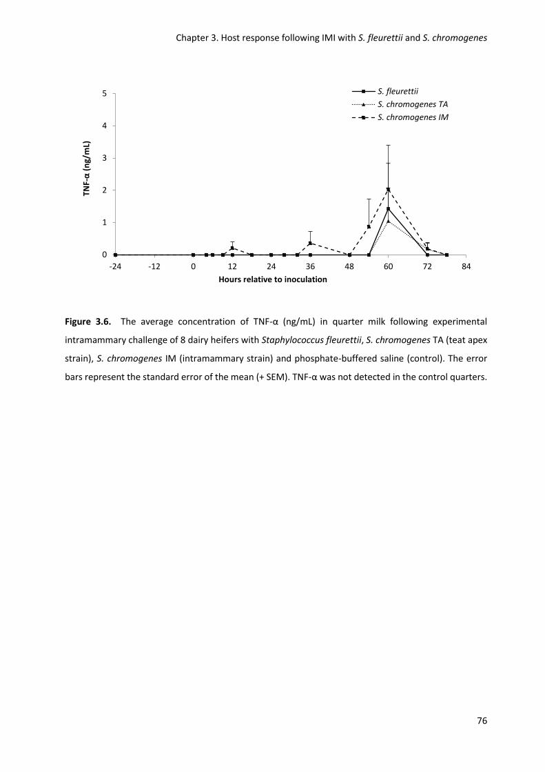
Chapter 3. Host response following IMI with S. fleurettii and S. chromogenes
76
Figure 3.6. The average concentration of TNF-α (ng/mL) in quarter milk following experimental
intramammary challenge of 8 dairy heifers with Staphylococcus fleurettii, S. chromogenes TA (teat apex
strain), S. chromogenes IM (intramammary strain) and phosphate-buffered saline (control). The error
bars represent the standard error of the mean (+ SEM). TNF-α was not detected in the control quarters.
0
1
2
3
4
5
-24 -12 0 12 24 36 48 60 72 84
TNF-
α(n
g/m
L)
Hours relative to inoculation
S. fleurettii
S. chromogenes TA
S. chromogenes IM

Chapter 3. Host response following IMI with S. fleurettii and S. chromogenes
77
Figure 3.7. The average concentration of IL-1β (pg/mL) in quarter milk following experimental
intramammary challenge of 8 dairy heifers with Staphylococcus fleurettii, S. chromogenes TA (teat apex
strain), S. chromogenes IM (intramammary strain) and phosphate-buffered saline (control). The error
bars represent the standard error of the mean (+ SEM). IL-1β was not detected in the control quarters.
0
10
20
30
40
50
60
70
80
-24 -12 0 12 24 36 48 60 72 84
IL-1
β(p
g/m
L)
Hours relative to inoculation
S. fleurettii
S. chromogenes TA
S. chromogenes IM

Chapter 3. Host response following IMI with S. fleurettii and S. chromogenes
78
3.6 Discussion
First, we wanted to compare the host responses in dairy cattle with subclinical mastitis caused by
representative isolates of either an “environmental” or a “host-associated” CNS species. For this
purpose, S. fleurettii was selected as the environmental species, and S. chromogenes as the host-
adapted species. The second objective was to study the host’s reaction to different strains of the same
species, in this case S. chromogenes. Due to the split-udder design, which partially circumvents variation
between heifers, the host response could be studied using only a limited number of experimental
animals. In contrast to other experimental CNS trials (Simojoki et al., 2009; Simojoki et al., 2011), all CNS
species found in the milk samples were identified with tDNA-PCR, in addition to conventional culturing
techniques. Through the molecular identification of the CNS species, a natural infection with S.
epidermidis was detected. The data of this particular quarter were subsequently discarded from the
analysis.
Based on the changes in SCC and bacterial shedding, all quarters were successfully challenged with the
different CNS strains, whereas the control quarters remained culture-negative throughout the entire
trial. Staphylococcus fleurettii was fairly quickly (i.e. within 12h) eliminated from the mammary gland,
and the bacterial shedding was lowest for this species. The latter finding was also observed in an
experimental trial in mice using the same CNS isolates (Breyne et al., 2015). Both S. chromogenes strains,
on the other hand, persisted in the mammary gland for at least 3 days, accentuating the host-adapted
nature of this species. Altogether, this is still a short period, considering that S. chromogenes IM was
originally isolated from a cow suffering from a persistent IMI lasting over 300 days (Piessens et al., 2011).
The bacterial count of S. chromogenes TA was significantly lower than S. chromogenes IM though. In
fact, S. chromogenes IM seemed to be the only strain able to multiply in the mammary gland in the first
6 hours after inoculation. The S. chromogenes IM strain also elicited a significantly larger increase in SCC
than S. chromogenes TA. The lower cellular response in the S. chromogenes TA-infected quarters could
possibly explain the slower bacterial clearance. These results might indicate a difference in pathogenicity

Chapter 3. Host response following IMI with S. fleurettii and S. chromogenes
79
and in vivo growth capacity between the different strains. Interestingly, in the murine experimental trial,
no differences in bacteriological shedding nor in neutrophil influx were observed between S.
chromogenes IM and S. chromogenes TA (Breyne et al., 2015).
The intramammary challenge had a significant effect on the apoptosis of PMN in milk, which was likely
the result of an influx of young, activated PMN from the blood stream to the site of infection along with
a potentially delayed PMN apoptosis. Aging PMN eventually undergo apoptosis (or programmed cell
death), impairing the functionality of the cells (Whyte et al., 1993; Van Oostveldt et al., 2002). Delayed
apoptosis might therefore contribute to a faster clearing of the bacterial infection (Mehrzad et al., 2004).
However, PMN apoptosis and subsequent phagocytosis by macrophages is a physiological necessity to
curb unbridled inflammatory responses that result in tissue damage (Whyte et al., 1993). Since the
lactation stage and parity may affect the survival of PMN (Van Oostveldt et al., 2001; Mehrzad et al.,
2004), only mid-lactation heifers were used in this experiment, allowing us to study the effect of the
infection (Boutet et al., 2004). Staphylococcus fleurettii evoked the greatest decrease in PMN apoptosis
during the first 48 p.i., at least partly explaining the higher (albeit insignificant) SCC increase compared
to S. chromogenes TA. Still, the PMN apoptosis was already decreased in all quarters right before the
inoculation. The pre-inoculation drop in apoptosis might have been caused by stress (Liles et al., 1995)
associated with the beginning the experiment, although this could not be confirmed. A longer
adaptation period (> 48h) in the tie-stall facility prior to the inoculation might have mitigated these
findings.
Pro-inflammatory cytokines, such as TNF-α and IL-1β, are involved in a plethora of immune functions on
a local and systemic level (e.g. the endothelial adhesion of immune cells, induction of fever or the
production of other cytokines) (Bannerman et al., 2004a; Schukken et al., 2011). The further recruitment
of PMN to the infection site is mediated by IL-8 and other chemokines (Harada et al., 1994). In contrast
to E. coli infections, S. aureus mastitis does not evoke a significant IL-8 or TNF-α response, which might
partially explain the chronic nature of S. aureus IMI (Riollet et al., 2000; Bannerman et al., 2004b). As
demonstrated in previous research (Simojoki et al., 2011), other CNS species (S. epidermidis and

Chapter 3. Host response following IMI with S. fleurettii and S. chromogenes
80
Staphylococcus simulans) appear to induce a clear pro-inflammatory response with TNF-α, IL-8 and IL-
1β nonetheless.
In this study, S. chromogenes IM induced the largest overall pro-inflammatory cytokine response,
starting with an increase in IL-8 at 9h p.i.. The production of IL-1β occurred later at 12h p.i., but lasted
longer (> 78h p.i.). The IL-1β response was less pronounced in the S. fleurettii-challenged quarters than
in the S. chromogenes IM quarters. The IL-8 response induced by these strains seemed to be smaller
than the response described for S. epidermidis or S. simulans (Simojoki et al., 2011). However, a higher
infection dose was used in the latter study. Furthermore, the authors transformed the IL-8 data by
multiplying it by 100, since they observed that the human IL-8 ELISA measures bovine IL-8 100-times less
efficiently. That might explain why their IL-8 results fall in the ng/mL range, whereas our results are
found in the pg/mL range. Also, we did not find a distinction between early (peaking at 12h) or late
(peaking at 30h) IL-8 responders as seen in (Simojoki et al., 2011). In fact, none of our animals displayed
any IL-8 response after 30 h p.i..
Staphylococcus chromogenes TA did not elicit any detectable cytokine response, except in one particular
heifer. In a similar murine infection trial, some mice displayed a disproportionate reaction to S.
chromogenes TA as well, resulting in a high IL-1β response and an intense clinical reaction (Breyne et al.,
2015). Another heifer showed no cytokine response at all to any CNS strain. Everything considered, the
pro-inflammatory cytokine response appeared to vary greatly between individual animals in our
research.
As discussed in other research (Sipka et al., 2014), the split udder design used in this experiment is
founded on within-cow comparisons. On the one hand, this study design partly circumvents high
variation between individual animals, and reduces the needed number of research animals substantially.
On the other hand, it assumes that all mammary quarters are completely separate anatomical entities
within the udder. This is not necessarily the case, as demonstrated by previous research that illustrated
that the immune response of the neighboring (uninfected) quarters is affected by an experimental
challenge with major mastitis pathogens (Jensen et al., 2013). This should be kept in mind when
Chapter 3. Host response following IMI with S. fleurettii and S. chromogenes
81
interpreting the results of this study. Nonetheless, we were still able to demonstrate different host
responses to each CNS strain while using the split-udder design.
None of the heifers in our study showed any local signs of clinical mastitis (such as clots in the milk or
swelling of the quarters), although 3 animals experienced a brief bout of fever in the first 12h after
inoculation. In a similar challenge study with S. chromogenes, the heifers only developed very mild
clinical signs of inflammation, even though the inoculation dose was more than double ours (2.1 x 10⁶
CFU versus 1.0 x 10⁶ CFU) (Simojoki et al., 2009). In the preliminary challenge trial conducted prior to
this study (Verbeke et al., 2015) higher doses of the same S. chromogenes strain evoked a more
pronounced immune response as opposed to lower doses with the appearance of mild clinical signs at
a dose of 2 x 10⁶ CFU. Observational research has demonstrated that approximately half of all cows with
an intramammary S. chromogenes infection showed clinical signs of mastitis (Taponen et al., 2006;
Waller et al., 2011). Notwithstanding a different infection dose or host-immune status, this could
suggest that not all S. chromogenes strains are equally pathogenic. This is in accordance with S. aureus,
where certain strains have also been linked to a more severe clinical outcome, with a reduced
persistence in the mammary gland (Haveri et al., 2005). When extrapolating the current results to
practice, it should be noted that natural infection doses might vary from doses used here, and that the
experimental strains were directly infused into the teat cistern (as opposed to natural infections, where
CNS have to overcome the teat barrier.) The results of our experimental trial with dairy heifers should
be interpreted with caution, since the outcome of an IMI may vary with parity, lactation stage and other
cow factors.
3.7 Conclusion
Even in case of large inoculation doses, bovine-associated S. chromogenes and environmental S.
fleurettii strains trigger a similar, relatively mild local response. The environmental CNS species, S.
fleurettii, evokes a profound cellular response in dairy heifers nonetheless, akin to the host-adapted

Chapter 3. Host response following IMI with S. fleurettii and S. chromogenes
82
species S. chromogenes. However, S. fleurettii is eliminated more rapidly from the mammary gland than
S. chromogenes. This might indicate that certain bovine-associated CNS species, like S. chromogenes,
are better able to withstand and thrive in the environment of the mammary gland. Still, the present in
vivo study also suggests that not all S. chromogenes strains exhibit the same degree of pathogenicity.
The clinical outcome of a natural S. chromogenes mastitis might therefore not only depend on the
infection pressure and the resistance of the cow, but also on the pathogenicity of the particular strain.
3.8 Acknowledgements
The authors would like to acknowledge Lars Hulpio, Marina Stevens and Dimitri Valckenier for their
assistance during the sampling. The work of Lydia Bommelé, Karel Vermeulen and André Turtelboom
at Biocentrum Agri-Vet is also greatly appreciated.

Chapter 3. Host response following IMI with S. fleurettii and S. chromogenes
83
3.9 References
Bannerman, D. D., M. J. Paape, W. R. Hare, and J. C. Hope. 2004a. Characterization of the bovine innate immune response to intramammary infection with Klebsiella pneumoniae. J Dairy Sci 87(8):2420-2432. Bannerman, D. D., M. J. Paape, J. W. Lee, X. Zhao, J. C. Hope, and P. Rainard. 2004b. Escherichia coli and Staphylococcus aureus elicit differential innate immune responses following intramammary infection. Clin Diagn Lab Immunol 11(3):463-472. Boutet, P., D. Boulanger, L. Gillet, A. Vanderplasschen, R. Closset, F. Bureau, and P. Lekeux. 2004. Delayed neutrophil apoptosis in bovine subclinical mastitis. J Dairy Sci 87(12):4104-4114. Braem, G., S. De Vliegher, K. Supré, F. Haesebrouck, F. Leroy, and L. De Vuyst. 2011. (GTG)5-PCR fingerprinting for the classification and identification of coagulase-negative Staphylococcus species from bovine milk and teat apices: a comparison of type strains and field isolates. Vet Microbiol 147(1-2):67-74. Breyne, K., S. De Vliegher, A. De Visscher, S. Piepers, and E. Meyer. 2015. Technical note: a pilot study using a mouse mastitis model to study differences between bovine associated coagulase-negative staphylococci. J Dairy Sci 98(2):1090-1100. De Visscher, A., S. Piepers, F. Haesebrouck, and S. De Vliegher. 2016. Teat apex colonization with coagulase-negative Staphylococcus species before parturition: Distribution and species-specific risk factors. J Dairy Sci 99(2):1427-1439. De Visscher, A., K. Supré, F. Haesebrouck, R. N. Zadoks, V. Piessens, E. Van Coillie, S. Piepers, and S. De Vliegher. 2014. Further evidence for the existence of environmental and host-associated species of coagulase-negative staphylococci in dairy cattle. Vet Microbiol 172(3-4):466-474. De Vliegher, S., H. Laevens, L. A. Devriese, G. Opsomer, J. L. M. Leroy, H. W. Barkema, and A. De Kruif. 2003. Prepartum teat apex colonization with Staphylococcus chromogenes in dairy heifers is associated with low somatic cell count in early lactation. Vet Microbiol 92(3):245–252. De Vliegher, S., G. Opsomer, A. Vanrolleghem, L. A. Devriese, O. C. Sampimon, J. Sol, H. W. Barkema, F. Haesebrouck, and A. de Kruif. 2004. In vitro growth inhibition of major mastitis pathogens by Staphylococcus chromogenes originating from teat apices of dairy heifers. Vet Microbiol 101(3):215-221. Fry, P. R., J. R. Middleton, S. Dufour, J. Perry, D. Scholl, and I. Dohoo. 2014. Association of coagulase-negative staphylococcal species, mammary quarter milk somatic cell count, and persistence of intramammary infection in dairy cattle. J Dairy Sci 97(8):4876-4885. Harada, A., N. Sekido, T. Akahoshi, T. Wada, N. Mukaida, and K. Matsushima. 1994. Essential involvement of interleukin-8 (IL-8) in acute inflammation. J Leukocyte Biology 56(5):559-564. Haveri, M., S. Taponen, J. Vuopio-Varkila, S. Salmenlinna, and S. Pyörälä. 2005. Bacterial genotype affects the manifestation and persistence of bovine Staphylococcus aureus intramammary infection. J Clin Microbiol 43(2):959-961. Hogan, J. S., R. N. González, R. J. Harmon, S. C. Nickerson, S. P. Oliver, J. W. Pankey, and K. L. Smith. 1999. Laboratory Handbook on Bovine Mastitis. Rev. ed. ed., Madison, WI.

Chapter 3. Host response following IMI with S. fleurettii and S. chromogenes
84
Hogan, J. S., D. G. White, and J. W. Pankey. 1987. Effects of Teat Dipping on Intramammary Infections by Staphylococci Other Than Staphylococcus-Aureus. J Dairy Sci 70(4):873-879. Jensen, K., J. Günther, R. Talbot, W. Petzl, H. Zerbe, H. J. Schuberth, H. M. Seyfert, and E. J. Glass. 2013. Escherichia coli- and Staphylococcus aureus-induced mastitis differentially modulate transcriptional responses in neighbouring uninfected bovine mammary gland quarters. BMC genomics 14:36. Liles, W. C., D. C. Dale, and S. J. Klebanoff. 1995. Glucocorticoids inhibit apoptosis of human neutrophils. Blood 86(8):3181-3188. Mehrzad, J., L. Duchateau, and C. Burvenich. 2004. Viability of milk neutrophils and severity of bovine coliform mastitis. J Dairy Sci 87(12):4150-4162. Mollet, C., M. Drancourt, and D. Raoult. 1997. rpoB sequence analysis as a novel basis for bacterial identification. Mol Microbiol 26(5):1005-1011. Nickerson, S. C. and R. L. Boddie. 1994. Effect of Naturally-Occurring Coagulase-Negative Staphylococcal Infections on Experimental Challenge with Major Mastitis Pathogens. J Dairy Sci 77(9):2526-2536. Park, J. Y., L. K. Fox, K. S. Seo, M. A. McGuire, Y. H. Park, F. R. Rurangirwa, W. M. Sischo, and G. A. Bohach. 2011. Comparison of phenotypic and genotypic methods for the species identification of coagulase-negative staphylococcal isolates from bovine intramammary infections. Vet Microbiol 147(1-2):142-148. Piccart, K., S. Piepers, J. Verbeke, N. M. de Sousa, J. F. Beckers, and S. De Vliegher. 2015. Milk prolactin response and quarter milk yield after experimental infection with coagulase-negative staphylococci in dairy heifers. J Dairy Sci. 98:1–8 Piepers, S., S. De Vliegher, K. Demeyere, B. N. Lambrecht, A. de Kruif, E. Meyer, and G. Opsomer. 2009. Technical note: Flow cytometric identification of bovine milk neutrophils and simultaneous quantification of their viability. J Dairy Sci 92(2):626-631. Piessens, V., E. Van Coillie, B. Verbist, K. Supré, G. Braem, A. Van Nuffel, L. De Vuyst, M. Heyndrickx, and S. De Vliegher. 2011a. Distribution of coagulase-negative Staphylococcus species from milk and environment of dairy cows differs between herds. J Dairy Sci 94(6):2933-2944. Pyörälä, S. and S. Taponen. 2009. Coagulase-negative staphylococci-emerging mastitis pathogens. Vet Microbiol 134(1-2):3-8. Riollet, C., P. Rainard, and B. Poutrel. 2000. Differential induction of complement fragment C5a and inflammatory cytokines during intramammary infections with Escherichia coli and Staphylococcus aureus. Clin Diagn Lab Immunol 7(2):161-167. Sampimon, O. C., H. W. Barkema, I. M. Berends, J. Sol, and T. J. Lam. 2009a. Prevalence and herd-level risk factors for intramammary infection with coagulase-negative staphylococci in Dutch dairy herds. Vet Microbiol 134(1-2):37-44. Sampimon, O. C., R. N. Zadoks, S. De Vliegher, K. Supré, F. Haesebrouck, H. W. Barkema, J. Sol, and T. J. Lam. 2009b. Performance of API Staph ID 32 and Staph-Zym for identification of coagulase-negative staphylococci isolated from bovine milk samples. Vet Microbiol 136(3-4):300-305. Schukken, Y. H., J. Günther, J. Fitzpatrick, M. C. Fontaine, L. Goetze, O. Holst, J. Leigh, W. Petzl, H. J. Schuberth, A. Sipka, D. G. Smith, R. Quesnell, J. Watts, R. Yancey, H. Zerbe, A. Gurjar, R. N. Zadoks, H. M.

Chapter 3. Host response following IMI with S. fleurettii and S. chromogenes
85
Seyfert, and c. members of the Pfizer mastitis research. 2011. Host-response patterns of intramammary infections in dairy cows. Veterinary immunology and immunopathology 144(3-4):270-289. Simojoki, H., T. Orro, S. Taponen, and S. Pyörälä. 2009. Host response in bovine mastitis experimentally induced with Staphylococcus chromogenes. Vet Microbiol 134(1-2):95-99. Simojoki, H., T. Salomaki, S. Taponen, A. Iivanainen, and S. Pyörälä. 2011. Innate immune response in experimentally induced bovine intramammary infection with Staphylococcus simulans and S. epidermidis. Vet Res 42. Sipka, A., S. Klaessig, G. E. Duhamel, J. Swinkels, P. Rainard, and Y. Schukken. 2014. Impact of intramammary treatment on gene expression profiles in bovine Escherichia coli mastitis. PLoS One 9(1):e85579. Souza, F. N., S. Piepers, A. M. Della Libera, M. B. Heinemann, M. M. Cerqueira, and S. De Vliegher. 2016. Interaction between bovine-associated coagulase-negative staphylococci species and strains and bovine mammary epithelial cells reflects differences in ecology and epidemiological behavior. J Dairy Sci. 99(4), 2867-2874. Supré, K., S. De Vliegher, O. C. Sampimon, R. N. Zadoks, M. Vaneechoutte, M. Baele, E. De Graef, S. Piepers, and F. Haesebrouck. 2009. Technical note: use of transfer RNA-intergenic spacer PCR combined with capillary electrophoresis to identify coagulase-negative Staphylococcus species originating from bovine milk and teat apices. J Dairy Sci 92(7):3204-3210. Supré, K., F. Haesebrouck, R. N. Zadoks, M. Vaneechoutte, S. Piepers, and S. De Vliegher. 2011. Some coagulase-negative Staphylococcus species affect udder health more than others. J Dairy Sci 94(5):2329-2340. Taponen, S., J. Björkroth, and S. Pyörälä. 2008. Coagulase-negative staphylococci isolated from bovine extramammary sites and intramammary infections in a single dairy herd. J Dairy Res 75(4):422-429. Taponen, S., H. Simojoki, M. Haveri, H. D. Larsen, and S. Pyörälä. 2006. Clinical characteristics and persistence of bovine mastitis caused by different species of coagulase-negative staphylococci identified with API or AFLP. Vet Microbiol 115(1-3):199-207. Tomazi, T., J. L. Gonalves, J. R. Barreiro, M. A. Arcari, and M. V. dos Santos. 2015. Bovine subclinical intramammary infection caused by coagulase-negative staphylococci increases somatic cell count but has no effect on milk yield or composition. J Dairy Sci 98(5):3071-3078. Van Oostveldt, K., M. J. Paape, H. Dosogne, and C. Burvenich. 2002. Effect of apoptosis on phagocytosis, respiratory burst and CD18 adhesion receptor expression of bovine neutrophils. Dom Anim Endocrinol 22(1):37-50. Van Oostveldt, K., F. Vangroenweghe, H. Dosogne, and C. Burvenich. 2001. Apoptosis and necrosis of blood and milk polymorphonuclear leukocytes in early and midlactating healthy cows. Vet Res 32(6):617-622. Vanderhaeghen, W., S. Piepers, F. Leroy, E. Van Coillie, F. Haesebrouck, and S. De Vliegher. 2014. Invited review: effect, persistence, and virulence of coagulase-negative Staphylococcus species associated with ruminant udder health. J Dairy Sci 97(9):5275-5293.

Chapter 3. Host response following IMI with S. fleurettii and S. chromogenes
86
Verbeke, J., K. Piccart, S. Piepers, M. Van Poucke, L. Peelman, A. De Visscher, and S. De Vliegher. 2015. Somatic cell count and milk neutrophil viability of dairy heifers with specific CXCR1 genotypes following experimental intramammary infection with Staphylococcus chromogenes originating from milk. Vet J. 204(3), 322-326. Waller, K. P., A. Aspan, A. Nyman, Y. Persson, and U. G. Andersson. 2011. CNS species and antimicrobial resistance in clinical and subclinical bovine mastitis. Vet Microbiol 152(1-2):112-116. Whyte, M. K., L. C. Meagher, J. MacDermot, and C. Haslett. 1993. Impairment of function in aging neutrophils is associated with apoptosis. J Immunol 150(11):5124-5134.

87
88
Chapter 4
The role of milk prolactin in intramammary infections
with coagulase-negative staphylococci

89
90
Chapter 4.1
Milk prolactin response and quarter milk yield after experimental infection with
coagulase-negative staphylococci in dairy heifers
K. Piccart1, S. Piepers1, J. Verbeke1, N.M. de Sousa2, J.F. Beckers2, and S. De Vliegher1
1M-team and Mastitis and Milk Quality Research Unit, Department of Reproduction, Obstetrics and Herd Health,
Faculty of Veterinary Medicine, Ghent University, 9820 Merelbeke, Belgium
2Laboratory of Animal Endocrinology and Reproduction, Faculty of Veterinary Medicine, University of Liege, 4000
Liege, Belgium
Journal of Dairy Science, 2015, 98:1–8

91

Chapter 4. Milk PRL response in CNS IMI
92
4.1.1 Abstract
Coagulase-negative staphylococci (CNS) are the most common bacteria involved in subclinical mastitis
in dairy cows. Remarkably, CNS-infected dairy heifers produce more milk than uninfected heifers.
Because the lactation hormone prolactin (PRL) is also involved in mammary gland immunity, we
investigated the milk PRL response and the mammary quarter milk yield following experimental CNS
challenge. Eight healthy Holstein-Friesian heifers in mid-lactation were experimentally infected using
a split-udder design with 3 different CNS strains: one Staphylococcus fleurettii (from sawdust bedding)
and 2 Staphylococcus chromogenes strains (one isolate from a teat apex, the other isolate from a
chronic intramammary infection). Three mammary quarters per heifer were simultaneously inoculated
with 1.0 × 10⁶ CFU, whereas the remaining mammary quarter was infused with sterile phosphate-
buffered saline, serving as a control. An existing radioimmunoassay was modified, validated, and used
to measure PRL frozen-thawed milk at various time points until 78 h after challenge. The mean milk
PRL level tended to be higher in the CNS-challenged mammary quarters compared with the control
mammary quarters (7.56 and 6.85 ng/mL, respectively). The increase in PRL over time was significantly
greater in the CNS-challenged mammary quarters than in the control mammary quarters. However,
no difference was found in the PRL response when comparing each individual CNS strain with the
control mammary quarters. The mean mammary quarter milk yield tended to be lower in the CNS-
infected mammary quarters than in the control mammary quarters (1.73 and 1.98 kg per milking,
respectively). The greatest milk loss occurred in the mammary quarters challenged with the
intramammary strain of S. chromogenes. Future observational studies are needed to elucidate the
relation between PRL, the milk yield, and the inflammatory condition, or infection status, of the
mammary gland.
Key words: coagulase-negative staphylococci, dairy heifer, experimental mastitis, prolactin

Chapter 4. Milk PRL response in CNS IMI
93
4.1.2 Introduction
Bovine mastitis, an inflammation of the mammary gland, creates a huge economic burden on the
global dairy industry (Bradley, 2002). Coagulase-negative staphylococci are the predominant group of
bacteria involved in subclinical mastitis (Pyorala and Taponen, 2009) and can cause clinical mastitis
with mild symptoms (Taponen et al., 2006). Thus far, more than 10 species of CNS have been isolated
from bovine milk (Piessens et al., 2011) with documented species-specific differences in putative
virulence (Vanderhaeghen et al., 2014), ecology, and epidemiology (Vanderhaeghen et al., 2015).
Contrary to what one might expect, various studies have observed a higher test-day milk yield in CNS-
infected dairy heifers and multiparous cows com- pared with noninfected cows (Compton et al., 2007;
Schukken et al., 2009; Piepers et al., 2010). Some studies have attributed a protective effect to pre-
existing CNS IMI against IMI with more virulent mastitis pathogens (e.g. Piepers et al., 2010). A meta-
analysis could not confirm this finding in observational studies, but nonetheless revealed a pronounced
protective effect in challenge trials (Reyher et al., 2012). Still, the positive effect on milk yield could be
an indirect result of the reduced incidence of clinical mastitis observed in CNS-infected animals
(Piepers et al., 2010). High-producing dairy cows might also be more susceptible to CNS IMI than low-
yielding animals (Compton et al., 2007). However, even after correcting for these factors, an
unexplained difference in milk yield of 2.0 kg/d remained between CNS-infected and uninfected herd
mates (Piepers et al., 2013), leaving the exact mechanism to be determined.
Prolactin has been associated with over 300 different biological actions, including lactation and
mammary gland development (Bole-Feysot et al., 1998). In dairy cattle, PRL is required to initiate
(Akers et al., 1981) and maintain the milk production after parturition (Lacasse et al., 2012). The
protein hormone also acts as a cytokine on molecular and functional levels (Goffin et al., 2002). The
ubiquitous PRL receptor belongs to the class I cytokine receptor superfamily, which also includes the
receptors of several interleukins and hematopoietic growth factors (Bazan, 1989; Bazan, 1990). The
hormone promotes the activity of macrophages (Edwards et al., 1987), inhibits the apoptosis of T-

Chapter 4. Milk PRL response in CNS IMI
94
lymphocytes caused by glucocorticoids (Krishnan et al., 2003), and stimulates the production of tumor
necrosis factor-α and IL-12 (Brand et al., 2004). Considering the immunomodulatory actions of PRL,
several studies have focused on its potential involvement in bovine mastitis. The periparturient PRL
peak coincides with the principal risk period for developing mastitis (Burton et al., 2001). The hormone
induces the in vitro synthesis of several cytokines in bovine MEC through the activation of nuclear
factor kappa B (Boutet et al., 2007). Although the circulating PRL level is not affected by acute, clinical
mastitis (Hockett et al., 2000; Vanselow et al., 2006), a positive correlation was found between SCC
and PRL concentration in milk of chronically infected mammary quarters (Boutet et al., 2007).
Because PRL is recognized as a pro-inflammatory cytokine, we hypothesize that milk PRL increases in
response to an IMI with CNS. Furthermore, we hypothesize that the quarter milk yield (QMY) also
increases after CNS IMI, assuming PRL stimulates the production of milk. To investigate this, an
experimental challenge trial was set up using 8 clinically healthy, mid- lactating dairy heifers using 3
different CNS strains. An existing radioimmunoassay for fresh milk was modified and subsequently
validated to measure bovine PRL in frozen-thawed milk samples. To assess the epithelial integrity of
the blood-milk barrier, the sodium, potassium, and chloride levels were also determined in milk.
4.1.3 Materials and methods
The study is in compliance with the European Directive 2010/63/EU and was approved by the ethical
committee of the Faculty of Veterinary Medicine, Ghent University (EC2012/73).
Animals
The study took place between December 2012 and May 2013 at the research dairy farm of Ghent
University (Biocentrum Agri-Vet, Melle, Belgium). Eight clinically healthy Holstein-Friesian heifers in
mid-lactation (78– 278 DIM) were selected. Heifers with a known history of clinical mastitis or
Chapter 4. Milk PRL response in CNS IMI
95
persistent high SCC (>150,000 cells/mL) were excluded from the trial. Milk samples were cultured
according to NMC guidelines 48 and 24 h before inoculation to ensure all mammary quarters were free
from IMI (NMC, 1999).
CNS Strains
All heifers were inoculated with 2 different wild strains of Staphylococcus chromogenes and 1
Staphylococcus fleurettii strain. The S. fleurettii isolate was recovered from sawdust bedding in a dairy
barn (Piessens et al., 2011; Breyne et al., 2015). The first S. chromogenes strain originated from a cow
suffering from a persistent IMI (hereafter referred to as S. chromogenes IM; Supré et al., 2011; Breyne
et al., 2015), whereas the second S. chromogenes isolate was cultured from the teat apex of a heifer
(hereafter referred to as S. chromogenes TA; De Vliegher et al., 2004; Breyne et al., 2015). The S.
chromogenes TA strain has the ability to inhibit the growth of several major pathogens under
laboratory conditions (De Vliegher et al., 2004), whereas the S. chromogenes IM strain does not. The
2 strains also elicit a different immune response in mice (Breyne et al., 2015), and the TA strain is
unable to grow in anaerobic iron-depleted medium unlike the IM strain (Souza et al., 2016). An
inoculum of 1.0 × 10⁶ CFU of each strain was prepared to induce an experimental infection. The live
number of CFU was determined by plating serial dilutions of the bacterial stock on tryptic soy agar.
Experimental Study Design
A split-udder design was used. The concept of the split-udder model is grounded on within-heifer
comparisons to reduce individual variation (Sipka et al., 2014). Following the morning milking, 3
mammary quarters of each heifer were instantaneously inoculated with the 3 aforementioned CNS
strains (one per mammary quarter) diluted in 5 mL of PBS using a sterile catheter (Vygon, Ecouen,
France). The fourth mammary quarter, serving as a control, was infused in the same manner with 5 mL
of sterile, pyrogen-free PBS. Milk samples for PRL analysis and microbiological culturing were collected
from each mammary quarter at 0, 4, 6, 9, 12, 18, 24, 28, 32, 36, 48, 54, 60, 72, and 78 h post-inoculation
Chapter 4. Milk PRL response in CNS IMI
96
(PI). Milk samples for ion analysis were collected at 0, 24, and 48 h PI. The milk SCC was determined
using a DeLaval Cell Counter (DeLaval, Tumba, Sweden). Bacteriological culturing was performed
according to NMC guidelines (NMC, 1999). The milk samples for the PRL and ion analysis were stored
at −20°C. The cows were milked twice a day with 12-h intervals, and QMY was registered using a
mammary quarter milking device. The cow were examined clinically at each sampling. Rectal
temperature, heart rate, respiratory rate, rumen motility, fecal consistency, and milk appearance were
registered.
Milk Analysis
Prolactin RIA. Milk PRL was determined by a double antibody, homologous RIA adapted from Malven
and McMurtry (1974). Because this protocol was developed for fresh, whole milk, we first modified
and validated the RIA for frozen milk samples in particular. For the validation, mammary quarter milk
samples (n = 4) were collected from randomly selected, multiparous Holstein-Friesian cows at a Belgian
commercial dairy farm. The samples were then stored at −20°C for 96 h. Afterward, the milk samples
were thawed in a warm water bath at 40°C for 30 min (Chew et al., 1977) and centrifuged at 1,800 × g
for 15 min at 20°C (Sigma 2–16K, Sigma Laborzentrifugen GmbH, Osterode am Harz, Germany). Bovine
PRL (NIH-B5) was used both as a standard and tracer. To generate the tracer, bovine PRL was
radiolabeled with 125I using the lactoperoxidase technique (Thorell and Johansson, 1971). A standard
curve ranging from 0.8 to 100 ng/ mL was prepared in Tris buffer (25 mM Tris; 0.01 mM MgCl2; 1.5
mM NaN3) containing 0.1% (wt/vol) BSA at pH 7.5. To analyze each standard concentration, 100 μL
was added to duplicate tubes containing 200 μL of Tris-BSA buffer. A sample volume of 50 μL was used
for milk to minimize potential incubation damage to the labeled antigen (Malven and McMurtry, 1974).
Afterward, 100 μL of tracer of approximately 30,000 cpm was added, followed by 100 μL of antiserum
(R#144). This antiserum was collected from a rabbit after injection with biological bovine PRL (NIH-B5;
Boutet et al., 2007). An antibody titer of 1:120,000 was previously selected for this assay, resulting in

Chapter 4. Milk PRL response in CNS IMI
97
the binding of circa 30 to 40% of the total amount of 125I-PRL. The amount of radiolabelled bPRL bound
to the antiserum in tubes containing no bPRL (B0) was designated as 100% 125I bound. After an
overnight incubation at room temperature, the antibody-bound fraction of PRL was precipitated using
a secondary antibody polyethylene glycol solution (Ayad et al., 2007). The radioactivity of the
precipitated 125I-PRL was measured with a gamma counter (LKB Wallac 1261 Multigamma automatic
counter, Breda, the Netherlands). Five parameters were taken into account to validate the RIA: (1)
inter- and intra-assay coefficient of variation, (2) minimal detection limit (MDL), (3) parallelism, (4)
accuracy, and (5) specificity. Three milk samples with a low (2.4 ng/mL), medium (12.9 ng/mL), and
high (19.7 ng/mL) PRL concentration were used to calculate the inter- and intra-assay coefficient of
variation. To assess the intra-assay coefficient of variation, the samples were assayed 10 times within
the same run. The interassay coefficient of variation was evaluated by measuring each sample in 10
consecutive assays. The MDL was calculated by measuring the mean concentration of 20 B0 (zero
standard) replicates minus twice the standard deviation within the same assay. For the evaluation of
parallelism, a sample dilution curve of a frozen-thawed sample was generated to detect any potential
interference of the sample matrix. The accuracy or spike-recovery of the assay was studied by adding
a series of known amounts of PRL to a sample with a low endogenous PRL concentration. To evaluate
potential cross-reactivity (specificity), 2 structurally similar compounds were tested: native ovine
growth hormone (oGH) and bovine placental lactogen (bPL). The validated RIA was then applied to all
mammary quarter milk samples from the 8 heifers in the present experimental trial.
Indicators of Epithelial Integrity. The milk samples were thawed and subsequently centrifuged at
16,000 × g for 30 min at 4°C. The sodium, potassium, and chloride concentrations were analyzed in the
fat-depleted fraction using an ion-selective electrode analyzer (Roche, Basel, Switzerland).

Chapter 4. Milk PRL response in CNS IMI
98
Statistical Analyses
The response of mammary quarter milk PRL after CNS challenge was evaluated using a linear mixed
regression model (SPSS 22.0, Chicago, IL), with the time of sampling, quadratic term of time of
sampling, and inoculum as predictor variables. Time of sampling and its quadratic term were included
as a continuous variable, whereas inoculum was considered as a categorical variable. The interaction
between inoculum and time of sampling was also tested, but only kept in the model when significant.
A similar model was constructed to assess the association between QMY, and time of sampling, its
quadratic term, and inoculum as predictor variables. For both outcome variables, the inoculum was
initially considered a dichotomous variable (challenged versus control). In a second approach, the
effect of all different CNS strains was examined (S. chromogenes IM, S. chromogenes TA, and S.
fleurettii). To determine the relationship between inoculation (challenge versus control) and the milk
ion concentration, comparable linear mixed regression models were constructed for sodium,
potassium, and chloride. Sampling time was included as a 3-level categorical variable (0, 24, and 48 h).
A natural logarithmic transformation of sodium and chloride was performed to obtain a normalized
distribution of the residuals. In all aforementioned analyses, heifer and mammary quarter were
included as random effects to account for the correlated nature of the data. Compound symmetry was
selected as a covariance pattern to account for the clustering of repeated samplings within mammary
quarter. Statistical significance was set at P ≤ 0.05.
4.1.4 Results
Experimental Infection
The data of one mammary quarter were omitted from the analysis due to a naturally occurring IMI.
Another mammary quarter was also excluded from the analysis for the same reason, but only 12 h
after inoculation. An increase in SCC was observed in all challenged mammary quarters, indicating the
establishment of IMI, whereas the SCC in the control mammary quarters remained low [geometric

Chapter 4. Milk PRL response in CNS IMI
99
mean of 47,000 cells/ mL; interquartile range (IQR) of 31,000–71,000 cells/ mL]. Twenty-four hours PI,
the S. fleurettii, S. chromogenes TA, and S. chromogenes IM challenged mammary quarters had a
geometric mean SCC of 2,400,000 cells/ mL (IQR: 442,000–3,420,000 cells/mL), 669,000 cells/ mL (IQR:
441,000–1,681,000 cells/mL), and 2,596,000 cells/mL (IQR: 1,662,000–4,775,000 cells/mL),
respectively. The challenged mammary quarters did not show any visual signs of clinical mastitis.
However, 3 heifers did experience a short bout of fever (>39.5°C) between 9 and 12 h PI. The S.
chromogenes IM and TA strains were recovered from milk up to 78 h PI. In all mammary quarters, the
S. fleurettii strain was eliminated within 9 h PI. No pathogens other than CNS were isolated during the
study. The PBS-infused mammary quarters remained free from infection during the entire study period
as demonstrated by bacteriological culture.
Prolactin
RIA Validation. The intra-assay coefficient of variation for samples with a low, medium, and high PRL
concentration was 9.7, 8.3, and 8.1%, respectively. The interassay coefficient of variation for the
corresponding samples was 11.9, 14.4, and 16.1%, respectively. The MDL of the assay was 0.536
ng/mL. Serial dilutions showed a dose-response curve parallel to the standard curve (data not shown).
The recovery ratio of the spiked PRL was consistently >85%. The binding of radiolabeled PRL remained
unaltered when testing the cross-reactivity, except for very high concentrations of oGH (starting at 6.2
× 103 ng/mL). This slightly interfered with the assay (B/B0 < 0.80), resulting in an apparent increase of
the PRL concentration.
Milk PRL. The overall mean PRL concentration tended to be higher in the challenged mammary
quarters than in the control mammary quarters (LSM = 7.56 and 6.85 ng/mL, respectively) throughout
the study (P = 0.10, Table 4.1.1). The PRL level varied over time in both challenged and control
mammary quarters, although not in a linear manner (P < 0.001). The evolution of PRL over time was
different between the challenged and the control mammary quarters (interaction inoculum × time of
sampling: P = 0.05, Figure 4.1.1). No significant difference was observed in the PRL response between

Chapter 4. Milk PRL response in CNS IMI
100
each of the 3 CNS strains and the control mammary quarters (P = 0.77), nor in the evolution over time
between strains (interaction inoculum × time of sampling: P = 0.14).
Quarter Milk Yield
The overall mean QMY per milking tended to be lower in the CNS challenged mammary quarters than
in the control mammary quarters (LSM = 1.73 and 1.98 kg, respectively) throughout the study (P =
0.06, Table 4.1.2). The decline of the QMY over time was greater in the CNS-challenged mammary
quarters than in the control mammary quarters (interaction inoculum × time of sampling: P < 0.001,
Figure 4.1.2). The difference in QMY compared with the control mammary quarters was more
pronounced in the mammary quarters challenged with S. chromogenes IM (−0.38 kg per milking) than
with S. chromogenes TA (−0.21 kg per milking) or S. fleurettii (−0.17 kg per milking; Table 4.1.2).
Blood-Milk Barrier
The concentration of potassium in milk was not significantly influenced by the challenge with CNS
strains (P = 0.37). A small, but significant increase in the natural logarithmic of sodium and chloride
was noted in the challenged quarters (interaction inoculum × time of sampling P < 0.05). However, the
sodium and chloride levels did not exceed the upper limit of the normal reference ranges (Figure 4.1.3;
Gaucheron, 2005).

Chapter 4. Milk PRL response in CNS IMI
101
Figure 4.1.1. The average prolactin (PRL) (± SEM, standard error of the mean) concentration in milk
following experimental inoculation in the challenged quarters (_
_) versus the control quarters (…□…).
The challenged quarters were inoculated with Staphylococcus fleurettii, the teat apex strain of S.
chromogenes and the intramammary strain of S. chromogenes.
0
2
4
6
8
10
-12 0 12 24 36 48 60 72 84
Milk
PR
L (n
g/m
L)
Hours relative to inoculation
Control
Challenged

Chapter 4. Milk PRL response in CNS IMI
102
Figure 4.1.2. Mean quarter milk yield (QMY) (±SEM, standard error of the mean) following
experimental inoculation in the challenged quarters (_
_) versus the control quarters (…□…). The
challenged quarters were inoculated with Staphylococcus fleurettii, the teat apex strain of S.
chromogenes and the intramammary strain of S. chromogenes.
0
1
2
3
-12 0 12 24 36 48 60 72 84
QM
Y (
kg)
Hours relative to inoculation
Control
Challenged
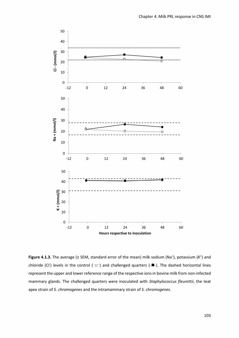
Chapter 4. Milk PRL response in CNS IMI
103
Figure 4.1.3. The average (± SEM, standard error of the mean) milk sodium (Na+), potassium (K+) and
chloride (Cl-) levels in the control (…□…) and challenged quarters (_
_). The dashed horizontal lines
represent the upper and lower reference range of the respective ions in bovine milk from non-infected
mammary glands. The challenged quarters were inoculated with Staphylococcus fleurettii, the teat
apex strain of S. chromogenes and the intramammary strain of S. chromogenes.
0
10
20
30
40
50
-12 0 12 24 36 48 60
Cl -
(mm
ol/
l)
0
10
20
30
40
50
-12 0 12 24 36 48 60
Na
+ (m
mo
l/l)
0
10
20
30
40
50
-12 0 12 24 36 48 60
K +
(m
mo
l/l)
Hours respective to inoculation

Chapter 4. Milk PRL response in CNS IMI
104
Tabel 4.1.1. Linear mixed regression model for milk prolactin after experimental infection, including all three CNS strains combined (model 1; left) and with
Staphylococcus chromogenes TA1, S. chromogenes IM² and S. fleurettii considered seperately (model 2; right).
Prolactin (ng/mL) Prolactin (ng/mL)
Predictor variable
β3 SE4 LSM5 P-value6
β SE LSM P-value
Intercept 7.56 0.01 … … 7.56 0.71 … …
Inoculum 0.10 0.77
Control Ref. … 6.85 … Ref. … 6.85 …
Challenge7 0.21 0.48 7.56 … … … … …
S. fleurettii … … … … 0.38 0.59 7.60 …
S. chromogenes TA … … … … -0.11 0.59 7.45 …
S. chromogenes IM … … … … 0.37 0.59 7.62 …
Time of sampling 0.005 0.01 … 0.28 0.018 0.013 … 0.17
Quadratic term of Time of sampling - 0.001 < 0.001 … < 0.001 -0.001 < 0.001 … < 0.001
Inoculum x Time of sampling … … … 0.05 … … … 0.14
1 Teat apex strain of S. chromogenes; ² Intramammary strain of S. chromogenes; ³ Regression coefficient; 4 Standard error; 5 Least square means; 6 P-value for overall
effect; 7 Quarters challenged with S. chromogenes TA, S. chromogenes IM and S. fleurettii combined.

Chapter 4. Milk PRL response in CNS IMI
105
Tabel 4.1.2. Linear mixed regression model for quarter milk yield after experimental infection, including all three CNS strains combined (model 1; left) and
with Staphylococcus chromogenes TA1, S. chromogenes IM² and S. fleurettii considered seperately (model 2; right).
Quarter milk yield (kg) Quarter milk yield (kg)
Predictor variable
β3 SE4 LSM5 P-value6
β SE LSM P-value
Intercept 1.86 0.18 … … 1.86 0.18 … …
Inoculum 0.06 0.44
Control Ref. … 1.98 … Ref. … 1.98 …
Challenge7 0.15 0.17 1.73 … … … … …
S. fleurettii … … … … 0.29 0.20 1.81 ...
S. chromogenes TA … … … … 0.15 0.20 1.77 …
S. chromogenes IM … … … … 0.02 0.20 1.60 …
Time of sampling -0.02 0.004 … < 0.001 -0.02 0.005 … 0.001
Quadratic term of Time of sampling < 0.001 < 0.001 … < 0.001 < 0.001 < 0.001 … < 0.001
Inoculum x Time of sampling … … … < 0.001 … … … 0.001
1 Teat apex strain of S. chromogenes; ² Intramammary strain of S. chromogenes; ³ Regression coefficient;4 Standard error; 5 Least square means;6 P-value for overall effect;
7 Quarters challenged with S. chromogenes TA, S. chromogenes IM and S. fleurettii combined.

Chapter 4. Milk PRL response in CNS IMI
106
4.1.5 Discussion
Because CNS-infected cows appear to produce more milk than non-infected cows (Compton et al.,
2007; Piepers et al., 2010; Schukken et al., 2009), we investigated the milk yield and the response of
milk PRL after CNS challenge in clinically healthy dairy heifers. There is a plethora of evidence
implicating PRL as a immunomodulating factor (Edwards et al., 1987; Krishnan et al., 2003; Brand et
al., 2004; Boutet et al., 2007). Therefore, we hypothesized that PRL increases in the milk after CNS
infection. Milk PRL might then simultaneously stimulate the secretion of milk as a galactopoetic
hormone (Lacasse et al., 2012) in a paracrine or autocrine manner (Clevenger et al., 1997), potentially
explaining the aforementioned milk yield increases in CNS-infected cows. First, the modified PRL
radioimmunoassay for frozen-thawed milk samples proved to be reproducible, accurate and specific.
Only high concentrations of GH interfered with the assay, resulting in an apparent increase of PRL. This
might be due either to cross-specificity, or to the presence of trace amounts of pituitary PRL in the
native GH preparation. Either way, any cross-reactivity is irrelevant since the endogenous GH amount
is limited in bovine milk (Burton et al., 1994).
This study confirmed that milk PRL indeed increases in challenged mammary quarters compared to
control mammary quarters after CNS inoculation. This corresponds with the results from Boutet et al.
(2007), demonstrating that the PRL level is elevated in chronically infected mammary quarters with
high SCC (Boutet et al., 2007). Even though others have observed an increased milk production in
naturally CNS-infected cows (Schukken et al., 2009; Piepers et al., 2013), this study reported a
substantial milk loss in both challenged and control mammary quarters. Then again, this observation
is not entirely unexpected. Unlike the previous observational studies, our experimental trial only
monitored the milk yield for a short period of time during the acute phase of inflammation. This
experimental challenge might not necessarily reflect a natural infection, based on the high infection
dose and the direct intracisternal inoculation of CNS. Also, the aforementioned observational studies
(Schukken et al., 2009; Piepers et al., 2013) focused on milk production at animal level, where

Chapter 4. Milk PRL response in CNS IMI
107
uninfected glands could compensate for the production loss in CNS-infected mammary quarters
(Leitner et al., 2004). Our study measured the production at the mammary quarter level, and found
transitory milk loss in all mammary quarters. The extent of production loss in the unchallenged
mammary quarters has previously been used to score the severity of experimental mastitis (Burvenich
et al., 2003). Since the production of the control mammary quarters was practically restored in all cows
after 48 hours, we conclude that the systemic and long-term effects of the induced CNS IMI -even
when using a high inoculum dose- were limited. As seen in other experimental infection trials, the CNS
in this study evoke a mild inflammatory response despite the high infectious dose (Simojoki et al.,
2009; 2011). It should, however, be noted that the overall milk yield during the experimental trial was
unexpectedly low in all mammary quarters for Holstein-Friesian heifers. We believe this could be
related to the different housing conditions and milking routines during the experimental trial.
Under physiological conditions, circulating pituitary PRL is transported from the bloodstream to the
milk compartment via transcytosis. After binding on the membrane receptor, PRL is internalized by the
mammary epithelial cell and subsequently released into the milk through secretory vesicles (Ollivier-
Bousquet, 1998). Mastitis increases tight junction permeability, hereby enabling the paracellular
transport of blood-borne components (Nguyen and Neville, 1998). Changes in milk ion concentration
can indicate the disruption of the blood-milk barrier (Stelwagen et al., 1994). In this study, the sodium
and chloride levels increased significantly in the challenged mammary quarters. This might imply that
the PRL merely leaks from the bloodstream into the milk compartment, as a result of an increased tight
junction permeability. Whether the milk PRL increase is entirely due to passive diffusion, can neither
be confirmed nor denied in this experimental set-up. The biological significance of plasma-borne
proteins in the milk could be questioned. Future research should address the origin of PRL in milk.
Other ruminants, such as sheep and goats (Le Provost et al., 1994), are able to produce extra-pituitary
PRL in the mammary gland. So far, this has never been demonstrated in cattle, but it would support
the hypothesis of PRL as a pro-inflammatory cytokine and autocrine lactation hormone. Although
Boutet et al. (2007) found no association between bacterial species and PRL concentration, we also

Chapter 4. Milk PRL response in CNS IMI
108
wonder if the release of PRL during IMI is pathogen dependent. Additional longitudinal studies on
mammary quarter level will shed more light on the true association between QMY and CNS infections,
and clarify the role of PRL in IMI.
4.1.6 Conclusion
This study demonstrated that milk PRL increases after an experimental intramammary CNS challenge
in dairy heifers. The mechanism behind this PRL response, and whether it is biologically relevant,
remains to be determined.
4.1.7 Acknowledgements
We would like to thank Dr. A.F. Parlow of the National Hormone and Peptide Program (NHPP) for kindly
providing the bovine PRL, as well as Aline Flora Tchimbou from the University of Liège and Lars Hulpio
from Ghent University for their excellent technical assistance.

Chapter 4. Milk PRL response in CNS IMI
109
4.1.8 References
Akers, R. M., D. E. Bauman, A. V. Capuco, G. T. Goodman, and H. A. Tucker. 1981. Prolactin regulation of milk secretion and biochemical differentiation of mammary epithelial-cells in periparturient cows. Endocrinol 109(1):23-30. Ayad, A., Sousa N. M., Sulon J., Hornick, L., Watts J., Lopez-Gatius F., Iguer-Ouada M. and Beckers, J.F. 2007. Influence of progesterone concentrations on secretory functions of throphoblast and pituitary during the first trimester of pregnancy in dairy cattle. Theriogenol 67(9): 1503-1511. Bazan, J. F. 1989. A novel family of growth-factor receptors - A common binding domain in the growth-hormone, prolactin, erythropoietin and Il-6 receptors, and the P75 Il-2 receptor beta-chain. Biochem Bioph Res Co. 164(2):788-795. Bazan, J. F. 1990. Structural Design and Molecular Evolution of a Cytokine Receptor Superfamily. P. Natl. Acad. Sci. USA 87(18):6934-6938. Bole-Feysot, C., V. Goffin, M. Edery, N. Binart and P. A. Kelly. 1998. Prolactin (PRL) and its receptor: Actions, signal transduction pathways and phenotypes observed in PRL receptor knockout mice. Endocrinol Rev. 19(3):225-268. Boutet, P., J. Sulon, R. Closset, J. Detilleux, J. F. Beckers, F. Bureau, and P. Lekeux. 2007. Prolactin-induced activation of nuclear factor kappa B in bovine mammary epithelial cells: Role in chronic mastitis. J Dairy Sci. 90(1):155-164. Bradley, A. J. 2002. Bovine mastitis: An evolving disease. Vet J. 164(2):116-128. Brand, J. M., C. Frohn, K. Cziupka, C. Brockmann, H. Kirchner, and J. Luhm. 2004. Prolactin triggers pro-inflammatory immune responses in peripheral immune cells. Eur Cytokine Netw. 15(2):99-104. Breyne K., De Vliegher S., De Visscher A., Piepers S. and Meyer E. 2014. A Pilot study using a mouse mastitis model to study the differences between bovine associated coagulase-negative staphylococci. J Dairy Sci. 98(2) 1090-1100. Burton, J. L., B. W. Mcbride, E. Block, D. R. Glimm, and J. J. Kennelly. 1994. A review of bovine growth hormone. Can J Anim Sci. 74: 167-201. Burton, J. L., S. A. Madsen, J. Yao, S. S. Sipkovsky, and P. M. Coussens. 2001. An immunogenomics approach to understanding periparturient immunosuppression and mastitis susceptibility in dairy cows. Acta Vet Scand:71-88. Burvenich C., Van Merris V., Mehrzad J., Diez-Fraile A. and Duchateau L. 2003. Severity of E. coli mastitis is mainly determined by cow factors. Vet Res. 34(5): 521-564. Chew, B. P., R. E. Erb, and P. V. Malven. 1977. Assay of Prolactin after Freezing Cow’s Milk. J Dairy Sci. 60(6):981-984. Clevenger C.V., Chang W.P., Ngo W., Pasha T.L., Montone K.T., Tomaszewski J.E. 1997. Expression of prolactin and prolactin receptor in human breast carcinoma. Evidence for an autocine/paracrine loop. Am J Pathol. 146(3); 695-705.
Chapter 4. Milk PRL response in CNS IMI
110
Compton, C. W. R., C. Heuer, K. Parker, and S. McDougall. 2007. Epidemiology of mastitis in pasture-grazed peripartum dairy heifers and its effects on productivity. J Dairy Sci. 90(9):4157-4170.
De Vliegher, S., G. Opsomer, A. Vanrolleghem, L. A. Devriese, O. C. Sampimon, J. Sol, H. W. Barkema, F. Haesebrouck, and A. de Kruif. 2004. In vitro growth inhibition of major mastitis pathogens by Staphylococcus chromogenes originating from teat apices of dairy heifers. Vet Microbiol 101(3):215-221. Edwards, C. K., J. M. Schepper, L. M. Yunger, and K. W. Kelley. 1987. Somatotropin and Prolactin Enhance Respiratory Burst Activity of Macrophages. J Neuroimmunol. 16(1):49-49. Gaucheron F. 2005. The minerals of milk. Reprod Nutr Dev. 45:473-483. Goffin, V., N. Binart, P. Touraine, and P. A. Kelly. 2002. Prolactin: The new biology of an old hormone. Ann Rev Physiol 64:47-67. Hockett, M. E., F. M. Hopkins, M. J. Lewis, A. M. Saxton, H. H. Dowlen, S. P. Oliver, and F. N. Schrick. 2000. Endocrine profiles of dairy cows following experimentally induced clinical mastitis during early lactation. Anim Reprod Sci. 58(3-4):241-251. Krishnan, N., O. Thellin, D. J. Buckley, N. D. Horseman, and A. R. Buckley. 2003. Prolactin suppresses glucocorticoid-induced thymocyte apoptosis in vivo. Endocrinol 144(5):2102-2110. Lacasse, P., V. Lollivier, F. Dessauge, R. M. Bruckmaier, S. Ollier, and M. Boutinaud. 2012. New developments on the galactopoietic role of prolactin in dairy ruminants. Domest. Anim. Endocrin. 43(2):154-160. Leitner G., Merin U. and Silanikove N. 2004. Changes in milk composition as affected by subclinical mastitis in goats. J. Dairy. Sci.87(6): 1719-1729. Le Provost F., Leroux C., Martin P., Gaye P. and Dijane J. 1994. Prolactin gene expression in ovine and caprine mammary gland. Neuroendocrinol 60(3): 305-313. Malven, P. V. and J. McMurtry. 1974. Measurement of prolactin in milk by radioimmunoassay. J. Dairy Sci. 57(4):411-415. National Mastitis Council. 1999. Laboratory Handbook on Bovine Mastitis. National Mastitis Counc. Inc., Madison, WI. Nguyen D.D. and Neville M.C. (1998) Tight junction regulation in the mammary gland. J. Mammary Gland Biol. Neoplasia 3(3): 233-246. Ollivier-Bousquet, M. 1998. Transferrin and prolactin transcytosis in the lactating mammary epithelial cell. J. Mammary Gland Biol. Neoplasia 3:303–313. Piepers, S., G. Opsomer, H. W. Barkema, A. de Kruif, and S. De Vliegher. 2010. Heifers infected with coagulase-negative staphylococci in early lactation have fewer cases of clinical mastitis and higher milk production in their first lactation than noninfected heifers. J. Dairy Sci. 93(5):2014-2024.

Chapter 4. Milk PRL response in CNS IMI
111
Piessens, V., E. Van Coillie, B. Verbist, K. Supré, G. Braem, A. Van Nuffel, L. De Vuyst, M. Heyndrickx, and S. De Vliegher. 2011. Distribution of coagulase-negative Staphylococcus species from milk and environment of dairy cows differs between herds. J. Dairy Sci. 94(6):2933-2944. Piepers, S., Y. H. Schukken, P. Passchyn, and S. De Vliegher. 2013. The effect of intramammary infection with coagulase-negative staphylococci in early lactating heifers on milk yield throughout first lactation revisited. J. Dairy Sci. 96(8):5095-5105. Pyörälä, S. and S. Taponen. 2009. Coagulase-negative staphylococci - Emerging mastitis pathogens. Vet. Microbiol. 134(1-2):3-8. Reyher, K.K., Haine, D., Dohoo, I.R., and Revie, C.W. 2012. Examining the effect of intramammary infections with minor mastitis pathogens on the acquisition of new intramammary infections with major mastitis pathogens--a systematic review and meta-analysis. J Dairy Sci. 95: 6483-6502. Schukken, Y. H., R. N. Gonzalez, L. L. Tikofsky, H. F. Schulte, C. G. Santisteban, F. L. Welcome, G. J. Bennett, M. J. Zurakowski, and R. N. Zadoks. 2009. CNS mastitis: Nothing to worry about? Vet. Microbiol. 134(1-2):9-14. Simojoki H., Orro T., Taponen S. and Pyörälä S. 2009. Host response in bovine mastitis experimentally induced with Staphylococcus chromogenes. Vet. Microbiol.134(1-2): 95-99. Simojoki H., Salomäki T., Taponen S., Iivanainen A. and Pyörälä S. 2011. Innate immune response in experimentally induced bovine intramammary infection with Staphylococcus simulans and S. epidermidis. Vet. Res. 42:49 Sipka A., Klaessig S., Duhamel G.E., Swinkels J., Rainard P. and Schukken Y. (2014) Impact of intramammary treatment on gene expression profiles in bovine Escherichia coli mastitis. PLoS ONE 9(1): e85579. Stelwagen K., Davis S.R., Farr V.C., Prosser C.G. an Sherlock C.G. (1994) Mammary epithelial cell tight junction integrity and mammary blood flow during an extended milking interval in goats. J Dairy Sci. 77(2):426-432. Supré, K., F. Haesebrouck, R. N. Zadoks, M. Vaneechoutte, S. Piepers, and S. De Vliegher. 2011. Some coagulase-negative Staphylococcus species affect udder health more than others. J Dairy Sci. 94(5):2329-2340. Taponen, S., H. Simojoki, M. Haveri, H. D. Larsen, and S. Pyörälä. 2006. Clinical characteristics and persistence of bovine mastitis caused by different species of coagulase-negative staphylococci identified with API or AFLP. Vet Microbiol 115(1-3):199-207. Thorell, J. I. and B. Johansson. 1971. Enzymatic iodination of polypeptides with I-125 to high specific activity. Biochim Biophys Acta 251(3):363-369. Vanderhaeghen, W., S. Piepers, F. Leroy, E. Van Coillie, F. Haesebrouck, and S. De Vliegher. 2014. Invited review: Effect, persistence, and virulence of coagulase-negative Staphylococcus species associated with ruminant udder health. J Dairy Sci. 97: 5275-5293. Vanderhaeghen, W., S. Piepers, F. Leroy, E. Van Coillie, F. Haesebrouck, and S. De Vliegher. 2015. Identification, typing and ecology of coagulase-negative staphylococci associated with ruminants. Vet. Vet J 203(1):44-51.

Chapter 4. Milk PRL response in CNS IMI
112
Vanselow, J., W. Yang, J. Herrmann, H. Zerbe, H. J. Schuberth, W. Petzl, W. Tomek, and H. M. Seyfert. 2006. DNA-remethylation around a STAT5-binding enhancer in the alpha S1-casein promoter is associated with abrupt shutdown of alpha S1-casein synthesis during acute mastitis. J Mol Endocrinol 37(3):463-477.

113

114
Chapter 4.2
Prolactin gene expression in bovine mammary epithelial cells after a challenge
with coagulase-negative staphylococci
K. Piccart1, M. Van Poucke2, Fernando N. Souza3, S. Piepers1, J. Verbeke1, L.Peelman2,
and S. De Vliegher1
1M-team and Mastitis and Milk Quality Research Unit, Department of Reproduction, Obstetrics and Herd Health,
Faculty of Veterinary Medicine, Ghent University, 9820 Merelbeke, Belgium
2Animal Genetics Laboratory, Department of Nutrition, Genetics, and Ethology, Faculty of Veterinary Medicine,
Ghent University, Heidestraat 19, Merelbeke 9820, Belgium.
3Department of Clinical Science, Faculty of Veterinary Medicine and Animal Science, University of São Paulo, São
Paulo 05508-270, Brazil.
Submitted to the Journal of Dairy Research

Chapter 4. Milk PRL response in CNS IMI
115

Chapter 4. Milk PRL response in CNS IMI
116
4.2.1 Abstract
This study addresses the hypothesis that the expression of the prolactin (PRL) gene increases in bovine
mammary epithelial cells (MEC) following an infection with coagulase-negative staphylococci (CNS).
Various studies have demonstrated in the past that dairy cattle with an intramammary infection caused
by CNS have a higher milk yield than non-infected dairy cows. It has been suggested that this could be
the result of an increased synthesis of the autocrine lactation hormone PRL, since PRL is also involved in
the inflammatory response of MEC during mastitis. Bovine MEC (more specifically MAC-T cells) were
therefore inoculated using three well-defined CNS strains from varying habitats: one strain of
Staphylococcus fleurettii originating from sawdust, and two different strains of Staphylococcus
chromogenes (one isolated from a heifer’s teat apex [Staphylococcus chromogenes TA], the other from
a persistent intramammary infection [S. chromogenes IM]). Although PRL was expressed in all samples,
the expression was not higher in CNS-challenged cells compared to the unchallenged control cells. The
elevated PRL level previously observed in milk from cows with CNS infection might rather be the result
of a disruption of the blood-milk barrier, although this needs further substantiation.
Key words: prolactin – expression – mammary – coagulase-negative staphylococci

Chapter 4. Milk PRL response in CNS IMI
117
4.2.2 Introduction
Coagulase-negative staphylococci (CNS) are one of the most frequently isolated bacteria in dairy cattle
with subclinical mastitis (Pyörälä and Taponen, 2009; Vanderhaeghen et al., 2014). Bovine-associated
CNS occupy a diverging range of habitats, varying from environmental sites in the dairy barn to host-
specific sites, such as the mammary gland and udder skin (Pyörälä and Taponen, 2009; De Visscher et
al., 2014). Even though it seems counterintuitive, several studies indicate that dairy cattle infected with
CNS have a higher milk yield than non-infected cows (Schukken et al., 2009; Piepers et al., 2010; Piepers
et al., 2013). Various hypotheses for this unexpected observation have been put forward. On the one
hand, high-producing dairy cows might be more prone to develop CNS mastitis compared to low-
yielding cows (Gröhn et al., 2004). On the other hand, the presence of CNS might protect the mammary
gland against major mastitis pathogens, such as S. aureus or E. coli, through competitive exclusion
(Hibbing et al., 2010), the activation of the immune system (Schukken et al., 1999), or the production
of antibacterial components (Braem et al., 2014). We previously postulated that the lactation hormone
prolactin (PRL) might stimulate the milk production in an autocrine manner in response to a CNS
infection (Piccart et al., 2015).
Besides being a key hormone in lactation (Akers et al., 1981; Lacasse et al., 2012), PRL also exhibits
immunomodulatory characteristics. For instance, PRL can activate human macrophages (Edwards et
al., 1987), stimulate the production of pro-inflammatory cytokines (Brand et al., 2004), and alter the
expression of multiple genes regulating the activity of leukocytes (Dogusan et al., 2001). Even though
the blood PRL level of dairy cows is not affected by intramammary infection (IMI [Hockett et al., 2000]),
the PRL concentration is higher in the milk of infected mammary quarters compared to healthy,
uninfected quarters (Boutet et al., 2007). Although the anterior pituitary gland is the major source of
PRL, the hormone can also be synthetized in the mammary gland of humans, rodents and small
ruminants (Fields et al., 1993; Le Provost et al., 1994; Lkhider et al., 1997). Gene expression profiling
data previously submitted to the NCBI Gene Expression Omnibus (GDS4406, GDS4437, GDS4009) show

Chapter 4. Milk PRL response in CNS IMI
118
that PRL is also expressed in the MEC of dairy cattle, even in cells challenged with the major mastitis
pathogen S. aureus (Brand et al., 2011; Günther et al., 2011).
Recent work has shown that the milk PRL concentration also increases when mammary quarters of
dairy heifers are inoculated with strains of S. chromogenes and S. fleurettii (Piccart et al., 2015). While
S. fleurettii is mostly found in the environment of dairy cattle, S. chromogenes is able to colonize the
skin of the cow’s teats (Piessens et al., 2011; De Visscher et al., 2014). At the same time, S.
chromogenes is one of the more common CNS species involved in IMI (Vanderhaeghen et al., 2014). It
has been demonstrated that these CNS strains originating from diverging habitats elicit a clearly
differential host response in mice (Breyne et al., 2015) and dairy heifers (Piccart et al., 2016), and differ
in their capacity to adhere to and invade MEC (Souza et al., 2016).
The objective of the present study was to determine whether the well-studied, epidemiologically
diverging strains of S. chromogenes and S. fleurettii can promote the PRL expression in bovine MEC.
4.2.3 Materials & Methods
Bacterial strains
Three different field strains of CNS were used to challenge the MAC-T cells: one S. fleurettii strain and
two S. chromogenes strains. The S. fleurettii-strain was isolated from sawdust bedding in a dairy barn,
whereas both S. chromogenes-strains were isolated from dairy cattle: one strain originating from the
skin of a heifer’s teat apex (referred to as S. chromogenes TA), and another strain from a cow with
persistent IMI (S. chromogenes IM). These particular strains have been used in other studies as well
(Breyne et al., 2015; Piccart et al., 2016; Souza et al., 2016). The growth conditions are described in
Souza et al., 2016.

Chapter 4. Milk PRL response in CNS IMI
119
Bovine mammary epithelial cells
An immortalized line of bovine MEC (MAC-T), originally established from mammary alveolar cells to
mimic bovine lactation (Huynh et al., 1991), was used to study the in vitro expression of PRL. The MAC-
T cells were cultured according to Souza et al., 2016 and incubated overnight in a humidified incubator
with 5% CO2 at 37°C.
Inoculation
Three replicate 6-well plates were prepared per inoculum (including three replicate unchallenged
controls) per sampling time. Each well was inoculated with 4 mL of the respective staphylococcal
suspension (3 x 10⁵ CFU/mL; 1:1 ratio of staphylococci to cells). A negative, unchallenged control was
set up in parallel. Following the inoculation, the cell cultures were incubated at 37°C in 5% CO2. The
MAC-T cells were harvested after 0.25% trypsin treatment (Gibco, Paisley, UK) at 1h, 3h, 6h and 12h.
Then, 1mL of Dulbecco’s modified Eagle’s medium (Gibco, Paisley, UK) supplemented with 10% fetal
calf serum was added to each well. The MAC-T cell suspensions were pooled accordingly (per sampling
time), centrifuged at 400 x g for 8 minutes and adjusted to 5 x 10⁶ cells using trypan blue staining and
a Neubauer chamber. The pooled samples were stored for less than 3 months at -20°C in 1 mL of total
RNA isolation reagent (TRIR; Ambion, Austin, USA).
RNA extraction
Following extraction and DNase treatment with the Aurum Total RNA Fatty and Fibrous Tissue kit (Bio-
Rad, Hercules, USA), the RNA concentration and purity was measured using a ND-1000
spectrophotometer (Nanodrop, Wilmington, USA). The RNA concentration ranged between 108 ng/µL
and 1.626 ng/µL, whereas the A260/280 ratio was ≥ 2.00 (range 2.09 – 2.18) for all RNA samples. The
RNA integrity of all samples was visually assessed using agarose gelelectrophoresis. Additionally, a
representative subset of RNA samples was subjected to microfluidic analysis (Experion; Bio-Rad,
Hercules, USA), revealing an RQI number of ≥ 7.5. All samples were free of gDNA contamination after

Chapter 4. Milk PRL response in CNS IMI
120
DNase treatment, as demonstrated by the minus reverse transcriptase (RT) control. The minus RT
control was performed using a PCR mix of 10 μL, containing 1 μL of diluted sample (corresponding with
approximately 30 ng RNA), 1 μL 10 × FastStart Taq DNA Polymerase Buffer (Roche Applied Science,
Indianapolis, USA), 0.2 μl dNTP Mix (10 mM each; Bioline, London, UK), 0.1 μL Taq DNA Polymerase (5
U/μl, Roche Applied Science, Indianapolis, USA), 0.5 μL forward primer [BtauPRL +1, 5 µM, 5’-
TGGAGCCAAAGAGACTGAGC-3’] (Integrated DNA Technologies, Leuven, Belgium) and 0.5 µL reverse
primer [BtauPRL -1, 5 µM, 5’-GCAGTTGTTGTTGTAGATGATTCTG-3’] (Integrated DNA Technologies,
Leuven, Belgium). The PCR program consisted of an initial 4-min denaturation step at 95°C, followed
by 40 cycles of denaturation (10s at 95°C), annealing (10s at 58°C) and synthesis (20s at 72°C), with a
final extension step of 2 minutes at 72°C. The final product was examined using gel electrophoresis on
ethidium bromide-stained agarose (2%) gel (150 V, 30 min).
cDNA synthesis
cDNA was synthetized with approximately 1 µg RNA using the enzyme Improm-II reverse transcriptase
(Promega, Fitchburg, USA). The reaction further contained 0.8 μL random hexamer primers (10 μM;
IDT, Coraville, USA) and 0.8 μL oligo(dT)15 primer (10 μM, IDT, Coraville, USA). The mix was incubated
for 5 min at 70°C, and subsequently incubated for 5 min on ice. Afterwards, 1 μL Improm-II reverse
transcriptase (20 U/μL), 4 μL Improm-II 5x reaction buffer, 2.4 μL MgCl2 (25 mM), 1 μL dNTP Mix (10
mM each; Bioline, London, UK) was added to the mix. The solution (20 µL) was incubated for 5 min at
25°C for primer annealing, followed by 60 min at 42°C for first-strand cDNA synthesis and heat-
inactivation of the reverse transcriptase at 70°C for 15 min. The cDNA samples were diluted 10 times
and stored at -20°C. The cDNA integrity was assessed using the same PCR conditions and primers as
described above for the minus RT control.
Quantitative real-time PCR

Chapter 4. Milk PRL response in CNS IMI
121
The relative expression level of PRL was determined using qPCR on the CFX96 Touch™ Real-Time PCR
Detection System (Bio-Rad, Hercules, USA) with FastStart Essential DNA Green Master (Roche
Diagnostics, Basel, Switzerland). Information on the primers, amplicons and amplification efficiency is
listed in Table 4.2.1. A PCR mix (10 µL) was prepared for each sample with 5 µL ready-made FastStart
Essential DNA Green Master (Roche Diagnostics, Basel, Switzerland), 1 µL forward and reverse PRL
primer (5 µM) and 2 µL cDNA sample. The PCR program, performed on a C1000 Touch Thermal Cycler
(Bio-Rad, Hercules, USA), consisted of an initiation step of 10 min at 95°C, followed by 40 cycles of
denaturation (20s at 95°C) and annealing-elongation (40s at the optimal annealing temperatures). A
melt curve was then generated by heating the samples from 75°C to 95°C in 0.5°C increments while
measuring the fluorescence. The optimal annealing temperature for the amplification of PRL and PCR
efficiency was determined by gradient qPCR of a 4-fold dilution series of the cDNA of a bovine uterine
tissue sample. Four reference genes (ubiquitin C [UBC], histone-2-alpha [H2A], succinate
dehydrogenase [SDHA], and ribosomal protein S15A [RPS15A]) were selected based on previous
research (Verbeke et al., 2015c) and evaluated via geNorm for normalization of the data from MAC-T
cells (Vandesompele et al., 2002). The PRL primers were designed using Primer3Plus (Untergasser et
al., 2007) based on the sequence available in NCBI Genbank. The pooled samples were analyzed in
triplicate.
Data analysis
The raw quantification cycles (Cq) of each sample were transformed to a relative quantity (Q) using
the ∆ΔCt-method. The expression data of the CNS-inoculated cells were normalized against the control
data for each sampling time, and a one sample t-test (SPSS 23.0, Armonk, USA) was used to determine
if the mean PRL gene expression was affected by the challenge with CNS for each sampling time. The
significance level was set at P ≤ 0.05.

Chapter 4. Milk PRL response in CNS IMI
122
4.2.4 Results & discussion
The aim of this research was to determine if the PRL gene expression increases in cells challenged with
S. fleurettii, S. chromogenes IM and S. chromogenes TA. RT-PCR revealed that PRL mRNA was present
in all tested samples, including the unchallenged MAC-T cells. The geNorm analysis showed that UBC,
H2A and RPS15A were the most stable reference genes in the MAC-T cells, and the geometric mean of
the expression level of these three genes was used to normalize the PRL gene expression data.
Figure 4.2.1. Overall average normalized PRL expression in mammary epithelial cells challenged with
Staphylococcus fleurettii, S. chromogenes TA (teat apex strain) and S. chromogenes IM (intramammary
strain). The mean is expressed relative to unchallenged control cells per sampling time. The dashed
line corresponds with the expression level of the control cells. The error bars represent the standard
deviation. The asterisk denotes a statistically significant difference (P-value ≤ 0.05) in the mean
expression between challenged and unchallenged cells.
0
1
2
3
1h 3h 6h 12h
Ave
rage
no
rmal
ized
PR
L ex
pre
ssio
n r
elat
ive
to c
on
tro
l
Hours after challenge
*

Chapter 4. Milk PRL response in CNS IMI
123
At 1h post-infection, the PRL expression was indeed higher in the CNS-challenged cells compared to
the control, but the increase was not statistically significant (P-value = 0.17; Figure 4.2.1). Conversely,
the PRL expression was slightly lower in the CNS-challenged cells at 3h, 6h and 12h after infection,
although the difference was only significant at 3h (respective P-values = 0.05, 0.84 and 0.22). It should
be noted that, due to the pooling of the 6-well plates per inoculum and per sampling time, this
experimental set-up was not suitable for comparing the PRL expression between the individual CNS
strains.
To our knowledge, this is the first study that demonstrates that MAC-T cells are capable of expressing
PRL. The MAC-T cell line is a frequently used immortalized mammary cell line for in vitro studies on
lactation or the mammary immune response (Jedrzejczak and Szatkowska, 2014; Günther et al., 2016).
Although the immortalized cell line consists of a heterogeneous cell population (Zavizion et al., 1995)
with a limited production of milk-specific proteins (Rose and McConochie, 2006), MAC-T cells respond
with the same kinetic (albeit weaker) to mastitis pathogens as primary MEC (Günther et al., 2016).
However, the cell line used in this study cannot account for other external factors that might have
otherwise influenced the PRL expression. For instance, the lactation stage and reproductive phase also
affect the expression of PRL and its receptor in the mammary glands of other animals (Iwasaka et al.,
2000; Morammazi et al., 2016). Prolactin and the PRL receptor might not only be synthetized by the
epithelial cells, but also by the stromal tissue of the mammary gland (Camarillo et al., 2001; Zinger et
al., 2003). Furthermore, the amount of mRNA does not necessarily correlate with the amount of
protein that is produced (Guo et al., 2008).
Normally, pituitary-derived PRL is transported through the MEC via transcytosis following the binding
on its respective membrane receptor (Ollivier-Bousquet, 1998). However, an intramammary
inflammation promotes the paracellular passage of macromolecules due to the disruption of the
blood-milk barrier (Nguyen and Neville, 1998). Based on the results of this study, we speculate that
the elevated PRL level found in the milk of CNS-challenged dairy cattle (Piccart et al. 2015) might rather
be the result of leaky tight junctions.

Chapter 4. Milk PRL response in CNS IMI
124
4.2.5 Conclusion
In conclusion, we observed that the PRL gene is indeed expressed in bovine MAC-T cells, but we did
not find a higher PRL expression in cells challenged with S. chromogenes TA, S. chromogenes IM or S.
fleurettii. The expression of PRL does not appear to be upregulated in MAC-T cells by these particular
CNS strains.
4.2.6 Acknowledgements
The authors would like to express their gratitude to Carolien Rogiers of the Department of Comparative
Physiology and Biometrics at the Faculty of Veterinary Medicine of the University of Ghent for her
great support and advice in the laboratory.

Chapter 4. Milk PRL response in CNS IMI
125
Table 4.2.1. Gene, primer, amplicon and qPCR information.
Gene
Symbol Gene name
NCBI
Gene
ID
Genbank accession
number Sequence (5’ to 3’)
Product
size (bp) Ta (°C) E (%) R2
H2A Histone-2-alpha 506900 NM_001205596.1 GTCGTGGCAAGCAAGGAG 182 60 91.3 0.999
GATCTCGGCCGTTAGGTACTC
RSP15A Ribosomal protein 337888 NM_001037443.2 AATGTCCTGGCTGATGCTCT 218 59 90.1 0.999
S15a GGGCTGATCACTCCACACTT
PRL Prolactin 280901 NM_173953.2 TGGAGCCAAAGAGACTGAGC 181 58 103.0 0.998
TGGAGCCAAAGAGACTGAGC
UBC Ubiquitin C 444874 NM_001206307.1 AGTTCAGTCTTCGTTCTTCTGTG 88 58 89.2 0.995
GGTTTTACCAGTGAGGGTCTT
ACTB Actin, beta 280979 NM_173979.3 CCTCACGGAACGTGGTTACA 87 58 90.2 0.996
TCCTTGATGTCACGCACAATTT
TA (°C) = Optimal annealing temperature; E (%) = PCR efficiency; R2 = squared correlation coefficient

Chapter 4. Milk PRL response in CNS IMI
126
4.2.7 References
Akers, R.M., Bauman D.E., Capuco A.V., Goodman G.T. and Tucker H.A. 1981. Prolactin Regulation of Milk Secretion and Biochemical Differentiation of Mammary Epithelial-Cells in Periparturient Cows. Endocrinol 109(1) 23-30. Boutet P., Sulon J., Closset R., Detilleux J., Beckers J.F., Bureau F. & Lekeux P. 2007. Prolactin-induced activation of nuclear factor kappaB in bovine mammary epithelial cells: role in chronic mastitis. J Dairy Sci 90(1) 155-164. Braem G., Stijlemans B., Van Haken W., De Vliegher S., De Vuyst L. and Leroy F. 2014. Antibacterial activities of coagulase-negative staphylococci from bovine teat apex skin and their inhibitory effect on mastitis-related pathogens. J App Microbiol 116(5) 1084-1093. Brand J.M., Frohn C., Cziupka K., Brockmann C., Kirchner H. and Luhm J. 2004. Prolactin triggers pro-inflammatory immune responses in peripheral immune cells. Euro Cytok Netw 15(2) 99-104. Breyne K., De Vliegher S., De Visscher A., Piepers S. and Meyer E. 2015. Technical note: a pilot study using a mouse mastitis model to study differences between bovine associated coagulase-negative staphylococci. J Dairy Sci 98(2) 1090-1100. Camarillo I.G., Thordarson G., Moffat J.G., Van Horn K.M., Binart N., Kelly P.A. and Talamantes F. 2001. Prolactin receptor expression in the epithelia and stroma of the rat mammary gland. J Endocrinol 171(1) 85-95. De Visscher A., Supré K., Haesebrouck F., Zadoks R.N., Piessens V., Van Coillie E., Piepers S. and De Vliegher S. 2014. Further evidence for the existence of environmental and host-associated species of coagulase-negative staphylococci in dairy cattle. Vet Microbiol 172(3-4) 466-474. Dogusan Z., Hooghe R., Verdood P. and Hooghe-Peters E.L. 2001. Cytokine-like effects of prolactin in human mononuclear and polymorphonuclear leukocytes. J Neuroimmunol 120(1-2) 58-66. Edwards C.K., Schepper J.M., Yunger L.M. and Kelley K.W. 1987. Somatotropin and Prolactin Enhance Respiratory Burst Activity of Macrophages. J Neuroimmunol 16(1) 49-49. Gröhn Y.T., Wilson D.J., Gonzalez R.N., Hertl J.A., Schulte H., Bennett G. and Schukken Y.H. 2004. Effect of pathogen-specific clinical mastitis on milk yield in dairy cows. J Dairy Sci 87(10) 3358-3374. Günther J., Koy M., Berthold A., Schuberth H.J. and Seyfert H.M. 2016. Comparison of the pathogen species-specific immune response in udder derived cell types and their models. Vet Res 47(1): 22. Guo Y.F., Xiao P., Lei S.F., Deng F.Y., Xiao G.G., Liu Y.Z., Chen X.D., Li L.M., Wu S., Chen Y., Jiang H., Tan L.J., Xie J.Y., Zhu X.Z., Liang S.P. and Deng H.W. 2008. How is mRNA expression predictive for protein expression? A correlation study on human circulating monocytes. Acta Biochim & Biophys Sin 40(5) 426-436. Hibbing M.E., Fuqua C., Parsek M.R. and Peterson S.B. 2010. Bacterial competition: surviving and thriving in the microbial jungle. Nature Rev Microbiol 8(1) 15-25.

Chapter 4. Milk PRL response in CNS IMI
127
Hockett M.E., Hopkins F.M., Lewis M.J., Saxton A.M., Dowlen H.H., Oliver S.P. and Schrick F.N. 2000. Endocrine profiles of dairy cows following experimentally induced clinical mastitis during early lactation. Anim Repro Sci 58(3-4) 241-251. Huynh H.T., Robitaille G. and Turner J.D. 1991. Establishment of Bovine Mammary Epithelial-Cells (Mac-T) - an In vitro Model for Bovine Lactation. Exp Cell Res 197(2) 191-199. Iwasaka T., Umemura S., Kakimoto K., Koizumi H. and Osamura Y.R. 2000. Expression of prolactin mRNA in rat mammary gland during pregnancy and lactation. J Histochem and Cytochem 48(3) 389-395. Jedrzejczak M. and Szatkowska I. 2014. Bovine mammary epithelial cell cultures for the study of mammary gland functions. In vitro Cell Dev Biol Anim 50(5) 389-398. Lacasse P., Lollivier V., Dessauge F., Bruckmaier R.M., Ollier S. and Boutinaud M. 2012. New developments on the galactopoietic role of prolactin in dairy ruminants. Domest Anim Endocrinol 43(2) 154-160. Morammazi S., Masoudi A.A., Torshizi R.V. and Pakdel A. 2016. Changes in the Expression of the Prolactin Receptor (PRLR) Gene in Different Physiological Stages in the Mammary Gland of the Iranian Adani Goat. Reproduction in Domestic Animals 51(4) 585-590. Nguyen D. and Neville M.C. 1998. Tight junction regulation in the mammary gland. Journal of Mammary Gland Biology and Neoplasia 3(3) 233-246. Ollivier-Bousquet M. 1998. Transferrin and prolactin transcytosis in the lactating mammary epithelial cell. Journal of Mammary Gland Biology and Neoplasia 3(3) 303-313. Piccart K., Piepers S., Verbeke J., de Sousa N.M., Beckers J.F. and De Vliegher S. 2015. Milk prolactin response and quarter milk yield after experimental infection with coagulase-negative staphylococci in dairy heifers. J Dairy Sci 98(7) 4593-4600. Piccart K., Verbeke J., De Visscher A., Piepers S., Haesebrouck F. and De Vliegher S. 2016. Local host response following an intramammary challenge with Staphylococcus fleurettii and different strains of Staphylococcus chromogenes in dairy heifers. Vet Res 47. Piepers S., Opsomer G., Barkema H.W., de Kruif A. and De Vliegher S. 2010. Heifers infected with coagulase-negative staphylococci in early lactation have fewer cases of clinical mastitis and higher milk production in their first lactation than noninfected heifers. J Dairy Sci 93(5) 2014-2024. Piepers S., Schukken Y.H., Passchyn P. and De Vliegher S. 2013. The effect of intramammary infection with coagulase-negative staphylococci in early lactating heifers on milk yield throughout first lactation revisited. J Dairy Sci 96(8) 5095-5105. Piessens V., Van Coillie E., Verbist B., Supré K., Braem G., Van Nuffel A., De Vuyst L., Heyndrickx M. and De Vliegher S. 2011. Distribution of coagulase-negative Staphylococcus species from milk and environment of dairy cows differs between herds. J Dairy Sci 94(6) 2933-2944. Pyörälä S. and Taponen S. 2009. Coagulase-negative staphylococci-Emerging mastitis pathogens. Vet Microbiol 134(1-2) 3-8. Rose M.T. and McConochie H. 2006. The Long Road to a Representative In vitro Model of Bovine Lactation. J Integr Field Sci 3: 67-72.

Chapter 4. Milk PRL response in CNS IMI
128
Schukken Y.H., Gonzalez R.N., Tikofsky L.L., Schulte H.F., Santisteban C.G., Welcome F.L., Bennett G.J., Zurakowski M.J. and Zadoks R.N. 2009. CNS mastitis: Nothing to worry about? Vet Microbiol 134(1-2) 9-14. Schukken Y.H., Leslie K.E., Barnum D.A., Mallard B.A., Lumsden J.H., Dick P.C., Vessie G.H. and Kehrli M.E. 1999. Experimental Staphylococcus aureus intramammary challenge in late lactation dairy cows: Quarter and cow effects determining the probability of infection. J Dairy Sci 82(11) 2393-2401 Souza F.N., Piepers S., Della Libera A.M., Heinemann M.B., Cerqueira M.M. and De Vliegher S. 2016. Interaction between bovine-associated coagulase-negative staphylococci species and strains and bovine mammary epithelial cells reflects differences in ecology and epidemiological behavior. J Dairy Sci 99(4) 2867-2874. Untergasser A., Nijveen H., Rao X., Bisseling T., Geurts R. and Leunissen J.A. 2007. Primer3Plus, an enhanced web interface to Primer3. Nucleic Acids Res 35 (W71-74). Vanderhaeghen W., Piepers S., Leroy F., Van Coillie E., Haesebrouck F. and De Vliegher S. 2014. Invited review: effect, persistence, and virulence of coagulase-negative Staphylococcus species associated with ruminant udder health. J Dairy Sci 97(9) 5275-5293. Vandesompele J., De Preter K., Pattyn F., Poppe B., Van Roy N., De Paepe A. and Speleman F. 2002. Accurate normalization of real-time quantitative RT-PCR data by geometric averaging of multiple internal control genes. Gen Biol 3(7). Verbeke J,. Van Poucke M., Peelman L. and De Vliegher S. 2015. Differential expression of CXCR1 and commonly used reference genes in bovine milk somatic cells following experimental intramammary challenge. BMC Genet 16-40. Zavizion B., Gorewit R.C. and Politis I. 1995. Subcloning the Mac-T Bovine Mammary Epithelial-Cell Line - Morphology, Growth-Properties, and Cytogenetic Analysis of Clonal Cells. J Dairy Sci 78(3) 515-527. Zinger M., McFarland M. and Ben-Jonathan N. 2003. Prolactin expression and secretion by human breast glandular and adipose tissue explants. J Clin Endocrinol and Metabol 88(2) 689-696.

Chapter 4. Milk PRL response in CNS IMI
129

130
Chapter 5
General discussion

131

Chapter 5. General discussion
132
5.1 Introduction
Coagulase-negative staphylococci are among the most prevalent bacteria involved in subclinical bovine
mastitis (Barkema et al., 1999; Pitkälä et al., 2004; Sampimon et al., 2009; Tenhagen et al., 2009;
Pankey et al., 2011; Piepers et al., 2011; Rall et al., 2014). Initially, bovine-associated CNS were
considered as one uniform group (Hogan et al., 1987; Nickerson and Boddie, 1994), even though they
cover a wide range of habitats; from the dairy cow’s surrounding environment to the external and
internal milieu of the mammary gland (De Visscher et al., 2014). Also, CNS-infected dairy cattle produce
more milk than their non-infected herd mates according to certain studies (Schukken et al., 2009;
Piepers et al., 2013).
Over the past decennium, numerous studies have shown that the clinical impact of CNS on bovine
udder health varies considerably between different species. However, knowledge on intraspecies
diversity is still limited. The main objective of this thesis was to develop a deeper understanding of
how a dairy heifer’s immune system responds to different CNS strains from diverging habitats by
means of a live challenge study. In addition, we examined the role of the lactation hormone PRL in CNS
mastitis. This work focused on three particular CNS strains, each representative for their particular
habitat: S. fleurettii from the dairy barn environment, S. chromogenes IM from a persistent IMI and S.
chromogenes TA from the teat apex of a heifer (Chapter 2). To characterize the host response of dairy
heifers infected with these CNS strains, eight Holstein heifers were subjected to an experimental
challenge (Chapter 3). The same experiment was used to study the milk PRL concentration in response
to CNS-infection (Chapter 4.1). Finally, the PRL expression in MEC following CNS-infection was
examined using an in vitro study with MAC-T cells (Chapter 4.2).
5.2 Response of heifers to host-adapted versus environmental CNS species
In terms of ecology, CNS can be roughly divided into “host-adapted” and “environmental” species or
strains (Piessens et al., 2012a; Fry et al., 2014; De Visscher et al., 2014). As the name suggests,
environmental CNS species are typically found at different sites in the farm environment (e.g. floors,
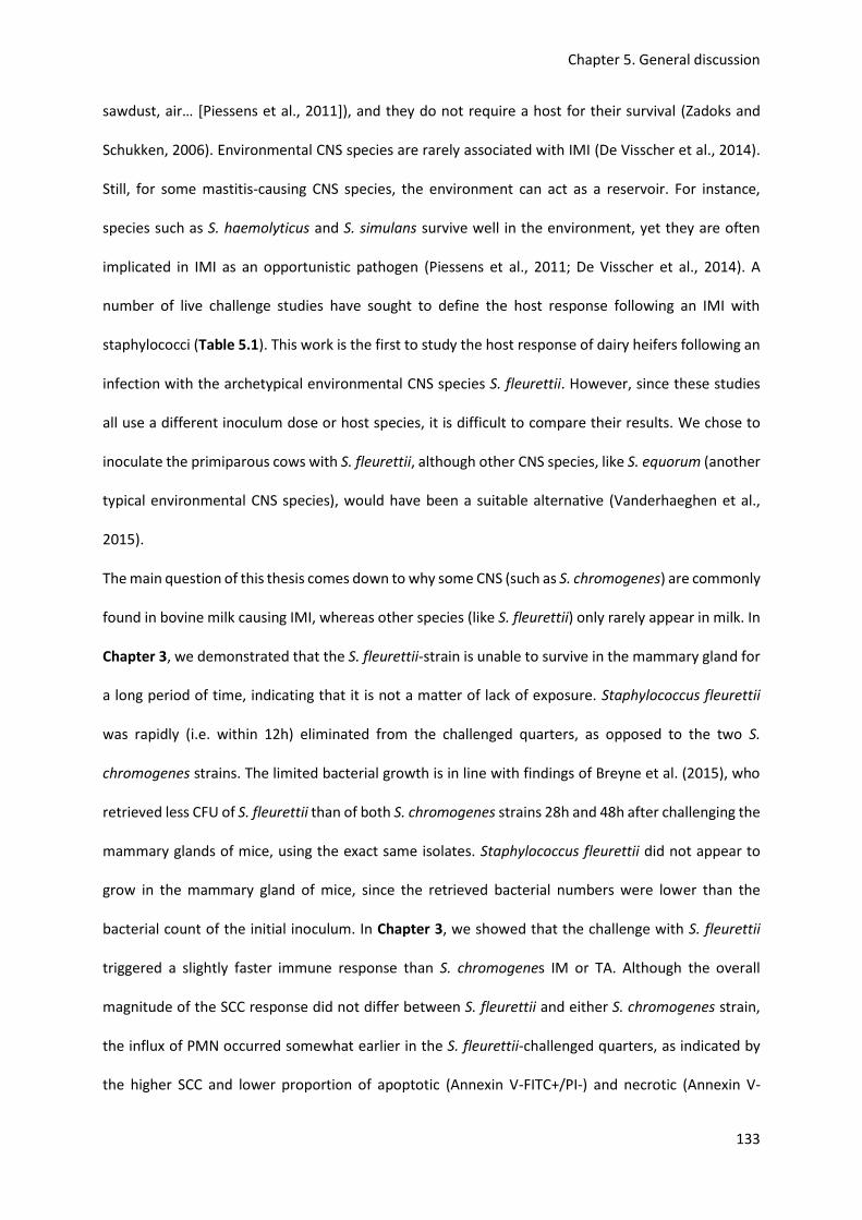
Chapter 5. General discussion
133
sawdust, air… [Piessens et al., 2011]), and they do not require a host for their survival (Zadoks and
Schukken, 2006). Environmental CNS species are rarely associated with IMI (De Visscher et al., 2014).
Still, for some mastitis-causing CNS species, the environment can act as a reservoir. For instance,
species such as S. haemolyticus and S. simulans survive well in the environment, yet they are often
implicated in IMI as an opportunistic pathogen (Piessens et al., 2011; De Visscher et al., 2014). A
number of live challenge studies have sought to define the host response following an IMI with
staphylococci (Table 5.1). This work is the first to study the host response of dairy heifers following an
infection with the archetypical environmental CNS species S. fleurettii. However, since these studies
all use a different inoculum dose or host species, it is difficult to compare their results. We chose to
inoculate the primiparous cows with S. fleurettii, although other CNS species, like S. equorum (another
typical environmental CNS species), would have been a suitable alternative (Vanderhaeghen et al.,
2015).
The main question of this thesis comes down to why some CNS (such as S. chromogenes) are commonly
found in bovine milk causing IMI, whereas other species (like S. fleurettii) only rarely appear in milk. In
Chapter 3, we demonstrated that the S. fleurettii-strain is unable to survive in the mammary gland for
a long period of time, indicating that it is not a matter of lack of exposure. Staphylococcus fleurettii
was rapidly (i.e. within 12h) eliminated from the challenged quarters, as opposed to the two S.
chromogenes strains. The limited bacterial growth is in line with findings of Breyne et al. (2015), who
retrieved less CFU of S. fleurettii than of both S. chromogenes strains 28h and 48h after challenging the
mammary glands of mice, using the exact same isolates. Staphylococcus fleurettii did not appear to
grow in the mammary gland of mice, since the retrieved bacterial numbers were lower than the
bacterial count of the initial inoculum. In Chapter 3, we showed that the challenge with S. fleurettii
triggered a slightly faster immune response than S. chromogenes IM or TA. Although the overall
magnitude of the SCC response did not differ between S. fleurettii and either S. chromogenes strain,
the influx of PMN occurred somewhat earlier in the S. fleurettii-challenged quarters, as indicated by
the higher SCC and lower proportion of apoptotic (Annexin V-FITC+/PI-) and necrotic (Annexin V-

Chapter 5. General discussion
134
FITC+/PI+) PMN at 6h after inoculation. This was in all likelihood the result of the earlier IL-8 response.
The chemotactic cytokine IL-8 was seen to be increased more rapidly (i.e. within 6h) in some of the S.
fleurettii-challenged quarters, although the cytokine response was generally very low and varied too
much between individual animals and quarters to draw any definite conclusions.
One potential immune evasion tactic of the staphylococci is to invade the host cells (Dego et al., 2002).
Based on the fast elimination of S. fleurettii in the mammary gland, one might expect that S. fleurettii
would invade MEC slower than S. chromogenes. Indeed, Souza et al. (2016) -using the exact same
isolates- observed that the adhesion and internalization capacity of S. fleurettii was lower than that of
S. chromogenes IM after 12h of incubation, yet higher than that of S. chromogenes TA. The efficacy
with which bacteria can attach to MEC is presumably also influenced by various host factors (Van
Belkum et al., 2002). As such, Hyvönen et al., (2009) could not find any differences in the in vitro
adhesion or internalization between CNS strains (including S. chromogenes, S. epidermidis and S.
haemolyticus) causing persistent IMI and transient IMI.
All in all, S. fleurettii evoked a mild clinical response in the dairy heifers. For one, there were no visible,
clinical signs of mastitis in any of the challenged quarters. Also, the overall loss in milk yield following
inoculation was less pronounced in S. fleurettii-challenged quarters compared to quarters challenged
with either S. chromogenes strain (Chapter 4.1).
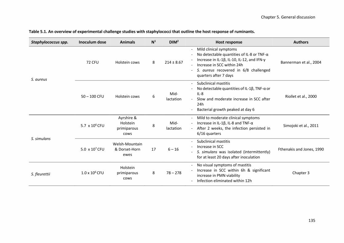
Chapter 5. General discussion
135
Table 5.1. An overview of experimental challenge studies with staphylococci that outline the host response of ruminants.
Staphylococcus spp. Inoculum dose Animals N1 DIM2 Host response Authors
S. aureus
72 CFU Holstein cows 8 214 ± 8.67
- Mild clinical symptoms - No detectable quantities of IL-8 or TNF-α - Increase in IL-1β, IL-10, IL-12, and IFN-γ - Increase in SCC within 24h - S. aureus recovered in 6/8 challenged
quarters after 7 days
Bannerman et al., 2004
50 – 100 CFU Holstein cows 6 Mid-
lactation
- Subclinical mastitis - No detectable quantities of IL-1β, TNF-α or
IL-8 - Slow and moderate increase in SCC after
24h - Bacterial growth peaked at day 6
Riollet et al., 2000
S. simulans
5.7 x 10⁶ CFU
Ayrshire & Holstein
primiparous cows
8 Mid-
lactation
- Mild to moderate clinical symptoms - Increase in IL-1β, IL-8 and TNF-α - After 2 weeks, the infection persisted in
6/16 quarters
Simojoki et al., 2011
5.0 x 10⁷ CFU Welsh-Mountain & Dorset-Horn
ewes 17 6 – 16
- Subclinical mastitis - Increase in SCC - S. simulans was isolated (intermittently)
for at least 20 days after inoculation
Fthenakis and Jones, 1990
S. fleurettii 1.0 x 10⁶ CFU Holstein
primiparous cows
8 78 – 278
- No visual symptoms of mastitis - Increase in SCC within 6h & significant
increase in PMN viability - Infection eliminated within 12h
Chapter 3

Chapter 5. General discussion
136
Staphylococcus spp. Inoculum dose Animals N1 DIM2 Host response Authors
S. epidermidis
3.3 x 10⁷ CFU Merino ewes 5 3 months
- Mild clinical symptoms - Increase in IL-8 from 4h until day 3 - Significant elevation of IL-1β in 1 ewe - SCC increased after 4h and peaked after 1
day - S. epidermidis was found sporadically in
the milk of 4 ewes until 10 weeks, while 1 ewe eradicated the infection after 8h
Winter et al., 2003
5.7 x 10⁶ CFU
Ayrshire & Holstein
primiparous cows
8 Mid-
lactation
- Mild to moderate clinical symptoms - Increase in IL-1β, IL-8 and TNF-α - After 2 weeks, the infection persisted in
5/16 quarters
Simojoki et al., 2011
2.8 x 10⁶ CFU Merino ewes 5 3 months
- Mild clinical symptoms - Increased leukocyte count in milk - Increase in IL-6, IL-8, IL-1β - S. epidermidis was shed intermittently
until the end of the trial (144h)
Winter and Colditz, 2002
S. chromogenes
2.1 × 10⁶ CFU Holstein
primiparous cows
6 4 weeks
- Mild clinical symptoms - Increase in SCC within 12h, peaking at 30h - S. chromogenes was eliminated within
46h, except in 1 cow that developed a persistent infection
Simojoki et al., 2009
1.0 x 10⁶ CFU Holstein
primiparous cows
8 78 – 278
- No visual symptoms of mastitis - Increase in SCC within 9h & significant
increase in PMN viability in S. chromogenes IM challenged quarters
- S. chromogenes still present in milk at 78h
Chapter 3
1 Number of challenged animals; 2 Days in lactation at the moment of inoculation.

Chapter 5. General discussion
137
5.3 Intraspecies diversity within Staphylococcus chromogenes
Thanks to the increasing availability of low-cost molecular identification techniques, we have recently
gained more insight into the strain-specific virulence of mastitis pathogens. The heterogeneity of the
mastitis pathogen S. aureus is well documented, in contrast to that of other staphylococci. Although
most of the S. aureus strains causing bovine mastitis can be traced back to a limited number of lineages
or clonal complexes (Kapur et al., 1995; Barkema et al., 2006; Delgado et al., 2011), the strains often
differ in their ability to invade MEC, produce biofilm, respond to antibiotic treatment, affect milk yield,
evoke a SCC response or spread from cow to cow (Barkema et al., 2006; Zadoks et al., 2011; Bardiau et
al., 2014). While some studies conclude that extramammary S. aureus strains (e.g. isolated from skin,
perineum, nares, etc.) are indistinguishable by molecular typing from strains involved in IMI (Haveri et
al., 2008; Capurro et al., 2010), others do report the existence of tissue-specific strains (Zadoks et al.,
2002; Van Leeuwen et al., 2005).
The intraspecies diversity of CNS, on the other hand, has long been overlooked. Although several studies
have used molecular typing (e.a. pulsed field gel electrophoresis [PFGE]) for strain differentiation of
bovine CNS (Thorberg et al., 2006; Mørk et al., 2012; Bexiga et al., 2014), there is not much known at
the present time about the clinical importance of these different individual strains. Chapter 3 of this
thesis therefore focused on the host response and clinical impact of different strains of S. chromogenes,
the most commonly isolated CNS species in dairy cattle with an IMI (Sampimon et al., 2009; Thorberg et
al., 2009; Piessens et al., 2011). To address the potential tissue-specificity of S. chromogenes, two
separate strains were selected; i.e. the so-called “teat apex strain” and “intramammary strain”. The S.
chromogenes strains challenged the innate immune system in varying degrees (Table 5.2). The S.
chromogenes IM strain appeared to be better equipped to invade and colonize the bovine mammary
gland than S. chromogenes TA. Consequently, S. chromogenes IM induced the largest cellular response
and resulted in the greatest production loss. Even though S. chromogenes IM was originally isolated
from a cow with a persistent IMI lasting for more than 10 months, both S. chromogenes-strains were
eliminated within 6 days. In another live challenge study with S. chromogenes (Simojoki et al., 2009),

Chapter 5. General discussion
138
one out of six heifers developed a persistent IMI, seeing that the bacteria were still present after 7 days
in the challenged quarter. It should be emphasized that the duration of the infection not only depends
on the virulence of the strain, but also on various host factors and the initial inoculum dose. The host
response of the mammary gland also responds to pathogens in a dose-dependent manner (Günther et
al., 2010). In case of experimental E. coli mastitis, lowering the inoculum dose results in a slower
bacterial clearance, and is preceded by excessive bacterial growth (Shuster et al., 1996; Vangroenweghe
et al., 2004). We assume that the high inoculum dose used in our experimental study (i.e. 1.0 x 10⁶ CFU)
might have triggered a more aggressive host response than is the case with natural infections, possibly
shortening the duration of infection.
Yet, S. chromogenes is also able to colonize the host (i.e. the cow's body) without causing damage (i.e.
mastitis). The species can be found on the teat skin, streak canal, hair coat, nares, vagina and perineum
of healthy dairy cattle (White et al., 1989; De Vliegher et al., 2003; Taponen et al., 2008; De Visscher et
al., 2016), though not necessarily on every cow (Braem et al., 2013; De Visscher et al., 2014). While S.
chromogenes is rarely found on the teat skin of lactating cows (Braem et al., 2013; De Visscher et al.,
2014), the species appears to be omnipresent on the teats of dry cows and pregnant heifers (De Visscher
et al., 2016b). Certain pulsotypes of S. chromogenes are found both in the milk and on extramammary
sites (Taponen et al., 2008). Some authors reported an association between the extramammary
colonization by S. chromogenes and IMI (Taponen et al., 2008; Quirk et al., 2012; De Visscher et al.,
2016a), while others found no such relation (De Vliegher et al., 2003). In fact, De Vliegher et al. (2003)
stated that teat apex colonization with S. chromogenes might be protective against IMI in early lactation,
although no strain typing was performed in this study.

Chapter 5. General discussion
139
Table 5.2. A basic summary of the known differences between strains of S. chromogenes isolated from
a teat apex (TA) and from an intramammary infection (IM) in terms of their interaction with the host.
Parameter Study object S. chromogenes TA S. chromogenes IM Author
Adhesion capacity MAC-T cells + + + Souza et al. (2016)
Invasion capacity MAC-T cells + + + Souza et al. (2016)
SCC increase Heifers + + + N.S. Chapter 3
% Apoptotic milk PMN Heifers + + N.S. + Chapter 3
Bacterial growth in the
mammary gland Heifers + + + Chapter 3
Milk yield Heifers − − −N.S. Chapter 4.1
Milk PRL increase Heifers + + Chapter 4.1
Local IL-1β level Mice + + N.S. + Breyne et al. (2015)
Heifers + D.S. + Chapter 3
Local TNF-α level Mice − − Breyne et al. (2016)
Heifers +D.S. + Chapter 3
Local IL-8 level Heifers − + +D.S. Chapter 3
N.S. No statistically significant difference.
D.S. Based on descriptive statistics.
5.4 The role of PRL in CNS mastitis
Although certain CNS species and strains are associated with persistent IMI and elevated SCC levels,
some studies suggest that CNS-infected dairy cattle produce more milk throughout the lactation period
than their non-infected herd mates (Compton et al., 2007; Schukken et al., 2009; Piepers et al., 2010).
Several hypotheses have been put forward, but there is still no sound biological explanation for this
unexpected observation (Piepers et al., 2013). In Chapter 4.1, the milk production was monitored for
only three days following the experimental challenge with S. fleurettii, S. chromogenes TA and S.
chromogenes IM. Even though the milk production was suppressed in all challenged quarters, the overall
production losses were minimal. These results align with the work of Pearson et al., 2013 and Tomazi et

Chapter 5. General discussion
140
al., 2015, who observed little or no decline in milk yield in dairy cattle following a CNS IMI. Still, the
experiment in Chapter 4.1 only focused on the acute phase of inflammation, and is therefore not
suitable for inferring definitive conclusions regarding the milk production throughout lactation following
an IMI with the 3 CNS strains.
The hypothesis that PRL stimulates the milk production in response to an infection with CNS was
examined in Chapter 4. It had previously been demonstrated that PRL is involved in the pathogenesis of
bovine mastitis by stimulating the inflammatory response in the MEC through the activation of the NF-
κB (Boutet et al., 2007). Consequently, this thesis postulated that the local production of PRL would
increase in the mammary gland following an infection with CNS. The inflammation-induced PRL might
then possibly stimulate autocrine milk production, although this association was not directly studied in
this work. Chapter 4.1 shows that, over time, PRL is higher in the milk of CNS-infected quarters compared
to uninfected quarters. No significant difference was found in the PRL level between the three different
CNS strains. Approximately 28 hours after the inoculation, the PRL concentration started to decline in
both the challenged and control quarters. The final PRL levels at the end of the experiment, 78h after
the inoculation, were lower than the initial, pre-challenge values. One possible explanation for the
overall decline in milk PRL, is that the sampling interval lengthens as the experiment progresses. It has
previously been demonstrated that a higher milking frequency leads to higher circulating PRL levels (Bar-
Peled et al., 1995). Therefore, the overall higher PRL concentration at the beginning of the experiment
might be the result of the higher sampling frequency. Another possibility is that the secretion of
(pituitary) PRL is controlled through a classic negative feedback loop (Anderson et al., 2006), either by
promoting the hypothalamic dopamine release or by directly inhibiting the release of PRL in the
lactotrophs (Bernard et al., 2015).
Chapter 4.2 confirmed that the bovine mammary gland is indeed able to synthesize PRL, similar to
humans, rats or goats. No increase in mRNA of PRL was found in MEC after challenging them with S.
chromogenes TA, S. chromogens IM or S. fleurettii. The results imply that the expression of the PRL gene
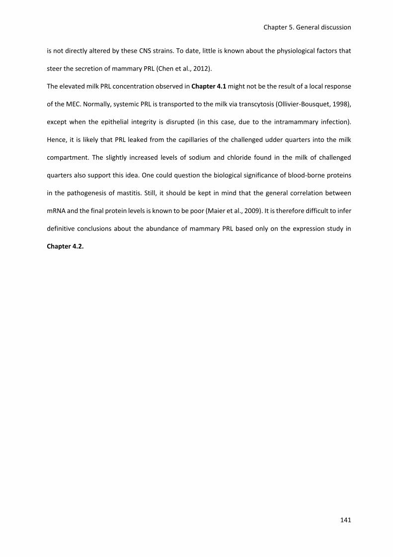
Chapter 5. General discussion
141
is not directly altered by these CNS strains. To date, little is known about the physiological factors that
steer the secretion of mammary PRL (Chen et al., 2012).
The elevated milk PRL concentration observed in Chapter 4.1 might not be the result of a local response
of the MEC. Normally, systemic PRL is transported to the milk via transcytosis (Ollivier-Bousquet, 1998),
except when the epithelial integrity is disrupted (in this case, due to the intramammary infection).
Hence, it is likely that PRL leaked from the capillaries of the challenged udder quarters into the milk
compartment. The slightly increased levels of sodium and chloride found in the milk of challenged
quarters also support this idea. One could question the biological significance of blood-borne proteins
in the pathogenesis of mastitis. Still, it should be kept in mind that the general correlation between
mRNA and the final protein levels is known to be poor (Maier et al., 2009). It is therefore difficult to infer
definitive conclusions about the abundance of mammary PRL based only on the expression study in
Chapter 4.2.

Chapter 5. General discussion
142
5.5 Evaluation of the experiments
5.5.1 In vivo experiment
During the in vivo experiment (Chapter 3 & 4.1), eight Holstein heifers received an intramammary
infusion with either S. fleurettii, S. chromogenes TA, S. chromogenes IM or PBS in each udder quarter.
Three quarters were inoculated with a bacterial dose of 1.0 x 10⁶ CFU suspended in 5 mL PBS, whereas
the fourth quarter –infused with an equal volume of sterile PBS- was used as an internal control. This
type of experimental set-up is referred to as a "split-udder design", which has its inherent strengths and
weaknesses.
One advantage of this particular study design is that the heifers act as their own control group, allowing
for a smaller number of required experimental animals and a reduction in variability owing to individual
differences. This is in agreement with the reduction principle of the “3Rs” (Replacement, Reduction and
Refinement), the ethical framework for humane experimentation. A split-udder design assumes that all
neighboring udder quarters are separate, individual anatomical entities within the same animal.
However, previous research has demonstrated that this is not necessarily the case. An experimental
infection of an udder quarter with E. coli or S. aureus results in changes in gene expression in
neighboring, uninfected quarters (Mitterhuemer et al., 2010; Jensen et al., 2013). But even though genes
associated with the local immune response are upregulated in untreated control quarters (Jensen et al.,
2013), the SCC or the PMN viability does not seem to increase significantly in uninfected quarters
compared to naturally infected quarters (Blagitz et al., 2015). In the experimental CNS-challenge
described in Chapter 3, the SCC nor the viability increased considerably, and the cytokines TNF-α, IL-8
or IL-1β were not detected in uninfected control quarters following the challenge with sterile PBS.
Therefore, we believe that the effect of any potential “cross-talk” between neighboring quarters on our
results is of minor importance.
In vivo experiments in dairy cattle also have certain disadvantages. For one, the animals have to be taken
out of their usual environment, which affects their stress level. It has been well documented that stress
can modulate the immune response and circulating PRL levels of animals (Carroll & Forsberg, 2007;

Chapter 5. General discussion
143
GarcÍa-Ispierto et al., 2009). Glucocorticoid hormones, associated with a stress response, can delay the
apoptosis in human PMN (Liles et al., 1995). This might explain why the proportion of apoptotic PMN
(Chapter 3) already decreased in all mammary quarters before the actual inoculation procedure had
taken place. For future in vivo experiments, we suggest incorporating a longer (> 48h) adaptation period
for letting the cows adjust to their new surroundings, or -if feasible- performing the experiment in their
usual setting together with familiar herd mates.
Another limitation of our study, is that only heifers in mid-lactation were challenged with the CNS
strains, making it difficult to extrapolate our results to multiparous cows, or cows in a different stage of
lactation. As mentioned in Chapter 2, we chose to only include primiparous cows in our study, because
the prevalence of CNS IMI is higher in primiparous cows than in multiparous cows (Sampimon et al.,
2009; De Visscher et al., 2016). The heifers were knowingly selected according to their CXCR1 genotype
for an additional experiment studying the effect of a polymorphism (c.980 A>G) in the CXCR1 gene on
the innate immune response (Verbeke et al., 2015). Although the PMN migration was slightly slower in
the S. chromogenes IM-infected quarters of the c.980AG heifers than the c.980GG heifers, it is unlikely
to have had any effect on the conclusions of this thesis. Whether or not the heifers had previously been
infected with either CNS strain under natural circumstances (and therefore might have established an
antigen-specific memory through the adaptive immune system [Rainard et al., 2016]) can never be
completely ruled out though.
Because of these limitations, alternative in vivo models for exploring bovine mastitis have been
developed in mice (Brouillette and Malouin, 2005). They are less expensive, less cumbersome and easier
to control. The mammary glands of mice and cows share certain anatomical characteristics. For one,
both species have one teat opening and one primary duct per mammary gland. Similar to cows, the
glands of mice are structurally independent from each other (Notebaert and Meyer, 2006). On the other
hand, the composition of murine milk is fundamentally different (Gors et al., 2009), and mice have less
resident phagocytic cells in their mammary glands (Notebaert and Meyer, 2006). All limitations
Chapter 5. General discussion
144
considered, we believe that an in vivo challenge in dairy cattle is still the most accurate method for
mimicking natural IMI in bovines.
5.5.2 In vitro experiment
In vitro or ex vivo studies, using an immortalized cell line (as in Chapter 4.2) or primary bovine cells, have
also contributed to our understanding of bovine mastitis. These types of studies are generally
inexpensive, fast and easy to handle, making it a popular model for studying intramammary infections.
However, in vitro studies cannot fully account for the various host factors that affect the course of an
infection, such as parity (Mehrzad et al., 2002), stage of lactation (Mallard et al., 1998), and genetic
variability (Rupp and Boichard, 2003). Primary cultures of bovine MEC, taken from mammary tissue, are
thought to be a better model for studying the in vivo conditions of the mammary gland (Rabot et al.,
2006). Although they reflect the physiological and metabolic processes of epithelial cells more closely,
primary MEC quickly lose their original characteristics after a number of passages (Matitashvili et al.,
1997). In Chapter 4.2, we chose to infect MAC-T cells to study the PRL expression in response to the 3
CNS strains. MAC-T cells, immortalized by originally transfecting mammary cells with Simian virus 40
(SV40), consist of a heterogeneous cell population (excluding myoepithelial cells [Zavizion et al., 1995]).
The MAC-T cell line has the ability to express milk proteins (i.e. β-casein, α-lactalbumin, etc.), GH-
receptors, and various cytokines in response to an infection (Zhou et al., 2008; Günther et al., 2016).
Although the cytokine response of MAC-T cells is weaker than that of primary MEC, the cell line responds
in a similar manner (Günther et al, 2016). Primary bovine MEC might have displayed a higher PRL
expression than MAC-T cells in Chapter 4.2, but we assume that this would not have affected the
conclusion of the study.
5.5.3 The inoculum dose
Simulating a natural IMI under experimental conditions is inherently difficult. One particular issue is
selecting an inoculum dose that corresponds with natural infections. The in vivo study of Chapter 3 and

Chapter 5. General discussion
145
Chapter 4.1 was preceded by a limited dose-response trial, in which a multiparous cow was challenged
in each quarter with increasing doses of S. chromogenes IM (2.5 x 10⁵, 5.0 x 10⁵, 1.0 x 10⁶ and 2.0 x 10⁶
CFU in 5 mL PBS) as described by Verbeke et al. (2015). Only the highest dose, 2.0 x 10⁶ CFU, resulted in
local, clinical symptoms (i.e. a swollen, reddish mammary quarter). However, in the quarter that was
challenged with the lowest inoculum dose, 2.5 x 10⁵ CFU, the bacteria were eliminated within 20h.
Unlike Simojoki et al. (2009), we did not necessarily want to induce clinical mastitis in the dairy heifers.
Generally speaking, CNS infections are mostly associated with subclinical or mild clinical symptoms
(Taponen et al., 2006; Supré et al., 2011). That is why we chose for the inoculum dose of 1.0 x 10⁶ CFU.
Still, only a limited number of strains were studied in this thesis, which is insufficient to represent entire
communities of CNS within the host-associated or environmental niche. Furthermore, considering the
strain differences we found in S. chromogenes, we cannot exclude that S. fleurettii strains are equally
diverse in terms of pathogenicity.
5.6 Conclusions and practical implications
In this work, we focused on the host response in dairy heifers following three CNS field strains from
diverging environments: S. chromogenes TA, S. chromogenes IM and S. fleurettii. The effects of these
strains have been studied in MEC (Chapter 4.2; Souza et al., 2016), mice (Breyne et al., 2015) and dairy
heifers (Chapter 3 and Chapter 4.1). These results confirm that (quarters of) dairy heifers respond
differently to host-adapted versus environmental CNS. Staphylococcus fleurettii (or at least the
particular strain used in this research) is less accustomed to the bovine mammary gland than the
typically host-adapted species S. chromogenes. Furthermore, strains within S. chromogenes can differ in
their ability to cause damage to the host (Chapter 3 and Chapter 4.1). The S. chromogenes strain derived
from an extramammary site, i.e. the teat apex, is less able to grow in the mammary gland than the other
S. chromogenes strain isolated from a mastitis case. Overall, S. chromogenes TA induced a less
pronounced inflammatory response. The results in Chapter 3, combined with experimental challenge
studies in mice and MEC, suggest that the strains within S. chromogenes show a variable degree of

Chapter 5. General discussion
146
pathogenicity and tissue-specificity. Of course, intraspecies diversity in terms of pathogenicity is not
unique to S. chromogenes, and has been observed in other mastitis bacteria as well, such as S. uberis
(Tassi et al., 2013), S. aureus (Middleton and Fox, 2002; Haveri et al., 2007) and E. coli (Dogan et al.,
2006; Lippolis et al., 2014). The degree in which bacteria cause damage to their host, is the result of a
complex interplay between both the microorganism and the host. Distinguishing pathogenic from less
pathogenic strains or isolates within one species, and anticipating their clinical impact, is therefore not
a straightforward process. In practice, it often boils down to the decision on whether or not to treat a
case of bovine mastitis, and whether or not to cull a chronically infected cow. However, in case of the
two S. chromogenes strains studied in this thesis, the difference in the clinical impact and loss in milk
yield in dairy heifers is ultimately limited (Chapter 3 and Chapter 4.1). Since the use of veterinary
antimicrobials is under scrutiny in the European Union, subclinical and mild cases of mastitis are often
left untreated, assuming that the infection is self-limiting. Spontaneous cure rates in CNS are generally
thought to be as high as 60 – 70% (McDougall, 1998; Wilson et al., 1999), although more recent studies
report lower numbers (i.e. 12 to 45% [Deluyker et al., 2005; Taponen et al., 2006]). Because of their
limited clinical importance, CNS are currently not routinely identified in veterinary laboratories at the
species level. Therefore, routine strain-typing of CNS in practice seems out of the question for now. Still,
addressing CNS infections can be of great value for dairy farms with a low bulk milk SCC that specifically
strive for an even lower bulk milk SCC. In herds with a bulk milk SCC below 200,000 cells/mL, CNS
infections contribute on average 17.9% of all somatic cells in the bulk tank (Schukken et al., 2009).
It was beyond the scope of this thesis to assess the effect of CNS as a bacterial group on the milk yield.
Although PRL does increase in the quarters challenged with the three CNS strains (Chapter 4.1), there is
inconclusive evidence that this is the result of a local PRL production in the mammary gland (Chapter
4.2).

Chapter 5. General discussion
147
5.7 Future research
The effect of CNS on the milk yield of dairy cattle remains unclear. A longitudinal study throughout
lactation on the association between the milk yield and CNS infections can offer more insights. It might
be opportune to monitor the milk production at quarter level (for instance at farms equipped with
automatic milking systems), so one can compare the milk production of CNS-infected quarters to
healthy, uninfected quarters. This minimizes the effect of confounding variables at the cow or herd level
(Pyörälä and Taponen, 2009; Tomazi et al., 2015). Future studies should ideally identify the involved CNS
at the subtype or strain level using genotypic methods. Strain typing is required in observational studies
to ascertain whether the CNS infections are transient or persistent in nature. Pulsed field gel
electrophoresis used to be considered the “gold standard”, although it is quite labor-intensive and
expensive (van Belkum et al., 2007). Depending on the study objective, multi locus sequence typing or
whole genome sequencing might be more appropriate to differentiate isolates (Smith et al., 2005; Köser
et al., 2012; Vanderhaeghen et al., 2015).
Strain typing is required in observational studies to ascertain whether the CNS infections are transient
or persistent in nature. The results from Chapter 3 also clearly demonstrate the existence of pathogenic
diversity within CNS species (in this case S. chromogenes). Species and strain differences might account
for the seemingly contradicting research findings regarding the milk production of CNS-infected cattle
(Gröhn et al., 2004; Piepers et al., 2010; Tomazi et al., 2015). Studies considering CNS as one
homogeneous group inevitably fall prey to ecological fallacy, i.e. the error that arises when drawing
conclusions about individuals based on the observations of groups (Morgenstern, 1982). The same
reasoning may hold true for studies focusing on the species level of CNS, given the strain diversity we
found in S. chromogenes.
Strain typing of S. chromogenes is essential to further elucidate the association between teat apex
colonization and IMI, or the inhibition of IMI. The teat apex strain used in this work, S. chromogenes TA,
is capable of inhibiting the in vitro growth of mastitis field isolates of S. aureus, S. uberis and S.
dysgalactiae to some extent (De Vliegher et al., 2004). This is likely the result of extracellular bacteriocins

Chapter 5. General discussion
148
produced by the S. chromogenes TA strain, as has been demonstrated in other CNS (Nascimento et al.,
2005; Braem et al., 2014). Bacteria produce these substances as a tool for defending their niche against
similar, competing microorganisms (Kommineni et al., 2015). It is still uncertain though whether teat
apex colonization with S. chromogenes TA, or any other strain with similar inhibitory properties, is
actually able to prevent IMI caused by Gram-positive mastitis pathogens in dairy cattle. Additional
challenge studies are needed to verify this assumption. One possible experimental set-up is an split-
udder or split-herd trial where the cows receive a “protective strain” on the skin of their teats (for
instance, through swabbing or dipping of the teats). By determining the incidence of new IMI in the
challenged and control teats over a period of time, one could determine the efficacy of the protective
strain. However, such a trial is cumbersome, and moreover, restricted by biosafety legislations (Van
Vaerenbergh et al., 2010). Therefore, this type of trial cannot be performed in commercial dairy farms.
Another possibility is to inoculate a protective strain of interest directly into the teat or gland cistern of
dairy cows (as described in this thesis), followed by an experimental superinfection with a major mastitis
pathogen.
All in all, the concept of using live microflora to combat bacterial infections is not new (Woodward et
al., 1987; Bouchard et al., 2015). One could wonder if certain strains of CNS can be utilized as an
alternative (preventive) therapy for bovine mastitis. At this time, there is insufficient scientific evidence
to presume that one or more CNS strains might be suitable as an intramammary probiotic. For one, the
purported protective effect in vivo has never been traced back to a particular species or strain.
Furthermore, their safety cannot be warranted. Multiple antimicrobial resistance genes (e.g. mecA, blaZ)
have been found in CNS from bovine milk (Frey et al., 2013), creating a potential reservoir for
transferring resistance to other bacteria (Piessens et al., 2012b). Even so, the notion of intramammary
probiotics remains a promising track to combat and prevent bovine mastitis, and should be further
investigated.

Chapter 5. General discussion
149
5.8 References
Anderson, S. T., J. L. Barclay, K. J. Fanning, D. H. L. Kusters, M. J. Waters, and J. D. Curlewis. 2006. Mechanisms underlying the diminished sensitivity to prolactin negative feedback during lactation: Reduced STAT5 signaling and up-regulation of cytokine-inducible SH2 domain-containing protein (CIS) expression in tuberoinfundibular dopaminergic neurons. Endocrinol 147(3):1195-1202. Bannerman, D. D., M. J. Paape, J. W. Lee, X. Zhao, J. C. Hope, and P. Rainard. 2004. Escherichia coli and Staphylococcus aureus elicit differential innate immune responses following intramammary infection. Clin Diagn Lab Immunol 11(3):463-472. Bar-Peled, U., E. Maltz, I. Bruckental, Y. Folman, Y. Kali, H. Gacitua, A. R. Lehrer, C. H. Knight, B. Robinzon, H. Voet, and et al. 1995. Relationship between frequent milking or suckling in early lactation and milk production of high producing dairy cows. J Dairy Sci 78(12):2726-2736. Bardiau, M., J. Detilleux, F. Farnir, J. G. Mainil, and I. Ote. 2014. Associations between properties linked with persistence in a collection of Staphylococcus aureus isolates from bovine mastitis. Vet Microbiol 169(1-2):74-79. Barkema, H. W., Y. H. Schukken, and R. N. Zadoks. 2006. Invited Review: The role of cow, pathogen, and treatment regimen in the therapeutic success of bovine Staphylococcus aureus mastitis. J Dairy Sci 89(6):1877-1895. Barkema, H. W., H.A. Deluyker, Y.H. Schukken, and T.J.G.M. Lam. 1999. Quarter-milk somatic cell count at calving and at the first six milkings after calving. Prevent Vet Med 38(1), 1-9. Bernard, V., J. Young, P. Chanson, and N. Binart. 2015. New insights in prolactin: pathological implications. Nat Rev Endocrinol 11(5):265-275. Bexiga, R., M. G. Rato, A. Lemsaddek, T. Semedo-Lemsaddek, C. Carneiro, H. Pereira, D. J. Mellor, K. A. Ellis, and C. L. Vilela. 2014. Dynamics of bovine intramammary infections due to coagulase-negative staphylococci on four farms. J Dairy Res 81(2):208-214. Blagitz, M. G., F. N. Souza, C. F. Batista, S. A. Diniz, L. F. F. Azevedo, M. X. Silva, J. P. A. Haddad, M. B. Heinemann, M. M. O. P. Cerqueira, and A. M. M. P. Della Libera. 2015. Flow cytometric analysis: Interdependence of healthy and infected udder quarters. J Dairy Sci 98(4):2401-2408. Bouchard, D. S., B. Seridan, T. Saraoui, L. Rault, P. Germon, C. Gonzalez-Moreno, F. M. Nader-Macias, D. Baud, P. Francois, V. Chuat, F. Chain, P. Langella, J. Nicoli, Y. Le Loir, and S. Even. 2015. Lactic Acid Bacteria Isolated from Bovine Mammary Microbiota: Potential Allies against Bovine Mastitis. PLoS One 10(12):e0144831. Boutet, P., J. Sulon, R. Closset, J. Detilleux, J. F. Beckers, F. Bureau, and P. Lekeux. 2007. Prolactin-induced activation of nuclear factor kappa B in bovine mammary epithelial cells: Role in chronic mastitis. J Dairy Sci 90(1):155-164. Braem, G., B. Stijlemans, W. Van Haken, S. De Vliegher, L. De Vuyst, and F. Leroy. 2014. Antibacterial activities of coagulase-negative staphylococci from bovine teat apex skin and their inhibitory effect on mastitis-related pathogens. J Appl Microbiol 116(5):1084-1093.

Chapter 5. General discussion
150
Breyne, K., S. De Vliegher, A. De Visscher, S. Piepers, and E. Meyer. 2015. Technical note: a pilot study using a mouse mastitis model to study differences between bovine associated coagulase-negative staphylococci. J Dairy Sci 98(2):1090-1100. Brouillette, E. and F. Malouin. 2005. The pathogenesis and control of Staphylococcus aureus-induced mastitis: study models in the mouse. Microb & Infect 7(3):560-568. Capurro, A., A. Aspan, H. Ericsson Unnerstad, K. Persson Waller, and K. Artursson. 2010. Identification of potential sources of Staphylococcus aureus in herds with mastitis problems. J Dairy Sci 93(1):180-191. Carroll, J. A., and N.E. Forsberg. 2007. Influence of stress and nutrition on cattle immunity. Vet Clin N
Am: Food Anim Pract. 23(1), 105-149.
Chen, C. C., D. B. Stairs, R. B. Boxer, G. K. Belka, N. D. Horseman, J. V. Alvarez, and L. A. Chodosh. 2012. Autocrine prolactin induced by the Pten-Akt pathway is required for lactation initiation and provides a direct link between the Akt and Stat5 pathways. Gene Dev 26(19):2154-2168. Compton, C. W. R., C. Heuer, K. Parker, and S. McDougall. 2007. Epidemiology of mastitis in pasture-grazed peripartum dairy heifers and its effects on productivity. J Dairy Sci 90(9):4157-4170.
Dego, O. Kerro, J. E. Van Dijk, and H. Nederbragt. 2002. Factors involved in the early pathogenesis of
bovine Staphylococcus aureus mastitis with emphasis on bacterial adhesion and invasion. A review.
Vet Q 24(4): 181-198.
De Visscher, A., K. Supré, F. Haesebrouck, R. N. Zadoks, V. Piessens, E. Van Coillie, S. Piepers, and S. De Vliegher. 2014. Further evidence for the existence of environmental and host-associated species of coagulase-negative staphylococci in dairy cattle. Vet Microbiol 172(3-4):466-474. De Visscher, A., S. Piepers, F. Haesebrouck, and S. De Vliegher. 2016a. Intramammary infection with coagulase-negative staphylococci at parturition: Species-specific prevalence, risk factors, and effect on udder health. J Dairy Sci 99(8):6457-6469. De Visscher, A., S. Piepers, F. Haesebrouck, and S. De Vliegher. 2016b. Teat apex colonization with coagulase-negative Staphylococcus species before parturition: Distribution and species-specific risk factors. J Dairy Sci 99(2):1427-1439. De Vliegher, S., H. Laevens, L. A. Devriese, G. Opsomer, J. L. Leroy, H. W. Barkema, and A. de Kruif. 2003. Prepartum teat apex colonization with Staphylococcus chromogenes in dairy heifers is associated with low somatic cell count in early lactation. Vet Microbiol 92(3):245-252. De Vliegher, S., G. Opsomer, A. Vanrolleghem, L. A. Devriese, O. C. Sampimon, J. Sol, H. W. Barkema, F. Haesebrouck, and A. de Kruif. 2004. In vitro growth inhibition of major mastitis pathogens by Staphylococcus chromogenes originating from teat apices of dairy heifers. Vet Microbiol 101(3):215-221. Delgado, S., P. Garcia, L. Fernandez, E. Jimenez, M. Rodriguez-Banos, R. del Campo, and J. M. Rodriguez. 2011. Characterization of Staphylococcus aureus strains involved in human and bovine mastitis. FEMS Immunol Med Microbiol 62(2):225-235.

Chapter 5. General discussion
151
Deluyker, H. A., S. N. Van Oye, and J. F. Boucher. 2005. Factors affecting cure and somatic cell count after pirlimycin treatment of subclinical mastitis in lactating cows. J Dairy Sci 88(2):604-614. Dogan, B., S. Klaessig, M. Rishniw, R. A. Almeida, S. P. Oliver, K. Simpson, and Y. H. Schukken. 2006. Adherent and invasive Escherichia coli are associated with persistent bovine mastitis. Vet Microbiol 116(4):270-282. Foster, T. J. 2005. Immune evasion by staphylococci. Nat Rev Microbiol 3(12):948-958. Fry, P. R., J. R. Middleton, S. Dufour, J. Perry, D. Scholl, and I. Dohoo. 2014. Association of coagulase-negative staphylococcal species, mammary quarter milk somatic cell count, and persistence of intramammary infection in dairy cattle. J Dairy Sci 97(8):4876-4885. Frey, Y., J. P. Rodriguez, A. Thomann, S. Schwendener, and V. Perreten. 2013. Genetic characterization of antimicrobial resistance in coagulase-negative staphylococci from bovine mastitis milk. J Dairy Sci 96(4):2247-2257. Fthenakis, G. C. and J. E. T. Jones. 1990. The Effect of Experimentally Induced Subclinical Mastitis on Milk-Yield of Ewes and on the Growth of Lambs. Brit Vet J 146(1):43-49. Gors, S., M. Kucia, M. Langhammer, P. Junghans, and C. C. Metges. 2009. Technical note: Milk composition in mice-Methodological aspects and effects of mouse strain and lactation day. J Dairy Sci 92(2):632-637. Gröhn, Y. T., D. J. Wilson, R. N. Gonzalez, J. A. Hertl, H. Schulte, G. Bennett, and Y. H. Schukken. 2004. Effect of pathogen-specific clinical mastitis on milk yield in dairy cows. J Dairy Sci 87(10):3358-3374. Günther, J., S. Liu, K. Esch, H. J. Schuberth, and H. M. Seyfert. 2010. Stimulated expression of TNF-alpha and IL-8, but not of lingual antimicrobial peptide reflects the concentration of pathogens contacting bovine mammary epithelial cells. Veterinary immunology and immunopathology 135(1-2):152-157. Günther, J., M. Koy, A. Berthold, H. J. Schuberth, and H. M. Seyfert. 2016. Comparison of the pathogen species-specific immune response in udder derived cell types and their models. Vet Res 47. Haveri, M., A. Roslo, L. Rantala, and S. Pyörälä. 2007. Virulence genes of bovine Staphylococcus aureus from persistent and nonpersistent intramammary infections with different clinical characteristics. J Appl Microbiol 103(4):993-1000. Haveri, M., M. Hovinen, A. Roslof, and S. Pyörälä. 2008. Molecular types and genetic profiles of Staphylococcus aureus strains isolated from bovine intramammary infections and extramammary sites. J Clin Microbiol 46(11):3728-3735. Hogan, J. S., D. G. White, and J. W. Pankey. 1987. Effects of Teat Dipping on Intramammary Infections by Staphylococci Other Than Staphylococcus Aureus. J Dairy Sci 70(4):873-879. Huynh, H. T., G. Robitaille, and J. D. Turner. 1991. Establishment of Bovine Mammary Epithelial-Cells (Mac-T) - an Invitro Model for Bovine Lactation. Experimental Cell Research 197(2):191-199. Hyvönen, P., S. Kayhko, S. Taponen, A. von Wright, and S. Pyörälä. 2009. Effect of bovine lactoferrin on the internalization of coagulase-negative staphylococci into bovine mammary epithelial cells under in-vitro conditions. J Dairy Res 76(2):144-151.
Chapter 5. General discussion
152
Jensen, K., J. Günther, R. Talbot, W. Petzl, H. Zerbe, H. J. Schuberth, H. M. Seyfert, and E. J. Glass. 2013. Escherichia coli- and Staphylococcus aureus-induced mastitis differentially modulate transcriptional responses in neighbouring uninfected bovine mammary gland quarters. BMC genomics 14. Kapur, V., W. M. Sischo, R. S. Greer, T. S. Whittam, and J. M. Musser. 1995. Molecular population genetic analysis of Staphylococcus aureus recovered from cows. J Clin Microbiol 33(2):376-380. Kommineni, S., D. J. Bretl, V. Lam, R. Chakraborty, M. Hayward, P. Simpson, Y. M. Cao, P. Bousounis, C. J. Kristich, and N. H. Salzman. 2015. Bacteriocin production augments niche competition by enterococci in the mammalian gastrointestinal tract. Nature 526(7575):719-722. Köser, C. U., M.J. Ellington, E.J. Cartwright, S.H. Gillespie, N.M. Brown, M. Farrington, M.T.G. Holden, G. Dougan, S.D. Bentley, J. Parkhill, and S.J. Peacock. 2012. Routine use of microbial whole genome sequencing in diagnostic and public health microbiology. PLoS pathogens, 8(8), e1002824. Liles, W. C., D. C. Dale, and S. J. Klebanoff. 1995. Glucocorticoids inhibit apoptosis of human neutrophils. Blood 86(8):3181-3188. Lippolis, J. D., B. W. Brunelle, T. A. Reinhardt, R. E. Sacco, B. J. Nonnecke, B. Dogan, K. Simpson, and Y. H. Schukken. 2014. Proteomic analysis reveals protein expression differences in Escherichia coli strains associated with persistent versus transient mastitis. J Proteomics 108:373-381. Maier, T., M. Guell, and L. Serrano. 2009. Correlation of mRNA and protein in complex biological samples. Febs Lett 583(24):3966-3973. Mallard, B. A., J. C. Dekkers, M. J. Ireland, K. E. Leslie, S. Sharif, C. L. Vankampen, L. Wagter, and B. N. Wilkie. 1998. Alteration in immune responsiveness during the peripartum period and its ramification on dairy cow and calf health. J Dairy Sci 81(2):585-595. Matitashvili, E., A. J. Bramley, and B. Zavizion (1997). An in vitro approach to ruminant mammary gland biology. Biotech Adv. 15(1), 17-41. McDougall, S. 1998. Efficacy of two antibiotic treatments in curing clinical and subclinical mastitis in lactating dairy cows. New Zeal Vet J 46(6):226-232. Mehrzad, J., L. Duchateau, S. Pyörälä, and C. Burvenich. 2002. Blood and milk neutrophil chemiluminescence and viability in primiparous and pluriparous dairy cows during late pregnancy, around parturition and early lactation. J Dairy Sci 85(12):3268-3276. Middleton, J. R. and L. K. Fox. 2002. Influence of Staphylococcus aureus strain on mammary quarter milk production. Vet Rec 150(13):411-413. Mitterhuemer, S., W. Petzl, S. Krebs, D. Mehne, A. Klanner, E. Wolf, H. Zerbe, and H. Blum. 2010. Escherichia coli infection induces distinct local and systemic transcriptome responses in the mammary gland. BMC genomics 11. Mollet, C., M. Drancourt, and D. Raoult. 1997. rpoB sequence analysis as a novel basis for bacterial identification. Mol Microbiol 26(5):1005-1011. Morammazi, S., A. A. Masoudi, R. V. Torshizi, and A. Pakdel. 2016. Changes in the Expression of the Prolactin Receptor (PRLR) Gene in Different Physiological Stages in the Mammary Gland of the Iranian Adani Goat. Reprod Domest Anim 51(4):585-590.

Chapter 5. General discussion
153
Morgenstern, H. 1982. Uses of Ecologic Analysis in Epidemiologic Research. Am J Public Health 72(12):1336-1344. Mørk, T., H. J. Jorgensen, M. Sunde, B. Kvitle, S. Sviland, S. Waage, and T. Tollersrud. 2012. Persistence of staphylococcal species and genotypes in the bovine udder. Vet Microbiol 159(1-2):171-180. Nascimento, J. D. S., P. C. Fagundes, M. A. V. D. Brito, K. R. N. dos Santos, and M. D. D. Bastos. 2005. Production of bacteriocins by coagulase-negative staphylococci involved in bovine mastitis. Vet Microbiol 106(1-2):61-71. Nickerson, S. C. and R. L. Boddie. 1994. Effect of Naturally-Occurring Coagulase-Negative Staphylococcal Infections on Experimental Challenge with Major Mastitis Pathogens. J Dairy Sci 77(9):2526-2536. Notebaert, S. and E. Meyer. 2006. Mouse models to study the pathogenesis and control of bovine mastitis. A review. Vet Q 28(1):2-13. Ollivier-Bousquet, M. 1998. Transferrin and prolactin transcytosis in the lactating mammary epithelial cell. J Mammary Gland Biol 3(3):303-313. Pankey, J. W., P.B. Pankey, R.M. Barker, J.H. Williamson, and M.W. Woolford. 1996. The prevalence of mastitis in primiparous heifers in eleven Waikato dairy herds. New Zealand Vet J 44(2), 41-44. Pearson, L. J., J. H. Williamson, S. A. Turner, S. J. Lacy-Hulbert, and J. E. Hillerton. 2013. Peripartum infection with Streptococcus uberis but not coagulase-negative staphylococci reduced milk production in primiparous cows. J Dairy Sci 96(1):158-164. Piccart, K., S. Piepers, J. Verbeke, N. M. de Sousa, J. F. Beckers, and S. De Vliegher. 2015. Milk prolactin response and quarter milk yield after experimental infection with coagulase-negative staphylococci in dairy heifers. J Dairy Sci 98(7):4593-4600. Piccart, K., J. Verbeke, A. De Visscher, S. Piepers, F. Haesebrouck, and S. De Vliegher. 2016. Local host response following an intramammary challenge with Staphylococcus fleurettii and different strains of Staphylococcus chromogenes in dairy heifers. Vet Res 47. Piepers, S., G. Opsomer, H. W. Barkema, A. de Kruif, and S. De Vliegher. 2010. Heifers infected with coagulase-negative staphylococci in early lactation have fewer cases of clinical mastitis and higher milk production in their first lactation than noninfected heifers. J Dairy Sci 93(5):2014-2024. Piepers, S., K. Peeters, G. Opsomer, H.W. Barkema, K. Frankena, and S. De Vliegher. 2011. Pathogen group specific risk factors at herd, heifer and quarter levels for intramammary infections in early lactating dairy heifers. Prevent Vet Med 99(2): 91-101. Piepers, S., Y. H. Schukken, P. Passchyn, and S. De Vliegher. 2013. The effect of intramammary infection with coagulase-negative staphylococci in early lactating heifers on milk yield throughout first lactation revisited. J Dairy Sci 96(8):5095-5105. Piessens, V., E. Van Coillie, B. Verbist, K. Supré, G. Braem, A. Van Nuffel, L. De Vuyst, M. Heyndrickx, and S. De Vliegher. 2011. Distribution of coagulase-negative Staphylococcus species from milk and environment of dairy cows differs between herds. J Dairy Sci 94(6):2933-2944.

Chapter 5. General discussion
154
Piessens, V., S. De Vliegher, B. Verbist, G. Braem, A. Van Nuffel, L. De Vuyst, M. Heyndrickx, and E. Van Coillie. 2012a. Intra-species diversity and epidemiology varies among coagulase-negative Staphylococcus species causing bovine intramammary infections. Vet Microbiol 155(1):62-71. Piessens, V., S. De Vliegher, B. Verbist, G. Braem, A. Van Nuffel, L. De Vuyst, M. Heyndrickx, and E. Van Coillie. 2012b. Characterization of coagulase-negative Staphylococcus species from cows' milk and environment based on bap, icaA, and mecA genes and phenotypic susceptibility to antimicrobials and teat dips. J Dairy Sci 95(12):7027-7038. Pitkälä, A., M. Haveri, S. Pyörälä, V. Myllys, and T. Honkanen-Buzalski. 2004. Bovine mastitis in Finland 2001 - prevalence, distribution of bacteria, and antimicrobial resistance. J Dairy Sci 87(8): 2433-2441. Pyörälä, S. and S. Taponen. 2009. Coagulase-negative staphylococci-Emerging mastitis pathogens. Vet Microbiol 134(1-2):3-8. Quirk, T., L. K. Fox, D. D. Hancock, J. Capper, J. Wenz, and J. Park. 2012. Intramammary infections and teat canal colonization with coagulase-negative staphylococci after postmilking teat disinfection: Species-specific responses. J Dairy Sci 95(4):1906-1912. Rabot, A., O. Wellnitz, H.H. Meyer, and R.M. Bruckmaier (2007). Use and relevance of a bovine mammary gland explant model to study infection responses in bovine mammary tissue. J Dairy Res. 74(01), 93-99. Rall V.L.M., E.S. Miranda, I.G. Castilho, C.H. Camargo, H. Langoni, F.F. Guimarães, J.P. Araújo Júnior, and A. Fernandes Júnior. 2014. Diversity of Staphylococcus species and prevalence of enterotoxin genes isolated from milk of healthy cows and cows with subclinical mastitis. J Dairy Sci 97(2): 829-837. Rainard, P., P. Cunha, and F. B. Gilbert. 2016. Innate and adaptive immunity synergize to trigger inflammation in the mammary gland. PLoS One 11(4):e0154172. Riollet, C., P. Rainard, and B. Poutrel. 2000. Differential induction of complement fragment C5a and inflammatory cytokines during intramammary infections with Escherichia coli and Staphylococcus aureus. Clin Diagn Lab Immunol 7(2):161-167. Rupp, R. and D. Boichard. 2003. Genetics of resistance to mastitis in dairy cattle. Vet Res 34(5):671-688. Sampimon, O. C., H. W. Barkema, I. M. Berends, J. Sol, and T. J. Lam. 2009. Prevalence and herd-level risk factors for intramammary infection with coagulase-negative staphylococci in Dutch dairy herds. Vet Microbiol 134(1-2):37-44. Schukken, Y. H., R. N. Gonzalez, L. L. Tikofsky, H. F. Schulte, C. G. Santisteban, F. L. Welcome, G. J. Bennett, M. J. Zurakowski, and R. N. Zadoks. 2009. CNS mastitis: Nothing to worry about? Vet Microbiol 134(1-2):9-14. Schukken, Y. H., J. Günther, J. Fitzpatrick, M. C. Fontaine, L. Goetze, O. Holst, J. Leigh, W. Petzl, H. J. Schuberth, A. Sipka, D. G. Smith, R. Quesnell, J. Watts, R. Yancey, H. Zerbe, A. Gurjar, R. N. Zadoks, H. M. Seyfert, and c. members of the Pfizer mastitis research. 2011. Host-response patterns of intramammary infections in dairy cows. Vet Immunol & Immunopathol 144(3-4):270-289.

Chapter 5. General discussion
155
Shuster, D. E., E. K. Lee, and M. E. Kehrli. 1996. Bacterial growth, inflammatory cytokine production, and neutrophil recruitment during coliform mastitis in cows within ten days after calving, compared with cows at midlactation. Am J Vet Res 57(11):1569-1575. Simojoki, H., T. Orro, S. Taponen, and S. Pyörälä. 2009. Host response in bovine mastitis experimentally induced with Staphylococcus chromogenes. Vet Microbiol 134(1-2):95-99. Simojoki, H., T. Salomaki, S. Taponen, A. Iivanainen, and S. Pyörälä. 2011. Innate immune response in experimentally induced bovine intramammary infection with Staphylococcus simulans and S. epidermidis. Vet Res 42. Smith E.M., L. E. Green, G. F. Medley, H. E. Bird, L. K. Fox, Y. H. Schukken, J. V. Kruze, A. J. Bradley, R. N. Zadoks and C. G. Dowson. 2005. J Clin Microbiol 43(9): 4737-4743. Souza, F. N., S. Piepers, A. M. Della Libera, M. B. Heinemann, M. M. Cerqueira, and S. De Vliegher. 2016. Interaction between bovine-associated coagulase-negative staphylococci species and strains and bovine mammary epithelial cells reflects differences in ecology and epidemiological behavior. J Dairy Sci 99(4):2867-2874. Supré, K., F. Haesebrouck, R. N. Zadoks, M. Vaneechoutte, S. Piepers, and S. De Vliegher. 2011. Some coagulase-negative Staphylococcus species affect udder health more than others. J Dairy Sci 94(5):2329-2340. Taponen, S., H. Simojoki, M. Haveri, H. D. Larsen, and S. Pyörälä. 2006. Clinical characteristics and persistence of bovine mastitis caused by different species of coagulase-negative staphylococci identified with API or AFLP. Vet Microbiol 115(1-3):199-207. Taponen, S., J. Björkroth, and S. Pyörälä. 2008. Coagulase-negative staphylococci isolated from bovine extramammary sites and intramammary infections in a single dairy herd. J Dairy Res 75(4):422-429. Taponen, S. and S. Pyörälä. 2009. Coagulase-negative staphylococci as cause of bovine mastitis - Not so different from Staphylococcus aureus? Vet Microbiol134(1-2):29-36. Tassi, R., T. N. McNeilly, J. L. Fitzpatrick, M. C. Fontaine, D. Reddick, C. Ramage, M. Lutton, Y. H. Schukken, and R. N. Zadoks. 2013. Strain-specific pathogenicity of putative host-adapted and nonadapted strains of Streptococcus uberis in dairy cattle. J Dairy Sci 96(8):5129-5145. Thorberg, B. M., I. Kuhn, F. M. Aarestrup, B. Brandstrom, P. Jonsson, and M. L. Danielsson-Tham. 2006. Pheno- and genotyping of Staphylococcus epidermidis isolated from bovine milk and human skin. Vet Microbiol 115(1-3):163-172. Thorberg, B. M., M. L. Danielsson-Tham, U. Emanuelson, and K. Persson Waller. 2009. Bovine subclinical mastitis caused by different types of coagulase-negative staphylococci. J Dairy Sci 92(10):4962-4970. Tomazi, T., J. L. Gonalves, J. R. Barreiro, M. A. Arcari, and M. V. dos Santos. 2015. Bovine subclinical intramammary infection caused by coagulase-negative staphylococci increases somatic cell count but has no effect on milk yield or composition. J Dairy Sci 98(5):3071-3078. Van Belkum, A., M. Kools-Sijmons, and H. Verbrugh. 2002. Attachment of Staphylococcus aureus to eukaryotic cells and experimental pitfalls in staphylococcal adherence assays: a critical appraisal. J Microbiol Meth 48(1):19-42.

Chapter 5. General discussion
156
Van Belkum, A., P. T. Tassios, L. Dijkshoorn, S. Haeggman, B. Cookson, N. K. Fry, V. Fussing, J. Green, E. Feil, P. Gerner-Smidt, S. Brisse, M. Struelens, Escmid, and Esgem. 2007. Guidelines for the validation and application of typing methods for use in bacterial epidemiology. Clin Microbiol Infec 13:1-46. Van Leeuwen, W. B., D. C. Melles, A. Alaidan, M. Al-Ahdal, H. A. M. Boelens, S. V. Snijders, H. Wertheim, E. van Duijkeren, J. K. Peeters, P. J. van der Spek, R. Gorkink, G. Simons, H. A. Verbrugh, and A. van Belkum. 2005. Host- and tissue-specific pathogenic traits of Staphylococcus aureus. J Bacteriol 187(13):4584-4591. Vanderhaeghen, W., S. Piepers, F. Leroy, E. Van Coillie, F. Haesebrouck, and S. De Vliegher. 2015. Identification, typing, ecology and epidemiology of coagulase negative staphylococci associated with ruminants. Vet J 203(1):44-51. Vangroenweghe, F., P. Rainard, M. Paape, L. Duchateau, and C. Burvenich. 2004. Increase of Escherichia coli inoculum doses induces faster innate immune response in primiparous cows. J Dairy Sci 87(12):4132-4144. Van Vaerenbergh, B., F. Koenen, K. Pauwels, K. Quanten , F. Boyen, K. Declercq , D. Desmecht , J. Thiry, and P. Herman. 2010. Methodology of the biological risk classification of animal pathogens in Belgium. Revue scientifique et technique-Office international des épizooties, 29(3), 513-522. Verbeke, J., K. Piccart, S. Piepers, M. Van Poucke, L. Peelman, A. De Visscher, and S. De Vliegher. 2015. Somatic cell count and milk neutrophil viability of dairy heifers with specific CXCR1 genotypes following experimental intramammary infection with Staphylococcus chromogenesoriginating from milk. Vet J 204(3):322-326. White, D. G., R. J. Harmon, J. E. S. Matos, and B. E. Langlois. 1989. Isolation and Identification of Coagulase-Negative Staphylococcus Species from Bovine Body Sites and Streak Canals of Nulliparous Heifers. J Dairy Sci 72(7):1886-1892. Wilson, D. J., R. N. Gonzalez, K. L. Case, L. L. Garrison, and Y. T. Gröhn. 1999. Comparison of seven antibiotic treatments with no treatment for bacteriological efficacy against bovine mastitis pathogens. J Dairy Sci 82(8):1664-1670. Winter, P. and I. G. Colditz. 2002. Immunological responses of the lactating ovine udder following experimental challenge with Staphylococcus epidermidis. Veterinary immunology and immunopathology 89(1-2):57-65. Winter, P., F. Schilcher, K. Fuchs, and I. G. Colditz. 2003. Dynamics of experimentally induced Staphylococcus epidermidis mastitis in East Friesian milk ewes. J Dairy Res 70(2):157-164. Woodward, W. D., T. E. Besser, A. C. Ward, and L. B. Corbeil. 1987. In vitro growth inhibition of mastitis pathogens by bovine teat skin normal flora. Can J Vet Res 51(1):27-31. Zadoks, R. N., W. B. van Leeuwen, D. Kreft, L. K. Fox, H. W. Barkema, Y. H. Schukken, and A. van Belkum. 2002. Comparison of Staphylococcus aureus isolates from bovine and human skin, milking equipment, and bovine milk by phage typing, pulsed-field gel electrophoresis, and binary typing. J Clin Microbiol 40(11):3894-3902. Zadoks, R. N. and Y. H. Schukken. 2006. Use of molecular epidemiology in veterinary practice. Vet Clin N Am Food A 22(1):229-261.

Chapter 5. General discussion
157
Zadoks, R. N., J. R. Middleton, S. McDougall, J. Katholm, and Y. H. Schukken. 2011. Molecular epidemiology of mastitis pathogens of dairy cattle and comparative relevance to humans. J Mammary Gland Biol Neoplasia 16(4):357-372. Zavizion, B., R. C. Gorewit, and I. Politis. 1995. Subcloning the Mac-T bovine mammary epithelial cell line - Morphology, growth-properties, and cytogenetic analysis of clonal cells. J Dairy Sci 78(3):515-527. Zhao, X. and P. Lacasse. 2008. Mammary tissue damage during bovine mastitis: causes and control. J Anim Sci 86(13 Suppl):57-65. Zhou, Y., R. M. Akers, and H. Jiang. 2008. Growth hormone can induce expression of four major milk protein genes in transfected MAC-T cells. J Dairy Sci 91(1): 100-108. Zinger, M., M. McFarland, and N. Ben-Jonathan. 2003. Prolactin expression and secretion by human breast glandular and adipose tissue explants. J Clin Endocr Metab 88(2):689-696.

158

159

160
Summary

Summary
161

Summary
162
Mastitis, an inflammation of the mammary gland, is a major issue in the dairy industry. It not only
undermines the welfare of cows, but it also leads to considerable financial losses for the dairy farmer.
Furthermore, mastitis is the primary indication for antimicrobial use in dairy cattle. That is why this
disease requires our continuous attention.
Chapter 1 illustrates the importance of coagulase-negative staphylococci (CNS), the most common
causative agent of mastitis in dairy cattle. Although CNS were initially considered as one large,
homogenous group of bacteria, recent epidemiological studies demonstrate that some CNS species
have a more severe clinical impact than others. However, the relative importance of intraspecies
differences in terms of pathogenicity is unexplored. All in all, little is known about the host response
of dairy cattle to different CNS species and strains.
Ecologically speaking, CNS can be divided into “host-adapted” and “environmental” species. Host-
adapted species, such as Staphylococcus chromogenes, are mainly found in the milk or on the cow’s
body. Environmental species, like Staphylococcus fleurettii, on the other hand, mostly occur in the
immediate surroundings of the cow and rarely in milk. Why some species thrive in the mammary gland,
as opposed to other body sites or the surrounding environment, is not entirely clear. Furthermore,
there are a number of observational studies showing that CNS-infected cows, against all odds, produce
more milk than their non-infected herd mates. However, the involved CNS were never identified at
species or strain level. The underlying mechanism for this apparent increased milk yield is still unclear.
Chapter 2 states the objectives of the thesis. The general purpose of this work is to broaden the existing
knowledge of the host-pathogen interactions between heifer and ecologically diverse CNS species and
strains, in order to explain the dichotomy between host-adapted and environmental CNS. This work
focused on 3 different field strains: one particular environmental S. fleurettii strain originating from
sawdust and two representative host-adapted S. chromogenes strains. One of those strains (S.
chromogenes IM) was initially isolated from the left hind quarter of a multiparous cow with a persistent
(> 300 days), while the other strain (S. chromogenes TA) was isolated from the teat apex of a heifer.
Furthermore, the latter strain is able to inhibit the in vitro growth of major mastitis pathogens. An

Summary
163
additional objective of this thesis is the examination of prolactin (PRL) as a potential autocrine lactation
hormone and immunomodulatory factor in CNS mastitis.
Eight healthy Holstein heifers in mid-lactation were inoculated with the three respective CNS strains
to examine the species differences between S. fleurettii and S. chromogenes on the one hand, and the
strain differences within S. chromogenes on the other hand (Chapter 3). Each mammary quarter was
inoculated with one strain, or a sterile phosphate-buffer saline solution as control. Despite the high
inoculum dose (1.0 x 10⁶ colony-forming units), the three CNS strains evoked a relatively mild and self-
limiting inflammatory response. These results confirm that the environmental species S. fleurettii is
less adjusted to the internal milieu of the mammary gland than the host-adapted S. chromogenes.
Contrary to S. chromogenes, S. fleurettii was rapidly (within 12h) eliminated from the quarters. The
influx of polymorphonuclear neutrophils occurred a little sooner (within 9h) in the S. fleurettii-infected
quarters, evidenced by the slightly higher somatic cell count (SCC) and the lower proportion of
apoptotic neutrophils. The two S. chromogenes strains also differed in pathogenicity. Staphylococcus
chromogenes IM showed a more distinct bacterial growth than S. chromogenes TA, and induced a
higher SCC response. Even though S. chromogenes IM was originally isolated from a chronically
infected quarter, the same exact isolate was not capable of colonizing the mammary tissue in this
study. After all, the strain was eliminated from the quarters within one week.
Chapter 4 evaluates the PRL level in the milk during an experimental challenge with the three
respective CNS strains. Chapter 4.1 is based on the same in vivo challenge described in the previous
chapter. During this trial, the quarter milk yield was determined and the milk PRL level was measured
using a radioimmunoassay in the following 78h after inoculation. This study showed that the milk
production decreased compared the control quarters, which is not entirely unexpected, since we only
measured the milk yield during the acute phase of inflammation. Staphylococcus chromogenes IM
resulted in the largest numeric production loss. The PRL-concentration was higher in the milk of CNS-
infected quarters, although we found no considerable difference between the CNS strains. After
inoculation, the sodium and chloride content of the milk slightly increased in the infected quarters,

Summary
164
indicating that the blood-milk barrier was somewhat disintegrated. This might indicate that the
increased PRL is the result of circulatory PRL leaking from the blood to the milk component.
Chapter 4.2 is consistent with these findings. We investigated in an in vitro study whether the MEC
respond to a CNS challenge by expressing the PRL gene. An immortalized epithelial cell line (MAC-T)
was challenged with the same three CNS strains (in a 1:1 ratio of staphylococci to cells). Next, the
expression of the PRL gene was quantified using qPCR. We found no evidence for a higher PRL
expression in the infected cells compared to the control cells. Although bovines can produce PRL locally
in the mammary gland, the expression of PRL does not appear to be regulated by an infection with
these strains. However, it should be noted that the general correlation between mRNA and the final
protein can be low, and that MAC-T cells might possibly not be the best reflection of a dairy cow’s
biological mammary tissue.
Finally, Chapter 5 provides a summary of the obtained results and relates these findings to the existing
body of research on CNS.

165
Samenvatting

Samenvatting
166

Samenvatting
167
Mastitis, oftewel ontsteking van het melkklierweefsel, is een van de belangrijkste aandoeningen in de
melkveesector. Het ondermijnt niet alleen het welzijn van de koeien, maar leidt ook tot zware
economische verliezen voor de melkveehouder. Bovendien is mastitis de primaire indicatie van
antimicrobiële middelen bij melkvee. Daarom verdient deze aandoening bijzondere aandacht.
In Hoofdstuk 1 wordt er dieper ingegaan op de coagulase-negatieve stafylokokken (CNS), de meest
voorkomende mastitisverwekkers bij melkvee. Hoewel de CNS aanvankelijk als één grote, homogene
groep bacteriën aanschouwd werden, tonen recente epidemiologische studies aan dat sommige CNS-
soorten een zwaardere klinische impact hebben dan andere. Of er binnen één species ook onderlinge
verschillen bestaan tussen stammen, is niet duidelijk. Bovendien is er weinig geweten over de
afweerreactie van vaarzen tijdens CNS-infecties.
Ecologisch gezien kunnen CNS ingedeeld worden in “gastheergebonden” en “omgevingsgebonden”
soorten. Gastheergebonden CNS-soorten, zoals Staphylococcus chromogenes, worden voornamelijk
teruggevonden in de melk of op het lichaam van de koe. Omgevingsgebonden soorten, zoals
Staphylococcus fleurettii, worden daarentegen zelden geïsoleerd uit melk, maar komen voornamelijk
voor in de stalomgeving. Waarom sommige CNS soorten vaker voorkomen in de uier dan anderen, is
echter niet geheel duidelijk. Daarnaast blijkt uit een aantal observationele onderzoeken dat CNS-
geïnfecteerde koeien -tegen alle verwachtingen in– een hogere melkgift hebben dan niet-
geïnfecteerde koeien, ook al werden de betrokken isolaten in deze studies meestal niet nader
geïdentificeerd op species- of stamniveau. Het onderliggende mechanisme achter deze zogenaamde
productieverhoging is vooralsnog onduidelijk.
In Hoofdstuk 2 worden de doelstellingen van de thesis aangehaald. Het overkoepelende doeleinde van
dit werk is het verbreden van de bestaande kennis rond de gastheer-pathogeen interactie tussen
vaarzen en ecologisch diverse CNS soorten en stammen, om de dichotomie tussen gastheer- en
omgevingsgebonden CNS soorten te helpen verklaren. Er wordt gefocust op drie verschillende
veldstammen, waarvan één specifieke omgevingsgebonden S. fleurettii-stam uit zaagsel en twee
representatieve gastheergebonden Staphylococcus chromogenes-stammen. De ene S. chromogenes-

Samenvatting
168
stam (IM) werd oorspronkelijk geïsoleerd uit het linker achterkwartier van een multipare koe met een
chronische infectie (> 300 dagen), terwijl de andere stam (TA) teruggevonden werd op de speentop
van een vaars. Tevens kan deze stam ook de groei van andere mastitiskiemen in cultuur inhiberen. Een
bijkomende doelstelling van deze thesis is het bestuderen van de rol van prolactine (PRL) in CNS
mastitis, als mogelijk autocrien lactatiehormoon en immunomodulerende factor.
Om enerzijds de soortverschillen tussen S. fleurettii en S. chromogenes, en anderzijds de
stamverschillen binnen S. chromogenes te bestuderen, werden acht gezonde Holstein vaarzen in mid-
lactatie geïnoculeerd met de respectievelijke stammen (Hoofdstuk 3). De vier uierkwartieren van elke
vaars werden simultaan geïnoculeerd met één van de drie beschreven CNS-stammen, of een steriele
fosfaatgebufferde zoutoplossing ter controle. Uit het experiment bleek dat, ondanks de hoge
infectiedosis van 1,0 x 10⁶ kolonievormende eenheden, de drie stammen een zelflimiterende infectie
en milde ontstekingsreactie uitlokten. De resultaten bevestigen dat de omgevingsgebonden kiem S.
fleurettii wel minder goed geadapteerd is aan het inwendige milieu van de uier dan de
gastheergebonden S. chromogenes. Staphylococcus fleurettii werd immers snel (binnen 12u)
geëlimineerd uit het kwartier, in tegenstelling tot S. chromogenes. Hoewel het celgetal (SCC) op
dezelfde manier doorheen de tijd evolueerde, kwam de influx van neutrofielen in de S. fleurettii-
geïnfecteerde kwartieren iets vroeger (binnen 9u) op gang. Dit blijkt uit het hogere SCC en het lagere
aandeel apoptotische neutrofielen in de melk. Ook tussen beide S. chromogenes-stammen blijkt er een
verschil te bestaan wat betreft de virulentie. Staphylococcus chromogenes IM vertoonde een meer
uitgesproken bacteriële groei dan S. chromogenes TA, en wekte eveneens een hogere SCC respons op.
Hoewel S. chromogenes IM oorspronkelijk geïsoleerd werd uit een chronisch geïnfecteerd kwartier,
was hetzelfde isolaat in deze studie niet in staat om het melkklierweefsel te koloniseren. Ongeveer
een week na inoculatie waren alle bacteriën immers verdwenen uit de geïnfecteerde kwartieren.
In Hoofdstuk 4 wordt het PRL gehalte in de melk bestudeerd tijdens de experimentele infectie met de
drie CNS-stammen. Hoofdstuk 4.1 stoelt op dezelfde in vivo studie die reeds aangehaald werd in het
vorige hoofdstuk. Tijdens deze proef werd ook de melkproductie per kwartier gemeten en het PRL

Samenvatting
169
gehalte in de melk bepaald tot 78u na inoculatie via een radioimmunoassay. Uit deze studie bleek dat
de melkproductie in de geïnfecteerde kwartieren daalde ten opzichte van de controlekwartieren. Dit
lag in de lijn der verwachtingen, aangezien we de melkproductie enkel tijdens de acute fase van de
inflammatie opgemeten hebben. Het productieverlies was op zich het grootst in de S. chromogenes
IM-geïnfecteerde kwartieren. Het PRL gehalte lag ook hoger in de melk van de CNS-geïnfecteerde
kwartieren dan in de niet-geïnfecteerde kwartieren, hoewel er geen noemenswaardig verschil
aangetoond kon worden tussen de CNS-stammen onderling. Na inoculatie, steeg het Na+ en Cl- gehalte
in de melk van de geïnfecteerde kwartieren, wat aangeeft dat de bloed-melk barrière enigszins
aangetast werd. Dit kan er mogelijks op wijzen dat PRL vanuit de bloedbaan naar het melk-
compartiment doorsijpelt.
Hoofdstuk 4.2 sluit aan op deze bevindingen. Of de epitheliale cellen van de melkklier überhaupt
kunnen reageren op een CNS-infectie door middel van expressie van het PRL-gen werd onderzocht in
een in vitro studie. Een geïmmortaliseerde epitheliale cellijn (MAC-T) werd namelijk geïnfecteerd met
dezelfde 3 CNS-stammen (volgens een 1:1 ratio van stafylokokken op cellen). Vervolgens werd de
expressie van het PRL gen gekwantificeerd via qPCR. Deze studie toont aan de PRL-expressie na infectie
met de 3 CNS-stammen niet hoger is dan in de controle cellen. Dit suggereert dat de aanmaak van PRL
weliswaar mogelijk is ter hoogte van de melkklier (zoals reeds aangetoond werd bij andere
diersoorten), maar dat de expressie niet noodzakelijk beïnvloed wordt door deze 3 CNS-stammen. Het
verhoogde PRL gehalte uit Hoofdstuk 4.1 is wellicht dus niet het gevolg van een verhoogde PRL
productie ter hoogte van de melkklier. Toch moet men zich er van bewust zijn dat de correlatie tussen
mRNA en het uiteindelijke eiwit notoir slecht is, en dat de MAC-T cellen in deze studieopzet mogelijks
niet de beste weerspiegeling zijn van het natuurlijke melkklierweefsel van koeien.
Hoofdstuk 5 is tenslotte een synthese van de verkregen onderzoeksresultaten, waarin deze
geïntegreerd in en afgetoetst worden aan de bestaande wetenschappelijke kennis.

170

171

172
Curriculum vitae & publications

173

174
Curriculum vitae
Kristine Piccart werd op 12 mei 1986 geboren in Hasselt. In 2004 behaalde ze haar diploma hoger
secundair onderwijs, meer bepaald in de richting Latijn-Wetenschappen, aan het Virga-Jesse College
in Hasselt. Datzelfde jaar startte ze ook de studie Diergeneeskunde aan de Universiteit Gent, die ze in
2010 succesvol afrondde.
Na een korte periode in het klinisch onderzoek, keerde Kristine terug naar de Faculteit
Diergeneeskunde en startte in 2011 een doctoraatsonderzoek bij de “Mastitis and Milk Quality
Research Unit” van de vakgroep Voortplanting, Verloskunde en Bedrijfsdiergeneeskunde. Het
doctoraat werd gefinancierd door het Bijzonder Onderzoeksfonds (BOF). Kristine Piccart is
(mede-)auteur van verschillende publicaties in wetenschappelijke “peer-reviewed” tijdschriften, en
presenteerde haar werk op diverse internationale congressen en symposia. Als lid van het M-team,
ondersteunde Kristine ook het onderwijs en het laboratorium voor bacteriologie.
Na haar tewerkstelling aan de Universiteit Gent, vervoegde Kristine het redactieteam van het
agrarische nieuwsblad Landbouwleven. Sinds augustus 2015 is ze echter actief als wetenschappelijk
medewerker aan het Instituut voor Landbouw-, Visserij- en Voedingsonderzoek (ILVO) in Merelbeke,
waar ze zich toelegt op precisieveeteelt.

175
Publications
Piccart K., Verbeke J., Piepers S., De Sousa N.M., Beckers J.F., and De Vliegher S. 2015. Milk prolactin response and quarter milk yield after experimental infection with coagulase-negative staphylococci in dairy heifers. J Dairy Sci 98(7): 4593-600. Verbeke, J., Piccart K., Piepers S., Van Poucke M., Peelman L., De Visscher A., and S. De Vliegher. 2015. Somatic cell count and milk neutrophil viability of dairy heifers with specific CXCR1 genotypes following experimental intramammary infection with Staphylococcus chromogenesoriginating from milk. Vet. J. 204:322- 326. Piccart K., Vasquez A., Piepers S., De Vliegher S., and Olofsson T. C. 2016. Short communication: Lactic acid bacteria from the honeybee inhibit the in vitro growth of mastitis pathogens. J Dairy Sci 99(4), 2940-2944. Piccart K., Verbeke J., De Visscher A., Piepers S., Haesebrouck F., and De Vliegher S. 2016. Local host response following an intramammary challenge with Staphylococcus fleurettii and different strains of Staphylococcus chromogenes in dairy heifers. BMC Vet Res 47. Van De Gucht, T., Saeys W., Van Nuffel A., Pluym L., Lauwers L., Piccart K., Vangeyte J. and S. Van Weyenberg. 2017. Farmers’ preferences for automatic lameness detection systems in dairy cattle. J Dairy Sci. Accepted. Piccart K., Van Poucke M., Souza F.N., Piepers S., Verbeke J., Peelman L. and S. De Vliegher. 2017. Prolactin gene expression in bovine mammary epithelial cells after a challenge with coagulase-negative staphylococci. J Dairy Res. In review.
Oral & poster presentations
Piccart K. and S. De Vliegher. 2012. Host response during experimental intramammary infection with S. chromogenes and S. fleurettii. Annual Meeting Mastitis Research Workers, November 2012, Chicago, USA. Piccart K., Verbeke J., Piepers S. and S. De Vliegher. 2013. Host response during experimental intramammary infection in dairy heifers with S. chromogenes and S. fleurettii - Preliminary results. Dutch Mastitis Research Workers’ Meeting, 16 April, Deventer, The Netherlands. Piccart K., Piepers S., De Vliegher S. 2013. The role of bovine prolactin in mammary gland immunity. Pregnancy, placenta, postpartum (PPP) meeting, 7 September 2013, Liège, Belgium. Piccart, K., Piepers S., Verbeke J., De Sousa N.M., Beckers J., and S. De Vliegher. 2014. Milk prolactin response after experimental infection with different coagulase-negative staphylococci in dairy heifers. 2014 Joint Annual Meeting, 20-24 July, Kansas City, Missouri.

176
Verbeke J., Piccart K., Piepers S., Van Poucke M., Peelman L., and S. De Vliegher. 2014. Experimental intramammary challenge with Staphylococcus chromogenes in dairy heifers with specific CXCR1 genotypes. 2014 Joint Annual Meeting, 20-24 July, Kansas City, Missouri. Piccart K., Piepers S., Verbeke J., De Souza N.M., Beckers J.F. and S. De Vliegher. 2014. Milk prolactin response after induced intramammary infection with coagulase-negative staphylococci in dairy heifers. National Mastitis Council Regional Meeting, 4-6 August, Ghent, Belgium. Van Weyenberg, S., Pluym L., Piccart K., Aernouts B., Huybrechts T., De Busser E., Mondelaers K. and W. Saeys. 2016. Farmers' preference towards heat detection sensor technology. Precision Dairy Farming 2016, 21-23 June, Leeuwarden, The Netherlands. Van De Gucht, T., Van Nuffel A., Pluym L., Piccart K., Ludwig L., Saeys W. and S. Van Weyenberg. 2016. Relation between sensor performance and willingness of farmers to pay for lameness detection systems. Precision Dairy Farming 2016, 21-23 June, Leeuwarden, The Netherlands. Adriaens I., Huybrechts T., Aernouts B., Piccart K., Geerinckx K., Daems D., De Ketelaere B. & Saeys, W. 2016. Mathematical characterization of the milk progesterone profile as a leg up to individualised monitoring of reproduction status in dairy cows. Precision Dairy Farming 2016, 21-23 June, Leeuwarden, The Netherlands.

177

178
Dankwoord - Acknowledgements

179

180
Elke koe is als kalf begonnen.
Uit “Dit is mijn Hof” – Chris De Stoop

181

182
Wanneer ik in wetenschappelijke, Engelstalige artikels verwijs naar “we” , “us” en “ours”, dan maak ik
geen gebruik van het koninklijk meervoud. Wetenschappelijk onderzoek is bovenal een
groepsgebeuren. Zonder de inzet en het inzicht van vele anderen had dit werk nooit tot stand kunnen
komen.
In de eerste plaats wil ik mijn promotoren bedanken die me van Arcanobacterium tot Zymophilus
begeleid hebben. Sarne, bedankt voor het vertrouwen dat je me van meet af aan geschonken hebt. Na
mijn studies vond ik de weg naar het M-team niet onmiddellijk, maar ik ben blij dat je de deur op een
kier hebt laten staan. Je hebt me de vrijheid en de mogelijkheid gegeven om “zotte dingen” te doen,
zoals koeien infecteren met levende bacteriën, naar Chicago reizen om m’n resultaten voor te stellen,
een paar maanden verdwijnen om stalen te analyseren in Luik, enzovoort. Je hebt ooit eens gezegd
dat ik “mijn kleinkinderen later nog zou vertellen over alle dingen die ik tijdens die proef gedaan heb”.
Toen lachte ik het misschien weg, maar nu begin ik te denken dat je wel eens gelijk kon hebben.
Sofie, bedankt voor alle uren die je in dit werk gestopt hebt, gaande van het melken van koeien in de
vroege uurtjes, tot het bijschaven van presentaties en statistische analyses. Ik kan me niet herinneren
dat ik je ooit “nee” heb horen zeggen op een vraag of een verzoek. Ik wens je dan ook veel succes met
je onderneming MEXTM. (De factuur voor deze sluikreclame mag je eerstdaags in de bus verwachten :))
Professor Haesebrouck, mijn interesse in microbiologie werd voor het eerst aangewakkerd tijdens uw
lessen (inmiddels al vele jaren geleden). Bedankt om de deuren van uw labo open te stellen, en dank
voor uw vakkundig inzicht bij het nalezen van deze thesis en andere artikels.
Mijn dank gaat tevens ook uit naar alle leden van de examencommissie, die tijd vrijgemaakt hebben
om dit werk grondig na te lezen en te verbeteren. Thank you for your contributions Dr. Suvi Taponen,
Dr. Otlis Sampimon, Prof. Evelyne Meyer, Dr. Bert Devriendt, Prof. Geert Opsomer en Prof. Catherine
Delesalle. Bedankt aan de voorzitter van de examencommissie, Prof. Luc Duchateau, om de
verdediging van dit doctoraat in goede banen te leiden.
In dit doctoraat heb ik gebruik gemaakt van verschillende analysetechnieken buiten de vertrouwde
muren van DI08, en dat had ik natuurlijk nooit kunnen doen zonder de juiste begeleiding. Un grand

183
merci à toute l’équipe de Physiologie de la Réproduction à ULg, nottament à Professeur Jean-François
Beckers, Noelita Melo de Sousa en Aline Flora Tchimbou. Dank aan de toffe bende van het labo voor
dierlijke genetica, met onder andere Prof. Luc Peelman, Mario Van Poucke en ook Caro Rogiers.
Dankjewel Kristel Demeyere en Koen Breyne van Biochemie voor alle flowcytometrische adviezen.
Thank you, Fernando Nogueira de Souza, for sharing your cell culture and for your skillful, relentless
aid. I also want to give a shout-out to the Honey group at the Division of Medical Microbiology at Lund
University, with Alejandra Vásquez, Tobias Olofsson, Sepideh Lamei, Éile Butler and Anette
Mårtensson. Tevens dank aan alle koeien die zo vriendelijk waren hun uier ter beschikking te stellen
aan de wetenschap, en dank aan de mensen die ze verzorgd hebben: Karel, André en Lydia van
Biocentrum Agri-Vet.
De mensen die me dag in dag uit gesteund hebben doorheen dit hele traject, zijn natuurlijk mijn oude
bureaugenootjes Joren, Dimtri, Reshat, Anneleen en Marina. Joren, jij hebt me bij het M-team
binnengeloodst (en indirect eigenlijk ook bij ILVO – is er sprake van een trend?). Jouw kurkdroge Brugse
humor werd ten zeerste geapprecieerd (de ranzige bureaubladachtergronden die je steeds opnieuw
op mijn pc instelde als ik het kantoor verliet misschien iets minder :p). Joren en Ilse, nog eens proficiat
met jullie prachtige dochtertje Lisette. Dimitri, bedankt voor al je hulp tijdens de infectieproef.
Misschien kan jij het prolactine-verhaal binnenkort wel ophelderen? Veel succes met je onderzoek, en
alles daarbuiten. Reshat, bij deze mijn excuses voor de geluidsoverlast op de bureau. Het ga je goed in
Canada!
En dan Anneleen en Marina, oftewel Visserke en Rina, mijn mede-musketiers. Het voelde vreemd aan
toen onze alles behalve heilige Drievuldigheid doorbroken werd op de bureau, maar intussen is
gebleken dat “uit het oog, uit het hart” niet altijd klopt. Rina -Limburger, Wereldburger- ik heb zoveel
respect voor jouw kracht en doorzettingsvermogen. Hoeveel keer heb ik geen tranen gelachen met
jouw verhalen… Ik kijk al uit naar de dag dat jij hier vooraan in dit auditorium zal staan. Die dag komt
sneller dan je denkt. Ik wens jou en Ruben dan ook het allerbeste toe. En Visserke, waar moet ik
beginnen met jou te bedanken? Jij bent één van de meest behulpzame mensen die ik ooit ontmoet
184
heb. Jij stond letterlijk op elk moment klaar met raad & daad (& koffie). Respect voor alles wat je de
laatste jaren opgebouwd hebt, en veel succes in dit nieuwe luik van je leven met Anthony en “mini”.
Ik wil gewoon nog één ding zeggen…. jij stinkende spons.
Ook dank aan de andere M-team leden (“emmertjes”) met wie ik al dan niet kortstondig een bureau
gedeeld heb: Ameline, Justine, Zyncke, Katelijne, Wannes, Pieter en Bert. De opvolging is intussen ook
al verzekerd met Chloë en Lisa. Bert, organisator van de befaamde wildfeestjes, ik kijk al uit naar de
piek van het scheerseizoen en hoop dat mijn rug het dit keer uithoudt!
Maar de vakgroep beslaat meer dan enkel het M-team. Dank aan alle collega’s voor de fijne babbels,
kerstquizzen, hilarische kettingmails (“zwaluwen: pro of contra?”), Eluna bezoekjes, en meer. Bedankt
om epjes te helpen labelen, Johan, ik ben het nog niet vergeten! Ilse en Lotte, merci dat ik mee op
uitstap mocht naar het slachthuis, een heuse afwisseling voor mij. Miel en Jenne, merci voor het
aangename gezelschap in Kansas, inderdaad één van de tofste congressen en tevens ook
huwelijksreizen die ik meegemaakt heb. Dan denk ik natuurlijk ook nog aan Vanessa, Mieke, Kristof,
Steven, Bénédicte, Merel, Jeroen, Ann, Dominique, Catharina, Maarten, Jan, Hilde, Eline, Kim, Valérie,
Rubèn, Osvaldo, en zovele anderen.
Het administratieve en technische personeel is echter de ruggengraat van de vakgroep. Dagelijks
zetten zij zich in om de zaak draaiende te houden. Leïla & Sandra, bedankt voor de goede zorgen (zelfs
nadat ik de Universiteit verlaten heb). Ria, bedankt voor je luisterend oor en spannende verhalen.
Veronique, op jou kon ik ook altijd rekenen. Lars, wat had ik zonder jou aangevangen in het labo? Ik
ken niet veel laboranten die bereid zijn om op zulke ongoddelijke uren melkstalen te gaan nemen voor
een doctoraatsstudent! Bedankt om alle uren die ik in het labo doorbracht wat kleur te geven. Steven,
jij was de steeds de redder in IT-nood. Els, jij knuffelbeer, bedankt voor alles en wie zien elkaar wel
weer in de Eurotuin. Natuurlijk wil ik ook Willy, Marnik, Dirk, Petra en Isabel in de spreekwoordelijke
bloemetjes zetten. De vakgroep is echter zo’n grote, dynamische groep… ik ben ongetwijfeld nog vele
belangrijke mensen vergeten. Mea culpa.

185
Daarnaast wil ook het team van Landbouwleven en Sillon Belge vermelden. Bedankt voor de weliswaar
korte, maar desalniettemin aangename samenwerking: André, Tim, Ivan, Marc, Delphine, Pierre-Yves,
Jérémy et tous les autres. En natuurlijk mogen de ILVO collega’s, die zich sinds augustus 2015 over mij
ontfermd hebben, niet ontbreken in dit dankwoord. In de eerste plaats wil ik Stephanie bedanken, die
altijd voor mij klaarstaat en mij de kans geeft om mezelf te ontplooien. Merci om me binnen te halen
bij ILVO: ik ben serieus met mijn gat in de boter gevallen! Mijn nieuwe (maar intussen al niet meer zo
nieuwe) bureaugenootjes –Jarissa, Tim, Olga, Zee – ik wil jullie bedanken voor alle dingen die ik
dagelijks van jullie opsteek, gaande van het veilig verwijderen van hoornaars tot communicatie
protocollen voor RFID. Fijn om deel te mogen uitmaken van zo’n diverse groep. Jürgen, Annelies, Ingrid,
Philippe, Pieterjan, Katrien B., Eva, Simon, Caroline, Raphaël, Lieve H., Dieter, Ingrid, Veerle, Ruben,
Gerlinde, Donald, Maarten, Jef, Ludwig, Tim D., Sophie, Elsy, Lieve D., Johan, Katrien V., Nathalie,
Claudia, Loes, Eric, Bart L., Lorenzo, Bart E., Marie-José, Marleen, Peter, Bert B., Davy, Brecht, Olav, Els,
Sofie, Ronny, Filip, Joren, Koen en alle andere collega’s: bij deze beloof ik jullie om opnieuw meer tijd
vrij te maken voor niet-werkgerelateerde personeelsactiviteiten. (Zei daar iemand “Feestpaleis”?)
Maar in de eerste plaats wil ik vanaf nu meer tijd maken voor mijn familie en vrienden. “Nee, ik moet
nog werken”, was zowat mijn standaardrespons in de afgelopen weken (maanden?). Aan mijn
Limburgse vrienden, bij wie ik de navelstreng nooit zal doorknippen, salute! De “Tunesian Girls” –
Hanne, Inge en Fee – van de kleuterklas tot in het rusthuis blijven jullie in m’n hart. (Ugh, zo melig.)
Bedankt om de vriendengroep te verbreden met de tofste aanhangsels denkbaar: Marco, Zander en
Fredje. Ook hier staat de volgende generatie al klaar met Alexander en Fema (een samentreksel van
Federica & Marco, bij gebrek aan een gepaste naam voor jullie nog ongeboren spruit). Jeroen, Annelies
en kleine Robin, ik kijk al uit naar het moment dat we samen de straten van Sint-Amandsberg gaan
onveilig maken, Campo Santo-style. Abbie, Lieve en Pia, jullie mogen altijd aansluiten bij onze crew!
Sofie en Joël, vanaf nu ga ik jullie deur platlopen, u weze gewaarschuwd. Stephen, het spijt me, maar
helaas heb ik geen tickets voor wetenschappelijke congressen rond kernenergie te geef. Noami, jou
bombardeer ik tot mijn gids in het Gentse nachtleven. Peter, jij wordt die voor Hasselt en daarbuiten.

186
Janneman en Sofie, waar blijft die dinner-date? Jullie gaan er niet onderuit kunnen muizen! Aan alle
andere leden van de Orde van de Rode Draak -met Aurelia, Joost, Peter, Thomas, Lotje - stay stong.
Brecht, onze logeerkamer is beschikbaar als je heimwee krijgt naar je studententijd in Gent. Merci ook
aan de CAVA-werkgroep voor al het professionele advies dat ik krijg, en bedankt aan alle
studiegenootjes die m’n tijd in Merelbeke zo plezierig maakten.
Dank aan mijn familie, een zotte collectie van tantes, nonkels, nichtjes en neefjes en een resem
aanhangsels. Als ik jullie allemaal apart zou bedanken, dan moet ik een tweede volume aan dit
doctoraat toevoegen :) Wel een speciale vermelding voor het allerliefste metekindje ter wereld, Oscar
Ignoul, en mijn grootouders (oma “van Ciet”, oma “van soep”, opa & Maria, en de opa die ik helaas
nooit heb mogen kennen). Ook dank aan mijn lieve schoonfamilie – een hechtere schoonfamilie zou ik
me niet kunnen inbeelden! Pieter en Rosalie, geniet nog van jullie studententijd– want voor je het
weet sta je vooraan een auditorium één of ander obscuur onderzoek te verdedigen :) Eef en Kevin, ik
hoop dat jullie binnenkort eens een weekendje plannen naar Gent (ook hier geldt het aanbod van het
logeerbed uiteraard!). En Kevin, als je per se wilt koken, hou ik je niet tegen! Ivo… ondanks het feit dat
je zelf door een enorm moeilijke periode gaat, sta je op ieder moment klaar om ons te helpen in het
huis en daarbuiten. Ik weet niet hoe we je ooit kunnen bedanken voor alles dat je voor ons, en de
andere kinderen, doet. Wat zou ik Carine er graag op deze dag bij gehad hebben. Ik ben er zeker van
dat ze fier zou geweest zijn op ons!
Natuurlijk wil ik ook mijn ouders bedanken, zonder wie dit werk er helemaal niet zou geweest zijn.
Dankuwel, mam & pap, voor alle financiële en morele steun, van de eerste dag dat jullie me gingen
afzetten aan mijn kot in Gent tot nu, in het huisje te Sint-Amandsberg. Bedankt om mij in al mijn keuzes
onvoorwaardelijk te steunen. Wie had kunnen denken dat het tot dit boek zou leiden? Ik ben blij dat
mijn slaapkamer nog steeds in ere bewaard wordt, en dat wij op ieder moment welkom zijn. Het
omgekeerde geldt vanaf nu ook, als jullie Gent eens willen bezoeken. (Ik blijf doorheen dit dankwoord
maar de logeerkamer aanprijzen…. Misschien moet ik een B&B openen.)

187
Soit, tijd om dit dankwoord, en bijgevolg ook dit doctoraat, af te ronden. Er blijft nog één persoon over
die ik per se wil bedanken, en dat is … Loopy!
Nee, mopje, natuurlijk ben jij dat, Hans! :) We hebben in die 14 jaar samen al zoveel meegemaakt,
maar het lijkt alsof we de laatste 2 jaar in een exponentiële stroomversnelling terecht gekomen zijn. Ik
ben zo ongelofelijk trots op al wat je doet, en wie je geworden bent. Wie had kunnen denken dat ik
met “die knappe jongen aan de bibliotheek” de rest van mijn leven zou delen… Puber-Kristine heeft
onmiddellijk de hoofdvogel afgeschoten! :p Bedankt voor alles wat je dag in dag uit voor mij doet.
Hopelijk mogen wij nog enkele decennia aan ons verhaal verderbreien. Ik ben al benieuwd waar de
toekomst ons heen zal leiden…


![Wipro Limited - Bangalore.ppt [Read-Only] · 2013-04-12 · – Making Wipro ecologically sustainablein every dimension – Providing our customers a diverse portfolio of green solutions](https://static.fdocuments.in/doc/165x107/5e6b7605e0e09739a9185162/wipro-limited-read-only-2013-04-12-a-making-wipro-ecologically-sustainablein.jpg)