The histologic evaluation of the Biodegradability and Tissue … · 2019-06-28 · iii ABSTRACT The...
30
The histologic evaluation of the Biodegradability and Tissue response of the absorbable collagen Sponge(ACS) following implantation on the 3 wall intrabony defects. Sang Min Lee The Graduate School Yonsei University Department of Dental Science
Transcript of The histologic evaluation of the Biodegradability and Tissue … · 2019-06-28 · iii ABSTRACT The...

i
The histologic evaluation of the
Biodegradability and
Tissue response of the absorbable collagen
Sponge(ACS) following implantation
on the 3 wall intrabony defects.
Sang Min Lee
The Graduate School
Yonsei University
Department of Dental Science
ii
The histologic evaluation of the
Biodegradability and
Tissue response of the absorbable collagen
Sponge(ACS) following implantation
on the 3 wall intrabony defects. 비교
연구
The Master's Thesis submitted to the Department of Dental Science and the Graduate School of Yonsei University
in partial fulfillment of the requirements for the degree of Master of Dental Science
Sang Min Lee
June 2009

iii
This certifies that the dissertation thesis
of Sang Min Lee is approved.
Thesis Supervisor:Chong-Kwan Kim
Jung-Kiu Chai
Chang-Sung Kim
The Graduate School
Yonsei University
June 2009

i
감사의 글
이 논문의 처음 시작 단계부터 완성에 이르기까지 자상한 관심과 가르침
을 주신 김종관 교수님께 깊은 감사의 마음을 전합니다. 바쁘신 와중에고
논문 교정과 심사를 맡아주신 채중규 교수님과 김창성 교수님께도 마음 깊
이 감사를 전합니다.
대학원 과정을 시작할 수 있도록 격려해 주시고 도전하게 해 주신 이지
윤 과장님께 깊은 고마움과 감사의 마음을 전하며 수련 기간동안 치주학에
대한 관심과 가르침을 주시고 부족한 것이 많은 저를 끝까지 지도해 주신
이학철 과장님, 정인원 원장님과 김기태 원장님께 존경과 감사의 마음을
전합니다.
대학원 생활에서도 병원에서도 항상 옆에서 조언과 관심을 가져주시고
학교생활과 병원 생활을 큰 어려움 없이 할 수 있도록 도와주시고 이해해
주신 유정아 과장님과 박필규 부장님께 감사드립니다.
그리고 실험과 수업 및 대학원 생활에서 많은 도움을 주신 치주과 의국
선생님들께 고마운 마음을 전합니다.
늘 곁에서 따뜻이 응원해 주는 사랑하는 시부모님 그리고 친정부모님,
은정, 상우, 은하 언니에게 감사와 사랑의 마음을 전합니다.
마지막으로 올해 놀라움과 신비로움을 느끼게 해준 나의 아들 혁민이에
게 엄마의 사랑을 전하며, 나의 신랑 혁이 선생님에게 깊은 사랑의 마음을
전하며 글을 마침니다.
2009년 6월
이 상 민
i
Table of Contents
ABSTRACT (ENGLISH) .............................................................................ⅲ
I. INTRODUCTION ...................................................................................... 1
II. MATERIALS AND METHODS ……………………………………... 3
1. Animals ………………………………………………………………………. 3
2. Surgical protocols …………………………………………………………..
3. Clinical procedures and histologic procedure ……………………………
4. Histologic analysis ……………….…………………………………………
III. RESULTS …………………………………………………………….
1. Clinical observations ………………………………………………………..
2. Histologic observations ……………………………………………………..
IV. DISCUSSION ………………………………………………………. 12
V. CONCLUSION ……………………………………………………….. 15
REFERENCES …………………………………………………………. 16
ABSTRACT (KOREAN) ………………………………………………. 21

ii
LIST OF FIGURES Figure 1. Photomicrographs of the sham surgery site ( 20 · · · · · · · · · · · · · · · · · · · · · · · ·7
Figure 1-1.Photomicrographs of the junctional epithelium ( 200 · · · · · · · · · · · · · · · 7
Figure 1-2 Photomicrographs of the connective tissue ( 200 · · · · · · · · · ·· · · · · · · · · · 7
Figure 1-3 Photomicrographs of the alveolar bone crest ( 200 · · · · · · · · · · · · · · · · · 7
Figure 2. Photomicrographs of the sham surgery site showing periodontal ligament space ( 400 · · · · · · · · · · · · · · · · · · · · · · · · · · · · · · · · · · · · · · · · · · · · · · · · ·· · · · · · · · · · · · · · · · · · 8
Figure 3. Photomicrographs of the sham surgery site showing the newly formed cementum ( 400 · · · · · · · · · · · · · · · · · · · · · ·· · · · · · · · · · · ·· · · · · · · · · · · · · · · · · · · ·· · · · · · · · · · · · · · 8
Figure 4. Photomicrographs of the ACS/buffer site ( 20 · · · · · · · · · · · · · · · · · · · · · · · · · ·10
Figure 4-1.Photomicrographs of the junctional epithelium ( 200 · · · · · · · · · · · · · · · 10
Figure 4-2.Photomicrographs of the connective tissue ( 200 · · · · · · · · · · ·· · · · · · · · 10
Figure 4-3.Phtomicrographs of the alveolar bone crest ( 200 · · · · · · · · ·· · · · · · · · · 10
Figure 5. Phtomicrographs of the ACS/buffer site showing osteoblasts and osteocyte at the alveolar bone crest ( 400 · · · · · · · · · · · · · · · · · · · · · · · · · · · · · · · · · · · · · · · · · · · · · · · · · · 11
Figure 6. Photomicrographs of the ACS/buffer site showing periodontal ligament space ( 400 · · · · · · · · · · · · · · · · · · · · · · · · · · · · · · · · · · · · · · · · · · · · · · · · · · · · · · · · · · · · · · · · · 11
Figure 7. Photomicrographs of remnant absorbable collagen sponge and osteoclasts ( 400 · · · · · · ·· · · · · · · · · · · · · · · · · · · · · · · · · · · · · · · · · · · · · · · · · · · · · · · · · · · · · · · · · · · · · · · · · · 11

iii
ABSTRACT
The histologic evaluation of the biodegradability and
tissue response of the absorbable collagen sponge(ACS)
following implantation
on the 3 wall intrabony defects.
Objective: Absorbable collagen sponge(ACS) has been extensively used as carriers
in bone tissue engineering due to its biocompatibility, biodegradability, and low
antigenicity. For evaluation of its biocompatibility, this study is compared the 8-
weeks histologic results of periodontal healing with open flap curettage implanted
buffer/Absorbable collagen sponge or open flap debridement only in the intrabony
defects.
Methods: Bilateral 3-wall intrabony periodontal defects were surgically induced in
the premolar region in the maxilla and mandible in 8 young adult Korean mongrel
dogs. The surgical control groups received a flap operation only, while the collagen
groups were treated with phosphate-buffered saline/Absorbable collagen sponge. The
subjects were sacrificed 8weeks after the operation, and a comparative histologic
evaluation was performed.

iv
Results: Clinical healing was uneventful without adverse effects. No meaningful
differences in histologic analysis about regenerated tissues were observed between
both groups. Absorbable Collagen Sponge was almost degraded in 8-week histologic
specimens. No sites exhibited ankylosis and few sites appeared root resorption.
Conclusions: Absorbable Collagen Sponge has been shown favorable, biocompatible
tissue response and relatively rapid biodegradation. So, It may be used a suitable
carriers for 3-wall intrabony defects treatment.
Key words: Carriers, Absorbable Collagen Sponge, 3-wall intrabony defects

1
The histologic evaluation of the biodegradability and
tissue response of the absorbable collagen sponge(ACS) following implantation
on the 3 wall intrabony defects.
Lee, Sangmin
Deptatment of Dental Science
Graduate School, Yonsei University
(Directed by Prof. Chong-Kwan Kim, D.D.S.,M.S.D.,PhD.)
I. INTRODUCTION
Periodontal therapy is directed at arresting the destruction of periodontium, with
the goal of stabilizing the long-term prognosis of the periodontium. More recently, it
has been directed towards obtaining a predictable regeneration of the periodontium.
Many reports have suggested various modes of therapies, including root debridement
with or without flap elevation, the placement of bone grafts or bone substituents, root
conditioning, and the use of barrier membranes. But they have limitations about
complete regeneration of supporting tissues of periodontium. (Wikesjo et al.1990 ,

2
Selvig et al.2001)
Recently, advances in the areas of cellular and molecular biology have allowed
the elucidation of functions of growth factors and their participation in different
phases of periodontal wound healing. Growth factors have been used to treat
periodontal defects in human and increased the possibility of periodontal regeneration.
(The 6th European workshop on periodontology)
Therapeutic application of growth factors requires a characterized carrier system
to ensure safe and effective delivery. Many prerequisites have been suggested for
the ideal scaffold material with regard to mechanical properties, biocompatibility, or
biodegradability. Nevertheless, none of the carriers evaluated can be considered
optimal in all these aspects. The carrier system is divided into synthetic and natural.
Synthetic polymers include the polylactides, such as polylactic acid(PLA),
polyglycolic acid(PGA), polyethylene oxide(PEO), poly(lactic-co-glycolic)PLGA.
Natural materials include type 1 collagen, hyaluronic acid, chitosan, starch.
Commonly used carriers for treatment of periodontal defects are collagen in the form
of a sponge, membrane or gel and gelatin with varying degrees of cross-linking.
Collagen has been extensively described as a beneficial material in tissue engineering
due to its biocompatibility, biodegradability, and low antigenicity. But this matrix
does not provide space and support the gingival flap in loaded areas. So it might have
applicability in cases where mechanical demand is low, such as contained defects.
The objectives of this study is to evaluate the biocompatibility and
biodegradability of collagen carrier system in the 3-wall intrabony defects in the dog.
3
II. MATERIALS AND METHODS
1. Animals
Four 2-year-old Korean mongrel dogs were used. The animals had intact dentition
and healthy periodontium. Animal selection and management, surgical protocol, and
preparation followed routines approved by the Instittutional Animal Care and Use
committee, Yonsei Medical Center, Seoul, Korea.
2. Surgical Protocol
The surgical procedure was performed under general anesthesia induced by an
intravenous injection of atropine(0.04mg/kg: Kwangmyung Pharmaceutical Ind. Co.,
LTD, Seoul, Korea), and the intramuscular induction with a compound of xylazin
(Rompun, Bayer Korea Co.,Seoul, Korea) and ketamin (Ketara, Yuhan Co., Seoul,
Korea) followed inhalation. Local infiltration with 2% lidocaine hydrochloride with
1/80,000 epinephrine was given for hemostasis and to reduce postoperative pain.
The maxillary second and mandibular third premolars had been extracted prior to
the experimental surgeries, and the extraction sites had been allowed to heal for
8weeks. The remaining dentition received oral prophylaxis in conjugation with the
extraction procedures.

4
At reconstructive surgery, buccal and lingual mucoperiosteal flaps were elevated
and 4*4*4mm3 3-wall intrabony defects were created at the distal aspect of maxillary
first and mandibular second premolars. Following root planing, a reference notch was
made with a 1/4round bur on the root surface at the base of the defect.
The bilateral maxillary and mandibular intrabony defects each received
alternately : buffer/ACS( experimental groups) , sham surgery ( control groups).
The defects of buffer groups were treated with ACS, cut into 25*25mm, then soaked
in a sufficient amount of PBS for 30min and again cut into 8*8mm sections and
folded twice for implantation. . The defects of sham surgery groups were performed
open flap curettage only and no filler or barrier was placed before suturing.
Primary ,tension-free wound closure was accomplished. Sutures were removed after 7
to 10 days. Postsurgery management included IM administration of antibiotics, soft
diet, and daily topical application of a 2% chlorhexidine solution throughout the
healing sequence. After 8weeks, the aninmals were euthanized
3. Clinical Procedures and Histologic Procedure
The aninmals were killed after 8weeks by an intravenous injection of concentrated
sodium pentobarbital(Entobar ○R Hanmim Pharmaceuticals Co., Seoul, Korea)
Tissue blocks, which included teeth, bone, and tissue were removed, rinsed in saline,
then fixed in 10% buffered formalin for 10days. Subsequently, the bolck sections
were decalified in 5% nitric acid for 7-8 days and embedded in paraffin. Serial

5
sections(5um thick) were made in the mesiodistal direction at intervals of 80um. They
were stained with hematoxylin/eosin for examination by light microscopy.
4. Histologic Analysis
Histologic evaluation of both groups was performed as follows:
Infiltration of inflammatory cells
Junctional epithelium migration
Connective tissue adhesion
The formation of new bone and new cementum
The absorption of the ACS
Arrangement of the PDL fibers and fibroblasts
Root resorption and Ankylosis
6
III. RESULTS
1. Clinical observations
Primary closure was successfully maintained in all defects. Clinical wound
healing was uneventful and no serious adverse events were reported.
2. Histologic observations
Sham group
The junctional epithelium(JE) extended apically to various degrees and composed
of thick cell layers thinning in a apical direction. Inflammatory cells infiltration was
minimal in all defect sites. (Fig.1-1)
At the point of junctional epithelium termination, a thin cementum layer was
observed with connective tissue fiber adhesion showing a parallel orientation.(Fig.1-2)
Newly formed bone was more or less observed on the coronal root surface notch.
The coronal thinning of new bone was apparent and many blood vessels are
developed at the periodontal ligament space around bone crest. (Fig.1-3)
Newly formed cementum with inserting collagen fibers was observed on the
instrumented root portions (Fig.2). It was in continuity with the original

7
Fig.1 Section of sham surgery site showing apical migration of the junctional epithelium(JE), connective tissue attachment of root surface and newly formed cementum and bone above reference notch(N). No signs of inflammatory reactions are visible. (H-E: original magnification 20) (BC: Bone Crest, NB: New Bone)
Fig.1-1 The apical migration of the junctional epithelium(JE) (H-E:original magnification 200)
Fig.1-2 The connective tissue fibers were apposed directly to the root planed dentin or newly formed cementum surface.(H-E: original magnification 200) (CTA:Connective Tissue Attachment)
Fig.1-3 Bone and cementum regeneration were seen on the bone crest. Irregularly oriented periodontal ligament fibers and many blood vessels were seen. (H-E: original magnification 200)
1
JE
CTA
BC
1-1
1-2
1-3
BC
NB
N
JE
8
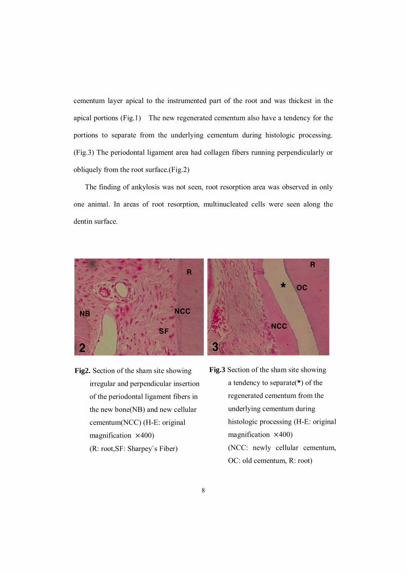
cementum layer apical to the instrumented part of the root and was thickest in the
apical portions (Fig.1) The new regenerated cementum also have a tendency for the
portions to separate from the underlying cementum during histologic processing.
(Fig.3) The periodontal ligament area had collagen fibers running perpendicularly or
obliquely from the root surface.(Fig.2)
The finding of ankylosis was not seen, root resorption area was observed in only
one animal. In areas of root resorption, multinucleated cells were seen along the
dentin surface.
Fig.3 Section of the sham site showing
a tendency to separate(*) of the
regenerated cementum from the
underlying cementum during
histologic processing (H-E: original
magnification 400)
(NCC: newly cellular cementum,
OC: old cementum, R: root)
Fig2. Section of the sham site showing
irregular and perpendicular insertion
of the periodontal ligament fibers in
the new bone(NB) and new cellular
cementum(NCC) (H-E: original
magnification 400)
(R: root,SF: Sharpey`s Fiber)
NCC
R
NB
*
NCC
R
OC
SF
2 3
9
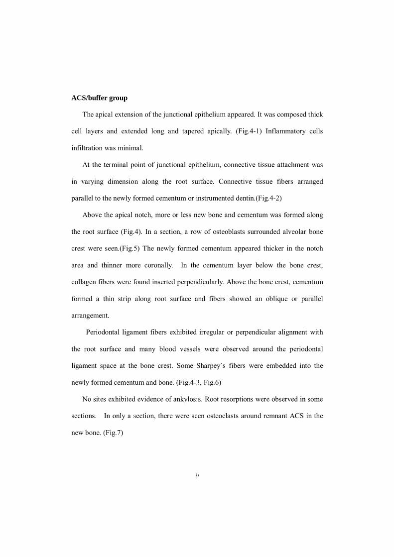
ACS/buffer group
The apical extension of the junctional epithelium appeared. It was composed thick
cell layers and extended long and tapered apically. (Fig.4-1) Inflammatory cells
infiltration was minimal.
At the terminal point of junctional epithelium, connective tissue attachment was
in varying dimension along the root surface. Connective tissue fibers arranged
parallel to the newly formed cementum or instrumented dentin.(Fig.4-2)
Above the apical notch, more or less new bone and cementum was formed along
the root surface (Fig.4). In a section, a row of osteoblasts surrounded alveolar bone
crest were seen.(Fig.5) The newly formed cementum appeared thicker in the notch
area and thinner more coronally. In the cementum layer below the bone crest,
collagen fibers were found inserted perpendicularly. Above the bone crest, cementum
formed a thin strip along root surface and fibers showed an oblique or parallel
arrangement.
Periodontal ligament fibers exhibited irregular or perpendicular alignment with
the root surface and many blood vessels were observed around the periodontal
ligament space at the bone crest. Some Sharpey`s fibers were embedded into the
newly formed cementum and bone. (Fig.4-3, Fig.6)
No sites exhibited evidence of ankylosis. Root resorptions were observed in some
sections. In only a section, there were seen osteoclasts around remnant ACS in the
new bone. (Fig.7)

10
Fig.4 Section of ACS/buffer site showing long junctional epithelium(JE), newly formed cementum(NC) and bone(NB). The new cementum is continuity with the original cementum layer and becomes gradually thinner in the coronal direction. (H-E : original magnification 20)(N: notch, BC: bone crest)
Fig.4-1 The long junctional epithelium adhered root surface. (H-E: original magnification 200)
Fig.4-2 Connective tissue contact to the root surface with a cementum formation. Collagen fibers are aligned parallel along the root surface. (H-E: original magnification 200)
Fig.4-3 Newly formed cellular cementum, alveolar bone formation may be observed, and many blood vessels are developed. (H-E: original magnification 200)
4 4-3
4-2
4-1
BC
NC
NC
CTA
JE
JE
BC
NB NC
N

11
Fig6. Section of the ACS/buffer site
showing periodontal ligament fibers
running irregular or perpendicular
(H-E: original magnification 400)
Fig7. Remnant ACS and osteoclasts were
observed.
(H-E: original magnification 400)
Fig5. Section of the ACS/buffer site
showing a row of osteoblasts and
a few osteocytes at the bone crest.
(H-E: original magnification 400)
5 6
7

12
IV. Discussion
The objective of this study was to evaluate the biocompatibility of absorbable
collagen sponge (ACS) in the 3 wall intrabony defects in the dogs. Some 4*4*4mm3 3
wall intrabony defects in the dogs implanted buffer/ACS and others received sham
surgery (open flap debridement). Clinical and histologic evaluations following 8 week
healing period showed no meaningful differences in regeneration of attachment
apparatus between defects receiving the buffer/ACS and defects receiving sham
surgery only.
In both groups, apical extension of junctional epithelium was observed. This
finding is similar to the previous histometric studies. (Choi et al.2002, Park et al.2003)
No marked differences in connective tissue adhesion and fibers arrangement
between the buffer/ACS groups and control groups are observed. At the termination
of junctional epithelium, gingival connective tissue contacts to the planed dentin or
cementum tightly and connective tissue fibers orient parallel along the root surface in
both groups.
Bone and cementum regeneration extending from above notch to coronal were
observed and no significant differences were found in both groups. Newly formed
bone was generally lined with osteoblast-like cells and osteoid, suggesting continued
bone remodeling after 8-week period. (Sigurdson et al. 1994) The regenerated
cementum appeared most cellular, usually covered with cementoblast-like cells and
cementoid materials. The cementum layer extended from notch to coronal of the

13
newly formed alveolar bone and was gradually thinner in the coronal direction.
Irregular or perpendicular arrangement of the periodontal ligament fibers in newly
formed bone and cementum was observed in both groups.
The evidence of root resorption was observed in both groups. In many previous
studies, it was explained that root resorption was more pronounced when the
connective tissue was directly opposed to dentin rather than when the root surface
was covered by cementum. (Wikesjo et al. 1994, Schroeder et al. 1992) Ankylosis
was not observed in any of the intrabony defects.
Carrier architecture is designed to used inherent regenerative capability of osseous
tissue by allowing osteoprogenitor cells to populate and mineralize in the carrier. And
it is to minimize local tissue response and allow replacement by newly formed bone
and allow for space maintenance in bony defect. Furthermore, the mechanical,
chemical, and biological properties of the scaffold should be suitable for the specific
application.
In this study, comparing the histologic observation of new attachment apparatus
formation in buffer/ACS groups and sham surgery groups, biocompatibility of
collagen as carrier system was evaluated. Histologic healing appearances of the both
groups are similar and the amount of regenerated tissues were not significantly
different by previous studies. (Park et al.2003, Choi et al.1993, Kim et al.1998)
Remnants of the collagen and signs of inflammation could not almost be seen at 8
week period.
Collagen may be resorbed without beneficial or harmful effect on the periodontal

14
healing. This property may make absorbable collagen sponge a suitable carrier for
study to evaluate osteogenic effect of a variety of growth factors.

15
V. CONCLUSION
Absorbable collagen sponge (ACS) has been shown favorable, biocompatible
tissue response and relatively rapid biodegradation. So, it may be used a suitable
carrier to study the effect of growth factors on the periodontal regeneration of the
contained defects.

16
REFERENCES
1. Pakr J-S, Choi S-H, Moon I-S, Cho K-S, Kim C-K(2003) Eight-week histological
analysis on the effect of chitosan on surgically created one-wall intrabony defects in
beagle dogs. J Clin periodontol 30, 443-453
2. Seong-Ho choi, Chong-Kwan Kim, Kyoo-Sung Cho(2002) Effect of recombinant
human bone morphogenic protein-2/ACS(rhBMP-2/ACS) on healing in 3-wall
intrabony defects in dogs. J Periodontol 73,63-72
3. William H.Hiatt, Robert G. Schallhorn, Albert James Aaronian(1978) The
Induction of new bone and cementum formation. J Periodontol Oct 495-512
4. Bartold P.M, Xiao Y, Lyngstaadas SP, Paine ML, Snead ML. Principles and
applications of cell delivery systems for periodontal regeneration. Periodontol 2000.
2006;41:123-135.
5. Lee CH, SinglaA, Lee Y. Biomedical applications of collagen. Int J Pharm
2001:221;1-22.
6. Bartold PM, McCulloch CA, Narayanan AS, Pitaru S. Tissue engineering: a new
paradigm for periodontal regeneration based on molecular and cell biology.
Periodontol 2000 2000;24:253-269.
7. Garrett S. Periodontal regeneration around natural teeth. Ann Periodontol
1996;1:621-666.

17
8. Boo JS, Yamada Y, Okazaki Y, Hibino Y, Okada K, Hata KI, Yoshikawa T, and a
biodegradable scaffold. J Craniofac Surg 2002;13:231-239.
9. Ronald E. Jung, Daniel S. Thoma and Christopf H. F. Hammerle. Assessment of
the potential of growth factors for localized alveolar ridge augmentation: a systemic
review. J Clin Periodontol 2008;35(Suppl.8):255-281
10. Boyne PJ, Marx RE, Nevins M., Triplett G.,Lazaro E., Lily LC., Alder M.,
Nummikoski P. A feasibility study evaluating rhBMP-2/absorbable collagen sponge
for maxillary sinus floor augmentation. Int J Periodontics Restorative Dent
1997;17(1):11-25
11. Cole RT, Crigger M., Bogle G., Egelberg J., Selvig KA: Connective tissue
regeneration to periodontally diseased teeth. J Periodont Res 1980;15:1
12. Fujimura K, Bessho K, Kusumoto K, Ogawa Y, Iizuka T. Experimental studies on
bone inducing activity of composites of atelopeptide type collagen as a carrier for
ectopic osteoinduction by rhBMP-2. Biochem Biophys Res Commun 1995;208:316-
322
13. Bessho K, Kusumoto K, Fujimura K, Konishi Y, Ogawa Y, Tani Y, Iizuka T.
Comparison of recombinant human bone morphogenetic protein. Br J Oral
Maxillofac Surg 1999;37:2-5
14. Isobe M, Yamazali Y, Oida S, Ishihara K, Nakabayashi N, Amagasa T. Bone
morphogenetic protein encapsulated with a biodegradable and biocompatible polymer.
J Biomed Mater Res 1996;32:433-438.
15. Bessho K, Carnes D.L, Ong J. L. Experimental studies on bone induction using

18
low-molecular-weight poly(DL-lactide-co-glycolide) as a carrier for recombinant
human bone morphogenetic protein-2. J Biomed Mater Res 2002;61:61-65
16. Holland, S.J., Tighe, B.J. & Gould, P.L.(1986) Polymers for biodegradable
medical device, I. The potential of polyesters as controlled macromolecular release
system. Journal of Controlled Release 4;155-180
17. Taba M Jr, Jin Q, Sugai JV, Giannobile WV. Current concepts in periodontal
bioengineering. Orthop Craniofacial Res. 2005;8:292-302
18. Zhao M, Jin Q, Berry JE, Nociti FH Jr, Giannobile WV, Somerman MJ.
Cementoblast delivery for periodontal tissue engineering. J Periodontol 2004;75:154-
61.
19. Giannobile W.V. Periodontal tissue engineering by growth factors. Bone
1996;19(1 Suppl): 235-375.
20. Ripamonti U, Reddi AH. Periodontal regeneration: potential role of bone
morphogenetic proteins. J Periodont Res 1994;29:225-235.
21. Lee CH, SinglaA, Lee Y. Biomedical applications of collagen. Int J Pharm
2001:221;1-22.
22. McPherson JM. The utility of collagen-based vehicles in delivery of growth
factors for hard and soft tissue wound repair. Clin Mater 1992;9:225-234.
23. Uludag H, Gao T, Porter TJ, Friess W, Wozney JM. Delivery systems for BMPs:
factors contributing to protein retention at an application site. J Bone Joint Surg Am
2001;83-A Suppl 1:S128-135.

19
24. Kim CS, Choi SH, Cho KS, Chai JK, Wikesjo UM, Kim CK. Periodontal healing
in one-wall intra-bony defects in dogs following implantation of autogenous bone or a
coral-derived biomaterial. J Clin Periodontol 2005;32:583-589.
25. Kim HY, Kim CS, Jhon GJ, et al. The effect of safflower seed extract on
periodontal healing of 1-wall intrabony defects in beagle dogs. J Periodontol
2002;73:1457-1466.
26. Whang K, Tsai DC, Nam EK et al. Ectopic bone formation via rhBMP-2 delivery
from porous bioabsorbable polymer scaffolds. J Biomed Mater Res 1998;42:491-499.
27. Fournier N, Doillon CJ. Biomaterial molecule-impregnated polyester: an in vivo
angiogenesis study. Biomaterials 1996;17:1659-1665.
28. Nilsson M, Fernandez E, Sarda S, Lidgren L, Planell JA. Characterization of a
novel calcium phosphate/sulphate bone cement. J Biomed Mater Res 2002;61(4):600-
607.
29. Kotani S, Fujita Y, Kitsugi T, Nakamura T, Yamamuro T, Ohtsuki C, KoKubo T.
Bone bonding mechanism of β-tricalcium phosphate. J Biomed Mater Res
1991;25:1303-1315.
30. Wiltfang J. Merten HA, Schlegel KA, Schultze-Mosgau S, Kloss FR, Rupprecht S,
Kessler P. Degradation characteristics of α and β tri-calcium-phosphate(TCP) in
minipigs. J Biomed Mater Res Appl Biomater 2002;63:115-121.
31. Klein CP, Dreissen AA, de Groot K, van den Hooff A. Biodegradation behavior of
various calcium phosphate materials in bone tissue. J Biomed Mater Res
1983;17:769-784.

20
32. Boden SD, Martin GJ, Mortone MA, Ugbo JL, Moskovitz PA. Posterolateral
lumbar intertransverse process spine arthrodesis with recombinant human bone
morphogenetic protein 2/hydroxyapatite-tricalcium phosphate after laminectomy in
the nonhuman primate. Spine 1999;24:1179-1185.
33. Frayssinet P, Trouillet JL, Rouquet N, Azimus E, Autefage A. Osseointegration of
macroporous calcium phosphate ceramics having a different chemical composition.
Biomaterials 1993;14:423-429.
34. Arnold U, Lindenhayn K, Perka C. In vitro-cultivatiob of human periosteum derived
cells in bioresorbable poymer-TCP-composites. Biometreials 2002;23:2303-2310.
35. Boo JS, Yamada Y, Okazaki Y, Hibino Y, Okada K, Hata KI, Yoshikawa T,
Sugiura Y, Ueda M. Tissue-engineered bone using mesenchymal stem cells and a
biodegradable scaffold. J Craniofac Surg 2002;13:231-239.
36. Gera I, Dori F, Keglevich T, Anton S, Szilagyi E, Windisch P. Experience with the
clinical use of b-Tricalcium phosphate(Cerasorb) as a bone replacement graft material
in human periodontal osseous defects. Fogorv Sz 2002;95:143-147.
37. Wikesjo UME, Nilveus RE. Periodontal repair in dogs: Healing patterns in large
circumferential periodontal defects. J Clin Periodontol 1991;81:49-59
38. Wikesjo UME, Nilveus RE. Periodontal repair in dogs: Effect of wound
stabilization on healing. J Periodontol 1990;61:719-724
39. Sigurdsson TJ, Lee MB, Kubota K, Turek TJ, Wozney JM, Wikesjo UME.
Periodontal repair in dogs: Recombinant human bone morphogenetic protein-2
significantly enhances periodontal regeneration. J Periodontol 1995;66:131-138

21
국문요약
삼벽성 골내낭에서 흡수성 콜라겐 스폰지의
흡수정도와 조직반응에 대한 조직학적 연구
<지도교수 김 종 관>
연세대학교 대학원 치의학과
이 상 민
목적: 흡수선 콜라겐 스폰지는 생체 적합성, 생체 흡수성, 낮은 알러지
반응으로 인해 조직 공학에서 carriers로서 널리 이용되고 있다. 그것의
생체 적합성을 평가하기 위해 이 연구는 3벽성 골내낭에서 흡수성 콜라겐
을 매식한 부위와 외과적 처치만을 한 부분의 치주조직 치유에 대한 8주
조직학적 결과를 비교해 보고자 한다.
방법: 8마리의 성견에서 양측성으로 상하악 소구치 부위에 삼벽성 골내
낭을 형성했다. 대조군으로는 외과적 처치만을 시행했으며 실험군으로는
콜라겐 스폰지를 외과적 처치와 함께 매식하였다. 8주 후 성견을 희생하여
조직학적 비교를 시행했다.

22
결과: 두 그룹간의 조직학적 차이는 거의 없었다. 임상적으로 부작용 없
이 치유가 일어났으며 8주 조직시편에서 흡수성 콜라겐 스폰지는 거의 흡
수가 일어났다. 유착이 일어난 곳은 없었고 치근 흡수를 보이는 시편도 거
의 없었다.
결론: 흡수성 콜라겐 스폰지는 양호하고 조직 친화적 반응을 가지며 비
교적 빠른 흡수가 일어난다. 따라서 3벽성 골내낭 처치시 안전한 carriers
로서 이용될 수 있을 것이다.
핵심이 되는 말: 운반체, 흡수성 콜라겐 스폰지, 삼벽성 골내낭