The Hexameric Structures of Human Heat Shock Protein · The Hexameric Structures of Human Heat...
14
The Hexameric Structures of Human Heat Shock Protein 90 Cheng-Chung Lee 1,2 , Ta-Wei Lin 1 , Tzu-Ping Ko 1,2 , Andrew H.-J. Wang 1,2 * 1 Institute of Biological Chemistry, Academia Sinica, Taipei, Taiwan, 2 Core Facility for Protein Production and X-ray Structural Analysis, Academia Sinica, Taipei, Taiwan Abstract Background: The human 90-kDa heat shock protein (HSP90) functions as a dimeric molecular chaperone. HSP90 identified on the cell surface has been found to play a crucial role in cancer invasion and metastasis, and has become a validated anti- cancer target for drug development. It has been shown to self-assemble into oligomers upon heat shock or divalent cations treatment, but the functional role of the oligomeric states in the chaperone cycle is not fully understood. Principal Findings: Here we report the crystal structure of a truncated HSP90 that contains the middle segment and the carboxy-terminal domain, termed MC-HSP90. The structure reveals an architecture with triangular bipyramid geometry, in which the building block of the hexameric assembly is a dimer. In solution, MC-HSP90 exists in three major oligomer states, namely dimer, tetramer and hexamer, which were elucidated by size exclusion chromatography and analytical ultracentrifugation. The newly discovered HSP90 isoform HSP90N that lacks the N-terminal ATPase domain also exhibited similar oligomerization states as did MC-HSP90. Conclusions: While lacking the ATPase domain, both MC-HSP90 and HSP90N can self-assemble into a hexameric structure, spontaneously. The crystal structure of MC-HSP90 reveals that, in addition to the C-terminal dimerization domain, the residue W320 in the M domain plays a critical role in its oligomerization. This study not only demonstrates how the human MC-HSP90 forms a hexamer, but also justifies the similar formation of HSP90N by using 3D modeling analysis. Citation: Lee C-C, Lin T-W, Ko T-P, Wang AH-J (2011) The Hexameric Structures of Human Heat Shock Protein 90. PLoS ONE 6(5): e19961. doi:10.1371/ journal.pone.0019961 Editor: Andreas Hofmann, Griffith University, Australia Received January 6, 2011; Accepted April 16, 2011; Published May 25, 2011 Copyright: ß 2011 Lee et al. This is an open-access article distributed under the terms of the Creative Commons Attribution License, which permits unrestricted use, distribution, and reproduction in any medium, provided the original author and source are credited. Funding: This work was supported by grants from the National Science Council of Taiwan (NSC97-3112-B-001-035-B4 to A.H.-J.W.). The funder had no role in study design, data collection and analysis, decision to publish, or preparation of the manuscript. Competing Interests: The authors have declared that no competing interests exist. * E-mail: [email protected] Introduction Heat shock protein 90 (HSP90) is an ATPase-dependent chaperone and the molecular chaperone functions as a dimer. HSP90 is responsible for managing protein folding and quality control in the crowded environment inside the cell. It participates in activating and stabilizing more than 200 ‘‘client’’ proteins involved in post-translational folding, protein stability, activation and maturation of cellular proteins, which are essential to cell- cycle control and signaling. HSP90, HSP70 and co-chaperones form a dynamic complex known as the HSP90 dynamic machine [1], which is regulated by co-chaperones and post-translational modification, e.g., phosphorylation, nitrosylation and acetylation for client protein interaction and ATPase activity. The yeast HSP90-Sba1 complex structure provides a view of HSP90 in the ATP-bound state, demonstrates the conformational changes in the N-terminal domain and reveals how the co-chaperone Sba1 recognizes the ‘‘closed’’ state of HSP90 dimer, that confirms the ATPase-coupled molecular clamp mechanism of HSP90 chaper- one [2–3]. Many oncoproteins are HSP90 client proteins, including EGFR, AKT, MMP2 and BCR-ABL. They depend on its protein folding machinery to avoid misfolding and degradation in cancer cells. HSP90 inhibition offers a great promise in the treatment of a wide variety of solid and haematological malignancies. Therefore, HSP90 has been a target for anticancer drugs, and several classes of compounds have been and are being developed to modulate its activity for therapeutic benefit [4–5]. The HSP90 proteins are highly conserved and five human isoforms have been identified, including two cytosolic isoforms HSP90a and HSP90b, a glucose-regulated protein (GRP94) in the endoplasmic reticulum, a tumor necrosis factor receptor-associated protein 1 (Hsp75/TRAP1) in the mitochondrial matrix, and a newly discovered isoform HSP90N [6]. These isoforms exhibit different domain structure and cellular location, and may have different client protein substrates [4,7]. Recent studies also indicate that many types of cells express HSP90 on the cell surface and secrete HSP90 into the extracellular space to carry out important extracellular functions [1,8]. The conserved HSP90 structure consists of three domains: an N-terminal (N) domain that contains the co-chaperone binding motif and an ATP and drug binding site that binds the natural compounds geldanamycin and radicicol; a middle (M) domain that is responsible for binding to co-chaperone and client proteins; and a C-terminal (C) domain that contains a dimerization motif, a second drug-binding site, and a conserved MEEVD pentapeptide at the C-terminus, which is recognized by the co-chaperone HSP70/HSP90 organizing protein (Hop) [9]. This C domain was predicted to contain a second nucleotide-binding site, which has PLoS ONE | www.plosone.org 1 May 2011 | Volume 6 | Issue 5 | e19961
Transcript of The Hexameric Structures of Human Heat Shock Protein · The Hexameric Structures of Human Heat...
The Hexameric Structures of Human Heat Shock Protein90Cheng-Chung Lee1,2, Ta-Wei Lin1, Tzu-Ping Ko1,2, Andrew H.-J. Wang1,2*
1 Institute of Biological Chemistry, Academia Sinica, Taipei, Taiwan, 2 Core Facility for Protein Production and X-ray Structural Analysis, Academia Sinica, Taipei, Taiwan
Abstract
Background: The human 90-kDa heat shock protein (HSP90) functions as a dimeric molecular chaperone. HSP90 identifiedon the cell surface has been found to play a crucial role in cancer invasion and metastasis, and has become a validated anti-cancer target for drug development. It has been shown to self-assemble into oligomers upon heat shock or divalent cationstreatment, but the functional role of the oligomeric states in the chaperone cycle is not fully understood.
Principal Findings: Here we report the crystal structure of a truncated HSP90 that contains the middle segment and thecarboxy-terminal domain, termed MC-HSP90. The structure reveals an architecture with triangular bipyramid geometry, inwhich the building block of the hexameric assembly is a dimer. In solution, MC-HSP90 exists in three major oligomer states,namely dimer, tetramer and hexamer, which were elucidated by size exclusion chromatography and analyticalultracentrifugation. The newly discovered HSP90 isoform HSP90N that lacks the N-terminal ATPase domain also exhibitedsimilar oligomerization states as did MC-HSP90.
Conclusions: While lacking the ATPase domain, both MC-HSP90 and HSP90N can self-assemble into a hexameric structure,spontaneously. The crystal structure of MC-HSP90 reveals that, in addition to the C-terminal dimerization domain, theresidue W320 in the M domain plays a critical role in its oligomerization. This study not only demonstrates how the humanMC-HSP90 forms a hexamer, but also justifies the similar formation of HSP90N by using 3D modeling analysis.
Citation: Lee C-C, Lin T-W, Ko T-P, Wang AH-J (2011) The Hexameric Structures of Human Heat Shock Protein 90. PLoS ONE 6(5): e19961. doi:10.1371/journal.pone.0019961
Editor: Andreas Hofmann, Griffith University, Australia
Received January 6, 2011; Accepted April 16, 2011; Published May 25, 2011
Copyright: � 2011 Lee et al. This is an open-access article distributed under the terms of the Creative Commons Attribution License, which permits unrestricteduse, distribution, and reproduction in any medium, provided the original author and source are credited.
Funding: This work was supported by grants from the National Science Council of Taiwan (NSC97-3112-B-001-035-B4 to A.H.-J.W.). The funder had no role instudy design, data collection and analysis, decision to publish, or preparation of the manuscript.
Competing Interests: The authors have declared that no competing interests exist.
* E-mail: [email protected]
Introduction
Heat shock protein 90 (HSP90) is an ATPase-dependent
chaperone and the molecular chaperone functions as a dimer.
HSP90 is responsible for managing protein folding and quality
control in the crowded environment inside the cell. It participates
in activating and stabilizing more than 200 ‘‘client’’ proteins
involved in post-translational folding, protein stability, activation
and maturation of cellular proteins, which are essential to cell-
cycle control and signaling. HSP90, HSP70 and co-chaperones
form a dynamic complex known as the HSP90 dynamic machine
[1], which is regulated by co-chaperones and post-translational
modification, e.g., phosphorylation, nitrosylation and acetylation
for client protein interaction and ATPase activity. The yeast
HSP90-Sba1 complex structure provides a view of HSP90 in the
ATP-bound state, demonstrates the conformational changes in the
N-terminal domain and reveals how the co-chaperone Sba1
recognizes the ‘‘closed’’ state of HSP90 dimer, that confirms the
ATPase-coupled molecular clamp mechanism of HSP90 chaper-
one [2–3]. Many oncoproteins are HSP90 client proteins,
including EGFR, AKT, MMP2 and BCR-ABL. They depend
on its protein folding machinery to avoid misfolding and
degradation in cancer cells. HSP90 inhibition offers a great
promise in the treatment of a wide variety of solid and
haematological malignancies. Therefore, HSP90 has been a target
for anticancer drugs, and several classes of compounds have been
and are being developed to modulate its activity for therapeutic
benefit [4–5].
The HSP90 proteins are highly conserved and five human
isoforms have been identified, including two cytosolic isoforms
HSP90a and HSP90b, a glucose-regulated protein (GRP94) in the
endoplasmic reticulum, a tumor necrosis factor receptor-associated
protein 1 (Hsp75/TRAP1) in the mitochondrial matrix, and a
newly discovered isoform HSP90N [6]. These isoforms exhibit
different domain structure and cellular location, and may have
different client protein substrates [4,7]. Recent studies also indicate
that many types of cells express HSP90 on the cell surface and
secrete HSP90 into the extracellular space to carry out important
extracellular functions [1,8].
The conserved HSP90 structure consists of three domains: an
N-terminal (N) domain that contains the co-chaperone binding
motif and an ATP and drug binding site that binds the natural
compounds geldanamycin and radicicol; a middle (M) domain that
is responsible for binding to co-chaperone and client proteins; and
a C-terminal (C) domain that contains a dimerization motif, a
second drug-binding site, and a conserved MEEVD pentapeptide
at the C-terminus, which is recognized by the co-chaperone
HSP70/HSP90 organizing protein (Hop) [9]. This C domain was
predicted to contain a second nucleotide-binding site, which has
PLoS ONE | www.plosone.org 1 May 2011 | Volume 6 | Issue 5 | e19961

been shown to bind to novobiocin, epilgallocatechin (ECGC) and
taxol [10]. However, neither the apo-form crystal structure nor
any complex structure has been reported for the M and C domains
of human HSP90. The isoform HSP90N is a plasma-membrane-
associated protein in poorly-differentiated colorectal cancers with
metastasis [11]. Specifically, HSP90N lacks the N-terminal
nucleotide-binding domain which is replaced by a 30-residue-
long hydrophobic motif, but it shares the 509 amino acids in the
C-terminal region with HSP90a [6,12–13].
HSP90 exists as a homodimer, and the dimers tend to associate
into tetramers, hexamers, and even higher oligomers. It has been
reported that the oligomeric forms of HSP90 are present in the
cytosol of mammalian cells [14]. The heat-induced HSP90
oligomer can bind to its substrates and prevent their irreversible
aggregation, and the self-oligomerization of HSP90 may have a
pivotal role in protecting cells from thermal damages by its
chaperone function [15–16]. HSP90 oligomerization can be
induced by heat treatment, the presence of nonionic detergents
or addition of divalent cations, such as magnesium and calcium.
The heat-induced oligomerization can be inhibited by ATP and
geldanamycin, both bind to the same pocket in the N-terminal
domain [15,17–18]. HSP90 association into tetramer, hexamer
and dodecamer forms, as induced by Mg2+ ion, demonstrated
that the building block for its oligomerization is a dimer. In
particular, the hexamer with a cozy nest-shape was obtained by
negative staining TEM tomography [18]. In addition, the divalent
cation-induced oligomerization is accompanied by the instan-
taneous loss of its molecular chaperone function [19]. The
functional roles of HSP90 oligomers in the chaperone cycle are
not clearly understood, therefore the oligomerization process and
the oligomeric structures are studied here. In this study, we
determined the crystal structure of a human HSP90 that contains
the M and C domains (MC-HSP90) in a hexameric assembly.
The oligomerization sites are located in the M domain and also
involve the dimerizing C domain. The hydrodynamic properties of
MC-HSP90 and HSP90N in solution were also examined. The
results clearly showed that MC-HSP90 exists in three major states:
dimer, tetramer and hexamer. HSP90N also showed a dimeric
and two higher oligomer states as did MC-HSP90 under similar
conditions. The hexameric structure of MC-HSP90 provides a
clue to how the possible oligomers of HSP90N establish their
functional architecture.
Results
Crystals and Structures of MC-HSP90The N-terminal truncated HSP90 (MC-HSP90, residues 293–
732) containing the middle segment and carboxy-terminal domain
was crystallized in three different forms, C2221, P21 and R32. A
fourth crystal form of P6322 was obtained using a modified MC-
HSP90-Ct (residues 293–697) in which the C-terminal 35 residues
were also truncated. The crystals were obtained using different
protein preparations or under different crystallization conditions.
For the C2221 and P21 crystals, obtained under similar conditions,
the protein solution was pre-incubated with cis-dichloro(ethylene-
diamine)platinum(II) (cis-DEP) and cisplatin, respectively, before
crystallization setup. The R32 crystals were obtained by using an
additive solution in the crystallization drops. The P6322 crystals
were grown under low pH conditions, also with an additive. (See
Materials and Methods for details.) All crystal structures were
determined by molecular replacement (MR) method, but only the
C2221 and P21 crystals were refined to 3.0 A and 3.05 A
resolutions, respectively (Table 1), with three and six molecules
of MC-HSP90 per asymmetric unit. The R32 and P6322 crystals
have one molecule per asymmetric unit. The protein molecules in
all crystal forms show a hexameric architecture with triangular
bipyramid geometry in the protein assembly, and in the latter two
forms (R32 and P6322) the molecular triad and dyads are further
expressed as crystallographic symmetry elements (Figure 1). If the
protein solutions were not pre-incubated with the platinum
compounds before the crystallization drop setup or the additive
solution were included only in the drops, the protein could still be
crystallized but with poor crystal quality; those crystals only
diffracted to about 6 A resolution and the structure could not be
determined.
The refined models of MC-HSP90 in the two different space
groups of C2221 and P21 contain nine independent polypeptide
chains in total. The N-termini start at residues 294–297, and the
C-termini stop at residues 697–699, depending on the corre-
sponding electron densities for the individual subunits. The residue
293 and the last 33 residues, plus the His-tag, were not observed,
probably because they were highly flexible. Other disordered
regions were located in residues 349–357, 394–404 and 611–629.
Three molecules had an additional disordered region in residues
558–613, which also showed high temperature factors in the other
molecules. In an attempt to obtain better crystals, we constructed a
C-terminal tail truncated MC-HSP90-Ct, but the resulting P6322
crystals did not show any improvement in the diffraction
resolution. The R32 crystal showed additional electron densities
for most of these regions, but they could not be modeled with
certainty due to the poor diffraction resolution. These two
structures could only be refined to 3.5 A resolution, yielding R
values of about 30% and Rfree of about 35%.
In the current structure, each molecule folds into three major
domains (Figure 2A): a large middle (LM) domain (residues 293–
469), a small middle (SM) domain (residues 470–547), and the C-
terminal (C) domain (residues 548–732). The structure of the LM
domain starts with a 310-helix, which is followed by a three-layer
architecture of a-b-a sandwich and a helical coil. It encompasses
two disordered loops comprising residues 351–358 and 396–404.
The SM domain is also folded into an a-b-a sandwich
architecture, and contains a sulfate-binding site. The sulfate was
hydrogen bonded to the side chains of R510, K513 and H514
within helix a7, which is close to the C domain (Figure 2A). The C
domain begins with an amphipathic loop, which shows high B-
values (Figure 3A), and contains a curved a-helix, a three-stranded
b-sheet, a three-helix coil and an extended disordered arm
between helix a10 and strand b11 (residues 617–629). Beyond
residue 697, the structure is disordered.
Structure-based Sequence AlignmentTwo determined 90-kDa chaperone structures, yeast HSP82
and canine GRP94 [3,20] were chosen for comparison with the
middle and C-terminal domains of human HSP90. Sequence
alignment shows that human MC-HSP90 and yeast MC-HSP82
have about 60% identity in 438 residues. The core domain and
loops regions are highly conserved, but the C-terminal extension is
variable (Figure 4B). The MC-HSP90 monomer superimposes
well on the MC domain of yeast HSP82 (PDB: 2CG9) and yields
an rmsd of 0.91 A for 219 Ca atoms. Human MC-HSP90 and
canine GRP94 (PDB: 2O1V) have only about 49% identity in 414
residues of the MC domain. When the domain structures are
superimposed, the rmsd value is 1.02 A for 241 Ca atoms. Both
yeast HSP82 and canine GRP94 show structural similarity to the
human HSP90 in the MC domain (Figure 3B).
In the absence of the N-terminal ATPase domain, co-chaperone
and client proteins, MC-HSP90 structure contains several
disordered regions, which correspond to four functional loops:
Structure of Human HSP90
PLoS ONE | www.plosone.org 2 May 2011 | Volume 6 | Issue 5 | e19961

an amphipathic loop (residues 347–360), a middle segment
catalytic loop (residues 395–406), an extended loop (residues
616–630) and the C-terminal tail (residues 698–732). To confirm
the presence of disordered loops and C-terminal tail in the crystal,
the C2221 crystal was harvested and dissolved for in-gel trypsin
digestion and MALDI-MS analysis. In this experiment, the N and
C-terminal tail segments and the residues comprising the
amphipathic loop, as well as the extended loop, can all be
identified in the MASS spectra. However, in the catalytic loop,
only the residues 395–400 could be identified (Figure 4B) by our
experimental procedure. In the yeast HSP82/Sba1 complex
structure, the equivalent regions of these disordered segments
were not observed either, except for the middle segment catalytic
loop, because it contained the catalytic residue R380 (equivalent to
R400 of human HSP90) bound to a nucleotide, which stabilized
the loop [3].
Structural Features and ComparisonIt has been proposed that HSP90 functions as a dimer, and the
C-terminal domain is involved in the major dimeric interactions
[21]. The previously determined 90-kDa chaperone structures also
supported this [3,20]. Here, in the truncated MC-HSP90
structure, the protein subunits are arranged into a parallel
homodimer with a left-handed helical twist. Each protomer
extends from its C-terminal helix coil at the central axis of the
dimer to form a ‘‘Twisted V’’ shape, which corresponds to the
‘‘open’’ state of HSP90 structure. In the open end, the distance is
about 30 A between the Ca atoms of N373 from the two
protomers, and it is about 70 A between the two N-terminal
residues K294 (Figure 2B). Helices a12 and a13 in the C-terminal
domain of two dyad symmetry related protomers are folded into a
four-helix bundle. The interaction interface consists of both
hydrophobic and hydrophilic contacts, which is the most
Table 1. Data collection and refinement statisticsa.
Additive compounds cis-DEP cisplatin
Data collection
Wavelength (A) 1.0 1.0
Space group C2221 P21
Cell dimensions (A, u) a = 162.70, b = 304.55, c = 87.62, a = 157.90, b = 90.87, c = 167.09,
b = 115.85
Resolution (A) 30-3.0 (3.11-3.0) 30-3.05 (3.16-3.05)
Observed reflections 186,922 291,396
Unique reflections 43,634 82,281
Rsym (%) 5.8 (45.1) 5.8 (36.2)
I/s(I) 17.9 (3.5) 19.6 (5.1)
Completeness (%) 99.6 (99.3) 98.8 (99.4)
Redundancy 4.3 (4.2) 3.5 (3.5)
Z 3 6
Refinement
Resolution (A) 30-3.0 30-3.05
No. of reflections Rwork/Rfree 39,180/2,185 72,417/4,017
Rwork/Rfree (%) 20.9/25.2 20.4/25.9
No. of atoms
Protein 9,197 17,827
SO4 15 30
Water 133 281
Avg B factor (A2)
Protein 90.7 82.3
SO4 138.6 124.3
Water 92.0 85.9
RMSD
Bond lengths (A) 0.012 0.012
Bond angles (u) 1.405 1.399
Ramachandran statistics (%)b
Most favored 88.6 89.9
Additionally allowed 9.6 9.2
Generously allowed 1.5 0.5
Disallowed 0.4 0.4
aValues corresponding to the highest resolution shell are shown in parentheses.bThe stereochemistry of models were validated with PROCHECK.doi:10.1371/journal.pone.0019961.t001
Structure of Human HSP90
PLoS ONE | www.plosone.org 3 May 2011 | Volume 6 | Issue 5 | e19961

important stabilizing element for HSP90 dimerization. A surface
area of about 1,267 A2 on each protomer is buried by its counter
protomer upon dimer formation (Figure 2C). Furthermore, when
the dimers are superimposed for the three-helix coil of C domain
(Figure 3C), the MC-HSP90 dimer shows an open-form in
contrast to the close-form yeast HSP82, differing from each other
by a large sway of the protomers. The loop region between the SM
and C domain, the ‘‘curved helix’’ (a9) and the extended loop
include helix a10 have large conformational changes, which result
in the domain shift of M domain. The flexible conformation of the
curved helix a9 and the connection loop between M and C
domain allow the middle domain to have a large domain shift for
the alternation between open and closed conformations of the
HSP90 clamp.
Quaternary Structure of MC-HSP90The P21 crystal has three open-form MC-HSP90 dimers in an
asymmetric unit, which assemble into a triangular bipyramid
architecture being arranged in a right-handed fashion around a
pseudo 3-fold axis (Figure 2C and D). The other crystals, the
C2221 and R32 crystals of MC-HSP90 and the P6322 crystal of
MC-HSP90-Ct have fewer than six molecules in an asymmetric
unit, but they also contain this kind of hexameric assembly in the
crystals, suggesting that the hexamer is not just a result of
stabilization by favourable crystal lattice contacts. A top view of
the hexameric structure shows a trefoil-like shape, with the N-
terminal end of the monomer facing up and the C-terminal end
directing radially away from the center. The top view shows a
compact arrangement of the three monomers, which belong to
three different dimers. The contact between the monomers around
the 3-fold axis is facilitated by the N-terminal part of LM domain.
One subunit is in contact with both 3-fold symmetry-related
subunits via the inter-dimer interface, that buries about 531 A2
surface area on a monomer for the hexamerization, the major
contact region is shown in Figure 5. The side chain of residues
W320 and R346 pack against a concave surface of the
neighbouring protomer (Figure 5A). The W320 side chain is
accommodated in a hydrophobic pocket formed by K294, P295
and W297, and also forms a hydrogen bond to the main chain
oxygen atom of R367 of the neighboring protomer. The side chain
of R346 is hydrogen bonded to the main chain oxygen atom of
R366 (Figure 5B). This interaction positions the LM domains
about the center of the complex, with N-terminnal end protruding
around the central 3-fold axis, three facing up and three facing
down. Viewed from the side, the hexameric structure appears to
have an elliptic shape. The middle domain is packed against one
another in the central core, and the C-terminal domain also
contacts one another at the rim.
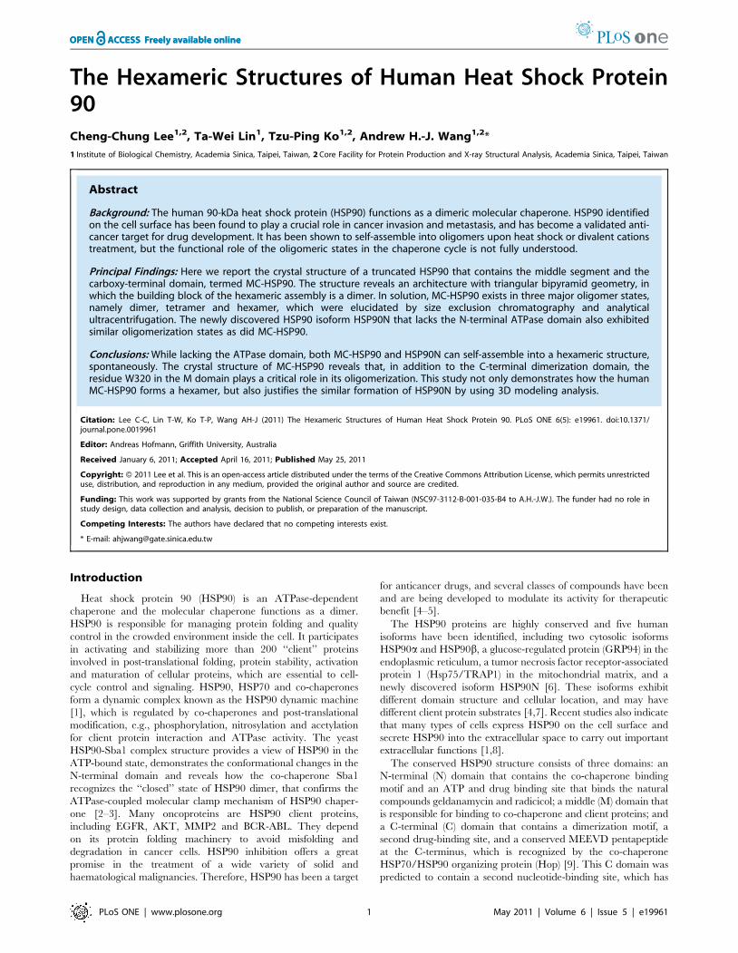
Oligomer States of MC-HSP90 in SolutionIn order to understand the oligomeric states of MC-HSP90 in
solution, size-exclusion chromatography and sedimentation veloc-
ity (SV) experiments were carried out to determine their molecular
size. The elution profile of size-exclusion chromatography for MC-
HSP90 shows two major peaks between the protein marker
thyroglobulin (669 kDa) and aldolase (158 kDa). It suggests that
MC-HSP90 contains at least two oligomeric states (Figure 6A). No
corresponding peak for the monomer was observed, indicating
that the monomeric species was not present or not detectable
under our experimental conditions. Based on the elution profile,
the two peaks were calculated to have molecular masses of about
252 and 408 kDa corresponding to approximately five and eight
subunits, respectively. However, MC-HSP90 is not a spherically
shaped globular protein and it is difficult to predict the molecular
Figure 1. Crystal packing of MC-HSP90 molceules. The picturesof the (A) orthorhombic, (B) monoclinic, (C) rhombohedral, and (D)hexagonal crystals are shown on the left side. The unit-cell contents ofthe corresponding crystals are shown on the right. All of these crystalscontain structurally similar hexamers, which are shown in three differentcolors and have a variety of arrangements in the unit cells. Although thegreen models appear to be disintegrated, they indeed form intacthexamers when associated with their symmetry-related mates from theneighboring unit cells. Only the orthorhombic and the hexagonalcrystals (A and D) show some resemblance in packing, but they containproteins with different C-termini.doi:10.1371/journal.pone.0019961.g001
Structure of Human HSP90
PLoS ONE | www.plosone.org 4 May 2011 | Volume 6 | Issue 5 | e19961

mass of its oligomeric states by size exclusion chromatography,
especially when it contains a flexible C-terminal tail. To obtain
further information of the oligomer state, the peak fractions and
those in the overlap region eluted from the size exclusion
chromatography were analyzed separately by analytical ultracen-
trifugation. Surprisingly, the continuous c(s) distribution suggests
that MC-HSP90 exists as three species in solution with
sedimentation coefficients of 5.3S, 8.3S and 11.6S (Figure 6B),
with a frictional ratio of 1.3. The calculated masses of 94.4 kDa,
189.6 kDa and 299.1 kDa, corresponding to dimer, tetramer and
hexamer, correlate very well with the actual molecular weights.
Based on the observed 3D structure of MC-HSP90 determined
in the P21 crystal, the sedimentation coefficient values of MC-
HSP90 at dimer and each oligomer states were calculated by using
Figure 2. Overall structure of MC-HSP90. (A) Ribbon representation of the MC-HSP90 monomer with a bound sulfate. The large middle (LM),small middle (SM) and C-terminal domains (C) are indicated. The inset figure shows a sulfate bound to R510, K513 and H514. (B) Parallel homodimerof MC-HSP90. Two protomers (blue and orange) extend from its C-terminal dimerization domain with ‘‘Twisted V’’ shape and are shown in ribbon andtranslucent surface. The inner and outer distances between the open ends are indicated. (C) (D) Top view and side view of the hexameric MC-HSP90structure. Six MC-HSP90 molecules assemble into a dextral hexamer. The N-termini are located on the top and bottom of the hexamer with 3-foldsymmetry. Each protomer is colored in separate colors (blue, green and orange) and half of hexamer is shown with translucent surface.doi:10.1371/journal.pone.0019961.g002
Structure of Human HSP90
PLoS ONE | www.plosone.org 5 May 2011 | Volume 6 | Issue 5 | e19961

the computer program HYDROPRO [22], and compared with
the experimental values from the sedimentation velocity experi-
ments. As shown in Figure 7A, the atomic models of monomer,
dimer, tetramer and hexamer yielded sedimentation coefficients of
3.7S, 5.7S, 8.8S and 11.9S, respectively. These HYDROPRO
predicted S values for MC-HSP90 dimer, tetramer and hexamer
models were similar to the experimental results (Table 2). The
agreement between the calculated and experimental values
confirms the existance of MC-HSP90 in these oligomeric states.
Furthermore, during the gel-filtration separation and sedimenta-
tion velocity experiments, the MC-HSP90 oligomers did not
undergo rapid interconversion between one another. Otherwise
the redistribution of protein in different states would result in
similar pattern of the sedimentation velocity profiles, for some
equilibrium state. It indicates that the association and dissociation
between each state is irreversible or the equilibrium rate is very
slow due to large energy barriers.
As shown above, the residue W320 may play an important role
in the hexamer assembly. Here it was mutated into an alanine and
the resulting mutant protein W320A of MC-HSP90 was subjected
Figure 3. Comparison of HSP90 structures. (A) Disordered regions of MC-HSP90. The protomers A and B in the C2221 crystal are superimposedwith RMSD of 0.637 A between 367 Ca atom pairs. The residue number of N- and C-termini and the terminal ends of disordered loops in protomer Aare indicated. The protein models are presented with ramped colors according to the B values. The start of the curved a-helix (a9) and the extendedarm (a10) at C domain show high temperature factors. (B) The monomer structure of human MC-HSP90 (orange) is superimposed on the MC domainsof closed-form yeast HSP82 (green) and open-form canine GRP94 (blue). (C) The dimer structure of human MC-HSP90 (orange) is superimposed onthe MC domains of close-form yeast HSP82 (green). (D) Stereo view of superposed hexameric structures. The human MC-HSP90 hexamer and the N-terminal truncated yeast HSP82 (PDB: 2CGE) hexamer are superimposed. The protomers of Human MC-HSP90 dimer are colored in blue and orange,respectively, and six protomers of yeast HSP82 are colored in green. See Video S1 for more comprehensive aspects.doi:10.1371/journal.pone.0019961.g003
Structure of Human HSP90
PLoS ONE | www.plosone.org 6 May 2011 | Volume 6 | Issue 5 | e19961

Figure 4. Sequence alignment. (A) Schematic representation of human HSP90 and HSP90N. The functional domains (N, M, and C) and the residuenumber are indicated. The N-terminal sequence (residues 1–99) of HSP90N is shown below and a predicted transmembrane helix is highlighted ingray. (B) Structure-based sequence alignment of the MC domains of human HSP90, yeast HSP82 (PDB: 2CG9) and canine GRP94 (PDB: 2O1V). Residuesof a-helices, b-strands, and disorders are shown in red, blue, and gray, respectively, and those of identical residues are highlighted by gray boxes. Thesecondary structural elements according to the human HSP90 structure are shown above the sequences, and the bars under the HSP90 sequenceindicate the segments which have been identified by MALDI-MS analysis. Two extra Ala residues at the N-terminus and the C-terminal His-tag alsohave been identified, but are not shown in the figure. (C) Part of the M domain sequence from human HSP90 is aligned with other 90-kDa heat shockproteins of Homo sapiens (Hs), Gallus gallus (Gg), Mus musculus (Mm), Macrocentrus cingulum (Mc), Drosophila melanogaster (Dm), Heterodera glycines(Hg), Arabidopsis thaliana (At), Saccharomyce scerevisiae (Ss) and Canis lupus (Cl) for the regions near the W320 binding site. The residues involved inM domain interaction in the human MC-HSP90 hexamer are marked with black stars.doi:10.1371/journal.pone.0019961.g004
Structure of Human HSP90
PLoS ONE | www.plosone.org 7 May 2011 | Volume 6 | Issue 5 | e19961

to similar analyses as was the wild-type protein for its molecular
size distribution. The size-exclusion chromatography profile
showed a large shift of the oligomeric states toward the dimer
(Figure 6C). The subsequent SV analyses showed sedimentation
coefficients of 5.4S, 8.2S and 10.8S for three major oligomer
states, with corresponding molecular mass of 92.6 kDa, 185.0 kDa
and 278.6 kDa for dimer, tetramer and hexamer, respectively
(Figure 6D and Table 2).
Oligomer States of HSP90NThe HSP90 isoform HSP90N lacks the N-terminal ATPase
domain, which is replaced by a 30-residue segment, but the other
related domains are the same as HSP90a. HSP90N has the same
N-terminal hydrophilic region (residues 31–100), the middle client
protein binding domain, and the C-terminal dimerization domain
as does HSP90a. Thus the hexameric structure of MC-HSP90
should somewhat resemble the quaternary structure of HSP90N.
To test this hypothesis, size exclusion chromatography and
sedimentation velocity experiments were also conducted on
HSP90N. The size-exclusion chromatography elution profile
showed two broad peaks with molecular weight of about 309
and 473 kDa (Figure 6E). When the elution profile was compared
with that of MC-HSP90, it shows that the apparent molecular
sizes in both peaks are larger than those of the corresponding
Figure 5. Protomer interactions in the M domain. (A) A protomer is shown as an electrostatic surface that contains a concave cavity for residuesW320 and R346 packing with the neighboring protomer. (B) The major contacts of M domain between protomers, colored in blue and green, areshown with stick models. (C) The corresponding region of M domain in the full-length yeast HSP82 (PDB 2CG9) is shown as a surface. F200 from the Ndomain appears to take over the place of W320. (D) The contacting M and N domains in yeast HSP82 are colored green and pink, and thecorresponding interactions are depicted with stick models. T273, F292 and Y344 (not shown) form part of the hydrophobic pocket but are notinvolved in direct contact.doi:10.1371/journal.pone.0019961.g005
Structure of Human HSP90
PLoS ONE | www.plosone.org 8 May 2011 | Volume 6 | Issue 5 | e19961
Structure of Human HSP90
PLoS ONE | www.plosone.org 9 May 2011 | Volume 6 | Issue 5 | e19961

oligomers of MC-HSP90. Protein solutions collected from the two
peaks and the overlap region were further analyzed for their
sedimentation coefficients. The sedimentation velocity profiles of
the low molecule weight peak and the overlap region show two
states with sedimentation coefficients of 6.3S and 8.7S have
calculated masses of about 118.8 kDa and 205.0 kDa (Figure 6F),
and have a frictional ratio of 1.3, which correspond to the dimer
and tetramer of HSP90N. However, the sample from the high
molecule weight peak has a broad distribution at SV analysis with
a major S value of 10.2S and has a frictional ratio of 1.30. The S
value is lower than the MC-HSP90 hexamer and corresponds to a
calculated mass of 280.4 kDa (Table 2). If the frictional ratio of
1.55 was used to analyze the S value of three oligomer states, it
yielded 6.32S, 8.43S and 10.43S for dimer, tetramer and hexamer
with molecular mass of 153.0, 267.4 and 371.7 kDa, respectively.
We surmise that the N-terminal 100 amino acid unstructural
region can endow HSP90N with a loose shape in the hexameric
state due to the flexible nature of the extra N-terminal segment,
and cause the density to decrease upon the formation of HSP90N
hexamer. The sedimentation velocity experiments also showed
that HSP90N exists in similar dimer and tetramer states as does
MC-HSP90.
The 3D models of hexameric HSP90NThe N-terminal extension region of HSP90N comprising of
residues 1–100 contains a hydrophobic helix, which was predicted
as a transmembrane helix by three web version prediction
programs. (See Materials and Methods for details.) The results
are highly consistent, though different prediction methods yielded
slight variations in the start and end points for the transmembrane
segment. According to the prediction results of the HMMTOP
program, the segment contains a transmembrane helix of 19
Figure 6. Oligomer states of MC-HSP90 and HSP90N. (A) (C) (E) The elution profiles of MC-HSP90, the mutant W320A, and HSP90N wererecorded using size exclusion chromatography column S300. The void volume (V0) and retention volume of protein molecular weight standards andthe positions of peaks (peak A and peak B) and overlap regions (region C) of proteins are shown above the curves. (B) (D) (F) The sedimentationcoefficient distribution profiles of the protein samples from peak A, peak C, and the overlap region (B) from S300. The predicted sedimentationcoefficients by SEDFIT for dimer, tetramer, and hexamer of MC-HSP90, W320A and HSP90N are indicated.doi:10.1371/journal.pone.0019961.g006
Figure 7. MC-HSP90 assembly and models of HSP90N hexamer. (A) Assembly of MC-HSP90 hexameric structure. The process begins with amonomer (orange) subunit associating with another monomer (green) to form a stable dimer as the building block. It continues with furtherassociation of dimers via the N-terminal interface of M domain to form a possible dimer of dimers (or tetramer) and a stable hexamer. The occurrenceof tetramer might represent an intermediate. In the models of MC-HSP90 dimer, tetramer and hexamer shown here, the building blocks are pairedand indicated by the labels AA9, BB9 and CC9. (B) Top view and side view of HSP90N hexamer with 12 flexible polypeptide segments hanging aroundthe core structure. The protomers of hexameric HSP90N are represented by different colors, green, orange, blue, yellow, red, and megenta. The N-terminal hydrophilic extension and C-terminal tail of the yellow protomer are labeled.doi:10.1371/journal.pone.0019961.g007
Structure of Human HSP90
PLoS ONE | www.plosone.org 10 May 2011 | Volume 6 | Issue 5 | e19961

amino acids at residues 12–30 (Figure 4A). Based on the above
conclusions derived from the sedimentation velocity experiments
and the predictions of transmenbrane helix, a model was
constructed for the quaternary structure of HSP90N. In the
model (Figure 7B), six protomers of HSP90N with N-terminal
extension and C-terminal tail are assembled into a hexameric
structure, which has an architecture of triangular bipyramid
geometry as was observed for the MC-HSP90 hexamer. The core
structure is built by middle and C-terminal domain comprise
residues 294 to 695 from the six protomers, The N-terminal
extensions with the transmembrane helix are extended from the
N-terminus of M domain around the cental 3-fold axis, three
segments facing up and the other three facing down. The C-
terminal tails extend from the dimerization domain toward the rim
of the hexamer.
Discussion
It is well established that HSP90 C-terminal domain is required
for its dimerization [21], and the dimer is the building block of its
oligomeric structures [18,23]. On the structural level, the HSP90
oligomerization process was not clearly understood. In this study,
we describe the hexameric structures of N-terminal truncated
human HSP90, MC-HSP90 and MC-HSP90-Ct crystallized in
four crystal forms, P21, C2221, R32 and P6322, which have six,
three or one molecules in their asymmetric unit. The protein
molecules were assembled into a virtually identical hexameric
structure with triangular bipyramid geometry in all crystal forms.
Other similar hexameric architecture had been observed in the
crystal structures of N-terminal truncated yeast HSP82 (PDB:
2CGE) and Leishmania major HSP83-1 (PDB: 3HJC), which have
rmsd of 1.167 A for 262 atoms (Figure 3D and Video S1) and
1.066 A for 319 atoms when compared with the MC-HSP90
hexamer (P21 crystal form), respectively. However, this kind of
hexamer structure was not present in the crystal structure of N-
terminal truncated GRP94 (PDB: 2O1T). The triangular-
bipyramid hexamer is assembled by using six MC-HSP90
molecules. The C-terminal dimerization and the interactions from
the M domain contact make the formation of MC-HSP90
hexamer; each protomer provides the interface of about 1267 A2
and 531 A2 for the dimerization of MC-HSP90 monomer and
trimerization of MC-HSP90 dimer. Presumably dimerization
occurs prior to trimerization for hexamerization. The residues
W320 and R346 play the key roles for associating MC-HSP90
dimers into the hexamer. These two residues are conserved in
other 90-kDa heat shock proteins, including yeast HSP82 and
Leishmania major HSP83-1, but not found in canine GRP94, these
two redidues are Ser in GRP94 (Figure 4B and 4C).
In fact, W320 of human HSP90, equvalent to W300 of yeast
HSP82, was found to be required for interaction with the client
protein PKA/AKt [24]. W320 packs against the W19 and Y23 of
Aha1 in the M-HSP90-N-Aha1 complex structure [25], and the
binding site was involved in N and M domain conmunication in
yeast HSP82 with similar interactions (Figure 5). The aromatic
side chain of F200 in the N-terminal domain has hydrophobic
interactions with the pocket formed by T273, P275, W277, F292
and Y344 in the middle domain [3]. These residues in the 90-kDa
heat shock chaperones are hightly conserved in many species
(Figure 4C). In human HSP90, the hydrophobic pocket formed by
P295, W297 and F312 may allow binding to the residue F213
(conserved as F200 in yeast HSP82) for the N and M domain
communication. In addition, the pocket provides a specific
concave site for MC-HSP90 oligomerization. That the mutant
W320A showed a shift toward dimer formation but the tetramer
and hexamer did not disappear suggests the conservation of an
intact hydrophobic pocket, which can still interact with the alanine
side chain, although in a significantly weaker manner.
Hydrodynamic analysis indicated that MC-HSP90 has three
oligomer states in solution. The crystal structures and the program
HYDROPRO predictions indicate that the three states are dimer,
tetramer and hexamer. These even-numbered oligomers are the
main species observed. The architecture for each oligomer states of
MC-HSP90 are shown in Figure 6A. In the oligomerization process,
two MC-HSP90 molecules associated into a stable dimer by their C-
terminal domain. The dimer structure is the building block of MC-
HSP90 oligomers. Two MC-HSP90 dimers can associate into a
tetramer by their N-terminal contacts of M domain. After the
tetramer intermediate state is formed, the third dimer can insert into
the tetramer to form the complete hexamer with triangular
bipyramid geometry. HSP90 oligomerizes into tetramers and
hexamers at high temperature, and the even-numbered oligomers
seem to be active in binding and chaperoning unfolded proteins
[15]. HSP90 existing as oligomers in the cytosol had been
demonstrated, and that MC-HSP90 dimer oligomerizes into
tetramer and hexamer is in agreement with previous reports. The
truncated form carrying residues 290–732 of HSP90a tended to
form oligomers even without heat treatment, and the C-terminal
200 amino acids were capable of forming oligomers [14]. The heat-
induced oligomerization occurs upon the heat treatment at 50uC,
and ATP and ADP inhibit the oligomerization by stabilizing a
dimeric structure of HSP90 that is less sensitive to heat [23]. High
temperatures usually increase the entropy and promote the
hydrophobic interactions between proteins. It has been pointed
out that HSP90 oligomerization at high temperature may be driven
by hydrophobic interactions [23]. The hydrophobic contacts as
shown above between the side-chains of non-polar residues in the M
domain may represent one of the specific temperature-sensitive
interactions to modulate the oligomerization of HSP90.
Lacking the ATPase domain, two MC-HSP90 molecules are
associated into an ‘‘open’’ form dimer by its C-terminal helices
(residues 640–695), and the C-terminal tail truncated MC-HSP90-
Ct in the hexagonal crystal also has the open-form dimer (data not
shown). The open-form dimer is required for hexamer packing
and is responsible for the ATPase-coupled molecular clamp
mechanism of HSP90 chaperone [3], whose opening and closing
by transient N-terminal dimerization are directly coupled to the
ATPase cycle [2], and also involves the co-chaperone p23/Sba1
recruitment [3]. The N domain and M domain of HSP90 are
separated by a flexible link of about 60 residues, thus the
interactions between these two domains are essential for the
formation of close-form HSP90. The linker allows N-domain
movements for different intersubunit interactions.
Table 2. Sedimentation coefficient results and moleculeweights.
MC-HSP90MC-HSP90W320A HSP90N
Sa S MWb S MWb S MWb
Dimer 5.71 5.33 94.4 5.42 92.6 6.27 118.8
Tetramer 8.92 8.34 189.6 8.16 185.0 8.70 205.0
Hexamer 11.87 11.61 299.1 10.84 278.6 10.20 280.4
aThe S values predicted by atomic coordinates model of MC-HSP90 using thesoftware HYDROPRO.
bThe calculated molecular weights by sedimentation velocity experiments.doi:10.1371/journal.pone.0019961.t002
Structure of Human HSP90
PLoS ONE | www.plosone.org 11 May 2011 | Volume 6 | Issue 5 | e19961
HSP90N also shows three oligomer states in solution. It lacks
the N-terminal ATPase domain and contains the amino acids
224–732 of HSP90a. But it retains the linker and has an additional
hydrophobic segment at its N-terminus. Based on the crystal
structure and hydrodynanic properties of MC-HSP90, the three
oligomer forms should be the dimer, tetramer and hexamer and
have similar quaternary structures to MC-HSP90. The hexameric
structure of HSP90N reconstructed by modeling is shown in
Figure 7. HSP90N hexamer has a similar core structure as MC-
HSP90, but the 12 flexible segments of additional N-terminal (100
residues) and C-terminal tails (35 residues) make it a disheveled
sphere, resulting in the decrease of protein density and
sedimentation coefficients in the hydrodynamic analysis. In the
present study of MC-HSP90 and HSP90N, it is shown that in the
lack of ATPase domain HSP90 self-oligomerizes into a hexameric
structure, and the presence of N-terminal transmembrane segment
does not affect the oligomerization of MC domains at room
temperature. Heat-induced oligomerization is affected by the
modulators ATP and ADP that are known to bind to the ATPase
domain and affect HSP90 function [23]. Thus, the ATPase
domain may play a central role in the heat-induced oligomer
dissociation for its chaperone function.
In summary, we have determined the crystal structures of
middle and C-terminal domain (MC-HSP90) of human HSP90a.
The structure reveals a hexameric assembly with triangular
bipyramid geometry, in which the building block is a dimer. In
addition to the four-helix bundle formed by the C-terminal
dimerization domain, the residue W320 and the hydrophobic
pocket in the middle domain provide the major interactions for the
oligomerization. Size exclusion chromatography and analytical
ultracentrifugation experiments indicated that MC-HSP90 not
only forms the functional dimer, but also exists in tetrameric and
hexameric states in solution. HSP90N in the dimer, tetramer and
hexamer states were also elucidated here. The hexamer structure
of MC-HSP90 presented here illustrates the possible oligomer
structures of HSP90N. For cancer therapies, inhibitor/drug
development has been focused on inhibition of the N-terminal
ATPase domain of HSP90, such as the purine mimetic PU24FC1,
geldanamycin (GA) and 17-allylamino-17demethoxygeldanamycin
(17-AAG) [26]. Here, the hexameric structure of MC-HSP90
reveals the possible oligomeric states of HSP90 for its chaperone
activity. The interaction sites at oligomerization and domain
communications could be the new targets for structure based
design of anti-cancer drugs to inhibit the HSP90 and HSP90N
activity.
Materials and Methods
Preparation of MC-HSP90, MC-HSP90-Ct, and HSP90NFor HSP90 production, a pCMV6-XL5 plasmid (OriGene
Technologies, Inc) containing a full length cDNA of the human
heat shock protein 90a gene (NM_005348.2) was used as the initial
DNA template. The DNA fragments of HSP90 with an N-terminal
Factor Xa cleavage site and a C-terminal His-tag were generated
using PCR amplification with primers 59-CGCGGATCCAT-
TGAGGGTCGCGCTGCTATGCCTGAGGAAACCC-39 (for-
ward, encoding the Factor Xa cleavage site and two extra Ala
residues (underlined)) and 59-CCGCTCGAGT TAGTGATGGT-
GATGGTGATGGTCTACTTCTTCCATG-39 (reverse, encod-
ing the C-terminal His-tag (underlined)), and then cloned into a
pGEX-6p-1 vector (GE Healthcare) at the BamH I and Xho I sites.
This full length HSP90 construction (residues 1–732), termed
pGEX-6p-1-HSP90, was used as the DNA template for making the
plasmids of MC-HSP90 (pGEX-6p-1-MC-HSP90), MC-HSP90-Ct
(pGEX-6p-1-MC-HSP90-Ct) and HSP90N (pGEX-6p-1-HSP90N).
The gene encoding MC-HSP90 (residues 293–732) and MC-
HSP90-Ct (residues 293–697) were cloned using the same
forward primer (59-CGCGGATCCATTGAGGGTCGCGCT-
GCTACAAAGCCCATCTGG-39) which also encodes a Factor
Xa cleavage site and two extra Ala residues. The HSP90N
plasmid (pGEX-6p-1-HSP90N) was constructed by overlap
PCR method, using six forward primers (59-TTATTTTCTT-
TATTGGAGTTATTTTAAGGAACGTGATAAAGAAGTAA-
GCGATGATGAGGC-39, 59-GTAAAGTTGAATGTAGTTT-
TCTTTATTTTCTTTATTGGAGTTATTTTAAGGAACG-39,
59-GTAATTGTAATTTTTGTGTTAGTAAAGTTGAATGT-
AGTTTTCTTTATTTTCTTTATTGG-39, 59-GGGTGAAAA-
AGCAAGTTATTGTAATTGTAATTTTTGTGTTAG-39, 59-
CGCGCTGCTATGGGTGAAAAAGCAAGTTATTGTAAT-
TG-39 59-CGCGGATCCATTGAGGGTCGCGCTGCTATG-
GGTGAA-39) to synthesize the DNA encoding the Factor Xa
cleavage site, the extra Ala residues, and the 30 N-terminal
amino residues of HSP90N. In both constructs of pGEX-6p-1-
MC-HSP90 and pGEX-6p-1-HSP90N, the pGEX-6p-1-HSP90
plasmid was used as the initial template, and a reverse primer,
59-CCGCTCGAGT TAGTGATGGTGATGGTGATGGTC-
TACTTCTTCCATG-39 was used. For pGEX-6p-1-MC-
HSP90-Ct a different reverse primer of 59-CCGCTCGAGT-
TAATGGTGATGATGGTGATGGTGATGACCCAGACCA-
AG -39 was used. Mutagenesis of MC-HSP90 W320A was
made using the Quick-change site directed mutagenesis kit
(Stratagene) on the recombinant plasmid pGEX-6p-1-MC-
HSP90 with the following pair of oligonucleotides: forward,
59-GAG CTTGACCAATGACGCTGAAGATCACTTGGCA-
G-39; reverse, 59-CTGCCAAGTGATCTTCAGCGTCATTG-
GTCAAGCTC-39.
Each of the HSP90 plasmids (pGEX-6p-1-MC-HSP90, pGEX-
6p-1-MC-HSP90-Ct, and pGEX-6p-1-HSP90N) was transformed
separately into Escherichia coli BL21 (DE3) cell (Novagen) for
protein overexpression. MC-HSP90 protein was pre-purified by
chromatography using a nickel-nitrilotriacetic acid (Ni-NTA)
column (Qiagen) and a glutathione column (GSTrap FF, GE
Healthcare). The glutathione S-transferase (GST) -tagged protein
was digested by Factor Xa protease at 4uC for 24 hours in
150 mM NaCl and 50 mM Tris, pH 8.0, and then the cleaved
MC-HSP90 was separated by ion-exchange chromatography on a
HiTrapTM 26/10 QFF column (GE Healthcare). The protein was
eluted at 350–400 mM NaCl, 50 mM Tris-HCl, pH 8.0.
Uncleaved MC-HSP90 and free GST were eliminated by flowing
through a GSTrap FF column, and pure MC-HSP90 was
collected in the flow-through fractions. The MC-HSP90-Ct and
HSP90N proteins were purified by similar protocols, in which the
first gluthathione column was omitted in both cases and the QFF
column was replaced by a second Ni-NTA column for MC-
HSP90-Ct. The mutant protein W320A was expressed and
pufiried as was the wild-type MC-HSP90. The purified proteins
were then dialyzed against a solution of 50 mM Tris-HCl, pH 8.0,
1 mM EDTA and 1 mM dithiothreiol (DTT), and then
concentrated using a 30 kDa filter (Amicon Ultra filter, Millipore)
to about 12 mg/ml for stock at 280uC.
Crystallization and Data CollectionThe crystals of MC-HSP90 were grown by the sitting-drop
vapor diffusion method. A solution of 2.5 M ammonium sulfate,
and 0.1 M Tri-sodium citrate at pH 5.4–5.8 was used as the
reservoir. Three crystal forms C2221, P21 and R32 were obtained
using different crystallization droplet preparations. For the C2221
and P21 crystals, the protein solutions were pre-incubated with
Structure of Human HSP90
PLoS ONE | www.plosone.org 12 May 2011 | Volume 6 | Issue 5 | e19961
solutions containing the platinum additive cis-dichloro(ethylene-
diamine)platinum (II) and cisplatin, respectively, at a molar ratio of
1:2 for one hour at 20uC. The mixed protein-Pt solutions (1.5 ml)
were then combined with the reservoir (1.0 ml) for crystallization
droplet set-up. The crystallization droplet of R32 crystal were
prepared by mixing 1.5 ml of the protein solution with 1 ml
reservoir solution and 0.5 ml additive solution, Silver Bullets
No. 18 which contains 0.33% w/v 2,6-Naphthalenedisulfonic acid
disodium salt, 0.33% w/v 2-Aminobenzenesulfonic acid, 0.33%
w/v m-benzenedisulfonic acid disodium salt, and 0.02 M HEPES
sodium pH 6.8 (Hampton Research). MC-HSP90-Ct crystals were
crystallized in the P6322 space group, with a reservoir solution of
2.6 M ammonium sulfate, 0.1 M Tri-sodium citrate at pH 5.0 and
the additive solution of Silver Bullets No. 70 (0.2% w/v anthrone,
0.2% w/v benzidine, 0.2% w/v N-(2-acetamido)-2-aminoethane-
sulfonic acid, 0.2% w/v phenylurea, 0.2% w/v b-Alanine, 0.02 M
HEPES sodium pH 6.8) (Hampton Research). All crystals were
flash-frozen with 13–15% glycerol (vol/vol) as a cryo-protectant
and the diffraction patterns were recorded at cryogenic temper-
atures. The diffraction data of C2221, P21 and R32 crystals of MC-
HSP90 were collected at the wavelength of 1.000 A from the
synchrotron beamline NW12 of Photon Factory (PF) in Japan,
using an ADSC Quantum-210 charge-coupled diode (CCD)
detector. Data of the P6322 crystal of MC-HSP90-Ct were
collected at the wavelength of 0.900 A from the synchrotron
beamline BL44XU of SPring-8 in Japan, using an MX-225 CCD
detector. All data were processed and scaled using the program
HKL2000 [27].
Structure Determination and RefinementThe C2221 crystal structure was determined by molecular
replacement using the program Molrep of the CCP4 program
suite [28] and the structure of yeast HSP82 MC domains (2CGE)
was used as a search model. The orthorhombic crystal form has
three molecules in an asymmetric unit.
The P21 and R32 crystal structures of MC-HSP90 and the
P6322 crystal structure of MC-HSP90-Ct were solved using
Molrep, with the partially refined C2221 crystal structure as the
search model. The monoclinic crystal form has six monomers in
an asymmetric unit, and both rhombohedral and hexagonal
crystal forms have one molecule in an asymmetric unit.
Throughout the refinement, 5% of randomly selected data were
set aside for cross validation with Rfree values. Manual
modifications of the models were performed using the program
XtalView [29] and Coot [30]. Difference Fourier (Fo-Fc) maps
were calculated to locate the solvent molecules.
The C2221 crystal structure was refined for individual atomic
positions and temperature factors, and the model yielded Rwork/
Rfree values of about 22/27 using the program Crystallography
and NMR System (CNS) version 1.2 [31]. The models derived
from the CNS refinement were further refined using Refmac5
[32], including individual isotropic B-factor refinement and
translation-libration-screw (TLS) refinement [33] and yielding
Rwork/Rfree values of 20.9/25.2.
The monoclinic crystal structure was refined with strong NCS
restraints and refined to Rwork/Rfree values of 24/30 for all data at
3.05 resolution. After addition of solvent molecules, NCS restraints
were removed and the model was refined for individual atomic
positions and temperature factors, yielding Rwork/Rfree values of
about 23/28 using the program CNS. The model was further
refinemet using Refmac5, including individual isotropic B-factor
refinement and TLS refinement, from which the Rwork/Rfree values
of 20.4/25.9 were obtained.
Data collection and final model statistics are shown in Table 1.
The molecular figures were produced by using UCSF Chimera
[34]. The atomic coordinates and structure factors of the C2221
and P21 crystal structures have been deposited in the Protein Data
Bank with accession codes 3Q6M and 3Q6N, respectively.
Size Exclusion ChromatographyThe protein solutions (100 ml) of MC-HSP90 and HSP90N at
final concentrations of 7–10 mg/ml were loaded separately onto a
Sephacryl S-300 HR (GE Healthcare) size exclusion chromatog-
raphy column of 1 cm6120 cm (Econo-column, Bio-Rad). The
column was equilibrated with a buffer solution of 50 mM Tris-
HCl, pH 8.0, and eluted at a flow rate of 0.5 ml/min at room
temperatures. OD280 was monitored for the eluted solution. The
elution volume corresponding to each oligomer types were then
selected for analytical ultracentrifugation analysis. Thyroglobulin
(669 kDa), aldolase (158 kDa) and ovalbumin (43 kDa) included in
the Gel Filtration Calibration Kits (GE Healthcare) were used as
the markers to estimate the molecular weight.
Analytical UltracentrifugationSedimentation velocity (SV) experiments were performed at
45,000 rpm using a 4-hole AnTi60 rotor at 20uC in a Beckman
Optima XL-I AUC equipped with absorbance optics. The MC-
HSP90 and HSP90N samples collected from the gel-filtration
column were diluted to a final concentration of 0.3 mg/ml using
100 mM Tris-HCl buffer. Standard 12-mm aluminum double-
sector centerpieces were filled with protein solution, and the
reference cell contained the blank buffer. Quartz windows were
used with absorbance optics (OD280) in a continuous mode
without averaging. No time interval was set between scans. Data
were analyzed with a c(s) distribution of the Lamm equation
solutions calculated by the program SEDFIT Version 12.1b
(http://analyticalultracentrifugation.com). The software Sednterp
(http://www.jphilo.mailway.com) was used to estimate protein
partial specific volume (Vbar), buffer density (0.99966 g/ml) and
buffer viscosity (0.010167 poise) at 20uC. The Vbar value of MC-
HSP90 is 0.7375 ml/g, and that for HSP90N is 0.7342 ml/g. For
the sedimentation coefficient (S) values predictions of MC-HSP90
oligomer states, the atomic coordinate models of MC-HSP90
dimer, tetramer and hexamer were extracted from the P21 crystal
structure and were used to calculate the S values by the software
HYDROPRO, in which all user adjustable parameters were left at
the default values [22].
In-gel Protein Digestion and Mass SpectrometryThe protein bands on a 1D SDS-PAGE gel were manually
excised to dehydrate with acetonitrile for 10 mins, vacuum dried,
rehydrated with 55 mM DTE in 25 mM ammonium bicarbonate,
pH 8.5, at 37uC for 1 h, and subsequently alkylated with 100 mM
iodoacetamide in 25 mM ammonium bicarbonate, pH 8.5, at
room temperature for 1 h. The pieces were then washed twice
with 50% acetonitrile in 25 mM ammonium bicarbonate, pH 8.5
for 15 min each time, dehydrated with acetonitrile for 10 min,
dried, and rehydrated with a total of 25 ng of sequencing grade,
modified trypsin (Promega, Madison, WI) in 25 mM ammonium
bicarbonate, pH 8.5, at 37uC for 16 hr. Following digestion,
tryptic peptides were extracted twice with 50% acetonitrile
containing 5% formic acid for 15 mins each time with moderate
sonication. The extracted solutions were pooled and evaporated to
dryness under vacuum. For MALDI-MS, the samples were pre-
mixed 1 : 1 with a matrix solution (5 mg/ml cyano-4-
hydroxycinnamic acid in 50% acetonitrile, 0.1% v/v TFA, and
2% w/v ammonium citrate) and spotted onto a 96-well format
Structure of Human HSP90
PLoS ONE | www.plosone.org 13 May 2011 | Volume 6 | Issue 5 | e19961

MALDI sample plate. The data-directed obtain on the quadrupole
time-of-flight (Q-TOF) Ultimat MALDI instrument.
Membrane Topology Prediction and 3D ModelingThe amino acid sequence for HSP90N (ACCESSION:
AAC25497) was downloaded from GenBank (http://ncbi.nlm.
nih.gov). The web-versions of three different topology prediction
methods were used to predict the topology of N-terminal residues
1–100 of the HSP90N protein: HMMTOP (http://www.enzim.hu/
hmmtop/), TMHMM (http://www.cbs.dtu.dk/services/TMHMM-
2.0/), and TMpred (http://bioweb.pasteur.fr/seqanal/interfaces/
toppred.html). All these methods were used in the single sequence
mode and all user adjustable parameters were left at their default
values. Graphics of the 3D models of the hexameric HSP90N was
produced with UCSF Chimera [34] using the solved MC-HSP90
hexamer structure of the P21 crystal as a template.
Supporting Information
Video S1 Hexamer structural comparison. The video
shows the superposition of human MC-HSP90 hexamer and the
N-terminal truncated yeast HSP82 (PDB: 2CGE) hexamer. The
protomers of human MC-HSP90 in the same dimer are colored in
blue and orange, and all protomers of yeast HSP82 are colored in
green.
(MOV)
Acknowledgments
We thank Dr. Shu-Chuan Jao and Yusen Wu (Biophysics Core Facility,
Scientific Instrument Center, Academia Sinica, Taiwan) for their advice in
the AUC experiments. We also thank Dr. Hui-Ju Chen (Center of
Translational Research on Traditional Medicine, China Medical Univer-
sity and Hospital, Taiwan) for many useful suggestions. We are grateful to
the National Synchrotron Radiation Research Center of Taiwan, as well as
SPring-8 and Photon Factory of Japan for beam time allocations.
Author Contributions
Conceived and designed the experiments: C-CL AH-JW. Performed the
experiments: C-CL T-WL. Analyzed the data: C-CL T-WL T-PK. Wrote
the paper: C-CL T-PK AH-JW.
References
1. Trepel J, Mollapour M, Giaccone G, Neckers L (2010) Targeting the dynamic
HSP90 complex in cancer. Nat Rev Cancer 10: 537–549.2. Prodromou C, Panaretou B, Chohan S, Siligardi G, O’Brien R, et al. (2000) The
ATPase cycle of Hsp90 drives a molecular ‘clamp’ via transient dimerization of
the N-terminal domains. EMBO J 19: 4383–4392.3. Ali MM, Roe SM, Vaughan CK, Meyer P, Panaretou B, et al. (2006) Crystal
structure of an Hsp90-nucleotide-p23/Sba1 closed chaperone complex. Nature440: 1013–1017.
4. Mahalingam D, Swords R, Carew JS, Nawrocki ST, Bhalla K, et al. (2009)
Targeting HSP90 for cancer therapy. Br J Cancer 100: 1523–1529.5. Powers MV, Clarke PA, Workman P (2009) Death by chaperone: HSP90,
HSP70 or both? Cell Cycle 15: 518–526.6. Schweinfest CW, Graber MW, Henderson KW, Papas TS, Baron PL, et al.
(1998) Cloning and sequence analysis of Hsp89alpha DeltaN, a new member oftheHsp90 gene family. Biochim Biophys Acta 1398: 18–24.
7. Powers MV, Workman P (2007) Inhibitors of the heat shock response: biology
and pharmacology. FEBS Lett 581: 3758–3769.8. Suzuki S, Kulkarni AB (2010) Extracellular heat shock protein HSP90beta
secreted by MG63 osteosarcoma cells inhibits activation of latent TGF-beta1.Biochem Biophys Res Commun 398: 525–531.
9. Scheufler C, Brinker A, Bourenkov G, Pegoraro S, Moroder L, et al. (2000)
Structure of TPR domain-peptide complexes: critical elements in the assemblyof the Hsp70–Hsp90 multichaperone machine. Cell 101: 199–210.
10. Donnelly A, Blagg BS (2008) Novobiocin and additional inhibitors of the Hsp90C-terminal nucleotide-binding pocket. Curr Med Chem 15: 2702–2717.
11. Milicevic Z, Bogojevic D, Mihailovic M, Petrovic M, Krivokapic Z (2008)Molecular characterization of hsp90 isoforms in colorectal cancer cells and its
association with tumour progression. Int J Oncol 32: 1169–1178.
12. Grammatikakis N, Vultur A, Ramana CV, Siganou A, Schweinfest CW, et al.(2002) The role of Hsp90N, a new member of the Hsp90 family, in signal
transduction and neoplastic transformation. J Biol Chem 277: 8312–8320.13. Zurawska A, Urbanski J, Bieganowski P (2008) Hsp90n - An accidental product
of a fortuitous chromosomal translocation rather than a regular Hsp90 family
member of human proteome. Biochim Biophys Acta 1784: 1844–1846.14. Nemoto T, Sato N (1998) Oligomeric forms of the 90-kDa heat shock protein.
Biochem J 330: 989–995.15. Yonehara M, Minami Y, Kawata Y, Nagai J, Yahara I (1996) Heat-induced
chaperone activity of HSP90. J Biol Chem 271: 2641–2645.16. Nemoto TK, Ono T, Tanaka K (2001) Substrate-binding characteristics of
proteins in the 90 kDa heat shock protein family. Biochem J 354: 663–670.
17. Csermely P, Schnaider T, Soti C, Prohaszka Z, Nardai G (1998) The 90-kDamolecular chaperone family: structure, function, and clinical applications. A
comprehensive review. Pharmacol Ther 79: 129–168.18. Moullintraffort L, Bruneaux M, Nazabal A, Allegro D, Giudice E, et al. (2010)
Biochemical and biophysical characterization of the Mg2+-induced 90-kDa heat
shock protein oligomers. J Biol Chem 285: 15100–15110.
19. Jakob U, Meyer I, Bugl H, Andre S, Bardwell JC, et al. (1995) Structural
organization of procaryotic and eucaryotic Hsp90. Influence of divalent cations
on structure and function. J Biol Chem 270: 14412–14419.
20. Dollins DE, Warren JJ, Immormino RM, Gewirth DT (2007) Structures of
GRP94-nucleotide complexes reveal mechanistic differences between the hsp90
chaperones. Mol Cell 28: 41–56.
21. Minami Y, Kimura Y, Kawasaki H, Suzuki K, Yahara I (1994) The carboxy-
terminal region of mammalian HSP90 is required for its dimerization and
function in vivo. Mol Cell Biol 14: 1459–1464.
22. Garcıa De La Torre J, Huertas ML, Carrasco B (2000) Calculation of
hydrodynamic properties of globular proteins from their atomic-level structure.
Biophys J 78: 719–730.
23. Chadli A, Ladjimi MM, Baulieu EE, Catelli MG (1999) Heat-induced
oligomerization of the molecular chaperone Hsp90. Inhibition by ATP and
geldanamycin and activation by transition metal oxyanions. J Biol Chem 274:
4133–4139.
24. Sato S, Fujita N, Tsuruo T (2000) Modulation of Akt kinase activity by binding
to Hsp90. Proc Natl Acad Sci U S A 97: 10832–10837.
25. Meyer P, Prodromou C, Liao C, Hu B, Roe SM, et al. (2004) Structural basis for
recruitment of the ATPase activator Aha1 to the Hsp90 chaperone machinery.
EMBO J 23: 1402–1410.
26. Koga F, Kihara K, Neckers L (2009) Inhibition of cancer invasion and
metastasis by targeting the molecular chaperone heat-shock protein 90.
Anticancer Res 29: 797–807.
27. Otwinowski Z, Minor W (1997) Processing of x-ray diffraction data collected in
oscillation mode. Methods Enzymol 276: 307–326.
28. Bailey S (1994) The CCP4 suite: programs for protein crystallography. Acta
Crystallogr. D Biol Crystallogr 50: 760–763.
29. McRee DE (1999) XtalView/Xfit – a versatile program for manipulating atomic
coordinates and electron density. J Struct Biol 125: 156–165.
30. Emsley P, Cowtan K (2004) Coot: model-building tools for molecular graphics.
Acta Crystallogr D Biol Crystallogr 60: 2126–2132.
31. Brunger AT, Adams PD, Clore GM, DeLano WL, Gros P, et al. (1998)
Crystallography and NMR system: a new software suite for macromolecular
structure determination. Acta Crystallogr D Biol Crystallogr 54: 905–921.
32. Murshudov GN, Vagin AA, Dodson EJ (1997) refinement of macromolecular
structures by the maximunlikelihood method. Acta Crystallogr D Biol Crystal-
logr 53: 240–255.
33. Winn MD, Isupov MN, Murshudov GN (2001) Use of TLS parameters to model
anisotropic displacements in macromolecular refinement. Acta Crystallogr D Biol
Crystallogr 57: 122–133.
34. Pettersen EF, Goddard TD, Huang CC, Couch GS, Greenblatt DM, et al.
(2004) UCSF Chimera–A visualization system for exploratory research and
analysis. J Comput Chem 25: 1605–1612.
Structure of Human HSP90
PLoS ONE | www.plosone.org 14 May 2011 | Volume 6 | Issue 5 | e19961


![Recent advances in hydrogen-bonded hexameric encapsulation ... · Fig. 1 Chemical structures of resorcin[4]arenes 1 and pyrogallol[4]arenes 2. A modeled hexameric form of 1 incorporates](https://static.fdocuments.in/doc/165x107/5f4a5dcc9a9e1a76956b0856/recent-advances-in-hydrogen-bonded-hexameric-encapsulation-fig-1-chemical-structures.jpg)