The Heart 4-chambered muscular double pump Right-side collects and delivers deO 2 blood to lungs...
14
The Heart 4-chambered muscular double pump Right-side collects and delivers deO 2 blood to lungs Left-side collects and delivers O 2 blood to the heart
-
Upload
matilda-gibbs -
Category
Documents
-
view
228 -
download
1
Transcript of The Heart 4-chambered muscular double pump Right-side collects and delivers deO 2 blood to lungs...
The Heart
4-chambered muscular double pump
Right-side collects and delivers deO2 blood to lungs
Left-side collects and delivers O2 blood to the heart

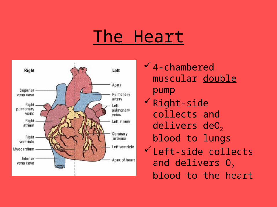
The Electrical Heart
The heart can contract and relax (i.e. beat) on its own
It is wired for action (SA node, AV node and Purkinje fibres)
The medulla oblongata of the brainstem controls that rate at which the heart beats
The amount of CO2 in the blood determines the heart rate
Electrical Activity of the Heart

Structures of the Heart
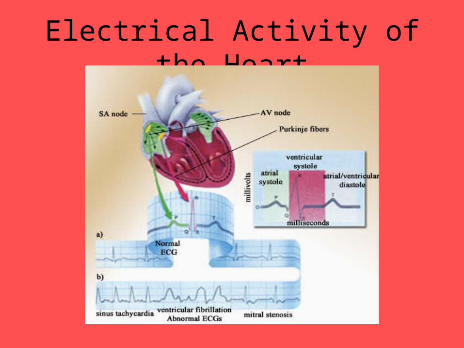
Structure & Function of HeartSuperior vena cava Returns deO2 blood to RA from upper body
Inferior vena cava Returns deO2 blood to RA from lower body
Right atrium (RA) Collects and pumps blood to RV
Right atrioventricular valve (tricuspid valve)
Closes to prevent backflow of blood into RA; responsible for LUB sound
Right ventricle (RV) Contracts and pumps blood to lungs
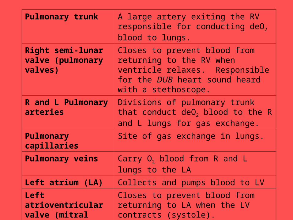
Pulmonary trunk A large artery exiting the RV responsible for conducting deO2 blood to lungs.
Right semi-lunar valve (pulmonary valves)
Closes to prevent blood from returning to the RV when ventricle relaxes. Responsible for the DUB heart sound heard with a stethoscope.
R and L Pulmonary arteries
Divisions of pulmonary trunk that conduct deO2 blood to the R and L lungs for gas exchange.
Pulmonary capillaries Site of gas exchange in lungs.
Pulmonary veins Carry O2 blood from R and L lungs to the LA
Left atrium (LA) Collects and pumps blood to LV
Left atrioventricular valve (mitral valve)
Closes to prevent blood from returning to LA when the LV contracts (systole). Responsible for LUB heart sound heard with a stethoscope.
Left ventricle (LV) Contracts and pumps (systole) blood to the body by way of the aorta
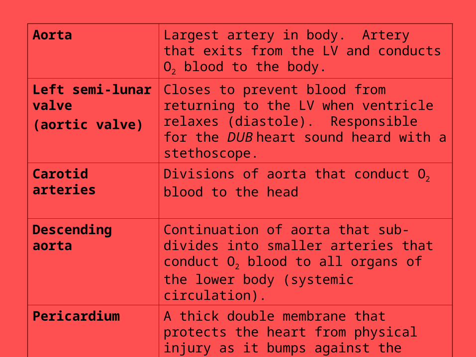
Aorta Largest artery in body. Artery that exits from the LV and conducts O2 blood to the body.
Left semi-lunar valve
(aortic valve)
Closes to prevent blood from returning to the LV when ventricle relaxes (diastole). Responsible for the DUB heart sound heard with a stethoscope.
Carotid arteries Divisions of aorta that conduct O2 blood to the head
Descending aorta Continuation of aorta that sub-divides into smaller arteries that conduct O2 blood to all organs of the lower body (systemic circulation).
Pericardium A thick double membrane that protects the heart from physical injury as it bumps against the ribcage. Keeps the heart moist.
Septum Thick muscular wall that subdivides the L and R sides of the heart
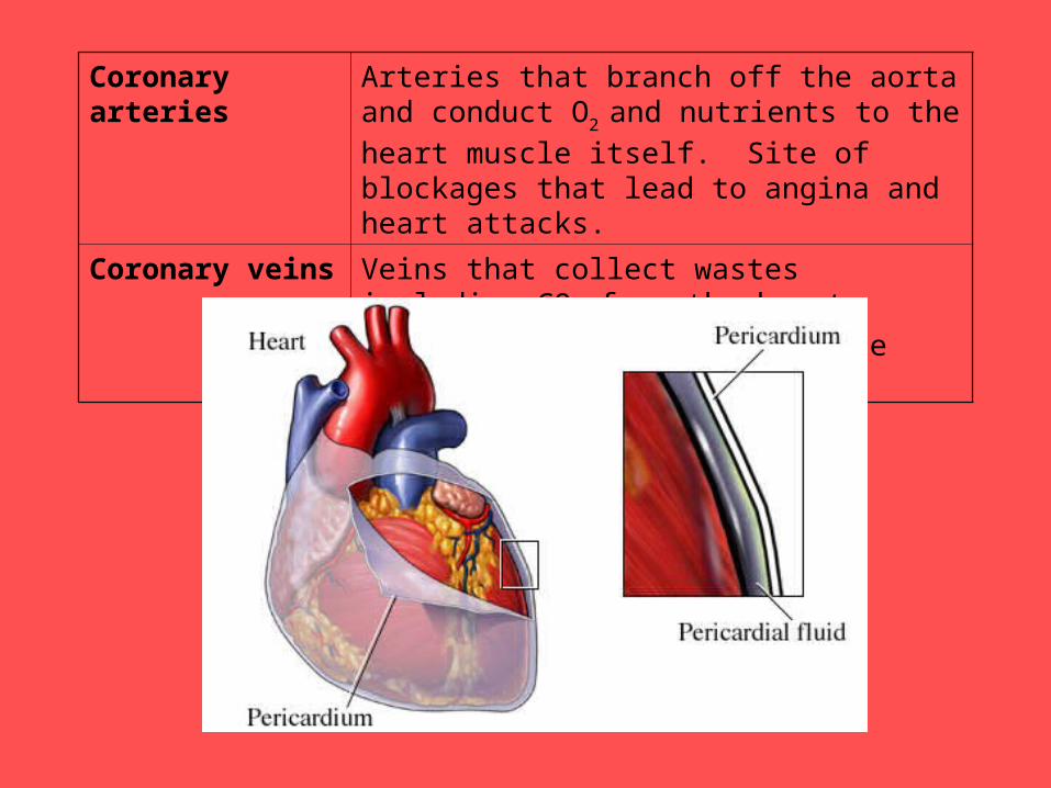
Coronary arteries Arteries that branch off the aorta and conduct O2
and nutrients to the heart muscle itself. Site of blockages that lead to angina and heart attacks.
Coronary veins Veins that collect wastes including CO2 from the
heart muscle and conduct them to the vena cavae
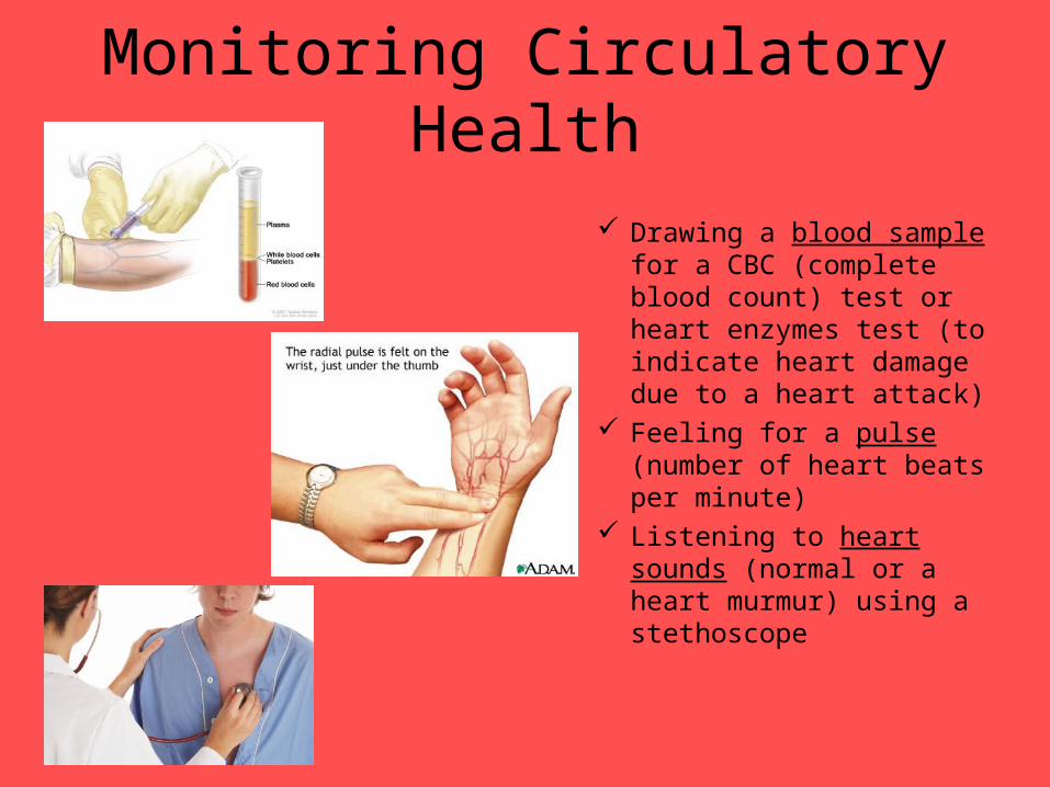
Monitoring Circulatory Health
Drawing a blood sample for a CBC (complete blood count) test or heart enzymes test (to indicate heart damage due to a heart attack)
Feeling for a pulse (number of heart beats per minute)
Listening to heart sounds (normal or a heart murmur) using a stethoscope

Monitoring Circulatory Health
Using a sphygmomanometer to measure blood pressure (looking for evidence of hypotension or hypertension)
Perform an electrocardiogram (ECG – to check out electrical activity of heart for unusual patterns)

Monitoring Circulatory Health
Perform an echocardiogram (using ultrasound to check on heart valve function)
Performing an angiogram (use dyes/contrasts to look for blockages of coronary arteries that may cause angina or a heart attack)
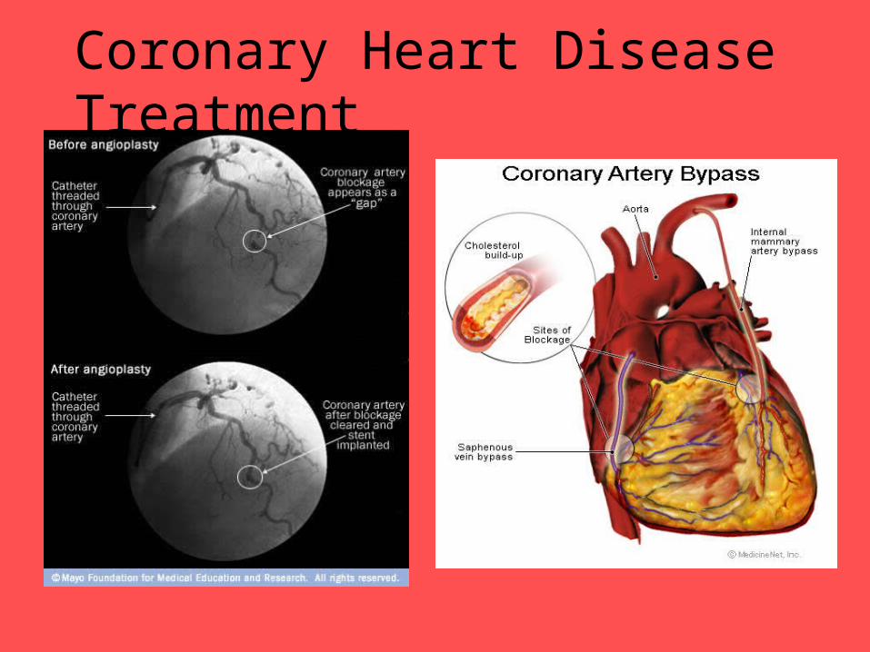
Coronary Heart Disease Treatment
•
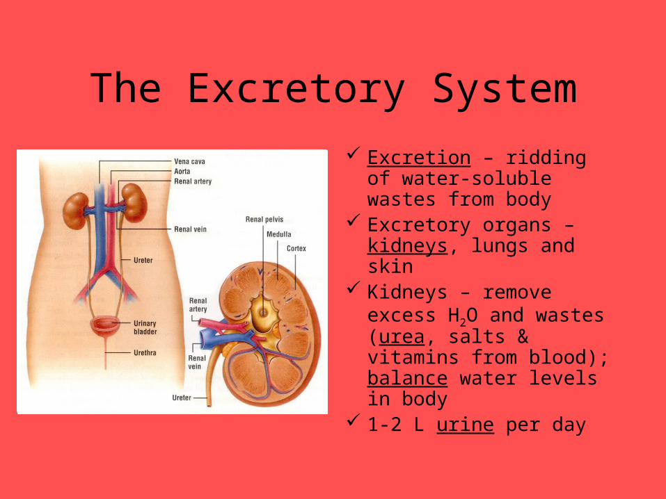
The Excretory System
Excretion – ridding of water-soluble wastes from body
Excretory organs – kidneys, lungs and skin
Kidneys – remove excess H2O and wastes (urea, salts & vitamins from blood); balance water levels in body
1-2 L urine per day
The End