The genus Acanthochitona (Mollusca: Polylacophora) in the ... - Bonfitto et al..pdfThe genus...
10
The genus Acanthochitona (Mollusca: Polylacophora) in the Mediterranean Sea: morphological and molecular data ANTONIO BONFITTO 1 , BRUNO DELL’ANGELO 2 , FRANCESCA EVANGELISTI 1 and BRUNO SABELLI 1 1 Dept. BES and Museum of Zoology, University of Bologna, via Selmi 3, 40126 Bologna, Italy. E-mail: [email protected] 2 Via Santelia 55, I-16153 Genova, Italy. SUMMARY: This work represents an attempt to resolve the confused and contradictory taxonomy of Mediterranean chitons of the genus Acanthochitona by analysing morphological (SEM observations of aesthetes, radula and girdle) and molecular data (COI, 12S, ITS1). Both analyses support the validity of the three species, Acanthochitona fascicularis, A. crinita and A. oblonga, the latter two of which were previously considered as synonymous. Keywords: SEM morphology, molecular systematics, Mediterranean Sea, Acanthochitona, Polyplacophora. RESUMEN: El género ACANTHOCHITONA (Mollusca: Polylacophora) en el mar Mediterráneo: datos morfológi- cos y moleculares. – En el presente trabajo se pretende resolver la confusa taxonomía de las especies mediterráneas de los quitones del género Acanthochitona a través de su estudio morfológico (observaciones al SEM de aestetes, rádula y cintura) y molecular (COI, 12S, ITS1). En ambos casos se confirma la validez de las tres especies Acanthochitona fascicularis, A. crinita y A. oblonga, las dos últimas consideradas previamente como sinónimas. Palabras clave: SEM morfología, sistemática molecular, mar Mediterráneo, Acanthochitona, Polyplacophora. SCIENTIA MARINA 75(1) March 2011, 171-180, Barcelona (Spain) ISSN: 0214-8358 doi:10.3989/scimar.2011.75n1171 INTRODUCTION Acanthochitona species are characterized by extreme variability of morphological features com- monly used in their systematics. Kaas (1985), in his review of the Polyplacophora of Mediterranean and Atlantic coasts, recognizes three living species: Acan- thochitona fascicularis (Linné, 1767) and A. crinita (Pennant, 1777) from the Mediterranean Sea, and A. discrepans (Brown, 1827) from the northeastern Irish coast and southern British coast. As regards the Medi- terranean Sea, Dell’Angelo and Smriglio (1999) more recently agreed with the view of Kaas. An additional taxon, Acanthochitona oblonga (Leloup, 1981), has a strong morphological relation to A. crinita with such different marginal issues that Kaas (1985) and Dell’Angelo and Smriglio (1999) considered them as cospecific; however, this issue remains to be demon- strated convincingly. The aim of this work is to try to resolve the confused systematics position of A. oblonga, compared to the other two Mediterranean species of Acanthochitona, by analyzing morphologi- cal and molecular features. MATERIALS AND METHODS The morphological analysis was performed with a scanning electron microscope Jeol jsm 5200 on some traditional systematic characters such as the morphol- ogy of granules of tegmentum, radulae and elements of the girdle. Hundreds of specimens from several Euro- pean and African localities were studied.
Transcript of The genus Acanthochitona (Mollusca: Polylacophora) in the ... - Bonfitto et al..pdfThe genus...

The genus Acanthochitona (Mollusca: Polylacophora) in the Mediterranean Sea: morphological and
molecular data
ANTONIO BONFITTO 1, BRUNO DELL’ANGELO 2, FRANCESCA EVANGELISTI 1 and BRUNO SABELLI 1
1 Dept. BES and Museum of Zoology, University of Bologna, via Selmi 3, 40126 Bologna, Italy. E-mail: [email protected]
2 Via Santelia 55, I-16153 Genova, Italy.
SUMMARY: This work represents an attempt to resolve the confused and contradictory taxonomy of Mediterranean chitons of the genus Acanthochitona by analysing morphological (SEM observations of aesthetes, radula and girdle) and molecular data (COI, 12S, ITS1). Both analyses support the validity of the three species, Acanthochitona fascicularis, A. crinita and A. oblonga, the latter two of which were previously considered as synonymous.
Keywords: SEM morphology, molecular systematics, Mediterranean Sea, Acanthochitona, Polyplacophora.
RESUMEN: El género AcAnthochitonA (Mollusca: Polylacophora) en el mar Mediterráneo: datos morfológi-cos y moleculares. – En el presente trabajo se pretende resolver la confusa taxonomía de las especies mediterráneas de los quitones del género Acanthochitona a través de su estudio morfológico (observaciones al SEM de aestetes, rádula y cintura) y molecular (COI, 12S, ITS1). En ambos casos se confirma la validez de las tres especies Acanthochitona fascicularis, A. crinita y A. oblonga, las dos últimas consideradas previamente como sinónimas.
Palabras clave: SEM morfología, sistemática molecular, mar Mediterráneo, Acanthochitona, Polyplacophora.
Scientia Marina 75(1)March 2011, 171-180, Barcelona (Spain)
ISSN: 0214-8358doi:10.3989/scimar.2011.75n1171
INTRODUCTION
Acanthochitona species are characterized by extreme variability of morphological features com-monly used in their systematics. Kaas (1985), in his review of the Polyplacophora of Mediterranean and Atlantic coasts, recognizes three living species: Acan-thochitona fascicularis (Linné, 1767) and A. crinita (Pennant, 1777) from the Mediterranean Sea, and A. discrepans (Brown, 1827) from the northeastern Irish coast and southern British coast. As regards the Medi-terranean Sea, Dell’Angelo and Smriglio (1999) more recently agreed with the view of Kaas. An additional taxon, Acanthochitona oblonga (Leloup, 1981), has a strong morphological relation to A. crinita with such different marginal issues that Kaas (1985) and
Dell’Angelo and Smriglio (1999) considered them as cospecific; however, this issue remains to be demon-strated convincingly. The aim of this work is to try to resolve the confused systematics position of A. oblonga, compared to the other two Mediterranean species of Acanthochitona, by analyzing morphologi-cal and molecular features.
MATERIALS AND METHODS
The morphological analysis was performed with a scanning electron microscope Jeol jsm 5200 on some traditional systematic characters such as the morphol-ogy of granules of tegmentum, radulae and elements of the girdle. Hundreds of specimens from several Euro-pean and African localities were studied.

172 • A. BONFITTO et al.
SCI. MAR., 75(1), March 2011, 171-180. ISSN 0214-8358 doi: 10.3989/scimar.2011.75n1171
Most molecular work was carried out at the mo-lecular laboratory at IRSNB, Brussels, Belgium. Total genomic DNA was isolated from a small piece of tis-sue taken from the foot of ethanol-preserved specimens. The extractions were carried out using the “Genomic DNA from tissue” kit (Macherey-Nagel) and the Chelex 100 procedure according to Nelson and Savannah River Ecology Lab protocol. When these two methods failed, or in all cases of a low yield of DNA extract, the CTAB method was used instead (Winnepenninckx et al., 1993). All the DNA extractions were kept at 4ºC for short-time use. Undiluted or different dilutions (from 1:10 to 1:50, based on the DNA concentration) of each DNA extraction were used as templates for PCR amplification of a portion of each of the three loci: the mitochondrial small subunit ribosomal DNA (mt-12S) and the cytochrome oxidase subunit 1 (mt-COI) genes, and the nuclear Internal Transcribed Spacer 1 (nu-ITS1) region. For the COI gene the primers used were LCO1490 (5’-GGTCAACAAATCATAAAGATATT-GG-3’) and HCO2198 (5’-TAAACTTCAGGGTGAC-CAAAAAATCA-3’) (Folmer, 1994), which amplified a region of 658 bp; the PCR conditions involved an initial denaturation step at 95ºC for 5 minutes; then 35 cycles of denaturation at 95ºC for 45 seconds, annealing at 45ºC for 45 seconds and extension at 72°C for 1 minute and 30 seconds; followed by a final extension step at 72°C for 5 minutes. For the 12S gene the primers used were 12SaL (5’-AAACTGGGATTAGATACCCCAC-TAT-3’) and 12SaH (5’-GAGGGTGACGGGCGGT-GTGT-3’) (Kocher, 1989), which amplified a region of 362 bp; the PCR conditions involved an initial de-naturation step at 95°C for 5 minutes; then 40 cycles of denaturation at 95°C for 45 seconds, annealing at 51°C for 45 seconds and extension at 72°C for 2 minutes; fol-lowed by a final extension at 72°C for 5 minutes. For the ITS1 region the primers used were ITS1L (5’-TCCG-TAGGTGAACCTGCGGAAGGAT-3’) and 58C (5’- TGCGTTCAAGATATCGATGTTCAA-3’) (Hillis and Dixon, 1991), which amplified a fragment of 641 bp; the PCR conditions involved an initial denaturation step at 95ºC for 5 minutes; then 35 cycles of denaturation at 95ºC for 1 minute, annealing at 55ºC for 1 minute and extension at 72°C for 2 minutes; followed by a final ex-tension step at 72°C for 5 minutes. PCR reactions were performed in a total volume of 25 µl including 2.5 µl of 2 mM of each dNTP (GE Healthcare), 2.5 µl of 10x load buffer-MgCl2 (Qiagen), 2.5 µl of 2 µM of each primer, 0.25 µl of 5 U/µl Taq DNA polymerase (Qiagen), 19 µl of demineralized water and 1 µl of the DNA template. Amplified products were purified using the GFX PCR DNA and GEL Band Purification Kit (GE Healthcare), or the NucleoFast PCR plates (Macherey-Nagel), fol-lowing the protocol provided for the purification of PCR products under vacuum. Cycle sequencing was carried out in both directions of the amplified regions using the Big Dye Terminator v1.1 cycle sequencing kit (Applied Biosystems) and Hitachi 3130x Genetic Analyser (Ap-plied Biosystems).
For each gene, the forward and reverse sequences obtained were imported and read in the program Chro-mas Lite version 2.01 (http://www.technelysium.com.au/chromas_lite.html), for visual inspection of chroma-tograms, and subsequently combined in the program Bioedit version 7.0.9.0 (Hall, T.A. 1999) in order to obtain consensus sequences to be used in subsequent analyses. All the sequences were identified by BLAST matching and assembled separately for each gene us-ing Bioedit. The external primer regions and blank ends were subsequently trimmed from each of the three groups of sequences. Each group was converted in MEGA format using the program DnaSP version 5 (Librado et al., 2009) and imported in the program MEGA version 4 (Tamura et al., 2007).
Three species were selected as outgroup taxa for the molecular analysis: two molluscs, the bivalve Crassos-trea gigas (Thunberg, 1793) and the gastropod Lottia digitalis (Rathke, 1833), and a more distant species, the crustacean Portunus pelagicus (Linnaeus, 1758). It was not possible to include other chitons or species belong-ing to the genus Acanthochitona due to the lack of data in GenBank; of the three genes considered, only COI sequences are published for chitons (Okusu et al., 2003; Wilson et al., 2010), but not for 12S and ITS1. Published sequences from GenBank for the three selected outgroups were downloaded and then added to each alignment (see Table 1 for their accession number and lengths). All the sequences for each gene were aligned with the multise-quence alignment Clustal W software (Thompson et al., 1994), as implemented in the program MEGA 4. The alignments were then refined by eye. Therefore, all the regions larger than 4 nucleotides which were ambigu-ously aligned with respect to the bordering regions were deleted while the gaps were included in the analysis and scored as missing characters. Pairwise evolution-ary divergence between sequences calculated between A. oblonga and A. crinita, between A. oblonga and A. fascicularis, and between A. crinita and A. fascicularis, were calculated using the Kimura-2-parameters method (Kimura, 1980) in MEGA 4, with pairwise deletion of gaps and missing data, and uniform rates among sites. The final three alignments were concatenated in MEGA 4 in all the possible combinations in order to create dif-ferent datasets to be used in the phylogenetic analysis. These combined data sets contain a smaller number of sequences with respect to the sequences of each single gene because specimens with no data for all three genes were excluded from the combined analyses.
Table 1. – GenBank accession numbers and sequence length values for each outgroup species used in the phylogenetic analysis.
Species COI 12S ITS1
Crassostrea gigas AY397686 EF484878 AB041761 551 bp 671 bp 602 bpLottia digitalis AB238464 AB238241 DQ248942 661 bp 346 bp 558 bpPortunus pelagicus FJ812293 FJ812311 AM410550 598 bp 374 bp 688 bp
MEDITERRANEAN ACANTHOCHITONA SYSTEMATICS • 173
SCI. MAR., 75(1), March 2011, 171-180. ISSN 0214-8358 doi: 10.3989/scimar.2011.75n1171
For each data set the phylogenetic analysis was performed in the program MrBayes 3.1.2 (Ronquist and Huelsenbeck, 2003) using the Bayesian Inference method (Holder and Lewis, 2007 and Huelsenbeck and Ronquist, 2001). Each data set was examined assuming the most complex evolution model (GTR+I+Γ) and us-ing the Markov Chain Monte Carlo (Yang and Ranna-la, 1997; Larget and Simon, 1999) as the tree sampling procedure. The analyses were run for three million gen-erations with every hundredth tree and parameter value stored. The plot of likelihood scores versus generations was generated to establish how many generations were necessary to reach stationarity. The likelihood values clearly plateaued after 20000 generations, that is, after 20000 generations the changes in the tree topology and parameter values did not continue to improve the tree likelihood scores. Therefore, the first 200 trees (from the first 20000 generations) were excluded from the analysis as burn-in and the remaining trees were used to make a 50% majority rule consensus and to estimate the Bayesian Posterior Probability (BPP) to give sup-port to tree nodes. All the trees were drawn with the program Dendroscope version 1.4 (Huson et al., 2007).
Abbreviations
MNHN Museum National d’Histoire Naturelle, Paris,CT Catania provence, SicilyLE Lecce provence, ApuliaME Messina provence, SicilyMZB Museum of Zoology, University of BolognaRSMNH Royal Scottish Museum of Natural History, EdinburghIRSNB Royal Belgian Institute of Natural SciencesTA Taranto provence, Apulia
RESULTS
Taxonomic remarks
Several papers deal with the systematics and mor-phology of the investigated species; here we summa-rize some data and the main references.
Acanthochitona fascicularis (Linné, 1767)
Chiton fascicularis Linné, 1767: 1106.Acanthochitona fascicularis Kaas, 1985: 585, Figs. 1-6 (bibliog-
raphy and synonymy). Dell’Angelo and Smriglio, 1999: 192, Figs. 113-123, pls 64-65 (bibliography and synonymy). Özturk et al., 2000: 72. Anistratenko V.V. and O.Y., 2001: 51, Fig. 17. Özturk et al., 2004: 52. Sigwart, 2005: 19. Peñas et al., 2006: 34, Fig. 419. Trono, 2006: 75. Cecalupo et al., 2008: 54, pl. 1, Figs. 10-12.
Type. The species is not preserved at the Linnean Society of London (fide Dodge, 1952). Neotype designated and figured by Kaas (1985: Fig. 1), MNHN.
Type locality. Algeria, Oran.
Material examined. Campomarino (TA): 5 specimens (used for the present study). Capo Molini (CT): 1 specimen (used for the present
study). Torre Ovo (TA): 6 specimens (used for the present study). Hundreds of specimens and valves from diverse European (Spain, France, Italy, Croatia, Greece, Turkey, Malta, Canary Islands), Afri-can (Morocco, Algeria, Tunisia) and eastern Mediterranean (Israel, Lebanon) localities (BDA, MZB).
Remarks. Acanthochitona fascicularis is a highly variable species with a puzzling synonymy; see Kaas (1985) and Dell’Angelo and Smriglio (1999) for a good description, synonymy and literature. Owing to the variability and convergence of the morphological characters of the Acanthochitona species it would be useful to refer to all specimens of A. fascicularis as be-ing mainly characterized by:
tegmentum with dense, round, rather small gran-ules arranged in arched lines; quadrangulars apophy-ses anteriorly well developed; girdle densely covered with small and thin spicules and some others scattered which are longer and thicker, with 18 tufts of white bristles; and rather small, triangular apical area.
The outline and profile of intermediate and tail valves are very variable, and a representative range of this vari-ability was nicely figured in Leloup (1941: Fig. 2).
Distribution. The Mediterranean Sea, the Black Sea, and the eastern Atlantic Ocean from the Channel and Britain to Azores and Canary Islands.
Acanthochitona crinita (Pennant, 1777)
Chiton crinitus Pennant, 1777: 71, pl. 36, Figs. 1, A1.Acanthochitona crinita: Kaas, 1985: 588, Figs. 7-50 (bibliography
and synonymy). Dell’Angelo and Smriglio, 1999: 198, Figs. 124-130, pls 66-68 (bibliography and synonymy). Özturk et al., 2000: 72. Özturk et al., 2004: 52. Sigwart, 2005: 19. Penas et al., 2006: 34. Trono, 2006: 75. Cecalupo et al., 2008: 24.
Type. The three syntypes of A. crinita are no longer with the Pennant collection. Neotype designated and figured by Kaas (1985: Fig. 27), RSMNH no. reg. 1978.052.02601.
Type locality. Hebrides Islands.
Material examined. Villafranca Tirrena (ME): 10 specimens (used for the present study). Hundreds of specimens and valves from diverse European (Portugal, Spain, France, Italy, Croatia, Greece, Turkey, Malta, Canary Islands), African (Morocco, Algeria, Tuni-sia) and eastern Mediterranean (Israel, Lebanon) localities (BDA, MZB).
Remarks. A. crinita is also a highly variable spe-cies with a puzzling synonymy. See Kaas (1985) and Dell’Angelo and Smriglio (1999) for a good descrip-tion, synonymy and literature. It differs from A. fas-cicularis mainly in the jugal area, which is generally wider, slightly elevated and not well separated from the lateropleural area, and the granules are less thickly arranged, more spaced, and shaped from oval to a more or less elongated drop.
Distribution. The Mediterranean Sea, a few records from the north African coast: e.g. Jerba Tunisia (Dell’Angelo and Cuppini, 1983; Cecalupo et al., 2008), The North American Atlantic coast

174 • A. BONFITTO et al.
SCI. MAR., 75(1), March 2011, 171-180. ISSN 0214-8358 doi: 10.3989/scimar.2011.75n1171
and the eastern Atlantic along the European coast from Norway to Azores, Madeira, Canary and Cape Verde Islands (Dell’Angelo and Smriglio, 1999). As reported by Dell’Angelo and Smfriglio (1999), A. crinita has been collected in the intertidal zone and as deep as 50 m, and there are records from a depth of 175 m deep. It lives under stones which are often partly buried in sand or pebbles. It may be associ-ated with the barnacle zone or with several kinds of algae.
Acanthochitona oblonga (Leloup, 1981)
Acanthochiton oblongus Leloup, 1981: 1, Figs. 1a-d, pl. 1. Kaas, 1985: 596, Figs. 39-43.
Acanthochitona oblonga Dell’Angelo and Cuppini, 1983: 77. Ga-glini, 1985: xviii, pl. 1, Fig. 6; 1989: 9. Savona and Carnieri, 1985: 36, Figs. 1-3. Dell’Angelo and Smriglio, 1999: 200, Figs. 124-130, pl. 67, Figs. K-L; pl. 68, Figs. Q-V.
Acanthochitona crinita f. oblonga Baschieri, 1994: 40, Fig. 2. Bas-chieri et al., 1992: 68, Fig. 4.
Type. Holotype at the Royal University of Malta.
Type locality. Malta, Salina Bay, “under rocks at 3 meters depth, July 1974”.
Material examined: San Foca (LE), 11 specimens (used for the present study); San Foca (LE), 20 specimens, leg. B. Dell’Angelo 08/2004 (BDA); Roca Li Posti (LE), 8 specimens, leg. L. Bas-chieri 08/1989 (BDA); Campomarino (TA): 6 specimens, leg. B. Dell’Angelo 08/2007 (BDA); Marina di Lizzano (TA), 20 speci-mens, leg. B. Dell’Angelo 08/2007 (BDA); Malta, Gnejna Bay, 3 specimens, ex coll. C. Mifsud (BDA); Tunisia, Djerba, Menzel, 2 specimens, ex coll. P. Piani (BDA); and Tunisia, Djerba, Sidi Gar-rus: 1 specimen, leg. A. Germanà 04/2007 (BDA).
Remarks. This species, described by Leloup based on 4 specimens collected at Salina Bay, Malta, is characterized by the very extended sharp granules of the tegmentum. After the original description, several specimens were reported from the Mediterranean Sea (Dell’Angelo and Cuppini, 1983; Baschieri, 1994), showing that this species is very common in some localities of the South Apulian Adriatic coast where specimens exhibit granules that vary from typical drop shaped to a shape that is even more extended than what was originally described by Leloup.
Distribution. Southern Mediterranean Sea in scat-tered Italian localities (Apulia, Sicily, Lampedusa Is.), Malta, Cyprus, Tunisia (Jerba) (Dell’Angelo and Cuppini, 1983; Dell’Angelo and Smriglio, 1999). It is worth noting that in Bruno Dell’Angelo’s collection, there are specimens of both A. crinita and A. oblonga from Jerba; however, he did not collect them directly and the information on the label does not specify either the locality along the coast of the isle, or the habitat. The two species are surely sympatric and are clearly discriminated by the shape of granules in S. Foca (LE) where they were collected on a sandy bottom 2 - 3 m deep with sparse rocky fissured tables. Green algae grow in these fissures and the two Acanthochitona spe-cies live under them.
A third species of Acanthochitona, A. discrepans (Brown, 1827), lives along the northeastern Irish and southern English coasts but not in the Mediterranean Sea. This species has been studied by Kaas (1985) and differs from A. crinita mainly due to:
valves carinated (not carinated in A. fascicularis and A. crinita); “velvety” perinotum covered by short spicules 40 µm long; and short tufts of spicules some-times with 1 to 3 extra tufts around the posterior valve.
Kaas (1985) writes that these extra tufts, which were considered typical of Chiton gracilis Jeffreys, 1859, are present only in a few specimens (mostly only one tuft) and “… on the other hand I possess a fine and in all respects normal specimen of crinita from Nor-mandy, with one extra tuft”.
Morphological analysis
In all Acanthochitona species the perinotum is char-acterized by rounded or drop shaped elevated granules; those of A. fascicularis (Fig. 1) are rounded, rather elevated, in some cases heart shaped, concave, with a characteristic incision on the anterior margin. The single central macroaesthete is generally surrounded by 1 to 5 microaesthetes, (sometimes 0) irregularly arranged.
In A.crinita the granules (Figs. 2A, B) are lower with respect to A. fascicularis, ovoid and drop-shaped, more or less elongated, flat or slightly concave. The single posteriorly located macroaesthete is surrounded by a different number of microaesthetes, ranging from 12 to over 16, irregularly grouped mainly in the antero-central part of the granule.
In A. oblonga, the granules (Figs. 3A, B) appear lower if compared to A. crinita, more elongated, narrow, lanceolate and slightly concave anteriorly. The single posteriorly located macroaesthete is surrounded by a lower number of microaesthetes, from 6 to 9, irregularly grouped mainly in the middle part of the granule.
Radulae
The symmetrical radulae (Figs. 4A, B, C) are quite similar with minor quantitative differences. The central tooth is squarish in outline, medially constricted with a strong median crista in each species and only in A. oblonga (Fig. 4C) appears very slightly bilobated at the apex. The centro-lateral tooth is smaller as usual, being very arched in A. fascicularis (Fig. 4A) and more or less straight in the other two species (Figs. 4B, C). Its tip is distinctly concave in A. fascicularis (Fig. 4A) and slightly concave in A. crinita (Fig. 4B) giving the impression of a truncated end, while it is convex and spatuliform in A. oblonga (Fig. 4C). The major lateral tooth has a large head with mesocone larger than the endo- and ectocone in A. fascicularis (Fig. 4A), while in both A. crinita (Fig. 4B) and A. oblonga (Fig. 4C) the endo- and mesocone are of similar size and the ec-tocone is smaller.

MEDITERRANEAN ACANTHOCHITONA SYSTEMATICS • 175
SCI. MAR., 75(1), March 2011, 171-180. ISSN 0214-8358 doi: 10.3989/scimar.2011.75n1171
Perinotum
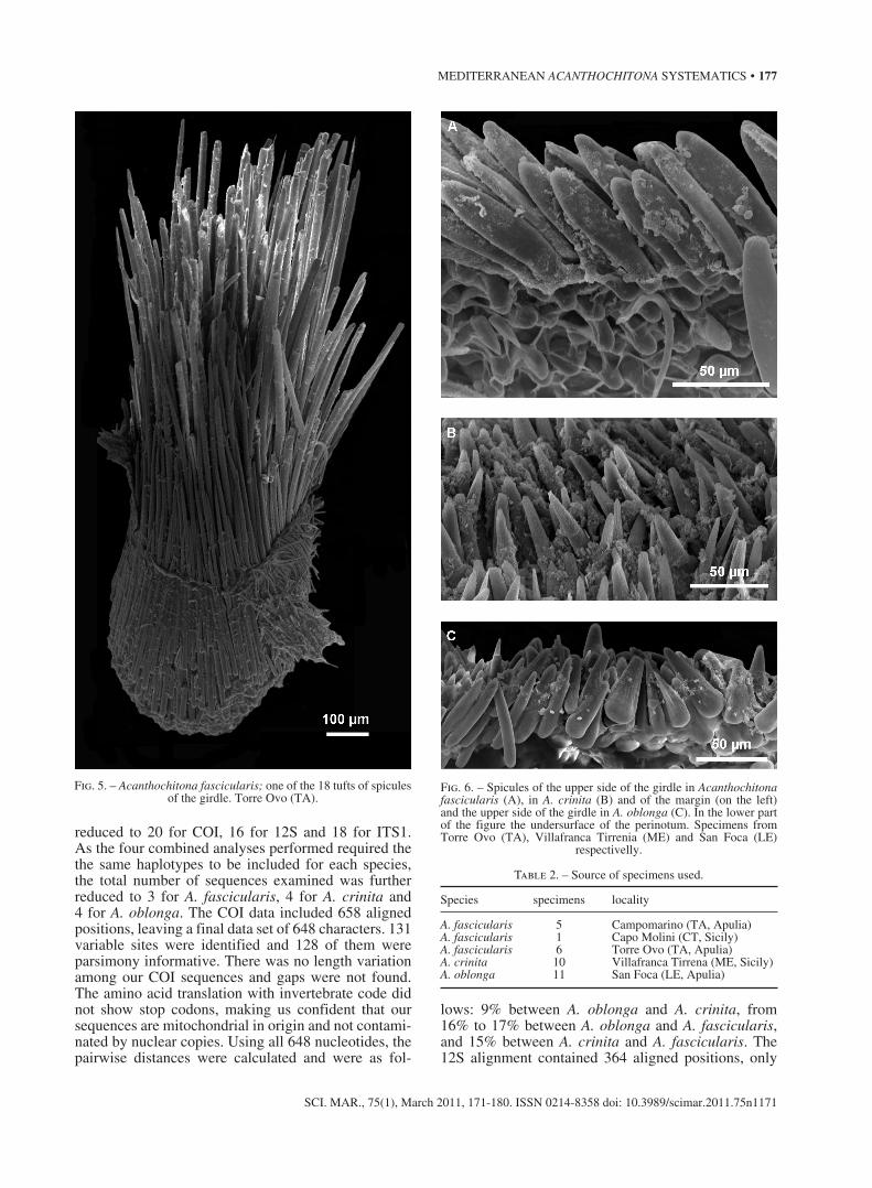
All species share a broad perinotum with 18 large tufts of usually colourless spines that vary in length from 500 to 1000 µm and more (Fig. 5). The dorsal
surface is covered by short more or less curved spi-cules among which other thicker and longer spicules are scattered. According to Dell’Angelo and Smriglio (1999) in A. fascicularis the short curved spicules are 100-150 mm in length, the others are up to 500 µm;
Fig. 2. – A, morphology of the granules of tegmentum in Acanthochitona crinita. B, close-up of a single granule of the same species. On the right side its proximal end. Specimens from Villafranca Tirrenia (ME).
Fig. 1. – Morphology of the granules of tegmentum in Acanthochitona fascicularis; on the right side their proximal end. Specimen from Torre Ovo (TA).
Fig. 3. – A, morphology of the granules of tegmentum in Acanthochitona oblonga. B, close-up of two granules in the same species; on the right side their proximal end. Specimens from San Foca (LE).

176 • A. BONFITTO et al.
SCI. MAR., 75(1), March 2011, 171-180. ISSN 0214-8358 doi: 10.3989/scimar.2011.75n1171
in A. crinita they are respectively 50 µm and 320 µm and in A. oblonga 60-70 µm and 400 µm. A marginal fringe of longitudinally finely striated spicules of 300-400 mm is always present and on the ventral surface the spicules are shorter and also finely striate. We can add that in A. fascicularis the perinotum doesn’t exhibit the marked difference between the two kinds of spicules as in the other two species. The specimens we examined (Figs. 6A, B, C) agree in general with this description
Molecular analysis, sequence data
A total of 33 DNA sequence fragments, 12 be-longing to A. fascicularis, 10 to A. crinita and 11 to A. oblonga, were obtained for COI, 12S and ITS1 loci (Table 2). In addition, for each gene, 3 sequences for the three selected outgroups (Crassostrea gigas, Lot-tia digitalis and Portunus pelagicus) were downloaded from GenBank. After removing identical haplotypes from each alignment, the number of sequences was
Fig. 4. – Portion of the radular ribbon of Acanthochitona fascicularis (A), A. crinita (B) and A. obonga (C). Specimens from Castiglioncello (LI), Arenzano (GE) and San Foca (LE) respectivelly.
MEDITERRANEAN ACANTHOCHITONA SYSTEMATICS • 177
SCI. MAR., 75(1), March 2011, 171-180. ISSN 0214-8358 doi: 10.3989/scimar.2011.75n1171
reduced to 20 for COI, 16 for 12S and 18 for ITS1. As the four combined analyses performed required the the same haplotypes to be included for each species, the total number of sequences examined was further reduced to 3 for A. fascicularis, 4 for A. crinita and 4 for A. oblonga. The COI data included 658 aligned positions, leaving a final data set of 648 characters. 131 variable sites were identified and 128 of them were parsimony informative. There was no length variation among our COI sequences and gaps were not found. The amino acid translation with invertebrate code did not show stop codons, making us confident that our sequences are mitochondrial in origin and not contami-nated by nuclear copies. Using all 648 nucleotides, the pairwise distances were calculated and were as fol-
lows: 9% between A. oblonga and A. crinita, from 16% to 17% between A. oblonga and A. fascicularis, and 15% between A. crinita and A. fascicularis. The 12S alignment contained 364 aligned positions, only
Table 2. – Source of specimens used.
Species specimens locality
A. fascicularis 5 Campomarino (TA, Apulia)A. fascicularis 1 Capo Molini (CT, Sicily)A. fascicularis 6 Torre Ovo (TA, Apulia)A. crinita 10 Villafranca Tirrena (ME, Sicily)A. oblonga 11 San Foca (LE, Apulia)
Fig. 5. – Acanthochitona fascicularis; one of the 18 tufts of spicules of the girdle. Torre Ovo (TA).
Fig. 6. – Spicules of the upper side of the girdle in Acanthochitona fascicularis (A), in A. crinita (B) and of the margin (on the left) and the upper side of the girdle in A. oblonga (C). In the lower part of the figure the undersurface of the perinotum. Specimens from Torre Ovo (TA), Villafranca Tirrenia (ME) and San Foca (LE)
respectivelly.

178 • A. BONFITTO et al.
SCI. MAR., 75(1), March 2011, 171-180. ISSN 0214-8358 doi: 10.3989/scimar.2011.75n1171
a seven sites region was excluded due to its ambigu-ity; therefore, the final data set was 357 bp. The align-ments were relatively simple as the sequence lengths were again conserved and only four gap regions (cor-responding to single nucleotide substitutions) were found. The number of variable sites was 53 and almost all of them (52) were parsimony informative. The val-ues of uncorrected divergences were 7% for A. oblonga and A. crinita, 14% for A. oblonga and A. fascicularis, and 13% for A. crinita and A. fascicularis. The ITS1 alignment shows considerable length variations (the maximum observed was equal to 624 positions (all four haplotypes of A. crinita) while the minimum was equal to 499 positions, belonging to A. oblonga haplo-type AO8. 124 sites were variable, 97 of which were informative. The sequence length was too variable to be globally aligned, particularly from position 255 to 280. Therefore, this and all the other highly variable regions were excluded and many gaps were added to align sequences locally. The final data set was 624 bp. The uncorrected divergences ranged from 5% to 10% between A. oblonga and A. crinita, from 12% to 15% between A. oblonga and A. fascicularis, and from 14% to 17% between A. crinita and A. fascicularis. The total number of sites considered in the combined analyses were as follows: 1005 (COI+12S), 1272 (COI+ITS1), 981 (12S+ITS1) and 1629 (COI+12S+ITS1).
Bayesian inference analysis
Phylogenetic Bayesian estimates made for each gene separately could not resolve well the phylogenetic relationship among A. oblonga, A. crinita and A. fas-cicularis. In all three trees obtained (data not shown), the three species were always separated as distinct monophyletic groups but with low levels of support
(BPP values less than 95%), especially in the 12S tree. The deepest internal nodes of the trees were found to be less robust.
However, the four combined Bayesian analyses provide good resolution among the three species and recover identical consensus tree topologies with nearly identical posterior probabilities for each clade. The consensus tree obtained through the Bayesian Infer-ence based on the combined data set of the three genes (COI+12S+ITS1) is shown in Figure 7. The tree clearly reveals two major clades, one consisting of A. fascicu-laris and one including A. crinita and A. oblonga, each supported by strong BPP values (100%). The latter clade clearly resolved into two branches: one with A. crinita and A. oblonga so that A. crinita and A. oblonga are clearly identified as monophyletic groups, again highly supported by 100% BPP scores.
DISCUSSION
After the SEM analysis, the morphology of the teg-mentum appears to be the most useful morphological character for distinguishing the three taxa. The shape of the granules and the distribution of the aesthetes of A. fascicularis were studied in detail by Fischer and Renner (1979), and Dell’Angelo and Smriglio (1999) also made some observations of both A. fascicularis and A. crinita; however, contrary to what they stated, the aesthetes are absent in the intertubercolar areas of the tegmentum in all examined specimens of each taxon.
Apart from the shape of the granules, based on which Leloup (1981) separated A. oblonga from A. crinita, which may exhibit some degree of variation, the distribution of anterocentral microaesthetes in the second species vs the central microaesthetes in the first
Fig. 7. – Bayesian Inference phylogram of the relationship among A. oblonga, A. crinita and A. fascicularis based on COI, 12S and ITS1 concatenated sequences. Fifty percent majority rule consensus tree, rooted using Portunus pelagicus as designate outgroup. Posterior prob-ability values (only values above 95% are statistically significant) provided at the branches. Numbers at the end of the haplotypes represent
the sample names used for this study.

MEDITERRANEAN ACANTHOCHITONA SYSTEMATICS • 179
SCI. MAR., 75(1), March 2011, 171-180. ISSN 0214-8358 doi: 10.3989/scimar.2011.75n1171
species and the completely different numerical macro/microaesthete ratio (1-5/1 in A fascicularis, 12-16/1 in A. crinita and 6-9/1 in A. oblonga) probably represent the best characters for identifying Mediterranean Acan-thochitona well.
Even if the differences in the radulas are not particu-larly remarkable, the three species can be distinguished mainly by the shape of the centrolateral teeth, which are only very arched in A. fascicularis and their tip is only convex and spatuliform in A. oblonga. Moreover, a slight difference can be observed in the three cusps of the major lateral teeth: the central cusp is larger in A. fascicularis while in A. crinita and A. oblonga it is the same size as the internal one and the external cusp is always the smallest.
The perinotum is the most variable character; Le-loup (1941, 1968), Kaas (1985) and Dell’Angelo and Smriglio (1999) described and illustrated in detail its spicules showing some differences, but we observed high variability in shape and also minor variability in the size of the dorsal spicules. This led us to consider this character to be of minor importance, at least for discriminating between the two disputed species A. crinita and A. oblonga.
The molecular analysis reported here represents the first estimate of the relationship within the genus Acan-thochitona (Gray, 1821). The uncorrected pairwise dis-tance values calculated among the three species show an evident close genetic relationship between A. oblon-ga and A. crinita, while A. fascicularis appears more distant. The topologies of the trees based on the four combined data sets show that A. oblonga and A. crinita always clustered separately from A. fascicularis and resolve each one as a monophyletic group. Therefore, all the genetic data definitely highlights that A. oblonga is closely related to A. crinita but it is an independent lineage. The pattern shown by the trees obtained from the analysis of the single genes can be explained by a rapid speciation (very low BPP values at deep nodes) followed by a slower process of differentiation of the three species (higher values of BPP). These data might be in agreement with the reports of many suspected neo-endemic species of the southern Mediterranean Sea and in particular the gulf of Gabes (Cecalupo et al., 2008). The lower BPP values observed in the 12S tree were expected due to the more conservative nature of this gene with respect to COI and ITS1. The fact that good resolution is obtained only by the combined data sets and not by the single genes was not surpris-ing as it has been recognized that a single marker is often insufficient for reconstructing the phylogeny of a group of living animals (Nichols, 2001). Combining data sets can in fact recover hidden signals because the true signal is not randomly distributed across data sets. In general, the results of our investigation clearly dem-onstrate the usefulness of molecular data to delineate the relationship among the species of the genus Acan-thochitona, and in particular the status of A. oblonga as a valid species.
ACKNOWLEDGEMENTS
We would like to thank all the staff of the Molecular Systematics laboratory, Department of Invertebrates, Royal Belgian Institute of Natural Sciences (IRSNB), Brussels, Belgium, where Francesca Evangelisti con-ducted most of the molecular work. In particular we are grateful to Professor Thierry Backeljau who gave helpful suggestions regarding the lab work and strong support for data interpretation; we are also grateful to Karin Breugelmans for her expert technical assistance in the genetic analysis, and also Vanya Prévot and Hilde Vrijders who helped in all lab procedures and operations.
REFERENCES
Anistratenko, V.V. and O.Y. Anistratenko. – 2001. Fauna Ukraine. Class Polyplacophora, Class Gastropoda- Cyclobranchia, Scutibranchia and Pectinibranchia (part). Volume 29: Mol-lusca. Fasc. 1. (in Russian)
Baschieri, L. – 1994. Un’insolita concentrazione di due specie di Poliplacofori. La Conchiglia, 26(270): 40-42.
Baschieri, L., B. Dell’Angelo and S. Palazzi. – 1992. Recenti ri-trovamenti di Polyplacophora anomali nel Mediterraneo. Boll. Malacol., 28: 65-68.
Cecalupo, A., G. Buzzurro and M. Mariani. – 2008. Contributo alla conoscenza della malacofauna del Golfo di Gabès (Tunisia). Quad. Civ. Staz. Idrobiol. Milano, 31: 1-177.
Dell’Angelo, B. and S. Cuppini. – 1983. Prima segnalazione di Acanthochitona oblonga Leloup, 1981 lungo le coste italiane. Boll. Malacol., 19: 77-78.
Dell’Angelo, B. and C. Smriglio. – 1999. Chitoni viventi del Medi-terraneo. Evolver, Roma. (English edition, 2001, Living Chi-tons from the Mediterranean Sea).
Folmer, O., M. Black, W. Hoeh, R. Lutz and R. Vrijenhoek. – 1994. DNA primers for amplification of mitochondrial cytochrome c oxidase subunit I from diverse metazoan invertebrates. Mol. Mar. Biol. Biotechnol., 3: 294-299.
Gaglini, A. – 1985. Classe Amphineura. In: F. Settepassi (ed.), (1972), Atlante Malacologico. I Molluschi Marini viventi nel Mediterraneo. Vol. III, 19 pp. INIVAG, Roma.
Gaglini, A. – 1989. I Polyplacophora delle coste italiane. Quad. Malacol., 1: 3-16.
Hall, T.A. – 1999. BioEdit: a user-friendly biological sequence alignment editor and analysis. Department of Microbiology. North Carolina State University.
Hillis, D.M. and M.T. Dixon. – 1991. Ribosomal DNA: molecular evolution and phylogenetic inference. Quart. Rev. Biol., 66: 411-453.
Holder, M. and P.O. Lewis. – 2003. Phylogeny estimation: tradi-tional and Bayesian approaches. Nature Rev. Gen., 4: 275-284.
Huelsenbeck, J.P. and F. Ronquist. – 2001. MrBayes: Bayesian in-ference of phylogeny. Bioinformatics, 17: 754-755.
Huson, D.H., D.C. Richter, C. Rausch, T. Dezulian, M. Franz and R. Rupp. – 2007. Dendroscope-an interactive viewer of large phylogenetic trees. BMC Bioinformatics, 8: 460.
Kaas, P. – 1985. The genus Acanthochitona Gray, 1821 (Mollusca, Polyplacophora) in the north-eastern Atlantic Ocean and in the Mediterranean Sea, with designation of neotypes of A. fascicu-laris (L., 1767) and of A. crinita (Pennant, 1777). Bull. Mus. Nat. Hist. Nat., (4)7(A3): 579-609.
Kimura, M. – 1980. A simple method for estimating evolutionary rate of base substitutions through comparative studies of nucle-otide sequences. J. Mol. Evol., 16: 111-120.
Kocher, T.D., W.K. Thomas, A. Meyer, S.V. Edwards, S. Paabo, F.X. Villablanca and A.C. Wilson. – 1989. Dynamics of mi-tochondrial DNA evolution in animals: Amplification and se-quencing with conserved primers. PNAS, 86: 6196-6200.
Larget, B. and D.L. Simon. – 1999. Markov Chain Monte Carlo algorithms for the Bayesian analysis of phylogenetic trees. Mol.

180 • A. BONFITTO et al.
SCI. MAR., 75(1), March 2011, 171-180. ISSN 0214-8358 doi: 10.3989/scimar.2011.75n1171
Biol. Evol., 16: 750-759.Leloup, E. – 1941. A propos de quelques Acanthochitons peu con-
nus ou nouveaux. II. Region Atlantique. Bull. Mus. R. Hist. Nat. Belgique, 17(43): 1-15.
Leloup, E. – 1968. Acanthochitons de la côte atlantique africaine. Mem. Junta Investigações Ultramar, 54: 55-84.
Leloup, E. – 1981. Acanthochiton oblongus sp.nov. Bull. Inst. Roy-ale sci. Nat. Belgique, 53(5): 1-3.
Librado, P. and J. Rozas. – 2009. DnaSP v5: A software for compre-hensive analysis of DNA polymorphism data. Bioinformatics, 25: 1451-1452.
Nichols, R. – 2001. Gene trees and species trees are not the same. Trends Ecol. Evol., 16: 358-364.
Okusu, A., E. Schwabe, D.J. Eernisse, and G. Giribet. – 2003. Towards a phylogeny of chitons (Mollusca, Polyplacophora) based on combined analysis of five molecular loci. Org. Diver-sity Evol., 4: 281-302.
Özturk, B., G. Buzzurro, and A. Benli. – 2004 (2003). Marine mol-luscs from Cyprus: new data and checklist. Boll. Malacol., 39: 49-78.
Öztürk, B., Z. Ergen, and M. Önen. – 2000. Polyplacophora (Mol-lusca) from the Aegean coast of Turkey. Zool. Middle East, 20: 69-76.
Penas, A., E. Rolán, A.A. Luque, J. Templado, D. Moreno, F. Ru-bio, C. Salas, A. Sierra and S. Gofas. – 2006. Moluscos marinos de la isla de Alborán. Iberus, 24: 23-151.
Ronquist, F. and J.P. Huelsenbeck. – 2003. MrBayes 3: Bayesian phylogenetic inference under mixed models. Bioinformatics, 19: 1572-1574.
Savona, S. and E. Carnieri. – 1985 (1983). Prima segnalazione di Acanthochitona oblonga Leloup, 1981 nelle acque toscane (Amphineura: Acanthochitonidae). Notiziario CISMA, 5: 36-38.
Sigwart, J.D. – 2005. Hidden biodiversity: Chitons in Ireland. Proc. ESAI ENVIRON, pp. 18-20.
Tamura, K., J. Dudley, M. Nei and S. Kumar. – 2007. MEGA4: Molecular Evolutionary Genetics Analysis (MEGA) software version 4.0. Mol. Biol. Evol. 24:1596-1599.
Thompson, J. D., D.G. Higgins and T.J. Gibson. – 1994. CLUSTAL W: improving the sensitivity of progressive multiple sequence alignment through sequence weighting, positions-specific gap penalties and weight matrix choice. Nucleic Acids Res., 22: 4673-4680.
Trono, D. – 2006. Nuovi dati sulla malacofauna del Salento (Puglia meridionale). Boll. Malacol., 42: 58-84.
Wilson, N.G., G.W. Rouse and G. Giribet. – 2010. Assessing the molluscan hypothesis Serialia (Monoplacophoa + Polyplacoph-ora) using novel molecular data. Mol. Phyl. Evol., 54: 187-193.
Winnepenninckx, B., T. Backeljau and R. De Wachter. – 1993. Complete small ribosomal subunit RNA sequence of the chi-ton Acanthopleura japonica (Lischke, 1873) (Mollusca, Poly-placophora). Nucleic Acids Res., 21(7):1670.
Yang, Z. and B. Rannala. – 1997. Bayesian phylogenetic inference using DNA sequences: a Markov Chain Monte Carlo method. Mol. Biol. Evol., 14: 717-724.
Scient. ed.: J. Templado.Received January 25, 2010. Accepted July 5, 2010.Published online January 10, 2011.