The eyes of Tullimonstrum reveal a vertebrate affinity · 00 MONTH 2016 | VOL 000 | NATURE | 1...
11
00 MONTH 2016 | VOL 000 | NATURE | 1 LETTER doi:10.1038/nature17647 The eyes of Tullimonstrum reveal a vertebrate affinity Thomas Clements 1 , Andrei Dolocan 2 , Peter Martin 3,4 , Mark A. Purnell 1 , Jakob Vinther 3,5 & Sarah E. Gabbott 1 Tullimonstrum gregarium is an iconic soft-bodied fossil from the Carboniferous Mazon Creek Lagerstätte (Illinois, USA) 1 . Despite a large number of specimens and distinct anatomy, various analyses over the past five decades have failed to determine the phylogenetic affinities of the ‘Tully monster’, and although it has been allied to such disparate phyla as the Mollusca 2 , Annelida 3,4 or Chordata 5 , it remains enigmatic 1–5 . The nature and phylogenetic affinities of Tullimonstrum have defied confident systematic placement because none of its preserved anatomy provides unequivocal evidence of homology, without which comparative analysis fails. Here we show that the eyes of Tullimonstrum possess ultrastructural details indicating homology with vertebrate eyes. Anatomical analysis using scanning electron microscopy reveals that the eyes of Tullimonstrum preserve a retina defined by a thick sheet comprising distinct layers of spheroidal and cylindrical melanosomes. Time- of-flight secondary ion mass spectrometry and multivariate statistics provide further evidence that these microbodies are melanosomes. A range of animals have melanin in their eyes, but the possession of melanosomes of two distinct morphologies arranged in layers, forming retinal pigment epithelium, is a synapomorphy of vertebrates. Our analysis indicates that in addition to evidence of colour patterning 6 , ecology 7 and thermoregulation 8 , fossil melanosomes can also carry a phylogenetic signal. Identification in Tullimonstrum of spheroidal and cylindrical melanosomes forming the remains of retinal pigment epithelium indicates that it is a vertebrate; considering its body parts in this new light suggests it was an anatomically unusual member of total group Vertebrata. The enigmatic T. gregarium from the Carboniferous Mazon Creek Lagerstätte (307 million years ago) is among the world’s most contro- versial fossils. Familiar to millions of people as the official state fos- sil of Illinois, reconstructions of the ‘Tully monster’ have graced the sides of U-Haul trailers across the USA. Yet the phylogenetic affinity of Tullimonstrum remains unresolved. In contrast with the Cambrian Chengjiang and Burgess Shale biotas, the Mazon Creek preserves fos- sils that are largely familiar (at least at the level of higher taxa), with Tullimonstrum being a notable anomaly in this respect. Tullimonstrum, a monotypic taxon known from several hundred specimens, is preserved as stains with some relief within Mazon Creek siderite nodules. Despite the uncertainty about its position in the tree of life, there is a surprisingly high level of agreement about the arrangement and shape of anatomical features (Fig. 1 and Table 1). The anatomical complexity, evident cardinal axes and the bilateral symmetry demonstrates that Tullimonstrum is a bilaterian 1–5 ; however, beyond this, it has defied systematic placement. Given the consensus about the shape and ana- tomical disposition of body parts, this might seem perplexing. But the issue is in fact quite simple: there is little agreement about its affinities because no study has identified unequivocal homologies/synapomor- phies upon which to base a solid comparative anatomical interpretation. This is a classic example of how, without the criterion of topological relations between body parts as a potential falsifier of character hypoth- eses, testing alternative hypotheses becomes problematical 9,10 . Different choices of extant anatomical comparator result in radically different hypotheses of homology and affinity for Tullimonstrum (Table 1), but evidence to test which hypothesis is correct remains elusive. Where top- ological data in fossils are equivocal, other homology criteria, normally subordinate to topology, assume greater importance 9–11 . Here we apply the criterion of the intrinsic properties of body parts (also referred to as ‘special qualities’ 10 or ‘correspondence of composition’ 11 ) to resolve the phylogenetic placement of Tullimonstrum. One of the defining characters of Tullimonstrum is the transverse bar. Associated with this in many specimens is a pair of dark structures that, regardless of the orientation of the fossil, occur at the distal ends of the bar (Figs 1 and 2 and Extended Data Fig. 1). The transverse bar is relatively straight, although it bends forwards or backwards in some specimens 3 ; it is preserved in relief, suggesting a relatively recalcitrant structure, but there is no evidence that it was biomineralized 3 . Scanning electron microscopy and energy-dispersive X-ray spectroscopy reveal that the dark structures comprise thick, multi-layered masses of tightly packed, micrometre-sized bodies composed of carbonaceous material (Fig. 2). They exhibit two distinct morphologies: highly cylindrical forms with rounded terminations (1.3–2.0 μm long and 0.3–0.4 μm wide), and oblate, almost spherical forms (0.4–0.7 μm diameter). There are at least two layers of bodies, with oblate and cylindrical types showing little intermixing (Fig. 2 and Extended Data Fig. 1). No other anatomy, even that composed of carbon, exhibits this microtexture (Extended Data Fig. 2). The composition, anatomical localization and fabrics indicate that the cylindrical and oblate bodies are layers of melanosomes; the range of shape and size compares closely with extant and fossilized 1 Department of Geology, University of Leicester, Leicester LE1 7RH, UK. 2 Texas Materials Institute, The University of Texas at Austin, Austin, Texas 78712, USA. 3 School of Earth Sciences, University of Bristol, Bristol BS8 1RJ, UK. 4 Interface Analysis Centre, HH Wills Physics Laboratory, University of Bristol, Bristol BS8 1TQ, UK. 5 School of Biological Sciences, University of Bristol, Bristol BS8 1TQ, UK. 3 1 and 2 4 5 6 Proboscis Figure 1 | T. gregarium fossil from the Mazon Creek Lagerstätte. Optical image (BMRP2014MCP1000) showing typical morphology and spatial relationships between the principal anatomical features: 1, appendage; 2, stylets in terminal structure; 3, transverse bar; 4, distal structures on transverse bar; 5, transverse sigmoidal bands on trunk; 6, extensions to the posterior body. Differing opinions on these anatomical characters in the literature are shown in Table 1. Scale bar, 40 mm. © 2016 Macmillan Publishers Limited. All rights reserved
Transcript of The eyes of Tullimonstrum reveal a vertebrate affinity · 00 MONTH 2016 | VOL 000 | NATURE | 1...

0 0 M O N T H 2 0 1 6 | V O L 0 0 0 | N A T U R E | 1
LETTERdoi:10.1038/nature17647
The eyes of Tullimonstrum reveal a vertebrate affinityThomas Clements1, Andrei Dolocan2, Peter Martin3,4, Mark A. Purnell1, Jakob Vinther3,5 & Sarah E. Gabbott1
Tullimonstrum gregarium is an iconic soft-bodied fossil from the Carboniferous Mazon Creek Lagerstätte (Illinois, USA)1. Despite a large number of specimens and distinct anatomy, various analyses over the past five decades have failed to determine the phylogenetic affinities of the ‘Tully monster’, and although it has been allied to such disparate phyla as the Mollusca2, Annelida3,4 or Chordata5, it remains enigmatic1–5. The nature and phylogenetic affinities of Tullimonstrum have defied confident systematic placement because none of its preserved anatomy provides unequivocal evidence of homology, without which comparative analysis fails. Here we show that the eyes of Tullimonstrum possess ultrastructural details indicating homology with vertebrate eyes. Anatomical analysis using scanning electron microscopy reveals that the eyes of Tullimonstrum preserve a retina defined by a thick sheet comprising distinct layers of spheroidal and cylindrical melanosomes. Time- of-flight secondary ion mass spectrometry and multivariate statistics provide further evidence that these microbodies are melanosomes. A range of animals have melanin in their eyes, but the possession of melanosomes of two distinct morphologies arranged in layers, forming retinal pigment epithelium, is a synapomorphy of vertebrates. Our analysis indicates that in addition to evidence of colour patterning6, ecology7 and thermoregulation8, fossil melanosomes can also carry a phylogenetic signal. Identification in Tullimonstrum of spheroidal and cylindrical melanosomes forming the remains of retinal pigment epithelium indicates that it is a vertebrate; considering its body parts in this new light suggests it was an anatomically unusual member of total group Vertebrata.
The enigmatic T. gregarium from the Carboniferous Mazon Creek Lagerstätte (307 million years ago) is among the world’s most contro-versial fossils. Familiar to millions of people as the official state fos-sil of Illinois, reconstructions of the ‘Tully monster’ have graced the sides of U-Haul trailers across the USA. Yet the phylogenetic affinity of Tullimonstrum remains unresolved. In contrast with the Cambrian Chengjiang and Burgess Shale biotas, the Mazon Creek preserves fos-sils that are largely familiar (at least at the level of higher taxa), with Tullimonstrum being a notable anomaly in this respect. Tullimonstrum, a monotypic taxon known from several hundred specimens, is preserved as stains with some relief within Mazon Creek siderite nodules. Despite the uncertainty about its position in the tree of life, there is a surprisingly high level of agreement about the arrangement and shape of anatomical features (Fig. 1 and Table 1). The anatomical complexity, evident cardinal axes and the bilateral symmetry demonstrates that Tullimonstrum is a bilaterian1–5; however, beyond this, it has defied systematic placement. Given the consensus about the shape and ana-tomical disposition of body parts, this might seem perplexing. But the issue is in fact quite simple: there is little agreement about its affinities because no study has identified unequivocal homologies/synapomor-phies upon which to base a solid comparative anatomical interpretation. This is a classic example of how, without the criterion of topological
relations between body parts as a potential falsifier of character hypoth-eses, testing alternative hypotheses becomes problematical9,10. Different choices of extant anatomical comparator result in radically different hypotheses of homology and affinity for Tullimonstrum (Table 1), but evidence to test which hypothesis is correct remains elusive. Where top-ological data in fossils are equivocal, other homology criteria, normally sub ordinate to topology, assume greater importance9–11. Here we apply the criterion of the intrinsic properties of body parts (also referred to as ‘special qualities’10 or ‘correspondence of composition’11) to resolve the phylogenetic placement of Tullimonstrum.
One of the defining characters of Tullimonstrum is the transverse bar. Associated with this in many specimens is a pair of dark structures that, regardless of the orientation of the fossil, occur at the distal ends of the bar (Figs 1 and 2 and Extended Data Fig. 1). The transverse bar is relatively straight, although it bends forwards or backwards in some specimens3; it is preserved in relief, suggesting a relatively recalcitrant structure, but there is no evidence that it was biomineralized3. Scanning electron microscopy and energy-dispersive X-ray spectroscopy reveal that the dark structures comprise thick, multi-layered masses of tightly packed, micrometre-sized bodies composed of carbonaceous material (Fig. 2). They exhibit two distinct morphologies: highly cylindrical forms with rounded terminations (1.3–2.0 μm long and 0.3–0.4 μm wide), and oblate, almost spherical forms (0.4–0.7 μm diameter). There are at least two layers of bodies, with oblate and cylindrical types showing little intermixing (Fig. 2 and Extended Data Fig. 1). No other anatomy, even that composed of carbon, exhibits this microtexture (Extended Data Fig. 2).
The composition, anatomical localization and fabrics indicate that the cylindrical and oblate bodies are layers of melanosomes; the range of shape and size compares closely with extant and fossilized
1Department of Geology, University of Leicester, Leicester LE1 7RH, UK. 2Texas Materials Institute, The University of Texas at Austin, Austin, Texas 78712, USA. 3School of Earth Sciences, University of Bristol, Bristol BS8 1RJ, UK. 4Interface Analysis Centre, HH Wills Physics Laboratory, University of Bristol, Bristol BS8 1TQ, UK. 5School of Biological Sciences, University of Bristol, Bristol BS8 1TQ, UK.
31 and 2
4 5 6Proboscis
Figure 1 | T. gregarium fossil from the Mazon Creek Lagerstätte. Optical image (BMRP2014MCP1000) showing typical morphology and spatial relationships between the principal anatomical features: 1, appendage; 2, stylets in terminal structure; 3, transverse bar; 4, distal structures on transverse bar; 5, transverse sigmoidal bands on trunk; 6, extensions to the posterior body. Differing opinions on these anatomical characters in the literature are shown in Table 1. Scale bar, 40 mm.
© 2016 Macmillan Publishers Limited. All rights reserved

2 | N A T U R E | V O L 0 0 0 | 0 0 M O N T H 2 0 1 6
LETTERRESEARCH
melanosomes6. To further test this hypothesis we employed time-of-flight secondary ion mass spectrometry (TOF–SIMS) and principal com-ponent analysis (PCA) to compare the relative intensity distribution of the melanin-specific peaks originating from fresh, artificially matured, fossil melanin and non-melanin samples (Extended Data Fig. 3).
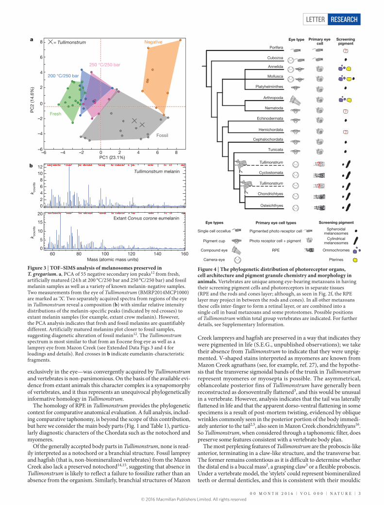
Spectra from Tullimonstrum and pure melanin samples12 show a simi-lar spectral composition (Fig. 3 and Extended Data Fig. 3). PCA show Tullimonstrum data plot among samples of fossil melanin12 (Fig. 3 and Extended Data Fig. 4), thus providing, in addition to anatomical localization and morphology, independent chemical evidence that the microbodies are melanosomes. An alternative interpretation is that microbodies are the remains of melanin-synthesizing bacteria or fungi. This scenario is unlikely because these microorganisms are not known to colonize decaying bodies, and their distribution in the fossils would require that they localized only to formerly melanin-synthesizing tissues.
Within the Mazon Creek Lagerstätte the only other fossils to possess paired, dark, ovoid structures are the numerous vertebrates (cyclos-tomes and gnathostomes), and a single putative coleoid13. In verte-brates, anatomical landmarks indicate that the dark structures are eyes (see, for example, refs 14–17 and Extended Data Fig. 5). Eyes in basal vertebrates are relatively decay resistant18,19 and pigment is one of the most decay resistant features in lampreys18,19. In Tullimonstrum, the dark structures are paired, bilaterally disposed and comprise thick, multi-layered masses of melanosomes. Together, these data constitute strong evidence that the dark structures are eyes.
Retinal pigments function as visual photoreceptors or as screen-ing pigments that act to prevent stray light from reaching the photo-receptive cells20. While all metazoans can synthesize melanin, ocular screening pigments are known to vary, and current data indicate that invertebrates chiefly employ ommochromes and pterines21. In annelids, molluscs and arthropods these pigments are contained in microbodies that are exclusively spherical or slightly oval, frequently facetted by abutting pigment granules and cell walls. There are a handful of inver-tebrate groups where melanin has been chemically identified as the screening pigment (planarian flatworms22, cubozoan cnidarians23 and ascidians24, phaeomelanin in the shell-eyes of chitons25). Significantly, the available ultrastructural data indicate that where these groups employ melanin, their melanosomes are exclusively ovoid (Fig. 4; see also Supplementary Information).
Chordates are unusual among metazoans in that their ocular screen-ing pigments are exclusively melanin23. In vertebrate eyes, the iris, choroid and retinal pigment epithelium (RPE) all contain melanosomes but the last tissue is distinct in having layers of ovoid and cylindrical melanosomes26. Tullimonstrum eyes comprise ovoid and cylindrical melanosomes that occur in distinct layers (that is, not intermixed; Fig. 2 and Extended Data Fig. 1) and we therefore interpret the melanosome layer as the remains of RPE. The possibility that this micro-anatomical complex—melanosomes of the same size and shape, arranged in layers,
Table 1 | Anatomical interpretations and affinity of T. gregarium in the literature
1 2 3 4 5 6
Reference Appendage Stylets in terminal structure
Transverse bar Distal structures on transverse bar
Transverse sigmoidal bands on trunk
Extensions to the posterior body
Proposed affinity
1 Jaw apparatus Stylets/teeth N/A Bar organs Segmentation Lateral tail fins Incertae sedis
3 Grasping claw Jaw
Stylets Sensory organs Otocysts Hydrodynamic stabilizers
Eyes Segmentation Lateral tail fins Nemertea Polychaeta Sipunculidea Arthropoda Echiuroidea
2 Buccal mass Teeth Eye stalks N/A Segmentation Muscle bands
Lateral tail fins Dorsoventral tail fins
Nemertea Polychaeta Mollusca
5 Grasping claw Jaw buccal mass
Teeth Paired copulatory organs Setae
N/A Segmentation Muscle bands
Dorsoventral tail fins
Mollusca Conodonta
4 N/A N/A N/A N/A Segmented muscles
Caudal appendage Nemertea Annelida
Proposed herein
Proboscis Tentative: teeth or dermal denticles?
Eye stalks Eyes Possible myomeres?
Dorsoventral tail fins
Non-osteichthyan total group vertebrate; possible total group gnathostome
Anatomical characters correspond with Fig. 1. ‘Proposed affinity’ lists the range of groups that Tullimonstrum has been allied to. Underlined groups indicate the phylogenetic placement favoured in each original study.
ey
tb
tb
a
b
l
c d
e f
Figure 2 | The ultrastructural details of the eyes of T. gregarium from the Mazon Creek Lagerstätte and an extant anchovy (Coilia nasus). a, Tullimonstrum (PE22061) with eye (ey) and transverse bar (tb). b, Eye in a, with possible lens (l) (see Extended Data Fig. 1). c–e, Scanning electron microscope images of melanosomes in Tullimonstrum eye (c, d, PE22061; e, PE22126). The boundary between layers is highlighted by blue applied to areas dominated by oblate melanosomes. f, Radial cross section (transmission electron microscope) of a larval anchovy retina with oblate and cylindrical melanosomes in the RPE (dark pigment granules). Image used with permission29. Decay-induced collapse of the RPE would result in a fossilized structure with oblate melanosomes overlying cylindrical as seen in c–e (see Extended Data Fig. 1), or vice versa, depending on specimen orientation. Scale bars, 10 mm (a); 2 μm (c–f).
© 2016 Macmillan Publishers Limited. All rights reserved
0 0 M O N T H 2 0 1 6 | V O L 0 0 0 | N A T U R E | 3
LETTER RESEARCH
exclusively in the eye—was convergently acquired by Tullimonstrum and vertebrates is non-parsimonious. On the basis of the available evi-dence from extant animals this character complex is a synapomorphy of vertebrates, and it thus represents an unequivocal phylogenetically informative homology in Tullimonstrum.
The homology of RPE in Tullimonstrum provides the phylogenetic context for comparative anatomical evaluation. A full analysis, includ-ing comparative taphonomy, is beyond the scope of this contribution, but here we consider the main body parts (Fig. 1 and Table 1), particu-larly diagnostic characters of the Chordata such as the notochord and myomeres.
Of the generally accepted body parts in Tullimonstrum, none is read-ily interpreted as a notochord or a branchial structure. Fossil lamprey and hagfish (that is, non-biomineralized vertebrates) from the Mazon Creek also lack a preserved notochord14,15, suggesting that absence in Tullimonstrum is likely to reflect a failure to fossilize rather than an absence from the organism. Similarly, branchial structures of Mazon
Creek lampreys and hagfish are preserved in a way that indicates they were pigmented in life (S.E.G., unpublished observations); we take their absence from Tullimonstrum to indicate that they were unpig-mented. V-shaped stains interpreted as myomeres are known from Mazon Creek agnathans (see, for example, ref. 27), and the hypothe-sis that the transverse sigmoidal bands of the trunk in Tullimonstrum represent myomeres or myosepta is possible. The asymmetrical, oblanceolate posterior fins of Tullimonstrum have generally been reconstructed as dorsoventrally flattened3, and this would be unusual in a vertebrate. However, analysis indicates that the tail was laterally flattened in life and that the apparent dorso-ventral flattening in some specimens is a result of post-mortem twisting, evidenced by oblique wrinkles commonly seen in the posterior portion of the body immedi-ately anterior to the tail2,5, also seen in Mazon Creek chondrichthyans16. So Tullimonstrum, when considered through a taphonomic filter, does preserve some features consistent with a vertebrate body plan.
The most perplexing features of Tullimonstrum are the proboscis-like anterior, terminating in a claw-like structure, and the transverse bar. The former remains contentious as it is difficult to determine whether the distal end is a buccal mass2, a grasping claw3 or a flexible proboscis. Under a vertebrate model, the ‘stylets’ could represent biomineralized teeth or dermal denticles, and this is consistent with their mouldic
b 1210
86420
k coun
ts
Tullimonstrum melanin
20
15
10
5
0
k coun
ts
1601401201008060Mass (atomic mass units)
Extant Corvus corone eumelanin
200 °C/250 bar
Fossil
= Tullimonstrum
Fresh
0 2 4 6 8–2–4–6
0
2
4
6
8
–2
–4
–6
a
PC1 (23.1%)
PC
2 (1
4.6%
)
250 °C/250 bar
Negative
Figure 3 | TOF–SIMS analysis of melanosomes preserved in T. gregarium. a, PCA of 55 negative secondary ion peaks12 from fresh, artificially matured (24 h at 200 °C/250 bar and 250 °C/250 bar) and fossil melanin samples as well as a variety of known melanin-negative samples. Two measurements from the eye of Tullimonstrum (BMRP2014MCP1000) are marked as ‘X’. Two separately acquired spectra from regions of the eye in Tullimonstrum reveal a composition (b) with similar relative intensity distributions of the melanin-specific peaks (indicated by red crosses) to extant melanin samples (for example, extant crow melanin). However, the PCA analysis indicates that fresh and fossil melanins are quantifiably different. Artificially matured melanins plot closer to fossil samples, suggesting diagenetic alteration of fossil melanin12. The Tullimonstrum spectrum is most similar to that from an Eocene frog eye as well as a lamprey eye from Mazon Creek (see Extended Data Figs 3 and 4 for loadings and details). Red crosses in b indicate eumelanin-characteristic fragments.
Pigmented photo receptor cell
Photo receptor cell + pigment
RPE
Spheroidalmelanosomes
Cylindricalmelanosomes
Ommochromes
Pterines
Single cell occellus
Pigment cup
Compound eye
Camera-eye
Primary eye cell
Porifera
Cubozoa
Annelida
Mollusca
Platyhelminthes
Arthropoda
Nematoda
Echinodermata
Cephalochordata
Tunicata
Cyclostomata
Chondrichtyes
Osteichthyes
Hemichordata
Tullimonstrum
Tullimonstrum
?
?
Screening pigment
?
Eye type
Primary eye cell types Screening pigment Eye types
Figure 4 | The phylogenetic distribution of photoreceptor organs, cell architecture and pigment granule chemistry and morphology in animals. Vertebrates are unique among eye-bearing metazoans in having their screening pigment cells and photoreceptors in separate tissues (RPE and the rods and cones layer; although, as seen in Fig. 2f, the RPE layer may project in between the rods and cones). In all other metazoans these cells inter-finger to form a retinal layer, or are combined into a single cell in basal metazoans and some protostomes. Possible positions of Tullimonstrum within total group vertebrates are indicated. For further details, see Supplementary Information.
© 2016 Macmillan Publishers Limited. All rights reserved

4 | N A T U R E | V O L 0 0 0 | 0 0 M O N T H 2 0 1 6
LETTERRESEARCH
preservation, comparable to biomineralized structures in Mazon Creek gnathostomes17. If the ‘claw’ is a buccal mass this might reflect anterior rostralization or posterior displacement of the eye. Perhaps more likely is the interpretation of this flexible rostral extension as a proboscis, similar to that of the Australian ghost shark, Callorhinchus milii (Holocephali). The unusual transverse bar we interpret as a stalked eye structure, on the basis of the presence of melanosomes and the remains of RPE. Stalked eyes occur in several animal groups including vertebrates (for exam-ple, larvae of several phylogenetically distinct teleost clades possessing eyes borne on stalks, up to one-quarter the length of the body28; the larvae of Idiacanthus fasciola28 and Stylopthalmus paradoxus resemble Tullimonstrum in having markedly stalked eyes and a rostral extension). Stalked eyes with well-developed RPE (and a possible lens, see Fig. 2 and Extended Data Fig. 1) suggests a camera-style eye capable of image for-mation, meaning that vision in Tullimonstrum involved more than sim-ple detection of light direction as is the case in non-vertebrate chordates.
None of the preserved anatomy of Tullimonstrum contradicts the hypothesis that it is a vertebrate, and in the absence of any other un equivocal indicators of homology we show that the intrinsic prop-erties of the eye, a character complex indicative of vertebrate RPE, provide compelling evidence that Tullimonstrum is a total group vertebrate. A dual-melanosome RPE evolved at some stage along the vertebrate stem and therefore does not constrain how near the base of the vertebrate tree Tullimonstrum might sit. However, if dual- melanosome RPE is a synapomorphy of crown vertebrates, and the stylets in the ‘claw’ prove to be the remains of biomineralized (phos-phatized) structures, the affinities of Tullimonstrum would lie with total group gnathostomes. Lacking any evidence of a bony skeleton, a placement within Osteichthyes is unlikely; however, without additional diagnostic characters, Tullimonstrum cannot currently be assigned to any more delineated clade.
Online Content Methods, along with any additional Extended Data display items and Source Data, are available in the online version of the paper; references unique to these sections appear only in the online paper.
Received 12 October 2015; accepted 9 March 2016.
Published online 13 April 2016.
1. Richardson, E. S. Jr. Wormlike fossil from the Pennsylvanian of Illinois. Science 151, 75–76 (1966).
2. Foster, M. in Mazon Creek Fossils (ed. Nitecki, M. H.) 269–301 (Academic, 1979).3. Johnson, R. G. & Richardson, E. S. Pennsylvanian invertebrates of the Mazon
Creek Area, Illinois: the morphology and affinities of Tullimonstrum. Fieldiana Geol. 12, 119–149 (1969).
4. Schram, F. in The Early Evolution of Metazoa and The Significance of Problematic Taxa (eds Conway-Morris, S. & Simonetta, A. M.) 35–46 (Cambridge Univ. Press, 1991).
5. Beall, B. in The Early Evolution of Metazoa and The Significance of Problematic Taxa (eds Conway-Morris, S. & Simonetta, A. M.) 271–286 (Cambridge Univ. Press, 1991).
6. Vinther, J., Briggs, D. E., Prum, R. O. & Saranathan, V. The colour of fossil feathers. Biol. Lett. 4, 522–525 (2008).
7. Clarke, J. A. et al. Fossil evidence for evolution of the shape and color of penguin feathers. Science 330, 954–957 (2010).
8. Lindgren, J. et al. Skin pigmentation provides evidence of convergent melanism in extinct marine reptiles. Nature 506, 484–488 (2014).
9. Donoghue, P. C. & Purnell, M. A. Distinguishing heat from light in debate over controversial fossils. BioEssays 31, 178–189 (2009).
10. Rieppel, O. & Kearney, M. Similarity. Biol. J. Linn. Soc. 75, 59–82 (2002).
11. Ruppert, E. E. Key characters uniting hemichordates and chordates: homologies or homoplasies? Can. J. Zool. 83, 8–23 (2005).
12. Colleary, C. et al. Chemical, experimental, and morphological evidence for diagenetically altered melanin in exceptionally preserved fossils. Proc. Natl Acad. Sci. USA 112, 12592–12597 (2015).
13. Kluessendorf, J. & Doyle, P. Pohlsepia mazonensis, an early ‘octopus’ from the Carboniferous of Illinois, USA. Palaeontology 43, 919–926 (2000).
14. Bardack, D. First fossil hagfish (Myxinoidea): a record from the Pennsylvanian of Illinois. Science 254, 701–703 (1991).
15. Bardack, D. & Zangerl, R. First fossil lamprey: a record from the Pennsylvanian of Illinois. Science 162, 1265–1267 (1968).
16. Sallan, L. C. & Coates, M. I. The long-rostrumed elasmobranch Bandringa zangerl, 1969, and taphonomy within a Carboniferous shark nursery. J. Vertebr. Paleontol. 34, 22–33 (2014).
17. Shabica, C. W. & Hay, A. Richardson’s Guide to The Fossil Fauna of Mazon Creek (eds Shabica, C. W. & Hay, A. H.) (Northeastern Illinois Univ., 1997).
18. Sansom, R. S., Gabbott, S. E. & Purnell, M. A. Decay of vertebrate characters in hagfish and lamprey (Cyclostomata) and the implications for the vertebrate fossil record. Proc. R. Soc. B 278, 1150–1157 (2011).
19. Sansom, R. S., Gabbott, S. E. & Purnell, M. A. Atlas of vertebrate decay: a visual and taphonomic guide to fossil interpretation. Palaeontology 56, 457–474 (2013).
20. Fein, A. & Szuts, E. Z. Photoreceptors, Their Role in Vision Vol. 5 (Cambridge Univ. Press, 1982).
21. Vopalensky, P. & Kozmik, Z. Eye evolution: common use and independent recruitment of genetic components. Phil. Trans. R. Soc. B 364, 2819–2832 (2009).
22. Hase, S. et al. Characterization of the pigment produced by the planarian, Dugesia ryukyuensis. Pigment Cell Res. 19, 248–249 (2006).
23. Kozmik, Z. et al. Assembly of the cnidarian camera-type eye from vertebrate-like components. Proc. Natl Acad. Sci. USA 105, 8989–8993 (2008).
24. Sato, S. & Yamamoto, H. Development of pigment cells in the brain of ascidian tadpole larvae: insights into the origins of vertebrate pigment cells. Pigment Cell Res. 14, 428–436 (2001).
25. Speiser, D. I., DeMartini, D. G. & Oakley, T. H. The shell-eyes of the chiton Acanthopleura granulata (Mollusca, Polyplacophora) use pheomelanin as a screening pigment. J. Nat. Hist. 48, 2899–2911 (2014).
26. Liu, Y. et al. Comparisons of the structural and chemical properties of melanosomes isolated from retinal pigment epithelium, iris and choroid of newborn and mature bovine eyes. Photochem. Photobiol. 81, 510–516 (2005).
27. Bardack, D. & Richardson, E. Jr. New agnathous fishes from the Pennsylvanian of Illinois. Fieldiana Geol. 33, 489–510 (1977).
28. Weihs, D. & Moser, H. Stalked eyes as an adaptation towards more efficient foraging in marine fish larvae. Bull. Mar. Sci. 31, 31–36 (1981).
29. Haacke, C., Hess, M., Melzer, R. R., Gebhart, H. & Smola, U. Fine structure and development of the retina of the grenadier anchovy Coilia nasus (Engraulididae, Clupeiformes). J. Morphol. 248, 41–55 (2001).
Supplementary Information is available in the online version of the paper.
Acknowledgements W. Simpson, P. Mayer, S. Williams, D. Rudkin and K. Seymour are thanked for specimen access and loans. Funding was through a Natural Environment Research Council studentship P14DF19 (to T.C.) and grant NE/K004557/1 (to M.A.P. and S.E.G.). We also acknowledge the National Science Foundation grant DMR-0923096 used to purchase the TOF–SIMS instrument at Texas Materials Institute, UTA. D. Murdock, C. Nedza, S. Wentges and A. Clements are thanked for proofreading. S. Furzeland is thanked for scanning electron microscope optimization. P. Smith is thanked for Adobe Illustrator tutorials.
Author Contributions S.E.G. and M.A.P. conceived the research programme of which this work is part. S.E.G., J.V. and T.C. designed and performed research. S.E.G., T.C., J.V., M.A.P. and A.D. wrote the manuscript. A.D. and J.V. undertook TOF–SIM analyses and interpretation. J.V., A.D. and M.A.P. conducted PCA. S.E.G., T.C., J.V. and P.M. operated and optimized the scanning electron microscope.
Author Information Reprints and permissions information is available at www.nature.com/reprints. The authors declare no competing financial interests. Readers are welcome to comment on the online version of the paper. Correspondence and requests for materials should be addressed to S.E.G. ([email protected]) or J.V. ([email protected]).
© 2016 Macmillan Publishers Limited. All rights reserved

LETTER RESEARCH
METHODSNo statistical methods were used to predetermine sample size. The experiments were not randomized. The investigators were not blinded to allocation during experiments and outcome assessment.
As part of a larger study on pigment preservation and taphonomy in the Mazon Creek, we investigated the dark elliptical patches at the terminations of the transverse bar in 12 specimens of T. gregarium from the Burpee Museum of Natural History, Illinois, and the Field Museum of Natural History, Illinois. We analysed textural and compositional data using a Hitachi S-3600N and Zeiss Sigma Environmental scanning electron microscope with an energy-dispersive X-ray spectroscopy system. Partial pressure was 20–30 Pa, working distance was between 9 and 12 mm, with an operating voltage of 15 kV. Specimens were uncoated. Specimens were optically imaged, using a Canon EOS 5D SLR camera and a Leica M205 C stereo microscope.
For TOF–SIMS analysis, one of the eyes in MCPX27C5369 (Burpee Museum of Natural History) was used. The specimen was placed in a TOF.SIMS 5 (ION-TOF, 2010) and secondary ion spectra were collected using a polyatomic analysis beam (Bi3
+, 30 keV, 0.9 pA sample current) to increase the yield of organic fragments, as previously employed in ref. 12. Two 500 μm × 500 μm areas were analysed in negative polarity with a resolution of 512 pixels × 512 pixels: one area included the eye and adjacent matrix, another region was selected within the main body of the eye. The acquired maps from within the eye showed no significant effect of topography and was analysed without further processing,
while a region of interest was chosen from the sampled area of the eye and sed-iment to minimize topographic-related artefacts. All spectra were mass cali-brated using the polyatomic fragment series of carbon (C-, C2-, C3-, C4-, C5-, C7-, C8-, C9-, C10-). The total count intensities of 55 select secondary ion peaks representative for melanin were used for PCA in conjunction with a previously collected data set12 of artificially matured melanin. Before PCA, each melanin- specific spectrum was normalized to its total intensity, the resulting data set was mean-centred and then standard-deviation-normalized across all samples for each composing mass30. The last process ensured that each melanin-specific peak was given the same weight in the PCA. The Tullimonstrum spectra are shown alongside extant reference melanin samples: black (eu)melanosomes from a glossy carrion crow (Corvus corone; Fig. 3b and Extended Data Fig. 3), reddish brown domestic chicken (Gallus gallus; Extended Data Fig. 3) and representative fossil samples (Jurassic ink sac and Eocene frog eye; Extended Data Fig. 3). Spatial mapping of the melanin-characteristic fragments of the melanosomes within the eye region show a clear separation at micrometre level between the cement (Extended Data Fig. 6e–h) and sediment (Extended Data Fig. 6i–l, u–x), while certain inorganic ions, attributed to calcium phosphates, occur associated with the melanosomes (Extended Data Fig. 6q–t).
30. Wagner, M. S., Graham, D. J., Ratner, B. D. & Castner, D. G. Maximizing information obtained from secondary ion mass spectra of organic thin films using multivariate analysis. Surf. Sci. 570, 78–97 (2004).
© 2016 Macmillan Publishers Limited. All rights reserved
LETTERRESEARCH
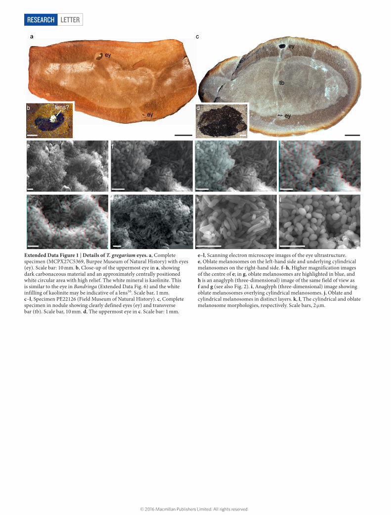
Extended Data Figure 1 | Details of T. gregarium eyes. a, Complete specimen (MCPX27C5369, Burpee Museum of Natural History) with eyes (ey). Scale bar: 10 mm. b, Close-up of the uppermost eye in a, showing dark carbonaceous material and an approximately centrally positioned white circular area with high relief. The white mineral is kaolinite. This is similar to the eye in Bandringa (Extended Data Fig. 6) and the white infilling of kaolinite may be indicative of a lens16. Scale bar, 1 mm. c–l, Specimen PE22126 (Field Museum of Natural History). c, Complete specimen in nodule showing clearly defined eyes (ey) and transverse bar (tb). Scale bar, 10 mm. d, The uppermost eye in c. Scale bar: 1 mm.
e–l, Scanning electron microscope images of the eye ultrastructure. e, Oblate melanosomes on the left-hand side and underlying cylindrical melanosomes on the right-hand side. f–h, Higher magnification images of the centre of e; in g, oblate melanosomes are highlighted in blue, and h is an anaglyph (three-dimensional) image of the same field of view as f and g (see also Fig. 2). i, Anaglyph (three-dimensional) image showing oblate melanosomes overlying cylindrical melanosomes. j, Oblate and cylindrical melanosomes in distinct layers. k, l, The cylindrical and oblate melanosome morphologies, respectively. Scale bars, 2 μm.
© 2016 Macmillan Publishers Limited. All rights reserved

LETTER RESEARCH
Extended Data Figure 2 | Tullimonstrum (BMRP2014MCP1000) with scanning electron microscope images of anatomical features. a, Complete specimen (anterior at top) with scanning electron microscope images showing the mode of preservation of the anatomy and that only the eyes contain melanosomes. Scale bar, 10 mm. b, Proboscis with small, dark, organic carbon patch (oC) which has a smooth texture. c, Distal
portion of the proboscis ‘claw’ showing pyrite crystals and framboids. d, Eye bar containing siderite and clay minerals. e, Eye showing melanosome texture. f, Dark transverse banding (possible myomeres) containing mainly siderite. g, The nodule matrix: siderite and detrital clay minerals. h, Main trunk: siderite and clay minerals. oC, organic carbon; sd, siderite; py, pyrite. Scale bars, 2 μm.
© 2016 Macmillan Publishers Limited. All rights reserved
LETTERRESEARCH
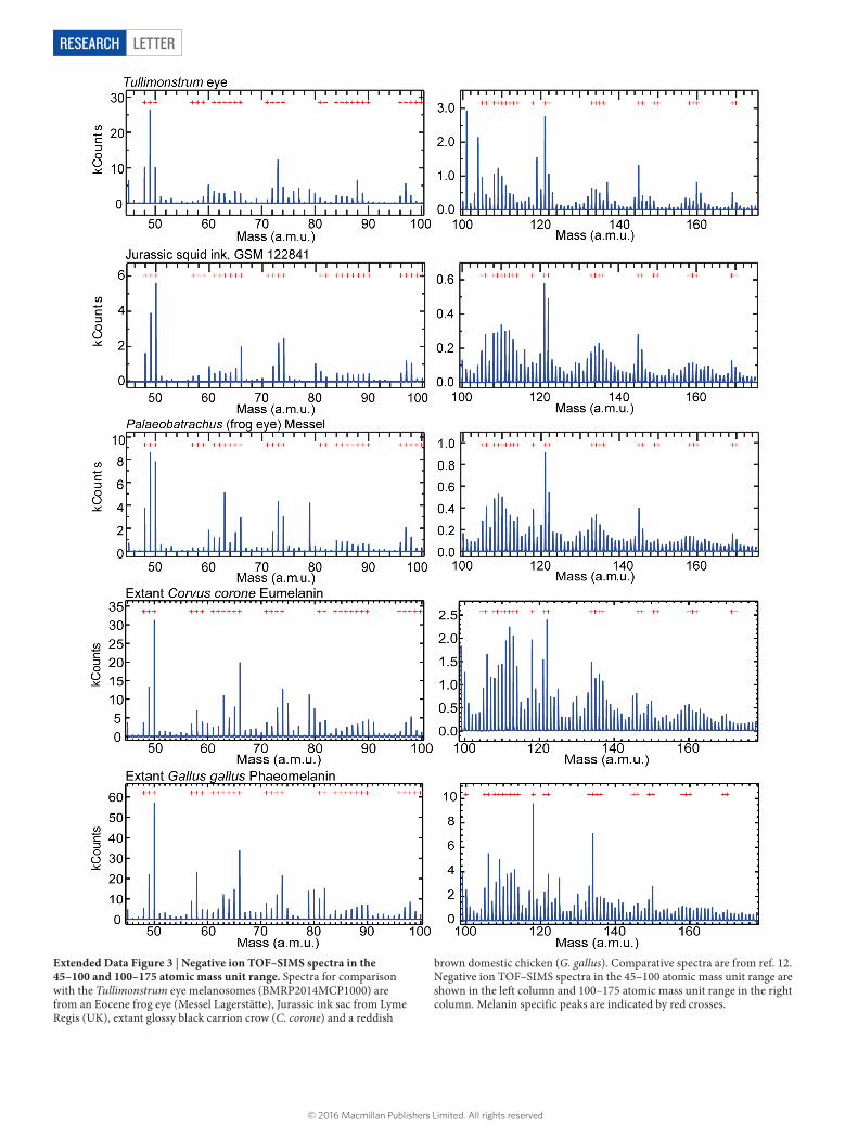
Extended Data Figure 3 | Negative ion TOF–SIMS spectra in the 45–100 and 100–175 atomic mass unit range. Spectra for comparison with the Tullimonstrum eye melanosomes (BMRP2014MCP1000) are from an Eocene frog eye (Messel Lagerstätte), Jurassic ink sac from Lyme Regis (UK), extant glossy black carrion crow (C. corone) and a reddish
brown domestic chicken (G. gallus). Comparative spectra are from ref. 12. Negative ion TOF–SIMS spectra in the 45–100 atomic mass unit range are shown in the left column and 100–175 atomic mass unit range in the right column. Melanin specific peaks are indicated by red crosses.
© 2016 Macmillan Publishers Limited. All rights reserved
LETTER RESEARCH
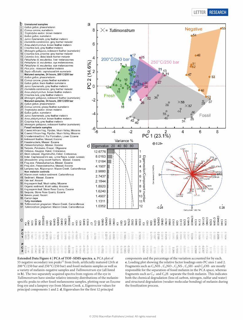
Extended Data Figure 4 | PCA of TOF–SIMS spectra. a, PCA plot of 55 negative secondary ion peaks12 from fresh, artificially matured (24 h at 200 °C/250 bar and 250 °C/250 bar) and fossil melanin samples as well as a variety of melanin-negative samples and Tullimonstrum eye (all listed in b). The two separately acquired spectra from regions of the eye in Tullimonstrum have similar relative intensity distributions of the melanin-specific peaks to other fossil melanosome samples, plotting near an Eocene frog eye and a lamprey eye from Mazon Creek. c, Eigenvector values for principal components 1 and 2. d, Eigenvalues for the first 12 principal
components and the percentage of the variation accounted for by each. e, Loading plot showing the relative factor loadings onto PC axes 1 and 2. Fragments such as CnNH-, CnNO-, CnNS-, CnSH- and CnOH- are mostly responsible for the separation of fossil melanin in the PCA space, whereas fragments such as Cn- and CnH- separate the fresh melanin. This indicates both the chemical degradation (loss of carbon, nitrogen, sulfur and water) and structural degradation (weaker molecular bonding) of melanin during the fossilization process.
© 2016 Macmillan Publishers Limited. All rights reserved
LETTERRESEARCH
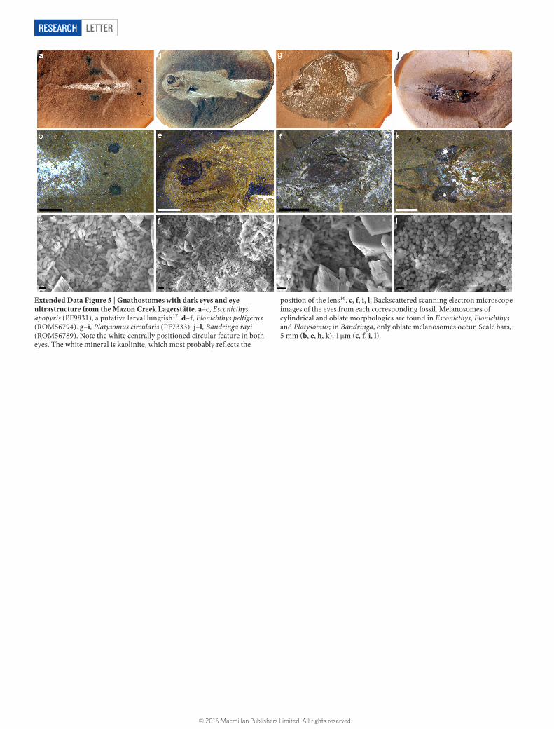
Extended Data Figure 5 | Gnathostomes with dark eyes and eye ultrastructure from the Mazon Creek Lagerstätte. a–c, Esconicthys apopyris (PF9831), a putative larval lungfish17. d–f, Elonichthys peltigerus (ROM56794). g–i, Platysomus circularis (PF7333). j–l, Bandringa rayi (ROM56789). Note the white centrally positioned circular feature in both eyes. The white mineral is kaolinite, which most probably reflects the
position of the lens16. c, f, i, l, Backscattered scanning electron microscope images of the eyes from each corresponding fossil. Melanosomes of cylindrical and oblate morphologies are found in Esconicthys, Elonichthys and Platysomus; in Bandringa, only oblate melanosomes occur. Scale bars, 5 mm (b, e, h, k); 1 μm (c, f, i, l).
© 2016 Macmillan Publishers Limited. All rights reserved

LETTER RESEARCH
Extended Data Figure 6 | TOF–SIMS intensity maps from eye region in Tullimonstrum, showing relative distribution of ions derived from melanin relative inorganic ions from the matrix. False-colour chemical mapping of the spatial distribution of several melanin-specific secondary ion fragments (a, e, i, m, q, u) compared with the maps of melanin-characteristic ions (b, c, n, o) and inorganic ions derived from the sediment (SiOn-, j, w; Al(Hn)On−, k, v) and the concretion cements (FeO2−, g; CaSOH-, f), which map distinctly from the melanin ions or co-occur with melanin (PO2−, r; PO2H−, s). The secondary ion CHO2−
is likely from carboxyl groups (o) and is a known constituent of melanin. It exhibits only a moderate overlap with melanin markers (p), which could be attributed to different diagenetic alterations of the melanin or some difference in composition. The right-hand column maps are composites of the tentatively assigned secondary ions in their respective row (that is, d is a composite of a–c). The distribution of inorganic and organic ions shows that the melanin and matrix ions are distinct contributions to the TOF–SIMS spectrum.
© 2016 Macmillan Publishers Limited. All rights reserved