The Extended Signal Peptide of the Trimeric ...jb.asm.org/content/193/24/6983.full.pdf · The...
12
JOURNAL OF BACTERIOLOGY, Dec. 2011, p. 6983–6994 Vol. 193, No. 24 0021-9193/11/$12.00 doi:10.1128/JB.05813-11 Copyright © 2011, American Society for Microbiology. All Rights Reserved. The Extended Signal Peptide of the Trimeric Autotransporter EmaA of Aggregatibacter actinomycetemcomitans Modulates Secretion X. Jiang, 1 T. Ruiz, 2 and K. P. Mintz 1 * Department of Microbiology and Molecular Genetics 1 and Department of Molecular Physiology and Biophysics, 2 University of Vermont, Burlington, Vermont 05405 Received 13 July 2011/Accepted 10 October 2011 The extracellular matrix protein adhesin A (EmaA) of the Gram-negative bacterium Aggregatibacter actino- mycetemcomitans is a fibrillar collagen adhesin belonging to the family of trimeric autotransporters. The protein forms antenna-like structures on the bacterial surface required for collagen adhesion. The 202-kDa protein monomers are proposed to be targeted and translocated across the inner membrane by a long signal peptide composed of 56 amino acids. The predicted signal peptide was functionally active in Escherichia coli and A. actinomycetemcomitans using truncated PhoA and Aae chimeric proteins, respectively. Mutations in the signal peptide were generated and characterized for PhoA activity in E. coli. A. actinomycetemcomitans strains expressing EmaA with the identical mutant signal peptides were assessed for cellular localization, surface expression, and collagen binding activity. All of the mutants impaired some aspect of EmaA structure or function. A signal peptide mutant that promoted alkaline phosphatase secretion did not allow any cell surface presentation of EmaA. A second mutant allowed for cell surface exposure but abolished protein function. A third mutant allowed for the normal localization and function of EmaA at 37°C but impaired localization at elevated temperatures. Likewise, replacement of the long EmaA signal peptide with a typical signal peptide also impaired localization above 37°C. The data suggest that the residues of the EmaA signal peptide are required for protein folding or assembly of this collagen adhesin. An important step in tissue colonization for many bacteria is binding to the extracellular matrix (54). Surface proteins are essential mediators for establishing these infectious foci. The extracellular matrix protein adhesin A (EmaA) is critical for the binding of the oral and systemic pathogen Aggregatibacter actinomycetemcomitans to fibrillar collagens (28). A. actinomy- cetemcomitans is a Gram-negative, capnophilic bacterium as- sociated with both adult and juvenile forms of periodontal disease (8, 44, 59). This bacterium is also associated with non- oral diseases including but not limited to pneumonia, bone disease, soft tissue abscesses, and infective endocarditis (6, 27, 32, 40). It has been shown that the loss of EmaA activity impairs the colonization of traumatized heart valves in a rabbit infective endocarditis model by A. actinomycetemcomitans (48). EmaA belongs to a specific family of autotransporter pro- teins and is secreted via the type V secretion pathway (17, 28). In general, autotransporters are targeted to the membrane by a signal peptide and translocated across the inner membrane using the Sec translocon (12, 17). Once in the periplasmic space, the carboxyl terminus of the protein is proposed to form an outer membrane pore for secretion of the passenger do- main through a yet undefined mechanism (10, 16). Following translocation across the outer membrane, the protein is either retained on the surface or proteolytically released into the milieu (12). EmaA and members of the type V c secretion pathway proteins differ from the traditional autotransporters by requiring three monomers to form the outer membrane pore for secretion (17). This adhesin is synthesized as 202-kDa monomers, which trimerize to form surface antenna-like struc- tures required for collagen binding (42). Inactivation of emaA results in a bacterial strain that does not display surface struc- tures and reduces the ability of the bacterium to bind to both purified and tissue collagens (42, 48). The biological activity of the EmaA structure has been assigned to the most distal do- main of the antenna-like structures, which is composed of the amino termini of the monomers, amino acids 70 to 386 (58). Recently, the three-dimensional architecture of the functional domain of this adhesin has been described and is composed of three subdomains (57, 58). In addition, oligosaccharides are suggested to be required for collagen binding, which are re- lated to the O polysaccharide (O-PS) of the lipopolysaccharide (LPS) of A. actinomycetemcomitans (49). Targeting to and translocation of the EmaA monomers across the inner membrane are predicted to be mediated by an amino-terminal signal peptide (28). The length of the pre- dicted EmaA signal peptide (56 amino acids) is much greater than that of typical signal peptides, which are composed of up to 25 amino acids (52). These long signal peptides are found predominantly associated with proteins secreted through the autotransporter, type V protein secretion pathway; long signal peptides are present in all of the serine protease autotrans- porters of members of the Enterobacteriaceae family (SPATEs) (47), some proteins of the two-partner secretion (TPS) path- way (19), select trimeric autotransporters (46), as well as the eukaryotic proteins interleukin 15 (IL-15) and plasminogen activator inhibitor 2 (3, 26). Amino-terminal signal peptides exhibit limited sequence similarity but are composed of clusters of charged or hydro- * Corresponding author. Mailing address: Department of Microbi- ology and Molecular Genetics, Room 118 Stafford Hall, University of Vermont, Burlington, VT 05405. Phone: (802) 656-0712. Fax: (802) 656-8749. E-mail: [email protected]. Published ahead of print on 14 October 2011. 6983 on July 22, 2018 by guest http://jb.asm.org/ Downloaded from
Transcript of The Extended Signal Peptide of the Trimeric ...jb.asm.org/content/193/24/6983.full.pdf · The...
JOURNAL OF BACTERIOLOGY, Dec. 2011, p. 6983–6994 Vol. 193, No. 240021-9193/11/$12.00 doi:10.1128/JB.05813-11Copyright © 2011, American Society for Microbiology. All Rights Reserved.
The Extended Signal Peptide of the Trimeric Autotransporter EmaAof Aggregatibacter actinomycetemcomitans Modulates Secretion�
X. Jiang,1 T. Ruiz,2 and K. P. Mintz1*Department of Microbiology and Molecular Genetics1 and Department of Molecular Physiology and Biophysics,2
University of Vermont, Burlington, Vermont 05405
Received 13 July 2011/Accepted 10 October 2011
The extracellular matrix protein adhesin A (EmaA) of the Gram-negative bacterium Aggregatibacter actino-mycetemcomitans is a fibrillar collagen adhesin belonging to the family of trimeric autotransporters. Theprotein forms antenna-like structures on the bacterial surface required for collagen adhesion. The 202-kDaprotein monomers are proposed to be targeted and translocated across the inner membrane by a long signalpeptide composed of 56 amino acids. The predicted signal peptide was functionally active in Escherichia coli andA. actinomycetemcomitans using truncated PhoA and Aae chimeric proteins, respectively. Mutations in thesignal peptide were generated and characterized for PhoA activity in E. coli. A. actinomycetemcomitans strainsexpressing EmaA with the identical mutant signal peptides were assessed for cellular localization, surfaceexpression, and collagen binding activity. All of the mutants impaired some aspect of EmaA structure orfunction. A signal peptide mutant that promoted alkaline phosphatase secretion did not allow any cell surfacepresentation of EmaA. A second mutant allowed for cell surface exposure but abolished protein function. Athird mutant allowed for the normal localization and function of EmaA at 37°C but impaired localization atelevated temperatures. Likewise, replacement of the long EmaA signal peptide with a typical signal peptide alsoimpaired localization above 37°C. The data suggest that the residues of the EmaA signal peptide are requiredfor protein folding or assembly of this collagen adhesin.
An important step in tissue colonization for many bacteria isbinding to the extracellular matrix (54). Surface proteins areessential mediators for establishing these infectious foci. Theextracellular matrix protein adhesin A (EmaA) is critical forthe binding of the oral and systemic pathogen Aggregatibacteractinomycetemcomitans to fibrillar collagens (28). A. actinomy-cetemcomitans is a Gram-negative, capnophilic bacterium as-sociated with both adult and juvenile forms of periodontaldisease (8, 44, 59). This bacterium is also associated with non-oral diseases including but not limited to pneumonia, bonedisease, soft tissue abscesses, and infective endocarditis (6, 27,32, 40). It has been shown that the loss of EmaA activityimpairs the colonization of traumatized heart valves in a rabbitinfective endocarditis model by A. actinomycetemcomitans(48).
EmaA belongs to a specific family of autotransporter pro-teins and is secreted via the type V secretion pathway (17, 28).In general, autotransporters are targeted to the membrane bya signal peptide and translocated across the inner membraneusing the Sec translocon (12, 17). Once in the periplasmicspace, the carboxyl terminus of the protein is proposed to forman outer membrane pore for secretion of the passenger do-main through a yet undefined mechanism (10, 16). Followingtranslocation across the outer membrane, the protein is eitherretained on the surface or proteolytically released into themilieu (12). EmaA and members of the type Vc secretionpathway proteins differ from the traditional autotransporters
by requiring three monomers to form the outer membranepore for secretion (17). This adhesin is synthesized as 202-kDamonomers, which trimerize to form surface antenna-like struc-tures required for collagen binding (42). Inactivation of emaAresults in a bacterial strain that does not display surface struc-tures and reduces the ability of the bacterium to bind to bothpurified and tissue collagens (42, 48). The biological activity ofthe EmaA structure has been assigned to the most distal do-main of the antenna-like structures, which is composed of theamino termini of the monomers, amino acids 70 to 386 (58).Recently, the three-dimensional architecture of the functionaldomain of this adhesin has been described and is composed ofthree subdomains (57, 58). In addition, oligosaccharides aresuggested to be required for collagen binding, which are re-lated to the O polysaccharide (O-PS) of the lipopolysaccharide(LPS) of A. actinomycetemcomitans (49).
Targeting to and translocation of the EmaA monomersacross the inner membrane are predicted to be mediated by anamino-terminal signal peptide (28). The length of the pre-dicted EmaA signal peptide (56 amino acids) is much greaterthan that of typical signal peptides, which are composed of upto 25 amino acids (52). These long signal peptides are foundpredominantly associated with proteins secreted through theautotransporter, type V protein secretion pathway; long signalpeptides are present in all of the serine protease autotrans-porters of members of the Enterobacteriaceae family (SPATEs)(47), some proteins of the two-partner secretion (TPS) path-way (19), select trimeric autotransporters (46), as well as theeukaryotic proteins interleukin 15 (IL-15) and plasminogenactivator inhibitor 2 (3, 26).
Amino-terminal signal peptides exhibit limited sequencesimilarity but are composed of clusters of charged or hydro-
* Corresponding author. Mailing address: Department of Microbi-ology and Molecular Genetics, Room 118 Stafford Hall, University ofVermont, Burlington, VT 05405. Phone: (802) 656-0712. Fax: (802)656-8749. E-mail: [email protected].
� Published ahead of print on 14 October 2011.
6983
on July 22, 2018 by guesthttp://jb.asm
.org/D
ownloaded from
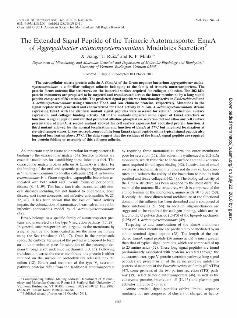
phobic amino acids that are required for interaction with theprotein secretory machinery in the cytoplasm (10, 51). Typicalsignal peptides are divided into three regions, with a variablenumber of amino acids. An uncommonly high number of pos-itively charged amino acids present after the start methionineconstitute the N region, followed by a region of hydrophobicamino acids (H region) adjacent to a sequence containing thecleavage site for the inner membrane-bound signal peptidase(C region). The C region usually contains small, slightly polaramino acids at the �1 and �3 positions of the signal peptidasecleavage site (53). The carboxyl-terminal half of long signalpeptides contains the canonical N, H, and C regions of typicalsignal peptides but displays high sequence variability (47).Conversely, the amino-terminal amino acids contain a unique,highly conserved sequence motif, referred to as the N-terminalextension or extended signal peptide region (ESPR) (Fig. 1Aand B) (11, 47). Some long signal peptides may be composedof two divergent NH regions followed by a C region, denotedN1H1N2H2C, with little sequence similarity between the indi-vidual N and H regions (11, 17). The role of the N-terminalextension remains to be elucidated.
In this study, we demonstrate that the first 56 amino acids ofthe EmaA sequence are required for protein secretion. Theresults of amino acid deletion analysis of the signal peptidesuggest that the residues are required for proper protein fold-ing and assembly of this collagen adhesin. Furthermore, wehave demonstrated that part of the long EmaA signal peptideis important for the secretion of this virulence determinant inresponse to changes in growth conditions.
MATERIALS AND METHODS
Bacterial strains and growth conditions. The bacterial strains and plasmidsused in this study are listed in Table 1. Unless noted, all A. actinomycetemcomi-tans strains were grown statically in 3% Trypticase soy broth supplemented with0.6% yeast extract (TSBYE) in a 10% CO2 humidified incubator at 37°C. Anti-biotics were added to a final concentration of 50 �g/ml kanamycin or 1 �g/mlchloramphenicol for A. actinomycetemcomitans, when necessary. All Escherichia
coli strains were grown with agitation in Luria-Bertani broth (LB) at 37°Ccontaining 50 �g/ml kanamycin, 20 �g/ml chloramphenicol, 50 �g/ml ampicillin,or 500 �g/ml erythromycin, as appropriate.
For some experiments, bacteria were adapted for growth at 39°C. Bacteria,from frozen stocks, were inoculated on TSBYE agar and grown at 39°C in 10%CO2. Colonies were subsequently inoculated into prewarmed TSBYE broth andgrown statically at 39°C. For heat shock experiments, the cells were initiallygrown in TSBYE at 37°C overnight. The cells were diluted, grown at 37°C for 30min in 10% CO2, sealed, and incubated at 42°C for 3 h before analysis. Bacterialgrowth at 39°C and 42°C was comparable to the growth of cells grown at 37°C forthe stated periods of time.
In-frame deletion constructs. A shuttle plasmid (pKM9) containing the full-length emaA sequence and 500 bp upstream of the start methionine (GenBankaccession number AY344064) was used to generate in-frame deletion constructsutilizing unique 5� SphI and 3� KpnI (within the emaA sequence) restriction sitesfor directional cloning (58). The deletion constructs were generated by overlap-ping PCR methodology. Three rounds of PCR were completed. The first roundutilized a forward primer (CBP1-5�up) and a reverse primer designed to containthe complementary sequence flanking the sequence of interest to encompass the5� SphI site. The second round utilized a forward primer complementary to thesequence of interest and the reverse primer CBP1-3131Rev to generate a prod-uct containing the unique 3� KpnI restriction site (Table 2). The PCR productswere purified using a QIAquick gel extraction kit (Qiagen, Hilden, Germany)and used as the template for the final round of PCR using the primer setCBP1-5�up and CBP1-3131Rev. The final PCR product was purified, treatedwith the respective enzymes, and ligated with purified pQEmaA (Table 1). Theligation product was transformed into E. coli DH10B cells by electroporation(45), and ampicillin-resistant colonies were selected. Plasmid DNA was digestedwith SphI and SmaI (located at the 3� end of the emaA gene), ligated with pKM1(Table 1) previously digested with SphI and HincII and transformed into E. coliDH10B. Kanamycin-resistant colonies were selected, and the purified plasmidcontaining the modified emaA sequence was transformed into the emaA mutantstrain of A. actinomycetemcomitans by electroporation as described previously(45). All PCR constructs were confirmed by DNA sequence analysis performedat the Vermont Cancer Center DNA Analysis Facility, University of Vermont.
Construction and analysis of phoA fusions. Alkaline phosphatase fusion con-structs were generated using pHRM104, a plasmid containing a truncated genefor alkaline phosphatase (phoA) that lacks a functional signal sequence, as thebackbone (36). The emaA signal peptide in-frame deletion constructs containingthe emaA promoter region were amplified with engineered BamHI sites (Table2). The purified PCR products were treated with BamHI and ligated withpHRM104 previously digested with BamHI. The ligation mixture was trans-formed into electrocompetent E. coli CC118 cells with mutant phoA genes, anderythromycin-resistant transformants were selected. The orientation of the insert
FIG. 1. Signal peptides of selected Gram-negative bacterial proteins. (A) Sequence alignment of long signal peptides of proteins from differentorganisms. Alignments were generated using ClustalW2 (7). Identical amino acids (�), highly conserved amino acids (:), and weakly conservedamino acids (.) are indicated below the sequence alignment. Gaps introduced to maximize sequence alignment are indicated by the dashes.(B) Predicted domain structure of the EmaA signal peptide and schematics of the in-frame EmaA signal peptide deletion constructs used in thisstudy. The arrow indicates the predicted signal peptidase cleavage site between amino acid 56 (Ala) and 57 (Tyr). (C) Sequence of the typical signalpeptide of Omp34 of A. actinomycetemcomitans.
6984 JIANG ET AL. J. BACTERIOL.
on July 22, 2018 by guesthttp://jb.asm
.org/D
ownloaded from
was verified by PCR using the requisite primer set (Table 2). The hia signalpeptide-phoA fusion was kindly provided by J. St. Geme, Duke University Med-ical Center, Durham, North Carolina (46).
PhoA activity was determined based on the colorimetric assay of Brickmanand Beckwith (5). Data were collected from three individual experiments per-formed in triplicate and statistically analyzed for significance using Student’s ttest for statistical significance (P � 0.05).
Heterologous signal peptide fusion constructs. Two heterologous signal pep-tide fusions using A. actinomycetemcomitans outer membrane proteins weredeveloped in this investigation. The nucleotide sequences corresponding to thesignal peptides of the outer membrane protein 34 (Omp34) (GenBank accessionnumber AB015936) (55) and EmaA was generated by overlapping PCR meth-odology. The omp34 signal peptide sequence was generated by PCR using therequisite primer set (Table 2) and genomic DNA as the template. The 500-bpsequence upstream of the emaA start codon and the 5� end of the emaA gene,after the signal peptide sequence, were amplified (Table 2) and purified. Amixture of the three products was used as template with the requisite primer set(Table 2), and the PCR product was treated with SphI and KpnI, ligated withpQEmaA previously digested with the corresponding enzymes, and transformedinto E. coli DH10B cells.
The signal peptide of EmaA was fused to the epithelial cell adhesin, Aae(GenBank accession number AY262734) (41), with the endogenous signal pep-tide deleted, by overlapping PCR methodology. The emaA upstream sequenceand the sequence for the signal peptide were amplified using the requisiteprimers, one of which contains the complementary sequence for the aae genedownstream of the signal peptide sequence. In a separate reaction, the aaesequence from bp 82 of the coding sequence (minus the signal peptide sequence)was amplified (Table 2). The purified PCR products were used as the templatewith the requisite primers (Table 2) to generate the fusion product. The purified
product was treated with SphI and SacI and ligated with the empty plasmid(pKM2) previously treated with the same enzymes. The ligation product wastransformed into the aae mutant strain VT1565 (41).
Surface detection of Aae. An enzyme-linked immunosorbent assay (ELISA)was developed based on a modified version of the method of Mintz and Fives-Taylor (30). Briefly, 1 � 108 CFU of mid-log-phase bacterial cells were adsorbedto microtiter plate wells, and surface-bound Aae was localized by the addition ofanti-Aae polyclonal antiserum. Immunoreactive complexes were detected by theaddition of enzyme-conjugated goat anti-rabbit serum (Sigma-Aldrich, Inc., St.Louis, MO) in the presence of a colorimetric substrate. Data were collected fromthree individual experiments in triplicate and statistically analyzed for signifi-cance using Student’s t test for significance (P � 0.05).
Generation of a chromosomal in-frame EmaA signal peptide deletion mutant.A mutant with a chromosomal deletion of nucleotides 46 to 117 of the emaAcoding sequence, corresponding to amino acids 16 to 39, was generated allelicreplacement of the wild-type sequence with a DNA fragment containing a se-lectable marker 500 bp upstream of the deleted sequence. The DNA fragmentfor homologous recombination was generated using overlapping PCR method-ology. A 500-bp DNA fragment of the coenzyme A (CoA) ligase gene, which isfound upstream of the emaA sequence, was amplified with an engineered 5�sequence of the aad9 selectable marker (GenBank accession number M69221)(Table 2). The aad9 selectable marker was amplified to generate DNA with endscomplementary to the CoA ligase and the intergenic sequence of emaA (Table2). A plasmid containing the in-frame deletion corresponding to amino acids 16to 39 was used as the template to generate the third DNA fragment for over-lapping PCR (Table 2). The PCR products were purified, combined, and used asthe template for PCR using the requisite primers (Table 2). The overlappingPCR product was cloned into the TA cloning vector TOPO (Invitrogen, Carls-bad, CA). The 2.7-kb fragment was excised from the plasmid by digestion with
TABLE 1. Bacterial strains and plasmids used in this study
Bacterial strain or plasmid Description or relevant genotype or phenotype Reference or source
A. actinomycetemcomitans strainsVT1169 Wild-type strain, a derivative of SUNY465 31KM73 emaA mutant strain 28VT1565 aae mutant strain 41KM333 In-frame deletion of emaA gene region corresponding to deletion of amino acids
16 to 39 of EmaA proteinThis study
E. coli strainsDH10B F� mcrA �(mrr-hsdRMS-mcrBC) �80dlacZ�M15 �lacX74 endA1 recA1 deoR
�(ara leu)7697 araD139 galU galK nupG rpsL ��15
DH5 � pir endA1 hsdR17(r� m) supE44 thi-1 recA1 gyrA1(Nalr) relA1 �(lacIZYA-argF)U169 deo ��80 �lacD(lacZ)M15� pir R6K
SM10 � pir thi-1 thr leu tonA lacY supE recA::RP4-2Tc::Mu l pir R6K 39CC118 phoA20 thi-1 rspE rpoB argE(Am) recA1 18
PlasmidspKM1 Shuttle vector; Kanr 42pKM2 Shuttle vector; Cmr 14pVT1460 Mobilization plasmid 29pVT1566 aae gene sequence cloned into pGEM-T Easy 41pKM9 Upstream promoter region and emaA in pKM1 42pQE30 Intermediate cloning vector; Ampr QiagenpHRM104 Plasmid containing truncated phoA 17pQEmaA Upstream promoter region and emaA in pQE30 This studypKM�2-23 In-frame deletion of bp 4 to 69 of emaA in pKM9 This studypKM�2-35 In-frame deletion of bp 4 to 105 of emaA in pKM9 This studypKM�24-53 In-frame deletion of bp 70 to 159 of emaA in pKM9 This studypKM�16-39 In-frame deletion of bp 46 to 117 of emaA in pKM9 This studypKMOmp34SP emaA signal peptide replaced by omp34 signal peptide in pKM9 This studypKMEmaASP-Aae aae containing the emaA signal peptide and upstream promoter region in pKM2 This studypHRMEmaASP emaA signal peptide and upstream promoter region in pHRM104 This studypHRMEmaASP�2-56 In-frame deletion of bp 4 to 168 of pHRMEmaASP This studypHRMEmaASP�2-23 In-frame deletion of bp 4 to 69 of pHRMEmaASP This studypHRMEmaASP�2-35 In-frame deletion of bp 4 to 105 of pHRMEmaASP This studypHRMEmaASP�24-53 In-frame deletion of bp 70 to 159 of pHRMEmaASP This studypHRMEmaASP�16-39 In-frame deletion of bp 46 to 117 of pHRMEmaASP This studypHRMHiaSP hia signal peptide and emaA upstream promoter region in pHRM104 This study
VOL. 193, 2011 ANALYSIS OF THE EXTENDED SIGNAL PEPTIDE OF EmaA 6985
on July 22, 2018 by guesthttp://jb.asm
.org/D
ownloaded from

EcoRI, purified, and ligated with the pVT1460, previously treated with EcoRI,for conjugation as described previously (29).
Quantitative real-time PCR. Total bacterial RNA was isolated using the Qia-gen RNeasy kit (Qiagen, Hilden, Germany) according to the manufacturer’sprotocol and used for reverse transcription using SuperScript III first-strandsynthesis system for reverse transcription-PCR (RT-PCR) (Invitrogen, Carlsbad,CA) according to the manufacturer’s protocol. Quantitative real-time PCR wasperformed as described previously (57) in the DNA Analysis Facility, VermontCancer Center, at the University of Vermont. The primers and the TaqManfluorogenic probes (Sigma Chemical Co.) used are present in Table 2. Theexpression of emaA was normalized to expression of the endogenous 16S rRNAfor variation in RNA quantity and quality.
The relative quantification of target gene expression was performed using thecomparative cycle threshold (CT) method (1a). The emaA mutant strain trans-formed with pKM9 was chosen as the calibrator. Results are presented as themeans standard deviations of three independent experiments.
Transmission electron microscopy. Bacterial samples were prepared by themethod of Ruiz et al. (42) using Nano-W (Nanoprobes, Yaphank, NY) as thestaining agent. Data collection was carried out using a Tecnai 12 electron mi-croscope (FEI, Hillsboro, OR) equipped with a LaB6 cathode, a 14-�m, 2,048-by 2,048-pixel charge-coupled-device (CCD) camera (TVIPS, Gauting, Ger-many) and a dual-axis tilt tomography holder (Fischione, Export, PA), operatingat 100 kV. Micrographs were recorded using the CCD camera at a nominal
magnification of �52,000, which corresponds to a 0.25-nm pixel size on thespecimen scale.
Isolation of bacterial membranes and aggregated proteins. Bacterial mem-branes were prepared following the protocol described by Mintz (28). Theprotein concentration was determined spectrophotometrically at a wavelength of280 nm following the addition of sodium dodecyl sulfate (SDS) to a final con-centration of 2% (wt/vol).
Aggregated proteins in the membrane fraction samples were isolated accord-ing to the protocol of Tomoyasu et al. (50). Briefly, membrane fraction sampleswere treated with 10% (vol/vol) NP-40, and the aggregated proteins were iso-lated by centrifugation and analyzed for EmaA aggregates by immunoblotting.
Immunoblot analysis. The amount of EmaA synthesized by the bacterialstrains was analyzed by immunodot blot analysis of membrane fragments follow-ing the procedure of Yu et al. (57) using a monoclonal antibody specific for thestalk region of EmaA. The nitrocellulose membranes were exposed to photo-graphic film or imaged using a Bio-Rad Molecular Imager Gel Doc XR system(Bio-Rad Laboratories). The intensity of the dots was quantified using theBio-Rad Quantity One software.
Membrane localization of Aae was determined by immunoblotting of SDS-polyacrylamide gels transferred to nitrocellulose membranes using polyclonalantisera raised against a recombinant protein corresponding to the passengerdomain of Aae (41).
The presence of PhoA in the bacterial whole-cell lysates was determined by
TABLE 2. Oligonucleotides used in this study
Oligonucleotidea Sequence (5�–3�)b
CBP1-5�up..................................................................................................ACATGCATGCAACAAATCGCCGTCATCGCCCBP1-3131Rev...........................................................................................GACTGCTAAATTCTTTCCTGCCEmaA�2-23For .........................................................................................CAAAAAGGAAAACATAAGATGAAAGCTTTTTCCCTTTCTACCACEmaA�2-23Rev.........................................................................................GTGGTAGAAAGGGAAAAAGCTTTCATCTTATGTTTTCCTTTTTGEmaA�24-53For .......................................................................................GAACTATCTTTTAATACCAACGCTTACATTGCTATAGGEmaA�24-53Rev.......................................................................................CCTATAGCAATGTAAGCGTTGGTATTAAAAGATAGTTCEmaA�16-39For .......................................................................................GGTGTAAAACATCTCAGACAATATTCATTGCTGCAGCCCCGEmaA�16-39Rev.......................................................................................CGGGGCTGCAGCAATGAATATTGTCTGAGATGTTTTACACComp34SPFor................................................................................................GCACCACAAGCAAACACTomp34SPRev ...............................................................................................CACTACGAATTAAAGCGGemaAP-omp34SPFor ..................................................................................CAAAAAGGAAAACATAAGATGAAAAGAACTGCAATCemaAP-omp34SPRev .................................................................................GATTGCAGTTCTTTTCATCTTATGTTTTCCTTTTTGomp34SP-emaAFor.....................................................................................GCAACAGTAGCACAGGCATACATTGCTATAGGTTCTomp34SP-emaARev....................................................................................AGAACCTATAGCAATGTATGCCTGTGCTACTGTTGCemaASP-aaeFor ..........................................................................................TCCTTTAATACCAACGCTTCAGAGTTTAATGCTCAAATAAATAATemaASP-aaeRev .........................................................................................ATTATTTATTTGAGCATTAAACTCTGAAGCGTTGGTATTAAAGGAaaeBamFor.................................................................................................GGATCCTTCAGAGTTTAATGCTCAAaaeSacRev ..................................................................................................GAGCTCTTACCAGTAGTAATTCAGHiaSP-EmaA5�For.....................................................................................CAAAAAGGAAAACATAAGATGAACAAAATTTTTAACGHiaSP-EmaA5�Rev ....................................................................................CGTTAAAAATTTTGTTCATCTTATGTTTTCCTTTTTGHiaSP-EmaA3�For.....................................................................................GTCCGCAACGGTTGAGGCGTTACATTGCTATAGGTTCTGHiaSP-EmaA3�Rev ....................................................................................CAGAACCTATAGCAATGTAACGCCTCAACCGTTGCGGACEmaASPBamHIRev ..................................................................................GGATCCAGCGTTGGTATTAAAEmaAPromoterBamHIRev......................................................................GGATCCCATCTTATGTTTTCCTTTTTGEmaA�24-53BamHIRev..........................................................................GGATCCAGATAGTTCAGATACGGCOmp34SPBamHIRev.................................................................................GGATCCTGCCTGTGCTACTGTTGCTGCHiaSPBamHIRev .......................................................................................GGATCCCGCCTCAACCGTTGCGGPhoA396Rev..............................................................................................AATATCGCCCTGAGCAGCCCoA1347StuIFor .......................................................................................GATGCAGGCCTCGACGGCAATTTATACATCCBP1-7StuIRev .........................................................................................GGAGAAGGCCTTTGACGCATCATCGCAAGCoA-aad95�For..........................................................................................GTATAACTAAATGATTCATCATCGATTTTCGTTCGTGAATACCoA-aad95�Rev.........................................................................................GTATTCACGAACGAAAATCGATGATGAATCATTTAGTTATACaad9-EmaAFor..........................................................................................CAATAAACCTTGCATATGGATTCATCATAAAAGTGCaad9-EmaARev .........................................................................................GCACTTTTATGATGAATCCATATGCAAGGTTTATTGSpc2Rev......................................................................................................CTCTTGCCAGTCACGTTACG16SrRNAfor...............................................................................................GAACCTTACCTACTCTTGACATCC16SrRNArev ..............................................................................................GGACTTAACCCAACATTTCACAAC16SrRNAprobe..........................................................................................6-FAM-CTGACGACAGCCATGCAGCACCTG-BHQ-1emaAstalkfor .............................................................................................GGTCAATAACGACGGTGTTACGemaAstalkrev .............................................................................................TTCCCTTTCGCAACGTTAGCemaAstalkprobe ........................................................................................6-FAM-CGGTCCAAGCATGACAAGCCACGG-BHQ-1
a Forward (For) and reverse (rev) oligonucleotide primers and probes.b 6-FAM, 6-carboxyfluorescein; BHQ-1, black hole quencher 1.
6986 JIANG ET AL. J. BACTERIOL.
on July 22, 2018 by guesthttp://jb.asm
.org/D
ownloaded from

immunoblotting of SDS-polyacrylamide gels transferred to nitrocellulose mem-branes using rabbit anti-PhoA antiserum (Rockland Immunochemicals Inc., Gil-bertsville, PA). The immune complexes were detected by horseradish peroxi-dase-conjugated goat anti-rabbit antibodies and exposed to photographic film.
Collagen binding assay. Collagen binding activity was determined as describedpreviously (58). Data were collected from three individual experiments in trip-licate and statistically analyzed for significance using Student’s t test (P � 0.05)or two-way analysis of variance (ANOVA) where appropriate.
RESULTS
The first 56 amino acids of EmaA act as a signal peptide forinner membrane translocation in E. coli. A signal peptidasecleavage site between amino acids 56 and 57 of the EmaAprotein sequence (Fig. 1B) is predicted by the Signal P algo-rithm (4, 33, 34). The membrane targeting and translocationactivity of this sequence was determined in E. coli using a phoAreporter construct, which lacks the native signal peptide andpromoter (36, 46). PhoA activity is detected only when theenzyme is translocated across the bacterial inner membrane(5). Alkaline phosphatase activity was clearly associated withthe emaASP-phoA construct as shown in Fig. 2. The activity ofthe strain expressing EmaASP (EmaA signal peptide corre-sponding to amino acids 1 to 56) was comparable to the activityassociated with the strain expressing the 49-amino-acid signalpeptide of the Haemophilus influenzae trimeric autotransporterepithelial cell adhesin Hia (46) fused to PhoA (HiaSP) used asa positive control. These activities are in contrast to the lack ofenzymatic activity associated with the construct that containsthe promoter but lacks the first 56 amino acids of the EmaAsequence (EmaASP�2-56). The presence of phosphatase activ-ity in the EmaASP-expressing strain suggested that the first 56amino acids of EmaA were sufficient to mediate the targetingand translocation of PhoA across the bacterial inner mem-brane.
The EmaA signal peptide drives secretion of the epithelialcell adhesin Aae in A. actinomycetemcomitans. Based on thesealkaline phosphatase studies, we generated a fusion constructto determine whether the first 56 amino acids of the EmaAprotein sequence also function as a signal peptide in A. acti-nomycetemcomitans. The predicted EmaA signal peptide se-quence was fused in-frame with the passenger domain of thetypical A. actinomycetemcomitans autotransporter Aae, aknown epithelial cell adhesin (41), and transformed into an aaemutant strain. Immunoblot analysis of membrane proteinsfrom the strain containing this construct (EmaASP-Aae) dem-onstrated the presence of immunoreactive material associatedwith the membrane fraction (Fig. 3A, lane 3). The stainingpattern is comparable to that of the A. actinomycetemcomitanswild-type membrane fraction (Fig. 3A, lane 2). Immunoreac-tivity was not associated with membrane proteins derived fromthe aae mutant strain (Fig. 3A, lane 1). The immunoreactivebands with a molecular mass lower than that of the intactprotein (130 kDa) (Fig. 3A, lanes 2 and 3) are proposed to bedegradation products of Aae.
In the strain expressing the Aae chimera, Aae was localizedto the bacterial surface, as indicated in Fig. 3B. Whole bacteriaimmobilized onto a solid surface and probed with anti-Aaeserum demonstrated a statistically significant difference in an-tibody binding activity between the strain expressing the Aaechimera and the aae mutant strain. A strain expressing wild-
type levels of Aae was used as the positive control for surfaceexpression of Aae. The Aae surface expression levels wereequivalent in both the wild-type strain and the Aae chimera-expressing strains (Fig. 3B).
The N-terminal extension and canonical signal peptide re-gions are required for protein secretion. The EmaA long signalpeptide can be divided into the N-terminal extension andthe canonical N, H, C region (Fig. 1B). Therefore, deletionmutants of the N-terminal extension (amino acids 2 to 23)and the canonical region (amino acids 24 to 56) were con-structed and investigated for membrane translocation. In E.coli, the strain expressing EmaASP�2-23 (EmaASP withamino acids 2 to 23 deleted) displayed PhoA activity but atlevels much lower than the full-length sequence (Fig. 2A).This is in contrast to the strain expressing EmaASP�24-53(EmaASP with amino acids 24 to 53 deleted) that lackedPhoA activity (Fig. 2A). The lack of enzyme activity corre-
FIG. 2. Alkaline phosphatase activity of signal peptide constructsin E. coli. Plasmids were constructed with the emaA promoter adjacentto DNA sequences that encode the entire signal peptide or regions ofthe emaA signal peptide fused to a truncated form of phoA(pHRM104). All constructs were transformed into strain CC118, an E.coli phoA mutant strain. (A) PhoA enzymatic activity represented asmicromoles of para-nitrophenyl generated per minute. HiaSP, signalpeptide from the epithelial cell adhesin Hia of H. influenzae (aminoacids 1 to 49); EmaASP, EmaA signal peptide corresponding to aminoacids 1 to 56; EmaASP�2-56, EmaA signal peptide with amino acids 2to 56 deleted; EmaASP�2-23, EmaA signal peptide with amino acids 2to 23 deleted; EmaASP�24-53, EmaA signal peptide with amino acids24 to 53 deleted; EmaASP�2-35, EmaA signal peptide with aminoacids 2 to 35 deleted; EmaASP�16-39, EmaA signal peptide with aminoacids 16 to 39 deleted. (B) Immunoblot analysis of alkaline phospha-tase expression. Whole-cell lysates, derived from the E. coli CC118strains expressing the PhoA constructs described above for panel A,were probed with anti-PhoA antiserum. The constructs are shown inthe same order as in panel A.
VOL. 193, 2011 ANALYSIS OF THE EXTENDED SIGNAL PEPTIDE OF EmaA 6987
on July 22, 2018 by guesthttp://jb.asm
.org/D
ownloaded from

lated with the absence of PhoA protein as determined byimmunoblot analysis in E. coli (Fig. 2B).
A. actinomycetemcomitans strains transformed with thesesignal peptide mutations in the emaA gene were assessed for
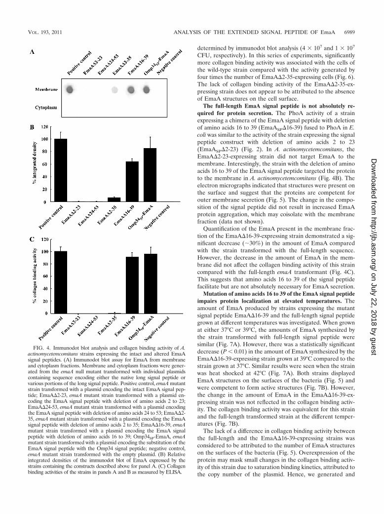
EmaA localization in the cytoplasm or membrane fractionsusing an anti-EmaA monoclonal antibody (58). EmaA formsaggregates upon solubilization and heating, which does notallow for reproducible entry and separation of the monomersin SDS-polyacrylamide gels (58). Immunoreactive EmaA isusually associated with the wells of the stacking gel, and only asmall amount of monomer is present at the expected molecularweight. Therefore, for a more reliable quantification of theamount of EmaA produced by the strains used in this study, animmunodot blot format was used. Fractionation of the wild-type bacterial cells into cytoplasm and membrane fractionsindicated that EmaA was localized to the membrane fraction,with little, if any, immunoreactive protein present in the cyto-plasm at the concentrations used (Fig. 4A). In A. actinomyce-temcomitans, EmaA was not detected in the cytoplasm orthe membrane fraction of the strains expressing eitherEmaA�2-23 or EmaA�24-53 sequences (Fig. 4B). In addition,EmaA structures were absent on the surfaces of these strains(Fig. 5). Quantitative real-time PCR (QRT-PCR) analysis ofthese constructs suggested the absence of any defect in tran-scription of emaA mRNA compared with the fully comple-mented strain (data not shown). Associated with the absenceof structures was the loss of collagen binding activity of thesestrains, which was similar to the emaA mutant strain containingthe empty vector (Fig. 4C). The data indicated that neither ofthe amino acid sequences 2-23 or 24-53 of the signal peptidecan support EmaA secretion alone.
Deletion of a portion of the canonical signal peptide regionallows for surface expression but abolished collagen bindingactivity. An in-frame EmaA signal peptide deletion construct,corresponding to deletion of amino acids 2 to 35, was gener-ated and assayed for PhoA activity in E. coli. The strain ex-pressing this construct (EmaASP�2-35) demonstrated mar-ginal PhoA activity, compared with the intact signal peptide(Fig. 2A). In A. actinomycetemcomitans, the strain expressingthe plasmid with this deletion in the emaA gene (EmaA�2-35)contained less than 10% of the EmaA protein associated withthe membrane fraction compared with the strain expressingthe full-length signal peptide (Fig. 4B). EmaA in the cytoplasmof this strain was not detected (Fig. 4A). Transcriptional ac-tivity of the EmaA�2-35 plasmid was similar to the plasmidcontaining the full-length signal peptide (data not shown).
Transmission electron microscopy images indicate the pres-ence of EmaA structures on the surface of this strain (Fig. 5).However, the reduced frequency of the visualization of EmaAstructures in these images suggested that the number of EmaAstructures on the surface of this strain was greatly diminishedcompared with the positive-control strain. Associated with thereduction of EmaA structures was the loss of collagen bindingactivity in the EmaA�2-35-expressing strain (Fig. 4C). Thebinding activity of the EmaA�2-35-expressing strain was sim-ilar to the binding activity of the emaA mutant strain, whichdoes not synthesize any detectable EmaA protein or show anystructures on the bacterial surface (Fig. 4C and 5). Since thecollagen binding activity of these strains was determined usingequal cell numbers in the ELISA, we posited that the differ-ence in collagen binding activity could be attributed to theamount of EmaA synthesized. Therefore, the number of cellsof the EmaA�2-35-expressing strain and wild-type strainadded to the assay was first normalized for EmaA protein, as
FIG. 3. Membrane and surface localization of the A. actinomyce-temcomitans epithelial cell adhesin Aae. (A) Immunoblot analysis ofbacterial membranes probed with a polyclonal antiserum specific forAae. Lane 1, aae mutant strain; lane 2, wild-type (WT) strain(VT1169); lane 3, aae mutant strain transformed with a replicatingplasmid expressing the EmaA signal peptide-Aae chimera (EmaASP-Aae). (B) Detection of Aae on the bacterial surface. Surface-exposedAae was detected using antibodies specific for Aae in an ELISA formatwith bacteria immobilized on the bottom of wells on 96-well microtiterplates.
6988 JIANG ET AL. J. BACTERIOL.
on July 22, 2018 by guesthttp://jb.asm
.org/D
ownloaded from
determined by immunodot blot analysis (4 � 107 and 1 � 107
CFU, respectively). In this series of experiments, significantlymore collagen binding activity was associated with the cells ofthe wild-type strain compared with the activity generated byfour times the number of EmaA�2-35-expressing cells (Fig. 6).The lack of collagen binding activity of the EmaA�2-35-ex-pressing strain does not appear to be attributed to the absenceof EmaA structures on the cell surface.
The full-length EmaA signal peptide is not absolutely re-quired for protein secretion. The PhoA activity of a strainexpressing a chimera of the EmaA signal peptide with deletionof amino acids 16 to 39 (EmaASP�16-39) fused to PhoA in E.coli was similar to the activity of the strain expressing the signalpeptide construct with deletion of amino acids 2 to 23(EmaASP�2-23) (Fig. 2). In A. actinomycetemcomitans, theEmaA�2-23-expressing strain did not target EmaA to themembrane. Interestingly, the strain with the deletion of aminoacids 16 to 39 of the EmaA signal peptide targeted the proteinto the membrane in A. actinomycetemcomitans (Fig. 4B). Theelectron micrographs indicated that structures were present onthe surface and suggest that the proteins are competent forouter membrane secretion (Fig. 5). The change in the compo-sition of the signal peptide did not result in increased EmaAprotein aggregation, which may coisolate with the membranefraction (data not shown).
Quantification of the EmaA present in the membrane frac-tion of the EmaA�16-39-expressing strain demonstrated a sig-nificant decrease (�30%) in the amount of EmaA comparedwith the strain transformed with the full-length sequence.However, the decrease in the amount of EmaA in the mem-brane did not affect the collagen binding activity of this straincompared with the full-length emaA transformant (Fig. 4C).This suggests that amino acids 16 to 39 of the signal peptidefacilitate but are not absolutely necessary for EmaA secretion.
Mutation of amino acids 16 to 39 of the EmaA signal peptideimpairs protein localization at elevated temperatures. Theamount of EmaA produced by strains expressing the mutantsignal peptide EmaA�16-39 and the full-length signal peptidegrown at different temperatures was investigated. When grownat either 37°C or 39°C, the amounts of EmaA synthesized bythe strain transformed with full-length signal peptide weresimilar (Fig. 7A). However, there was a statistically significantdecrease (P � 0.01) in the amount of EmaA synthesized by theEmaA�16-39-expressing strain grown at 39°C compared to thestrain grown at 37°C. Similar results were seen when the strainwas heat shocked at 42°C (Fig. 7A). Both strains displayedEmaA structures on the surfaces of the bacteria (Fig. 5) andwere competent to form active structures (Fig. 7B). However,the change in the amount of EmaA in the EmaA�16-39-ex-pressing strain was not reflected in the collagen binding activ-ity. The collagen binding activity was equivalent for this strainand the full-length transformed strain at the different temper-atures (Fig. 7B).
The lack of a difference in collagen binding activity betweenthe full-length and the EmaA�16-39-expressing strains wasconsidered to be attributed to the number of EmaA structureson the surfaces of the bacteria (Fig. 5). Overexpression of theprotein may mask small changes in the collagen binding activ-ity of this strain due to saturation binding kinetics, attributed tothe copy number of the plasmid. Hence, we generated and
FIG. 4. Immunodot blot analysis and collagen binding activity of A.actinomycetemcomitans strains expressing the intact and altered EmaAsignal peptides. (A) Immunodot blot assay for EmaA from membraneand cytoplasm fractions. Membrane and cytoplasm fractions were gener-ated from the emaA null mutant transformed with individual plasmidscontaining sequence encoding either the native long signal peptide orvarious portions of the long signal peptide. Positive control, emaA mutantstrain transformed with a plasmid encoding the intact EmaA signal pep-tide; EmaA�2-23, emaA mutant strain transformed with a plasmid en-coding the EmaA signal peptide with deletion of amino acids 2 to 23;EmaA�24-53, emaA mutant strain transformed with a plasmid encodingthe EmaA signal peptide with deletion of amino acids 24 to 53; EmaA�2-35, emaA mutant strain transformed with a plasmid encoding the EmaAsignal peptide with deletion of amino acids 2 to 35; EmaA�16-39, emaAmutant strain transformed with a plasmid encoding the EmaA signalpeptide with deletion of amino acids 16 to 39; Omp34SP-EmaA, emaAmutant strain transformed with a plasmid encoding the substitution of theEmaA signal peptide with the Omp34 signal peptide; negative control,emaA mutant strain transformed with the empty plasmid. (B) Relativeintegrated densities of the immunodot blot of EmaA expressed by thestrains containing the constructs described above for panel A. (C) Collagenbinding activities of the strains in panels A and B as measured by ELISA.
VOL. 193, 2011 ANALYSIS OF THE EXTENDED SIGNAL PEPTIDE OF EmaA 6989
on July 22, 2018 by guesthttp://jb.asm
.org/D
ownloaded from
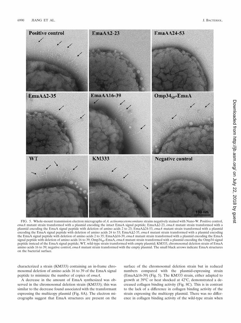
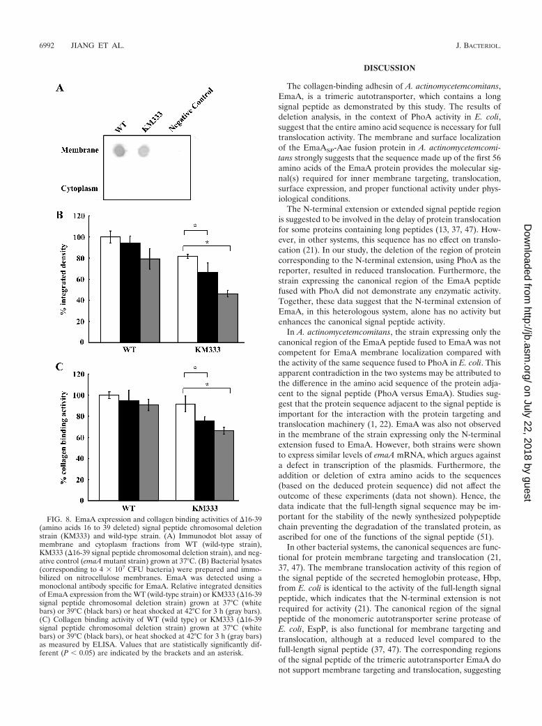
characterized a strain (KM333) containing an in-frame chro-mosomal deletion of amino acids 16 to 39 of the EmaA signalpeptide to minimize the number of copies of emaA.
A decrease in the amount of EmaA synthesized was ob-served in the chromosomal deletion strain (KM333); this wassimilar to the decrease found associated with the transformantexpressing the multicopy plasmid (Fig. 8A). The electron mi-crographs suggest that EmaA structures are present on the
surface of the chromosomal deletion strain but in reducednumbers compared with the plasmid-expressing strain(EmaA�16-39) (Fig. 5). The KM333 strain, either adapted togrowth at 39°C or heat shocked at 42°C, demonstrated a de-creased collagen binding activity (Fig. 8C). This is in contrastto the lack of a difference in collagen binding activity of thestrain expressing the multicopy plasmid. There was no differ-ence in collagen binding activity of the wild-type strain when
FIG. 5. Whole-mount transmission electron micrographs of A. actinomycetemcomitans strains negatively stained with Nano-W. Positive control,emaA mutant strain transformed with a plasmid encoding the intact EmaA signal peptide; EmaA�2-23, emaA mutant strain transformed with aplasmid encoding the EmaA signal peptide with deletion of amino acids 2 to 23; EmaA�24-53, emaA mutant strain transformed with a plasmidencoding the EmaA signal peptide with deletion of amino acids 24 to 53; EmaA�2-35, emaA mutant strain transformed with a plasmid encodingthe EmaA signal peptide with deletion of amino acids 2 to 35; EmaA�16-39, emaA mutant strain transformed with a plasmid encoding the EmaAsignal peptide with deletion of amino acids 16 to 39; Omp34SP-EmaA, emaA mutant strain transformed with a plasmid encoding the Omp34 signalpeptide instead of the EmaA signal peptide; WT, wild-type strain transformed with empty plasmid; KM333, chromosomal deletion strain of EmaAamino acids 16 to 39; negative control, emaA mutant strain transformed with the empty plasmid. The small black arrows indicate EmaA structureson the bacterial surface.
6990 JIANG ET AL. J. BACTERIOL.
on July 22, 2018 by guesthttp://jb.asm
.org/D
ownloaded from

grown at the different temperatures, suggesting a role for thissequence of the signal peptide in the expression of EmaA atelevated temperatures.
A typical signal peptide is sufficient for EmaA secretion. Afusion construct composed of the signal peptide of Omp34 (55)and the passenger domain of EmaA (Omp34SP-EmaA) wasgenerated to determine whether a typical signal peptide (N, H,C region) would support EmaA protein secretion. In the emaAmutant strain transformed with the Omp34SP-EmaA fusionconstruct, EmaA was located in the membrane fraction (Fig.4A and B) in amounts similar to the strain expressing thecomplete emaA gene.
Electron micrographs of the Omp34SP-EmaA-expressingstrain clearly demonstrated the ability of the translocated pro-teins to form EmaA antenna-like structures on the bacterialsurface (Fig. 5). This is in stark contrast to the surface of theemaA mutant strain, which does not display any EmaA surfacestructures (Fig. 5, negative control). The proper oligomeriza-tion of the EmaA monomers to form a functionally activestructure, in terms of collagen binding, was also determined forthese strains. The collagen binding activity of the Omp34SP-EmaA-expressing strain was similar to the emaA mutant straintransformed with the entire emaA gene on the same plasmidbackbone (Fig. 4C).
The long signal peptide of EmaA was required for maximumsecretion at elevated temperatures (Fig. 7). To determine if atypical signal peptide responds to elevated temperatures sim-ilar to the long signal peptide, the Omp34SP-EmaA-expressingstrain was grown under heat shock conditions and analyzed forthe amount of EmaA present in the membrane. As demon-strated in Fig. 7C, there was a statistically significant decreasein the amount of membrane-localized EmaA in the heat-shocked strain compared with the same strain grown at 37°C.
FIG. 6. Collagen binding activities of A. actinomycetemcomitansstrains. Collagen binding activity of 1 � 107 CFU WT (wild-type strain)or 4 � 107 CFU EmaA�2-35 (emaA mutant strain transformed withpKM�2-35) as measured by ELISA.
FIG. 7. EmaA immunodot blot and collagen binding activities oftransformed A. actinomycetemcomitans strains. (A) Bacterial lysates(corresponding to 4 � 107 CFU bacteria) were prepared and immo-bilized on nitrocellulose membranes. EmaA was detected using amonoclonal antibody specific for EmaA in cells grown at 37°C (blackbars) or 39°C (white bars) or heat shocked at 42°C (gray bars). Positivecontrol, emaA mutant strain expressing the intact EmaA protein (thestrain was transformed with pKM9); EmaA�16-39, emaA mutantstrain expressing the EmaA protein with amino acids 16 to 39 deleted(the strain was transformed with pKM�16-39). (B) Collagen bindingactivity of the strain expressing intact EmaA (positive control) or strainEmaA�16-39 (emaA mutant strain transformed with a plasmid encod-ing the EmaA signal peptide with amino acids 16 to 39 deleted) grownat 37°C (black bars) or 39°C (white bars) or heat shocked at 42°C (graybars) as measured by ELISA. (C) Integrated intensities of the immu-noreactive protein in the membrane of cells grown at 37°C (white bars)or heat shocked at 42°C (black bars). Positive control, emaA mutantstrain expressing the intact EmaA protein (the strain was transformedwith pKM9); Omp34SP-EmaA, emaA mutant strain transformed with aplasmid encoding the Omp34 signal peptide instead of the EmaAsignal peptide.
VOL. 193, 2011 ANALYSIS OF THE EXTENDED SIGNAL PEPTIDE OF EmaA 6991
on July 22, 2018 by guesthttp://jb.asm
.org/D
ownloaded from
DISCUSSION
The collagen-binding adhesin of A. actinomycetemcomitans,EmaA, is a trimeric autotransporter, which contains a longsignal peptide as demonstrated by this study. The results ofdeletion analysis, in the context of PhoA activity in E. coli,suggest that the entire amino acid sequence is necessary for fulltranslocation activity. The membrane and surface localizationof the EmaASP-Aae fusion protein in A. actinomycetemcomi-tans strongly suggests that the sequence made up of the first 56amino acids of the EmaA protein provides the molecular sig-nal(s) required for inner membrane targeting, translocation,surface expression, and proper functional activity under phys-iological conditions.
The N-terminal extension or extended signal peptide regionis suggested to be involved in the delay of protein translocationfor some proteins containing long peptides (13, 37, 47). How-ever, in other systems, this sequence has no effect on translo-cation (21). In our study, the deletion of the region of proteincorresponding to the N-terminal extension, using PhoA as thereporter, resulted in reduced translocation. Furthermore, thestrain expressing the canonical region of the EmaA peptidefused with PhoA did not demonstrate any enzymatic activity.Together, these data suggest that the N-terminal extension ofEmaA, in this heterologous system, alone has no activity butenhances the canonical signal peptide activity.
In A. actinomycetemcomitans, the strain expressing only thecanonical region of the EmaA peptide fused to EmaA was notcompetent for EmaA membrane localization compared withthe activity of the same sequence fused to PhoA in E. coli. Thisapparent contradiction in the two systems may be attributed tothe difference in the amino acid sequence of the protein adja-cent to the signal peptide (PhoA versus EmaA). Studies sug-gest that the protein sequence adjacent to the signal peptide isimportant for the interaction with the protein targeting andtranslocation machinery (1, 22). EmaA was also not observedin the membrane of the strain expressing only the N-terminalextension fused to EmaA. However, both strains were shownto express similar levels of emaA mRNA, which argues againsta defect in transcription of the plasmids. Furthermore, theaddition or deletion of extra amino acids to the sequences(based on the deduced protein sequence) did not affect theoutcome of these experiments (data not shown). Hence, thedata indicate that the full-length signal sequence may be im-portant for the stability of the newly synthesized polypeptidechain preventing the degradation of the translated protein, asascribed for one of the functions of the signal peptide (51).
In other bacterial systems, the canonical sequences are func-tional for protein membrane targeting and translocation (21,37, 47). The membrane translocation activity of this region ofthe signal peptide of the secreted hemoglobin protease, Hbp,from E. coli is identical to the activity of the full-length signalpeptide, which indicates that the N-terminal extension is notrequired for activity (21). The canonical region of the signalpeptide of the monomeric autotransporter serine protease ofE. coli, EspP, is also functional for membrane targeting andtranslocation, although at a reduced level compared to thefull-length signal peptide (37, 47). The corresponding regionsof the signal peptide of the trimeric autotransporter EmaA donot support membrane targeting and translocation, suggesting
FIG. 8. EmaA expression and collagen binding activities of �16-39(amino acids 16 to 39 deleted) signal peptide chromosomal deletionstrain (KM333) and wild-type strain. (A) Immunodot blot assay ofmembrane and cytoplasm fractions from WT (wild-type strain),KM333 (�16-39 signal peptide chromosomal deletion strain), and neg-ative control (emaA mutant strain) grown at 37°C. (B) Bacterial lysates(corresponding to 4 � 107 CFU bacteria) were prepared and immo-bilized on nitrocellulose membranes. EmaA was detected using amonoclonal antibody specific for EmaA. Relative integrated densitiesof EmaA expression from the WT (wild-type strain) or KM333 (�16-39signal peptide chromosomal deletion strain) grown at 37°C (whitebars) or 39°C (black bars) or heat shocked at 42°C for 3 h (gray bars).(C) Collagen binding activity of WT (wild type) or KM333 (�16-39signal peptide chromosomal deletion strain) grown at 37°C (whitebars) or 39°C (black bars), or heat shocked at 42°C for 3 h (gray bars)as measured by ELISA. Values that are statistically significantly dif-ferent (P � 0.05) are indicated by the brackets and an asterisk.
6992 JIANG ET AL. J. BACTERIOL.
on July 22, 2018 by guesthttp://jb.asm
.org/D
ownloaded from
a difference in the function of the long signal peptide of EmaAin A. actinomycetemcomitans. Studies of the signal peptide ofother members of the trimeric autotransporter family have notbeen reported.
The N-terminal extension and the canonical region of thelong signal peptide of EmaA are not sufficient for membranelocalization of EmaA in A. actinomycetemcomitans. However, asequence within the canonical region (amino acids 36 to 56)demonstrated activity in both bacterial systems, albeit at re-duced levels. The presence of EmaA structures and the ab-sence of collagen binding activity associated with this mutantsuggest that this sequence allows for normal EmaA localiza-tion but abolishes the function of the adhesin. Studies proposethat the secondary structure of the signal peptide modulatesthe cleavage specificity of the signal peptidase (20, 38). There-fore, a change in the secondary structure of the EmaA�2-35signal peptide may alter the peptidase cleavage site, resultingin modified EmaA monomers that are not competent to foldinto an active conformation (58). Alternatively, the lack ofcollagen binding of this strain may be due to a reduction in thenumber of EmaA surface structures per cell.
Inspection of the long signal peptide sequence of EmaAsuggested that deletion of amino acids 16 to 39 of the full-length sequence would result in a peptide that resembles atypical signal peptide (Fig. 1B). Our data clearly demonstratethat in this strain, EmaA is transported to the membrane anddisplays surface structures that interact with collagen. The datasuggest that amino acids 16 to 39 facilitate maximal secretionbut are not completely essential for protein targeting andtranslocation. Furthermore, the data indicate that amino acids40 to 53, in context with the N-terminal extension, are impor-tant or required for the interaction of the peptide with theprotein secretion machinery.
A distinct role for long signal peptides has not yet beenaddressed in other organisms. We hypothesize that the addi-tional amino acids (amino acids 16 to 39) in the EmaA signalpeptide function to ensure the secretion of this adhesin underphysiological stress. These stresses may include changes in theenvironment of the gingival sulcus, which occurs during thetransition between health and disease (2, 35, 43, 44). Theseconditions may contribute to a stress response in the bacte-rium, which translates to an overall change in the transcrip-tional activity of specific gene products, including heat shockproteins or stress-induced proteins (56). These molecularchaperones modulate polypeptide folding, assembly, degrada-tion, and translocation (9, 24, 25). We observed a reduction inthe amount of EmaA and collagen binding of the chromosomalEmaA�16-39 mutant strain (KM333) grown at increased tem-peratures compared with the same strain grown at 37°C. Thisdifference in the amount of EmaA protein and collagen bind-ing activity was not observed with the wild-type strain grownunder identical conditions. Interestingly, we have demon-strated that a typical signal peptide can drive secretion ofEmaA. However, in this strain, the reduction in the amount ofEmaA secreted at 42°C (Fig. 7C) supports the hypothesis thatamino acids 16 to 39 of the long signal peptide are important,in part, for the proper protein folding of this adhesin at ele-vated temperatures. The experimental data using the typicalsignal peptide of Omp34 further corroborate our hypothesis.
The binding of A. actinomycetemcomitans to collagen, me-
diated by EmaA, is important for the initiation of infectiveendocarditis (48) and may contribute to the pathogenicity ofthis bacterium in periodontal diseases. EmaA is the only pro-tein that contains an extended signal peptide of the membraneproteins in A. actinomycetemcomitans that have been charac-terized, such as Aae, Omp34, and Omp100 (23, 41, 55). Thedata presented in this study suggest that the amino acids of theextended signal peptide of EmaA are required for proteinstability in the cytoplasm and the proper assembly of themonomers to form a functional adhesin. Furthermore, specificamino acids of the long signal peptide may be important forthe presentation of this adhesin on the bacterial surface to bindand colonize the oral cavity under changing environmentalconditions.
ACKNOWLEDGMENTS
We thank Chris Lennox for his contribution to this project.This research was supported by National Institutes of Health-Na-
tional Institute of Dental and Craniofacial Research (NIH-NIDCR)grant RO1-DE13824.
REFERENCES
1. Andrews, D. W., E. Perara, C. Lesser, and V. R. Lingappa. 1988. Sequencesbeyond the cleavage site influence signal peptide function. J. Biol. Chem.263:15791–15798.
1a.Applied Biosystems. 1997. ABI PRISM 7700 sequence detection system:user bulletin #2. Relative quantification of gene expression. Applied Bio-systems, Foster City, CA.
2. Baab, D. A., A. Oberg, and A. Lundstrom. 1990. Gingival blood flow andtemperature changes in young humans with a history of periodontitis. Arch.Oral Biol. 35:95–101.
3. Belin, D., S. Bost, J. D. Vassalli, and K. Strub. 1996. A two-step recognitionof signal sequences determines the translocation efficiency of proteins.EMBO J. 15:468–478.
4. Bendtsen, J. D., H. Nielsen, G. von Heijne, and S. Brunak. 2004. Improvedprediction of signal peptides: SignalP 3.0. J. Mol. Biol. 340:783–795.
5. Brickman, E., and J. Beckwith. 1975. Analysis of the regulation of Esche-richia coli alkaline phosphatase synthesis using deletions and phi80 trans-ducing phages. J. Mol. Biol. 96:307–316.
6. Carlile, J. R., E. N. Beckman, and R. M. Arensman. 1984. Actinobacillusactinomycetemcomitans pneumonia. Clin. Pediatr. (Phila.) 23:578–580.
7. Chenna, R., et al. 2003. Multiple sequence alignment with the Clustal seriesof programs. Nucleic Acids Res. 31:3497–3500.
8. Christersson, L. A. 1993. Actinobacillus actinomycetemcomitans and local-ized juvenile periodontitis. Clinical, microbiologic and histologic studies.Swed. Dent. J. Suppl. 90:1–46.
9. Collier, D. N., V. A. Bankaitis, J. B. Weiss, and P. J. Bassford, Jr. 1988. Theantifolding activity of SecB promotes the export of the E. coli maltose-binding protein. Cell 53:273–283.
10. Dautin, N., and H. D. Bernstein. 2007. Protein secretion in gram-negativebacteria via the autotransporter pathway. Annu. Rev. Microbiol. 61:89–112.
11. Desvaux, M., et al. 2006. The unusual extended signal peptide region of thetype V secretion system is phylogenetically restricted. FEMS Microbiol. Lett.264:22–30.
12. Desvaux, M., N. J. Parham, and I. R. Henderson. 2004. Type V proteinsecretion: simplicity gone awry? Curr. Issues Mol. Biol. 6:111–124.
13. Desvaux, M., et al. 2007. A conserved extended signal peptide region directsposttranslational protein translocation via a novel mechanism. Microbiology153:59–70.
14. Gallant, C. V., M. Sedic, E. A. Chicoine, T. Ruiz, and K. P. Mintz. 2008.Membrane morphology and leukotoxin secretion are associated with a novelmembrane protein of Aggregatibacter actinomycetemcomitans. J. Bacteriol.190:5972–5980.
15. Grant, S. G., J. Jessee, F. R. Bloom, and D. Hanahan. 1990. Differentialplasmid rescue from transgenic mouse DNAs into Escherichia coli methyla-tion-restriction mutants. Proc. Natl. Acad. Sci. U. S. A. 87:4645–4649.
16. Hegde, R. S., and H. D. Bernstein. 2006. The surprising complexity of signalsequences. Trends Biochem. Sci. 31:563–571.
17. Henderson, I. R., F. Navarro-Garcia, M. Desvaux, R. C. Fernandez, and D.Ala’Aldeen. 2004. Type V protein secretion pathway: the autotransporterstory. Microbiol. Mol. Biol. Rev. 68:692–744.
18. Herrero, M., V. de Lorenzo, and K. N. Timmis. 1990. Transposon vectorscontaining non-antibiotic resistance selection markers for cloning and stablechromosomal insertion of foreign genes in gram-negative bacteria. J. Bacte-riol. 172:6557–6567.
VOL. 193, 2011 ANALYSIS OF THE EXTENDED SIGNAL PEPTIDE OF EmaA 6993
on July 22, 2018 by guesthttp://jb.asm
.org/D
ownloaded from
19. Jacob-Dubuisson, F., C. Locht, and R. Antoine. 2001. Two-partner secretionin Gram-negative bacteria: a thrifty, specific pathway for large virulenceproteins. Mol. Microbiol. 40:306–313.
20. Jain, R. G., S. L. Rusch, and D. A. Kendall. 1994. Signal peptide cleavageregions. Functional limits on length and topological implications. J. Biol.Chem. 269:16305–16310.
21. Jong, W. S., and J. Luirink. 2008. The conserved extension of the Hbpautotransporter signal peptide does not determine targeting pathway speci-ficity. Biochem. Biophys. Res. Commun. 368:522–527.
22. Kajava, A. V., S. N. Zolov, A. E. Kalinin, and M. A. Nesmeyanova. 2000. Thenet charge of the first 18 residues of the mature sequence affects proteintranslocation across the cytoplasmic membrane of gram-negative bacteria. J.Bacteriol. 182:2163–2169.
23. Komatsuzawa, H., et al. 2002. Identification of six major outer membraneproteins from Actinobacillus actinomycetemcomitans. Gene 288:195–201.
24. Kumamoto, C. A., and J. Beckwith. 1985. Evidence for specificity at an earlystep in protein export in Escherichia coli. J. Bacteriol. 163:267–274.
25. Kumamoto, C. A., and P. M. Gannon. 1988. Effects of Escherichia coli secBmutations on pre-maltose binding protein conformation and export kinetics.J. Biol. Chem. 263:11554–11558.
26. Kurys, G., Y. Tagaya, R. Bamford, J. A. Hanover, and T. A. Waldmann. 2000.The long signal peptide isoform and its alternative processing direct theintracellular trafficking of interleukin-15. J. Biol. Chem. 275:30653–30659.
27. Mauff, A. C., S. Miller, V. Kuhnle, and M. Carmichael. 1983. Infections dueto Actinobacillus actinomycetemcomitans. A report of 3 cases. S. Afr. Med. J.63:580–581.
28. Mintz, K. P. 2004. Identification of an extracellular matrix protein adhesin,EmaA, which mediates the adhesion of Actinobacillus actinomycetemcomi-tans to collagen. Microbiology 150:2677–2688.
29. Mintz, K. P., C. Brissette, and P. M. Fives-Taylor. 2002. A recombinaseA-deficient strain of Actinobacillus actinomycetemcomitans constructed byinsertional mutagenesis using a mobilizable plasmid. FEMS Microbiol. Lett.206:87–92.
30. Mintz, K. P., and P. M. Fives-Taylor. 1994. Adhesion of Actinobacillusactinomycetemcomitans to a human oral cell line. Infect. Immun. 62:3672–3678.
31. Mintz, K. P., and P. M. Fives-Taylor. 1999. Binding of the periodontalpathogen Actinobacillus actinomycetemcomitans to extracellular matrix pro-teins. Oral Microbiol. Immunol. 14:109–116.
32. Muhle, I., J. Rau, and J. Ruskin. 1979. Vertebral osteomyelitis due toActinobacillus actinomycetemcomitans. JAMA 241:1824–1825.
33. Nielsen, H., J. Engelbrecht, S. Brunak, and G. von Heijne. 1997. Identifica-tion of prokaryotic and eukaryotic signal peptides and prediction of theircleavage sites. Protein Eng. 10:1–6.
34. Nielsen, H., and A. Krogh. 1998. Prediction of signal peptides and signalanchors by a hidden Markov model. Proc. Int. Conf. Intell. Syst. Mol. Biol.6:122–130.
35. Page, R. C., and H. E. Schroeder. 1976. Pathogenesis of inflammatory peri-odontal disease. A summary of current work. Lab. Invest. 34:235–249.
36. Pearce, B. J., Y. B. Yin, and H. R. Masure. 1993. Genetic identification ofexported proteins in Streptococcus pneumoniae. Mol. Microbiol. 9:1037–1050.
37. Peterson, J. H., R. L. Szabady, and H. D. Bernstein. 2006. An unusual signalpeptide extension inhibits the binding of bacterial presecretory proteins tothe signal recognition particle, trigger factor, and the SecYEG complex.J. Biol. Chem. 281:9038–9048.
38. Pratap, J., and K. L. Dikshit. 1998. Effect of signal peptide changes on theextracellular processing of streptokinase from Escherichia coli: requirementfor secondary structure at the cleavage junction. Mol. Gen. Genet. 258:326–333.
39. Priefer, U. B., R. Simon, and A. Puhler. 1985. Extension of the host range ofEscherichia coli vectors by incorporation of RSF1010 replication and mobi-lization functions. J. Bacteriol. 163:324–330.
40. Reider, J., and J. Wheat. 1979. Endocarditis caused by Actinobacillus acti-nomycetemcomitans. South. Med. J. 72:1219–1220.
41. Rose, J. E., D. H. Meyer, and P. M. Fives-Taylor. 2003. Aae, an autotrans-porter involved in adhesion of Actinobacillus actinomycetemcomitans to ep-ithelial cells. Infect. Immun. 71:2384–2393.
42. Ruiz, T., C. Lenox, M. Radermacher, and K. P. Mintz. 2006. Novel surfacestructures are associated with the adhesion of Actinobacillus actinomycetem-comitans to collagen. Infect. Immun. 74:6163–6170.
43. Slots, J., and M. A. Listgarten. 1988. Bacteroides gingivalis, Bacteroides in-termedius and Actinobacillus actinomycetemcomitans in human periodontaldiseases. J. Clin. Periodontol. 15:85–93.
44. Slots, J., H. S. Reynolds, and R. J. Genco. 1980. Actinobacillus actinomyce-temcomitans in human periodontal disease: a cross-sectional microbiologicalinvestigation. Infect. Immun. 29:1013–1020.
45. Sreenivasan, P. K., D. J. LeBlanc, L. N. Lee, and P. Fives-Taylor. 1991.Transformation of Actinobacillus actinomycetemcomitans by electroporation,utilizing constructed shuttle plasmids. Infect. Immun. 59:4621–4627.
46. St. Geme, J. W., III, and D. Cutter. 2000. The Haemophilus influenzae Hiaadhesin is an autotransporter protein that remains uncleaved at the C ter-minus and fully cell associated. J. Bacteriol. 182:6005–6013.
47. Szabady, R. L., J. H. Peterson, K. M. Skillman, and H. D. Bernstein. 2005.An unusual signal peptide facilitates late steps in the biogenesis of a bacterialautotransporter. Proc. Natl. Acad. Sci. U. S. A. 102:221–226.
48. Tang, G., T. Kitten, C. L. Munro, G. C. Wellman, and K. P. Mintz. 2008.EmaA, a potential virulence determinant of Aggregatibacter actinomycetem-comitans in infective endocarditis. Infect. Immun. 76:2316–2324.
49. Tang, G., and K. P. Mintz. 2010. Glycosylation of the collagen adhesinEmaA of Aggregatibacter actinomycetemcomitans is dependent upon the li-popolysaccharide biosynthetic pathway. J. Bacteriol. 192:1395–1404.
50. Tomoyasu, T., A. Mogk, H. Langen, P. Goloubinoff, and B. Bukau. 2001.Genetic dissection of the roles of chaperones and proteases in proteinfolding and degradation in the Escherichia coli cytosol. Mol. Microbiol.40:397–413.
51. von Heijne, G. 1990. The signal peptide. J. Membr. Biol. 115:195–201.52. von Heijne, G. 1985. Signal sequences. The limits of variation. J. Mol. Biol.
184:99–105.53. von Heijne, G. 1989. The structure of signal peptides from bacterial lipopro-
teins. Protein Eng. 2:531–534.54. Westerlund, B., and T. K. Korhonen. 1993. Bacterial proteins binding to the
mammalian extracellular matrix. Mol. Microbiol. 9:687–694.55. White, P. A., S. P. Nair, M. J. Kim, M. Wilson, and B. Henderson. 1998.
Molecular characterization of an outer membrane protein of Actinobacillusactinomycetemcomitans belonging to the OmpA family. Infect. Immun. 66:369–372.
56. Yamamori, T., and T. Yura. 1980. Temperature-induced synthesis of specificproteins in Escherichia coli: evidence for transcriptional control. J. Bacteriol.142:843–851.
57. Yu, C., K. P. Mintz, and T. Ruiz. 2009. Investigation of the three-dimensionalarchitecture of the collagen adhesin EmaA of Aggregatibacter actinomyce-temcomitans by electron tomography. J. Bacteriol. 191:6253–6261.
58. Yu, C., T. Ruiz, C. Lenox, and K. P. Mintz. 2008. Functional mapping of anoligomeric autotransporter adhesin of Aggregatibacter actinomycetemcomi-tans. J. Bacteriol. 190:3098–3109.
59. Zambon, J. J. 1985. Actinobacillus actinomycetemcomitans in human peri-odontal disease. J. Clin. Periodontol. 12:1–20.
6994 JIANG ET AL. J. BACTERIOL.
on July 22, 2018 by guesthttp://jb.asm
.org/D
ownloaded from