The Estuarine Crocodile Gavialosuchus Carolinensis n.sp ...
50
Paleoenvironmental reconstruction of an Atlantic Ocean strandline zone during Chattian time in South Carolina. The occurrence of the large estuarine crocodile Gavialosuchus carolinensis portends later faunal associations in both North America and Europe. Also shown are a stranded squalodont and a large pseudodontorn. Illustration by A. Hage.
Transcript of The Estuarine Crocodile Gavialosuchus Carolinensis n.sp ...
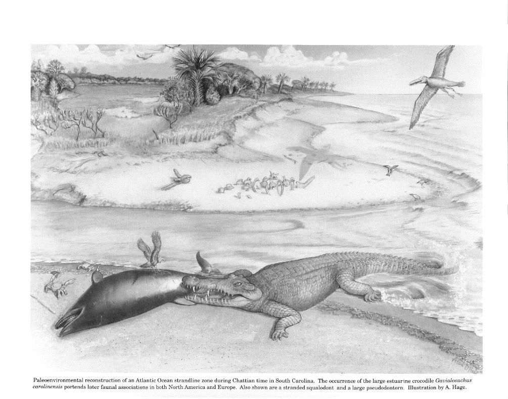
Paleoenvironmental reconstruction of an Atlantic Ocean strandline zone during Chattian time in South Carolina. The occurrence of the large estuarine crocodile Gavialosuchus carolinensis portends later faunal associations in both North America and Europe. Also shown are a stranded squalodont and a large pseudodontorn. Illustration by A. Hage.

THE ESTUARINE CROCODILE GAVIALOSUCHUS CAROLINENSIS n.sp.
(CROCODYLIA: EUSUCHIA) FROM THE LATE OLIGOCENE
OF SOUTH CAROLINA, NORTH AMERICA
BRUCE R. ERICKSON
AND
GLEN T. SAWYER
MONOGRAPH
VOLUME 3: PALEONTOLOGY
Published by
THE SCIENCE MUSEUM OF MINNESOTA ST. PAUL April 3, 1996
MONOGRAPH OF THE SCIENCE MUSEUM OF MINNESOTA
VOLUME3
International Standard Book Number 911338-84-5

CONTENTS Page
ILLUSTRATIONS ......... , .....................................2
ABSTRACT ....................................................3
INTRODUCTION .............................................. .4
INSTITUTIONAL ABBREVIATIONS ...............................5
ACKNOWLEDGMENTS ..........................................5
SYSTEMATIC PALEONTOLOGY ..................................6
DESCRIPTION ................................................10
Skull ......................................................10
Major Cranial Openings .......................................10
Bones of the Skull ............................................11
Mandible ...................................................15
Postcranial Skeleton ..........................................16
Vertebral Column ............................................19
Ribs .......................................................24
Pectoral Girdle ..............................................24
Forelimb ...................................................26
Manus .....................................................27
Pelvic Girdle ................................................27
Hind Limb ..................................................28
Scalation ...................................................30
TAXONOMIC RELATIONSHIPS ..................................31
GEOGRAPHIC AND GEOLOGIC OCCURRENCE ....................35
PALEOPATHOLOGY ...........................................36
PALEOECOLOGY AND TAPHONOMY .............................38
DISCUSSION ................................................ .43
BIBLIOGRAPHY ..............................................44

ILLUSTRATIONS
FIGURES Page
l. Gavialosuchus carolinensis, holotype skull in dorsal and ventral views .............7 2. Gavialosuchus carolinensis, holotype skull in dorsal, ventral, and
left lateral views ........................................................8 3. Gavialosuchus carolinensis n.sp., G. eggenburgensis type species, and
G. americanus skulls in posterior views ..................................... .9 4. Gavialosuchus carolinensis, paratype skull in dorsal and ventral views ............11 5. Reconstructed mandible, Gavialosuchus carolinensis holotype ...................14 6. Mandible, Gavialosuchus carolinensis paratype ...............................15 7. Atlas/axis complex of Gavialosuchus carolinensis holotype ......................19 8. Cervical vertebrae of Gavialosuchus carolinensis holotype ......................20 9. Dorsal vertebrae of Gavialosuchus carolinensis holotype ........................21 10. Sacrum and caudal vertebrae of Gavialosuchus carolinensis holotype .............22 11. Mounted skeleton of Gavialosuchus carolinensis n.sp ..........................23 12. Gavialosuchus carolinensis n.sp., left cervical rib series ........................24 13. Right scapula and right coracoid of Gavialosuchus carolinensis holotype ...........25 14. Left coracoids of Gavialosuchus carolinensis paratype and G. americanus ..........25 15. Right humerus of Gavialosuchus carolinensis holotype .........................26 16. illnae, G. americanus and G. carolinensis paratype; radiale and interclavicle,
G. carolinensis n.sp .....................................................26 17. Pelvic bones, Gavialosuchus carolinensis holotype .............................27 18. Right femur, Gavialosuchus carolinensis holotype .............................28 19. Right femora of Gavialosuchus americanus and G. carolinensis n.sp ..............29 20. Right tibia/fibula of Gavialosuchus carolinensis holotype .......................29 21. Osteoscutes of Gavialosuchus carolinensis n.sp ...............................30 22. Map, North American species of Gavialosuchus ...............................30 23. Skulls of Gavialosuchus and Tomistoma .....................................32 24. Erosional limits of the Ashley and Chandler Bridge formations on the
South Carolina coastal plain ..............................................34 25. Gavialosuchus carolinensis n.sp., bitten right epipodials ........................36 26. Alignment of skull teeth with punctures in tibia ..............................37 27. Pathologic left coracoid of Gavialosuchus carolinensis n.sp ......................38 28. Femur of Gavialosuchus in situ ............................................39 29. Carapace of a dermochelyid in situ ........................................ .40 30. Diagrammatic section of the Ashley/Chandler Bridge contact ................... .41
TABLES
I. Measurements of the skull of Gauialosuchus carolinensis n.sp ...................12 II. Measurements of the vertebrae ofGavialosuchus carolinensis n.sp ...............17 III. Measurements of the postcranial elements excluding vertebrae ..................18

THE ESTUARINE CROCODILE GAVIALOSUCHUS CAROLINENSIS n.sp.
(Crocodylia: Eusuchia) from the late Oligocene
of South Carolina, North America
by
Bruce R. Erickson and Glen T. Sawyer Department of Paleontology
The Science Museum of Minnesota USA
ABSTRACT
Skeletal remains from the late Oligocene deposits of the coastal plain of South Carolina represent a heretofore unrecognized species of eusuchian crocodile. These remains are the earliest record known for the cosmopolitan genus Gauialosuchus. Two partly complete skeletons provide materials for a thorough description of its osteology which include plesiomorphic characters that establish its ancestral relationship to later members of the genus. Aspects of its paleoecology and paleopathology are also discussed with behavioral implications for this estuarine crocodile.
KEY WORDS: Gauialosuchus, Tomistoma, Ashley/Chandler Bridge formations, Chattian, estuarine crocodile, osteology, paleoecology and paleopathology.

4 SCIENCE MUSEUM OF MINNESOTA MONOGRAPH VOL.3: PALEONTOLOGY
INTRODUCTION
The type species Gavialosuchus eggenburgensis (Taula and Kail, 1885) was founded on a skull and a few associated fragments from the "Laegendsande" early Miocene of Austria. In 1915 Sellards described a similar crocodile from the Miocene of Florida, USA which he called Tomistoma americanus. The type specimen (USNM 8816) consists only of the anterior part of the rostrum. Additional materials were assigned to this form by Sellards (1915b, 1916). Mook (1921a) reassigned Sellards' material to the genus Gavialosuchus. Subsequent additional material from North America (Mook, 1924;Auffenberg, 1954) gives detailed accounts of the skull and a number of postcranials of G. americanus (Sellards).
Recent discovery of another large longirostrine crocodile in the late Oligocene of coastal South Carolina was noted by Sanders (1980). Preparation and analysis of this discovery have now been completed and the results are herein reported. The new material includes several specimens in the collections of The Charleston Museum, one in the South Carolina State Museum and one in a private collection. These specimens are the basis for recognition of a new
species and possess plesiomorphic characters which distinguish them from later taxa. In the process of examining the collections in The Charleston Museum a reevaluation of some specimens from the South Carolina "Phosphate Beds" was necessary. These fragmentary materials: ChM 55.108.186; 55.108.187; 29.139.1; 13895; 13942; and 137 45, a skull section, regarded as Miocene in age, were referred to G. americanus by Auffenberg (1957). In as much as the "Phosphate Beds" of the Charleston area (Rogers, 1914; Malde, 1959) have been determined as being late Oligocene by Sanders (1980) and Sanders et al. (1979, 1982) and the specimens obtained from these beds show greatest likeness to the new form, they are referred to the new taxon. No crocodilian remains in younger strata of the state have yet been identified as G. americanus. This Miocene/Pliocene species may not have ranged into South Carolina. Gauialosuchus carolinensis n.sp. appears to have occupied the northernmost limit of the genus at least in North America. As the earliest known and most primitive stage of the genus it offers some interesting points of comparison with later species and the closely related Tomistoma.

5 ERICKSON/SAWYER: GAVIALOSUCHUS
INSTITUTIONAL ABBREVIATIONS
ChM=Charleston Museum; SCSM=South Carolina State Museum; SMM=Science Museum of Minnesota; UF=University of Florida; USNM=United States National Museum.
ACKNOWLEDGEMENTS
The writers wish to express their gratitude to A. Sanders for allowing us to study specimens in the collections ofThe Charleston Museum, for the hospitality extended on numerous occasions and for lengthy exchanges about Oligocene vertebrates, in addition to many trips to collect Oligocene sites in South Carolina. We are especially grateful to F. Steininger for the privilege of studying the type species of Gavialosuchus at the Institut fur Palaontologie in Vienna, for introducing one of us (ERE) to the Krahuletz Museum in Eggenberg as well as for valuable discussions about local stratigraphy and paleoecology at the type locality at Eggenberg, Austria.
Thanks are extended also to J. Knight for the long-term loan ofSCSM 90.93.1 and to L. Eberle, of Summerville, South Carolina, who collected and permitted us to study the referred skeleton of Gavialosuchus in addition to showing us the site of its discovery.
Finally, the completion of this publication was greatly assisted by the help ofR. Spading, SMM, who did all the photographic work and collaborated on field research. A. Hage provided illustrations 1, 5, 7, 12, 13, 15, 16C, 16D, 17, 18, 20, 21A, 21B, 23A, 24, 25, 26, 27 and the frontispiece. J. Janke provided illustrations 3A, 3C, 14A, 16A, 16B, 21C, 22, 23, and 30. N. Petschauer proofed and edited the manuscript, J. Singer-Johnson did word processing of various drafts, and M. Hawkinson proofread the final draft.

6 SCIENCE MUSEUM OF MINNESOTA MONOGRAPH VOL.3: PALEONTOLOGY
SYSTEMATIC PALEONTOLOGY
Order CROCODYLIA Gmelin, 1788 Family CROCODYLIDAE Cuvier, 1807
Sub Family THORACOSAURINAE Nopcsa, 1928
Genus GAVIALOSUCHUS Taula and Kail, 1885
Gavialosuchus carolinensis n.sp.
Holotype. - ChM PV 4279 skeleton and skull
Horizon. - Chandler Bridge Formation late Oligocene ( Chattian)
Locality. - 32° 51.9'N 800 02.0' W (USGS. Johns Island, 7.5 quad.) Charleston County, South Carolina
Paratypes. - SCSM 90.93.l skeleton and skull collected 1990, Chandler Bridge Fm., Crowfield Plantation, Berkeley Co., So. Car.;
ChM PV 4282 mandible collected 1978, Ashley Fm., Dorchester Co., So. Car.; ChM PV 4281 femur collected from Chandler Bridge Fm., Ladson, Charleston Co., So. Car.
Referred Specimens. - Two specimens in the collections of The Charleston Museum: ( 1) PV 4280 cranial fragment from marl pit at Lambs, Charleston Co., So. Car.; (2) 13745 partial skull from phosphate beds at Lambs, Charleston Co., So. Car.; and a partly complete skeleton from the Chandler Bridge Fm., Summerville, So. Car., in the private collection of L. Eberle, Summerville, So. Car.
Diagnosis. - Differs from other species of the genus in the following combination ofcharacters: (1) more massive skull and mandible; (2) broader and shorter rostrum; (3) only 16 teeth in dentary; (4) shorter mandibular symphysis and splenial contact; and (5) greater size differentiation of teeth.

7 ERICKSON/SAWYER: GAVIALOSUCHUS
A
Figure L Skull ofGavialosuchus carolinensis holotype specimen (Chl\f PV 4279) inA, dorsal and B, ventral view. Abbreviations: ch, choanae; ec, ectopterygoid; f, frontal; fi, incisive foramen; fpl, palatal fenestra; j, jugal; 1, lacrimal; m, maxilla; n, nasal; occ, occipital condyle; p, parietal; pal, palatine; pm, premaxilla; po, postorbital; prf, prefrontal; pt, pterygoid; q, quadrate; qj, quadratojugal; so, supraoccipital; sq, squamosal. Scale bar equals 10 cm.
8 SCIENCE MUSEUM OF MINNESOTA MONOGRAPH VOL.3: PALEONTOLOGY
I
A B
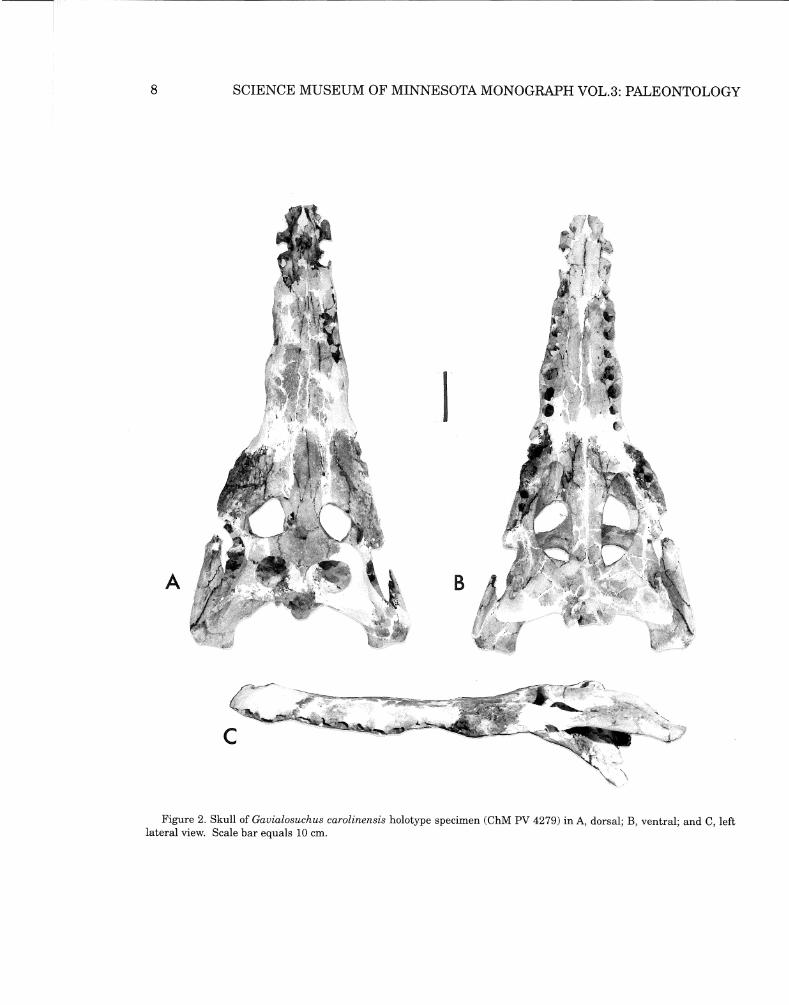
Figure 2. Skull of Gavialosuchus carolinensis holotype specimen (ChM PV 4279) in A, dorsal; B, ventral; and C, left lateral view. Scale bar equals 10 cm.

B
~-- I
C
/
9 ERICKSON/SAWYER: GAVIALOSUCHUS
Figure 3. Skulls ofA, Gavialosuchus carolinensis n.sp. SCSM 90.93.1; B, G. eggenburgensis type species (from Toula and Kail, 1885); C, G. americanus (SMM P 86.8.1). All in posterior view. Abbreviations: bo, basioccipital; eo, exoccipital; £Eu, eustachian foramen; fm, foramen magnum; occ, occipital condyle; p, pa~ietal; q, quadrate; qj, quadratojugal; so, supraoccipital; sq, squamosal Scale bar equals approximately 10 cm.

10 SCIENCE MUSEUM OF MINNESOTA MONOGRAPH VOL.3: PALEONTOLOGY
DESCRIPTION
Skull
The new species is a long-snouted crocodile wherein the rostrum makes up about twothirds of the total length of the skull. It narrows forward to a smoothly rounded anterior end. Lateral undulations have greatest expression at the fifth maxillary alveoli and at the crocodyloid notch where the premaxillae and maxillae are in contact laterally. Large cranial openings, mild pitting ofthe cranial table, a flat face, and large conical teeth with unimpressive carinae further characterize this form (Figs. IA, B; Figs. 2A,B,C; and Fig. 3A).
Major Cranial Openings
External nares: This single opening is large, subcircular and undivided with only a slight intrusion along midline on the posterior edge where the premaxillae join one another. The opening is situated behind a broadly rounded anterior rim of the snout.
Choanae: Aspects of this important diagnostic feature are preserved in both the type and paratype skulls (Figs. lB, 2B, 4B). The opening is located on midline along the posterior edge of the pterygoid plates. A derived character state that distinguishes the genus is this subtriangular choanal opening, which is broad and open posteriorly and narrowed to a blunt point anteriorly. Its lateral borders are slightly elevated. The shape ofthis opening was first described by Mook (1921a, 1924), later noted by Auffenberg (1954), and is again
recognized in the present form as a unique character.
Supratemporal fenestrae: Large and close set, the upper temporal openings occupy much of the surface of the cranial table behind the frontal. They are ovaloid and more laterally expanded in outline as viewed from above. Space between these openings is greater than in either of the other species of Gauialosuchus.
Infratemporal fenestrae: A short, blunt spina quadratojugalis is situated posterolaterally in the opening as in the other species.
Orbits: The orbits are ovate in dorsal outline, slightly larger than the supratemporal fenestrae and spaced somewhat farther apart than in other species ofGavialosuchus. Therefore there is a wider interorbital bridge and borders that are not everted as seen in the other species of the genus.
Recessus oticus externus: The opening is prominent but unremarkable.
Postemporal fenestrae: These openings are closed and poorly preserved in all material.
Foramen magnum: This is of note because of its shape. In the type as well as in paratype SCSM 90.93.1 and the referred Eberle specimen the opening has a straight dorsal side with lateral sides curving outward before converging ventrally to form a subtriangular opening. A similar condition is observed in G. americanus but the height ofthe opening is greater. In G. eggenburgensis the foramen magnum has a circular outline (Fig. 3).
A B
11 ERICKSON/SAWYER: GAVIALOSUCHUS
Palatine vacuities: These features are large and ovaloid in outline. From the level of the 10th maxillary alveoli they reach caudad beyond the anterior edge of the supratemporal fenestrae above and the pterygoid-palatine suture below (Figs. lB, 2B). Posteriorly the opening is angular rather than rounded as in the other species of Gavialosuchus.
Incisive foramen: Unlike that ofother species, the foramen is large and diamond-shaped.
Bones of the Skull (Table I)
Premaxillae: The premaxillary bones are long and elevated anteriorly, forming a moderately bulbous tip that encloses a large dorsally-positioned external narial orifice. They
are united behind the orifice and separate the nasals from the maxillaries at the level of the third maxillary alveoli forward. Ventrally the premaxillary-maxillary sutures cross a short distance between the fifth and sixth alveoli, angle caudad to the level of the sixth alveoli and turn medial to converge at midline. Five alveoli are present in each element. The first two are subequal in size and small. The third and fourth are much larger and also similar in size. The fifth alveoli are reduced to about the size of numbers one and two. Spacing between the alveoli is relatively greater in the smaller paratype skull. The diastema between the fifth premaxillary and first maxillary alveoli is wide but considerably less than in G. americanus which has a longer rostrum.
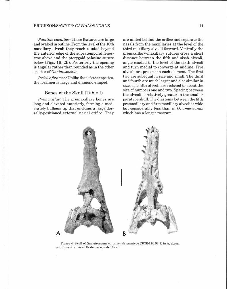
Figure 4. Skull of Gauialosuchus carolinensis paratype (SCSM 90.93.1) in A, dorsal and B, ventral view. Scale bar equals 10 cm.
12 SCIENCE MUSEUM OF MINNESOTA MONOGRAPH VOL.3: PALEONTOLOGY
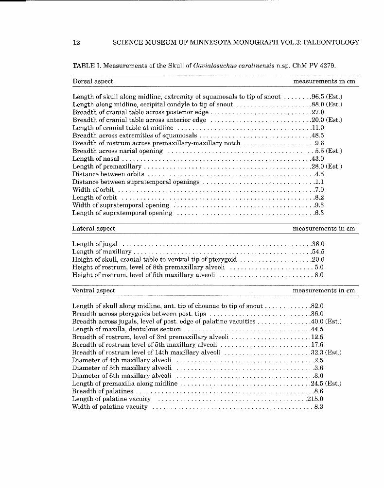
TABLE I. Measurements of the Skull of Gavialosuchus carolinensis n.sp. ChM PV 4279.
Dorsal aspect measurements in cm
Length of skull along midline, extremity of squamosals to tip of snout ........96.5 (Est.) Length along midline, occipital condyle to tip of snout .....................88.0 (Est.) Breadth of cranial table across posterior edge ............................27.0 Breadth of cranial table across anterior edge ............................20.0 (Est.) Length of cranial table at midline .....................................11.0 Breadth across extremities of squamosals .............................. .48.5 Breadth of rostrum across premaxillary-maxillary notch ....................9.6 Breadth across narial opening ........................................ 5.5 (Est.) Length of nasal ................................................... .43.0 Length of premaxillary ..............................................28.0 (Est.) Distance between orbits ............................................. .4.5 Distance between supratemporal openings .................. , ............1.1 Width oforbit ......................................................7.0 Length oforbit .....................................................8.2 Width of supratemporal opening .......................................9.3 Length of supratemporal opening ......................................6.3
Lateral aspect measurements in cm
Length of jugal ....................................................36.0 Length of maxillary .................................................54.5 Height of skull, cranial table to ventral tip of pterygoid ....................20.0 Height of rostrum, level of 8th premaxillary alveoli ....................... 5.0 Height of rostrum, level of 5th maxillary alveoli .......................... 8.0
Ventral aspect measurements in cm
Length of skull along midline, ant. tip of choanae to tip of snout .............82.0 Breadth across pterygoids between post. tips ............................36.0 Breadth across jugals, level of post. edge of palatine vacuities .............. .40.0 (Est.) Length of maxilla, dentulous section .................................. .44.5 Breadth of rostrum, level of 3rd premaxillary alveoli ......................12.5 Breadth of rostrum level of 5th maxillary alveoli .........................17.6 Breadth of rostrum level of 14th maxillary alveoli ........................32.3 (Est.) Diameter of 4th maxillary alveoli ......................................2.5 Diameter of 5th maxillary alveoli ......................................3.6 Diameter of 6th maxillary alveoli ......................................3.0 Length of premaxilla along midline ....................................24.5 (Est.) Breadth of palatines .................................................8.6 Length of palatine vacuity .........................................215.0 Width of palatine vacuity ............................................ 8.3

13 ERICKSON/SAWYER: GAVIALOSUCHUS
Maxillae: In both type and paratype skulls damage has occurred near the base of the rostrum (Figs. 1, 2, 4). In the type skull there is evidence of only 13 alveoli; however, from the length of the dentulous portion of the skull, it is likely that 14 alveoli were present. Fourteen alveoli are also found in the paratype skull. A single tooth resides in the twelfth alveolus of the left maxilla in the type. A number of teeth are present in the paratype (Fig. 4B). Alveoli one to four are of similar size. Number five is considerably larger. Number six is smaller than the fifth but larger than other anterior teeth. From here on posteriorly the alveoli decrease in diameter except for the tenth which are larger than the rest approaching number five in size.
Nasals: Paired nasals are long and taper gradually forward. Their lateral margins are relatively straight, lacking undulations seen in the other species of the genus. They wedge between the posterior ends of the premaxillae from the level of the sixth to the eighth alveoli. Posteriorly the nasals reach beyond the anterior rim ofthe orbit to the eighteenth alveoli in the paratype skull.
Prefrontals: They are small and diamondshaped. Compared to the lacrimals they form a minor part of the anteromedial orbital edge. Their surface areas are about one-fourth that of the lacrimals. They contact the frontal for one-half of their medial length, the lacrimals for their entire lateral length and posterolateral margins of the nasals.
Lacrimals: Larger than prefrontals, the lacrimals make up the anterior margin of the orbits. Their common border angles medially from the rim ofthe orbits to the posterior angle of the nasals. The lateral edges are irregular and reach anteriorly to the fourteenth alveoli before angling caudad to meet nasals.
Frontal: Unlike G. eggenburgensis and G. americanus, the frontal is rather smooth and shows little pitting. This element has a short
forward process which reaches as far as the prefrontals where it divides the nasals along midline. Ventrally it extends somewhat beyond the anterior end of the lacrimals. The frontal also forms the posteromedial wall of the orbits and a medial trough is shallow and wide.
Cranial table: The table is large and rectangular with nearly parallel lateral edges. Expanded postorbitals with squared anterolateral shoulders overhang heavy postorbital bars. The postorbital meets the squamosal on the dorsal surface in a narrow bridge at about mid-point of the supratemporal opening. Contact between squamosals and parietals is also short behind the supratemporal opening.
Pitting of the surface is minimal on the type and somewhat more pronounced on the paratype skull. This is consistent with the condition described for the vertebral column wherein neural arches and centra are co-ossified to a greater degree in the smaller, presumably more mature, paratype skeleton (SCSM 90.93.1) than in the larger, presumably less mature, holotype specimen (ChM PV 4279). Iordansky (1976) has noted that sculpturing of the skull tends to be less in younger animals and develops progressively with age. This suggests that the smaller paratype is older as well as more mature than the holotype specimen.
In posterior view the plain ofthe table is flat in contrast to G. eggenburgensis and shows no sign of the dorsal crests at the posterolateral angles of the table that distinguish the later species (Fig. 3B).
Jugal, quadratojugal and quadrate: This complex is unremarkable and is characterized by a slightly elevated edge of the jugal along the lateral border ofthe orbit, whereas G. eggenburgensis and G. americanus display a conspicuous elevation of this edge. A modest spina quadratojugalis, slight pitting of the surface of the jugal and a long caudad extension of the quadratojugal and quadrate further describe this complex.

14 SCIENCE MUSEUM OF MINNESOTA MONOGRAPH VOL.3: PALEONTOLOGY
Palatines: Palatines are broad and widened anteriorly. A large foramen is located in each element near the maxillary suture on the margin of the palatine vacuity. Anteriorly they reach to the level ofthe seventh maxillary alveoli, whereas in the other species they end near the eighth alveoli. They have a long oblique contact with the maxillae, join at midline and have a simple transverse junction with the pterygoids.
Pterygoids: These are wide, laterally expanded, flat bones and remarkable in being deeply incised along both front and rear edges. Posterior incising is deepest toward midline and
~~ (\ ~1\-f\_(\_ (\ ---~U{ ~ Ll,"-A~C'.._ A._~
A
/--- ---- --- -=-
C:) a"'-c.Jl__Q __d__ Q__{j_Qi_c5,
B
- -•
an
reaches the level of the choanal aperture as a continuation ofthe low elevation along the lateral sides of the aperture.
Ectopterygoids: Anteriorly the bones lie along the maxillae medial to the last two alveoli. They overlap the outer edge of the pterygoids to the level of the anterior edge of the choanal aperture.
Occipital group: The occipital group is indistinctive. The posterior face of the basioccipital beneath the occipital condyle containing the eustachian canal varies appreciably in breadth and texture (Fig. 3A).
Figure 5. Reconstructed mandible of Gavialosuchus carolinensis holotype (ChM PV 4279). A, left lateral view and B, occlusal view. Abbreviations: an, angular; ar, articular; d, dentary; fme, external mandibular fenestra; s, splenial; san, surangular. Scale bar equals 10 cm.

15 ERICKSON/SAWYER: GAVIALOSUCHUS
Mandible
The mandible of the type specimen including most of the symphysis is incomplete anteriorly (Fig. 5). Paratype ChM PV 4282, which has the anterior half of the mandible intact with nearly all of the teeth in place, is the basis for. the reconstruction of the type mandible (Fig. 6A). A second paratype, SCSM 90.93.1, is a nearly complete mandible lacking teeth and
C
Figure 6. Mandible ofGavialosuchus carolinensis paratype (ChM PV 4282) in A, dorsal view of anterior half; B, right articular, retroarticular and surangular in lateral view; C, joined splenials in dorsal view. Scale bar equals 5 cm.
is distorted by dorsoventral compression. A rather complete mandible may be described from this·material. The mandible has an estimated length of 111.0 cm with the dentulous portion estimated at 64.0 cm and the symphysial contact at 43.0 cm.
Openings: Two major openings occur in each dentary. A large lateral, ovaloid mandibular fenestra and a small eliptical internal mandibular fenestra are present.
Dentary: In articulation the dentaries together with the splenials form a medial symphysis for the distance between the anterior end of the jaw and the first ten teeth. By comparison G. americanus has a longer tooth row and a larger symphysis spanning the first 11 teeth. G. carolinensis n.sp. has 16 alveoli and G. americanus has 17 alveoli in the dentary.
Surangulars: The surangulars reach from the sixteenth alveoli to about mid-length of the retroarticular process. They contribute the posterodorsal portion of the external mandibular fenestrae. Their surfaces are smooth.
Angulars: These elements are longer than the surangulars covering most of the distance between the sixteenth alveoli and the back end of the retroarticular processes. They form the posteroventral edges of the external mandibular fenestrae.
Splenials: None of the splenials in the type series are complete; however, their structure is readily determined. Right and left elements unite at midline to form that part of the symphysis between the seventh and tenth alveoli (Fig. 6C). Posteriorly the splenials extend to the level of the internal mandibular fenestrae. Their height is low, reflecting the shallow silhouette of the whole mandible (Fig. 5A).
Articulars: As preserved only in the type specimen, the articulars are like those of G. americanus except for a deeper concavity immediately posterior to the quadrate surface on the medial side of the base of the retroarticu-

16 SCIENCE MUSEUM OF MINNESOTA MONOGRAPH VOL.3: PALEONTOLOGY
lar process. A complete retroarticular is lacking. The best preserved fragment (Fig. 6B) illustrates its stout upswept form.
Hyoid: The hyoid is unknown.
m 14 Dentition: PM 5 ---
d 16
The generalized form ofthe individual tooth is heavy and conical. There is a poorly developed carina for the largest teeth as well as the posterior teeth. Smaller teeth in the anterior region of the mandible and premaxillae are longer and recurved. All of the known species of Gavialosuchus have five premaxillary teeth in each element. Fourteen are present in the maxilla of G. americanus and the new species and 15 are found in the type species. The teeth are variable in size. For example, the new skull indicates that the largest teeth by far are numbers one, four, ten and eleven. In G. americanus one and eleven are only slightly greater in size than the rest.
The holotype skull ChM PV 4279 is damaged in the area of the eighth to the tenth alveoli and all but one tooth has been separated from its alveolus. Clusters ofassociated teeth, some within the unprepared skull itself, assisted in relocating most of the teeth in their proper places. The paratype skull SCSM 90.93.1 retains few of its teeth as well. However, its alveolar rows are intact and it can be shown that each tooth occupied a discrete alveolus adjacent to a wide interalveolar space with the exception of two groups of three teeth each. These are numbers ten, eleven and twelve and numbers fourteen, fifteen and sixteen. A sim-
ilar condition is found in G. americanus except for the seventeenth tooth that is included in the last group.
Teeth in the dentary number sixteen and seventeen respectively for G. carolinensis n.sp. and G. americanus and the former show greater variability in size. The large conical teeth nearly always have characteristic encircling color bands of buff, tan and sienna. These bands provide an unmistakable code for field recognition in the Ashley and Chandler Bridge deposits.
Postcranial Skeleton (Tables II and III)
In 1954Auffenberg described the postcranial skeleton of Gavialosuchus americanus from a large sample of bones collected at the village ofHaile in Florida. Much of the material available to him consisted of disassociated bones of individuals of various sizes derived from several localities. A partly complete specimen UF 6225, provided the only articulated evidence and the basis for his comparative measurements. A number of postcranials were described, but for the most part they were unillustrated.
Recent discovery of two more-or-less intact skeletons of the new taxon Gavialosuchus carolinensis n.sp. now affords the opportunity for a more thorough description ofGavialosuchus which further characterizes the genus. This description is augmented by an illustrated osteology and comparisons with the other species of Gavialosuchus as well as the Crocodylia in general (Mook, 1921b).

17 ERICKSON/SAWYER: GAVIALOSUCHUS
TABLE II. Measurements (in mm) of the Vertebrae of Gavialosuchus carolinensis n.sp. ChM PV 4279: Cervicals (CJ; Dorsals (D); Sacrals (S).
Maximum Transverse Vertical Maximum Maximum Maximum Vertebra Height, Tip of Diameter of Diameter of Centrum Centrum Height Centrum
Neural Spine to Neural Canal, Neural Canal, Length, Excluding Breadth at Ventral Face of at Midpoint at Midpoint -Ventral Face Hypophysis, Midpoint, Centrum, Anteriorly Anteriorly Anteriorly
C3 152. 7 (Est.) 20.3 23.6 72.5 50.0 50.5
C4 165.4 21.6 26.3 74.4 51.4 53.8
C5 C6
C7 cs 183.3 (Est.) 23.3 27.6 74.5 52.6 (Est.) 55.4
C9 192.4 24.1 28.1 71.2 51.9 57.2
Dl 193.6 22.2 27.2 77.1 54.5 58.8
D2 191.8 (Est.) 18.9 26.5 81.3 57.9 66.3
D3 188.6 (Est.) 18.9 28.8 82.8 (Est.) 60.7 63.4
D4 173.3 (Est.) 20.4 27.7 86.2 (Est.) 58.1 55.8 (Est.)
D5 172.2 17.6 28.3 87.9 62.4 60.8 (Est.)
D6 171.4 (Est.) 20.6 27.6 (Est.) 84.3 65.0 55.5
D7 165.7 21.3 27.2 81.0 60.9 51.9
D8 161.5 20.3 (Est.) 22.5 84.2 62.3 56.6 (Est.)
D9 162.2 (Est.) 18.9 24.7 85.0 61.0 (Est.) 57.7 (Est.)
DlO 155.6 18.7 22.0 86.2 60.5 57.0 (Est.)
Dll 155.8 (Est.) 20.8 (Est.) 20.1 (Est.) 84.5 (Est.) 60.3 (Est.) 57.7 (Est.)
D12 152.0 (Est.) 17.4 20.8 86.9 57.7 (Est.) 58. 7 (Est.)
D13 D14 152.0 18.6 18.3 79.7 55.5 62.3
D15 147.6 17.7 21.0 77.3 52.4 55.2
D16 147.2 19.2 21.2 72.9 51.5 57.8
S1 148.0 19.3 21.0 64.0 (Est.) 51.0 (Est.) 74.0 (Est.)
S2 143.0 (Est.) 19.2 22.0 62.0 (Est.)

18 SCIENCE MUSEUM OF MINNESOTA MONOGRAPH VOL.3: PALEONTOLOGY
TABLE III. Measurements (in mm) of Postcranial Elements Excluding Vertebrae
G. carolinensis n.sp. G. carolinensis n.sp. Element ChM PV 4279 SCSM 90.93.1
Seapula Length 236.0 (Est.) 147.0 (Est.) Breadth of blade Breadth of base 67.0 (Est.) Max. diameter shaft 35.3 25.3
Coracoid Length 204.0 (Est.) 153.5 Breadth, distal end 88.0 (Est.) 66.0 (Est.) Breadth, proximal end 98.0 (Est.) Max. diameter shaft at narrowest point 32.5 25.0 (Est.)
Humerus Length 298.0 (Est.) Breadth, distal end 78.5 56.3 Breadth, proximal end Max. diameter shaft at midlength 36.1 27.4
Ulna Length 117.8 Breadth, distal end 24.0 (Est.) Breadth, proximal end 35.0 (Est.) Max. diameter shaft at midlength 14.0
Ilium Height 116.0 (Est.) 86.4 Length of blade 189.2 136.6
Ischium Length 208.0 (Est.) 146.0 (Est.) Breadth, distal end 84.0 (Est.) Breadth, proximal end Max. diameter shaft at narrowest point 42.4 30.4
Pubis Length 172.0 (Est.) 130.7 Breadth, distal end 111.0 (Est.) 84.0 (Est.) Breadth, proximal end 47.3 30.8 Max. diameter shaft at narrowest point 23.5 20.4
Femur Length 334.2 Breadth, distal end 77.7 41.5 Breadth, proximal end 78.2 Max. diameter shaft at midlength 44.9 31.2
Tibia Length 219.5 154.3 (Pathologic) Breadth, distal end 52.0 (Est.) 47.0 (Est.-Pathologic) Breadth, proximal end 68.0 (Est.) 49.0 (Est.-Pathologic) Max. diameter shaft at midlength 26.5 29.2 (Pathologic)
Fibula Length 221.0 (Est.) 162. 7 (Pathologic) Breadth, distal end 30.5 (Pathologic) Breadth, proximal end 43.4 33.4 (Pathologic) Max. diameter shaft at midlength 18.5 18.5 (Pathologic)

A
D C
ERICKSON/SAWYER: GAVIALOSUCHUS 19
Vertebral Column
Based on the holotype and paratype skeletons ofG. carolinensis n.sp. the presacral series lacks two cervicals and one dorsal vertebra. Both sacrals are present in each specimen and about 19 total caudal vertebrae are present. Although co-ossification is advanced in each skeleton anterior to the sacrum, suturing between the neural arch and the centrum is more complete and therefore less visible in the paratype, which is a smaller individual but evidently somewhat more mature.
Cervical vertebrae: The proatlas is unknown. The atlas/axis complex is otherwise complete in the type and missing only the neurocentra in the paratype. Viewed anteriorly (Fig. 7 A) with the neurocentra in place above the atlas intercentrum, a large depression for the occipital
condyle makes up most of the anterior surface of these elements. The articular surface of each neurocentrum is about one-third as large as that ofthe intercentrum. The upper opening between the neurocentra is fully twice as large as the axis neural canal behind it.
Through the lower opening between the neurocentra the axis intercentrum (odontoid) is visible. Lateral to the atlas intercentrum, diapophyses protrude on either side. Above the level of the neurocentral arch the wing-like postzygapophyses and the low, narrow neural spine of the axis are visible.
In lateral view (Fig. 7B) the size of the neurocentrum is about equal to that of the atlas intercentrum which it overlaps slightly dorsally. The posteriorly projecting wing of the neurocentrum is short, anteriorly blunt and
Figure 7. Atlas/axis complex of Gavialosuchus carolinensis holotype (ChM PV 4279) in A, anterior; B, left lateral; and C, ventral view. D, axis and intercentrum of paratype (SCSM 90.93.1) in left lateral view. Scale bars equal 5 cm.

A
20 SCIENCE MUSEUM OF MINNESOTA MONOGRAPH VOL.3: PALEONTOLOGY
tapered posteriorly. An incipient process is located at either end. The atlas centrum is fused to the axis and the suture is distinct. The condyle of the axis centrum is small and deflected ventrally. Overall, the vertebra is somewhat shorter than the length ofthe assembled atlas/axis complex. As shown by paratype SCSM 90.93.1 (Fig. 7D), the neural spine has a straight superior border, is squared-off posteriorly and overhangs anteriorly. A small prezygapophysial structure opposes the noted process on the posterior wing of the neurocentrum and the postzygapophysis of the axis is
large and ovaloid in outline.
In ventral view (Fig. 7C) the atlas intercentrum is nearly as wide as the axis centrum. A low, rounded centrally-located hypapophysis runs from mid-length of the axis forward and is expressed in lower relief on the atlas intercentrum. The axis centrum is constricted medially. Posteriorly postzygapophyses and the tip of the neural spine are visible.
Other cervical vertebrae (Fig. 8) exhibit the following characters: procoelous centra with
Figure 8. Cervical vertebrae of Gavialosuchus carolinensis holotype (ChM PV 4279) in anterior (upper) and right lateral (lower) views. A, third; B, eighth; and C, ninth cervical vertebrae. Scale bar equals 5 cm.

21 ERICKSON/SAWYER: GAVIALOSUCHUS
deep cotyles; ventral hypapophyses; large parapophyses; a high neural arch with a large neural canal and dorsal spines that increase in height posteriorly, taper distally and angle posteriorly; numbers eight and nine are smaller at their tips, and all of the neurocentral sutures are still visible.
Dorsal vertebrae: Of the probable 16 dorsal vertebrae, 15 are available. Number thirteen is missing. Three elements are shown in Figure 9. The preserved elements exhibit the following characters: procoelous centra with large
condyles which are geometrically greater than hemispheres; correspondingly deep cotyles; ventral hypapophyses on the first three elements; parapophyses on the first two dorsals; centra decrease in height from the first through the sixteenth vertebra; neural spines increase in anteroposterior width to the tenth, and thereafter remain more or less uniform in width; all spines are slightly expanded distally; spines are uniform in height except for the first and second which are somewhat longer; there are prominent tubercular rib facets on
Figure 9. Dorsal vertebrae of Gavialosuchus carolinensis holotype (ChM PV 4279) in anterior (upper) and right lateral (lower) views. A, first; B, fourth; and C, fourteenth dorsal vertebrae. Scale bar equals 5 cm.
22 SCIENCE MUSEUM OF MINNESOTA MONOGRAPH VOL.3: PALEONTOLOGY
the third through the tenth vertebrae, and rib facets on the eleventh to the sixteenth are diminished or absent.
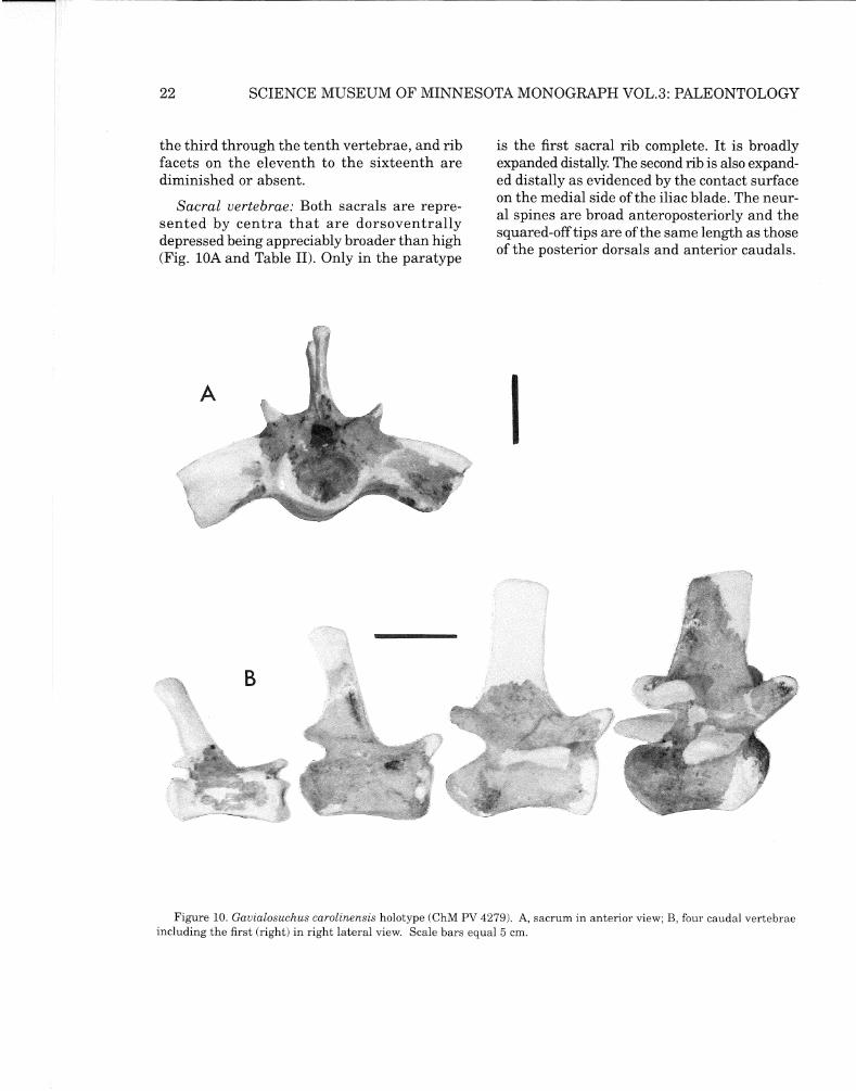
Sacral vertebrae: Both sacrals are represented by centra that are dorsoventrally depressed being appreciably broader than high (Fig. lOA and Table II). Only in the paratype
I
is the first sacral rib complete. It is broadly expanded distally. The second rib is also expanded distally as evidenced by the contact surface on the medial side of the iliac blade. The neural spines are broad anteroposteriorly and the squared-off tips are of the same length as those of the posterior dorsals and anterior caudals.
Figure 10. Gauialosuchus carolinensis holotype (ChM PV 4279). A, sacrum in anterior view; B, four caudal vertebrae including the first (right) in right lateral view. Scale bars equal 5 cm.

23 ERICKSON/SAWYER: GAVIALOSUCHUS
Figure 11. Mounted skeleton (cast) of Gavialosuchus carolinensis n.sp. in A, left posterolateral and B, right anterolateral views. Skeleton in resting posture with limbs outstretched. Total length of skeleton 537 cm (17.3 ft.).
Caudal vertebrae: Twenty-two of an estimated 43 caudal vertebrae are present. Of these the first through the tenth were articulated. All neurocentral sutures are obliterated in both type and paratype. Near the base of the tail, the first spines are long. The longest were probably on numbers six and seven. Lateral
spines occur on the first dozen or so vertebrae. Four elements are shown in Figure lOB.
Chevrons: Only fragments of about a dozen elements remain. As reconstructed (Fig. 11), 23 chevrons are present.

24
-
SCIENCE MUSEUM OF MINNESOTA MONOGRAPH VOL.3: PALEONTOLOGY
Figure 12. Left cervical rib series of Gavialosuchus carolinensis n.sp. Scale bar equals 5 cm.
Ribs
Cervical ribs: All cervical ribs are accounted for with the exception of number two. Only those of the left side are shown in Figure 12. Of the most anterior ribs only number one remains. The proximal two-thirds of this element shows a straight, flat and holocephalous form with a prominent uncinate elevation at mid-length. The third element is larger but similar in form to numbers four through seven. The eighth and ninth each lack distal ends. They are similar to one another, the last being about twice the size of the anterior rib.
Dorsal ribs: Only the third right rib is intact, however many partly complete elements reveal the form of the ribs. The dorsal ribs may be characterized as being stout, having moderately curved shafts with circular to slighty oval cross sections. The first four or five ribs have well defined capitular and tubercular features.
Gastralia: A few scraps of gastralia were associated with the type specimens. These are slender rod-like structures with both circular and flattened cross sections. No rib associations can be determined.
Pectoral Girdle
Scapula: The right scapula and both scapulae are accounted for in the type and paratype skeletons respectively. Their general form agrees with that of G. americanus, and the scapular length is greater than that of the coracoid (Figs. 13A, B).
Coracoid: Both coracoids and the right coracoid are preserved in the type and paratype skeletons respectively (Figs. 13C, D). This element is strikingly different than in G. americanus (Fig. 14). In lateral view both anterior and posterior margins of the blade have distinct curvatures. In G. carolinensis n.sp. the anterior edge is deeply incised and there is an abrupt forward angulation at the base of the blade. In G. americanus the anterior margin is a smooth, continuous curve that is shallower than in the former taxon. Auffenberg (1954) contrasts this feature with Crocodylus americanus and shows it to be distinctive. Posteriorly the new taxon shows a nearly straight margin beyond the glenoid region to the tip of the blade. The Miocene/Pliocene species lacks its posterior edge and its exact shape is unknown (Auffenberg, 1954). Although the blade is missing its posterior extremity, expansion of the shaft below sug-

25 ERICKSON/SAWYER: GAVIALOSUCHUS
Figure 13. Gavialosuchus carolinensis holotype (ChM PV 4279). Right scapula in A, lateral and B, posterior view. Right coracoid in C, ventral and D, anterior view. Scale bar equals 10 cm.
gests that the tip of the blade was anteroposteriorly broad. The superior border was apparently more acutely arched in the new species and the base through the coracoid foramen is relatively shorter anteroposteriorly.
Sternum: The interclavicle is absent in the holotype. This element preserved in the paratype skeleton is, as far as known, the only record for the genus. Its extremities are weathered but the bone can be characterized as being uniformly narrow and diminished posteriorly with a slight constriction near mid-shaft. Just anterior to this the ventral lateral borders are incised for a short distance, presumably for the cartilaginous sternum. On the visceral face there is a central ridge the length of the shaft and at the level of the constriction there is also a minor midline prominence (Fig. 16D).
Figure 14. Left coracoids of A, Gavialosuchus carolinensis paratype (SCSM 90.93.1) and G. americanus (from Auffenberg, 1954) in medial view.
B

B
B
26 SCIENCE MUSEUM OF MINNESOTA MONOGRAPH VOL.3: PALEONTOLOGY
Forelimb
Humerus: All of the humeri belonging to the two skeletons are incomplete. The right humerus of the type specimen (Fig. 15) has been reconstructed at its proximal end based on information from the other humeri. By comparison with G. americanus the most notable difference is the location of the deltoid crest which in the new taxon is heavier and situated lower on the shaft. The distal end is appreciably expanded and the shaft has a conspicuous sigmoid curve.
Figure 15. Right humerus of Gauialosuchus carolinensis holotype (ChM PV 4279) in A, posterior and B, ventral view. Scale bar equals 10 cm.
Radius and ulna: The radius is missing; however, the ulna is present in the paratype.
Figure 16. Left ulnae ofA, G. americanus and B, Gauialosuchus carolinensis paratype (SCSM 90.93.1); C, radiale ofG. carolinensis n.sp. (SCSM 90.93.1); D, interclavicle of G. carolinensis n.sp. (SCSM 90.93.1). Scale bar equals 2 cm.
When compared with the ulna of G. americanus they are strikingly similar (Figs. 16A, B). The ulna is short in both species, approximating the length of the longest metatarsals. By comparison with the other crocodilians such as Crocodylus sinensis, Leidyosuchus formidabilis and even Alligator this indicates a relatively shorter forelimb for the new taxon. At its proximal end it is greatly enlarged with broad articular areas. Distally it is moderately enlarged.
Carpus: Unfortunately the bones of the forefeet are mostly absent. The radiale is available for comparison with G. americanus (Fig. 16C).

27 ERICKSON/SAWYER: GAVIALOSUCHUS
Compared to the younger species its length is greater and its breadth nearly the same but its articular surfaces are somewhat less developed especially along the proximal medial side.
Manus
Only metacarpal V remains. It has a pathologic condition showing what is evidently a healed fracture.
Pelvic Girdle
Bones of the pelvic girdle are shown in Figure 17.
Ilium: The superior border of the iliac blade is shorter than that of G. americanus. It is a robust bone with a large posteriorly projecting blade that has a constriction towards its posterior end as found in later crocodilians. The acetabulum occupies about half of the lateral area of this bone. There also remains a suggestion of an anterodorsal tubercle near the base of the blade.
Pubis: This bone matches the approximate size of the other pubic bones and is like that of the younger species.
Ischium: This bone also closely resembles that of the other species.
C
Figure 17. Pelvic bones of Gauialosuchus carolinensis holotype (ChM PV 4279). A, left ilium, lateral view; B, right pubis, visceral view; C, left ischium, anteroventral view. Scale bars equals 5 cm.

28 SCIENCE MUSEUM OF MINNESOTA MONOGRAPH VOL.3: PALEONTOLOGY
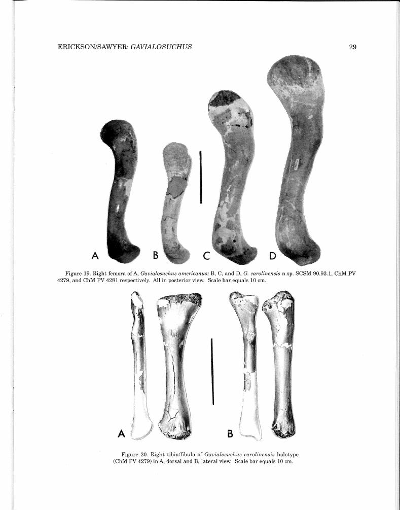
Hind Limb ed to articular surface. The noted large right element from ChM PV 4281 (Fig. 19D) represents what we judge to be near the upper size limit for this taxon. The mounted skeleton, a cast ofholotype ChM PV 4279) (Fig. 11), has a hind limb length of 86 cm ( combined maximum lengths of propodial, epipodials and pes) and a total skeleton length of 537 cm (17.3 ft.). By comparison the femur from ChM PV 4281 belonged to an individual with a possible overall length of about 639 cm (21 ft.)
Femur: Several femora are available for the type series, both from the holotype: the right element from SCSM 90.93.1; and a large right element from ChM PV 4281 (Figs. 18, 19). The femur is approximately ten percent longer than the humerus. It is a heavy bone with a large bulbous fourth trochanter and expanded extremities. The proximal end shows greatest development and most of its surface is devot-
Tibia I fibula: Tibiae and the right fibula are present in the type specimen (Fig. 20). Right elements are present in the paratype SCSM 90.93.1. The later epipodials show a pathologic condition that suggests behavior. These elements are discussed in the paleopathology section of this paper. The tibia of the type has lost much of its surface detail at the extremities; however, its form is clearly straight-sided except for a medial curve to join the widened proximal end of the shaft. The shaft of the fibula is circular at mid-length and flattened proximally. Distally it is not preserved in any of the available material.Although incomplete, the epipodials are judged to have been about two-thirds the length of the femur. This is consistent with the observed proportions in the forelimb.
Tarsus: Only the calcaneum survives. Its fragmentary nature provides little that is noteworthy for description.
Pes: All metatarsals are accounted for with the exception of the fifth. Only two phalanges including one ungual remain.
Figure 18. Right femur of Gavialosuchus carolinensis holotype (ChM PV 4279) in A, posterior and B, ventral view. Scale bar equals 10 cm.
29 ERICKSON/SAWYER: GAVIALOSUCHUS
A B
Figure 19. Right femora of A, Gavialosuchus americanus; B, C, and D, G. carolinensis n.sp. SCSM 90.93.1, ChM PV 4279, and ChM PV 4281 respectively. All in posterior view. Scale bar equals 10 cm.
A
Figure 20. Right tibia/fibula of Gavialosuchus carolinensis holotype (ChM PV 4279) in A, dorsal and B, lateral view. Scale bar equals 10 cm.

South Carolina
Georgia
Gavialosuchus carolinensis
Gavialosuchus Floridaundet.
Gavialosuchus americanus
30 SCIENCE MUSEUM OF MINNESOTA MONOGRAPH VOL.3: PALEONTOLOGY
Scalation
Scalation in living crocodilians incorporates ossified and unossified scales which may vary interspecifically as well as among individuals of the same species. Intraspecific variation is common and may be reflected by the shape and number of scales and the number of scale rows as demonstrated by Ross and Roberts (1979). They further note that no differences in scalation between sexes ofthe American alligator were found.
Osteoderms: Approximately 80 scales that were directly associated with present specimens are ossified (osteoscutes). They range in size from 2.5 cm to 9 cm across and from <1 cm to >1.5 cm in thickness and they are relatively thinner than those of G. americanus (Auffenberg, 1954). None possess a dorsal keel and the dorsal surface has large, well-spaced, circular and subcircular pits. Some of the more rectangular, dorsal elements are beveled along their longest edges. The osteoscutes separate into four general groups: 1) the largest with more-or-less parallel margins (Fig. 21A); 2) sizable elements having a near-circular outline (Fig. 21B); 3) elements having a subtriangularto-square outline; and 4) those that are smaller, rounded to irregular in outline and bear incipient or prominent spikes on one edge (Fig. 21C). Precise location of any ofthese osteoscutes in life is conjectural.
Figure 21. Osteoscutes of Gavialosuchus carolinensis n.sp. in dorsal view. A, large dorsal element; B, subcircular back element; C, spiked flank or back element. Scale bar equals 2 cm.
Those in groups one, two and three, however, are regarded as major dorsal, flank and nuchal elements which provide the dermal armor ofthe animal. Those in category four are more random in their occurrence. Osteoscutes of the kind which would have been positioned at the junction of the head and neck (postoccipitals) do not appear to be represented. It is possible that postoccipital osteoscutes were absent as they often are in estuarine crocodiles (Wermuth, 1953; Neill, 1971).
During the course of this investigation a number of osteoscutes collected from Miocene (Hawthorne Group) burrow pits in coastal Georgia by private collectors were examined. This material is ofinterest because these osteoscutes are of comparable shape and size to those of
Figure 22. Map of known occurrences of the North American species ofGavialosuchus. G. carolinensis n.sp., late Oligocene (stippled); G. americanus Miocene/Pliocene (diagonal lines); G. sp. undet., early Miocene (cross hatched).

31 ERICKSON/SAWYER: GAVIALOSUCHUS
the new taxon but are appreciably thicker. They have flat unkeeled surfaces and are otherwise similarly ornamented. Auffenberg (1954) states that flat, unkeeled osteoscutes such as these occurring in the southeastern U.S. belong to the genus Gavialosuchus. Considering the known fossil crocodilian materials from the southeastern coastal plain, we tend to agree with this notion. Furthermore the age of the Georgia material and its noticeable morphological distinction suggests that the Miocene fossils may represent another species ofGavialosuchus that is intermediate, at least in time in North America, between the new form and G.
americanus. Additional materials from South Carolina currently under study by the authors are considered to represent the same undetermined gavialosuchid. The possible presence ofa form different from the new species as well as from G. americanus, whichAuffenberg (1957) regards as late Miocene and, with reservations, as Pliocene, is suggested. The recognized ages are based on studies of the Hawthorne Group by Pirkle (1956, 1957). The occurrence of this undetermined gavialosuchid is indicated in Figure 22. The evidence strongly indicates that Gavialosuchus was present in North America from the late Oligocene throughout the Miocene.
TAXONOMIC RELATIONSHIPS
Hecht and Malone (1972) discuss the morphological convergence of the rostrum in longirostrine crocodiles once they have invaded the gavialoid adaptive zone wherein the rostrum becomes streamlined. In the true gavials, differentiation is extreme. Longirostrine crocodiles lacking extreme differentiation such as Gavialosuchus americanus are considered to be at or near the tomistomine stage of organization. G. carolinensis n.sp. presents an even less extreme stage of organization with its broader skull (Fig. 23).
The genus Gavialosuchus is retained as valid and distinct from Tomistoma to which it has been most often allied. Characters that show it to be distinct are: 1) its subtriangular choanal opening noted by Mook (1924) andAuffenberg (1954); 2) pterygoids deeply incised anteriorly by palatine vacuities; and 3) absence of keeled osteoscutes as noted by Auffenberg (1954).
The shorter, broader rostrum which borders
on the tomistomine stage of organization has: 1) shorter premaxillae; 2) longer nasals that are nearly half the length of the skull; 3) small prefrontals and large broad lacrimals; 4) long anterior projections of paired palatines; 5) Vshaped premaxillary-maxillary suture on the palate as discussed by Mook (1921a); 6) only 14 to 15 maxillary alveoli rather than 20 to 21 as in Tomistoma; and 7) five rather than the normal four premaxillary alveoli indicated by Steel (1973) for Tomistoma.
Three species of Gavialosuchus are recognized (Fig. 23). The type species, G. eggenburgensis (Toula and Kail, 1885), consists of a wellpreserved skull. Originally a few postcranial elements were also included in the type description; however, these elements could not be located, either in Vienna at the Institut for Palaontologie or in the Krahuletz Museum in Eggenburg, during the course of this study. The type specimen was collected from early Miocene strata, Eggenburgian stage, "Laegendsande"

( ·' ,t
:~ ·" ...·•·• ,
A ' B ~ •·• ,,
'c
.'
j
C D
32 SCIENCE MUSEUM OF MINNESOTA MONOGRAPH VOL.3: PALEONTOLOGY
Figure 23. Skulls of Gavialosuchus and Tomistoma in dorsal (left) and ventral (right) aspects. A, Gavialosuchus carolinensis n.sp; B, G. americanus; C, G. eggenburgensis (from Toula and Kail, 1885); and D, 1bmistoma (from Iordansky).

33 ERICKSON/SAWYER: GAVIALOSUCHUS
lithostratigraphic unit. The type locality is at Eggenburg near Horn, Austria on the right-ofway of the Franz Joseph Bahn. Associated nannoplankton at the base ofthe Burdigalian are NH2 (Steininger, pers. comm., 1988).
A second species, G. americanus (Sellards, 1915a), is known from numerous remains including a partly complete skeleton (UF 6225) from the Bone Valley Gravel Formation, HemphillianAge in Polk and Alachua counties, Florida, USA.
Finally, the new taxon G. carolinensis n.sp. is known from two relatively complete skeletons (ChM PV 4279 and SCSM 90.93.1), a less complete skeleton in a private collection, and various isolated elements from the Ashley and Chandler Bridge formations, Chattian Age, Charleston and Dorchester counties, South Carolina, USA. Greatest variability of these three species is in the morphological organization of the rostrum.
Characters that distinguish G. carolinensis n.sp. from its fellow species are: a broader more robust skull with a shorter rostrum; pronounced size differences of alveoli; shorter interalveolar distances; and fewer teeth in the dentary. Notable postcranial differences from G. americanus are found in the form of the coracoid blade, morphological and proportional features of the forelimb, and osteoscute pitting. As the postcranials of the type species are not available, comparisons cannot be made.
Other materials referred to the genus Gavialosuchus include the following specimens which are herein reevaluated. The first ofthese is a partial maxilla in The Charleston Museum bearing number 13745. This specimen was collected in 1920 during the phosphate mining period from the Ashley beds which at that time were thought to be Miocene in age. Auffenberg (1957) referred it to G. americanus primarily because of its presumed age and its close similarity to the species from Florida. Sanders et. al. (1982) redefined and assigned the Ashley
beds to the latest Oligocene (Chattian). In light of this and the rather extensive amount of materials belonging to the new taxon from these deposits, it is appropriate to reassign 13745 to G. carolinensis n.sp.
Two further specimens in the collections of The Charleston Museum (PV 2495 and PV 2504) from Pleistocene deposits of Edisto Island, South Carolina, were identified by Roth and Laerm (1980) as right humeri of cf. Gavialosuchus. Neither of these specimens is a humerus nor do either agree with Gavialosuchus morphologically. They are both left femora and referable to Alligator.
A taxon originally allocated to G. americanus var. lusitanica n.v. (Vianna and Moraes, 1941) and subsequently assigned to Tomistoma lusitanica by Telles Amtunes (1961) is reassessed and assigned to the type species G. eggenburgensis on the basis that both are Miocene and possess the following combination of shared characters which also separates it from Tomistoma: 1) a triangular outline of choanal opening; 2) concave cranial table with dorsal elevations on its lateral margins above the otic opening; 3) a deeply pitted cranial roof; 4) pronounced lateral crocodyloid notch at premaxilla-maxillary contact; 5) palatine/maxillary suture extending forward to the level of the ninth alveoli; 6) number of teeth; and 7) supratemporal fenestrae separated only by a thin web of bone.
Gavialosuchus also includes Mega/ode/phis magnidens, a so-called giant dolphin. Morgan ( 1986) demonstrates that the holotype of M. magnidens, which Kellogg described in 1944 as a whale, is actually a partial mandible of Gauialosuchus americanus and is therefore a junior synonym of this taxon. As with G. carolinensis n.sp. from the late Oligocene, the Miocene/Pliocene species occurs in direct association with a variety of cetacean remains (Webb and Tessman, 1968).

34 SCIENCE MUSEUM OF MINNESOTA MONOGRAPH VOL.3: PALEONTOLOGY
Scrcd.e of KilOineiers 0 50 WO
Figure 24. Erosional limits of the Ashley (heavy line) and Chandler Bridge (stippled) formations on the South Carolina coastal plain. The known geographic ranges of Gavialosuchus carolinensis n. sp. during Ashley and Chandler Bridge times are also defined by these limits.

35 ERICKSON/SAWYER: GAVIALOSUCHUS
GEOGRAPHIC AND GEOLOGIC OCCURRENCE
The coastal plain of South Carolina has remained basically unaltered since the early Paleogene with a peidmont escarpment bisecting the state in a general northeast-southwesterly direction about 100 miles inland (Ward, et al., 1979). The coastal plain has experienced a long history oferosional episodes during this time that has resulted in continuously shifting shorelines (Cooke, 1936; Colquhoun, 1965). Shoreline sediments in Berkeley, Colleton, Charleston and Dorchester counties reflect transgressive stages of the sea during late Oligocene times. The Ashley and Chandler Bridge formations make up a series of distinct lithographic units that occupy what previously may have been a shallow embayment some 100 miles or more across (Fig. 24). Erosional limits of the Ashley and Chandler Bridge are based on the known distribution of various vertebrate taxa and descriptions ofthe geology by Weems and Sanders (1986). The abundant vertebrate fossils contained in these sediments comprise a variety of fishes, marine turtles and mammals. Less well represented forms are birds and the crocodile which is the subject of this paper. The known geographic ranges of this new crocodile are also defined by the known limits of the Ashley and Chandler Bridge formations (Fig. 24).
It is highly probable that like some other large estuarine crocodiles such as the Cretaceous form Teleorhinus with a range that included the interior of North America and Central
Europe (Buffetaut and Wellnhoffer, 1980) and the living species Crocodylus porosus of the Inda-Australian region, Gauialosuchus carolinensis n.sp. likely had a range vastly greater than the Carolinian coastal plain. The genus in the northern hemisphere ranged between the southeastern coastal plain ofNorthAmerica and the Mediterranean Region. During the latest Oligocene a species closely allied to the new taxon invaded Europe by way of the Mediterranean Tethys seaway. G. eggenburgensis was established by the early Miocene Eggenburgian chronostratigraphic stage in Austria and a second possible location in the middle Miocene of Yugoslavia (Pejovic, 1951). Subsequent closure of the western Mediterranean gateway during the late Miocene (Rog! and Steininger, 1984) isolated this taxon and eventually caused its disappearance.
Continuation of the genus in North America by the Miocene/Pliocene species G. americanus may be best explained by evidence of a yet undetermined gavialoid crocodile from the Miocene strata of Georgia. This material, largely in private collections, includes several large unkeeled osteoscutes comparable in size and form to the new species. They are distinct, however, in their greater thickness. The same material includes several frontal bones which are highly sculptured with large deep pits. Except for being black rather than brown or tan, teeth associated with these elements are indistinguishable from those of the new species.

36 SCIENCE MUSEUM OF MINNESOTA MONOGRAPH VOL.3: PALEONTOLOGY
PALEOPATHOLOGY
Among the most remarkable bioerosional traces found in fossil crocodilians are bite marks. Their makers are sometimes difficult, however, to identify and related behavior is always conjectural because of inter-, and intraspecific interactions. The occurrence of a number of puncture wounds in the right tibia and fibula
of SCSM 90.93.1 (Fig. 25) is interpreted as injury inflicted by an individual of the same species and of similar size. Distress to these bones is present as four discrete punctures of the tibia and at least two gouges on the fibula. In addition, healed mid-shaft fractures of both epipodials are indicated.
Figure 25. Bitten right epipodials of Gauialosuchus carolinensis n.sp. (SCSM 90.93.1) showing penetration and distortion of the tibia/fibula. A, distorted dorsal view; B, distorted more lateral view. Approximately 5/6 natural size.

ERICKSON/SAWYER: GAVIALOSUCHUS 37
Each of four in-line punctures of the tibia is a crater-like depression. The distal one cannot be measured accurately due to erosion of the end of the bone. Others vary in size from 0.5 X 0.8 cm to the largest puncture measuring 1.5 X 1.2 cm at the surface ofthe bone. The largest wound extends through the center of the shaft and communicates with a probable fistula that is associated with a healed mid-shaft fracture. The fistula suggests a compound fracture with post traumatic infection at the site. The fracture healed with angulation of the shaft and mild callus-formation.
The fibula has two probable bite marks represented by gouges on the dorsolateral surface near the distal end of the bone (Fig. 25). These are shallow features which correspond with two neighboring bite marks on the tibia. The fibula also has a well healed shaft fracture with angulation and mild callus formation but no fistula.
Size, depth and location ofthese depressions provide a precise match with the premaxillary teeth of the skull of this specimen. Starting from the distal puncture the four depressions correspond, to the position and form ofthe first, second, third and fourth teeth respectively. The first, second and fourth depressions are shallow and bowl-shaped as noted. Number three is much deeper and penetrates the entire thickness of the shaft. It has a recurved interior which agrees in detail with the shape and size of the third premaxillary tooth (Fig. 26).
When the tibia and fibula are juxtaposed, as normally associated, and the four punctures aligned with their respective teeth in the skull, other markings on the fibula match tooth positions in the lower jaw. Pressure exerted by the occluding jaws resulted in tooth penetration of the epipodials and caused fracturing of the lower limb. The character ofthese wounds indicates that flexing of the limb and rolling of the body occurred during the encounter which resulted in the injury. Evidence of healing
proves that these were severe but not fatal wounds. It is therefore our interpretation that intraspecific fighting took place, probably in the water by young males. Such behavior is known to be common in living crocodiles (Cott, 1961; Meyer, 1984; and observations by one of us [BRED.
Other possible bite wounds were found on: the right distal humerus; right distal metatarsal I; two osteoscutes with penetrated margins; and a section of rib all belonging to this specimen. Apart from bite wounds, other affected elements in SCSM 90.93.1 include two dorsal vertebrae exhibiting healed fractures of right lateral spines and metatarsal V, also with a healed fracture at mid-shaft and callus formation.
Figure 26. Alignment of skull teeth with punctures in tibia showing relationship to other bones of the right hind limb and lower jaw of Gauialosuchus carolinensis n.sp. (SCSM 90.93.11.

38 SCIENCE MUSEUM OF MINNESOTA MONOGRAPH VOL.3: PALEONTOLOGY
Prominent pathology noted in the type specimen ChM PV 4279 is a left coracoid fracture just above mid-shaft. As a result of healing, a callus has greatly thickened the bone in this area. Thickening is especially prominent laterally (Fig. 27). Probably because the fractured ends moved into a side to side position prior to callus formation, the overall length of the coracoid was decreased by about ten percent. This did not noticeably twist the shaft and the bone shows no evidence of associated infection.
Cott (1961) states that post traumatic pathology is the most common pathology found in the Nile crocodile Crocodylus niloticus. All of the noted pathology in the present specimens was also post traumatic with the possible exception of the noted condition of the coracoid. It is possible that this element was already diseased prior to fracturing and was indeed more susceptible to fracturing because of it. There is, however, no good evidence for disease other than the fracture itself and the bony response to the fracture.
A B
Figure 27. Pathologic left coracoid of Gavialosuchus carolinensis n.sp. (ChM PV 4279) in A, ventral and B, anterior view. Scale bar equals 10 cm.
PALEOECOLOGY AND TAPHONOMY
The depositional environment of Gavialosuchus carolinensis n.sp. was a beach face of marine transgression during the late Oligocene. Sediments containing skeletal remains of Gavialosuchus are punctuated by evidence of strand lines strewn with bones. Fossils most frequently encountered are those of several different cetacean taxa that were stranded or washed ashore. Skulls, mandibles and postcranials of squalodonts indicate the presence of
modest-sized individuals of four to five meters in length. Langston (1973) notes that the predator role may have been the most influential factor favoring giant size in crocodilians and cites large crocodilians that are associated with large animals such as dinosaurs and some aquatic mammals. Dugongids, sharks, rays and a variety of bony fishes are also numerous in these deposits. All of these recently collected materials are housed in The Charleston Museum.

39 ERICKSON/SAWYER: GAVIALOSUCHUS
Figure 28. A, Gavialosuchus femur (ChM PV 4281) overlying B, plastral element of sea turtle Carolinochelys; C, caudal vertebra of squalodont. All bones in situ, Chandler Bridge Formation of coastal South Carolina. Photo from files of ChM.
Several sea turtles occur as associated forms. Carolinochelys, a common chelonian a meter or less in length, is plentiful as fragmentary evidence and a few partly complete shells are known. A typical occurrence is shown (Fig. 28) in association with a femur of Gavialosuchus. Specimens of larger dermochelyids such as Dermochelys and an unidentified taxon are found as somewhat intact carapaces (Fig. 29). Procolpochelys and Syllomus have also been identified (pers. comm., A. Sanders, 1994). In all, five taxa of sea turtles visited the shallow embayments of the Chattian coastline. The abundance of individuals suggests that the lagoonal beach fronts of the Chandler Bridge were especially suitable nesting grounds. This notion is consistent with the stratigraphic information about the Chandler Bridge deposits
provided by Weems and Sanders (1986). Healed punctures of the carapace, such as that in the seventh costal of one specimen of Carolinochelys, speak of possible encounters with the estuarine crocodile Gavialosuchus, who also may have utilized the available shores for nesting as well as a place to feed (frontispiece).
Today, females of large estuarine crocodiles such as Crocodylus porosus nest within or close to the region in which they spend most of the year (Webb, 1977). If the Chattian coastline was utilized for nesting during Chandler Bridge time by Gavialosuchus, such behavior would have taken place on the shore above high water levels and likely in protected locations ofheavy vegetation and shade such as in tidal sections of the rivers adjoining the bays.

40 SCIENCE MUSEUM OF MINNESOTA MONOGRAPH VOL.3: PALEONTOLOGY
Figure 29. Seven foot long carapace of a dermochelyid in situ. Excavation in the Chandler Bridge Formation of coastal South Carolina.
A large pseudodontorn is also represented by several specimens in the collections of ChM and USNM. The most complete Charleston Museum specimen indicates that this large flying bird possessed a wing span of about six meters. Its occurrence in the middle Chandler Bridge as well as the Ashley establishes it as a contemporary of Gavialosuchus despite lack of direct physical association.
Nearly all of the Ashley/Chandler Bridge vertebrates consist of mostly disarticulated skeletons, and separate, but complete, skeletal elements. This is expected in shoreline and lagoonal deposits (Shipman, 1981). Exceptions are a few nearly complete cetacean skulls, the noted chelonid materials and the two crocodile skeletons
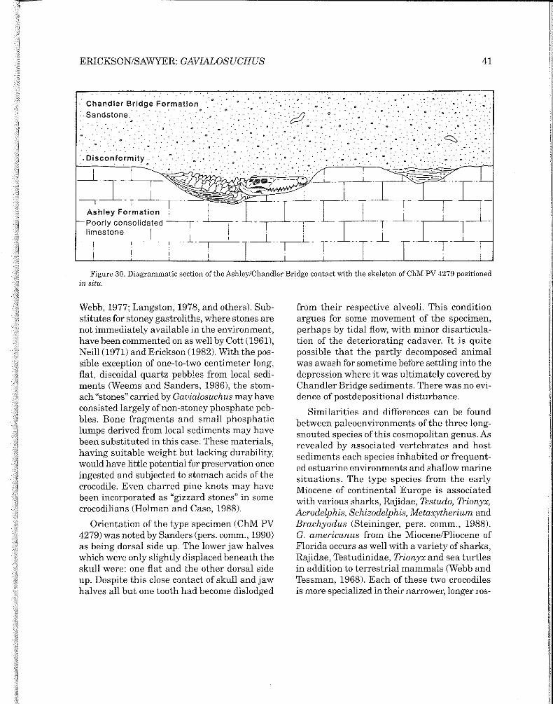
of the present type series. Most complete of these is the type specimen ChM PV 4279. Its relatively intact state can be attributed largely to its deposition in one of the many depressions or pits which characterize the upper surface of the Ashley Formation (Fig. 30). Weems and Sanders (1986) suspect that these depressions or pits represent tree stump holes where trees were rooted into the Ashley sediments. Another explanation might involve the dissolution of large phosphate blocks that are so abundant in the Ashley (Leidy, 1877). Lime phosphate which is present in the Ashley (up to 15 percent) occurs as small phosphatic lumps (Cook, 1936) and is characterized as highly calcareous marl or soft limestone (Cooke, 1943). These depressions range in size from less than ten centimeters to over two meters across as based on personal observations (1987-93) made during low tide along the Ashley River and other watercourses which dissect the Ashley Formation. Disposition of the skeleton in one of the larger depressions could have resulted from the cadaver awash in the tidal flow. As discussed by Erickson (1990) confinement within a depression and subsequent fossilization resulted in the specimen's relatively intact condition. There is no evidence of the specimen having been scavenged before fossilization except perhaps by invertebrates, because of the integrity of the semi-articulated skeleton. The absence of many foot bones, rib and gastralia sections, as well as other small elements may be explained by decomposition and biological erosion which also removed much of the thin articular surfaces of many bones. The apparent lack of gastroliths with such a large crocodile, which would presumably have had large stomach "stones", is puzzling. However, individual crocodilians and sometimes suites of associated crocodile skeletons totally lack associated gastroliths or stomach "stones" (Erickson, 1982).
Whether for ballast or food processing the occurrence of stomach "stones" has been widely discussed (Krumbiegel, 1959; Cott, 1961;
41ERICKSON/SAWYER: GAVIALOSUCHUS
... Chandler Bridge Formation •..
·.sandstone.· • •
·. Disconformity. · ·
_J_
T I
. . .
. . 0 .
..
. . ,, . .. . . . .
. . . .
.. .s,,·.. ·
Figure 30. Diagrammatic section of the Ashley/Chandler Bridge contact with the skeleton of ChM PV 4279 positioned in situ.
Webb, 1977; Langston, 1978, and others). Substitutes for stoney gastroliths, where stones are not immediately available in the environment, have been commented on as well by Cott (1961), Neill (1971) and Erickson (1982). With the possible exception of one-to-two centimeter long, flat, discoidal quartz pebbles from local sediments (Weems and Sanders, 1986), the stomach "stones" carried by Gavialosuchus may have consisted largely of non-stoney phosphate pebbles. Bone fragments and small phosphatic lumps derived from local sediments may have been substituted in this case. These materials, having suitable weight but lacking durability, would have little potential for preservation once ingested and subjected to stomach acids of the crocodile. Even charred pine knots may have been incorporated as "gizzard stones" in some crocodilians (Holman and Case, 1988).
Orientation of the type specimen ( ChM PV 4279) was noted by Sanders (pers. comm., 1990) as being dorsal side up. The lower jaw halves which were only slightly displaced beneath the skull were: one flat and the other dorsal side up. Despite this close contact of skull and jaw halves all but one tooth had become dislodged
from their respective alveoli. This condition argues for some movement of the specimen, perhaps by tidal flow, with minor disarticulation of the deteriorating cadaver. It is quite possible that the partly decomposed animal was awash for sometime before settling into the depression where it was ultimately covered by Chandler Bridge sediments. There was no evidence of postdepositional disturbance.
Similarities and differences can be found between paleoenvironments of the three longsnouted species of this cosmopolitan genus. As revealed by associated vertebrates and host sediments each species inhabited or frequented estuarine environments and shallow marine situations. The type species from the early Miocene of continental Europe is associated with various sharks, Rajidae, Testudo, Trionyx, Acrodelphis, Schizodelphis, Metaxytherium and Brachyodus (Steininger, pers. comm., 1988). G. americanus from the Miocene/Pliocene of Florida occurs as well with a variety of sharks, Rajidae, Testudinidae, Trionyx and sea turtles in addition to terrestrial mammals (Webb and Tessman, 1968). Each of these two crocodiles is more specialized in their narrower, longer ros-
42 SCIENCE MUSEUM OF MINNESOTA MONOGRAPH VOL.3: PALEONTOLOGY
tra and greater uniformity of tooth size. Mook (1932) noted a direction of specialization for Gauialis that included lengthening the snout and reducing tooth size, while increasing their number, as an adaptation for a more picivorous habit. Langston (1973) also discusses the long snout with widely spaced teeth as being highly functional for striking and impaling prey. The new taxon, by contrast, with its shorter rostrum and appreciably larger teeth, was more suited for disemboweling large prey such as stranded cetaceans (Erickson, 1990). Its teeth are extremely robust yet relatively short for their basal circumferences. The fifth maxillary tooth is the largest in the skull, as
it is in the other American species G. americanus, whereas in G. eggenburgensis the fifth maxillary tooth is essentially the same size as those anterior and posterior to it. Other aspects of the skull shared by the North American species but not by their European counterpart are much closer tooth spacing in the maxillae and a low, flat cranial table. The type species possesses a cranial table situated well above the base of the rostrum. Its surface is also depressed medially and prominent elevations (spurs) occur at its posterolateral corners (Fig. 3). These configurations of skull and dentition indicate a more gavial-like adaptation than either of the American forms.

43 ERICKSON/SAWYER: GAVIALOSUCHUS
DISCUSSION
With a general amelioration of the climate by Chattian times (Lillegraven, 1972) the coastal plain of Southeastern North America provided a new environmental setting to be exploited by large estuarine crocodiles. One of these forms was Gavialosuchus. The earliest record of this genus is the new taxon G. carolinensis n.sp. from the late Oligocene ofcoastal South Carolina. The next earliest record ofthis genus is G. eggenburgensis from the early Miocene ofAustria. The most recent occurrence is G. americanus from the Miocene/Pliocene of Florida. The two American species are each represented by ample materials including partly complete skeletons which provide us with a rather complete osteology for the group. Distribution of the American species is shown in Figure 22.
Knowledge of the Austrian species derives from the skull of the type specimen. Its original preparation was completed shortly after its discovery by Krahuletz and inclusion in his private collection. The original description by Toula and Kail (1885) included what had been originally collected and prepared when the specimen was placed in the newly established Krahuletz Museum in Eggenburg. Examination of the skull by Erickson in 1987, however,
revealed the loss of the pterygoids and other ventral portions of the skull. These were fortunately figured in the original description. This was especially critical regarding the shape of choanal opening. Although incomplete the opening was narrowed to a point anteriorly and was evidently triangular when complete (Fig. 23). Seemingly a second preparation also had been attempted, either before or after the loss, which incorrectly replaced two teeth and misaligned the cranium. Most recently ( 1988) restoration of the type skull was completed in Vienna by the Institut for Palaontologie after Professor Steininger kindly permitted one of us (ERE) to make recommendations for its restoration.
In all of the specimens a shared character state is a subtriangular opening ofthe choanae. The diagnostic value of this shape has been debated (Muller, 1927; Auffenberg, 1954; and others) and it may be a character that varies somewhat. Yet its persistence within the described suite of species is regarded as diagnostic especially when grouped with another persistent character, the large, relatively thin, unkeeled osteoscutes with large pits.

44 SCIENCE MUSEUM OF MINNESOTA MONOGRAPH VOL.3: PALEONTOLOGY
BIBLIOGRAPHY
AUFFENBERG, W. 1954. Additional specimens of Gavialosuchus americanus (Sellards) from a new locality in Florida. Quart. Jour. Fla. Acad. Sci., 17(4): 185-209.
______. 1957. Notes on fossil crocodilians from the southern United States. Quart. Jour. Fla. Acad. Sci., 20(2): 107-113.
BUFFETAUT, E. and P. WELLNHOFFER. 1980. Der Krokodilier Teleorhinus Osborn, 1904 (Mesosuchia, Pholidosauridae) in Regensburger Grunsandstein (Obercenoman). Mitt. Bayer. Staatsslg. Pali!ont. Hist. Geo/., 20: 83-94 Taf. 8, Munchen.
COLQUHOUN, D.J. 1965. Terrace sediment complexes in central South Carolina,Atlantic Coastal Plain Geo. Assn. Field Conf., Univ. S.C. 1965.
COOKE, C.W. 1936. Geology of the coastal plain of South Carolina. U.S. Geol. Sur., Bull. 867: 1-196.
______. 1943. Geology of the coastal plain of Georgia. U.S. Geo/. Sur., Bull. 941: 1-121.
COTT, H.B. 1961. Scientific results of an inquiry into the ecology and economic status of the Nile crocodile (Crocodylus niloticus) in Uganda and Northern Rhodesia. Trans. Zoo/. Soc. Land., 29: 211-356.
ERICKSON, B.R. 1982. The Wannagan Creek Quarry and its reptilian fauna [Bullion Creek Formation, Paleocene] in Billings County, North Dakota. No. Dak. Geo/. Sur., Rept. Invest. 72: 1-17.
_____. 1990. Paleoecology ofcrocodile and whale-bearing strata of Oligocene Age in North America. Hist. Biol., 4: 1-14.
HECHT, M. and B. MALONE. 1972. On the early history of the Gavialid Crocodylians. Herpetologica, 28(3): 281-284.
HOLMAN, J.A. and G.R. CASE. 1988. Reptiles from the Eocene Tallahatta Formation ofAlabama. Jour. Vert. Paleontol., 8(3): 328-333.
IORDANSKY, N.H. 1976. The skull of the crocodile. Biol. ofReptilia, Acad. Press, ed. C. Gans and T.S. Parsons, 4 Morph D: 201-262.
KELLOGG, R. 1944. Fossil cetaceans from the Florida Tertiary. Bull. Mus. Comp. Zoo!., 94: 432-471.
KRUMBIEGEL, G. 1959. Die tertiare Pflanzen-und Tierwelt der Braunkohle des Geisel tales. Die Neve Brehm-Bucherei, Geiseltalmuseum, Museum fur Mitteldeutsche Erdgeschichte, Halle, A.Z. Verlag. Wittenberg, Lutherstadt.

45 ERICKSON/SAWYER: GAVIALOSUCHUS
LANGSTON, W., Jr. 1973. The crocodylian skull in historical perspective. Biol. ofReptilia, Acad. Press, ed. C. Gans and T.S. Parsons, 4 Morph D: 263-284.
______. 1978. A yearling crocodylian from the Middle Eocene Green River Formation of Colorado. Jour. Paleontol., 52: 122-125.
LEIDY, J. P. 1877. Description of vertebrate remains, chiefly from the Phosphate Beds of South Carolina. Jour. Acad. Nat. Sci., Phil. ser. 2, 8 pt. 3(9): 209-262.
LILLEGRAVEN, J.A. 1972. Ordinal and familial diversity of Cenozoic mammals. Taxon, 21 (2/3): 261-274.
MALDE, E.E. 1959. Geology of the Charleston phosphate area, South Carolina. U.S. Geol. Sur., Bull. 1079: 1-105.
MEYER, E.R. 1984. Crocodylians as living fossils. In: N. Eldredge and S.M. Stanley Editors, Living Fossils, Springer-Verlag, Berlin: 105-131.
MOOK, C.C. 1921a. Skull characters and affinities of the extinct Florida gavial Gavialosuchus americanus (Sellards). Bull. Amer. Mus. Nat. Hist., 44: 33-42.
_____. 1921b. Notes on the postcranial skeleton of the Crocodylia. Bull. Amer. Mus. Nat. Hist., 44: 67-100.
_____. 1924. Further notes on the skull characters of Gavialosuchus americanus (Sellards) Amer. Mus. Nat. Hist. Nov., 155: 1-2.
_____. 1932. A new species of fossil gavial from the Siwalik Beds. Amer. Mus. Nat. Hist., Nov. 514: 1-5.
MORGAN, G.S. 1986. The so-called giant Miocene dolphin Megalodelphis magnidens Kellogg (Mammalia: Cetacea) is actually a crocodile (Reptilia: Crocodylia). Jour. Paleontol., 60(2): 411-417.
MULLER, L. 1927. Ergebnisse der Forschungereisen Prof. E. Stromers in dem WustenAegyptens. Vol. 5. Tertiare Wirbeltiere. No. 1. Beitrage zur Kenntnis der Krokodilien des aegyptischen Tertiars. Ahb. Akad. wiss. Munchen. Mathnatur. 31: 1-97.
NEILL, W.T. 1971. The last of the ruling reptiles, alligators, crocodiles and their kin. Columbia Univ. Press, N.Y., pp. 1-486.
PEJOVIC, D. 1951. Ueber den Fund des Unterkiefers eines fossilen Krokodile in Cementmergel von Popovac bei Paracin. Zborn. Radova Geol. Inst. Beograd, 16: 103-107.
PIRKLE, E.C. 1956. The Hawthorne and Alachua formations ofAlachua County, Florida. Quart. Journ. Fla. Acad. Sci., 19(4): 197-240.
_____. 1957. Hawthorne exposures southeast of Gainesville Florida. Quart. Journ. Fla. Acad. Sci., 20(2) 137-142.

46 SCIENCE MUSEUM OF MINNESOTA MONOGRAPH VOL.3: PALEONTOLOGY
ROGERS, S.C. 1914. The phosphate deposits of South Carolina. U.S. Geol. Sur., Bull. 580: 183-200.
ROGL, F. and F.F. STEININGER. 1984. Neogene Paratethys Mediterranean and Indo-Pacific Seaways, implications for the paleobiogeography of marine and terrestrial biotas. Fossils and Climate. ed. P. Brenchley, Wiley & Sons Ltd., pp. 171-200.
ROSS, C.A. and C.D. ROBERTS. 1979. Scalation of the American alligator. U.S.D.I. Spec. Sci. Rept., Wildlife, 225: 1-8.
ROTH, J.A. and J. LAERM. 1980. A Late Pleistocene vertebrate assemblage from Edisto Island, South Carolina. Brimleyana, 3: 1-29.
SANDERS, A.E. 1980. Excavation of Oligocene marine fossil beds near Charleston, South Carolina. Natl. Geog. Soc. Res. Rept., 12: 601-621.
SANDERS, A.E., E.M. LEMON, Jr., and R.E. WEEMS. 1979. A newly recognized post-Cooper/preHawthorne stratagraphic unit in the lower coastal plain, Dorchester County, South Carolina. (abs) Geo/. Soc. Amer. Abst. w/prog., 11(4): 1-210.
SANDERS, A.E., R.E. WEEMS, and E.M. LEMON, Jr. 1982. Chandler Bridge Formation-Anew Oligocene stratigraphic unit in the lower coastal plain of South Carolina. U.S. Geol. Sur., Bull. 1529-H, cont. to strat., pp. 105-124.
SELLARDS, E.H. 1915a. A new gavial from the late Tertiary of Florida. Amer. Jour. Sci., 4(40): 135-138.
_______. 1915b. The pebble phosphates of Florida.Ann. Rept. Fla. Geol. Sur., (7): 25-116.
_______. 1916. Anew tortoise and a supplementary note on the gavial, Tomistoma americana. Amer. Jour. Sci., pp. 235-240.
SHIPMAN, P. 1981. Life history of a fossil, an introduction to taphonomy and paleoecology. Harvard Univ. Press, Lond., pp. 1-222.
STEEL, R. 1973. Crocodylia. Encyclopedia ofPaleoherpetology 16: 95-96.
TELLES AMTUMES, M. 1961. Tomistoma lusitanica, crocodilien du Miocene du Portugal. Rev. Facul. Cien. Lisboa (2), C9(1): 5-88.
TOULA, F. and J.A. KAIL. 1885. Uber einen Krokodil-Schadel aus den Tertiarablagerungen von Eggenberg in Niederiisterreich, Denkschr. Acad. wiss. Vienna, L: 299-356.
VIANNA, A. and A. MO RAES. 1941. Sur un crane de crocodile fossile decouvert dans le Miocene de Lisbonne. Version francoise de la communication presentee au Ier Congres Natl. des Sci. Nat., Lisbonne.
WARD, L.W., E.W. BLACKWELDER, G.S. GO LIN and R.E. POORE. 1979. Stratigraphic revision of Eocene, Oligocene and lower Miocene formations of South Carolina. So. Car. Div. Geol., Geol. Notes., 23(1): 2-32.

47 ERICKSON/SAWYER: GAVIALOSUCHUS
WEBB, G.T.W. 1977. The natural history ofCrocodylus porosus, habitat and nesting, growth, movement, river distribution and general comments. A study of Crocodylus porosus in Northern Australia, ed. Messel and Butler, Sydney pp. 237-312.
WEBB, S.D. and N. TESSMAN. 1968. A Pliocene vertebrate fauna from low elevation in Manatee County, Florida. Amer. Jour. Sci., 266: 777-811.
WEEMS, R.E. andA.E. SANDERS. 1986. The Chandler Bridge Formation (Upper Oligocene) of the Charleston, South Carolina region. Geol. Sur. Amer. Centennial Field Guide - So. Sec., pp. 323-326.
WERMUTH, H. 1953. Systematik der rezenten Krokodile. Mitt. Zool. Mus. Berlin, 29: 375-514.