The epigenetic impact of weaning on craniofacial morphology during growth
11
THE JOURNAL OF EXPERIMENTAL ZOOLOGY 276:243-253 (1996) The Epigenetic Impact of Weaning on Craniofacial Morphology During Growth J.W. HELM AND R.Z. GERMAN Department of Biological Sciences, University of Cincinnati, Cincinnati, Ohio 45221 ABSTRACT Miniature pigs (Sus scrofa) were used as a model to investigate whether the time of weaning (a nongenetic factor) affects skeletal growth rates for both pre- and postweaning time periods. Control litters were weaned at the normal time of 32 days. Two litters were weaned early (at 20 days) and two late (at 46 days). We digitized cranial landmarks from radiographs taken three times a week for a total of 70 days. We used analysis of covariance to test for differences in growth rates between pre- and post-weaning periods, as well as differences in growth rates among treatments. In both the late weaned pigs and the controls, facial length, facial width, basicranial length, and basicranial width growth rates slowed significantly at the time of weaning. However, in the early weaned pigs, there were no significant changes in growth rates for any of the facial or basicranial measurements at weaning. Furthermore, the postweaning rates of growth were differ- ent among treatments. One possible implication is that early growth rates could be under tight genetic control while later growth rates can be epigenetically regulated through nutritional changes. 0 1996 Wiley-Liss, Inc. The significance of genetic inheritance to mor- phology has been a central theme in evolution- ary theory since the time of Darwin. However, processes outside of genetic control can play a role in the evolutionary change of morphology. Evolu- tionary biologists are still debating the role and relative importance of development and other pri- marily nongenetic mechanisms in the determina- tion of form. Yet, it is difficult to understand the significance of development to evolutionary change without first detailing the morphological changes that take place between the zygote and the adult (McKinney and McNamara, '91). Epigenetics refers to nongenetic changes in morphology produced through environmental and internal stimuli. Her- ring ('93a) has proposed two reasons for the recent resurgence of interest in epigenetic regulation, particularly of skull morphology. First, studies of heritability demonstrate an environmental com- ponent for variation in craniofacial characters, and second, experimental data show the ease of cra- nial alteration by environmental stimuli. Epigenetic influences over bone growth have been the subject of much study in the last decade (Totsuka et al., '83; Vilmann et al., '85, '89; Her- ring, '93b); however, few of these studies have used longitudinal growth designs and/or non-pathologi- cal models. A number of studies show that mus- cular loading of bone is an epigenetic mechanism for altering growth (Herring and Scapino, '73; 0 1996 WILEY-LISS, INC. Corruccini and Beecher, '82; Vilmann et al., '85, '89; Herring, '92, '93b). Herring ('93b) hypothesizes that skull loading is far more dynamic and vari- able than assumed by current models for predict- ing changes in morphology. She further argues that "cranial development is epigenetic" because factors such as mechanical loads influence cra- nial morphology. The impact of biomechanical function on cranial development occurs through muscular loading (Herring, '93b). Herring sup- ports these hypotheses of an epigenetic component to cranial morphology with a number of examples where the mechanical forces that act on bone were altered, producing changes in shape. For example, anencephalic infants lack the mechanical loading needed to stimulate the bones of the neurocra- nium to grow, forming a cranial vault. Most studies examining the impact of muscu- lar loads on skeletal growth involve a pathologi- cal alteration of normal growth mechanisms, such as muscular dystrophy, to see the effects on bone growth (Totsuka et al., '83; Vilmann et al., '85, '89; Lightfoot, '94). Beyond these examples, there are no studies in the literature that closely exam- ine the effects of muscular loading or other epige- Received January 3,1996; accepted February 25,1996. Address reprint requests to R.Z. German, Department of Biologi- cal Sciences, ML-06, University of Cincinnati, Cincinnati, OH 45221.
Transcript of The epigenetic impact of weaning on craniofacial morphology during growth

THE JOURNAL OF EXPERIMENTAL ZOOLOGY 276:243-253 (1996)
The Epigenetic Impact of Weaning on Craniofacial Morphology During Growth
J.W. HELM AND R.Z. GERMAN Department of Biological Sciences, University of Cincinnati, Cincinnati, Ohio 45221
ABSTRACT Miniature pigs (Sus scrofa) were used as a model to investigate whether the time of weaning (a nongenetic factor) affects skeletal growth rates for both pre- and postweaning time periods. Control litters were weaned at the normal time of 32 days. Two litters were weaned early (at 20 days) and two late (at 46 days). We digitized cranial landmarks from radiographs taken three times a week for a total of 70 days. We used analysis of covariance to test for differences in growth rates between pre- and post-weaning periods, as well as differences in growth rates among treatments. In both the late weaned pigs and the controls, facial length, facial width, basicranial length, and basicranial width growth rates slowed significantly a t the time of weaning. However, in the early weaned pigs, there were no significant changes in growth rates for any of the facial or basicranial measurements at weaning. Furthermore, the postweaning rates of growth were differ- ent among treatments. One possible implication is that early growth rates could be under tight genetic control while later growth rates can be epigenetically regulated through nutritional changes. 0 1996 Wiley-Liss, Inc.
The significance of genetic inheritance to mor- phology has been a central theme in evolution- ary theory since the time of Darwin. However, processes outside of genetic control can play a role in the evolutionary change of morphology. Evolu- tionary biologists are still debating the role and relative importance of development and other pri- marily nongenetic mechanisms in the determina- tion of form. Yet, it is difficult to understand the significance of development to evolutionary change without first detailing the morphological changes that take place between the zygote and the adult (McKinney and McNamara, '91). Epigenetics refers to nongenetic changes in morphology produced through environmental and internal stimuli. Her- ring ('93a) has proposed two reasons for the recent resurgence of interest in epigenetic regulation, particularly of skull morphology. First, studies of heritability demonstrate an environmental com- ponent for variation in craniofacial characters, and second, experimental data show the ease of cra- nial alteration by environmental stimuli.
Epigenetic influences over bone growth have been the subject of much study in the last decade (Totsuka et al., '83; Vilmann et al., '85, '89; Her- ring, '93b); however, few of these studies have used longitudinal growth designs and/or non-pathologi- cal models. A number of studies show that mus- cular loading of bone is an epigenetic mechanism for altering growth (Herring and Scapino, '73; 0 1996 WILEY-LISS, INC.
Corruccini and Beecher, '82; Vilmann et al., '85, '89; Herring, '92, '93b). Herring ('93b) hypothesizes that skull loading is far more dynamic and vari- able than assumed by current models for predict- ing changes in morphology. She further argues that "cranial development is epigenetic" because factors such as mechanical loads influence cra- nial morphology. The impact of biomechanical function on cranial development occurs through muscular loading (Herring, '93b). Herring sup- ports these hypotheses of an epigenetic component to cranial morphology with a number of examples where the mechanical forces that act on bone were altered, producing changes in shape. For example, anencephalic infants lack the mechanical loading needed to stimulate the bones of the neurocra- nium to grow, forming a cranial vault.
Most studies examining the impact of muscu- lar loads on skeletal growth involve a pathologi- cal alteration of normal growth mechanisms, such as muscular dystrophy, to see the effects on bone growth (Totsuka et al., '83; Vilmann et al., '85, '89; Lightfoot, '94). Beyond these examples, there are no studies in the literature that closely exam- ine the effects of muscular loading or other epige-
Received January 3,1996; accepted February 25,1996. Address reprint requests to R.Z. German, Department of Biologi-
cal Sciences, ML-06, University of Cincinnati, Cincinnati, OH 45221.

244 J.W. HELM AND R.Z. GERMAN
netic mechanisms on bone growth in both a longi- tudinal and a nonpathogenic manner.
One area where an epigenetic effect has been demonstrated for a non -pathological mechanism, using cross-sectional data, is the impact of dietary regulation on growth (Elouvier and Hylander, '81; Corruccini and Beecher, 82). Bouvier and Hylander ('81) show that macaque mandibular shape and remodeling can be altered with changing levels of bone strain due to a switch in dietary consistency. Corruccini and Beecher ('82) support the premise that a change in dietary force can alter bone shape, reporting a strong relationship between di- etary consistency and occlusal development in pfi- mates. A soft diet Wafs shown to reduce bone growth significantly in the palatine region of the skull through decreased jaw forces (Corruccini and Beecher, '82). Most longitudinal studies examin- ing dietary effects on growth only measure body weight, ignoring changes of size or shape (Hughes and Tanner, '70). Those studies that describe form or body composition generally do so for the soft tissues and not bone (Feiiton and Cam, '51; Collins and Cheek, '83; Cothran et al., '85; Reeds et al., '93). One example comes from Hughes and Tan- ner ('701, who attribute a small decrease in the growth rates of rat body length and tail length to a decrease in the amount of milk available and a switch to supplement their diet with solid food. These results support the hypothesis that the regulation of dietary intake, in both nutritional value and muscular loading, can alter growth rates epigenetically.
The results presented here address the ques- tion of variation in piglet size, shape, and rates of growth produced by the epigenetic regulation of weaning during early growth. Specifically, how does epigenetically varying the time of wean- ing affect the rate change that normally occurs at weaning? The morphological variation elic- ited in this study resulted from a change in the timing of a normal event, weaning. This shift produced two immediate impacts: a change in the forces generated by chewing and a differ- ence in nutritional value. Since this study was longitudinal in nature, it included precise rates of growth for each individual and, therefore, ac- counted for variation among pigs. Results of these experiments showed one link between bone growth and dietary regulation in postem- bryonic development. Few epigenetic studies quantify growth in this fashion or measure the exact impact of one variable in a nonpatho- logical and controlled method.
MATERIALS AND METHODS This study used six litters of pigs, Sus scrofa
(mini Hanford breed), each with three individu- als, obtained from Charles River Laboratories (Wilmington, MA). To control for any sexual di- morphism, only females were examined. Two lit- ters were used for each treatment, and two litters comprised the control. The normal weaning time, when all piglets are pulled from the mother, is approximately 30-32 days of age (personal com- munication, Don Moody, Charles River Laborato- ries). Therefore, control weaning was at 32 days of age. One treatment weaned individuals at 20 days, and another treatment weaned individuals at 46 days. Prior to weaning, the diet consisted of milk replacer (Purina #2007, St. Louis, MO) with a 25% protein content and a 13% fat content, spe- cially formulated for infant pigs. Pigs were fed ev- ery 3-4 h during the first week, after which this frequency was gradually reduced to three times a day as volume was increased. All pigs were trained to lap the formula from bowls. From the day of weaning to the 70th day, pigs were fed a minipig starter chow (Purina #5080, St. Louis, MO) with a 20% protein content and a 4% fat content.
Radiographs were taken (three times a week) to measure cranial dimensions in both the dors- oventral and lateral views. Weights were taken daily during the entire study. Measurements in- cluded mass in grams, as well as the lengths and widths of skull dimensions in millimeters. A Bennett mammography machine was used to col- lect the radiographs while the animals were anes- thetized using an Ohio 4000 Compact Anesthesia machine (Madison, WI) with isoflurane gas. The anesthetic was administered using a cone mask, and individuals remained sedated for no more than 30 min. Animals were permitted to wake be- fore being returned to their pen. From a preliminary study, the x-ray dosage producing the greatest reso- lution was found to be 49 KV at 150 mas. Weight data were collected using an electronic scale. All pro- cedures were followed in accordance with IACUC regulations and protocol number 91-05-27-01.
We measured the radiographs with a Numonics flatbed digitizing tablet with an accuracy of 0.127 mm and the program DIGIT (written by D. Hertweck). We chose points from the radio- graphs for their biological meaning and their repeatability. Measurements consisted of ho- mologous landmarks defined by suture lines and bone projections (Table 1). These measurements included lengths, widths, and heights of the skull. Dorsoventral skull measurements included facial

EPIGENETIC IMPACT OF WEANING 245
TABLE 1. Craniofacial measurements
Label Measurement Descriution
Dorsoventral 1
2
3
4
5
6
7
8
9
10
Lateral 1
2
3
4
5
6
7
Maxillary length
Palate length
Total length
Maxillary width
Mandibular width
Occipital length
Parietal length
Total basicranial length
Max basicranial width
Min basicranial width
Nasal length
Frontal length
Ma xi 11 a ry length
Nasal height
Total facial height
Total b a d neurocranial length Total basil neurocranial height
Distance from most anterior end of pre- maxilla to anterior end of hard palate at palatine suture Distance from most anterior end of hard palate at palatine suture to most posterior end of hard palate Distance from most anterior end of premaxilla to most posterior end of hard palate Distance between most anterior left and right edges of maxilla Distance between left and right edges of mandible at the point where the jaw first crosses under the maxilla Distance from anterior edge of the basioccipi- tal bone to foramen magnum Distance from temporal- parietal suture to parietal-occipital suture Distance from anterior edge of basioccipital bone to posterior end of skull Distance between left and right temporal- parietal sutures Distance between left and right parietal- occipital sutures
Distance from anterior tip of the nasal bone to nasal-frontal suture Distance from anterior frontal bone to frontal- interparietal suture Distance from anterior premaxilla to palatine suture Distance from anterior tip of the nasal bone to anterior premaxilla Distance from nasal- frontal suture to maxilla-palatine suture Distance from frontal- interparietal suture to nucal ridge Distance from frontal- interparietal suture to posterior bulla (just in front of the styloid urocess)
lengths-maxillary, palate, and total-and ba- s icranial widths-maximum a n d minimum (Fig. 1A). Lateral skull measurements included facial lengths-nasal, frontal, and maxillary-and facial heights-nasal and total. Basicranial mea- surements included total height and total poste- rior neurocranial length (Fig. 1B).
For all analyses, individuals within each litter, as well as litters within each treatment, were grouped. However, replication of individuals and litters was accounted for in the statistical model fit to the data. Linear regression for preweaning and postweaning data gave estimates of growth rates for each treatment. The dependent variable was a skull measurement, such as mandibular length, while the independent measure was age or the log of weight. Significant differences among these growth rates, pre- and postweaning within a given treatment as well as among treatments, were tested with analysis of covariance (ANCOVA). All ANCOVAs were a mixed model, partitioning the variation to include individuals nested within lit- ters and litters nested within treatments. Most likely due to the small sample size, there were significant differences between individuals and lit- ters. However, the mixed model ANCOVA tested
A
Fig. 1. Diagrammatic representation of a pig skull made from tracings of actual radiographs in A dorsoventral view and B lateral view. In A anterior is up and in B anterior is left. Straight lines indicate measurements digitized using re- peatable points. Measurements are defined in Table 1.

246 J.W. HELM AND R.Z. GERMAN
for treatment and timing differences given sig- nificant variation in individuals and litters. The ANCOVA model included one of the cranial mea- surements, such as maxillary height, and a cova- riate, such as age or weight. Age was the covariate used in the analysis of all lateral measurements, and the log of weight was used for the dorsoven- tral measurements to maximize the total varia- tion accounted for by the model. The first set of ANCOVA models tested for significant slope dif- ferences within each treatment between pre- and postweaning rates. The second set of ANCOVA models tested the homogeneity of slopes among treatments for preweaning rates. If the slopes did not differ significantly, i.e., the lines were paral- lel, then a linear model was used to test for any treatment effects, i.e., differences in intercepts. If either pre- or postweaning growth rates were not equal, then the lines were not parallel and test- ing for treatment effects would be meaningless. These analyses were done separately for pre- and postweaning data.
A full-model ANCOVA tested for homogeneity of the entire slope without separating into pre- and postweaning data sets. This model also in- cluded treatment and weaning effects to see which was significant: the difference in the growth rates among treatments (treatment effect) or the dif- ference between pre- and postweaning slopes (weaning effect). An example model statement is:
face - length = constant + age + wean + treat + pig {litter) + litter {trei~tl + wean * age + treat * age + wean * treat * age
Both the weaning and treatment effects tested whether there was a significant interaction be- tween the covariate and either the different treat- ments or the pre- vs. postweaning growth rates.
We used an ANCOVA including the log of weight, and the covariate age was used to test for significant differences in the amount of growth (the log of weight) among the three treatments. ANCOVA corrects for tbe initial size differences between the various litters so that the amount of growth was compared atmong the treatments.
Finally, we did bivariate regressions of frontal length and total facial height, maxillary length and maxillary width, maxillary length and total facial height, palate len,@h and mandibular width, nasal length and nasal height, basiheurocranial length and total basiheurocranial height, parietal length and minimum basicranial width, and total basicranial length to maximum total basicranial
width, to test for allometric scaling differences among treatments. ANCOVA tested the homogene- ity of slopes among treatments to confirm any shape differences over the various regions of the skull.
In summary, the analyses done were: 1. For each treatment (weaning time), differ-
ences between pre- and postweaning rates were tested.
2. For preweaning growth rates, differences among treatments (weaning times) were tested.
3. For postweaning growth rates, differences among treatments (weaning times) were tested.
4. A complete ANCOVA model was used to test the homogeneity of the entire juvenile slope, including tests for treatment and weaning effects.
5. Differences in juvenile size among treatments (weaning times) were tested.
6. Allometric shape differences among treat- ments were tested with ANCOVA.
RESULTS Pre- us. postweaning differences
within treatment For most skull measurements, the preweaning
slope differed significantly from the postweaning slope in both the control and late weaned treat- ments. However, there were no significant slope differences between the pre- and postweaning rates for most measurements of the early weaned treatment (Table 2). One example, facial length as a function of age, showed a distinct rate change that occurred around the time of weaning for the control and late treatments, but there was no rate change that occurred in the early weaned treat- ment (Fig. 2). The only two measurements of the face which did not follow this pattern were palate length and total facial length (Table 2). Within the palate, there was no significant slope differ- ence between the pre- and postweaning rates for the late weaned treatment. For total facial length, there was a significant slope difference between the pre- and postweaning rates of the early weaned treatment.
Basicranial lengths, widths, and heights showed the same trends as in the face (Table 2). For ex- ample, the rate change between pre- and postwean- ing slopes for basicranial length is evident in the control and late treatments but not in the early (Fig. 3). The only basicranial measurements that did not follow this pattern were occipital length, maximum total width, and basiheurocranial to-

EPIGEIWTIC IMPACT OF WEANING 247
TABLE 2. Significant differences (p values) for ANCOVA between pre- and postweaning growth rates for
dorsoventral and lateral measurements
Measurement Early Control Late
Facial lengths Dorsoventral
Maxillary Palate Total
Lateral Nasal Frontal Maxillary
Facial widths Dorsoventral
Maxillary Mandibular
Facial heights Lateral
Nasal Total
Basicranial lengths Dorsoventral
Occipital Parietal Total
Basicranial widths Dorsoventral
Max total Min total
Basiineurocranial Later a 1
Total length Total height
0.071 0.000 0.280 0.000 0.012 0.000
0.650 0.000 0.599 0.016 0.627 0.000
0.216 0.000 0.554 0.001
0.832 0.001 0.619 0.048
0.415 0.460 0.212 0.000 0.142 0.000
0.441 0.000 0.937 0.014
0.540 0.560 0.455 0.051
0.001 0.215 0.000
0.000 0.005 0.000
0.000 0.026
0.007 0.000
0.000 0.039 0.015
0.938 0.009
0.144 0.000
tal length. Occipital length did not have a pre- vs. postweaning slope difference for the early weaned treatment or the control; however, there was a significant slope change for the late weaned treatment. For the maximum total basicranial width, there was no significant slope difference between the pre- and postweaning growth rates for the late weaned treatment. Within the total basi/neurocranial length, there was no significant slope difference between the pre- and postwean- ing growth rates for any of the three treatments.
Preweaning differences among treatments ANCOVA for the homogeneity of preweaning
slopes found no difference in slope among treat- ments for any of the facial lengths, facial widths, or basicranial lengths (Table 3). Therefore, all of these lines were parallel, making it valid to test for differences in intercepts (treatment effect). For each set of lines tested, there was no significant difference among intercepts, implying that the lines overlapped. The only preweaning slope,
I ! I I 1
! 25 1 30
control
*I :. i
D
20 I 1
35 r 30 I
age (days)
Fig. 2. Facial length (mm) vs. age (days) among treat- ments (early weaning, control weaning, and late weaning). Vertical line represents the time of weaning for each treat- ment. Rate changes occur around the time of weaning only for the control and late treatments. No rate change occws at the early weaning time.
which differed significantly among treatments, was the maximum basicranial width, where the late weaning treatment had the highest slope. In summary, the preweaning slopes (with one excep- tion) of each treatment were equal, with the lines parallel and lying on top of one another.
Postweaning differences among treatments ANCOVA for slope homogeneity of postweaning
growth rates among treatments showed that there was a significant difference between the treatment slopes for all the facial and basicranial measure-

J.W. HELM AND R.Z. GERMAN
44
42
. . 1 . *:* ; . * a o
40-
* . 42
50 r ! . 8
control
I
- t I * . I .. *. I 0 , : .:. .: :i .. f . .1.
early
late
age (days)
Fig. 3. Basicranial width (mm) vs. age (days) among treat- ments (early weaning, control weaning, and late weaning). Vertical line represents the time of weaning for each treat- ment. Rate changes occur around the time of weaning only for the control and late treatments. No rate change occurs at the early weaning time.
ments with one exception (Table 4). For each of these measurements, there were at least two sig- nificantly different treatments, and, in most cases, all three treatments differed significantly, with the late weaning slope being highest. Because these measurements had nonequal slopes, testing for an intercept difference (treatment effect) was mean- ingless. The only excelption was the total length of the badneurocranium, where all treatments had equal growth rates. However, there was no treatment effect; i.e., all intercepts were the same. In summary, all of the postweaning growth rates
TMLE 3. Significant differences (p values) for ANCOVA among treatments for preweaning growth rates
Homogeneity Measurement of slopes Treatment effect
Facial lengths Dorsoventral
Maxillary 0.189 0.199 Palate 0.141 0.281 Total 0.248 0.250
Nasal 0.336 0.169 Frontal 0.233 0.532 Maxillary 0.511 0.232
Lateral
Facial widths Dorsoventral
Maxillary 0.476 0.466 Mandibular 0.640 0.358
Facial heights Lateral
Nasal 0.334 0.387 Total 0.360 0.587
Basicranial lengths Dorsoventral
Occipital 0.731 0.715 Parietal 0.293 0.298 Total 0.850 0.971
Basicranial widths Dorsoventral
Max total 0.001 -
Min total 0.352 0.369 Basiheurocranial
Lateral Total length 0.166 0.331 Total height 0.824 0.827
(with one exception) were significantly different among treatments.
Slope differences among treatments with entire ANCOVA model
ANCOVA for the homogeneity of slopes among treatments, when both the weaning effect (differ- ences between pre- and postweaning growth rates) and treatment effect (difference due to variation in weaning time) have been accounted for, indi- cated that for most measurements the slopes were not equal (Table 5). Therefore, the lines (each treatment's growth rate over the entire time mea- sured) were not parallel. For each measurement with unequal slopes, or growth rates, the inter- cepts were significantly different, implying signifi- cant differences among treatments, beyond the pre- vs. postweaning differences. Total b a d neurocranial length was the only measurement which had equal slopes among treatments and was the exception to this pattern.
Looking at the weaning effect (whether there

EPIGENETIC IMPACT OF WEANING 249
TABLE 4. Significant differences (p ualues) for ANCOVA among treatments for uostweaning growth rates
Homogeneity Measurement of slopes Treatment effect
Facial lengths Dorsoventral
Maxillary 0.000 - Palate 0.000 - Total 0.000 -
Nasal 0.000 - Frontal 0.000 - Maxillary 0.001 -
Maxillary 0.000 - Mandibular 0.000 -
Lateral
Facial widths Dorsoventral
Facial heights Lateral
Nasal 0.003 - Total 0.000 -
Basicranial lengths Dorsoventral
Occipital 0.000 - Parietal 0.002 -
Total 0.000 -
Basicranial widths Dorsoventral
Max total 0.000 - Min total 0.040 -
Basilneurocranial Lateral
Total length 0.328 0.059 Total height 0.003 -
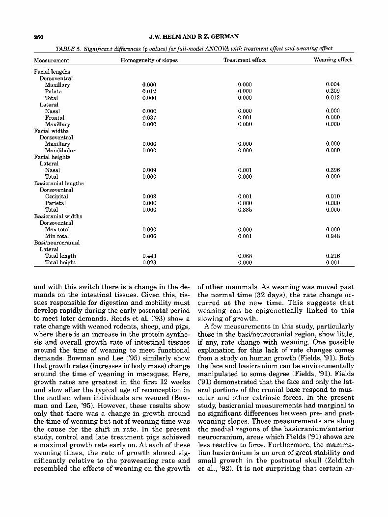
was a significant pre- vs. post-weaning slope dif- ference), all but four measurements had prewean- ing growth rates that were significantly different from postweaning growth rates when partition- ing the variation to include the treatment effect (difference due to variation in weaning time among treatments). Palate length, nasal height, minimum total basicranial width, and total b a d neurocranial height did not have significant pre- vs. postweaning growth rate differences (Table 5).
Final size differences among treatments
The ANCOVA model, including the log of weight and age, corrected for the initial size difference among litters so that variation in the amount of growth (changes in size) indicated significant dif- ferences among the treatments. No difference ex- isted between the control and early treatments ( p = 0.4991, but the late weaned treatment was dif- ferent from both the early ( p = 0.000) and control (p = 0.028) treatments in the amount of growth
occurring. Therefore, the late weaned treatment grew the most.
Allometric shape differences among treatments
In general, most measurements scaled the same, so there were few significant shape differences among treatments. The only notable exceptions were facial lengths relative to the other skull mea- surements. Within the face, lengths changed much faster with age than widths. The early weaned treatment had facial lengths which changed faster, relative to other skull measurements, than the other two treatments (Fig. 4). The control and late weaned treatments, however, had identical allo- metric scaling of facial length to other skull mea- surements. Thus, the faces of the early weaned treatment grew longer in length and at a faster rate than those of the control or late weaned treatments.
DISCUSSION
Changes in growth rates through ontogeny Among mammals, growth rates are maximal
soon after birth and slow within the first year (Laird et al., '65; Hughes and Tanner, '70; Fleagle and Samonds, '75; Fields, '91). Laird et al. ('65) describe a general pattern for mammalian growth; the rate of growth slows with the age of the ani- mal, until it approaches the adult size character- istic of the species and its individual genetic constitution. Similar patterns of growth exist for head measurements in the cebus monkey, for which cranial dimensions begin to slow in growth rate around 50 days of age and at 1 year are close to adult size (Fleagle and Samonds, '75). Fleagle and Samonds ('75) propose that "differences in growth schedule of various bones can be impor- tant to determine which bones can be affected by growth disturbances like malnutrition or illness." This is consistent with earlier work by Laird et al. ('65), showing that epigenetic influences be- come evident in later growth as all bones begin to slow in growth rate. These results taken to- gether suggest that as an individual moves along its growth trajectory, there is increasing poten- tial for processes outside of genetic control to act on growth.
Changes in growth around the time of weaning suggest that weaning can have an epigenetic influ- ence (Reeds et al., '93; Bowman and Lee, '95). Reeds et al. ('93) discuss the relevance of weaning to in- testinal and functional demands. Weaning is asso- ciated with a major switch in dietary composition,
260 J.W. HELM AND R.Z. GERMAN
TABLE 5. Significax.t differences (p values) for full-model ANCOVA with treatment effect and weaning effect
Measurement Homogeneity of slopes Treatment effect Weaning effect
Facial lengths Dorsoventral
Maxillary 0.000 0.000 0.004 Palate 0.012 0.000 0.209 Total 0.000 0.000 0.012
Nasal 0.000 0.000 0.000 Frontal 0.037 0.001 0.000 Maxillary 0.000 0.000 0.000
Maxillary 0.000 0.000 0.000
Lateral
Facial widths Dorsoventral
Mandibular 0.000 0.000 0.000 Facial heights
Lateral Nasal 0.009 0.001 0.396 Total 0.000 0.000 0.000
Basicranial lengths Dorsoventral
Occipital 0.009 0.001 0.010 Parietal 0.000 0.000 0.000 Total 0.000 0.335 0.000
Basicranial widths Dorsoventral
Max total 0.000 0.000 0.000 Min total 0.006 0.001 0.948
BasUneurocranial Lateral
Total length 0.443 0.068 0.216 Total height 0.023 0.000 0.001
and with this switch there is a change in the de- mands on the intestinal tissues. Given this, tis- sues responsible for digestion and mobility must develop rapidly during the early postnatal period to meet later demands. Reeds et al. ('93) show a rate change with weaned rodents, sheep, and pigs, where there is an increiase in the protein synthe- sis and overall growth rate of intestinal tissues around the time of weaning to meet functional demands. Bowman and Lee ('95) similarly show that growth rates (increases in body mass) change around the time of weaning in macaques. Here, growth rates are greatest in the first 12 weeks and slow after the typical age of reconception in the mother, when individuals are weaned (Bow- man and Lee, '95). However, these results show only that there was a change in growth around the time of weaning but not if weaning time was the cause for the shifl, in rate. In the present study, control and late treatment pigs achieved a maximal growth rate early on. At each of these weaning times, the rate of growth slowed sig- nificantly relative to the preweaning rate and resembled the effects of weaning on the growth
of other mammals. As weaning was moved past the normal time (32 days), the rate change oc- curred a t the new time. This suggests that weaning can be epigenetically linked to this slowing of growth.
A few measurements in this study, particularly those in the basilneurocranial region, show little, if any, rate change with weaning. One possible explanation for this lack of rate changes comes from a study on human growth (Fields, '91). Both the face and basicranium can be environmentally manipulated to some degree (Fields, '91). Fields ('91) demonstrated that the face and only the lat- eral portions of the cranial base respond to mus- cular and other extrinsic forces. In the present study, basicranial measurements had marginal to no significant differences between pre- and post- weaning slopes. These measurements are along the medial regions of the basicraniudanterior neurocranium, areas which Fields ('91) shows are less reactive to force. Furthermore, the mamma- lian basicranium is an area of great stability and small growth in the postnatal skull (Zelditch et al., '92). It is not surprising that certain ar-

EPIGENETIC IMPACT OF WEANING 251
Early ..... .......
Control -
30 20 25 30 35 40
Total Facial Length (rnrn)
Fig. 4. Scaling of frontal length (mm) vs. total facial height (mm) among treatments (early weaning, control weaning, and late weaning). Frontal length changes a t a faster rate for the early weaning treatment than the other two treatments. Both the control weaning treatment and the late weaning treat- ment scale the same.
eas in the basicraniudneurocranium, especially along the midline, were little affected by an epi- genetic factor like weaning.
Potential causes for the mammalian change in growth rate
Nonheritable environmental effects are one com- ponent of Atchley's ('93) hierarchy of developmen- tal control. The shift in the muscular loading of cranial bones caused by a change in dietary con- sistency could produce changes in growth rates and could have been one mechanism of environ- mental effect. Weaning causes such a shift, when the infant is switched from a liquid diet of milk or formula to a diet of hard food. The major force is due to crushing of food (Bouvier and Hylander, '81; Hylander et al., '87). The mastication of hard food increases the forces exerted by the jaw muscles on the facial bones (Zelditch and Charmichael, '89; Fields, '91). Previous studies demonstrate that the muscular loading of bone can influence the growth of morphological structures (Herring and Scapino, '73; Corruccini and Beecher, '82; Vilmann et al., '85, '89; Herring, '92, '93b; Lightfoot, '94). Suck- ling, however, makes little use of facial muscles (German and Crompton, '96). If the change in growth rate around the time of weaning in this
study resulted from a shift in muscular forces, then an increase in growth rate after weaning would be expected due to the higher force gener- ated to crush the solid food. However, our data demonstrated a decline in the rate of growth af- ter the time of weaning, suggesting that a differ- ent mechanism from loading is responsible for growth rate differences.
Prior to weaning, pigs were fed a minipig milk replacer formula with a higher protein and fat con- tent than the chow given to postweaned pigs. This decrease in fat and protein content may be re- sponsible for the rate change. A number of stud- ies have examined the effects of nutrition on growth and overall body size in many different taxa (Fenton and Carr, '51; Collins and Cheek, '83; Cothran et al., '85; Bernays, '86; Reeds et al., '93). Short-tailed opossums (Monodelphis domestica) were fed three different preweaning diets that varied in nutri- tional value. Opossums fed the high-protein diet had significantly lower weaning weights than those in the other two treatments (Cothran et al., '85). These results suggest that dietary regulation can influence the overall size reached by affect- ing growth rates, although in the opposite direc- tion from the results in the present study on pigs. Two possible causes for the lower weaning weight while on the more rich diet, not discussed by Cothran et al. ('85), could be that the higher pro- tein diet did not meet the minimum nutritional requirements of the opossums or that there was some adverse reaction to this diet as there was a Significantly higher (50%) mortality rate and lower growth rate among members of this treatment. A similar pattern exists in weanling mice which were fed a high-protein diet prior to weaning and never achieved the high weaning weight or final body size of mice fed lower protein diets (Fenton and Carr, '51). Our miniature pigs, in contrast to findings in opossums and mice, grew faster on a diet with high fat and protein, a result consistent with those of Reeds et al. ('93).
Genetic control of early growth rates No rate change occurred when weaning was
moved to 21 days of age. One explanation is that the early rate is more genetically constrained and not as easily perturbed as later growth rates. This is consistent with Atchley's ('93) view that com- plex morphological structures arise from a coor- dination of activities at different hierarchical levels. Atchley ('93) maintains that the genetic component or level is important early in develop- ment and only subsequent change is subject to

252 J.W. HELM AND R.Z. GERMAN
epigenetic forces. Cowley and Atchley ('92) simi- larly assert that heritable traits have strong ef- fects on progeny phenotype at an early age. Much of the research examining the extent of genetic control over developmerit and growth has centered around malformations produced by single gene mutations which are easily created and manipu- lated (Gruneberg, '63; Neuberg and Merker, '75; Johnson, '86). This work shows the ease of gener- ating morphological change by genetic manipula- tion early in development. However, work by Samonds and Hegsted ('78) on protein- and calorie- restricted diets in young cebus monkeys contradicts these studies, showing that some epigenetic effects, like malnutrition, can be manifested early on. It ap- pears that genetics and epigenetics are two interre- lated factors which cart both regulate growth at different stages in development (Atchley and Hall, '91; Cowley and Atchley, '92). Vogl et al. ('94), in their study analyzing two inbred strains of mice for mor- phological differences in the mandible, demonstrate this complex interaction between genetics and epigenetics. Their result,s show distinct morphologi- cal differences at 10 and 25 days of age in postnatal development of the mandible. However, at 20 days of age, no differences were seen between the coro- noid processes in the two strains of mice. Vogl et al. ('94) suggest that the development of morphological differences, such as in the mandible, among closely related taxa is not a uniform process. They conclude that this erratic pattern of growth is expected if de- velopment of the mandible is tightly integrated by genetic and epigenetic pi-ocesses.
The results of this study indicate that during early growth in miniature pigs genetic regulation of growth might predominate for a short period when survival of the infant is most uncertain. As an infant grows and the chances for survival in- crease? it seems that the potential for the epige- netic impact on growth rates also increases. Postnatal maternal effects and epigenetic phenom- ena can exhibit more control over the expression of genes that dictate the development of certain morphological traits as the genetic control of ear- lier growth stages becomes less tight (Atchley, '93). The time of weaning is an example of one epige- netic phenomenon which can affect the rates of growth of some morphological structures.
ACKNOWLEDGMENTS We thank D. Hertweck, G. Choi, J. Faller, P.
Lightfoot, M. Maunz, and C. McCluskey for all of their assistance. This work was supported by NIH DE9967 to R.Z.G.
LITERATURE CITED Atchley, W.R. (1993) Genetic and developmental aspects of
variability in the mammalian mandible. In: The Skull, vol. 1. J. Hanken and B.K. Hall, eds. University of Chicago Press, Chicago, pp. 207-247.
Atchley, W.R., and B.K. Hall (1991) A model for development and evolution of complex morphological structures. Biol. Rev.
Bernays, E.A. (1986) Diet-induced head allometry among fo- liage-chewing insects and its importance for graminivores. Science 231:495497.
Bouvier, M., and W.L. Hylander (1981) Effect of bone strain on cortical bone structure in macaques (Macaca mulatta). J. Morphol., 167:l-12.
Bowman, J.E., and P.C. Lee (1995) Growth and threshold weaning weights among captive rhesus macaques. Am. J. Phys. Anthropol., 96:159-175.
Collins, J.P., and J.E. Cheek (1983) Effect of food and den- sity on development of typical and cannibalistic sala- mander larvae in Ambystoma tigrinum nebulosum. Am. 2001. 23:77-84.
Corruccini, R.S., and R.M. Beecher (1982) Occlusal varia- tion related to soft diet in a nonhuman primate. Science
Cothran, E.G., M.J. Aivaliotis, and J.L. Vandeberg (1985) The effects of diet on growth and reproduction in grey short- tailed opossums (Monodelphis domestica). J. Exp. Zool.,
Cowley, D.E., and W.R. Atchley (1992) Quantitative genetic models for development, epigenetic selection, and pheno- typic evolution. Evolution 46:495-518.
Fenton, P.F., and C.J. Cam (1951) The nutrition of the mouse X. Studies on the utilization of high and moderately low protein diets for growth in four strains of mice. J. Nutr., 43:441-450.
Fields, H.W. (1991) Craniofacial growth from infancy through adulthood. Pediatr. Clin. North Am., 38:1053-1088.
Fleagle, J.C., and K.W. Samonds (1975) Physical growth of cebus monkeys (Cebus albifrons) during the first year of life. Growth 39:35-52.
German, R.Z., and A.W. Crompton (1996) Ontogeny of feed- ing in infant mammals. Am. J. Phys. Anthropol., 22:lll.
Gruneberg, H. (1963) The Pathology of Development. Blackwell Scientific, Oxford.
Herring, S.W. (1992) Muscles of mastication: Architecture and functional organization. In: The Biological Mechanisms of Tooth Movement and Craniofacial Adaptation. Z. Davidovitch, ed. Ohio State University, Columbus, pp. 541-548.
Herring, S.W. (1993a) Influences on skull growth. In: The Skull, vol. 1. J. Hanken and B.K. Hall, eds. University of Chicago Press, Chicago, pp. 153-206.
Herring, S.W. (199313) Formation of the vertebrate face: Epi- genetic and functional influences. Am. Zool., 33:472-483.
Herring, S.W., and R.P. Scapino (1973) Physiology of feeding in miniature pigs. J. Morphol., 141:427460.
Hughes, P.C.R., and J.M. Tanner (1970) A longitudinal study of the growth of the black-hooded rat: Methods of measure- ment and rates of growth for skull, limbs, pelvis, nose-rump and tail lengths. J. Anat., 106:349-370.
Hylander, W.L., K.R. Johnson, and A.W. Crompton (1987) Loading patterns and jaw movements during mastication in Macaca fascicularis: A bone-strain, electromyographic, and cineradiographic analysis. Am. J . Phys. Anthropol.
66:lOl-157.
218:74-76.
236:103-114.
72:287-314.

EPIGENETIC IMPACT OF WEANING 253
Johnson, D.R. (1986) The Genetics of the Skeleton. Clarendon Press, Oxford.
Laird, A.K., S.A. Tyler, and A.D. Barton (1965) Dynamics of normal growth. Growth 29:233-248.
Lightfoot, P. (1994) Craniofacial heterochrony and allometry in muscular dystrophic mice. Am. Zool., 34:90A.
Mckinney, M.L., and K.J. McNamara (1991) Heterochrony: The Evolution of Ontogeny. Plenum Press, New York.
Neuberg, D., and H.J. Merker (1975) New Approaches to the Evolution of Abnormal Embryonic Development. Georg Thieme, Stuttgart.
Reeds, P.J., D.G. Burrin, T.A. Davis, and M.L. Fiorotto (1993) Postnatal growth of gut and muscle: Competitors or col- laborators. Proc. Nutr. SOC. 52:57-67.
Samonds, K.W., and D.M. Hegsted (1978) Protein deficiency and energy restriction in young cebus monkeys. Proc. Natl. Acad. Sci. USA 75:1600-1604.
Totsuka, T., K. Watanabe, and I. Uramoto (1983) A bone- muscle imbalance hypothesis for the pathogenesis for mu-
rine muscular dystrophy. In: Muscular Dystrophy: Biomedi- cal Aspects. S. Ebashi and E. Ozawa, eds. Japan Scientific Society Press, Tokyo, pp. 29-38.
Vilmann, H., M. Jhul, and S. Kirkeby (1985) Bone-muscle interactions in the muscular dystrophic mouse. Eur. J.
Vilmann, H., S. Kirkeby, and M.L. Moss (1989) Skull devel- opment in the muscular dystrophic mouse. Eur. J. Orthod.,
Vogl, C., W.R. Atchley, and S. Xu (1994) The ontogeny of mor- phological differences in the mandible in two inbred strains of mice. J. Craniofac. Genet. Dev. Biol., 14:97-110.
Zelditch, M.L., and A.C. Carmichael (1989) Growth and in- tensity of integration through postnatal growth in the skull of Sigrnodon fuluiventer. J. Mamol., 70:477-484.
Zelditch, M.L., F.L. Bookstein, and B.L. Lundrigan (1992) Ontogeny of integrated skull growth in the cotton rat Sigrnodon fuluiuenter. Evolution, 46:1164-1180.
Orthod., 7:185-192.
11 :206-2 13.