The Embryonic Development of the Stick- Insect, Carausius morosus
29
The Embryonic Development of the Stick- Insect, Carausius morosus. By A. J. Thomas, B.A. (Madras). 1 Huxley Laboratory, Imperial College of Science and Technology, London. With Plates 25 and 26. I. INTRODUCTION. THIS paper is a contribution to our knowledge of the develop- ment of the germ layers in the stick-insect (Carausius morosus Br.). The problem was suggested to me by Professor E. W. MacBride, who thought that in view of the contradictory state- ments that had been made in recent years concerning the forma- tion of the germ layers in insects, a revision of the whole subject was called for. (a) Material.—I was fortunate enough to obtain an abundant and regular supply of eggs of the Phasmid, Caraus- ius (Dixipus) morosus, Br., the stick-insect of South India, from Miss D. Sladden, D.I.C., of the Imperial College of Science and Technology, who was breeding them in large numbers in order to investigate the inheritance of some of their char- acters. C a r a u s i u s is a parthenogenetic insect belonging to the Orthoptera. The eggs of C a r a u s i u s m o r o s u s are very large, measuring about 5x3 mm. They develop parthenogenetically and hatch- ing takes place in about 90-120 days after dehiscence from the ovary, the rate of development varying greatly and depending on the temperature. Eggs kept at ordinary room temperatures in the summer show so great differences in the rate of develop- ment that the eggs of a given age may be at very different stages of development. The egg is enclosed in a hard chitinous capsule 1 We regret to announce that the author died on May 15th, 1934, shortly after the completion of this paper.
Transcript of The Embryonic Development of the Stick- Insect, Carausius morosus

The Embryonic Development of the Stick-Insect, Carausius morosus.
By
A. J. Thomas, B.A. (Madras).1
Huxley Laboratory, Imperial College of Science and Technology, London.
With Plates 25 and 26.
I. INTRODUCTION.
THIS paper is a contribution to our knowledge of the develop-ment of the germ layers in the stick-insect (Carausiusmorosus Br.).
The problem was suggested to me by Professor E. W.MacBride, who thought that in view of the contradictory state-ments that had been made in recent years concerning the forma-tion of the germ layers in insects, a revision of the whole subjectwas called for.
(a) Mater ia l .—I was fortunate enough to obtain anabundant and regular supply of eggs of the Phasmid, Caraus -ius (Dixipus) morosus , Br., the stick-insect of SouthIndia, from Miss D. Sladden, D.I.C., of the Imperial College ofScience and Technology, who was breeding them in large numbersin order to investigate the inheritance of some of their char-acters. Caraus ius is a parthenogenetic insect belonging tothe Orthoptera.
The eggs of Caraus ius morosus are very large, measuringabout 5x3 mm. They develop parthenogenetically and hatch-ing takes place in about 90-120 days after dehiscence from theovary, the rate of development varying greatly and dependingon the temperature. Eggs kept at ordinary room temperaturesin the summer show so great differences in the rate of develop-ment that the eggs of a given age may be at very different stagesof development. The egg is enclosed in a hard chitinous capsule
1 We regret to announce that the author died on May 15th, 1934, shortlyafter the completion of this paper.

488 A. J. THOMAS
showing the place of its previous attachment to the ovariantubule by a ridge on the ventral side. At the anterior pole is acap or operculum, attached to the capsule by serrated teeth. Apale rounded prominence is found on this cap. Inside the capsulethere is a thin but almost impermeable membrane which is thechorion.
(b) Technique.—Owing to the large amount of yolkpresent double embedding in celloidin and paraffin was necessary.The method followed was mostly Newth's (1919) with modi-fications.
The eggs were placed in diaphanol for a few minutes to softenthe chitin and the caps were removed. After washing in 63 percent, alcohol in order to remove the diaphanol the eggs were fixedin hot (65°-70° C.) Bouin's picro-formol with a few crystals ofurea and kept in this fluid for 10-12 hours; the chorion waspierced with a needle some time after the fluid had cooled.Slight shrinkage of the chorion resulted from this treatment andthis enabled the eggs to be dissected out without suffering anydamage. They were washed in 70 per cent, alcohol for at least24 hours, and stored in 80 per cent, alcohol with a few drops ofglycerine.
After the chorion had been removed the embryonic part of theegg was cut out, in order to facilitate the penetration of thecelloidin solution, and stained in light green, then immersed ina mixture of equal parts of ether and alcohol for about 15minutes, and transferred with a few drops of ether and alcoholto the top of a thick solution of celloidin (8 per cent.) in a deepdish. Gradual mixing took place and the material slowly sankinto the celloidin. After at least 36 hours it was hardened invapour of chloroform for 3-4 hours, and the embryo was cutout of the mass with just a little celloidin adhering to it, clearedin liquid chloroform, and re-embedded for 40-60 min. in paraffin(56° C). Light green helps in orientating the embryo and can beeasily washed out of the sections on the slides. The curvature ofthe posterior end of the embryo makes it impossible to obtain acomplete series of either sagittal or transverse sections from thesame embryo.
Sections 6, 7, or 8 /x in thickness were treated as follows:

DEVELOPMENT OF CABAUSIUS 489
floated in a film of water on the slide, celloidin softened invapour of ether and alcohol, water removed, stretched and fixed,paraffin dissolved in xylol, placed for less than 40 seconds in oilof cloves to dissolve most of the celloidin, transferred to 90per cent, alcohol in which the residual celloidin was hardened toform an adhesive film, stained, mostly in Ehrlich's or Delafield'shaematoxylin, and counterstained in eosin. Early stages werestained in Heidenhain's iron haematoxylin in order to bring outthe nuclear structure. Double staining in borax carmine andpicro-nigrosin gave very satisfactory results for later stages ofdevelopment. To obtain whole mounts the eggs were stained inbulk in borax carmine.
This work was carried out in the Huxley Research Laboratoryof the Imperial College of Science and Technology at the sugges-tion and under the direction of Professor E. W. MacBride,P.E.S., to whom I wish to express my deepest gratitude for thevery great help and encouragement he has given me throughoutthe whole period of my work. I am deeply indebted to Miss D.Sladden for supplying the material and to Mr. H. R. Hewer forthe suggestions and criticisms he has always willingly given mein the preparation of this paper.
II. ORGANIZATION OF THE EGG.
Sections of the ovarian tubules show the eggs in various stagesof development. The maturation is immediately followed hvthe formation of the cleavage nucleus, its division, and theformation of the blastoderm.
In the youngest stage (fig. 1, PL 25) the egg is 5 mm. inlength and 3-7-4-0 mm. wide. The nucleus is in the centre of theegg and surrounded by cytoplasm. It measures 22 /xxlS /x.The anterior end of the egg (i.e. the end lying towards theanterior end of the ovarian tubule) is conical; the cytoplasm isundifferentiated and homogeneous.
When development begins the cytoplasm greatly increases inbulk, the nucleus being still in the centre. In transverse sectionsthe shape is roughly polygonal. The cytoplasm is still homo-geneous, but the part near the nucleus is clearer than the peri-pheral part. In longitudinal sections the anterior end of the egg

490 A. J . THOMAS
is seen to have a denser cytoplasm than the rest. This isidentified as the polar plasm, containing the cast-off polarbody. Very soon a structureless vitelline membrane is formedclosely adherent to the layer of cytoplasm beneath it. Thecortical layer of cytoplasm so closely follows the contour ofthe vitelline membrane that it is thicker at the angles thanelsewhere.
The outer part of the cortical layer of cytoplasm is more com-pact (dense) and absorbs stain more readily than the inner half.The cytoplasm inside rapidly loses its homogeneous nature andbecomes converted into what may be termed in the broad sense'yolk' (deutoplasm). Yolk-granules and vacuoles appear in it.The yolk is interlaced by very fine cytoplasmic strands continu-ous with the cytoplasm of the cortical layer, in the meshes ofwhich the vacuoles are distributed. The nucleus has shifted tothe anterior end and is situated on one side of the egg (probablythe ventral) very near the periphery.
In the stage described above, therefore, three distinct elementsare seen in the egg other than the nuclei. (1) The cytoplasmforming the cortical layer and the interlacing network inside;(2) yolk-granules in the meshes; (3) fat globules, the spaceoccupied by them being vacant, owing to their being dissolvedby the various reagents used.
In the next stage (fig. 3, PL 25) the vitelline membrane isvery clearly seen to be in continuity with the cortical layer,which is very thin owing to the yolk-granules taking up allavailable space. At this stage there appears to be a centrifugalflowing of the cytoplasm which leaves the centre of the egg freefrom any cytoplasmic material. The chorion is secreted round thevitelline membrane and an outer horny capsule has been formedround the chorion. The egg is laid in this condition.
In the early stages of development the nucleus lies in thecentre of the egg, but later migrates towards the anterior end.
Even though a very large number of eggs were sectioned soonafter oviposition, the process of cleavage was not observed. Inan egg about 10 days old the blastoderm is completed and theembryo formed. Cells are seen to be budded off from the middleof the germ-band (fig. 5, PI. 25).

DEVELOPMENT OF CARAUSIUS 491
The blastoderm is a very thin layer of flattened cells. Thenuclei are arranged in groups and placed wide apart.
III. CHANGES IN THE EXTEENAL FORM OF THE EMBRYO.
The embryo is formed from a specialized part of the blasto-derm, termed the germ-band. When first differentiated from therest of the blastoderm (fig. 7 a, PI. 25) it is heart-shaped. Thechanges in shape undergone are: increase in length, decrease inwidth in the posterior region, increase in thickness, and flexureof the posterior end. The flexure of the posterior end of the germ-band disappears and the embryo straightens itself, the anteriorend drawing the rest of the germ-band anteriorwards. It isfound on the ventral side of the egg, its anterior end almostreaching the anterior pole of the egg. Later on the posterior endof the germ-band becomes flexed towards the ventral side.
The changes in shape undergone by the embryo of C a r a u s -i u s are the same as have been observed in B1 a 11 a by Wheeler(1889) and are comparable to the shifting of the embryo throughthe yolk in X i p h i d i u m (Wheeler, 1893).
IV. FORMATION OF THE MBSODERM.
Examination of sections of a large number of eggs in whichthe blastoderm is just formed fails to reveal any trace of primaryyolk-cells or their nuclei. The central yolk is one homogeneousmass and does not show any division except at the periphery.A very thin layer of cytoplasm surrounds the yolk. The absenceof a trace even of nuclei in the yolk at this stage is ampleevidence that the peripheral migration of the products of divisionof the original cleavage nucleus is complete. Heymons (1897)found primary yolk nuclei in all Orthopterans except in G r y 1 -lot alp a, but Hammerschmidt (1910) and Leuzinger (1926),as in the present investigation, found no primary yolk-cellsin Caraus ius . Wheeler (1889) observed in B l a t t a all thederivatives of the cleavage nucleus migrating to the surface toform the blastoderm, but states that at the same time certaincells migrate back to the yolk from the blastoderm.
Closely following the completion of the blastoderm the germ-band appears on the ventral side of the egg near the posterior

492 A. J. THOMAS
pole. Its cells are arranged as a regular columnar epithelium.This is brought about by a contraction of the cytoplasm withits contained nuclei from the dorsal and lateral sides of the egg,as Avell as from the anterior end of the ventral side. The cellsat the anterior end of the germ-band spread sideways formingthe rudiments of the cephalic lobes; the unaffected median partforms the anterior ventral groove (fig. 7 b, PI. 25). Elsewhereon the germ-band there is no trace of any groove or imagination.It should be emphasized that in this stage also the yolk-nucleiare absent.
Very soon cells are given off from the middle of the germ-bandstarting from just behind the anterior ventral groove. Thisproliferation continues to the caudal end of the embryo and thecells migrate into the yolk singly as loose elements (fig. 4, PI. 25).Only the most anterior and posterior ends of the germ-band donot undergo this loss of cells.
These cells fuse to form a membrane between the germ-bandand the yolk. This has been termed by Hammerschmidt (1910)the ' Dotterzellenlamella' (figs. 6, 8, PI. 25). Some of these cellswander farther into the yolk. These yolk-cells are the largestcells found in the embryo, and they take no part in the formationof the mesoderm. They are the primary endoderm cells inCaraus ius , and represent the evanescent endoderm of Pieris(Eastham, 1927) and Ca landra (Mansour, 1927). The signi-ficance of this proliferation is far-reaching (see section on theyolk-nuclei).
The formation of the anterior ventral groove has already beendescribed. The middle part of the ventral plate posterior to thecephalic lobes shows no groove. This part is thinner than thelateral sides of the germ-band owing to the movement of endo-dermal cells into the yolk.
As soon, however, as this thinning is effected the middle partof the germ-band becomes slightly unfolded throughout itslength, so that in section it becomes concave. This infoldingproceeds from before backwards and is continuous with theanterior ventral groove (figs. 7 b,l c, PI. 25). The infolding isbroader and shallower at the cephalic end. This infoldingI regard as homologous to the gastrular furrow. Neither

DEVELOPMENT OF CARAUSIUS 493
Leuzinger (1926) nor Hammerschmidt (1910) has observed it,but it can be recognized in Leuzinger's figures.
The mesodermal cells are proliferated from the middle partof the ventral plate, i.e. from the apex of the arch of the gastrularfurrow from very near the anterior tip of the germ-band toalmost the posterior extremity (fig. 6, PI. 25). The cells aregiven off singly or in batches of two and three and the processtakes place from before backwards. These cells slide over oneanother in a transverse direction and occupy the space betweenthe ventral plate and the yolk-cell membrane (fig. 17, PI. 26).They very soon join together and form a layer one cell thick.Active division of these cells takes place and this results in amulti-layered, band of tissue. This is the mesoderm. When thisproliferation of mesoderm is completed, the ectoderm is formedfrom the rest of the ventral plate. In Ca l and ra (Mansour,1927) the mesodermal cells remain unconnected with each otherfor a very long time.
From the above description it is seen that in Ca raus iusthe formation of the mesoderm is effected by a process of cellmigration which takes place from cells forming the bottom ofthe gastrular furrow (or apex of the arch, according to the wayin which one looks at it).
In Pterygote insects invagination must have been the funda-mental method of lower layer formation, and the gastrularfurrow seems to represent a vestige of the process of invagina-tion seen in some other insects.
The following instances support this contention very clearly:Orthoptera.—Bruce (1887) finds a gastrular groove in
Mantidae and Graber (1888) in Acridiidae, where cells arebudded off along the entire length of the germ-band. 'In allfamilies of Orthoptera examined, except in Phasmidae, thegastrula is invaginate' (Wheeler, 1893). But, as we have seen,a gastrular furrow is found in the Phasmid, C a r a u s i u s ,from which the mesoderm arises.
0 d o n a t a.—Tschuproff (1903) finds in Libellulids mesodermarising from a longitudinal groove.
Hemiptera.—Will (1888) finds a longitudinal gastrular furrowfrom the sides of which the mesoderm is formed by immigration.

494 A. J. THOMAS
Tr ichopte ra .—In Phryganids, Patten (1884) finds a gas-trular furrow which 'when closing from behind forwards'encloses a few cells between the yolk- and the germ-band whichare the rudiments of the mesoderm.
Lepidoptera.—Bastham (1927) finds proliferation andimagination before the overgrowth of the middle plate by thelateral plates of ectoderm. Brobretsky (1878) describes proli-feration of mesoderm cells from an invaginated groove inP o r t h e s i a . The proliferation of cells observed by Eastham(1927) in P ie r is does not, however, as he supposes, correspondto the proliferation of mesoderm in Orthoptera, but is really therudiment of the endoderm.
Coleoptera.—Longitudinal imagination of the medianpart of the germ-band gives rise to a groove or tube from thewalls of which mesoderm cells are proliferated. (It is a groove inC a l a n d r a , Mansour, 1927.)
Dip t era.—Kowaleksky (1886) found in Musca an inva-gination of the middle part of the germ-band from the walls ofwhich the mesoderm was formed.
Hymenop te ra .—In Cha l icodoma (Carrierre, 1890)gastrulation is by invagination. In the drone bee a rolling up ofthe edges of the middle plate is observed, but an invaginationwas not found in the worker eggs of Apis (Nelson, 1915).
In the representatives of the various insect orders cited above,whatever be the final method of germ-layer formation it ispreceded by some invagination, and therefore it can be con-cluded that the fundamental method of gastrulation in insectsis by invagination, as suggested by Wheeler (1893), and not byproliferation of cells, as stated by Heymons (1895).
Though the lower layer is formed by immigration of cellssingly or in batches, these early become aggregated into smallgroups of cells, which are then arranged in segmental masses. Inthe anterior region of the germ-band the segmental masses areseparated from each other entirely. Transverse sections throughthe intersegmental grooves show no trace of mesoderm (fig.22 b, PI. 26). Towards the posterior end the segmental massesare not cut off in a like manner: the mesoderm is found on thelateral margins of the intersegmental spaces, only the median

DEVELOPMENT OF CAEAUSIU3 495
portions of these spaces being devoid of it (figs. 22 b, 22 d,PI. 26). The band of mesoderm gets wider as one proceedsbackwards, a condition very much to be expected, inasmuchas the farther back one goes the less strongly marked is the
Later changes in the mesoderm are mostly concerned withorganogeny. The segmental mesoderm extends laterally, and themedian portions of the segmental masses become narrow through-out. At this time increase in the area of the mesoderm is indi-cated by numerous mitoses so placed that the new cells movetowards the sides.
The mesodermal cells divide rapidly within these somites anda small cavity is left in the middle by the cells moving awayventrally and dorsally (fig. 22 c, PI. 26). This is the coelomiccavity of the mesoblastic somite. Only the lateral portions ofthe mesoderm form the somites, the median part remainingundifferentiated. ' The undifferentiated median part of themesoderm is formed by the precocious breaking down of thesomites' (Heymons, 1895).
The middle of the ectoderm now exhibits a deep groove whichnearly separates the ectoderm as well as the mesoderm into twoparts (figs. 22 c, 22 d, PI. 26). This is the neural groove. Largecells are differentiated from the ectoderm on either side of thisgroove. These are the neuroblast cells, from which the nerve-chain originates. Tracheal invaginations can now be seen in thesegments in which stigmata occur in the adult. At this stagethese invaginations are ventrolateral in position.
V. FORMATION OF THE ENDODEBM.
There is no subject in insect embryology which has receivedmore attention than the formation of the endoderm (the rudi-ments of the mid-gut epithelium). The interpretations and con-clusions are so varied that it is impossible to harmonize theresults. It is hard to say whether these differences are the resultsof errors in observation or interpretation, or whether thedevelopmental processes differ widely in closely allied ordersof insects. During the last fifty years this problem has been thesubject of study of numerous investigators, and their views have
NO. 311 K k

496 A. J. THOMAS
been discussed by Korschalt and Heider (1899), Nelson (1915),Eastham (1930), Henson (1931), and others.
Hammerschmidt (1910) found that in Ca raus ius thesplanchnic layer of the mesoderm gave rise to the lining of themid-gut. The cells proliferated from the middle of the ventralplate form the yolk-cell lamella. This is endodermal in natureand transitory. Strindberg (1914) in the same material found asplanchnic mesodermal origin for the mid-gut epithelium. Hefound that the cells passed inwards are endo-mesoderm, theendoderm being formed by delamination along the entire lengthof the germ-band. In 1926 Leuzinger, Weismann, and Lehmannidentified the yolk-cell lamella of Caraus ius as the trueendoderm. The lining of the gut is, according to them, formed byproliferation of cells from the median margins of the segmentallyarranged coelomic sacs as well as from the blind ends of thestomodaeum and proctodaeum. These cells gradually replacedthe endodermal cells of the yolk-cell membrane. They maintainthat in the post-embryonic stages of the stick-insect the trueendoderm is absent and that the adult mid-gut is ectodermal.
Descriptive.—Two rudiments which appear as heaps ofcells and arise at the anterior and posterior ends of the germ-band in the regions of the future mouth and anus give rise tothe epithelium of the mid-gut in C a r a u s i u s . These rudi-ments are endodermal in nature.
The A n t e r i o r E n d o d e r m Rudiment .—It has beenpointed out that the anterior extremity of the germ-band failsto keep pace in growth with the lateral edges when the cephaliclobes are formed, thus giving rise to the Anterior ventral groove.This part of the germ-band is about 500 /x in width, whereas therest is only about 250/^-300/u. The gastrular furrow is formedposteriorly to the cephalic lobes. The stomodaeum has not asyet appeared.
This is the place where the anterior endoderm rudiment isformed. It manifests itself as a rapid proliferation of cells whichresults in the formation of a rounded mass or heap in the middleof the cephalic lobe (fig. 8, PI. 25). The regular columnar natureof the ventral plate is disturbed and cells are passed towardsits inner side. The area of this proliferation is about 150 ju. in

DEVELOPMENT OF CAEAUSIUS 497
diameter. The cells spread in all directions between the ventralplate and the yolk-cell membrane. The latter is very thin andcovers the yolk in the region of the ventral plate. The rudimentpushes against this membrane and the yolk behind it.
In transverse and sagittal sections, a central region is foundwhere the epithelium of the ventral plate is confluent with thenewly formed endoderm. Cells in this region are seen to migrateinto this mass from the ventral plate (fig. 9, PI. 25). The lineof demarcation between the plate-cells and the endoderm is notprecise and never becomes so. The area of proliferation islimited. The endodermal part is easily distinguished by theshape and size of the contained nuclei and the relative amount ofstain taken. The endodermal nuclei are rounder and smaller, asa rule, than the elongated and columnar nuclei of the plate-cells.The peripheral cells of the anterior rudiment are sharply dennedfrom the ventral plate-cells over which they have spread. Theseperipheral cells have been shown to be partly mesodermal innature (Eastham, 1927; Henson, 1932).
It is seen in sagittal sections that the anterior end of themesoderm, when it is differentiated from the ventral plate, is incontinuity with the posterior end of the endodermal rudiment(fig. 11, PI. 25). Here we have the ectoderm, endoderm, andmesoderm running indistinguishably into one another, a condi-tion similar to that which was observed by Sedgwick (1885) atthe proliferating oral blastoporic area of P e r i p a t u s .
Soon after the formation of the endoderm rudiment an in-vagination of the ventral plate beneath the proliferating areaoccurs which pushes the rudiment still farther inwards into theyolk. This is the stomodaeum (figs. 10, 11, PL 25). The proli-ferating area diminishes in size. The endoderm is then foundas two masses on the ventro-lateral sides of the still elongatingstomodaeum just posterior to its tip (fig. 12, PI. 25).
The P o s t e r i o r E n d o d e r m Eudiment .—The develop-ment of the posterior endoderm rudiment differs in no essentialsfrom its anterior counterpart. The inner margin of the posteriorend of the germ-band gives rise to the posterior rudiment(fig. 13, PI. 26). This develops very much later than the anteriorrudiment and appears to be a process continuous in time and

498 A. J. THOMAS
space with the proliferation of the primary endoderm cells. Therudiment can be distinguished from the proliferation along themiddle line by the fact that its constituent cells do not migratetowards the yolk, but are all aggregated together and form aheap of cells like the anterior rudiment. The anterior end ofthis endoderm rudiment is continuous with the posterior endof the mesoderm and is placed in the last abdominal segment(fig. 14, PL 26). Very soon the tip of the embryo bends andburies its tail in the yolk. The rudiment also shifts its position(fig. 20, PL 26).
As soon as the posterior rudiment has begun to differentiate(fig. 15, PL 26) the proctodaeum starts as an inpushing of theectoderm impinging against this mass. Owing to the very rapiddevelopment of the proctodaeal invagination the proliferatingmass of endoderm is carried forward on the tip of the procto-daeum far from the place of its origin. Finally it is found as twosmall masses of cells placed ventrally very near the blind end ofthe proctodaeum (figs. 16, 19, PI. 26).
The anterior and posterior rudiments thus produced latergive rise to the definitive lining of the mid-gut by rapid divisionand spreading of cells.
Observations in Ca raus ius morosus on the formation ofthe endodermal rudiments of the mid-gut thus agree very closelywith the results of Eastham (1927, 1930) for P i e r i s r a p a e ,Henson (1932) for P i e r i s b r a s s i c a e , Nelson (1915) inApis , Wheeler (1889, 1893) in B l a t t a , &c, Hirschler (1909,1912) in Ca toca l a , &c. Nusbaum and Fulinski (1909) inP h y l l o d r o m i a and P e r i p l a n a t a . But the results ofHeymons (1894, 1895) for Orthoptera, Graber (1891), Korot-neff (1891), Mansour (1927) in C a l a n d r a , Hammerschmidt(1910), Strindberg (1914), and Leuzinger, Weismann, and Leh-mann (1926) in Ca raus ius itself are contrary to the aboveobservations.
It is interesting to note that most of the recent investigatorson the development of the mid-gut rudiments in insects aredivided into two camps, viz., those advocating an ectodermalorigin from the blind ends of the stomodaeum and procto-daeum, and those advocating an endodermal origin from two

DEVELOPMENT OF CARAUSIUS 499
proliferating areas of the blastoderm either dependent or inde-pendent of the lower layer.
Eastham (1927, 1930) found in P i e r i s r a p a e anterior andposterior endoderm rudiments which take part in the formationof the mid-gut. Eastham and Henson (1932) found evanescentendoderm along the middle line of the ventral plate. The lattercompared these cells to the cells which form the lips of the longslit-like blastopore of P e r i p a t u s (Sedgwick, 1884), and theanterior and posterior proliferating centres of blastoderm to theremnants of this blastopore which form mouth and anus. Man-sour (1927) found in Ca land ra a median wave of cell proli-feration which he regarded as evanescent endoderm. This wasthe condition for which Wheeler (1893) was searching, viz.,endoderm formation along the whole embryo, the anterior andposterior portions alone persisting, while the median partdegenerates after exercising its embryonic function. Kowalewsky(1886) compared the insects to Sagitta as far as the manner of gas-trulation was concerned, and supposed that the endoderm wasdeveloped from end to end continuously between the lateralinvaginations of the mesoderm. In insects, owing to the greatelongation of the gastrula, the endoderm was suppressed exceptat the two extremities. This view is greatly strengthened byWheeler's (1893) observations in S t a g m o n a n t i s , where thegastrular groove is so short that the oral and anal endodermalcentres are almost confluent. Separation of endoderm into twopieces is easily explained by the rapid elongation of the ecto-derm and mesoderm with which the endoderm does not keep pace.When the embryo shortens again it obviates the necessity forthe third layer also to be shortened to accommodate itself inthe space available on the yolk surface.
Fernando (1934) found in Archipsocus Pernandi anterior andposterior endoderm masses giving rise to a middle mass of endo-derm cells which form a lining on the ventral side of the nutri-tive mass. This is supplemented by further immigration of cellsfrom the two rudiments. These cells form the definitive liningof the mid-gut. The anterior and posterior masses themselvesform evanescent endoderm vesicles which are digested by thegut after the completion of the latter.

500 A. J. THOMAS
Nelson's (1915) conclusions on the honey-bee are very interest-ing, since the final result that could be arrived at depends onthe interpretation. First, the mesenteron may be regarded as themesoderm, owing to the fundamental similarity of origin of the twoorgans, but on the other hand the subsequent fate of the endo-derm is very different from that of the mesoderm.
As against these views Heymons (1895) and his supportersmaintain that the adult mid-gut of insects is ectodermal.According to them true endoderm does not enter into the post-embryonic stages of insects. In .the recent work of Mansour(1927) on Ca landra and of Leuzinger (1926) on Caraus iusmorosus both these authors support this view. In bothinsects the authors assert that the mid-gut arises from tissueother than endodermal. Eastham (1927, 1930) suggests thatthere may not be very great distinction between a mid-gut whichdevelops from anterior and posterior endoderm rudiments andone which arises by proliferation from the blind ends of thestomodaeum and proctodaeum, and hence is interpreted asectodermal. It is difficult to visualize how there can be so greata difference in such a vital matter as the development of themid-gut in a group of animals which exhibit so close a uniformityof structure.
We can imagine the germ-band of insects to consist of severalparts and their cells to be indistinguishable from each other inthe early stages. On the surface is the material from which theectoderm will arise (i.e. the primordium of the ectoderm), andwithin this there are three longitudinal bands, the median one,enlarged at either end, forming the middle strand from whichthe endoderm rudiments develop, and two lateral bands repre-senting the mesoderm rudiments (fig. 18, PL 26).
In some insects, at the time of gastrulation, the endodermaland mesodermal strips including the extremities of the endo-derm (mid-gut rudiments) are pushed inwards. Here the mid-gut may be formed from the two ends of the endoderm only(e.g. Apis , G r y l l o t a l p a , B l a t t a , P i e r i s , &c, andCarausius) or the middle strand also may take part in its forma-tion (e.g. P e r i p l a n a t a , P h y l l o d r o m i a , Catoca la , &c).
On the other hand, at the time of gastrulation the terminal

DEVELOPMENT OF CABAUSIUS 501
parts of the endoderm may be retarded in development and leftbehind till they are carried inwards on the tip of the stomodaealand proctodaeal invaginations. Here we get a condition producingthe appearance of an ectodermal proliferation similar to thatobserved by Mansour (1927) in C a l a n d r a . In other words,in those insects which do not show an obvious endodermal pro-liferation the endoderm is retarded in development, and hencethe tips of the stomodaeum and proctodaeum of such insectsare not purely ectodermal but carry endodermal elements also.The difference between the two types of development as typifiedby C a r a u s i u s and C a l a n d r a would therefore only concernthe relative times at which the mesenteron rudiments are formedby proliferation and the stomodaeum and proetodaeum formedby invagination of the ectoderm.
In 1909 Nusbaum and Fulinski suggested that, in insects'hitherto regarded as having an ectodermal mid-gut, either theendoderm rudiments were formed late, or the stomodaeum andproctodaeum developed early, so that the endoderm appears tohave an ectodermal origin in the stomodaeum and proctodaeum'(quoted from Eastham's ' Eeview', 1930). They show in supportof this that there are seven types of development of the mid-gutrudiment depending on the precocity or lateness in the appear-ance of the stomodaeum and proctodaeum compared with themid-gut. This view is fully supported by the conclusions of thepresent author, produces uniformity of interpretation wherenone existed before, and brings into line all the different theorieson the development of the mid-gut rudiments.
VI. THE YOLK AND YOLK-NUCLEI.
When the blastoderm is completed the yolk presents a peculiarstructure. The central part is a homogeneous mass and nocleavage of the yolk is seen. But the surface-layer of the yolkis cut up into numerous yolk-globules with large spaces inbetween them. It has already been mentioned that at this timethe deep-lying as well as the peripheral masses of yolk aredevoid of any yolk-nuclei.
A secondary cleavage of the yolk has been described for manyOrthoptera and Dermaptera (Heymons, 1895). Wheeler (1889)

502 A. J. THOMAS
found in B1 a 11 a that the segmentation of the yolk took placeonly very late. In higher orders of insects the cleavage takesplace very early and proceeds from the anterior end of the eggbackwards (Eastham, 1927). In these the yolk-nuclei are dis-tributed in the yolk-segments. These are derived from thecleavage nucleus and are called the 'primary yolk-nuclei'. InC a r a u s i u s the yolk does not show any cleavage and theprimary yolk-nuclei are absent; for, as we have already noted,the entire products of the cleavage nucleus migrate to the surfaceto form the blastoderm (Wheeler, 1889; Patten, 1884; Korot-neff, 1885; Hammerschmidt, 1910; and Leuzinger, 1926).
Even in those insects where the yolk-nuclei are present,observations on the fate of these nuclei are few and incon-clusive, primarily because the early emhryologists believed themto be the definite endoderm which gave rise to the mid-gut;and then, when another mode of origin had been assigned for thelatter, the interest in the yolk-nuclei waned.
Soon after the germ-band is differentiated from the blasto-derm a tangential proliferation of cells is observed (fig. 4, PL 25)from the middle of the germ-band. These cells, as alreadymentioned, form the yolk-cell membrane, which was supposedby Hammerschmidt (1910) and Leuzinger (1926) to give riseto the lining of the mid-gut. Some of these cells wander fartherinwards into the yolk, retaining a connexion with the membraneby cytoplasmic strands. These latter cells are smaller in sizeand of a distinctly different shape from those forming themembrane.
The yolk-cells increase in size very rapidly and become thelargest cells in the yolk at this time. Their outline is irregular,and one or two nucleoli are found inside them. More commonlythe chromatin granules are scattered (fig. 5, PI. 25). The cellswere never observed to divide. These cells take no part in theformation of the germ-layers. They disintegrate separately inthe yolk, and cells in all stages of disintegration are found. Thegranular nature of the cytoplasm is lost, vacuoles appear, thecells shrivel up and take stain less readily. Finally they arefound as weakly stained circular patches enclosing dark-stainedcrescentic bodies (fig. 5, PI. 25), the degenerated remains of
DEVELOPMENT OF OARAUSIUS 503
nuclear material. The yolk-nuclei probably function in the earlyembryonic stages by liquefying the yolk, and rendering itcapable of being absorbed by the embryo.
These cells are therefore endodermal in nature and evanes-cent in character. These are the only yolk-cells found inC a r a u s i u s .
Graber (1871, 1878) was the first to observe the immigrationof cells from the germ-band into the yolk, and Heymons (1895)has given a complete account of this process. Korotneff (1885,in Gry l lo ta lpa ) and Wheeler (1889, in B l a t t a ) observedthis phenomenon. Heymons paid special attention to these cellsin the Orthoptera and called them' Paracytes'. He distinguishedthem from the other embryonic cells by the dissolution of thenucleus and the very characteristic separation of the chromatin.The same has been found in other groups of insects also (e.g. inMusca , wall-bee, honey-bee, P i e r i s , &c). The observationsof Hammerschmidt (1910), Strindberg (1914), and Leuzinger(1926) on these cells are similar to mine.
The presence of the yolk-cell membrane in the stick-insectclosely recalls the condition found in Hymenoptera (Nelson,1915), and that found by Mme Tschuproff-Heymons (1899) inthe 0 d o n a t a. This membrane might represent the vestigesof an ancient mid-gut epithelium which was primitively formedfrom the yolk-cells. In the Psocoptera (Fernando, 1934), endo-dermal cells in the nutritive mass which correspond to the yolk-cells of other insects take part in the mid-gut formation.
In the Orthoptera the primitive method of mid-gut formationmight have been by proliferation from the gastrular groovealong the entire embryo, the cells in the middle taking an activepart. This condition is still found in P e r i p l a n a t a , P h y l l o -d r o m i a , &c. Later, when the yolk-nuclei separated off fromthe middle region of the blastoderm earlier than at the extremi-ties, they tried to form the mid-gut by themselves, but in theirattempt they reached no farther than the embryonic stages,where they function as vitellophags. The yolk-cell membranerepresents this stage of development. When the mid-gutoriginates from the two endoderm rudiments, the primary endo-derm (i.e. the yolk-cells) disappears. It is worth noting that

504 A. J. THOMAS
something very similar to this occurs in the yolky eggs of theCephalopoda. Here also a yolk-membrane is formed, but themid-gut arises independently of it from a group of cells whichalso give rise to the mesoderm.
VII. THE DEVELOPMENT OF THE GUT.
The alimentary canal develops in three sections, the fore-,mid-, and hind-gut. The mid-gut is of the greatest interestbecause of its peculiarities of development. As has alreadybeen pointed out, an examination of the literature on this sub-ject brings out with great force that lack of agreement dependsmore on differences in interpretation than on differences in theactual mode of development.
The Pore-gut .—The stomodaeum starts development asa very shallow invagination in the centre of the anterior part ofthe germ-band, impinging against the proliferation of the endo-derm rudiment (fig. 10, PI. 25). This is placed at right anglesto the surface; then, as the prae-oral ectoderm grows, it gets bentbackwards. It grows rapidly and becomes in later stages asimple tube folded once or twice. No valves or folds are formed.The tip of the stomodaeum pushes against the endodermal cellsand carries parts of the latter inwards. This tip changes inshape; the floor of the stomodaeum becomes thin and graduallywidens out so that it assumes a club-shaped form (fig. 12, PI. 25).Sections show that its dorsal and ventral walls are unequallythickened and that the tip is extremely thin. The stomodaeumis surrounded by the mesoderm of the head segments, the ventralside being comparatively free. In this region the sub-oesophagealbody is found.
The Hind-gut .—The proctodaeum is very similar to thestomodaeum but it is very much delayed in development. Itsplace of origin is in the last abdominal segment a little in frontof the posterior endoderm rudiment (fig. 15, PL 25). Theinvagination is directed anteriorly. The proctodaeum developsso rapidly that it carries the proliferating mass of endodermalong with it at its tip. It very soon reaches the same stage ofdevelopment as the stomodaeum. The proctodaeum is investedwith a uniform covering of mesodermal tissue. This is the

DEVELOPMENT OF CARAUSIUS 505
mesoderm of the last abdominal segment which was carried awayfrom its place of origin by the growing proctodaeum. In theadult, the last abdominal segment is almost devoid of mesoderm.
The Mid-gut.—The anterior and posterior mid-gut rudi-ments differ from the mesoderm in their mode of formation andtheir nuclei are distinct. The time of development of the anteriorand posterior masses differs, the former marking the beginning,and the latter the close, of gastrulation process. Consequently,at the close of gastrulation, the anterior rudiment is much largerthan the posterior one. When the stomodaeurn and proctodaeumappear, proliferation has almost ceased at the anterior end, whileit has only just started at the posterior end, so that the posteriorrudiment on superficial observation appears to develop from theectoderm of the proctodaeum. The anterior rudiment spreadsbackwards, its two lateral edges moving very rapidly betweenthe yolk and the mesodermal somites. These are in the form oftwo tongues of cells (fig. 12, PL 25), and become closely appliedto the splanchnic mesoderm. Cell divisions take place in adirection transverse to the plane of growth. The closeness of themid-gut cells to the mesoderm gives rise to the erroneous im-pression that the former is delaminated from the latter. Thedevelopment of the posterior strands of endoderm in no waydiffers from the anterior ones (fig. 16, PI. 26). From the twopairs of endodermal strands thus produced the entire mid-gutis formed. The anterior and posterior bands meet each other aboutthe third abdominal segment. By rapid cell division they spreadbetween the mesoderm and the yolk in the form of a plate, laterbecoming a gutter-shaped groove. The lateral edges of this groovegrow dorsalwards and form a tube surrounding theyolk. The floorsof the stomodaeum and proctodaeum form lamellae dividing thecavity of the mid-gut from the outside (fig. 19, PI. 26).
VIII. THE MALPIGHIAN TUBULES.
The partition separating the cavities of the mid-gut and hind-gut is not broken down till very near the time of hatching.Henson (1932) asserts that in the Lepidoptera examined by himthis partition is endodermal in nature (v. his Text-fig. 8), butthe examination of sections of C a r a u s i u s embryos at various
506 A. J. THOMAS
stages of development makes it clear that the endoderm ofthe mid-gut does not extend into this partition which is formedexclusively from the ectodermal lining of the hind-gut. Thislining is prolonged forward along the sides of the mid-gut as twoplates of ectoderm. On these plates the rudimentary Malpighiantubules open, and, as the cells of these tubules are identical withthose forming the plates, it is clear that the tubules are ofectodermal origin (fig. 19, PI. 26). Statements that the tubulesare of endodermal origin appear to be based on the circumstancethat these ectodermal plates flanking the mid-gut have beenoverlooked.
The S u b - o e s o p h a g e a l Body is found as a row or twoof very large vacuolated cells ventral to the blind end of thestomodaeum (fig. 12, PI. 25), which stain only very faintly.Their development was not followed sufficiently to warrant apositive statement about their origin; but from the evidence onhand they appear to be mesodermal. Some authors have assertedan endodermal origin for these cells. They may represent themesoderm of the pre-mandibular segment (Wheeler, 1893;Eastham, 1930).
IX. AMNION AND SEEOSA.
The cells of the germ-band are columnar and they differ fromthe cells of the rest of the blastoderm. The latter are squamous,roughly polygonal, and very widely distributed. This extra-embryonic blastoderm forms the serosa (fig. 7 d, PL 25). It isin contact with the embryonic rudiment on all sides.
The junction of the serosa and the germ-band is the seat ofvery rapid cell proliferation. This proliferation begins at theanterior end. As a result the edges of the embryo gradually sinkinto the yolk and the serosa grows backwards as a sheath. Thesunk-in part of the rudiment still retains its connexion with thegrowing serosa by a layer of cells which constitutes the rudimentof the amnion (fig. 11, PI. 25, fig. 13, PL 26). These cells arerounder than the cells of the embryo and the serosa, and are dif-ferentiated from the edge of the embryonic rudiment. This con-nexion also is made at the anterior end of the germ-band. Theserosa and the amnion grow backwards; but the serosa soon over-grows the accompanying amnion, leaving the latter far behind.

DEVELOPMENT OF CARAUSIUS 507
By the time the serosa has covered about half the embryonicrudiment the amnion is formed in the posterior end of the germ-band. Eventually the serosa covers the germ-band entirely.The amnion is still rudimentary, being round only along thesides of the embryo. When the embryo undergoes rapid changesin shape the amnion is completed and it is then found as a verythin membrane investing the embryo and enclosing the amnioticcavity between it and the germ-band (fig. 20, PL 26).
The amnion when formed is in close contact with the innerside of the serosal epithelium (fig. 21, PL 26), but when theembryo sinks down it is usually stated that granules of yolkpass in between the two embryonic coverings and the amnionthus becomes completely separated off from the serosa. Theprocess of separation was not observed i n C a r a u s i u s . In theLepidoptera Eastham (1927) shows it to be due to progressivedelamination of the one from the other. In the Orthopterainvestigated by Wheeler (1893) and Heymons (1895) thedevelopment of the amnion bears very important relations tothe revolutions of the embryo in the yolk. The same is true forC a r a u s i u s also, though not to such a high degree as in theother Orthoptera.
X. SUMMARY.
1. The maturation of the egg takes place in the ovarian tube,and is immediately followed by the formation of the cleavage-nucleus and its division into many nuclei.
2. The entire products of the cleavage-nucleus migrate to thesurface to form the blastoderm. Cleavage of the yolk was notobserved even in late stages. Yolk-cells are absent when theblastoderm is being formed.
3. Primitive endodermal cells are proliferated from the middleof the germ-band, and form a membrane between the germ-bandand the yolk. The membrane is present only in embryonicstages; some of the cells proliferated wander into the yolk andact as vitellophags.
4. Mesoderm is formed by proliferation of cells from the ven-tral plate. It is preceded by the formation of a shallow gas-trular furrow, and from the bottom of this furrow proliferation

508 A. J. THOMAS
takes place. The mesoderm becomes arranged in segmentalmasses.
5. Two masses of cells proliferated at the anterior and posteriorends of the germ-band are shown to be the endodermal rudi-ments from which the mid-gut epithelium is formed. Theinvaginations of the stomodaeum and proctodaeum grow againstthese masses and carry parts of the proliferating areas neartheir blind ends. It is shown that the various methods ofmid-gut formation which have been described could be recon-ciled with the process described in C a r a u s i u s .
6. The hinder end of the mid-gut is flanked by two plates ofectoderm which are forward extensions of the proctodaeum. Intothese extensions the Malpighian tubules open, and, as theirhistology is identical with that of these extensions and widelydifferent from that of the mid-gut, these tubules must beectodermal in nature.
7. The formation of the amnion and serosa are described.
XI. LITERATURE REFERRED TO.Ayers, H. (1884).—"Development of Oecanthus niveus and its parasite,
Teleas", 'Mem. Boston Soc. Nat. Hist.', 3.Balfour, F. M. (1880).—'A Treatise on Comparative Embryology', 1.
London.Baldwin, Spencer (1885).—"Urinary Organs of Amphipoda", 'Quart.
Journ. Micr. Sci.', 25.Bordas, L. (1902).—"Structure des tubes de Malpighi, &c, des Gryllidae",
'Bull. Soc. Ento., France'.(1913).—"Considerations anatomiques et histologiques sur les tubes
de Malpighi de quelques orthopt^res", 'Compt. Rend. Acad. Sci.,Paris', 156.
Bruce, A. T. (1887).—'Observations on the Embryology of Insects andArachnids'. Baltimore.
Cholodkowski, N. A. (1888).—"Bildung des Entoderms bei Blatta ger-manica", 'Zool. Anz.', 11.
(1891).—"Embryonalentwicklung von Phyllodromia germanica",'Mem. Acad. Imp. Sci., St. Petersb.', 38.
Davis, A. C. (1927).—"Anatomy and Histology of Stenopelmatus", 'Univ.Calif. Publi. Enro.', 4.
Eastham, L. E. S. (1927).—"Contribution to the Embryology of Pierisrapae", 'Quart. Journ. Micr. Sci.', 71.
(1930).—"Embryology of Pieris rapae: Organogeny", 'Phil. Trans.Boy. Soc.', Ser. B, 219.

DEVELOPMENT OF CAEAUSIUS 509
Eastham, L. E. S. (1930).—"Formation of Germ Layers in Insects",' Cambridge Biol. Reviews ' ,5 . [N.B. This Paper gives a full Bibliography.]
Fernando, W. (1934).—"The early embryology of a viviparous Psocid",' Quart. Journ. Micr. Soi.', 77.
Hammersohmidt, J. (1910).—"Entwicklung der Phasmatiden", 'Zts. wiss.Zool.', 95 (2).
Henson, H. (1931).—"Structure and Post-Embryonic Development ofVanessa urticae. I. Larval Alimentary Canal", 'Quart. Journ. Micr.Sci.', 74.
(1932).—"Development of the Alimentary Canal in Pieris Brassicaeand the Endodermal Origin of the Malpighian tubules of Insects",ibid., 75.
Heymons, R. (1894).—"Bildung der Keimblatter bei den Insecten",'Sitzber. der Acad. Wiss. Berlin', 1.
(1895).—'Embryonalentwicklung von Dermaptera und Orthopteraunter besonderer Berucksichtigung der Keimblatterbildung.' G. Fisher,Jena.
Hirschler, M. J. (1909).—"Embryonalentwicklung von Donacia crassipes,Linn.", 'Zeit. wiss. Zool.', 92.
Imms, A. D. (1924).—' General Text-Book of Entomology.' Methuen & Co.,London.
Korotneff, A. (1885).—"Embryologie der Gryllotalpa", 'Zeit. f. wiss.Zool.', 41.
Korschelt, E., and Heider, K. (1899).—'Text-Book of Embryology ofInvertebrates', 3. English translation. London.
Kowalewsky, A. (1871).—"Embryologische Studien an Wiirmern undArthropoden", 'Mem. Acad. Imp. Sci., St. Petersb.', Ser. 7,16.
'• (1886).—"Zur embryonalen Entwicklung der Musciden", 'Biol.Centralbl.', 6.
Leuzinger, H., Weismann, R., and Lehmann, F. E. (1926).—' Anatomie undEntwicklungsgeschichte der Stabheuschrecke (Carausius morosus, Br.).'G. Fisher, Jena.
MacBride, E. W. (1914).—'Text-book of Embryology, Invertebrates.' Mac-millan & Co., London.
Mansour, K. (1927).—"Development of the Larval and Adult Mid-gut ofCalandra oryzae", 'Quart. Journ. Micr. Sci.', 71.
Nelson, J. A. (1915).—'Embryology of the Honey-bee.' Princeton Univ.Press.
Newth, F. G. (1919).—"On the orientation of minute objects for themicrotome", 'Quart. Journ. Micr. Sci.', 63.
Nusbaum, J., and Fulinski, B. (1909).—"Entwicklungsgeschichte desDarmdrusenblattes bei Gryllotalpa vulgaris", 'Ztsch. wiss. Zool.',93 (2).
Patten, Wm. (1884).—"Development of the Phryganids (Neophalax)"'Quart. Journ. Micr. Sei.', 24.
510 A. J. THOMAS
Sedgwick, A. (1885-8).—"Development of the Cape species of Peripatus",'Quart. Journ. Micr. Sci.', 25-8.
Strindberg, H. (1914).—"Bntwicklung der Orthopteren, Dixipus morosus,Br.", 'Zool. Anz.', 1.
Toyoma, K. (1902).—"Embryology of the Silk worm", 'Bull. Coll. Agri.,1
Tokyo Imp. Univ.', 5.Tschuproff, Mme H. H. (1903).—"Entwicklung der Keimblatter bei den
Libellen", 'Zool. Anz.', 27.Will, L. (1888).—"Entwieklungsgeschichte der Aphiden", 'Zeit. f. wiss.
Zool.', 40.Wheeler, W. M. (1889).—"Embryology of Blatta germanica and Dory-
phora decemlineata", 'Journ. Morph.', 3.(1893).—'Contribution to Insect Embryology', ibid., 8.
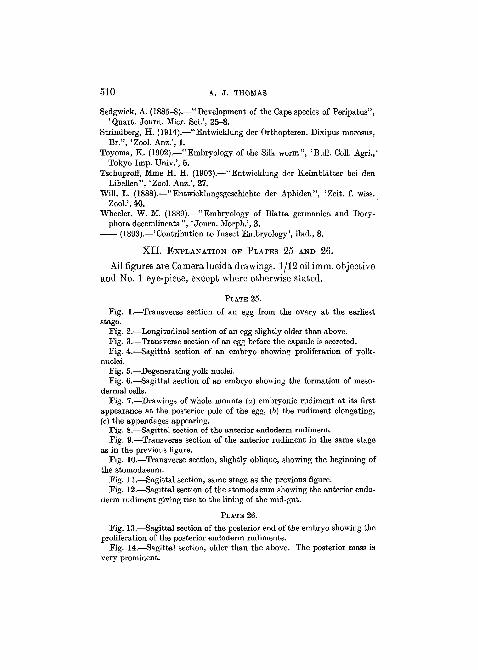
X I I . EXPLANATION OF PLATES 25 AND 26.
All figures are Camera lucida drawings. 1/12 oilimm. objectiveand No. 1 eye-piece, except where otherwise stated.
PLATE 25.
Fig. 1.—Transverse section of an egg from the ovary at the earlieststage.
Fig. 2.—Longitudinal section of an egg slightly older than above.Fig. 3.—Transverse section of an egg before the capsule is secreted.Fig. 4.—Sagittal section of an embryo showing proliferation of yolk-
nuclei.Fig. 5.—Degenerating yolk-nuclei.Fig. 6.—Sagittal section of an embryo showing the formation of meso-
dermal cells.Fig. 7.—Drawings of whole mounts (a) embryonic rudiment at its first
appearance at the posterior pole of the egg, (6) the rudiment elongating,(c) the appendages appearing.
Fig. 8.—Sagittal section of the anterior endoderm rudiment.Fig. 9.—Transverse section of the anterior rudiment in the same stage
as in the previous figure.Fig. 10.—Transverse section, slightly oblique, showing the beginning of
the stomodaeum.Fig. 11.—Sagittal section, same stage as the previous figure.Fig. 12.—Sagittal section of the stomodaeum showing the anterior endo-
derm rudiment giving rise to the lining of the mid-gut.
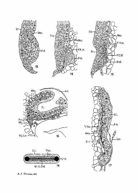
PLATE 26.
Fig. 13.—Sagittal section of the posterior end of the embryo showing theproliferation of the posterior endoderm rudiments.
Fig. 14.—Sagittal section, older than the above. The posterior mass isvery prominent.

DEVELOPMENT OF CARAUSIUS 511
Fig. 15.—Sagittal section, showing the proctodaeum as a very shallowdepression.
Fig. 16.—Sagittal section showing the posterior endodermal rudimentgiving rise to the mid-gut cells.
Kg. 17.—Transverse section showing the mesodermal cells spreadingsideways.
Fig. 18.—Hypothetical drawing of the arrangement of the primary germlayers.
Fig. 19.—Longitudinal section of the posterior end of the embryo, show-ing the ectodermal nature of the Malpighian tubules (1/6 obj.; eyepiece 4).
Fig. 20.—Transverse section of the posterior endoderm mass.Fig. 21.—Longitudinal section showing the arrangement of the meso-
derm.Fig. 22.—Transverse sections showing the arrangement in different parts
of the body:(a) Segmental mesoderm in anterior region.(b) Mesoderm absent in intersegmental region of anterior part.
(1/6 obj.; eyepiece 3.)(c) Segmental mesoderm and coelomic cavity in posterior region.
(1/6 obj.; eyepiece 3.)(d) Intersegmental mesoderm in posterior region. (1/6 obj.; eyepiece 3.)
ABBREVIATIONS.
Am., amnion; Ant.Mg., anterior mid-gut rudiment; A.V.Or., anteriorventral groove; App., appendage rudiment; Bl.Go., Blochmann's corpus-cles; Ceph.L., cephalic lobes; Ch., chorion; Cor., cortical layer of cyto-plasm; Deg.Y.Nuc, degenerating yolk nucleus; Ed., ectoderm; End.,endoderm; Fl.T., flexed tail of embryo; Gast.F., gastrular furrow; Intseg.mes., intersegmental mesoderm; Mal.T., malpighian tubules; Mal.T.Op.,opening of the Malpighian tubule; Mes., mesoderm; Mes.So., mesodermalsomites; Mg., mid-gut; Mg.Lin., mid-gut epithelium, i.e. the thin,flat cells covering the central part of the mid-gut which are proliferatedfrom the anterior and posterior endoderm rudiments; M.st.End., middlestrand of the endoderm; N.Or., neural groove; Nu., nucleus; Pos.Mg.,posterior mid-gut rudiment; Pp., polarplasm; Pr., Proctodaeum; Seg.mes.,segmental mesoderm; Ser., serosa; JS'J., stomodaeum; Sub.oe.by., sub-oesophageal body; V., vacuoles; V.P., ventral plate of blastoderm; Vit.in.,vitelline membrane; Y., yolk-granules; Y.C.M., yolk-cell membrane;Y.Nuc, yolk nucleus.
NO. 311 Ll

,pp-
Ch.
A. J. Thomas, del.

Quart. Journ. Micr. Sci. Vol. 78, N. S., PI 25
Deg.YNuc.
End.
10
Mes.
End.
•YC.M.
Mes.Mg.Lin. Ant.Mg
12Sub.oe.by. YNuc.
OMes.
Mg.Lin
Ect. Mes.
15
Ect.
M.st.End. 1 8
A. J. Thomas, del.

Quart, Journ. Micr. Sci Vol. 78, N. S., PI 26
Seg.Mes.
Mg.Lin.-
Intseq.Mes.
Ect. N.Gr Am.