The efficiency of surface-plasmon coupled emis- sion for
11
The efficiency of surface-plasmon coupled emis- sion for sensitive fluorescence detection Jörg Enderlein 1 and Thomas Ruckstuhl 2 1 Institute for Biological Information Processing 1 Forschungszentrum Jülich 52425 Jülich, Germany [email protected] 2 National Centre for Sensor Research, Dublin City University, Dublin 9, Ireland [email protected] Abstract: Surface-plasmon coupled emission (SPCE) has emerged as a new and potentially powerful tool for highly sensitive fluorescence detec- tion. In the case of SPCE, the fluorescence is collected through a semi- transparent thin metal film deposited on glass. We present a theoretical analysis of SPCE, studying the potential enhancement of the fluorescence collection efficiency, brightness, quantum-yield, and photostability. The re- sults are compared with fluorescence detection on a pure glass surface. It is shown that SPCE does not lead to any improvement, but that the metal film actually reduces the sensitivity of fluorescence detection. © 2005 Optical Society of America OCIS codes: (240.6680) Surface plasmons; (300.6280) Spectroscopy, fluorescence and lumines- cence; (260.2110) Electromagnetic theory. References 1. T. Hirschfeld “Total reflection fluorescence,” Can. Spectroscopy 10, 128 (1965). 2. D. Axelrod, T. P. Burghardt, N. L. Thompson “Total internal reflection fluorescence,” Ann. Rev. Biophys. Bioeng. 13, 124-268 (1984). 3. T. Ruckstuhl, J. Enderlein, S. Jung, S. Seeger “Forbidden light detection from single molecules,“ Anal. Chem. 72, 2117-2123 (2000). 4. T. Ruckstuhl, M. Rankl, S. Seeger „Highly sensitive biosensing using a supercritical angle fluorescence (SAF) instrument“ Biosens. Bioelectron. 18, 1193-99 (2003). 5. A. Krieg, S. Laib, T. Ruckstuhl, S. Seeger „Fast detection of single nucleotide polymorphisms (SNPs) by primer elongation using supercritical angle fluorescence,“ ChemBioChem 4, 1680-1685 (2004). 6. T. Liebermann and W. Knoll “Surface-plasmon field-enhanced fluorescence spectroscopy,” Colloids Surf. A 171, 115-130 (2000). 7. F. Yu, D. F. Yao, W. Knoll “Surface-plasmon field-enhanced fluorescence spectroscopy studies of the inter- action of the interaction of an antibody and its surface-coupled antigen,” Anal. Chem. 75, 2610-2617 (2003). 8. G. Stengel, W. Knoll “Surface plasmon field-enhanced fluorescence spectroscopy,“ Nucleic Acids Res. 33, e69 (2005). 9. J. R. Lakowicz “Radiative decay engineering 3. Surface plasmon-coupled directional emission,“ Anal. Bio- chem. 324, 153-169 (2004). 10. I Gryczynski, J. Malicka, Z. Gryczynski, J. R. Lakowicz “Radiative decay engineering 4. Experimental stud- ies of surface plasmon-coupled directional emission,” Anal. Biochem. 324, 170-182 (2004). 11. E. Matveeva, Z. Gryczynski, I. Gryczynski, J. Malicka, J. R. Lakowicz “Myoglobin immunoassay utilizing directional surface plasmon-coupled emission,” Anal. Chem. 76, 6287-6292 (2004). 12. W. H. Weber and C. F. Eagen “Energy transfer from an excited dye molecule to the surface plasmons of an adjacent metal,” Opt. Lett. 4, 236-238 (1979). 13. F. D. Stefani, K. Vasilev, N. Bocchio, N. Stoyanova, M. Kreiter “Surface-plasmon-mediated single-molecule fluorescence through a thin metallic film,” Phys. Rev. Lett. 94, 023005 (2005). 14. N. Calander “Theory and simulation of surface plasmon-coupled directional emission from fluorophores at planar structures,” Anal. Chem. 76, 2168-2173 (2004). 15. A. Sommerfeld Partial differential equations in physics (Academic Press, 1949). 16. G.W. Ford, W.H. Weber “Electromagnetic interactions of molecules with metal surfaces” Phys. Rep. 113, 195-287 (1984). #8650 - $15.00 USD Received 1 September 2005; revised 18 October 2005; accepted 18 October 2005 (C) 2005 OSA 31 October 2005 / Vol. 13, No. 22 / OPTICS EXPRESS 8855
Transcript of The efficiency of surface-plasmon coupled emis- sion for
The efficiency of surface-plasmon coupled emis-sion for sensitive fluorescence detection
Jörg Enderlein1 and Thomas Ruckstuhl2 1Institute for Biological Information Processing 1 Forschungszentrum Jülich 52425 Jülich, Germany
[email protected] 2National Centre for Sensor Research, Dublin City University, Dublin 9, Ireland
Abstract: Surface-plasmon coupled emission (SPCE) has emerged as a new and potentially powerful tool for highly sensitive fluorescence detec-tion. In the case of SPCE, the fluorescence is collected through a semi-transparent thin metal film deposited on glass. We present a theoretical analysis of SPCE, studying the potential enhancement of the fluorescence collection efficiency, brightness, quantum-yield, and photostability. The re-sults are compared with fluorescence detection on a pure glass surface. It is shown that SPCE does not lead to any improvement, but that the metal film actually reduces the sensitivity of fluorescence detection.
© 2005 Optical Society of America
OCIS codes: (240.6680) Surface plasmons; (300.6280) Spectroscopy, fluorescence and lumines-cence; (260.2110) Electromagnetic theory.
References
1. T. Hirschfeld “Total reflection fluorescence,” Can. Spectroscopy 10, 128 (1965). 2. D. Axelrod, T. P. Burghardt, N. L. Thompson “Total internal reflection fluorescence,” Ann. Rev. Biophys.
Bioeng. 13, 124-268 (1984). 3. T. Ruckstuhl, J. Enderlein, S. Jung, S. Seeger “Forbidden light detection from single molecules,“ Anal. Chem.
72, 2117-2123 (2000). 4. T. Ruckstuhl, M. Rankl, S. Seeger „Highly sensitive biosensing using a supercritical angle fluorescence (SAF)
instrument“ Biosens. Bioelectron. 18, 1193-99 (2003). 5. A. Krieg, S. Laib, T. Ruckstuhl, S. Seeger „Fast detection of single nucleotide polymorphisms (SNPs) by
primer elongation using supercritical angle fluorescence,“ ChemBioChem 4, 1680-1685 (2004). 6. T. Liebermann and W. Knoll “Surface-plasmon field-enhanced fluorescence spectroscopy,” Colloids Surf. A
171, 115-130 (2000). 7. F. Yu, D. F. Yao, W. Knoll “Surface-plasmon field-enhanced fluorescence spectroscopy studies of the inter-
action of the interaction of an antibody and its surface-coupled antigen,” Anal. Chem. 75, 2610-2617 (2003). 8. G. Stengel, W. Knoll “Surface plasmon field-enhanced fluorescence spectroscopy,“ Nucleic Acids Res. 33,
e69 (2005). 9. J. R. Lakowicz “Radiative decay engineering 3. Surface plasmon-coupled directional emission,“ Anal. Bio-
chem. 324, 153-169 (2004). 10. I Gryczynski, J. Malicka, Z. Gryczynski, J. R. Lakowicz “Radiative decay engineering 4. Experimental stud-
ies of surface plasmon-coupled directional emission,” Anal. Biochem. 324, 170-182 (2004). 11. E. Matveeva, Z. Gryczynski, I. Gryczynski, J. Malicka, J. R. Lakowicz “Myoglobin immunoassay utilizing
directional surface plasmon-coupled emission,” Anal. Chem. 76, 6287-6292 (2004). 12. W. H. Weber and C. F. Eagen “Energy transfer from an excited dye molecule to the surface plasmons of an
adjacent metal,” Opt. Lett. 4, 236-238 (1979). 13. F. D. Stefani, K. Vasilev, N. Bocchio, N. Stoyanova, M. Kreiter “Surface-plasmon-mediated single-molecule
fluorescence through a thin metallic film,” Phys. Rev. Lett. 94, 023005 (2005). 14. N. Calander “Theory and simulation of surface plasmon-coupled directional emission from fluorophores at
planar structures,” Anal. Chem. 76, 2168-2173 (2004). 15. A. Sommerfeld Partial differential equations in physics (Academic Press, 1949). 16. G.W. Ford, W.H. Weber “Electromagnetic interactions of molecules with metal surfaces” Phys. Rep. 113,
195-287 (1984).
#8650 - $15.00 USD Received 1 September 2005; revised 18 October 2005; accepted 18 October 2005
(C) 2005 OSA 31 October 2005 / Vol. 13, No. 22 / OPTICS EXPRESS 8855
17. J. Enderlein “Fluorescence detection of single molecules near a solution/glass interface – an electrodynamic analysis,“ Chem. Phys. Lett. 308, 263-266 (1999).
18. J. Enderlein “Single molecule fluorescence near a metal layer,“ Chem. Phys. 247, 1-9 (1999). 19. J. Enderlein “Theoretical study of detecting a dipole emitter through an objective with high numerical aper-
ture,” Opt. Lett. 25, 634-636 (2000). 20. J. Enderlein “A theoretical investigation of single molecule fluorescence detection on thin metallic layers,”
Biophys. J. 78, 2151-2158 (2000). 21. C. D. Geddes, I. Gryczynski, J. Malicka, Z. Gryczynski “Directional surface plasmon coupled emission,” J.
Fluoresc. 14, 119-123 (2004). 22. J. R. Lakowicz, J. Malicka, I. Gryczynski, Z. Gryczynski “Directional surface plasmon-coupled emission: a
new method for high sensitivity detection,” Biophys. Biochem. Res. Comm. 307, 435-439 (2003). 23. J. Enderlein, T. Ruckstuhl, S. Seeger “Highly efficient optical detection of surface-generated fluorescence“
Appl. Opt. 38, 724-732 (1999). 24. T. Ruckstuhl and S. Seeger WO 009946596 (1999) 25. E. Matveeva, J. Malicka, I. Gryczynski, Z. Gryczynski J. R. Lakowicz, “Multi-wavelength immunoassays us-
ing surface plasmon-coupled emission” Biophys. Biochem. Res. Comm. 313, 721-726 (2004).
1. Introduction
Fluorescence-based detection is one of the most important techniques for sensitively detect-ing low concentrations of biomolecules that are either fluorescently tagged or autofluores-cent. One of its applications is found in affinity-based assays, where surface-bound receptor molecules are used to capture ligands from a solution, thus concentrating them at the surface. These heterogeneous assays play a central role in DNA analysis, biotechnology and pharma-ceutical research, where fluorescence detection assures high sensitivity and a low background signal. An additional requirement in these assays is the surface selectivity of the detection technique, i.e. that background fluorescence from the bulk solution is efficiently suppressed in comparison with fluorescence generated by surface-captured molecules. A widely em-ployed technique for achieving such surface-selectivity is total internal-reflection fluores-cence (TIRF) excitation [1,2]. Recently, several alternative approaches have been proposed with the goal of achieving effective surface-restricted fluorescence detection without com-promising the signal-to-background ratio, namely supercritical angle fluorescence (SAF) [3-5], surface plasmon field-enhanced fluorescence (SPFS) [6-8] and surface plasmon-coupled emission (SPCE) [9-11].
The core idea of SPCE is the use of a semi-transparent thin metal film that is deposited on a glass substrate to enhance fluorescence excitation and to improve fluorescence collec-tion for molecules close to the surface. It is well-known that for plane wave illumination from the glass side at an incident angle close to the angle of surface-plasmon resonance (SPR), the local electric-field intensity near the surface can be significantly enhanced compared to a similar configuration without metal. It was also shown that fluorescence emission can couple into the surface plasmon (SP) mode of the metal film and subsequently be re-radiated into the glass around angles near the SPR angle [12]. Even single molecule detection has been re-ported using SPCE [13]. This raised expectations of improved fluorescence excitation and detection for a wide range of surface-restricted applications of fluorescence detection. In a recent publication [14], theoretical results were presented suggesting a significant improve-ment in fluorescence detection performance when using SPCE. This would make SPCE a potentially important tool for many biophysical or bioanalytical applications requiring high fluorescence detection sensitivity and good discrimination between surface and bulk gener-ated fluorescence. In the present paper, we will focus on a theoretical analysis of the effi-ciency and usefulness of SPCE for such applications. We present a detailed analysis of SPCE and provide quantitative results on fluorescence collection efficiency, brightness, fluores-cence quantum-yield, and photostability enhancements.
#8650 - $15.00 USD Received 1 September 2005; revised 18 October 2005; accepted 18 October 2005
(C) 2005 OSA 31 October 2005 / Vol. 13, No. 22 / OPTICS EXPRESS 8856
2. Theoretical background
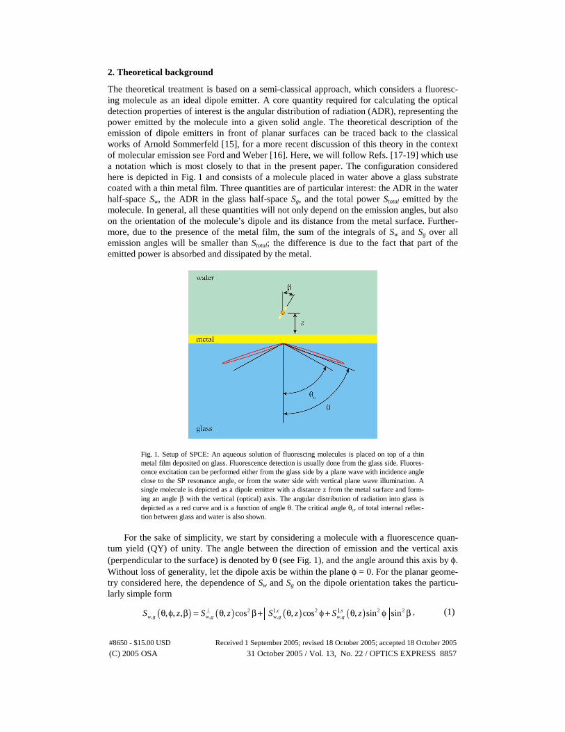
The theoretical treatment is based on a semi-classical approach, which considers a fluoresc-ing molecule as an ideal dipole emitter. A core quantity required for calculating the optical detection properties of interest is the angular distribution of radiation (ADR), representing the power emitted by the molecule into a given solid angle. The theoretical description of the emission of dipole emitters in front of planar surfaces can be traced back to the classical works of Arnold Sommerfeld [15], for a more recent discussion of this theory in the context of molecular emission see Ford and Weber [16]. Here, we will follow Refs. [17-19] which use a notation which is most closely to that in the present paper. The configuration considered here is depicted in Fig. 1 and consists of a molecule placed in water above a glass substrate coated with a thin metal film. Three quantities are of particular interest: the ADR in the water half-space Sw, the ADR in the glass half-space Sg, and the total power Stotal emitted by the molecule. In general, all these quantities will not only depend on the emission angles, but also on the orientation of the molecule’s dipole and its distance from the metal surface. Further-more, due to the presence of the metal film, the sum of the integrals of Sw and Sg over all emission angles will be smaller than Stotal; the difference is due to the fact that part of the emitted power is absorbed and dissipated by the metal.
Fig. 1. Setup of SPCE: An aqueous solution of fluorescing molecules is placed on top of a thin metal film deposited on glass. Fluorescence detection is usually done from the glass side. Fluores-cence excitation can be performed either from the glass side by a plane wave with incidence angle close to the SP resonance angle, or from the water side with vertical plane wave illumination. A single molecule is depicted as a dipole emitter with a distance z from the metal surface and form-ing an angle β with the vertical (optical) axis. The angular distribution of radiation into glass is depicted as a red curve and is a function of angle θ. The critical angle θcr of total internal reflec-tion between glass and water is also shown.
For the sake of simplicity, we start by considering a molecule with a fluorescence quan-
tum yield (QY) of unity. The angle between the direction of emission and the vertical axis (perpendicular to the surface) is denoted by θ (see Fig. 1), and the angle around this axis by φ. Without loss of generality, let the dipole axis be within the plane φ = 0. For the planar geome-try considered here, the dependence of Sw and Sg on the dipole orientation takes the particu-larly simple form
( ) ( ) ( ) ( )2 , 2 , 2 2, , , ,, , , , cos , cos , sin sinc s
w g w g w g w gS z S z S z S z⊥ ⎡ ⎤θ φ β = θ β + θ φ + θ φ β⎣ ⎦� � , (1)
#8650 - $15.00 USD Received 1 September 2005; revised 18 October 2005; accepted 18 October 2005
(C) 2005 OSA 31 October 2005 / Vol. 13, No. 22 / OPTICS EXPRESS 8857

where β is the angle between the dipole axis and the vertical axis, z is the dipole’s distance from the metal surface, and
,w gS ⊥ and ,w gS � are functions of θ and z only. For Stotal(z,β), a simi-
lar relation holds:
( ) ( ) ( )2 2, cos sintotal total totalS z S z S z⊥β = β + β� . (2)
The probability density that a photon is emitted along direction (θ,φ) is given by the ratio Sw,g(θ,φ,z,β)/Stotal(z,β), where the index, w, refers to the water half space and the index, g, to the glass half space. In what follows we will consider only the special cases of vertical (β = 0) and horizontal (β = π/2) dipole orientation – the general case can easily be derived using the above relations. Thus, the energy emitted into the glass within an angular region θ0 ≤ θ ≤ π/2 is given by
( ) ( )( )
0
,2,
,
, ,2 sin g
gtotal
S zI z d
S z
⊥π⊥
⊥θ
θ φ= π θ θ∫
�
�
�
, (3)
where Sg|| = (Sg
||,c + Sg||,s)/2, and the superscript ┴ refers to the vertical dipole orientation, and
the superscript || to the horizontal dipole orientation. If one sets θ0 = 0 in Eq. (3), emission into all angles of the glass half-space is considered. If one chooses θ equal to the critical an-gle of TIR, only SAF detection is considered. In order to obtain the emission into the water half space, one has to replace the subscript g by w in Eq. (3). Finally, the energy absorbed and dissipated within the metal film is calculated as the difference
( ) ( ) ( ) ( )2
, , , ,
0
2 sin , ,diss total g wI z S z d S z S zπ
⊥ ⊥ ⊥ ⊥⎡ ⎤= − π θ θ θ + θ⎣ ⎦∫� � � � . (4)
The situation becomes more complex when considering molecules with a fluorescence QY below unity. The emission power as calculated above refers only to the radiative compo-nent of the transition from the excited to the ground state and not to the non-radiative part, which remains unaffected by the electromagnetic interaction between the emitting molecule and its environment. Thus, if one starts with a molecule having fluorescence QY Q0 and total electromagnetic emission power S0 when placed in water and not in close proximity to any interface, its changed emission power Stotal near the surface leads to the following fluores-cence QY Q
0 0
0 0 01total
total
Q S SQ
Q Q S S=
− +, (5)
where Q here is understood as the part of the excited state energy, that is emitted electromag-netically, regardless of the further fate of this energy. The ratio Stotal/S0 is directly propor-tional to the radiative transition rate of the molecule near the interface and in water, i.e. to the inverse ratio of the fluorescence lifetime that would be measured for a molecule with a QY of unity,
( ), 00 ,
,total
Sz
S⊥
⊥τ β = τ�
�
. (6)
Thus, one has an intrinsic relationship between the enhanced radiative transition rate and an enhanced fluorescence QY. Taking this changed QY into account, the total energy emitted into the glass is now given by the integral of the product of Q and the ratio Sg/Stotal:
#8650 - $15.00 USD Received 1 September 2005; revised 18 October 2005; accepted 18 October 2005
(C) 2005 OSA 31 October 2005 / Vol. 13, No. 22 / OPTICS EXPRESS 8858
( ) ( ),2
0,,
0 0 00
, , 2 sin1
gg
total
S QI z Q d
Q S Q S
⊥π⊥
⊥β = π θ θ− +∫
�
�
�
. (7)
Finally, let us turn to the photostability of fluorescing molecules. If one assumes that any photodestructive process can occur only as long as the molecule is in the excited state, any change in excited state lifetime will lead to a changed photostability. We may assume that the average number of photons that can be extracted from a molecule before photobleaching is inversely proportional to the excited state lifetime. For simplicity, we will treat the photosta-bility only for molecules with fluorescence QY of unity, where this effect will be most pro-nounced. In this case, the average detectable number of photons until bleaching is estimated as
( )0
, 2,
0 0
2sing
g
N zd S
N S
⊥ π⊥
θ
π= θ θ∫�
� , (8)
where N0 denotes the average total number of photons emitted by a molecule in water until bleaching.
3. Results and discussion
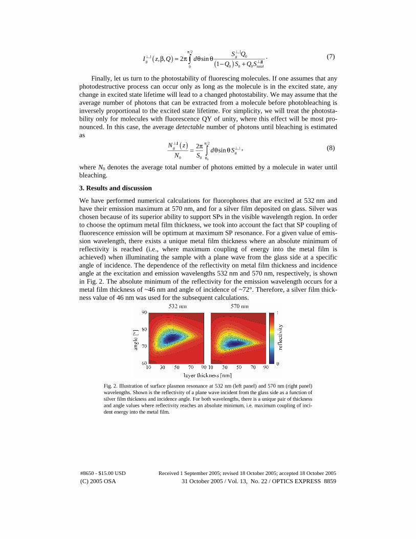
We have performed numerical calculations for fluorophores that are excited at 532 nm and have their emission maximum at 570 nm, and for a silver film deposited on glass. Silver was chosen because of its superior ability to support SPs in the visible wavelength region. In order to choose the optimum metal film thickness, we took into account the fact that SP coupling of fluorescence emission will be optimum at maximum SP resonance. For a given value of emis-sion wavelength, there exists a unique metal film thickness where an absolute minimum of reflectivity is reached (i.e., where maximum coupling of energy into the metal film is achieved) when illuminating the sample with a plane wave from the glass side at a specific angle of incidence. The dependence of the reflectivity on metal film thickness and incidence angle at the excitation and emission wavelengths 532 nm and 570 nm, respectively, is shown in Fig. 2. The absolute minimum of the reflectivity for the emission wavelength occurs for a metal film thickness of ~46 nm and angle of incidence of ~72°. Therefore, a silver film thick-ness value of 46 nm was used for the subsequent calculations.
Fig. 2. Illustration of surface plasmon resonance at 532 nm (left panel) and 570 nm (right panel) wavelengths. Shown is the reflectivity of a plane wave incident from the glass side as a function of silver film thickness and incidence angle. For both wavelengths, there is a unique pair of thickness and angle values where reflectivity reaches an absolute minimum, i.e. maximum coupling of inci-dent energy into the metal film.
#8650 - $15.00 USD Received 1 September 2005; revised 18 October 2005; accepted 18 October 2005
(C) 2005 OSA 31 October 2005 / Vol. 13, No. 22 / OPTICS EXPRESS 8859
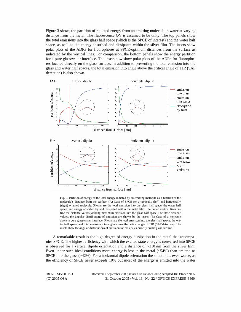
Figure 3 shows the partition of radiated energy from an emitting molecule in water at varying distance from the metal. The fluorescence QY is assumed to be unity. The top panels show the total emissions into the glass half space (which is the SPCE of interest) and the water half space, as well as the energy absorbed and dissipated within the silver film. The insets show polar plots of the ADRs for fluorophores at SPCE-optimum distances from the surface as indicated by the vertical lines. For comparison, the bottom panels show the energy partition for a pure glass/water interface. The insets now show polar plots of the ADRs for fluoropho-res located directly on the glass surface. In addition to presenting the total emission into the glass and water half spaces, the total emission into angle above the critical angle of TIR (SAF detection) is also shown.
Fig. 3. Partition of energy of the total energy radiated by an emitting molecule as a function of the molecule’s distance from the surface. (A) Case of SPCE for a vertically (left) and horizontally (right) oriented molecule. Shown are the total emission into the glass half space, the water half space, and energy absorbed by and dissipated within the metal film. The dotted vertical lines de-fine the distance values yielding maximum emission into the glass half space. For these distance values, the angular distributions of emission are shown by the insets. (B) Case of a molecule above a pure glass/water interface. Shown are the total emission into the glass half space, the wa-ter half space, and total emission into angles above the critical angle of TIR (SAF detection). The insets show the angular distributions of emission for molecules directly on the glass surface.
A remarkable result is the high degree of energy dissipation in the metal that accompa-
nies SPCE. The highest efficiency with which the excited state energy is converted into SPCE is observed for a vertical dipole orientation and a distance of ~110 nm from the silver film. Even under such ideal conditions more energy is lost in the metal (~54%) than emitted as SPCE into the glass (~42%). For a horizontal dipole orientation the situation is even worse, as the efficiency of SPCE never exceeds 10% but most of the energy is emitted into the water
#8650 - $15.00 USD Received 1 September 2005; revised 18 October 2005; accepted 18 October 2005
(C) 2005 OSA 31 October 2005 / Vol. 13, No. 22 / OPTICS EXPRESS 8860
half-space. Many analytical applications involve the detection of randomly oriented fluoro-phores. In such a case, the best achievable SPCE is observed for molecules at roughly 110 nm above the silver film, emitting on average 18% of their energy into SPCE modes.
In comparison, isotropically orientated fluorophores bound at a bare water/glass interface emit 73% of their energy into the glass. Consequently, the presence of the silver film signifi-cantly attenuates the emission into the glass and the SPCE modes contain much less energy than the emission from a plain water/glass interface.
SPCE has been described as a potentially powerful tool for monitoring binding assays due to the surface-confined detection volume [11] but an even stronger surface confinement is achieved using a bare water/glass interface in conjunction with SAF detection method. For isotropically oriented fluorophores at a water/glass interface SAF amounts to 34% of the total fluorescence emission [4]. Consequently, the SAF detection from a water/glass interface is significantly more efficient than the complete collection of SPCE through the silver film. These results are all the more remarkable as SPCE has been advertised as an approach that yields fluorescence enhancements of up to 1000-fold [9,18].
SPCE has been described as highly directional [9-11,21,22], which suggests that an effi-cient collection of the signal is straightforward. This is rather misleading as the SPCE is emit-ted into a narrow range of surface angles far above the critical angle of TIR, namely at an angle of 144° for the parameters considered here. A consequence of this is that even a micro-scope objective with a numerical aperture of 1.45 is insufficient for the collection of SPCE from an aqueous analyte. In order to achieve a complete collection of light emitted into the very large angles encountered in SPCE and SAF, special optical elements have to be em-ployed such as high aperture parabolic collectors [23,24].
An important issue is metal-induced QY enhancement. As shown in the top panel of Fig. 4, the presence of the silver film strongly increases the radiative transition rate for mole-cules near the surface. When taking into account this tremendous increase of the radiative transition rate, one may expect a strong QY enhancement, in particular for molecules with low intrinsic QY. However, the QY enhancement is counterbalanced by energy absorption and dissipation by the metal film, as shown in the center and bottom panels of Fig. 4. The numerical results for the total emission into the glass half space are shown for three different values of QY. Even for the lowest QY considered (10 %), no distinct improvement in detect-able signal is observed with SPCE. This is due to the fact that a substantial increase in radia-tive transition rate is only obtained for emitters very close to the silver film, where most of the emitted energy is absorbed by the metal. Compared to the pure water/glass system, the introduction of the silver coating clearly reduces the emission into the glass even for fluoro-phores with low QY.
Other important fluorescence properties to be studied are photostability and brightness. Due to the enhanced radiative transition rate for molecules close to the metal, one may expect that they become more photostable (due to the reduced time they spend in the excited state), resulting in a larger amount of fluorescence signal that can be collected until bleaching. The top panel of Fig. 5 shows the average number of photons that can be extracted from a mole-cule (with unity QY) until bleaching as function of its distance from the surface. As can be seen, the presence of the silver film does indeed lead to an increased number of detectable fluorescence photons until bleaching for molecules sufficiently close to the surface (a slight increase is also observed for the plain water/glass interface). However, this enhanced photo-stability cannot be considered without considering also the observable fluorescence bright-ness (i.e. signal strength). In case the price for an enhanced photostability is a significant re-duction in fluorescence intensity, the low signal-to-background ratio nullifies any potential benefit of enhanced photostability. Fluorescence brightness is calculated as the product of excitation intensity times the efficiency of fluorescence detection into the glass.
#8650 - $15.00 USD Received 1 September 2005; revised 18 October 2005; accepted 18 October 2005
(C) 2005 OSA 31 October 2005 / Vol. 13, No. 22 / OPTICS EXPRESS 8861

Fig. 4. (A) Fluorescence lifetime for molecules with unity QY of fluorescence as a function of dis-tance from the surface. Solid lines refer to a pure water/glass interface, shaded lines to the SPCE case. (B) Total emission into the glass half space for a vertically oriented dipole as a function of distance from the surface for three different values of QY. Solid lines refer to pure water/glass in-terface, shaded lines to SPCE. (C) Same as (B), but for horizontally oriented dipole.
#8650 - $15.00 USD Received 1 September 2005; revised 18 October 2005; accepted 18 October 2005
(C) 2005 OSA 31 October 2005 / Vol. 13, No. 22 / OPTICS EXPRESS 8862
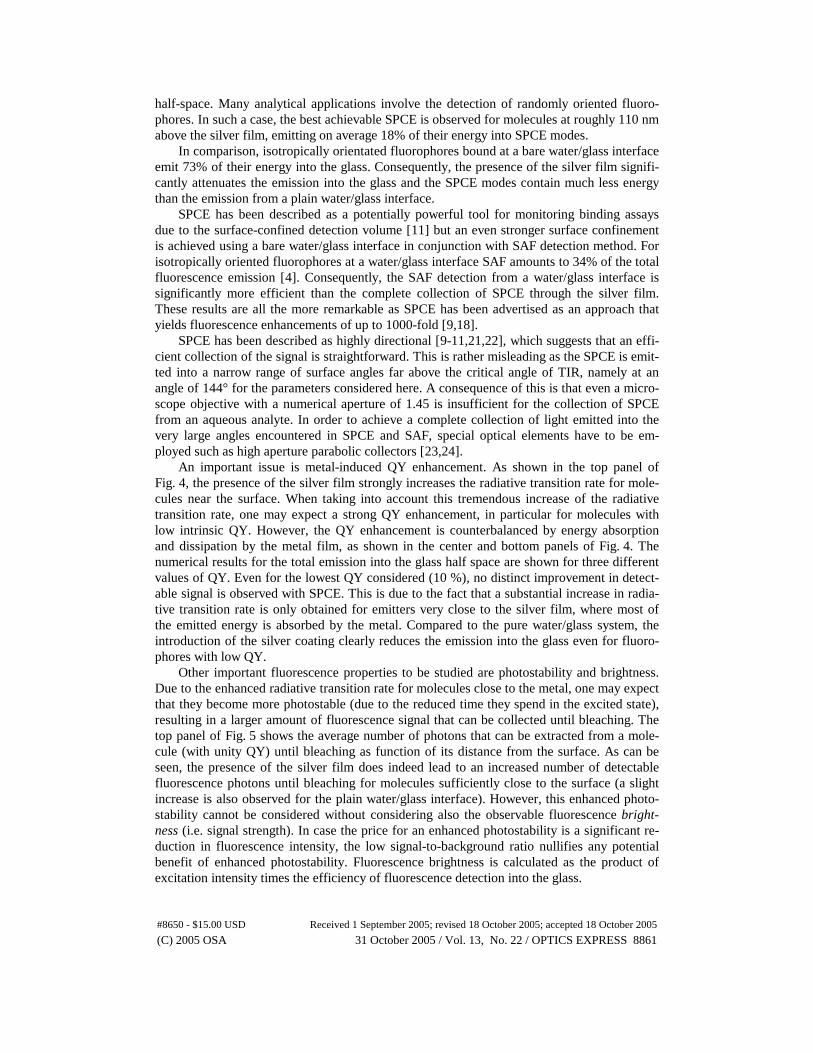
Fig. 5. (A) Average number of emitted photons until photobleaching as a function of distance from the surface for molecules with unity QY. (B) Maximum detectable fluorescence intensity when collecting over the whole glass half space. Lines (a) and (b) refer to SPCE, where (a) shows the result for plane wave excitation from the glass side at the incidence angle giving maximum electric field intensity on the metal surface, and (b) shows the result for perpendicular plane wave excita-tion from the water side. Lines (c) and (d) refer to a pure water/glass interface, where (c) shows the result for perpendicular plane wave excitation from the glass side, and (d) for plane wave exci-tation from the glass side with incidence angle just below the critical angle of TIR.
For molecules above a metal film, two basic excitation configurations are reasonable:
wide field excitation by a plane wave incident from the water side perpendicular to the silver film (reversed Kretschmann configuration), and SP coupled excitation by a plane wave inci-dent from the glass side at the plasmon resonance angle (Kretschman configuration) [10]. For the pure water/glass system, we will consider plane wave excitation from the glass side per-pendicular to the surface and excitation with an incidence angle close to the TIR angle. The dependence of the detectable fluorescence brightness on surface distance is shown in the bot-tom panel of Fig. 5. For all settings, an identical intensity of excitation light and randomly oriented emitters were assumed. By illuminating the silver film at the SP resonance angle, the intensity of excitation is enhanced by a factor of up to 16. This enhancement is only achieved directly on the film, a region where the efficiency of SPCE detection is nearly zero.
For SPR excitation, the intensity decays rapidly with surface distance so that, at the op-timum distance for SPCE of 110 nm, the excitation enhancement factor is only 3. Excitation in the reversed Kretschmann configuration turns out to be even more unfavourable. With a 46 nm thickness, the silver film acts as an efficient mirror creating a standing wave of excita-tion light above the surface. Coincidentally, the excitation intensity is near a minimum at a 110 nm surface distance, the region of highest SPCE, resulting in low fluorescence signals. In
#8650 - $15.00 USD Received 1 September 2005; revised 18 October 2005; accepted 18 October 2005
(C) 2005 OSA 31 October 2005 / Vol. 13, No. 22 / OPTICS EXPRESS 8863

the case of a water/glass interface, illumination close to the critical angle of TIR leads to an enhancement by a factor of up to 4 compared with perpendicular illumination. The highest excitation enhancement is achieved directly on the glass surface, which is simultaneously the location of highest collection efficiency. This leads to a higher fluorescence signal than ever obtainable with SPCE. Even with perpendicular plane-wave illumination, the detectable fluo-rescence brightness without the silver film is clearly superior to SPCE. Inspecting both panels of Fig. 5 together shows that the apparent increase of photostability by the metal is mostly negated by the low signal-to-background ratio due to decreased observable brightness. It should be also taken into account that the effect of increased photostability is lower for mole-cules with a QY smaller than one, so that the top panel of Fig. 5 already shows the most fa-vourable situation.
Fig. 6. Partition of emitted energy between radiation into glass half space, into water half space, and energy absorbed by and dissipated within the metal film for the glass/metal/waveguide/water system. Functional dependence on metal film and waveguide layer thickness is shown. The refrac-tive index of the dielectric layer is 2, the position of the emitting molecules is directly on the sur-face of the waveguide. The top panel shows the results for vertically oriented dipoles, the bottom panel for horizontally oriented ones.
Finally, we consider a modification of SPCE where an additional dielectric layer of high
refractive index is deposited on top of the metal film. It is well known that emitting molecules close to a refractive-index discontinuity emit the larger part of their energy into the medium with the higher refractive index. Thus, it may be hoped that an additional layer of high refrac-tive index serves to redirect the fluorescence emission towards the metal film, where it is then converted into SPCE modes. Additionally, the layer serves as a spacer between the fluoresc-
#8650 - $15.00 USD Received 1 September 2005; revised 18 October 2005; accepted 18 October 2005
(C) 2005 OSA 31 October 2005 / Vol. 13, No. 22 / OPTICS EXPRESS 8864
ing molecules and the metal surface, thereby eliminating direct quenching of molecules in close proximity to the metal. Numerical results for a dielectric layer with refractive index of 2.0 are presented in Fig. 6, where the partition of emitted energy between the glass half-space, the water-half space and the metal are shown as a function of metal and dielectric layer thickness. Remarkably, the vertical and horizontal dipole orientations display optimum of fluorescence detection conditions for completely different parameters. The optimum for the vertical dipole orientation is achieved for a metal film thickness of ~20 nm and a ~210 nm dielectric film thickness, whereas the optimum for the horizontal dipole orientation is achieved for the lowest considered film thickness of ~5 nm and a dielectric layer thickness of ~60 nm.
In both cases, maximum achievable fluorescence emission into the glass half-space is lower than when detecting fluorescence directly at a pure water/glass interface. We repeated the calculations also for a higher refractive index of 2.5, but found similarly unfavourable results. Therefore, introduction of an additional dielectric layer does not yield improved per-formance of SPCE compared with fluorescence detection on pure glass.
4. Conclusion
The sharp angular maximum of SPCE is a striking phenomenon and is in stark contrast to the familiar fluorescence behaviour inside a homogeneous medium. In a series of recent publica-tions, SPCE was introduced as an extremely powerful and straightforward new method for sensitive detection in analytical chemistry. It was anticipated that SPCE could increase the fluorescence signals by a factor of up to 1000 [9,21], which would be a revolutionary im-provement for analytical assays indeed. Here, we have theoretically evaluated the perform-ance of SPCE-based fluorescence detection and compared it with conventional fluorescence detection on pure glass. It was demonstrated that significantly less energy is coupled through a silver film into the glass than in the case no silver film is present. We have shown that the detectable fluorescence brightness with SPCE is worse than that obtainable on a bare wa-ter/glass interface, with the result that SPCE does not enhance the detectable fluorescence signal. On the contrary, light collection through the metal film clearly attenuates the fluores-cence signal and should be avoided in analytical applications where sensitivity is of crucial importance. However, it should be emphasized that the results presented here were calculated for the ideal case of perfectly planar substrates. Surface roughness of the metal coating may lead to additional effects such as more enhanced electric fields and thus fluorescence excita-tion, which are not accounted for by our theory.
There may be special applications where SPCE can be advantageous with respect to more conventional detection schemes. First, the high directionality of fluorescence emission in SPCE may help to better discriminate between fluorescence and any other background which does not show a similar directionality in emission (potentially Rayleigh and Raman scatter-ing). Second, the angular position of the SPCE emission peak is strongly wavelength depend-ent (due to the strong dispersion of metals), which makes it possible to use SPCE as a spec-trally resolving technique. This makes parallel detection of spectrally different fluorophores possible without the use of additional dispersive elements [25]. Third, SPCE emission is highly polarized, in contrast to emission generated at a glass/water interface. These polariza-tion properties of SPCE emission may be useful under special circumstances.
Acknowledgements
We are much obliged to Prof. U. Benjamin Kaupp and Prof. Brian D. McCraith for their gen-erous support of our work. We thank Prof. Joe R. Lakowicz for the numerous helpful discus-sions on SPCE, which have inspired the present work. Financial support by the Deutsche For-schungsgemeinschaft and the Science Foundation of Ireland is gratefully acknowledged.
#8650 - $15.00 USD Received 1 September 2005; revised 18 October 2005; accepted 18 October 2005
(C) 2005 OSA 31 October 2005 / Vol. 13, No. 22 / OPTICS EXPRESS 8865


![SIGNAL VARIANCE AS A CHATTER INDICATOR ......However, acoustic emis-sion (AE) sensors[27] and continuous monitoring of spindle current, voltage, and speed to calculate the instantaneous](https://static.fdocuments.in/doc/165x107/60e7a535ddc3e4306f61388a/signal-variance-as-a-chatter-indicator-however-acoustic-emis-sion-ae.jpg)