The Efficacy of LY293558 in Blocking Seizures and ... · The Efficacy of LY293558 in Blocking...
203
The Efficacy of LY293558 in Blocking Seizures and Associated Morphological, and Behavioral Alterations Induced by Soman in Immature Male Rats and the Role of the M1 Muscarinic Acetylcholine Receptor in Organophosphate Induced Seizures by Steven Lawrence Miller Dissertation submitted to the Faculty of the Neuroscience Graduate Program Uniformed Services University of the Health Sciences In partial fulfillment of the requirements for the degree of Doctor of Philosophy, 2014
Transcript of The Efficacy of LY293558 in Blocking Seizures and ... · The Efficacy of LY293558 in Blocking...
The Efficacy of LY293558 in Blocking Seizures and Associated Morphological, and
Behavioral Alterations Induced by Soman in Immature Male Rats and the Role of the M1
Muscarinic Acetylcholine Receptor in Organophosphate Induced Seizures
by
Steven Lawrence Miller
Dissertation submitted to the Faculty of the
Neuroscience Graduate Program Uniformed Services University of the Health Sciences
In partial fulfillment of the requirements for the degree of Doctor of Philosophy, 2014

UNIFORMED SERVICES UNIVERSITY, SCHOOL OF MEDICINE GRADUATE PROGRAMS Graduate Education Office (A 1045), 4301 Jones Bridge Road, Bethesda, MD 20814
FINAL EXAMlNA TION/PRIVATE DEFENSE FOR THE DEGREE OF DOCTOR OF PHILOSOPHY IN THE NEUROSCIENCE GRADUATE PROGRAM
Name of Student: Steven Lawrence Miller
Date of Examination: January 30, 20 15
Time: I O:OOam
Place: C2095
DECISION OF EXAMINATION COMMITTEE MEMBERS:
PASS FAIL
Committee Member
~ !% / Dr. Thomas Flagg DEPARTMENT OF ANATOMY, PHYSIOLOGY AND GENETICS Committee Member I Dr ereshteh Nu0 nt D ·PARTMENT OF ANATOMY, PHYSIOLOGY AND GENETICS Committee Member
Gregory P. Mueller, Ph.D., Associate Dean II www.usuhs.mil/graded II [email protected] Toll Free: 800-772-1747 II Commercial : 301-295-3913 I 9474 II DSN : 295-9474 II Fax: 301-295-6772
UNIFORMED SERVICES UNIVERSITY, SCHOOL OF MEDICINE GRADUATE PROGRAMS
Graduate Education Office (A 1045), 4301 Jones Bridge Road, Bethesda, MD 20814
APPROVAL OF THE DOCTORAL DISSERTATION IN THE NEUROSCIENCE GRADUATE PROGRAM
Title of Dissertation: "The Efficacy of L Y293558 in Blocking Seizures and Associated Morphological, and Behavioral Alterations Induced by Soman in Immature Male Rats and the Role of the MI Muscarinic Acetylcholine Receptor in Organophosphate Induced Seizures"
Name of Candidate: Steven Lawrence Mi ller Doctor of Phi losophy Degree January 30, 20 15
DISSERTATION AND ABSTRACT APPROVED:
DATE:
D ·. oseph McCabe DEPARTMENT 0 ANATOMY, PHYSIOLOGY AND GENETICS Committee Cha· pers n
113o/1S Dr. aria Br ga DE ARTME ;r OF ANATOMY, PHYSIOLOGY AND GENETICS Ltion Aavisor
1run.il~ p .. ;,,-> DEPARTMENT OF NEUROLOGY Committee Member
~~ ~o Dr. Thomas Flagg '--DEPARTMENT OF ANATOMY, PHYSIOLOGY AND GENETICS Committee Member
~---
· D . Fereshteh Nu ent D PARTMENT OF ANATOMY, PHYSIOLOGY AND GENETICS Committee Member
Gregory P. Mueller, Ph.D., Associate Dean II www.usuhs.mil/graded II [email protected] Toll Free: 800-772-1747 II Commercial: 301-295-3913 I 9474 II DSN: 295-9474 II Fax: 301-295-6772

iii
ACKNOWLEDGMENTS
One of the most important lessons I have learned in my career in science so far is
that science is a team effort. I could not be where I am today without the help of everyone
I have met during my career thus far. Thank you to my CSU Fresno faculty who really
started my career and instilled my passion for science and my curiosity: Drs. Jason Bush,
James Prince, Madhusudan Katti, Eric Pearson, David Frank and Fred Schreiber. And, to
Rowena Chu who started my "at-the-bench" training.
I truly appreciate Drs. Sharon Juliano and He Li for giving me the opportunity to
rotate in their labs before I decided upon doing my dissertation work in the lab of Dr.
Maria Braga. Thank you Dr. Braga for your extensive training you have provided to me
from the day that I joined your lab. Your mentorship has been a critical part of propelling
me through the neuroscience program at USUHS and I believe that I have excelled
because of it. Thank you for accepting me into your lab and for your guidance in training
me to become a principal investigator. I believe a great deal of my future success will
result from the extensive career advice you have given me, thank you so much.
Thank you to Drs. Vassiliki Aroniadou-Anderjaska, Volodymyr Pidoplichko and
Taiza Figueiredo. You have all been invaluable members in my hands-on training and a
special thanks to you Dr. Figueiredo who helped me conduct so many experiments up at
Aberdeen Proving Grounds. Thank you to my collaborator Dr. James Apland for his
guidance in the difficult field of research that we work in. And, thank you to his former
laboratory technician, Andrew Borrell for his enthusiasm in helping me conduct
experiments at Aberbeen Proving Grounds. Thank you to the other current and previous

iv
lab members of the Braga lab including Drs. Eric Prager, Camila Almeida-Suhett, Franco
Rosetti, as well as Katia Rosetti and Adriana Souza.
Thank you to the members of my dissertation committee which include my chair,
Dr. Joseph McCabe, as well as Drs. Ann Marini, Fereshteh Nugent and Thomas Flagg. I
have greatly appreciated the support and encouragement you've all provided.
The graduate education office has provided support I have been immensely
appreciative of. Thank you to Drs. Lee Metcalf and Gregory Mueller for the providing
the specialized support the PhDs-in-training need.
To my many friends, you have all been instrumental in helping me finish out one
of the greatest challenges of my life. Thank you all for your support and know that you
will forever have my love, gratitude and appreciation for it: Drs. Aimee Starosciak,
Kristen Hamilton, Eric Prager, LTC Jennifer Coyner, MAJ John Buonora, Jeremy
Ullmann, Clifton Dalgard, Anna Zecharia; Stephen Nasto, Andrew Smith, Chris Franklin,
Elizabeth Watson, Sam Moseley, Fiona Brabazon, Steve Dash, Josh Royall, Michael
Neill, Mike Rutherford, Stephanie Eftimiades, Justin Jung, Alex Biacsi, Maria Carrizales,
Ryan Shepard, all of my classmates, my friends from the Society for Neuroscience, and
my friends from both years of the Molecular Neuroanatomy Course in 2013 and 2014.
Thank you all for your unwavering belief in me.
To my family: Ashly McHatton, Westley Firschein, Miles Firschein, Marshall
Torre, Lawrence Miller and to my mother, Lynn Gygax, it is unclear to me where I would
be without you all. I cannot think of a group of people who believe in me more than you
and have made tremendous sacrifices to ensure that I made it to where I am. Thank you is
completely inadequate, I love you all so much.

v
DEDICATION
I dedicate this work to Lynn Gygax, the best mother any son could ask for. No
one is more selfless than you in putting my needs in front of yours. Thank you from the
bottom of my heart for all that you've done for me.

The author hereby certifies that the use of any copyrighted material in the thesis manuscript entitled:
The Efficacy of L Y293558 in Blocking Seizures and Associated Morphological and Behavioral
Alterations Induced by Soman in Immature Male Rats and the Role of the MI Muscarinic
Acetylcholine Receptor in Organophosphate Induced Seizures
is appropriately acknowledged and, beyond brief excerpts, is with the permission of the copyright owner.
~~ Steven Lawrence Miller Program in Neuroscience Department of Anatomy, Physiology & Genetics Uniformed Services University 02/20/2015

vii
ABSTRACT
Title of Dissertation: The Efficacy of LY293558 in Blocking Seizures and Associated
Morphological, and Behavioral Alterations Induced by Soman in Immature Male Rats
and the Role of the M1 Muscarinic Acetylcholine Receptor in Organophosphate Induced
Seizures
By
Steven L. Miller, Doctor of Philosophy, 2015
Thesis directed by: Dr. Maria Braga, D.D.S., Ph.D.,
Professor, Department of Anatomy, Physiology, and Genetics
Department of Psychiatry, Program in Neuroscience
Poisoning by organophosphorous compounds (OPs) can produce severe
symptoms including seizures or status epilepticus (SE), and if left untreated results in
long-term brain damage and neuropsychiatric symptoms or death. OPs produce their
toxic effects by irreversibly inhibiting the enzyme acetylcholinesterase (AChE), which
subsequently causes a hyperstimulation of the muscarinic acetylcholine receptors
(mAChRs) and the nicotinic acetylcholine receptors (nAChRs). The use of OP nerve
agents in attacks in Syria recently highlighted the importance of developing treatments
for all ages, but specifically, this attack highlighted the importance of developing
treatments for seizures induced by OP intoxication for children. We developed an
immature rat model appropriate for testing novel anticonvulsants against the nerve agent
viii
soman. Using postnatal day 21 male rats (P21), we found them to be highly susceptible to
seizure induction and mortality induced by soman exposure. Soman exposure in P21 rats
produced profound reduction in the activity of AChE in numerous brain regions but
suggested that this reduction must occur specifically in the basolateral amygdala (BLA)
to produce seizures as animals that did not experience seizures still retained higher AChE
activity within the BLA. Seizures, if treated within 20 min or 60 min post-soman
exposure could be arrested with the administration of atropine sulfate (ATS) or the
GluK1-subunit containing Kainate (GluK1KR)/α-Amino-3-hydroxy-5-methyl-4-
isoxazolepropionic Acid (AMPA) Receptor Antagonist LY293558, respectively. We also
found an additional GluK1KR antagonist, UBP302 to be efficacious in arresting soman-
induced seizures when treatment was administered at 60 min post-exposure. Delayed
posttreatment with LY293558 blocked volumetric reductions in the amygdala and
hippocampus induced by soman exposure that was observed 30 or 90 days post-soman
exposure in rats that did not receive LY293558 treatment. LY293558 treatment prevented
an increase in anxiety-like behavior and deficits in fear conditioning at 30 days post-
soman exposure. These results provide a rodent model relevant to the pediatric
population for studying novel anticonvulsants for soman intoxication and identify
treatments that are efficacious against soman-induced seizures and in preventing the
associated long-term effects.
Hyperstimulation of mAChRs, within the BLA, is thought to be responsible for
seizure induction following OP intoxication. To date, it has not been elucidated what
specific subtypes of mAChRs are involved in seizure induction, nor how their stimulation
produces an excitatory state. We found that the OP paraoxon (POX), through stimulation

ix
of the M1 mAChR subtype, facilitates a net increase in the ratio of spontaneous excitatory
postsynaptic currents to spontaneous inhibitory postsynaptic currents within the BLA.
We also found that antagonizing M1 mAChRs in vivo in rats suppressed the severity of
seizures induced by either POX or soman, indicating a critical role for these receptors for
seizure induction following OP intoxication.

x
TABLE OF CONTENTS
LIST OF TABLES ........................................................................................................... xiv
LIST OF FIGURES .......................................................................................................... xv
CHAPTER 1: Introduction ................................................................................................. 1
Biochemical Mechanism of OP Action .......................................................................... 1 Properties of Organophosphates: Nerve Agents and Pesticides ..................................... 2
Organophosphate Pesticides ....................................................................................... 4 Organophosphate Nerve Agents ................................................................................. 5
Mechanism of organophosphate-induced seizures ......................................................... 5 Treatment of OP-Exposure ............................................................................................. 7
Atropine .................................................................................................................. 7 Oximes .................................................................................................................... 8 Anticonvulsants....................................................................................................... 9
Long-term Neurotoxicological Effects of OP Exposure ............................................... 12 Long-Term Toxicological Effects in Humans .......................................................... 12 Long-Term Toxicological Effects in Animals .......................................................... 13
Children and OP Poisoning ........................................................................................... 15 Conclusions ................................................................................................................... 16
CHAPTER 2: A Rat Model of Nerve Agent Exposure Applicable to the Pediatric Population: The Anticonvulsant Efficacies of Atropine and GluK1 Antagonists ............ 18
Abstract ......................................................................................................................... 19 Introduction ................................................................................................................... 20 Materials and Methods .................................................................................................. 23
Animals ..................................................................................................................... 23 Seizure induction and assessment ............................................................................. 23 Median lethal dose determination ............................................................................. 24 Groups and drug treatments ...................................................................................... 24 Acetylcholinesterase assay ........................................................................................ 24 Fixation and tissue processing .................................................................................. 25 FJC staining and analysis .......................................................................................... 26 Volumetric analysis .................................................................................................. 26 Context- and cue-dependent fear conditioning ......................................................... 27 The open field test ..................................................................................................... 28 Statistical analysis ..................................................................................................... 28
Results ........................................................................................................................... 29 Calculation of the LD50 of soman in immature P21 male rats .................................. 29 Latency to seizure onset and comparison with adults ............................................... 31 Baseline AChE Activity in immature rats and comparison with adult rats .............. 32 Inhibition of AChE activity in the basolateral amygdala plays a critical role in seizure induction following exposure of immature rats to soman ............................ 33

xi
Efficacy of ATS against soman-induced seizures in immature versus adult rats ..... 34 Efficacy of the GluK1KR antagonists LY293558 or UBP302 against soman-induced seizures in immature rats .......................................................................................... 36 Immature rats do not undergo neuronal degeneration after soman-induced SE ....... 37 Amygdalar and hippocampal volume is significantly reduced 30 days and 90 days after exposure of immature rats to soman, and this is prevented by LY293558 treatment ................................................................................................................... 38 Soman-exposed immature rats display behavioral deficits 30 days post-exposure, which are prevented by LY293558 treatment ........................................................... 41
Discussion ..................................................................................................................... 44 Acknowledgments......................................................................................................... 50
CHAPTER 3: M1 Muscarinic Receptors Play a Key Role in Seizure Initiation After Exposure to Paraoxon or Soman: Mechanisms of Their Involvement ............................. 51
Abstract ......................................................................................................................... 52 Introduction ................................................................................................................... 54 Methods......................................................................................................................... 55
Animals ..................................................................................................................... 55 POX and soman exposure ......................................................................................... 56 Electrophysiology ..................................................................................................... 57 Statistical Analysis .................................................................................................... 58
Results ........................................................................................................................... 58 Pretreatment with VU0255035 reduced seizure severity following paraoxon exposure .................................................................................................................... 58 Pretreatment with VU0255035 reduced seizure severity following exposure to soman ........................................................................................................................ 59 Effects of POX on spontaneous synaptic activity and the role of M1 receptors ....... 59
Discussion and conclusions .......................................................................................... 65 Acknowledments........................................................................................................... 68 Conflict of interest ........................................................................................................ 68
CHAPTER 4: Discussion .................................................................................................. 69
Development of a Rat Model for Testing Anticonvulsants Against Nerve Agent-Induced Seizures and its Relevance to the Pediatric population ................................... 70 The Role of the M1 Muscarinic Receptor in Seizures Induced by Organophosphates 76 Conclusions and Future Directions ............................................................................... 79
Appendix 1: Semi-Quantitative Analysis of the Expression of mAChR Subtypes Throughout Development in Mouse Basolateral Amygdala ............................................ 81
Appendix 2: ASIC1a Activation Enhances Inhibition in the Basolateral Amygdala and Reduces Anxiety ............................................................................................................... 82
Abstract ......................................................................................................................... 83 Introduction ................................................................................................................... 83 Methods......................................................................................................................... 86
Electrophysiological Experiments. ........................................................................... 86

xii
Behavioral experiments ............................................................................................ 89 Subjects ..................................................................................................................... 89 Surgical procedure .................................................................................................... 89 Microinjection procedure .......................................................................................... 89 Open Field test .......................................................................................................... 90 Light-Dark Box test .................................................................................................. 91 Histology ................................................................................................................... 91 Statistical Analysis .................................................................................................... 91
Results ........................................................................................................................... 92 ASIC1a channels are present on both interneurons and principal cells .................... 92 The effects of ASIC1a activation or blockade on spontaneous inhibitory activity .. 97 ASIC1a activation increases glutamatergic excitation of interneurons .................. 102 The net effect of ASIC1a activation on BLA excitability ...................................... 105 The net effect of ASIC1a blockade on BLA excitability ........................................ 108 Effects of ASIC1a activation or blockade on anxiety-like behavior ...................... 110
Discussion ................................................................................................................... 113 Appendix 3: The Limitations of Diazepam as a Treatment for Nerve Agent-Induced Seizures and Neuropathology in Rats; Comparison with UBP302 ............................ 117
Abstract ................................................................................................................... 118 Introduction ............................................................................................................. 119 Materials and Methods ........................................................................................... 122
Animals ............................................................................................................... 122 Soman administration and drug treatment .......................................................... 122 Electrode implantation and EEG recordings....................................................... 124 Fixation & tissue processing ............................................................................... 125 Fluoro-Jade C staining and analysis ................................................................... 125 Stereological quantification ................................................................................ 126 GAD67 immunohistochemistry .......................................................................... 128 Behavioral experiments ...................................................................................... 129 Statistical analysis ............................................................................................... 130
Results ..................................................................................................................... 131 Neuronal loss and degeneration, 1 day after soman administration ................... 134 Neuronal loss and degeneration, 7 days after soman administration .................. 138 Neuronal loss and degeneration, 30 days after soman administration ................ 141 Behavioral alterations 30 days after soman administration ................................ 145
Discussion ............................................................................................................... 146 The disadvantages of DZP treatment as an anticonvulsant ................................ 147 Targeting the GluK1KRs to control seizures ...................................................... 149 Neuropathology and associated behavioral deficits ............................................ 151 Authorship contributions .................................................................................... 153
Appendix 4: NEURO.TV: Neuroscience Education on the Internet .......................... 155 Abstract ....................................................................................................................... 156 An Online Program to Disseminate Knowledge about Brain and Mind .................... 156
REFERENCES ............................................................................................................... 160

xiii
xiv
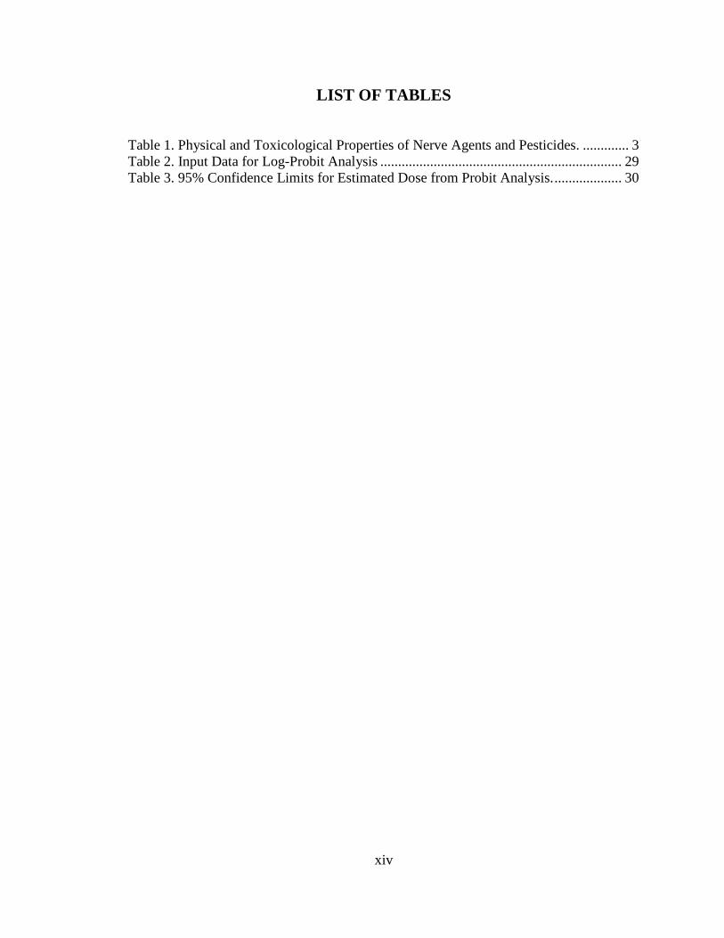
LIST OF TABLES
Table 1. Physical and Toxicological Properties of Nerve Agents and Pesticides. ............. 3 Table 2. Input Data for Log-Probit Analysis .................................................................... 29 Table 3. 95% Confidence Limits for Estimated Dose from Probit Analysis. ................... 30

xv
LIST OF FIGURES
Figure 1. Determination of the LD50 of soman for P21 male rats. Fifty rats (10 rats per dose) were injected subcutaneously with soman at the following doses (μg/kg): 40, 55, 57.5, 62.5, and 70. Mortality rates were recorded at 24 hr following soman injection and used as the input data into the log-probit method of the IBM SPSS Statistics 20 package to determine the LD50. The plot shows the predicted mortality rates at different doses of soman at P21. The LD50 was 62.02 μg/kg (dashed line; p = 0.00414). ................................................................................................................ 31
Figure 2. The latency to SE onset after soman injection is shorter in P21 rats compared to adults. P21 rats (n = 12) and young-adult rats (n = 20) were injected with the appropriate soman dose corresponding to 1.2 X LD50. ***p < 0.001 (Student’s t-test). ........................................................................................................................... 32
Figure 3. Baseline AChE activity in P21 rats is lower in the prefrontal cortex, piriform cortex and hippocampus, but not in the amygdala, compared to adult rats. For P21 rats, n = 5, and for the young-adult rats, n = 15. **p < 0.01, ***p < 0.001 (Student's t-test). ........................................................................................................................ 33
Figure 4. Reduction of AChE activity in P21 rats after injection of 1.2 X LD50 soman. Soman-exposed rats that did not develop SE were sacrificed 20 min after soman injection (No-SE group, n = 7). Rats that developed SE were sacrificed at the onset of SE (SE-onset group, n = 7). The soman-exposed rats had significantly lower AChE activity in all brain regions, compared to the control group (n = 5). AChE activity in the basolateral amygdala of the SE-onset group was significantly lower than that in the No-SE group. ***p < 0.001 compared to the control group, and *p < 0.05 between the no-SE and the SE-onset groups (One-way ANOVA with Tukey HSD post-hoc). ......................................................................................................... 34
Figure 5. Atropine sulfate (ATS) in immature but not in adult rats arrests generalized seizures induced by soman exposure. Young-adult (n = 12) and P21 (n = 10) rats were exposed to a soman dose corresponding to 1.2 X LD50 (P21 rats: 74.4 μg/kg, young-adult rats: 132 μg/kg). A. Administration of HI-6 to P21 rats, at 20 min after soman injection, had no effect on seizures. B. Administration of 2.0 mg/kg ATS, at 20 min after soman injection, terminated seizures in the P21 rats, but not in the adult rats. ***p < 0.001 when seizure severity score is compared between P21 and adult rats at 40, 50, and 60 min after soman exposure (MANOVA, Bonferroni correction).................................................................................................................................... 36
Figure 6. Delayed post-treatment with the GluK1R antagonists LY293558 or UBP302 arrests soman-induced seizures in P21 rats. Rats were exposed to 1.2 X LD50 soman (74.4 μg/kg) and treated with ATS (0.5 mg/kg) and HI-6 (125 mg/kg) at 1 min post-exposure. At 1 hr post-exposure, rats received LY293558 (20 mg/kg; n = 15), or UBP302 (250 mg/kg; n = 18), or the drug vehicle (n = 16). ***p < 0.001 for the difference in seizure score of the LY293558-treated and UBP302-treated groups compared to the vehicle-treated group (MANOVA, Bonferroni post-hoc test). ...... 37
Figure 7. Immature rats do not suffer neuronal degeneration, 1 day and 7 days after soman-induced SE. A. Cresyl violet photomicrographs outline the brain regions (amygdala in red; hippocampus in yellow) from where the FJC photomicrographs (B

xvi
and C) were taken (the specific areas shown in the photomicrographs are outlined with black rectangles). Immature and adult rats were exposed to the age-specific 1.2X LD50 of soman. In contrast to the adult rats (C), immature rats (B) did not display any degenerating cells. Magnification in A is 200x. Scale bar (for B and C) is 50 μm. .................................................................................................................... 38
Figure 8. A reduction in amygdalar volume, 30 and 90 days after soman exposure, is prevented by LY293558 treatment. A, B, C. Tracings of the amygdala in series of slices (left) and representative photomicrographs (right) from control animals (A, n = 8), soman-exposed animals that received only ATS (0.5 mg/kg) and HI-6 at 1 min post-exposure (B, n = 8), and soman-exposed animals that received LY293558 (20 mg/kg) at 1 h after soman injection (C, n = 8). D. Group data showing the estimated volume of the amygdala for all three groups, 30 days after the exposure. E. Group data showing the estimated volume of the amygdala for all three groups, 90 days after the exposure. *p < 0.05 (ANOVA, LSD post-hoc test). ................................. 40
Figure 9. A reduction in hippocampal volume, 30 and 90 days after soman exposure, is prevented by LY293558 treatment. A, B, C: Tracings of the hippocampus in series of slices (left) and representative photomicrographs (right) from control animals (A, n = 8), soman-exposed animals that received only ATS (0.5 mg/kg) and HI-6 at 1 min post-exposure (B, n = 8), and soman-exposed animals that received LY293558 (20 mg/kg) at 1 h after soman injection (C, n = 8). D: Group data showing the estimated volume of the hippocampus for all three groups, 30 days after the exposure. E: Group data showing the estimated volume of the hippocampus for all three groups, 90 days after the exposure. *p < 0.05 (ANOVA, LSD post-hoc test). 41
Figure 10. LY293558 treatment prevents behavioral deficits 30 and 90 days after soman exposure. A and B show that total freezing time of the contextual fear-conditioned responses, 30 and 90 days after soman exposure, for the soman-exposed rats who received only ATS (0.5 mg/kg) and HI-6 at 1 min after exposure (n = 13), similarly treated rats who received also LY293558 at 1 h after soman exposure (n = 11), and controls (n = 17). C and D show the total freezing time for the auditory fear-conditioned responses, for the same groups, at 30 and 90 days after exposure. E and F are the results from the open field test, showing the time spent in the center, for the same three groups, at 30 and 90 days after exposure. *p < 0.05 (ANOVA, Dunnett post-hoc test). .............................................................................................. 44
Figure 11. Pretreatment with a selective M1 receptor antagonist, VU0255035, reduces seizure severity after exposure to paraoxon or soman. Administration of VU0255035, 15 min before exposure to paraoxon (A) or soman (B) reduced the Racine score of seizure severity, averaged over 5 min to 60 min after exposure. For the paraoxon experiments, n = 8 for the vehicle group, and n = 10 for the VU0255035-pretreated group (P < 0.01). For the soman experiments, n = 4 for each group (the vehicle-pretreated and the VU0255035-pretreated group; P < 0.001). ... 59
Figure 12. Paraoxon, at three different concentrations, enhances both spontaneous inhibitory currents (sIPSCs) and spontaneous excitatory currents (sEPSCs), with a greater, lasting effect on the sEPSCs. Whole cell simultaneous recordings of sIPSCs and sEPSCs were obtained from principal cells in the BLA (Vh = -58). (A) The traces shown are representative examples of the effects of paraoxon at 0.1 μM, 1 μM, and 10 μM. Upward currents are GABAergic and downward currents are

xvii
glutamatergic. (B) The bar graphs show the ratio of the charge transferred by glutamatergic currents over the charge transferred by GABAergic currents, during a 20 s time-window, in control conditions and after bath application of paraoxon (left graph: at 17 min after paraoxon application; middle graph, at 9 min after paraoxon application; right graph, at 6 min after paraoxon application). *P < 0.05, **P < 0.01.................................................................................................................................... 61
Figure 13. The effects of paraoxon are prevented when slices are pre-exposed to atropine or VU0255035. Whole cell simultaneous recordings of sIPSCs and sEPSCs were obtained from principal cells in the BLA (Vh = -58). (A) Representative traces showing that 10 μM paraoxon had no significant effect when applied in the presence of atropine (1 μM, top row of traces) or VU0255035 (10 μM, second row of traces). (Β) Εffects of 10 μM paraoxon on sIPSCs and sEPSCs under three conditions: pretreatment with atropine (left bar graph), pretreatment with VU0255035 (middle bar graph), and in the absence of any muscarinic receptor antagonist (right bar graph). The total charge transferred by sIPSCs or sEPSCs during a 20 s time-window, 10 min after paraoxon application, was expressed as a percentage of the total charge transferred, during 20 s, in control conditions. Only when there was no atropine or VU0255035 in the slice medium, the total charge transferred by sIPSCs and sEPSCs was increased by paraoxon, and the difference between the increase in sIPSCs and the increase in sEPSCs was statistically significant (***P < 0.001). .... 64
Figure 14. Semi-quantitative analysis of mouse mAChR gene expression. Density and intensity was analyzed using the semi-quantitative scale published on the website for the Allen Institute for Brain Science (3). A - Representative images of sagittal sections of mouse BLA for each age and gene analyzed in the analysis. B - Quantification of the density and intensity for each age and gene in the analysis. Chrm5 was not detected in the analysis. "Chrm" designation indicates mouse gene names (e.g. Chrm1 refers to mAChR subtype 1). ..................................................... 81
Figure 15. ASIC1a channels are present on BLA interneurons. (a) Typical linear currents (Ih is absent) recorded from interneurons in response to hyperpolarizing voltage steps in voltage-clamp (v-clamp) mode (left), and an example of fast, non-accommodating spiking of interneurons in response to current injection in the current-clamp (c-clamp) mode (right). (b) Brief (200 ms) pressure application of acidified solution (left), or 40 mM ammonium (right) induced inward currents in interneurons, which were increased by lowering the bath temperature. (c) Currents evoked in interneurons by acidified solution or 40 mM ammonium were blocked by 2 mM flurbiprofen. (d) In the current clamp mode, bath application of 5 mM ammonium induced high-frequency firing of interneurons. In (b) and (c), holding potential is -70 mV. In (b), (c), and (d), recordings are in the presence of CNQX (10 μM), D-AP5 (50 μM), bicuculline (20 μM), and SCH50911 (10 μM). .................... 93
Figure 16. ASIC1a channels are present on BLA principal neurons. (a) Currents recorded from principal neurons in response to hyperpolarizing voltage steps in voltage-clamp (v-clamp) mode (left; notice the presence of Ih), and an example of accommodating firing in response to current injection in the current-clamp (c-clamp) mode (right). (b) Pressure application (200 ms) of acidified solution (left), or 40 mM ammonium (right) induced inward currents in principal cells, which were increased by lowering the bath temperature. (c) Currents evoked in principal cells by acidified solution or
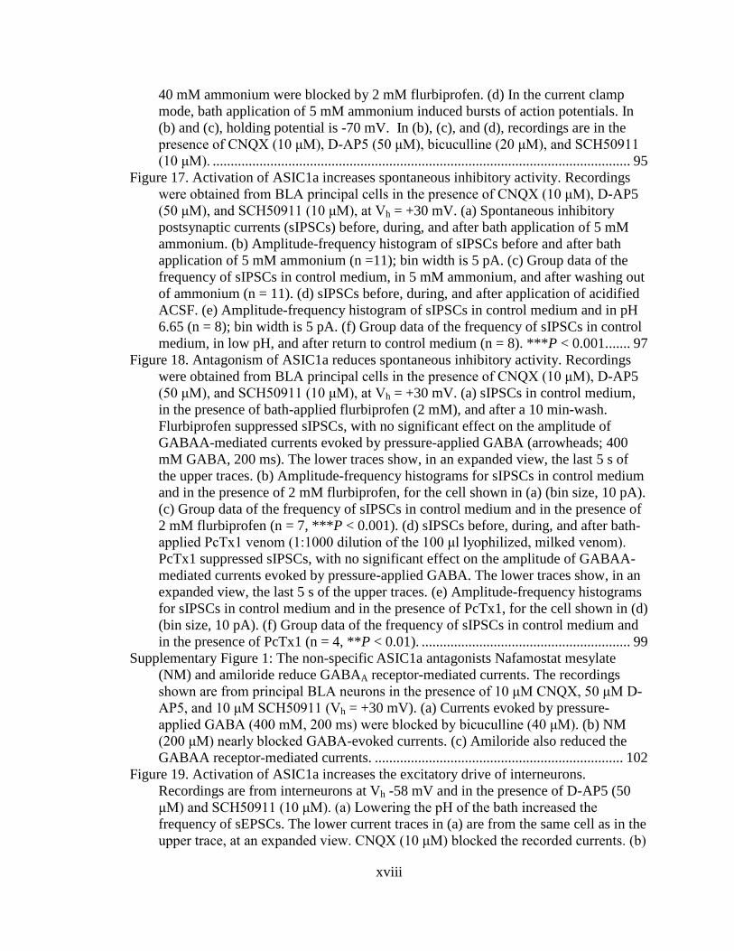
xviii
40 mM ammonium were blocked by 2 mM flurbiprofen. (d) In the current clamp mode, bath application of 5 mM ammonium induced bursts of action potentials. In (b) and (c), holding potential is -70 mV. In (b), (c), and (d), recordings are in the presence of CNQX (10 μM), D-AP5 (50 μM), bicuculline (20 μM), and SCH50911 (10 μM). .................................................................................................................... 95
Figure 17. Activation of ASIC1a increases spontaneous inhibitory activity. Recordings were obtained from BLA principal cells in the presence of CNQX (10 μM), D-AP5 (50 μM), and SCH50911 (10 μM), at Vh = +30 mV. (a) Spontaneous inhibitory postsynaptic currents (sIPSCs) before, during, and after bath application of 5 mM ammonium. (b) Amplitude-frequency histogram of sIPSCs before and after bath application of 5 mM ammonium (n =11); bin width is 5 pA. (c) Group data of the frequency of sIPSCs in control medium, in 5 mM ammonium, and after washing out of ammonium (n = 11). (d) sIPSCs before, during, and after application of acidified ACSF. (e) Amplitude-frequency histogram of sIPSCs in control medium and in pH 6.65 (n = 8); bin width is 5 pA. (f) Group data of the frequency of sIPSCs in control medium, in low pH, and after return to control medium (n = 8). ***P < 0.001. ...... 97
Figure 18. Antagonism of ASIC1a reduces spontaneous inhibitory activity. Recordings were obtained from BLA principal cells in the presence of CNQX (10 μM), D-AP5 (50 μM), and SCH50911 (10 μM), at Vh = +30 mV. (a) sIPSCs in control medium, in the presence of bath-applied flurbiprofen (2 mM), and after a 10 min-wash. Flurbiprofen suppressed sIPSCs, with no significant effect on the amplitude of GABAA-mediated currents evoked by pressure-applied GABA (arrowheads; 400 mM GABA, 200 ms). The lower traces show, in an expanded view, the last 5 s of the upper traces. (b) Amplitude-frequency histograms for sIPSCs in control medium and in the presence of 2 mM flurbiprofen, for the cell shown in (a) (bin size, 10 pA). (c) Group data of the frequency of sIPSCs in control medium and in the presence of 2 mM flurbiprofen (n = 7, ***P < 0.001). (d) sIPSCs before, during, and after bath-applied PcTx1 venom (1:1000 dilution of the 100 μl lyophilized, milked venom). PcTx1 suppressed sIPSCs, with no significant effect on the amplitude of GABAA-mediated currents evoked by pressure-applied GABA. The lower traces show, in an expanded view, the last 5 s of the upper traces. (e) Amplitude-frequency histograms for sIPSCs in control medium and in the presence of PcTx1, for the cell shown in (d) (bin size, 10 pA). (f) Group data of the frequency of sIPSCs in control medium and in the presence of PcTx1 (n = 4, **P < 0.01). .......................................................... 99
Supplementary Figure 1: The non-specific ASIC1a antagonists Nafamostat mesylate (NM) and amiloride reduce GABAA receptor-mediated currents. The recordings shown are from principal BLA neurons in the presence of 10 μM CNQX, 50 μM D-AP5, and 10 μM SCH50911 (Vh = +30 mV). (a) Currents evoked by pressure-applied GABA (400 mM, 200 ms) were blocked by bicuculline (40 μM). (b) NM (200 μM) nearly blocked GABA-evoked currents. (c) Amiloride also reduced the GABAA receptor-mediated currents. ..................................................................... 102
Figure 19. Activation of ASIC1a increases the excitatory drive of interneurons. Recordings are from interneurons at Vh -58 mV and in the presence of D-AP5 (50 μM) and SCH50911 (10 μM). (a) Lowering the pH of the bath increased the frequency of sEPSCs. The lower current traces in (a) are from the same cell as in the upper trace, at an expanded view. CNQX (10 μM) blocked the recorded currents. (b)

xix
Currents evoked by pressure application (arrowhead, 200 ms) of acidified ACSF in the absence of CNQX, displayed “riding” EPSCs. (c) Pressure application of ammonium (40 mM, 500 ms) increased the frequency of sEPSCs. (d) Bath application of flurbiprofen (1 mM) decreased the frequency of sEPSCs. .............. 104
Figure 20. The net effect of ASIC1a activation is suppression of BLA excitability. (a) and (b) Simultaneous recordings of sIPSCs (outward currents) and sEPSCs (inward currents) were obtained from principal cells at Vh -58 mV, and in the presence of D-AP5 (50 μM) and SCH50911 (10 μM). Bath application of 5 mM ammonium (a), or acidified solution (b) increased the charge transferred by sIPSCs and sEPSCs; the increase in the charge transferred by sIPSCs was significantly greater than the increase in charge transferred by sEPSCs. Example traces are shown in the upper panels of (a) and (b), and group data are shown in the bar graphs; n = 21 in (a) and n = 9 in (b), ***P < 0.001. (c) Upper panel shows field potentials evoked in the BLA by single-pulse stimulation of the external capsule, and lower panel shows spontaneous field activity recorded in gap-free mode. Recordings are in medium containing 7 mM K+ and zero Mg++, which induced epileptiform activity. Bath application of 8 mM ammonium reduced the evoked field potentials and blocked epileptiform activity. Each of the three field potentials shown in the upper panel is an average of 10 sweeps; the stimulus artifacts have been truncated for clarity. The equidistant vertical lines in the traces of spontaneous activity are stimulus artifacts, as evoked field potentials were sampled during gap-free recordings, by stimulation applied every 20 sec. ............................................................................................... 107
Figure 21. The net effect of ASIC1a antagonism is reduction of inhibition and increased excitability. (a) Simultaneous recordings of sIPSCs (outward currents) and sEPSCs (inward currents) were obtained from principal cells at Vh -58 mV, and in the presence of D-AP5 (50 μM) and SCH50911 (10 μM). Bath application of 2 mM flurbiprofen decreased the frequency of sIPSCs to a greater extent than that of sEPSCs. An example is shown in the upper panel, and group data in the bar graphs (n = 9, ***P < 0.001). (b) Field potentials evoked in the BLA by stimulation of the external capsule. Bath application of 2 mM flurbiprofen reversibly increased the amplitude of the evoked responses. Each trace is an average of 10 sweeps. .......... 109
Figure 22. In vivo activation of ASIC1a in the BLA suppresses anxiety-like behavior, while antagonism of ASIC1a increases anxiety. (a) In the open field test, the rats spent significantly more time in the center, after microinjection of ammonium bilaterally into the BLA (left graph), and significantly less time in the center, after microinjection of psalmotoxin into the BLA (right graph), compared to the time they spent in the center of the open field when injected with the vehicle. (b) In the light-dark box test, rats microinjected with ammonium bilaterally into the BLA took a significantly longer time to enter the dark compartment, and spent more total time in the light compartment compared to rats injected with the vehicle. *P < 0.05. ....... 111
Figure 23. DZP terminates soman-induced SE, but does not reduce the total duration of SE within the 24 h period after soman exposure, as seizures return; UBP302 reduces the total duration of SE within 24 h. (A) and (B) Example traces from EEG recordings showing that both DZP and UBP302–administered 1h after soman–terminated the SE induced by soman, but seizure activity returned after DZP administration. (C) Duration of initial SE and total duration of SE within 24 h after

xx
soman exposure, when DZP and UBP302 were administered at 1 h after soman injection. The three bars on the left show the duration of the initial SE (the SE that started 5 to 15 min after soman exposure and was terminated by DZP or UBP302, or spontaneously in the SOMAN group), while the three bars on the right show the total duration of SE. SOMAN, n = 4; SOMAN+DZP, n = 6; SOMAN+UBP302, n = 8. (D) Duration of initial SE and total duration of SE within 24 h after soman exposure, when DZP and UBP302 were administered at 2 h after soman injection. The three bars on the left show the duration of the initial SE, while the three bars to the right show the total duration of SE. SOMAN, n = 4; SOMAN+DZP, n = 4; SOMAN+UBP302, n = 4. *P < 0.05, **P < 0.01 and ***P < 0.001 in comparison to the SOMAN group (ANOVA followed by Bonferroni post-hoc test for the initial SE and ANOVA followed by Games-Howell post-hoc test for total SE). *P < 0.05, **P < 0.01 for the comparisons between the DZP-treated and the UBP302-treated groups (ANOVA followed by Fisher’s LSD test). ................................................. 133
Figure 24. The number of convulsive seizures that recurred in the DZP-treated rats after termination of the initial SE was greater than in the UBP302-treated rats. (A) EEG baseline before soman exposure. (B) Representative recording of a convulsive seizure recurring after termination of the initial SE by DZP, and its correspondence with the behavioral seizure observations. (C) Number of convulsive seizures that occurred after cessation of the initial SE, within the remaining time of the 24 h period after soman exposure. SOMAN, n = 4; SOMAN+DZP, n = 6; SOMAN+UBP302, n = 8. **P < 0.01, significantly higher compared to the SOMAN+UBP302 group and the SOMAN group (ANOVA followed by Holm-Sidak post-hoc test). ................................................................................................ 134
Figure 25. UBP302, but not DZP, administered 1 h after soman exposure, reduced neuronal degeneration in the amygdala, hippocampus, and neocortex, 1 day after the exposure. (A) and (B) Panoramic photomicrographs of Nissl-stained sections showing the brain regions evaluated by FJC staining. (C) Representative photomicrographs of FJC-stained sections from the brain regions where neuronal degeneration was evaluated, for the SOMAN, SOMAN+DZP, and SOMAN+UBP302 groups. Total magnification is 100x. Scale bar is 50 μm. (D) Neuropathology scores (median and interquartile range) for the SOMAN, SOMAN+DZP, and SOMAN+UBP groups (n = 6 for each group) for the amygdala (Amy), piriform cortex (Pir), entorhinal cortex (Ent), the CA1, CA3, and hilar areas of the ventral hippocampus, and neocortex (neo-Ctx). *P < 0.05, **P < 0.01 in comparison to the SOMAN group (Mann-Whitney U test). ................................... 136
Figure 26. UBP302, but not DZP, administered 1 h after soman exposure, reduced neuronal loss in the BLA and the CA1 hippocampal area, 1 day after the exposure. (A) Panoramic photomicrographs of Nissl-stained half hemispheres outlining the amygdalar nucleus and the hippocampal subfield where stereological analysis was performed. (B) Representative photomicrographs of Nissl-stained sections showing BLA and CA1 cells from the CONTROL, SOMAN, SOMAN+DZP and SOMAN+UBP302 groups. Total magnification is 630x and scale bar is 50 µm. (C) and (D) Group data (mean and standard error; n = 6 for each group) of stereological estimation of the total number of Nissl-stained neurons in the BLA (left) and CA1

xxi
area (right). **P < 0.01, ***P < 0.001 in comparison to CONTROL, ##P < 0.01 in comparison to the SOMAN group (ANOVA, Dunnett post-hoc test). ................... 137
Figure 27. UBP302, but not DZP, administered 1 h after soman exposure, reduced neuronal degeneration in the amygdala, CA1 and CA3 dorsal hippocampal areas, and entorhinal cortex, 7 days after the exposure. (A) and (B) Panoramic photomicrographs of Nissl-stained sections showing the brain regions evaluated by FJC staining. (C) Representative photomicrographs of FJC-stained sections from the brain regions where neuronal degeneration was evaluated, for the SOMAN, SOMAN+DZP, and SOMAN+UBP302 groups. Total magnification is 100x. Scale bar is 50 μm. (D) Neuropathology scores (median and interquartile range) for the SOMAN, SOMAN+DZP, and SOMAN+UBP groups (n = 6 for each group), for the amygdala (Amy), piriform cortex (Pir), entorhinal cortex (Ent), the CA1, CA3 and hilar areas of the ventral hippocampus, and neocortex (neo-Ctx). *P < 0.05, **P < 0.01 in comparison to the SOMAN group (Mann-Whitney U test). ...................... 139
Figure 28. UBP302, but not DZP, administered 1 h after soman exposure, reduced neuronal loss in the BLA and the CA1 hippocampal area, 7 days after the exposure. (A) Panoramic photomicrographs of Nissl-stained half hemispheres outlining the amygdalar nucleus and the hippocampal subfield where stereological analysis was performed. (B) Representative photomicrographs of Nissl-stained sections showing BLA and CA1 cells from the CONTROL, SOMAN, SOMAN+DZP and SOMAN+UBP302 groups. Total magnification is 630x and scale bar is 50 µm. (C) and (D) Group data (mean and standard error; n = 6 for each group) of stereological estimation of the total number of Nissl-stained neurons in the BLA (left) and CA1 area (right). *P < 0.05, **P < 0.01, ***P < 0.001 in comparison to CONTROL, #P < 0.05 and ##P < 0.01 in comparison to the SOMAN group (ANOVA, Dunnett post-hoc test). .................................................................................................................. 140
Figure 29. UBP302, but not DZP, administered 1 h after soman exposure, prevented GABAergic interneuronal loss in the BLA, 7 days after the exposure. Group data (mean and standard error; n = 6 for the each group) of stereological estimation of the total number of GAD67+ neurons in the BLA for the CONTROL, SOMAN, SOMAN+DZP and SOMAN+UBP302 groups. Representative photomicrographs of GAD67+ interneurons are shown in the lower panel. Total magnification is 630x and scale bar is 50 µm. **P < 0.01 in comparison to CONTROL (ANOVA, Dunnett post-hoc test). .......................................................................................................... 141
Figure 30. UBP302, administered 1 h after soman exposure, reduces neuronal degeneration in the amygdala, piriform cortex, and CA1 hippocampal area, while DZP reduces neurodegeneration in the CA1 area, 30 days after the exposure. (A) and (B) Panoramic photomicrographs of Nissl-stained sections showing the brain regions evaluated by FJC staining. (C) Representative photomicrographs of FJC-stained sections from the brain regions where neuronal degeneration was evaluated, for the SOMAN, SOMAN+DZP, and SOMAN+UBP302 groups. Total magnification is 100x. Scale bar is 50 μm. (D) Neuropathology scores (median and interquartile range) for the SOMAN , SOMAN+DZP, and SOMAN+UBP groups (n = 6 for each group), for the amygdala (Amy), piriform cortex (Pir), entorhinal cortex (Ent), the CA1, CA3 and hilar areas of the dorsal hippocampus, and neocortex (neo-Ctx). *P < 0.05 in comparison to the SOMAN group (Mann-Whitney U test). .... 143

xxii
Figure 31. UBP302, but not DZP, administered 1 h after soman exposure, reduced neuronal loss in the BLA and the CA1 hippocampal area, 30 days after the exposure. (A) Panoramic photomicrographs of Nissl-stained half hemispheres outlining the amygdalar nucleus and the hippocampal subfield where stereological analysis was performed. (B) Representative photomicrographs of Nissl-stained sections showing BLA and CA1 cells from the CONTROL, SOMAN, SOMAN+DZP and SOMAN+UBP302 groups. Total magnification is 630x and scale bar is 50 µm. (C) and (D) Group data (mean and standard error; n = 6 for each group) of stereological estimation of the total number of Nissl-stained neurons in the BLA (left) and CA1 area (right). *P < 0.05, **P < 0.01, ***P < 0.001 in comparison to CONTROL; ##P < 0.01 in comparison to the SOMAN group (ANOVA, Dunnett post-hoc test). ... 144
Figure 32. UBP302, but not DZP, administered 1 h after soman exposure, protected against the development of anxiety, 30 days after the exposure. (A) Distance traveled (Mean ± SE) in the open field. (B) Percentage of time spent in the center of the Open Field. (C) Amplitude of startle responses to 120 dB and 110 dB acoustic stimuli. CONTROL, n = 8; SOMAN, n = 10; SOMAN+DZP, n = 9; SOMAN+UBP302, n = 12. *P < 0.05, **P < 0.01 and ***P < 0.001 (ANOVA, Dunnett post-hoc comparison to CONTROL). ....................................................... 146

1
CHAPTER 1: Introduction
Over 1,400 civilians were killed by rockets loaded with military-grade sarin
during attacks in Damascus, Syria. The victims of this attack, which occurred in August,
2013, resulted in the death of 426 children and exemplifies one of the deadliest uses of
chemical weapons in recent history (81). Nerve agents, like sarin, are categorized as OP
compounds and also include some widely used pesticides. OPs can be extremely lethal
upon exposure; approximately 10% of people exposed to an OP will die from the acute
toxic side effects resulting from exposure (26; 60), indicating the significant public health
concern of OP exposure.
OP compounds exert their toxic effects through their action as irreversible
inhibitors of the enzyme AChE in the peripheral and central nervous system. Irreversible
inhibition of AChE indirectly results in a "cholinergic crisis" and induces a collection of
symptoms, including salivation, lacrimation, urination, defecation, gastrointestinal
motility, emesis, and miosis. In cases of severe poisoning, or if treatment is not
immediately administered, respiratory distress/failure and seizures/SE may result.
Following the binding of an OP compound to AChE, a biochemical process referred to as
"aging" occurs, where the OP becomes permanently bound to AChE, resulting in an
inability to restore AChE enzymatic activity. Time to AChE aging widely varies
depending on the OP compound, but nerve agents, especially the nerve agent soman,
display extremely short times to AChE aging, which makes the window for therapeutic
intervention limited.
BIOCHEMICAL MECHANISM OF OP ACTION

2
AChE is a critically important enzyme in the peripheral and central nervous
system due to its role in the termination of cholinergic neurotransmission. The canonical
role of AChE is the catalytic hydrolysis of acetylcholine (ACh) into acetate and choline
(272). AChE contains a catalytic triad of histidine, glutamate and serine deep within the
core of the enzyme and requires that the substrate for hydrolysis travel down into the
enzyme to be hydrolyzed (263; 272). OPs act as a substrate for AChE; once they interact
with the enzyme, the phosphorous group on the OP forms a covalent bond with the serine
residue within the active site (144). AChE can spontaneously recover its activity with the
OP potentially dissociating, but the rate at which AChE can recover activity varies
depending on the particular AChE-OP conjugate. If bound long enough to an OP, the
active site of AChE undergo dealkylation, which in turn makes this AChE-OP conjugate
permanent and AChE permanently inactive (144). The half-time to AChE aging varies
widely by the specific OP studied, but the rate of aging for AChE-OP conjugates is
highest for the nerve agent class of OPs (265). The aging half-time from the binding of
soman to human AChE has been reported to be as fast as 1.87 min which makes soman
one of the most lethal nerve agents (251) and is the nerve agent studied in this
dissertation.
PROPERTIES OF ORGANOPHOSPHATES: NERVE AGENTS AND PESTICIDES
Pesticides and nerve agents share a similar, primary mechanism of action: the
irreversible inhibition of AChE. Both pesticides and nerve agent organophosphates pose a
high risk to humans due to their potency and exert toxic effects through all potential
routes of exposure. However, the biochemical, chemical and physical properties of these
compounds vary dramatically and must be considered to understand the differences in

3
their relative toxicity. See Table 1 for a comparison of the relative toxicities between
pesticides and nerve agents.
Table 1. Physical and Toxicological Properties of Nerve Agents and Pesticides.
Tabun Sarin Soman VX Parathion Dichlorvos Chlorpyrifos Malathion
OP Classification Nerve
Agent
Nerve
Agent
Nerve
Agent
Nerve
Agent Pesticide Pesticide Pesticide Pesticide
Subclassification G-Series G-Series G-Series V-Series Diethyl Diethyl Dimethyl Dimethyl
Molecular Weight
(g/mol) 162.13 140.09 182.17 267.37 291.26 220.28 350.59 330.36
Vapor Pressure
(mm Hg at 20°C) 490 22,000 3,900 10.5 6.68 X 10⁻⁶ 1.2 X 10⁻² 1.9 X 10-5* 3.38 X 10-6
Rat Dermal LD50
(mg/kg) 18 N/A N/A N/A 6.8-49.4 75-107 202 >4,400
Rat Oral LD50
(mg/kg) 3.7 0.55 N/A 0.012 2-22 56-80 155 1000-1375
Human Dermal
LD50 (mg/70 kg) 1 0.7 0.35
0.001-
0.006 N/A N/A N/A N/A
Human Oral LD50
(mg/70 kg) N/A 8-12 7-12 5 50-200 500-1000 grams grams
References: (24; 206; 211; 245; 285). *mm Hg at 25°C.

4
Organophosphate Pesticides
Organophosphate pesticides are categorized into two groups: diethyl and dimethyl
pesticides. Commonly used dimethyl pesticides include dichlorvos and malathion
whereas commonly used diethyl pesticides include chlorphyrifos and parathion. Of these
pesticides, the World Health Organization (WHO) ranks parathion as the most hazardous,
while dichlorvos, chlorpyrifos, and malathion are less hazardous (144). Hazard rankings
are determined by the WHO based on dermal and oral acute toxicity in the rat, where
dermal medial lethal dose (LD50) values dictate the hazardous classification of a pesticide
(123). Additionally, if toxic effects in humans are found to be more pronounced,
pesticides are classified into more hazardous categories (310). Enhanced toxicity of
diethyl pesticides can be explained by a relatively slow spontaneous reactivation of
AChE following diethyl pesticide binding to the active site of AChE (100). This also
explains why dimethyl pesticides are classified as less toxic or hazardous due to the faster
spontaneous reactivation of AChE (100). Some of the OP pesticides require a metabolic
conversion to the active, toxic metabolite. Following exposure to these pesticides, liver
oxidases rapidly convert these compounds to an 'oxon' form, which is the form that binds
to and irreversibly inhibits AChE (286). The precursor, parent compounds are relatively
inactive and exert minimal toxicity on their own prior to conversion into their oxon form
(287). Some of the parent compounds are not substrates that are capable of interacting
with AChE until metabolic conversion to the oxon form (287). Parathion, for example, is
converted by oxidative metabolism to paraoxon and is highly toxic compared to the
parent compound (286). Synthesized in 1944, parathion is one of the most lethal
pesticides (287) and has been used as a chemical weapon in war (189). Specifically, the
Rhodesian Army produced a lethal mixture of parathion by mixing it with dimethyl
5
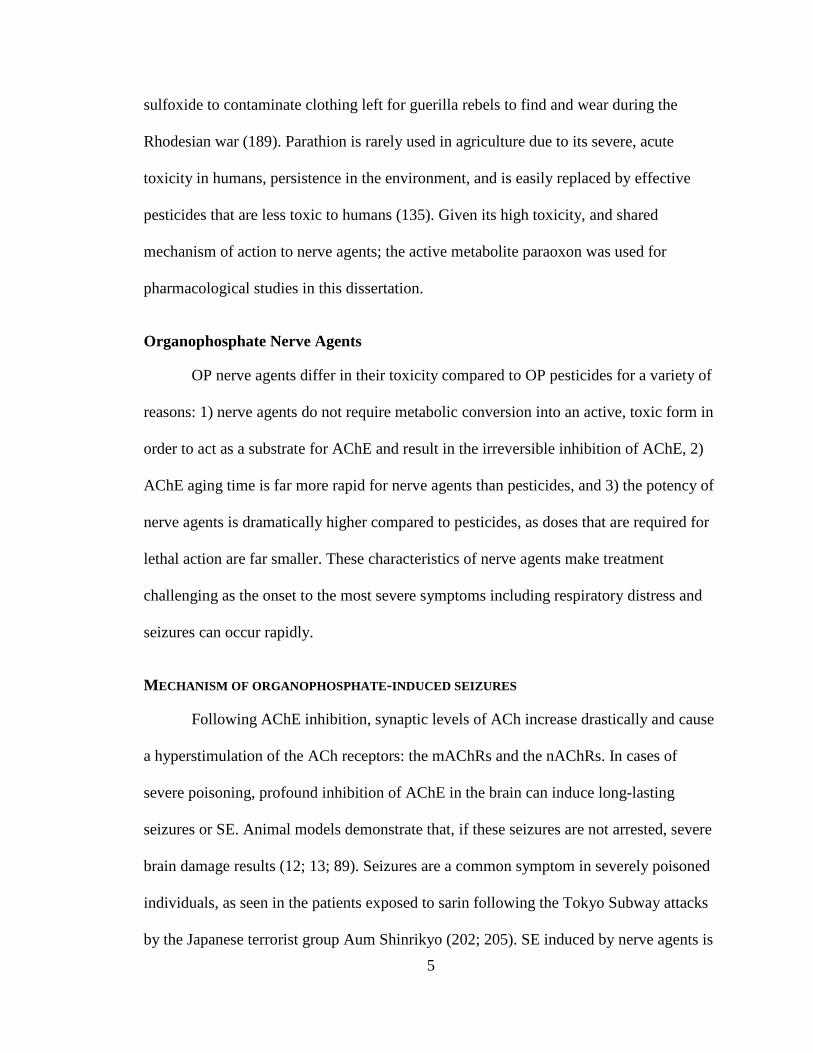
sulfoxide to contaminate clothing left for guerilla rebels to find and wear during the
Rhodesian war (189). Parathion is rarely used in agriculture due to its severe, acute
toxicity in humans, persistence in the environment, and is easily replaced by effective
pesticides that are less toxic to humans (135). Given its high toxicity, and shared
mechanism of action to nerve agents; the active metabolite paraoxon was used for
pharmacological studies in this dissertation.
Organophosphate Nerve Agents
OP nerve agents differ in their toxicity compared to OP pesticides for a variety of
reasons: 1) nerve agents do not require metabolic conversion into an active, toxic form in
order to act as a substrate for AChE and result in the irreversible inhibition of AChE, 2)
AChE aging time is far more rapid for nerve agents than pesticides, and 3) the potency of
nerve agents is dramatically higher compared to pesticides, as doses that are required for
lethal action are far smaller. These characteristics of nerve agents make treatment
challenging as the onset to the most severe symptoms including respiratory distress and
seizures can occur rapidly.
MECHANISM OF ORGANOPHOSPHATE-INDUCED SEIZURES
Following AChE inhibition, synaptic levels of ACh increase drastically and cause
a hyperstimulation of the ACh receptors: the mAChRs and the nAChRs. In cases of
severe poisoning, profound inhibition of AChE in the brain can induce long-lasting
seizures or SE. Animal models demonstrate that, if these seizures are not arrested, severe
brain damage results (12; 13; 89). Seizures are a common symptom in severely poisoned
individuals, as seen in the patients exposed to sarin following the Tokyo Subway attacks
by the Japanese terrorist group Aum Shinrikyo (202; 205). SE induced by nerve agents is

6
notoriously difficult to treat. Evidence from experimental models suggest that the longer
an episode of nerve agent induced-SE lasts, the greater the resulting brain damage and
associated symptoms.
Seizures induced by severe poisoning by OPs are a biochemical consequence of
the irreversible inhibition of AChE, which in turn leads to a dramatic increase in synaptic
ACh. A three-phase model of seizure initiation and maintenance following
organophosphate poisoning proposed by Shih and Mcdonough (181) suggests that the
increase in synaptic ACh initiates seizure activity, primarily through the hyperstimulation
of mAChRs. Neuropharmacological evidence supports this hypothesis as pre- or rapid
post-treatment with nonselective mAChR antagonists such as atropine sulfate or
scopolamine exert potent anticonvulsant efficacy (153; 181; 259; 261; 266). However, as
seizure activity continues over time, treatment with mAChR antagonists exert either no or
little anticonvulsant efficacy and require dramatically higher doses in order to do so.
Following an early phase of seizure activity mediated primarily by the increase in ACh in
peripheral and central synapses, excitatory amino acids appear to sustain and reinforce
ongoing seizure activity induced by OPs (181).
The development of successful immediate or delayed post-treatment of OP-
induced seizures/SE requires an understanding of the mechanism by which AChE
inhibition results in seizure induction, and experimental models that are relevant to all
populations and ages to test novel therapies. While evidence suggests mAChRs play a
critical role in seizure induction following OP poisoning, a clear mechanism for seizure
induction and their maintenance remains to be elucidated. Additionally, a majority of the
literature examining the long-term effects of organophosphates has largely been
7
examined in adult laboratory animals. Dramatic differences exist in the induction,
maintenance, and treatment of seizure activity in the immature brain. However, no
experimental models currently exist to examine 1) the long-term effects of these seizures
in immature animals, 2) if novel therapies can arrest OP-induced seizures/SE in immature
animals, 3) whether novel anticonvulsants are successful in blocking any associated long-
term side effects of OP-induced SE, and 4) how seizures are induced by OPs in the
immature brain. To address this gap, this dissertation has aimed to develop a rodent
model relevant to the pediatric population for testing treatments to arrest seizures induced
by the nerve agent soman, to verify whether new treatments can protect against the
associated long-term toxic effects; and determine the cellular and molecular mechanism
for seizure induction following severe OP poisoning.
TREATMENT OF OP-EXPOSURE
Following decontamination of the victim of OP intoxication, several
pharmacological treatments are currently available depending on the severity of
intoxication, symptoms present, and time at which therapies are administered.
Atropine
Atropine, a nonselective muscarinic receptor antagonist, is one of the first lines of
care in the treatment of OP intoxication and is used as a countermeasure against the
hyperstimulation of muscarinic receptors in the periphery (24). While animal studies
suggest that atropine may be efficacious as an anticonvulsant, it has low availability to
the central nervous system and may only be efficacious if given immediately after the
onset of seizure activity, which is unrealistic for mass casualty scenarios (182). Atropine
is extremely effective in alleviating some of the severe peripheral symptoms associated

8
with OP intoxication, including apnea, and bronchoconstriction, and can induce a drying
of secretions associated with mAChR hyperstimulation (24). Tolerance for atropine may
be very high as it has been estimated that in cases of severe OP intoxication, a safe
therapeutic range can be 35-210 mg per 70 kg in humans (24). This recommended dose is
much higher than the standard carried in the United States military nerve agent antidote
kits which contains 2.1 mg atropine sulfate (184). Doses of atropine contained in
autoinjectors designed for adult treatment following OP exposure have been found to be
well-tolerated in children. During the Persian Gulf Crisis, for example, the Medical Corps
of the Israeli Defense Forces distributed autoinjectors to the Israeli population containing
antidotes to nerve agent exposure, including atropine. A total of 410 children
unintentionally poisoned themselves with these atropine autoinjectors and were evaluated
in two separate studies (7; 142) but only a small number of cases presented with
symptoms of severe atropine poisoning but no deaths resulted.
Oximes
Oximes are also first line treatment for OP poisoning and are used in both clinical
and military settings. Oximes are nucleophilic compounds that attack the phosphorous
bond created between the AChE-OP adduct within the active site of AChE (145; 240).
This reaction can restore the activity of AChE if the enzyme has not already undergone
the aging process. Unfortunately, oximes do not exert broad-spectrum efficacy in
restoring AChE function for all AChE-OP adducts. In the United States, pralidoxime
chloride (2-PAM) is the standard civilian and military treatment used after OP
intoxication; 2-PAM is predominantly effective in restoring AChE function when sarin or
VX is the OP bound to the enzyme, but is relatively ineffective in restoring function

9
when the OP is tabun, cyclosarin or soman (240). These compounds are typically
incapable of penetrating the blood-brain barrier and as a result have little efficacy as
anticonvulsants (240). However, development of new oxime compounds with broad
spectrum efficacy in restoring AChE function, which are capable of crossing the blood-
brain barrier, is an active area of investigation and has yielded promising results with the
compound MMB4 (46; 240).
Anticonvulsants
Atropine is only an effective anticonvulsant against seizures induced by OP
exposure when administered rapidly following seizure induction; typically within the first
20 minutes (153; 261; 266). This may be due to a transition from mAChR-mediated
seizure activity to glutamate-receptor hyperactivation that sustains ongoing seizure
activity for OP-induced seizures (181). The United States Food and Drug Administration
(FDA) approved diazepam for the treatment of seizures induced by OPs. Enhancing the
activity of the inhibitory γ-aminobutyric acid A-receptor (GABAAR) has been
consistently used as a therapeutic approach for the treatment of seizures. Thus, the
commercially available anticonvulsant, diazepam was investigated as a potential
treatment for OP-induced seizures and has been implemented as the standard
anticonvulsant in military autoinjectors designed for treatment of OP intoxication (181).
Though numerous studies suggest that diazepam is an effective anticonvulsant for nerve
agent-induced seizures, none of these studies found a complete protection of brain
damage associated with nerve agent-induced seizures (37; 39; 65; 110; 115; 128; 129;
161-163; 169; 178; 180; 195; 218; 256; 257). Corroborating these findings, our group has
found that diazepam administered 1 h following soman exposure offers either no or

10
minimal neuroprotection against seizure-induced cellular loss, depending on the brain
structure studied (13). Diazepam treatment suppresses the initial episode of SE induced
by soman exposure, but over a 24 h period diazepam treatment offers no protection
against the total duration of SE. In juxtaposition, diazepam can enhance the 24 h duration
of SE if treatment is delayed to 2 h post-soman exposure and it was found to increase the
number of returning convulsive seizures (13).
It has been suggested that during a later phase of OP-induced seizure activity,
ongoing activity is primarily mediated by the excitatory amino acid receptors (181), and
as such, antagonists to the N-methyl-D-aspartate (NMDA) receptor have been thoroughly
investigated. This has been substantiated by recent research suggesting the NMDA
receptor antagonist MK-801 exerts some anticonvulsant efficacy against nerve agent-
induced seizures and this efficacy increases when treatment is delayed to further
timepoints (256). However, MK-801 is ineffective in blocking seizure induction or
arresting seizure activity when administered soon after induction by soman (43; 256). As
the sole treatment for seizures induced by soman in rats, high dose treatment of MK-801
increased mortality or enhanced the lethal effects of soman (256). In this study, Shih also
found that treatment with MK-801 at doses necessary to exert anticonvulsant efficacy
actually produces numerous adverse events including severe respiratory depression (256).
A novel class of glutamate receptor antagonists, GluK1KR antagonists, are
promising candidates for arresting seizures and protecting the brain against the
neuropathology associated with nerve agent exposure. GluK1KR antagonists have been
found to block the induction or arrest ongoing seizures induced by pilocarpine–a
nonselective mAChR agonist that has comparable mechanisms of action for seizure

11
induction and maintenance following exposure (269; 284). Topiramate, an anticonvulsant
that has antagonistic properties for the GluK1KRs, suppresses excitability in the BLA
(41), a key structure involved in the induction of seizures induced by OP exposure (227).
Our group and others have hypothesized a critical role for GluK1KRs in maintaining and
reinforcing further seizure activity during an episode of OP-induced SE. As such, we
have investigated the role of GluK1KR antagonists as treatments for nerve agent-induced
SE at delayed timepoints. Stopping seizures induced by nerve agents at delayed
timepoints following seizure induction (e.g. 1 h post-seizure onset) is extremely difficult,
but model realistic windows for therapeutic intervention for mass casualty scenarios
involving civilians or military personnel.
LY293558, a GluK1KR/AMPA receptor antagonist; and UBP302, a GluK1KR
antagonist, are promising candidates for the treatment of OP-induced SE. When SE is
induced by soman in adult male rats and treatment is delayed to 1 h post-soman exposure,
LY293558 treatment: 1) increases survival to 100% , 2) reduces the initial episode of SE,
3) reduces the duration of SE over a 24 hr period, 4) protects against neuronal loss in
BLA and CA1 at 1 and 7 d post-soman exposure, and 5) protects against the loss of
interneurons in the BLA at 7 d post-soman exposure (12; 89). By comparison, UBP302
administration 1 h post-soman exposure: 1) increases survival to 96%, 2) reduces the
initial episode of SE, 3) reduces the duration of SE over a 24 h period, 4) reduces
neuronal loss in the BLA and CA1 at 1 and 7 d post-soman exposure, 5) protects against
the loss of interneurons in the BLA at 7 d post-soman exposure, and 6) protects against
the development of anxiety-like behavior induced by soman exposure (13). These results
indicate that GluK1KR antagonist may be leading candidate in the treatment of OP-

12
induced SE as they are highly effective in increasing survival, arresting SE, and
protecting against neuropathology and behavioral deficits induced by OP exposure.
While both LY293558 and UBP302 show similar effectiveness, LY293558 reaches peak
brain levels more rapidly following administration (12) and can arrest SE more rapidly
following soman exposure. As such, LY293558 is the primary GluK1KR antagonist used
in the experiments described below.
LONG-TERM NEUROTOXICOLOGICAL EFFECTS OF OP EXPOSURE
Long-Term Toxicological Effects in Humans
What is known about the long-term effects of nerve agents in humans has largely
been derived from the long-term studies conducted on patients exposed to sarin in the
early 1990's in Japan. Largely, the long-lasting symptoms resulting from sarin exposure
are neuropsychiatric in nature (118), although some memory impairments have been
reported (198; 200; 204). The most common diagnosis or clinical finding in long-term
studies is the presentation of post-traumatic stress disorder (PTSD). A variety of studies
that performed long-term follow-ups found a higher frequency of PTSD or suspected
diagnoses of PTSD in sarin-exposed patients than in the general population, which was
positively correlated with the severity of sarin exposure (134; 194; 200; 203; 204).
Gray and white matter analyses have been conducted on a collection of patients
exposed to sarin from the attacks (238; 314). In one study, 38 patients treated for severe
sarin intoxication had structural MRI scans and variety of structural alterations were
identified up to six years post-sarin intoxication (314). In addition to widespread white
matter integrity disruption in the parietal lobe, temporal lobe and brainstem, gray matter
volume was reduced in the insular cortex, temporal cortex and the hippocampus (314).

13
Notably, the authors also found that the volume of insular white matter was positively
correlated with serum cholinesterase (ChE) suggesting that more pronounced structural
alterations are directly correlated to the severity of sarin intoxication (314). In a separate
structural study of victims of the sarin attacks, 25 patients were scanned for differences in
the volume of the amygdala and hippocampus. Remarkably, only patients who currently
or had a history of PTSD displayed reductions in bilateral volumes of the amygdalae and
specifically, a reduction in the volume of the left amygdala, which was negatively
correlated with PTSD severity (238). Given the critical role of the amygdala in regulating
anxiety and emotional processing (219; 221); along with the function of the hippocampus
in memory processes (239), reductions in the volumes of these structures may impair
their function and explain the observed, long-term clinical sequela of sarin poisoning.
Long-Term Toxicological Effects in Animals
The long-term consequences of OP and specifically nerve agent poisoning have
been studied exhaustively in a variety of laboratory species including guinea pigs, mice
and rats (70; 74; 90; 166; 188; 218; 228; 233). Exposing guinea pigs to sublethal (0.6 or
0.8 X LD50) doses of soman resulted in anxiety-like behavior that persisted up to 3
months following a single exposure (166). Similarly, in mice exposed to soman, anxiety-
like behavior was observed up to 3 months post-exposure and responses in both cue and
context dependent fear conditioning were impaired (74). Additionally, both mice and rats
have been found to have long-term deficits in performance on behavioral measures of
learning and memory (90; 233). These behavioral studies suggest that a single, acute
exposure to an OP can cause long-lasting anxiety and behavioral deficits.

14
Gross neuropathology is a common finding following OP-induced seizures in
animals (12-14; 23; 62; 69; 89; 132; 133; 158; 176; 178; 181; 194; 216; 218; 228). The
presence and severity of observed neuropathology appears to be directly related to the
expression of seizures and their duration (177; 227; 228; 258). Of the variety of brain
structures damaged following OP-induce seizures, which include the amygdala,
hippocampus, entorhinal cortex, neocortex, piriform cortex, and thalamus, the amygdala,
hippocampus and piriform cortex sustain the most severe damage (12-15; 53; 62; 70; 76;
89; 216). Given the role of the amygdala in regulating anxiety and its role in mediating
cue-dependent fear conditioning, along with the role of the hippocampus in mediating
context-dependent of fear conditioning and mediating some behavioral tasks of learning
and memory, it would suggest that the neuropathology routinely observed in these
structures following an OP exposure may explain the long-term behavioral impairments.
Acute neuropathology from OP-induced seizures may be long-lasting and could
explain the long-term neuropsychiatric symptoms observed in humans following sarin
poisoning. In mice receiving a convulsive dose of soman, the number of cells in the
amygdala is still reduced compared to controls up to 90 days post-soman exposure (70)
and correlates with impairments in amygdala-dependent behaviors such as cue-dependent
fear conditioning, lasting up to 30 days post-exposure and increases in anxiety-like
behavior up to 90 days post-exposure (74). Long-term neuropathology and impairments
in hippocampal-dependent behaviors are also evident following soman intoxication. A
single injection of soman in mice reduces the number of cells in the CA1 subfield of
hippocampus by 50% and this reduction persists up to 90 days post-exposure (68). More
severe intoxication by soman produces greater weight loss in mice and correlates the

15
extent of neuropathology observed in CA1 (90); as well as impairments in T-maze or the
Morris water maze task up to 90 days post-exposure (90). Animal models provide an
excellent means to study the mechanisms and time-course of the neuropathology and
behavioral alterations resulting from OP intoxication. Information obtained from these
studies may help in the development of novel treatments for OP intoxication, but limited
studies have been performed to understand the effects of OPs, especially the effects of
nerve agents on immature animals. This information is critically important for the
development of treatments suitable for children during mass casualty scenarios involving
nerve agent intoxication.
CHILDREN AND OP POISONING
Unlike adults, children require special consideration in the poisoning and
treatment following intoxication by OPs. Children are highly susceptible to the toxic
effects of OPs due to a variety of physiological, developmental and medical reasons. In
the event of dermal exposure to OPs, children are poisoned faster and more severely due
to thinner skin with a greater permeability compared to adults and a larger surface-area to
body mass ratio (116). Greater respiratory minute volume – the total air either inhaled or
exhaled in one minute – and their height, where vapor density of volatilized OPs is
greatest, also make them more vulnerable to respiratory exposure (116). Levels of AChE
in the brain are also lower compared to that of adults rats (Miller et al., Unpublished),
characteristic of incomplete development of the cholinergic system (75), and therefore
lower doses of OPs can elicit more severe symptoms. This is especially true in regards to
seizures due to the increased susceptibility to seizure induction in children (1; 113).
Standard decontamination methods, which do not include the use of heated water may

16
result in hypothermia following the decontamination of children also due to their higher
surface-area to body-mass ratio (116). Medical guidelines for the treatment of children
following nerve agent poisoning do not have a national consensus and therefore would
likely result in chaos, overwhelming first responders and causing a high mortality rate for
children during a mass casualty scenario involving nerve agents (92).
Given the special considerations for treating children intoxicated by OPs, it is
surprising that more basic research has not been conducted in this area. To our
knowledge, only one study has investigated developmental effects of nerve agent
intoxication in animal models. Specifically, this study by Sterri and Colleagues (279)
examined the mortality rates of rats exposed to varying doses of soman across
development. They found that younger rats were more susceptible to death following
soman exposure at lower doses when compared to adult rats only one to three weeks
older (279). For example, in their study, when post-natal day 5 (P5) rats were exposed to
20 μg/kg soman (s.c.) this caused 100% mortality, but this same dose only resulted in a
25% mortality rate in P12 rats (279). This single study in rats and the deaths of the 426
children during the sarin attack in Syria highlight the importance of investigating the
mechanisms of OP action in immature animals to develop treatment for children. With
this knowledge, treatments may be developed to protect against the acute and long-term
side effects associated with OP exposure.
CONCLUSIONS
The effects of OPs, their mechanism of action, treatment, as well as the acute and
long-term effects are well studied in adult animals and humans. Though it is well
accepted that the mechanism of action by which OPs induce their toxicity, and in
17
particular seizures, is through the inhibition of AChE and the subsequent
hyperstimulation of mAChRs and nAChRs, the specific receptors responsible for seizure
induction remain to be elucidated. The standard of care for seizures induced by OPs is
diazepam, but recent evidence suggests diazepam may increase SE duration and mortality
rate (13) and it is unclear if this therapy would be efficacious in children or protect them
against the long-term effects associated OP-induced seizures. Therefore, the purpose of
this dissertation is to develop a rodent model of nerve agent exposure that is relevant to
the pediatric population. In particular, in immature rats we will: 1) test novel
anticonvulsants to arrest seizures induced by nerve agents; 2) study the long-term
deleterious effects associated with nerve agent induced seizures; 3) study whether novel
anticonvulsants protect against these long-term neuropathological effects resulting from
nerve agent-induced seizures; and 4) clarify the molecular and cellular mechanism by
which OPs induce seizure activity.

18
CHAPTER 2: A Rat Model of Nerve Agent Exposure Applicable to the
Pediatric Population: The Anticonvulsant Efficacies of Atropine and GluK1 Antagonists
Steven L. Millera,c, Vassiliki Aroniadou-Anderjaskaa,b,c, Taiza H. Figueiredoa , Eric M. Pragera,c, Camila P. Almeida-Suhetta,c, James P. Aplandd, Maria F.M. Bragaa,b,c*
aDepartment of Anatomy, Physiology, and Genetics, bDepartment of Psychiatry, and
cProgram in Neuroscience, Uniformed Services University of the Health Sciences, 4301 Jones Bridge Road, Bethesda, MD 20814, USA
dNeurotoxicology Branch, U.S. Army Medical Research Institute of Chemical Defense, Aberdeen Proving Ground, Maryland, 21010, USA
*Corresponding Author: Maria F.M. Braga, D.D.S., Ph.D. Department of Anatomy, Physiology, and Genetics F. Edward Hébert School of Medicine, Uniformed Services University of the Health Sciences 4301 Jones Bridge Road Bethesda, MD 20814, USA Phone: (301) 295-3524 Fax: (301) 295-3566 Email: [email protected]
19
ABSTRACT
Inhibition of AChE after nerve agent exposure induces SE, which causes brain
damage or death. The development of countermeasures appropriate for the pediatric
population requires testing of anticonvulsant treatments in immature animals. In the
present study, exposure of P21 rats to different doses of soman, followed by probit
analysis, produced an LD50 of 62 μg/kg. The onset of SE was accompanied by a dramatic
decrease in brain AChE activity; rats who did not develop SE had significantly less
reduction of AChE activity in the basolateral amygdala than rats who developed SE. ATS
at 2 mg/kg, administered 20 min after soman exposure (1.2XLD50), terminated seizures.
ATS at 0.5 mg/kg, given along with an oxime within 1 min after exposure, allowed
testing of anticonvulsants at delayed time-points. The AMPA/GluK1 receptor antagonist
LY293558, or the specific GluK1 antagonist UBP302, administered 1 h post-exposure,
terminated SE. There were no degenerating neurons in the soman-exposed P21 rats, but
both the amygdala and the hippocampus were smaller than in control rats at 30 and 90
days post-exposure; this pathology was not present in rats treated with LY293558.
Behavioral deficits present at 30 days post-exposure, were also prevented by LY293558
treatment. Thus, in immature animals, a single injection of atropine is sufficient to halt
nerve agent-induced seizures, if administered timely. Testing anticonvulsants at delayed
time-points requires early administration of ATS at a low dose, sufficient to counteract
only peripheral toxicity. LY293558 administered 1 h post-exposure, prevents brain
pathology and behavioral deficits.

20
INTRODUCTION
Nerve agents are potent, OP toxins that act primarily by inhibiting the activity of
AChE. The resulting accumulation of acetylcholine at synaptic junctions produces
peripheral cholinergic crisis (excessive salivation, lacrimation, rhinorrhea, bronchorrhea,
cardiorespiratory suppression, eventual muscle paralysis, etc.), and, in the brain, induces
seizures and SE. Without timely pharmacological intervention, death will ensue, or if
death is prevented but the SE is not controlled, brain damage will result, with long-term
neurological and behavioral consequences (13; 14; 74; 88-90; 228; 229; 315). The nerve
agent sarin that was released during a terrorist attack in Syria, in August 2013, resulted in
the death of over 1,400 civilians, 426 of which were children (81). Mass casualties during
terrorist attacks that employ nerve agents are expected to disproportionately affect
children due to their greater body surface area-to-body mass ratio, increased skin
permeability, faster respiration rate, breathing at a level where nerve agent vapor density
would be highest, and an increased susceptibility to seizures (1). The higher vulnerability
of children necessitates the availability of effective countermeasures that will protect
their lives.
Pharmacological treatments to counteract nerve agent toxicity exist for the adult
population, and the scientific community is actively seeking to improve them by testing
the efficacy of novel compounds in mitigating both the short-term and the long-term
health consequences of nerve agent exposure. Specifically, for the control of the
peripheral effects of nerve agent exposure, the FDA has approved the use of atropine, a
muscarinic receptor antagonist, along with an oxime (2-PAM), which reactivates the
inhibited AChE (24; 25; 168; 297). In addition, diazepam has been approved for the
cessation of nerve agent-induced SE. Improvements to this regimen are likely to be made
21
in the future, as more effective anticonvulsants are being discovered (12; 49; 88; 89; 97),
and, as recent studies suggest, diazepam does not protect against brain damage and
behavioral deficits associated with nerve agent exposure (13; 196), while other
anticonvulsant compounds have high neuroprotective efficacy (13). Due to the nature of
the research, nerve agent studies can be carried out only in animal models. Do the
findings from studies in adult animals apply to the young age as well, and only the dose
has to be adjusted according to body weight? There is very limited information on
immature animals, and although it is possible that adult and immature animals or humans
would respond similarly to pharmacological countermeasures against nerve agents, the
differences between a developing brain and an adult brain, and their implications, must
be considered. For example, the cholinergic system‒which plays a central role in the
mechanisms of nerve agent action‒ is still at a developing stage in early postnatal life
(75). In addition, differences in the blood-brain barrier (BBB) between developing and
adult animals (243; 294) could affect the pharmacokinetics of the injected drugs.
Furthermore, there is evidence suggesting that immature animals differ from adult
animals both in seizure susceptibility and in the extent and nature of neuropathology that
seizures can induce (30; 113; 277; 302); it is unclear what type of neuropathology nerve
agents induce in immature animals, and, therefore, it is also unclear what is the nature of
neuroprotection expected by drugs that terminate nerve agent-induced seizures in
immature animals.
In the present study, we describe findings in P21 rats. It is difficult to precisely
ascertain the corresponding age in humans. If we use the conversion factor for the early
developmental phase (10; 250), P21 would correspond to about a 6-month-old human;

22
however, because the developmental stage of the brain in the two species must be taken
into account (8), and synaptogenesis‒which is a basic parameter of brain development‒is
completed within the first 3 weeks of life in the rat and about 3.5 years in the human
(230), a P21 rat may correspond to a human close to 4 years of age. In the P21 rats, we
determined the LD50 of the nerve agent soman, measured AChE activity after soman
exposure in brain regions that play an important role in seizure generation, and
determined the effects of atropine administration on ongoing SE. In addition, because we
have shown previously in young-adult rats that antagonists of kainate receptors
containing the GluK1 subunit (formerly known as GluR5Rs, see (66; 124)) are very
effective anticonvulsants and neuroprotectants against soman exposure (12; 13; 89), we
also examined the efficacy of these compounds in the immature, P21 rats.
23
MATERIALS AND METHODS
Animals
Sprague-Dawley rats were obtained from Charles River Laboratories
(Wilmington, MA). Male, immature rats (weighing 50-60g at the time of soman exposure
on P21) were housed in groups of 5 with a surrogate mother and weaned on the day of
experiments. Young-adult male rats (150-200g at the time of soman exposure) were
housed in pairs until the day of experiments. All rats were provided with food and water
ad libitum. Experiments were carried out with the approval of the Institutional Animal
Care and Use Committees of the Uniformed Services University of the Health Sciences
and the U.S. Army Institute of Chemical Defense.
Seizure induction and assessment
Rats were exposed to soman (pinacoyl methylphosphonofluoridate; obtained from
the Edgewood Chemical Biological Center, Aberdeen Proving Ground, Edgewood, MD)
diluted in cold physiological saline, by subcutaneous injection over the right hind-quarter.
The LD50 was determined for the immature rats (see next section), and the two age-
groups (immature and young-adult rats) were administered a dose that corresponded to
1.2 X LD50. Seizures were classified according to a minimally modified version of the
Racine scale (232) as we have previously described (89): stage 0, no behavioral response;
stage 1, behavioral arrest; stage 2, oral/facial movements, chewing, head nodding; stage
3, unilateral/bilateral forelimb clonus without rearing, Straub tail, extended body posture;
stage 4, bilateral forelimb clonus plus rearing; stage 5, rearing and falling; and stage 6,
full tonic seizures. Following soman injection, rats that went on to develop Stage 3
seizures or above were considered to have SE.

24
Median lethal dose determination
Using the log-probit method (164), we determined the LD50 of soman for the
immature rats. P21 rats were randomly assigned into five groups of ten animals per
group. Animals in each group received doses ranging from 40 to 70 µg/kg soman.
Groups and drug treatments
Another group of P21 male rats were administered 1.2 X LD50 soman (74.4 μg/kg,
s.c.) followed by 0.5 mg/kg ATS (i.m) and 125 mg/kg HI-6 (i.p.) at 1 min post-soman
exposure; HI-6 is a bispyridinium oxime that reactivates inhibited acetylcholinesterase,
primarily in the periphery (131; 186). At 60 min after soman injection, these rats were
administered the GluK1/AMPA receptor antagonist LY293558 (20 mg/kg, i.m.; kindly
provided by Raptor Pharmaceutical Corp., Novato, CA; Bleakman et al., 1996), or the
GluK1 receptor antagonist UBP302 (250 mg/kg, i.p.; Tocris, Bristol, UK; (190)), or the
drug vehicle.
Acetylcholinesterase assay
Following soman exposure, rats that went on to develop SE were sacrificed
immediately (at the onset of SE). Rats that did not develop SE were sacrificed 20 min
after soman exposure. Total AChE activity was measured in the prelimbic cortex, BLA,
piriform cortex, and hippocampus, using an established spectrophotometric protocol (83;
207). Rats were anesthetized with 3-5% isoflurane and rapidly decapitated. The brain was
removed and placed in ice-cold phosphate buffer (0.1 M, pH 8.0). Coronal brain slices
(500 µm-thick) containing the prelimbic cortex (Bregma 5.16 mm to 2.52 mm), BLA
(Bregma -2.28 mm to -3.72 mm), piriform cortex (Bregma -1.72 mm to -3.00 mm), and
hippocampus (Bregma -2.28 mm to -4.68 mm) were cut using a vibratome (series 1000;

25
Technical Products International, St. Louis, MO). The subsequent procedure has been
described previously (227; 228). Briefly, each structure was isolated by hand and placed
in an Eppendorf tube. They were homogenized in phosphate buffer (0.1M, pH 8.0) +
Triton 10%, centrifuged, and the supernatant was placed into a separate Eppendorf tube.
Glutathione was used to construct a standard curve in the presence of DTNB [5.5’-dithio-
bis(2-nitrobenzonic acid)]. Then, tissue homogenate supernatants (10 µL per well) were
added to either 10 µL eserine or ethoprozine, in the presence of 5 μL acetylthiocholine (1
μΜ) and 175 μL DTNB (1 mM; all purchased from Sigma-Aldrich, St. Louis, MO).
Samples were read by the Softmax Pro 5.2 kinetics every 20 sec for 4 min. The total
butyrylcholinesterase inhibition was subtracted from the absorbance sample to provide a
difference score, which was multiplied by the slope and intercept of the standard curve to
provide the total AChE activity. AChE specific activity was calculated by dividing the
total activity by the calculated protein concentration assayed by the Bradford method (40)
using a protein assay dye reagent (Biorad, Cat # 500-006).
Fixation and tissue processing
One day, 7, 30, or 90 days after soman administration, groups of rats were deeply
anesthetized with pentobarbital (75–100 mg/kg, i.p.) and transcardially perfused with
PBS (100 ml) followed by 4% paraformaldehyde (200 ml). The brains were removed and
postfixed overnight at 4°C, then transferred to a solution of 30% sucrose in PBS for 72 h,
and frozen with dry ice before storage at 80°C until sectioning. A 1-in-5 series of sections
from the rostral extent of the amygdala to the caudal extent of the entorhinal cortex was
cut at 40 μm on a sliding microtome. One series of sections was mounted on slides
(Superfrost Plus; Daigger, Vernon Hills, IL) in PBS for Nissl staining with cresyl violet.
26
Two adjacent series of sections were also mounted on slides for Fluoro-Jade C (FJC)
staining.
FJC staining and analysis
FJC (Histo-Chem, Jefferson, AR) was used to identify irreversibly degenerating
neurons (247) in the brains of P21 soman-exposed rats, at 24 h and 7 days after exposure,
as described previously (12; 89). Mounted sections were air-dried overnight and then
immersed in a solution of 1% sodium hydroxide in 80% ethanol for 5 min. The slides
were then rinsed for 2 min in 70% ethanol and 2 min in distilled water (dH20), and then
incubated in 0.06% potassium permanganate solution for 10 min. After a 2 min rinse in
dH20, the slides were transferred to a 0.0001% solution of FJC dissolved in 0.1% acetic
acid for 10 minutes. Following three 1-minute rinses in dH20, the slides were dried on a
slide warmer, cleared in xylene for at least 1 min, and coverslipped with DPX (Sigma-
Aldrich). To assess the extent of neurodegeneration, we used a series of adjacent Nissl-
stained sections to trace the regions of interest. The tracings from the Nissl-stained
sections were superimposed on the FJC-stained sections, using the Stereo Investigator 9.0
(MicroBrightField, Williston, VT). The rating system used to assess the extent of
neurodegeneration has been described previously (14; 89).
Volumetric analysis
Volumetric analysis was performed 30 and 90 days after soman exposure. Nissl-
stained sections containing the hippocampal formation (sections were 400 µm apart) or
the amygdala (sections were 200 µm apart) were used to estimate stereologically the
volume of these structures based on the previously described Cavalieri principle
(Gundersen et al., 1988). Sections were viewed with a Zeiss Axioplan 2ie fluorescent
27
microscope with a motorized stage (Oberkochen, Germany), interfaced with a computer,
running StereoInvestigator 9.0 (MicroBrightField, Williston, VT). The hippocampus and
the amygdala were identified on slide-mounted sections under a 2.5x objective, based on
the atlas of Paxinos and Watson (215), and traced using the Stereo Investigator 9.0. The
volume was calculated by using the stereological probe called Cavalieri Estimator. An
overlay of a rectangular lattice with a grid size of 300 µm was placed over the brain
region (amygdala or hippocampus) tracings, and each point marked was counted to
estimate the volume. For each animal, the coefficient of error (CE) was calculated to
assure sufficient accuracy of the estimate (CE < 0.05).
Context- and cue-dependent fear conditioning
Fear conditioning took place 30 or 90 days after soman exposure. Rats were
placed in Context A, which was a rodent-conditioning chamber made of Plexiglas and
metal grid floor (Coulbourn Instruments, Lehigh Valley, PA, USA). The chamber was
dimly illuminated by a single house-light (2–3 lux) and enclosed within a sound-
attenuating chamber (background dB = 55). The chamber was cleaned between testing
runs with a 70 % EtOH solution and thoroughly dried. Prior to presentation of the stimuli,
rats were left to explore the chamber for 5 min. Then, they were presented with three
pairings of an auditory conditioning stimulus (CS; 5 kHz, 75 dB, 20 s) that co-terminated
with a foot-shock unconditioned stimulus (US; 1.0 mA, 500 ms); the intervals between
CS presentations were random. One and two days after the fear conditioning, rats were
placed in Context A for 20 min, with no presentations of CS or US, in order to test for
context-dependent fear acquisition. Three and four days after the fear conditioning, rats
were placed into a novel context (Context B), and were presented with twenty auditory

28
CS (5 kHz, 75 dB, 20 s) at random intervals and without electric shock, in order to test
for cue-dependent fear acquisition. Context B consisted of plastic flooring covered with
fresh bedding; it had a geometry different from that of context A, contained different
spatial cues (red and black tape), and was cleaned with 1 % acetic acid solution. Freezing
behavior during testing of context or cue-dependent fear acquisition was scored from
digitized videos. Freezing was defined as the absence of all movements except those
related to respiration (Fanselow, 1980). For the cue-dependent test, freezing was scored
only during the CS presentations, while for the context-dependent test, freezing was
scored throughout the session. Total freezing time during test sessions was used as a
measurement of the extent of fear acquisition (the impact of the fear conditioning).
The open field test
Thirty and 90 days after soman exposure, anxiety-like behavior was assessed in
the open field apparatus (40X40X30 cm clear Plexiglas arena), as described previously
(17; 228). One day prior to testing (on day 29 or 89 after soman exposure), animals were
acclimated to the apparatus for 20 min. On the test day, the rats were placed in the center
of the open field, and activity was measured and recorded for 20 min, using an Accuscan
Electronics infrared photocell system (Accuscan Instruments Inc., Columbus, OH). Data
were automatically collected and transmitted to a computer equipped with “Fusion”
software (from Accuscan Electronics). Locomotion (distance traveled in cm), total
movement time, and time spent in the center of the open field were analyzed. Anxiety
behavior was measured as the ratio of the time spent in the center over the total
movement time, expressed as a percentage of the total movement time.
Statistical analysis
29
All data are expressed as mean ± standard error. Differences in latency to seizure
onset between adult and P21 rats were tested for statistical significance using the
Student’s t-test. Results from the AChE assay were analyzed using either One-way
ANOVA followed by Dunnett’s post-hoc test, or one-way ANOVA followed by Tukey
HSD post hoc test. Results from the behavioral seizures were analyzed using MANOVA
followed by Bonferroni post-hoc test. One-way ANOVA with a Dunnett’s post hoc test
was used to analyze the results from the behavioral tests and from the volumetric
analysis. Statistical analyses were made using the software package PAWS SPSS 22
(IBM, Armonk, NY, USA). Differences were considered significant with p < 0.05.
Sample size “n” refers to the number of animals.
RESULTS
Calculation of the LD50 of soman in immature P21 male rats
The doses of soman (40-70 μg/kg, n = 10 rats/dose) and the corresponding
mortality rates (Table 2) were the input data for the log-probit method of calculating the
LD50. Using the probit analysis function of the IBM SPSS Statistics 20 package, the
estimated dose of soman expected to result in 50% mortality rate was calculated to be
62.02 μg/kg. The estimated soman doses and mortality rates (Table 3) were used to
produce a dose-response curve for soman in P21 male rats (Fig. 1).
Table 2. Input Data for Log-Probit Analysis
Soman Dose (µg/kg) Number of Rats Mortality Rate 40 10 0% 55 10 40% 57.5 10 30% 62.5 10 50% 70 10 70%

30
Table 3. 95% Confidence Limits for Estimated Dose from Probit Analysis.
Estimated Mortality Soman Dose Estimate (μg/kg) Lower Bound
Upper Bound
.010 38.085 14.149 46.280
.020 40.325 16.924 47.951
.030 41.814 18.956 49.054
.040 42.970 20.640 49.909
.050 43.934 22.117 50.622
.060 44.771 23.455 51.243
.070 45.519 24.691 51.799
.080 46.199 25.851 52.306
.090 46.826 26.951 52.777
.100 47.411 28.002 53.219
.150 49.911 32.767 55.157
.200 51.991 37.045 56.872
.250 53.845 41.040 58.552
.300 55.566 44.817 60.340
.350 57.210 48.353 62.398
.400 58.815 51.557 64.926
.450 60.411 54.325 68.130
.500 62.023 56.633 72.148
.550 63.679 58.561 77.027
.600 65.407 60.235 82.802
.650 67.241 61.771 89.582
.700 69.231 63.258 97.594
.750 71.444 64.776 107.258
.800 73.991 66.407 119.332
.850 77.076 68.272 135.306
.900 81.140 70.606 158.672
.910 82.153 71.172 164.919
.920 83.268 71.788 171.993
.930 84.512 72.468 180.132
.940 85.923 73.230 189.689
.950 87.561 74.105 201.222
.960 89.525 75.140 215.687
.970 92.001 76.425 234.926
.980 95.398 78.158 263.217
.990 101.008 80.948 314.959

31
Figure 1. Determination of the LD50 of soman for P21 male rats. Fifty rats (10 rats per dose) were injected subcutaneously with soman at the following doses (μg/kg): 40, 55, 57.5, 62.5, and 70. Mortality rates were recorded at 24 hr following soman injection and used as the input data into the log-probit method of the IBM SPSS Statistics 20 package to determine the LD50. The plot shows the predicted mortality rates at different doses of soman at P21. The LD50 was 62.02 μg/kg (dashed line; p = 0.00414).
Latency to seizure onset and comparison with adults
Soman, at 1.2 X LD50, was administered to 191 P21 rats (74.4 μg/kg), of whom
156 developed SE, as well as to 24 young-adult rats (132 μg/kg), of whom 16 developed
SE. Mortality rates depended on the treatment and are reported below in the appropriate
section. The latency to initiation of generalized seizures (stage 3 of the Racine scale) was
significantly shorter in the P21 rats (2.15 ± 0.31 min, n = 20) compared to the young-
adults (8.94 ± 0.25 min, n = 16, p < 0.001, Fig. 2).

32
Figure 2. The latency to SE onset after soman injection is shorter in P21 rats compared to adults. P21 rats (n = 12) and young-adult rats (n = 20) were injected with the appropriate soman dose corresponding to 1.2 X LD50. ***p < 0.001 (Student’s t-test).
Baseline AChE Activity in immature rats and comparison with adult rats
Baseline AChE activity (in nmol/min/ng) in the prelimbic cortex, BLA, piriform
cortex, and hippocampus was measured in naïve P21 rats (n = 5) and compared with
young-adult rats (n = 15). As in the young-adult rats ((228), and Fig. 3), AChE activity in
the immature rats was significantly higher in the BLA (932.5 ± 132.2) than in the
prelimbic cortex (193.3 ± 11.8), piriform cortex (250.8 ± 37.2), and hippocampus
(196.8 ± 16.7; p < 0.001, Fig. 3). Between the two age groups, there was no statistically
significant difference for the BLA (932.5 ± 132.2 for the P21 group and 1134.8 ± 92.1 for
the adult group; p = 0.244), but in the prelimbic cortex (193.3 ± 11.8 in the P21 rats and
351.8 ± 32.4 in the adults; p < 0.001), piriform cortex (250.9 ± 37.2 in the P21 rats and
473.4 ± 58.6 in the adults; p < 0.01), and hippocampus (196.8 ± 16.7 in the P21 rats and
425.2 ± 45.0 in the adults; p < 0.001), AChE activity was significantly lower in the P21

33
rats (Fig. 3).
Figure 3. Baseline AChE activity in P21 rats is lower in the prefrontal cortex, piriform cortex and hippocampus, but not in the amygdala, compared to adult rats. For P21 rats, n = 5, and for the young-adult rats, n = 15. **p < 0.01, ***p < 0.001 (Student's t-test).
Inhibition of AChE activity in the basolateral amygdala plays a critical role in seizure induction following exposure of immature rats to soman
The AChE activity in the prelimbic cortex, BLA, piriform cortex, and
hippocampus of naïve P21 rats (Control Group, n = 5) was compared to that of P21 rats
that were exposed to soman but did not develop seizures (No-SE Group, n = 7; AChE
activity was measured at 20 min after soman injection) and P21 rats that were exposed to
soman and developed seizures (SE-Onset Group, n = 7; AChE activity was measured at
the onset of stage 3 seizures). We found that, compared to the control group (for control
values see baseline activity in the immature group in the previous section), AChE activity
was significantly lower in all four brain regions in the rats that were exposed to soman (p
< 0.001; Fig. 4). In the No-SE group, AChE activity was 12.2 ± 7.2 nmol/min/ng in the

34
prelimbic cortex, 360.4 ± 33.7 in the basolateral amygdala, 36.2 ± 24.2 in the piriform
cortex, and 29.7 ± 17.4 in the hippocampus. In the SE-Onset group, AChE activity was
13.4 ± 3.2 in the prelimbic cortex, 100.1 ± 30.3 in the basolateral amygdala, 15.9 ± 3.1 in
the piriform cortex, and 4.9 ± 0.8 in the hippocampus The only significant difference
between the SE-Onset group and the No-SE group was the greater reduction of AChE
activity in the basolateral amygdala of the former group (p < 0.05; Fig. 4).
Figure 4. Reduction of AChE activity in P21 rats after injection of 1.2 X LD50 soman.
Soman-exposed rats that did not develop SE were sacrificed 20 min after soman injection (No-SE group, n = 7). Rats that developed SE were sacrificed at the onset of SE (SE-onset group, n = 7). The soman-exposed rats had significantly lower AChE activity in all brain regions, compared to the control group (n = 5). AChE activity in the basolateral amygdala of the SE-onset group was significantly lower than that in the No-SE group. ***p < 0.001 compared to the control group, and *p < 0.05 between the no-SE and the SE-onset groups (One-way ANOVA with Tukey HSD post-hoc).
Efficacy of ATS against soman-induced seizures in immature versus adult rats
Testing of anticonvulsant treatments at different time points after nerve agent
exposure requires control of the peripheral cholinergic crisis in order to prevent rapid
death. For this purpose, most animal models employ administration of ATS within 1 min
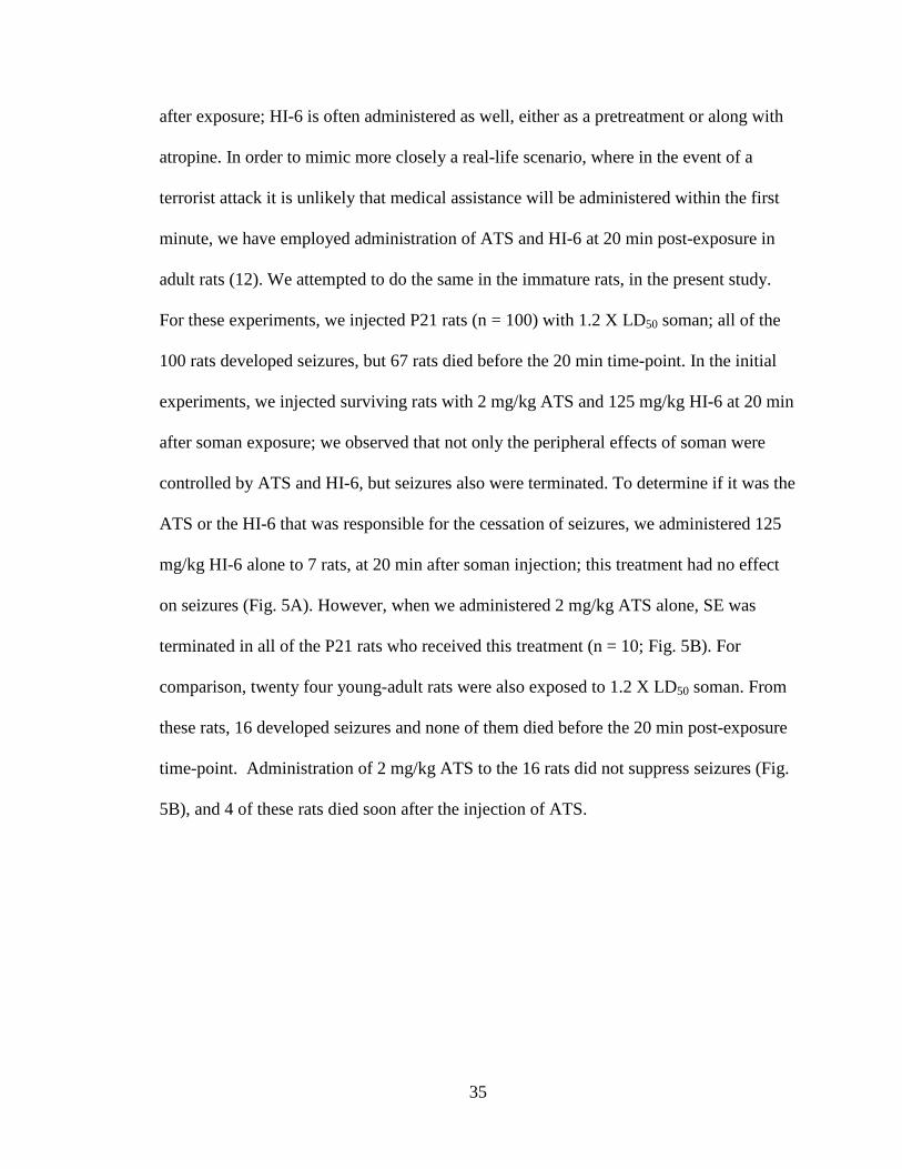
35
after exposure; HI-6 is often administered as well, either as a pretreatment or along with
atropine. In order to mimic more closely a real-life scenario, where in the event of a
terrorist attack it is unlikely that medical assistance will be administered within the first
minute, we have employed administration of ATS and HI-6 at 20 min post-exposure in
adult rats (12). We attempted to do the same in the immature rats, in the present study.
For these experiments, we injected P21 rats (n = 100) with 1.2 X LD50 soman; all of the
100 rats developed seizures, but 67 rats died before the 20 min time-point. In the initial
experiments, we injected surviving rats with 2 mg/kg ATS and 125 mg/kg HI-6 at 20 min
after soman exposure; we observed that not only the peripheral effects of soman were
controlled by ATS and HI-6, but seizures also were terminated. To determine if it was the
ATS or the HI-6 that was responsible for the cessation of seizures, we administered 125
mg/kg HI-6 alone to 7 rats, at 20 min after soman injection; this treatment had no effect
on seizures (Fig. 5A). However, when we administered 2 mg/kg ATS alone, SE was
terminated in all of the P21 rats who received this treatment (n = 10; Fig. 5B). For
comparison, twenty four young-adult rats were also exposed to 1.2 X LD50 soman. From
these rats, 16 developed seizures and none of them died before the 20 min post-exposure
time-point. Administration of 2 mg/kg ATS to the 16 rats did not suppress seizures (Fig.
5B), and 4 of these rats died soon after the injection of ATS.
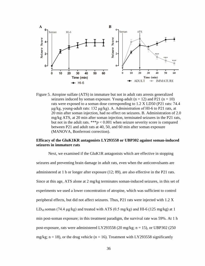
36
Figure 5. Atropine sulfate (ATS) in immature but not in adult rats arrests generalized seizures induced by soman exposure. Young-adult (n = 12) and P21 (n = 10) rats were exposed to a soman dose corresponding to 1.2 X LD50 (P21 rats: 74.4 μg/kg, young-adult rats: 132 μg/kg). A. Administration of HI-6 to P21 rats, at 20 min after soman injection, had no effect on seizures. B. Administration of 2.0 mg/kg ATS, at 20 min after soman injection, terminated seizures in the P21 rats, but not in the adult rats. ***p < 0.001 when seizure severity score is compared between P21 and adult rats at 40, 50, and 60 min after soman exposure (MANOVA, Bonferroni correction).
Efficacy of the GluK1KR antagonists LY293558 or UBP302 against soman-induced seizures in immature rats
Next, we examined if the GluK1R antagonists which are effective in stopping
seizures and preventing brain damage in adult rats, even when the anticonvulsants are
administered at 1 h or longer after exposure (12; 89), are also effective in the P21 rats.
Since at this age, ATS alone at 2 mg/kg terminates soman-induced seizures, in this set of
experiments we used a lower concentration of atropine, which was sufficient to control
peripheral effects, but did not affect seizures. Thus, P21 rats were injected with 1.2 X
LD50 soman (74.4 μg/kg) and treated with ATS (0.5 mg/kg) and HI-6 (125 mg/kg) at 1
min post-soman exposure; in this treatment paradigm, the survival rate was 59%. At 1 h
post-exposure, rats were administered LY293558 (20 mg/kg; n = 15), or UBP302 (250
mg/kg; n = 18), or the drug vehicle (n = 16). Treatment with LY293558 significantly

37
suppressed seizures within 20 min, while UBP302 also significantly suppressed seizures
but with a slower time course (p < 0.001; Fig. 6). In the rats who received only the drug
vehicle, SE continued for at least the 2 h of observation.
Figure 6. Delayed post-treatment with the GluK1R antagonists LY293558 or UBP302
arrests soman-induced seizures in P21 rats. Rats were exposed to 1.2 X LD50 soman (74.4 μg/kg) and treated with ATS (0.5 mg/kg) and HI-6 (125 mg/kg) at 1 min post-exposure. At 1 hr post-exposure, rats received LY293558 (20 mg/kg; n = 15), or UBP302 (250 mg/kg; n = 18), or the drug vehicle (n = 16). ***p < 0.001 for the difference in seizure score of the LY293558-treated and UBP302-treated groups compared to the vehicle-treated group (MANOVA, Bonferroni post-hoc test).
Immature rats do not undergo neuronal degeneration after soman-induced SE
In adult rats, nerve agent-induced SE causes significant neuronal degeneration in
many brain regions (12; 14; 88; 89) and particularly in the amygdala and the
hippocampus (14). In the immature rats, the effects of the soman-induced SE on neuronal
degeneration was examined at 24 h and 7 days after soman exposure in the group that
received 0.5 mg/kg ATS and 125 mg/kg HI-6 at 1 min post-exposure, but no
anticonvulsant treatment. There was no evidence for neuronal degeneration at either time

38
point, in the amygdala or the hippocampus; we also inspected the rest of the brain on the
FJC-stained sections, throughout the rostro-caudal extent, but, as in control rats, there
were no FJC-stained cells in any brain region. To ensure that the staining procedure
worked properly, we also processed sections from adult rats who had been exposed to
1.2XLD50 soman, receiving 2 mg/kg ATS and 125 mg/kg HI-6 at 1 min after exposure,
but no anticonvulsant treatment; neurodegeneration was pronounced in these rats (Fig. 7).
Figure 7. Immature rats do not suffer neuronal degeneration, 1 day and 7 days after
soman-induced SE. A. Cresyl violet photomicrographs outline the brain regions (amygdala in red; hippocampus in yellow) from where the FJC photomicrographs (B and C) were taken (the specific areas shown in the photomicrographs are outlined with black rectangles). Immature and adult rats were exposed to the age-specific 1.2X LD50 of soman. In contrast to the adult rats (C), immature rats (B) did not display any degenerating cells. Magnification in A is 200x. Scale bar (for B and C) is 50 μm.
Amygdalar and hippocampal volume is significantly reduced 30 days and 90 days after exposure of immature rats to soman, and this is prevented by LY293558 treatment
Despite the lack of neuronal degeneration after soman-induced SE, volumetric
measurements of the amygdala and the hippocampus, 30 and 90 days after soman
exposure, indicated that the size of these structures was reduced in the soman-exposed

39
rats that received only ATS (0.5 mg/kg) and HI-6 at 1 min after exposure, but no
anticonvulsant treatment. This type of pathology was not observed in the group that
received LY293558 at 1 h after soman exposure (Figs. 8 and 9). Thus, at 30 days post-
exposure, the size of the amygdala in the soman-exposed rats was 10.13 ± 0.49 µm3 (n =
8), while the control size was 11.86 ± 0.27 mm3 (n = 8, p < 0.05); in the soman-exposed
rats treated with LY293558, the size of the amygdala (11.72 ± 0.3; n = 8) did not differ
from the control (p = 0.08; Fig 8D). At 90 days post-exposure, the size of the amygdala
in the soman group was 12.10 ± 0.32 mm3 (n = 8), which was still significantly smaller
than the control (p < 0.05); in the LY293558-treated group the size of the amygdala
(11.98 ± 0.27, n = 8) did not differ from the control (p = 0.07; Fig. 8E). The size of the
hippocampus at 30 days post-exposure was 48.4 ± 2.2 mm3 in the soman-exposed group
(n = 8) versus 62.5 ± 1.7 mm3 in the control group (n = 8, p < 0.05); in the soman-
exposed rats treated with LY293558, the size of the hippocampus (59.8 ± 2.3, n = 8) did
not differ from the control (p = 0.09; Fig. 9D). At 90 days post-exposure, the
hippocampus was still significantly smaller in the soman group (41.2 ± 2.1, n = 8)
compared to control (p < 0.05); in the LY293558-treated group the size of the
hippocampus (62.4 ± 2.6, n = 8) again did not differ from the control (p = 0.08; Fig. 9E).

40
Figure 8. A reduction in amygdalar volume, 30 and 90 days after soman exposure, is
prevented by LY293558 treatment. A, B, C. Tracings of the amygdala in series of slices (left) and representative photomicrographs (right) from control animals (A, n = 8), soman-exposed animals that received only ATS (0.5 mg/kg) and HI-6 at 1 min post-exposure (B, n = 8), and soman-exposed animals that received LY293558 (20 mg/kg) at 1 h after soman injection (C, n = 8). D. Group data showing the estimated volume of the amygdala for all three groups, 30 days after the exposure. E. Group data showing the estimated volume of the amygdala for all three groups, 90 days after the exposure. *p < 0.05 (ANOVA, LSD post-hoc test).
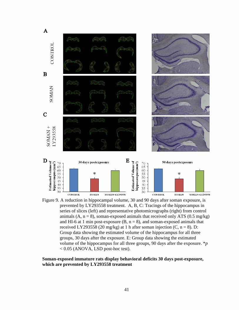
41
Figure 9. A reduction in hippocampal volume, 30 and 90 days after soman exposure, is
prevented by LY293558 treatment. A, B, C: Tracings of the hippocampus in series of slices (left) and representative photomicrographs (right) from control animals (A, n = 8), soman-exposed animals that received only ATS (0.5 mg/kg) and HI-6 at 1 min post-exposure (B, n = 8), and soman-exposed animals that received LY293558 (20 mg/kg) at 1 h after soman injection (C, n = 8). D: Group data showing the estimated volume of the hippocampus for all three groups, 30 days after the exposure. E: Group data showing the estimated volume of the hippocampus for all three groups, 90 days after the exposure. *p < 0.05 (ANOVA, LSD post-hoc test).
Soman-exposed immature rats display behavioral deficits 30 days post-exposure, which are prevented by LY293558 treatment
42
P21 rats that were exposed to soman and received only ATS (0.5 mg/kg) and HI-6
at 1 min after exposure, along with soman-exposed rats that were also treated with
LY293558 at 1 h after exposure, as well as control rats, were tested for context- and cue-
dependent fear conditioning. Fear conditioning took place at 30 days after exposure, and
testing for context-dependent and cue-dependent acquisition of fear took place within the
following 4 days (see methods). In the context-dependent test, the total freezing time for
the soman-exposed rats was 250 ± 15 s (n = 13), significantly lower than that of the
control group (523 ± 24 s, n = 17, p <0.05); for the soman-exposed rats treated with
LY293558, the total freezing time (452 ± 28 s, n = 11) did not differ from the control (p =
0.07, Fig.10A). In the cue-dependent fear acquisition test, the freezing time for the
soman-exposed rats (214 ± 14 s) was significant lower compared to control (354 ± 20 s, p
< 0.05), while for the soman-exposed rats that received LY293558, the freezing time (331
± 21 s) did not differ from the control (p = 0.08; Fig. 10C). At 90 days post-exposure,
there were no differences among the groups (Fig. 10B and D). Thus, the total freezing
time in the context-dependent test was 489 ± 20 s for the soman-exposed group, 554 ± 35
s for the control group, and 513 ± 29 s for the soman-exposed rats treated with LY293558
(p = 0.084). The total freezing time in the cue-dependent test was also similar among the
groups (p = 0.09). Thus, freezing time was 301 ± 40 s for the soman-exposed rats, 389 ±
25 s for the control rats, and 391 ± 19 s for the soman-exposed, LY293558-treated rats.
The same groups of rats were also tested in the open field. At 30 days after soman
exposure the distance traveled in the open field was not significantly different between
the soman-exposed rats (2522 ± 246 cm, n = 13), the soman-exposed rats that received
LY293558 (2741 ± 254 cm, n = 11) and the controls (2293 ± 158 cm, n = 17). However,

43
the time spent in the center of the open field was significantly less in the soman-exposed
rats that did not receive anticonvulsant treatment (8.6 ± 1.06 % of the total movement
time, n = 13, p = 0.02) compared to the time spent by the control rats (12.51 ± 1.12 % of
the total movement time, n = 17), or the soman-exposed that were treated with LY293558
(11.95 ± 1.08% of the total movement time, n = 11; Fig. 10E). At 90 days after exposure,
the distance traveled in the open field was not significantly different between the soman-
exposed rats (2851 ± 193 cm, n= 11), the soman-exposed rats that received LY293558
(2698 ± 244 cm, n = 12) and the controls (2752 ± 201 cm, n = 12). The time spent in the
center of the open field also did not differ significantly between the soman-exposed rats
that did not receive anticonvulsant treatment (11.7 ± 1.25 % of the total movement time,
n = 11) and the control rats (13.36 ± 1.03% of the total movement time, n = 12), or the
soman-exposed, LY293558-treated rats (13.68 ± 1.08% of the total movement time, n =
12, p = 0.08; Fig. 10F).

44
Figure 10. LY293558 treatment prevents behavioral deficits 30 and 90 days after soman
exposure. A and B show that total freezing time of the contextual fear-conditioned responses, 30 and 90 days after soman exposure, for the soman-exposed rats who received only ATS (0.5 mg/kg) and HI-6 at 1 min after exposure (n = 13), similarly treated rats who received also LY293558 at 1 h after soman exposure (n = 11), and controls (n = 17). C and D show the total freezing time for the auditory fear-conditioned responses, for the same groups, at 30 and 90 days after exposure. E and F are the results from the open field test, showing the time spent in the center, for the same three groups, at 30 and 90 days after exposure. *p < 0.05 (ANOVA, Dunnett post-hoc test).
DISCUSSION
The present study shows that 1) immature rats appear to be more susceptible to
the toxicity of nerve agents, as suggested by the LD50 of soman for the P21 rats, which
was lower than the LD50 for the adult rats; 2) immature rats are more prone to generating
seizures after nerve agent exposure, as suggested by the time to seizure onset which was
significantly shorter in the P21 rats compared to the adults; 3) as in adult rats, reduction
of brain AChE activity appears to be the primary mechanism of seizure generation in the

45
P21 rats, and the BLA plays a key role in that respect; 4) ATS, at a dose that does not
affect nerve agent-induced seizures in adult rats, promptly terminates seizures in P21 rats;
5) the GluK1 antagonists LY293558 and UBP302 are effective in terminating soman-
induced seizures in P21 rats, even when administered at 1 h after exposure; 6) soman-
induced seizures do not cause neuronal degeneration in the brain of P21 rats, but they
reduce the volume of the amygdala and the hippocampus, 30 and 90 days after exposure;
7) soman exposure results in fear-learning deficits and increased anxiety at 30 days after
exposure; 8) both the brain pathology and the behavioral deficits are prevented if exposed
rats are administered LY293558 at 1 h after exposure.
Since nerve agents induce seizures by inhibiting AChE, it seems reasonable to
speculate that the faster onset of seizures upon exposure to soman in the P21 rats
compared to adults is due to the lower concentration of the enzyme in some brain regions
(Fig. 3), which may result in more rapid reduction of AChE activity in the brain. This is
not a very plausible explanation, however, for two reasons: a) AChE activity was also
dramatically reduced in the prelimbic cortex, the piriform cortex, and the hippocampus of
the P21 rats that did not develop SE; the only difference of these rats from those who
developed SE was that AChE in their BLA was reduced to a lesser extent (Fig. 4). b)
AChE activity in the BLA, which appears to play a key role in the induction of seizures
in the P21 rats, as in the adult rats (227), did not differ significantly between the two ages
in the basal state (Fig. 3). Therefore, the shorter latency to seizure onset in the P21 rats is
likely due to reasons that are not related to the baseline concentration of AChE in the
brain. It is well recognized that the immature brain has a greater propensity for seizure
generation regardless of the triggering stimulus, in both humans and animals (112; 120).

46
The reasons are not quite clear, but may include the high input resistance of neurons
during development and the prevalence of gap junctions‒which facilitate bursting and
synchronous activity‒and the immaturity of the GABAergic and glutamatergic system
(30; 120). Contribution of possible differences between P21 and adult rats in the
pharmacokinetics of the injected soman must also be considered.
An unexpected finding in the present study was that ATS, at 2 mg/kg, a
concentration that has no effect on nerve agent-induced seizures in adult animals (Fig. 5B
and (12; 89; 181; 229; 260)), terminated soman-induced seizures in the P21 rats. After
nerve agent exposure, seizures are initiated by hyperstimulation of muscarinic receptors,
as evidenced by the anti-seizure effects of muscarinic antagonists when administered
shortly after exposure (152; 260; 261). In adult rats, ATS, a non-selective muscarinic
antagonist, suppresses nerve agent-induced seizures, but with an ED50 of more than 50
mg/kg (261). The BBB is weaker early in life (33; 173; 244) and appears to be more
susceptible to increased capillary leakiness induced by organophosphorus agents (271);
this may explain the anti-seizure efficacy of relatively low concentrations of ATS in the
immature rats. However, a weaker BBB in the P21 rats was not sufficient to confer
efficacy to HI-6, which had no effect on seizures when administered at 20 min after
exposure (Fig. 5A); it is likely though that the inefficacy of HI-6 had to do with the rapid
aging of soman-inhibited AChE (168; 265; 282), which renders it non-reactivatable by
oximes (24; 168). In the rat whole brain, the activity of choline acetyltransferase, the
concentration of ACh, and the number of mAChRs do not mature until after the fourth
postnatal week (75). Therefore, in addition to a weaker BBB, differences between the

47
P21 and the adult rats in the function of the muscarinic cholinergic system may be
involved in the differential anticonvulsant response to ATS.
Seizures did not return after the cessation of SE by 2 mg/kg ATS, at least for the
two hours that we continued observing the rats. We did not examine, in the present study,
whether ATS would also be effective if administered at 1 h after exposure or at more
delayed time points. However, from studies in adult rats, it has become clear that
muscarinic receptors are involved only in the initiation of seizures after exposure, while it
is excessive glutamatergic activity that reinforces and sustains SE (181). Accordingly,
soman-induced SE can be terminated by certain glutamate receptor antagonists.
Specifically, LY293558, an AMPA/GluK1R antagonist (36; 124), or UBP302, a GluK1R
antagonist (190), terminate soman-induced seizures even when administerd at 1 h or 2 h
after exposure (13; 89). The efficacy of these two compounds may lie on the fact that
GluK1Rs play an important role in the regulation of neuronal excitability of at least two
highly seizure-prone regions, the amygdala (15; 16; 18; 42; 237) and the hippocampus
(54; 99; 225; 242); both GABAergic and glutamatergic synaptic transmission is
modulated by GluK1Rs in different brain regions (124), but the net effect of GluK1R
activation appears to be an increase in excitability in both the BLA (17) and the
hippocampus (269). During development, there are changes taking place in the
distribution (22) and the function of GluK1Rs (249). Although it is unclear how the
fuction of GluK1Rs may differ in the brain of a P21 rat from an adult rat, the present
study shows that in P21 rats, LY293558 and UBP302 are effective in stopping soman-
induced seizures when administered at 1 h after exposure, with UBP302 having a slower
time course of action, as we had observed in the adult rats (13).

48
It is becoming increasingly understood that prolonged seizures early in life may
not cause neuronal death/loss, yet, they produce long-term pathological alterations,
inhibiting brain growth, modifying neuronal circuits, and leading to lasting behavioral
deficits (119; 121; 276). The present study shows that this is also the case when
prolonged seizures are induced by soman exposure in P21 rats. There was no irreversible
neuronal degeneration at either 24 h or 7 days post-exposure, suggesting that there was
no neuronal loss. This is in contrast to the severe neuronal degeneration that is seen in
young-adult rats at 24 h and 7 days after soman exposure (12-14; 227; 228). The
resistance of immature neurons to seizure-induced degeneration and death is attributed in
part to their lower vulnerability to oxidative stress (214) and glutamate toxicity (35; 165;
167). However, despite the absence of neuronal degeneration, the volumes of the
amygdala and the hippocampus were significantly smaller in the P21 rats than in control
rats, at 30 days and 90 days post-exposure. Considering that prolonged seizures induce
neurogenesis in the hippocampus (121; 210) and, at least in some models, in the
amygdala as well (212), it may seem surprising that the volume of these structures
decreased. However, significant atrophy of temporal lobe structures has also been
observed after P12 and P25 rats were subjected to lithium/pilocarpine-induced SE (148).
Furthermore, amygdalar and hippocampal atrophy is a common pathology in temporal
lobe epilepsy (16; 56; 57), and a smaller amygdala volume was also found in a significant
number of the victims of the sarin attack in Tokyo, in 1995 (238). It appears, therefore,
that prolonged seizures or chronic recurrent seizures can result in atrophy of temporal
lobe structures, and when prolonged seizures occur early in life, atrophy may ensue
despite the absence of neuronal loss; in this latter case, perhaps a smaller number or

49
thickness of synaptic connections contribute to the reduction in the total volume, and/or
death of glia cells.
Abnormalities in the amygdala and the hippocampus may also explain the
behavioral deficits seen at 30 days after exposure. The amygdala plays a key role in fear-
conditioning (127), and the ability for “fear-learning”, as reflected in the total freezing
time, was impaired in the rats exposed to soman at P21. The amygdala, but the
hippocampus as well, also play a central role in anxiety (5; 84; 149), and anxiety-like
behavior was increased 30 days after exposure of P21 rats to soman. These behavioral
deficits were absent 2 months later, despite that there was no reversal of the amygdalar
and hippocampal atrophy. It is possible that during the 2-month-period, which
corresponds to more than 8 human years (10; 250), synaptic or biochemical adjustments
rectified these behavioral deficits. Others have found persistent cognitive deficits, as
revealed by the Morris water-maze test, 3 months after P12 rats and P25 rats experienced
lithium/pilocarpine-induced SE (148). Thus, whether or not recovery will occur may
depend, in addition to age, on other factors such as the nature of the behavioral deficit.
For example, in adult mice exposed to soman-induced SE, increased anxiety-like
behavior was still present 90 days after the exposure, but deficits in auditory and
contextual fear-conditioned responses recovered during the 30 to 90 day post-exposure
period (74).
The present study provides support to the increasing evidence that prolonged
seizures early in life may not be benign in long-term, even when they have not produced
irreversible neuronal degeneration. The data suggest that children are likely to be more
prone to developing seizures upon exposure to nerve agents, but a single injection of

50
atropine will be sufficient to halt seizures, if administered timely. Control of seizures is
necessary in order to prevent long-term brain pathologies and behavioral deficits, and
administration of LY293558 is an effective anticonvulsant and neuroprotectant even
when administered at 1 h after exposure. Testing of anticonvulsants at delayed time-
points after nerve agent exposure in immature animal models requires timely
administration of ATS at a low dose, which will alleviate peripheral toxicity and reduce
mortality, without preventing or halting seizures.
ACKNOWLEDGMENTS
We thank Dr. Cara H. Olsen for her expert advice on the procedure for the
estimation of LD50. This work was supported by the CounterACT Program, National
Institutes of Health, Office of the Director and the National Institute of Neurologic
Disorders and Stroke [Grant Number 5U01NS058162-07].
The authors have no competing financial interests.
The views expressed in this article are those of the authors and do not reflect the
official policy of the Department of Army, Department of Defense, or the U.S.
Government.

51
CHAPTER 3: M1 Muscarinic Receptors Play a Key Role in Seizure Initiation After Exposure to Paraoxon or Soman: Mechanisms of Their
Involvement
Steven L. Miller1,3, Vassiliki Aroniadou-Anderjaska1,2,3, Volodymyr I. Pidoplichko1, Taiza H. Figueiredo1, James P. Apland4, Maria F. M. Braga1,2,3*
1Department of Anatomy, Physiology and Genetics, 2Department of Psychiatry, and
3Program in Neuroscience, F. Edward Hébert School of Medicine, Uniformed Services University of the Health Sciences, Bethesda, MD, 20814, USA
4Neurotoxicology Branch, U.S. Army Medical Research Institute of Chemical Defense, Aberdeen Proving Ground, Maryland, 21010, USA
*Corresponding Author: Maria F.M. Braga, D.D.S., Ph.D. Department of Anatomy, Physiology, and Genetics F. Edward Hébert School of Medicine, Uniformed Services University of the Health Sciences 4301 Jones Bridge Road Bethesda, MD 20814, USA Phone: (301) 295-3524 Fax: (301) 295-3566 Email: [email protected]

52
ABSTRACT
BACKGROUND AND PURPOSE
Exposure to OP toxins induces seizures and SE. The evidence is clear that
seizures are initiated by hyperstimulation of mAChR, subsequent to inhibition of AChE
by the OP; this is followed by glutamatergic hyperactivity, which reinforces and sustains
seizures. It is unknown, however, which muscarinic receptor subtypes are involved in
seizure initiation, or how their stimulation leads to hyperexcitability.
EXPERIMENTAL APPROACH
We tested whether a selective M1 mAChR antagonist, VU0255035, prevents the
development of SE after exposure of rats to POX or soman. In brain slices, we studied the
effects of POX on inhibitory and excitatory activity in the BLA, and the involvement of
M1 mAChRs.
KEY RESULTS
Pretreatment of the rats with VU0255035 significantly suppressed the intensity of
seizures after POX or soman exposure. In the basolateral amygdala, bath application of
POX elicited a barrage of spontaneous inhibitory postsynaptic currents, lasting about 1
min, and followed by an increase in the ratio of the total charge carried by spontaneous
glutamatergic postsynaptic currents over GABAergic currents. The effects of POX were
prevented by pretreatment of the slices with atropine or VU0255035.
CONCLUSIONS AND IMPLICATIONS: Previous studies have pointed out
that the basolateral amygdala plays a key role in seizure initiation after OP exposure. The
present study provides an insight into the mechanisms involved. Activation of M1
receptors tips the balance between GABAergic and glutamatergic activity. The resulting

53
hyperexcitability provides the background over which seizures commence and propagate
to other brain regions.

54
INTRODUCTION
It is well understood that OPs‒used as insecticides or in chemical warfare (nerve
agents)‒induce seizures by hyperstimulation of mAChRs. Thus, phosphorylation of
AChE by these toxins inactivates the enzyme (265), resulting in a built-up of synaptic
and extrasynaptic ACh. Although both nAChRs and mAChRs are probably activated by
the elevated acetylcholine, it is the activation of mAChRs that raises excitatory activity in
neuronal networks and initiates seizures. Evidence for that is provided by studies
showing that mAChR antagonists but not nAChR antagonists prevent the induction of
seizures if administered before OP exposure (19; 111; 179; 259; 303), and can arrest
seizures if administered shortly after exposure (9; 50; 178). mAChR antagonists are
ineffective if administered at time points longer than 10 to 20 min after seizure onset
(181) because mAChR activation is involved only in the initiation of seizures, and it is
the imbalance between GABAergic and glutamatergic activity that reinforces and
sustains seizures (77; 125; 181; 303). Little is known, however, about the mechanisms
involved in the transition from mAChR hyperstimulation to glutamatergic
hyperexcitation, or which mAChR subtypes mediate the triggering of hyperactivity.
Uncovering these mechanisms may allow the development of more selective and safer
pharmacological means to prevent the development of SE after OP exposure.
There are five subtypes of mAChRs, M1 to M5. They all are G-protein coupled;
M2 and M4 are preferentially coupled to the Gi class of G proteins, while M1, M3, and
M5 are coupled to the Gq family (147; 305). Although it is unknown which of these
receptor subtypes are primarily involved in seizure initiation after OP exposure, seizures
induced by convulsive doses of the mAChR agonist pilocarpine are dependent on

55
M1 receptor activation (47; 109; 137; 252). Since both pilocarpine and OPs induce
seizures by mAChR hyperstimulation, it is possible that the M1 receptor also mediates
seizure initiation by OPs. As there is now a new, selective M1 receptor antagonist
available (252), in the present study we examined if the M1 receptors are involved in the
generation of seizures by exposure to POX (O,O-diethyl O-p-nitrophenyl phosphate), the
active metabolite of the insecticide parathion (95), or exposure to the nerve agent soman.
To begin to understand how mAChR activation by an OP leads to hyperexcitability of
neuronal networks and seizure initiation, we also examined, in brain slices, the effects of
POX on spontaneous GABAergic and glutamatergic activity, and whether M1 receptors
mediate these effects. These in vitro studies were performed in the BLA, a seizure-prone
brain region (16) that plays a key role in the initiation of seizures by nerve agent exposure
(179; 227).
METHODS
Animals
Male, Sprague Dawley rats (Charles River Laboratories, Wilmington, MA) were
housed in an environmentally controlled room (20 –23°C, 12 hr light/dark cycle, lights on
06:00 A.M.), with food and water available ad libitum. For the in vivo experiments, adult
rats were pair-housed upon arrival. For the in vitro electrophysiology experiments,
animals were housed with a surrogate mother until they were 21 days-old. After 21 days
of age, all rats were pair-housed. The animal care and use programs at the Uniformed
Services University of the Health Sciences and the U.S. Army Medical Research Institute
of Chemical Defense are accredited by the Association for Assessment and Accreditation
of Laboratory Animal Care International. All animal experiments were conducted

56
following the Guide for the Care and Use of Laboratory Animals, by the Institute of
Laboratory Animal Resources, the National Research Council USA, and were approved
by the Institutional Animal Care and Use Committees of both of our institutions.
POX and soman exposure
Ten-week-old rats were administered the selective M1 receptor antagonist
VU0255035 (25 mg.kg-1, i.p.; Tocris Bioscience, Ellisville, MO) or the vehicle (DMSO,
1 ml.kg-1, i.p.; Sigma-Aldrich, St Louis, MO), 15 min before exposure to POX
(paraoxon-ethyl; Sigma-Aldrich, St Louis, MO) or soman (pinacoyl
methylphosphonofluoridate; obtained from the Edgewood Chemical Biological Center,
Aberdeen Proving Ground, Edgewood, MD). For the POX experiments, animals were
subcutaneously injected with 4 mg.kg-1 POX. In order to control the peripheral
cholinergic effects of POX exposure, the rats were intramuscularly injected with 2 mg.kg-
1 ATS (a non-selective mAChR antagonist that has no effect on seizures at this
concentration; Sigma-Aldrich, St Louis, MO) and 50 mg.kg-1 2-PAM (Sigma-Aldrich, St
Louis, MO), within 1 min after POX injection; this drug administration protocol
replicates the conditions of the DeLorenzo rat survival model of POX intoxication (79).
For the soman experiments, animals were subcutaneously injected with 1.8 X LD50
soman (198 μg.kg-1, diluted in cold physiological saline), and within 1 min after soman
injection, the rats received 2 mg/kg atropine methylnitrate (i.m.), a non-selective mAChR
antagonist that does not cross the blood-brain barrier (Wedgewood Pharmacy,
Swedesboro, NJ). Seizures were monitored behaviorally, and their severity was scored
for 1 h, using a modified version of the Racine scale (232), as we have described

57
previously (89). Behavioral seizures above stage 3, correspond to electrographically
recorded status epilepticus (76; 89).
Electrophysiology
Rats, 18 to 25 days-old, were anesthetized with isoflurane prior to decapitation for
slice preparation. Coronal slices (400 μm thick) containing the amygdala were cut as
described previously (17). Recording solution (artificial cerebrospinal fluid, ACSF)
consisted of the following (in mM): 125 NaCl, 2.5 KCl, 1.25 NaH2PO4, 21 NaHCO3, 2
CaCl2, 1 MgCl2, and 11 D-glucose. All solutions were saturated with 95% O2, 5% CO2 to
achieve a pH near 7.4. The slice chamber (0.7 ml capacity) had continuously flowing
ACSF (~8 ml/min) at a temperature of 31–32°C. The osmolarity of the external solution
was adjusted to 325 mOsm with D-glucose. Tight-seal (>1 GΩ), whole-cell recordings
from principal neurons in the BLA were performed as described previously (221); BLA
principal cells were identified by their pyramidal shape and the presence of the
hyperpolarization-activated cationic current (Ih; (17)). Borosilicate glass patch electrodes
had a resistance of 3.5-4.5 MΩ when filled with an internal solution of the following
composition (in mM): 60 CsCH3SO3, 60 KCH3SO3, 10 EGTA, 10 HEPES, 5 KCl, 5 Mg-
ATP, and 0.3 Na3GTP, pH 7.2, 290 mOsm. The 5 mM KCl allowed the simultaneous
recordings of spontaneous inhibitory postsynaptic currents (sIPSCs) and excitatory
postsynaptic currents (sEPSCs). Access resistance (15–24 MΩ) was regularly monitored
during recordings, and cells were rejected if the resistance changed by >15% during the
experiment. Ionic currents were amplified and filtered (1 kHz) using the Axopatch 200B
amplifier (Axon Instruments, Foster City, CA) with a four-pole, low-pass Bessel filter,
digitally sampled (up to 2 kHz) using the pClamp 10.2 software (Molecular Devices,

58
Sunnyvale, CA), and further analyzed using the Mini Analysis Program (Synaptosoft,
Fort Lee, NJ) and Origin (OriginLab, Northampton, MA). Drugs used for the in vitro
experiments were paraoxon-ethyl and atropine sulphate, as well as D-AP5, an N-methyl-
D-aspartate receptor antagonist (Tocris Bioscience, Ellisville, MO), SCH50911, a
GABAB receptor antagonist (Tocris Bioscience), and LY341495, a metabotropic
glutamate group II/III receptor antagonist (Tocris Bioscience).
Statistical Analysis
Seizure scores were analyzed using independent-samples Student’s t-test.
Electrophysiological results were analyzed using paired Student’s t-tests. Results were
considered statistically significant when P < 0.05. Data are represented as means ±
standard error of the mean. Sample size “n” refers to the number of animals for the in
vivo experiments and the number of cells for the whole-cell experiments.
RESULTS
Pretreatment with VU0255035 reduced seizure severity following paraoxon exposure
Eighteen rats were randomly divided into two groups: a group that was injected
with VU0255035 (25 mg.kg-1, n = 10) and a group injected with the vehicle (DMSO; 1
ml.kg-1, n = 8), at 15 min before exposure to paraoxon (4 mg.kg-1). All rats developed
seizures, but the seizure severity in the VU0255035-pretreated group (average Racine
score from 5 min to 60 min after exposure = 3.15 ± 0.03) was significantly lower than the

59
seizure severity in the vehicle-pretreated animals (average Racine score = 3.57 ±
0.08, P < 0.01; Fig. 11Α).
Pretreatment with VU0255035 reduced seizure severity following exposure to soman
Eight rats were randomly divided into two groups: a group that was injected with
VU0255035 (25 mg.kg-1, n = 4) and a group injected with the vehicle (DMSO; 1 ml.kg-1,
n = 4), at 15 min before exposure to soman (198 μg.kg-1). All rats developed seizures, but
the seizure severity in the VU0255035-pretreated group (average Racine score = 2.56 ±
0.20) was significantly lower than the seizure severity in the vehicle-pretreated animals
(average Racine score = 4.58 ± 0.74, P < 0.001; Fig. 11Β).
Figure 11. Pretreatment with a selective M1 receptor antagonist, VU0255035, reduces
seizure severity after exposure to paraoxon or soman. Administration of VU0255035, 15 min before exposure to paraoxon (A) or soman (B) reduced the Racine score of seizure severity, averaged over 5 min to 60 min after exposure. For the paraoxon experiments, n = 8 for the vehicle group, and n = 10 for the VU0255035-pretreated group (P < 0.01). For the soman experiments, n = 4 for each group (the vehicle-pretreated and the VU0255035-pretreated group; P < 0.001).
Effects of POX on spontaneous synaptic activity and the role of M1 receptors

60
The in vivo experiments suggested that M1 receptors play a key role in seizure
initiation after exposure to POX or soman. To obtain some insight into the mechanisms
by which M1 receptor activation increases the excitability of neuronal networks
culminating in seizure generation, we examined the effects of POX on spontaneous
inhibitory (GABAergic) and excitatory (glutamatergic) activity in the BLA. Whole-cell
simultaneous recordings of sIPSCs and sEPSCs were obtained from principal BLA
neurons, at Vh of -58 mV, in the presence of D-AP5 (50 μM), SCH50911 (10 μM), and
LY341495 (3 μM); under these conditions, upward currents are mediated by GABAA
receptors (they are blocked by bicuculline), while downward currents are mediated by
AMPA/kainate receptors (they are blocked by CNQX; not shown). Bath application of
POX, at three different concentrations (0.1, 1, and 10 μM) induced a transient barrage of
large-amplitude sIPSCs with a duration of 1.1 ± 0.1 min (n = 21), which was followed by
a lasting enhancement of sEPSCs, in 21 out 34 neurons (Fig. 12A). POX took effect
(appearance of the barrage of sIPSCs) within 2 to 8 min after application, depending on
the concentration (the lower concentration of POX was slower to take effect). To quantify
the relative increase of excitatory synaptic activity by POX, we calculated the charge
transferred by the excitatory and the inhibitory currents, during a 20 s time-window, in
control conditions, and then again after POX had taken effect; the charge transferred, in
picoCoulombs, was calculated as the area delimited by the excitatory or inhibitory
current and the baseline. The ratio of the charge transferred by sEPSCs to the charge
transferred by sIPSCs was increased significantly in the presence of POX. When 0.1 μM
POX was applied, the ratio of sEPSC/sIPSC charge-transferred was increased from 0.6 ±
0.1 in control conditions to 1.4 ± 0.3 at 17 min after POX application (P < 0.05, n = 7).

61
When 1 μM POX was applied, this ratio was increased from 0.6 ± 0.1 in control
conditions to 0.9 ± 0.1 at 9 min after POX application (P < 0.05, n = 4). Finally, when 10
μM POX was applied, the ratio of sEPSC/sIPSC charge-transferred was increased from
0.8 ± 0.1 in control conditions to 1.1 ± 0.1 at 6 min after POX application (P < 0.01, n =
10; Fig. 12B).
Figure 12. Paraoxon, at three different concentrations, enhances both spontaneous inhibitory currents (sIPSCs) and spontaneous excitatory currents (sEPSCs), with a greater, lasting effect on the sEPSCs. Whole cell simultaneous recordings of sIPSCs and sEPSCs were obtained from principal cells in the BLA (Vh = -58). (A) The traces shown are representative examples of the effects of paraoxon at 0.1 μM, 1 μM, and 10 μM. Upward currents are GABAergic and downward currents are glutamatergic. (B) The bar graphs show the ratio of the charge transferred by glutamatergic currents over the charge transferred by GABAergic currents, during a 20 s time-window, in control conditions and after bath application of paraoxon (left graph: at 17 min after paraoxon application; middle graph, at 9 min after paraoxon application; right graph, at 6 min after paraoxon application). *P < 0.05, **P < 0.01.
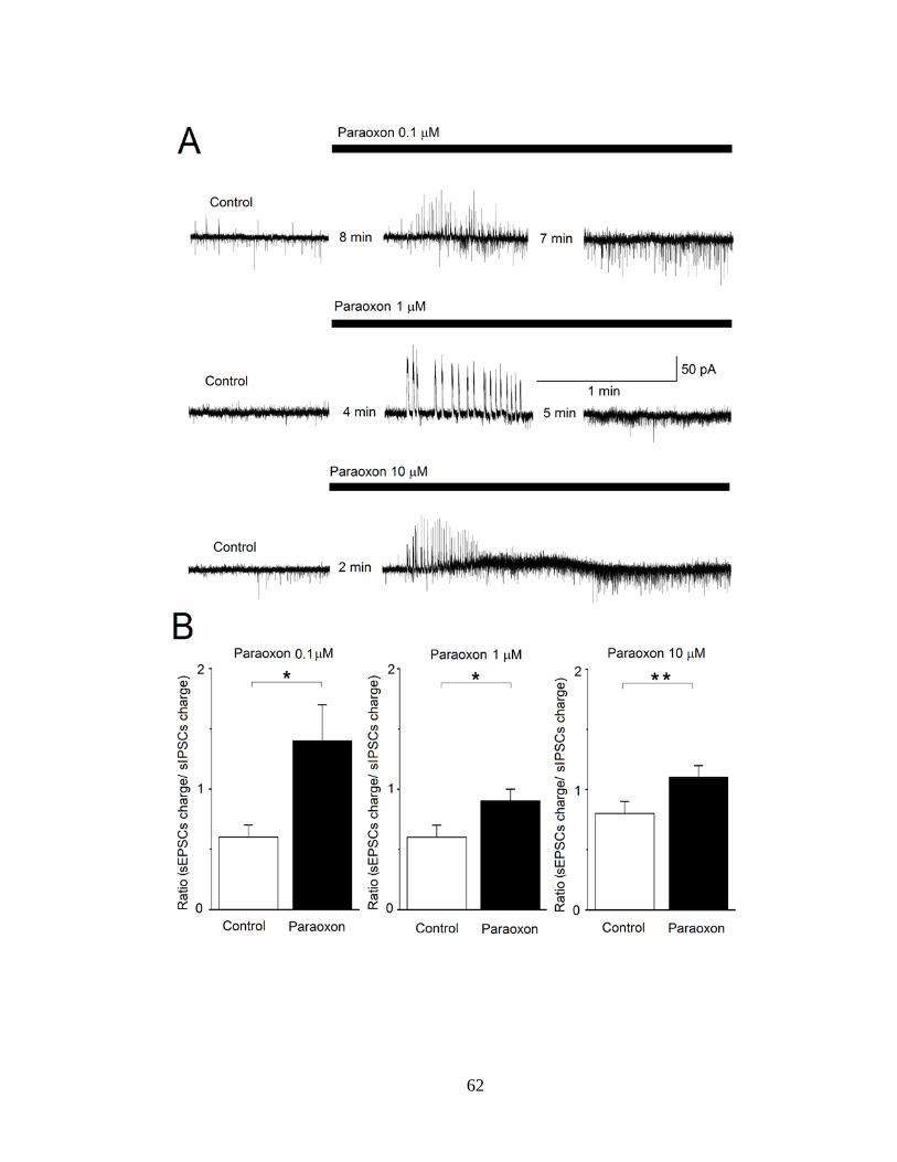
62

63
Next, we pretreated the slices with atropine (1 μM) or VU0255035 (10 μM), for
about 4 min, and then applied 10 μM POX, while recording from principal BLA neurons.
The holding potential was again -58 mV, and D-AP5 (50 μM), SCH50911 (10 μM), and
LY341495 (3 μM) were present in the slice medium. Examples of simultaneous sIPSC
and sEPSC recordings under these pharmacological conditions are shown in Fig. 13A.
Figure 13B shows the effects of 10 μM POX separately on sIPSCs and sEPSCs under the
three conditions: pretreatment with atropine (left bar graph), pretreatment with
VU0255035 (middle bar graph), and when no muscarinic receptor antagonist is present
(right bar graph). The total charge transferred by sIPSCs or sEPSCs during a 20 s time-
window, 10 min after POX application, was expressed as a percentage of the total charge
transferred, during 20 s, in control conditions. When 1 μM atropine was present in the
slice medium, the charge transferred by sIPSCs after 10 min in POX was 72 ± 25% of the
control (n = 7, P = 0.203), while the charge transferred by sEPSCs was 79 ± 23% of the
control (n = 7, P = 0.0441). When 10 μM VU0255035 was present in the slice medium,
the charge transferred by sIPSCs after 10 min in POX was 75 ± 17% of the control (n = 7,
P = 0.1561), while the charge transferred by sEPSCs was 81 ± 16% of the control (n = 7,
P = 0.3892). In contrast, in the absence of mAChR antagonists, the charge transferred by
sIPSCs was increased by POX to 275 ± 57% of the control (n = 10, P = 0.0001), and the
charge transferred by sEPSCs was increased by POX to 435 ± 80% of the control (n = 10,
P = 0.00002). The difference between the increase in sIPSCs and the increase in sEPSCs
was statistically significant (P < 0.001).

64
Figure 13. The effects of paraoxon are prevented when slices are pre-exposed to atropine
or VU0255035. Whole cell simultaneous recordings of sIPSCs and sEPSCs were obtained from principal cells in the BLA (Vh = -58). (A) Representative traces showing that 10 μM paraoxon had no significant effect when applied in the presence of atropine (1 μM, top row of traces) or VU0255035 (10 μM, second row of traces). (Β) Εffects of 10 μM paraoxon on sIPSCs and sEPSCs under three conditions: pretreatment with atropine (left bar graph), pretreatment with VU0255035 (middle bar graph), and in the absence of any muscarinic receptor antagonist (right bar graph). The total charge transferred by sIPSCs or sEPSCs during a 20 s time-window, 10 min after paraoxon application, was expressed as a percentage of the total charge transferred, during 20 s, in control conditions. Only when there was no atropine or VU0255035 in the slice medium, the total charge transferred by sIPSCs and sEPSCs was increased by paraoxon, and the difference between the increase in sIPSCs and the increase in sEPSCs was statistically significant (***P < 0.001).

65
DISCUSSION AND CONCLUSIONS
The present study demonstrated that pretreatment with a selective M1 receptor
antagonist interfered with seizure initiation and significantly decreased seizure severity
after exposure to POX or soman. The in vitro experiments suggested that one of the
mechanisms via which M1 receptors are involved in seizure initiation by OP exposure is
by mediating a derangement in the balance between GABAergic and glutamatergic
activity in the BLA, favoring glutamatergic hyperactivity.
The BLA plays a key role in seizure initiation after OP exposure (15). Thus, when
McDonough et al. (179) microinjected nerve agents into different brain regions, they
observed that convulsions were elicited only when the nerve agent was injected into the
BLA. Recently, Prager et al. (227) found that in the small proportion of rats that do not
develop seizures when exposed to high doses of soman, the activity of
acetylcholinesterase (AChE) is reduced in the hippocampus and the piriform cortex to a
similar extent as in the rats that develop seizures, but to a significantly lesser extent in the
BLA; this suggests a crucial role of AChE reduction in the BLA for seizure initiation.
The importance of the amygdala in nerve agent-induced seizure generation is also
highlighted by the findings that the amygdala displays the earliest increase in
extracellular glutamate after exposure to soman (151), and generates ictal-like discharges
after in vitro application of soman (11). It has remained unclear, however, what are the
synaptic events in the BLA that are triggered by AChE inhibition and lead to
glutamatergic hyperactivity.
The BLA receives a very dense cholinergic innervation from the basal forebrain
(31; 51; 140; 187; 192; 197), and has a higher basal AChE activity in comparison to other

66
brain regions in both adult (31; 227; 228) and 21-day old rats (Miller et al., unpublished).
Cholinergic nerve terminals synapse on both pyramidal cells and interneurons (51; 192;
201), targeting nicotinic and/or muscarinic acetylcholine receptors, which are abundantly
present in the amygdala (117; 171; 192; 248; 281; 293; 319). When acetylcholine levels
rise subsequent to inhibition of AChE by an OP (150), both nicotinic and muscarinic
receptors can be expected to be activated. The lack of contribution of nicotinic receptors
to seizure initiation by OP exposure may relate to the increase of inhibitory rather than
excitatory activity when these receptors are activated in the BLA (224; 319), or to the
rapid desensitization of nicotinic receptors (98) in the persistent presence of elevated
acetylcholine.
From the mAChRs, both the M1 and M2 subtypes are present in the rat BLA (71;
171; 172; 275), as well as the M3 and/or M4 receptors (267), but the M1 receptors
predominate (44). Immunocytochemistry along with electron microscopy have revealed
that M1 receptors in the rat BLA are located on somata, dendritic shafts, and, to a lesser
extent, spines of pyramidal neurons, as well as on aspiny dendrites with morphological
features typical of interneurons (193). In addition, M1 immunoreactivity was found on
presynaptic terminals most of which formed asymmetrical synapses with dendritic spines,
but some of which formed symmetrical synapses with neuronal somata and dendritic
shafts (193). It appears, therefore, that M1 receptors in the rat BLA are in a position to
affect the activity of both glutamatergic and GABAergic neurons, by postsynaptic or
presynaptic mechanisms. Knowledge of their function has been hampered by the lack of
selective M1 agonists and antagonists. The availability of the selective M1 antagonist
VU0255035 (252) has allowed us, in the present study, to discover that these receptors

67
mediate the increase of glutamatergic activity in the BLA upon exposure to POX. The
first event observed when POX took effect was a dramatic but short-lasting increase of
GABAergic inhibitory activity, which did not occur in the presence of atropine or
VU0255035. Whether this was due to direct depolarization‒by the elevated
acetylcholine‒of GABAergic interneurons, facilitation of GABA release from
GABAergic presynaptic terminals, or depolarization of glutamatergic neurons or
terminals synapsing onto inhibitory interneurons cannot be ascertained by the present
study; presynaptic facilitation of GABA release may be the least likely of the three
possibilities, as it has been previously suggested that in the rat lateral amygdala M1
receptors mediate inhibition of GABA release (280). GABAergic activity subsided within
about 1 min, which could imply either that the drive of interneuronal depolarization was
reduced, or the availability of presynaptic GABA-containing vesicles was decreased.
Negative feedback inhibition via GABAB autoreceptors is not a possibility, since
recordings were performed in the presence of a GABAB antagonist. We should note here
that we also performed some experiments in the absence of GABAB, NMDA, and
metabotropic glutamate receptor antagonists, and the sequence of events upon application
of POX was the same as in the presence of the antagonists.
After the initial barrage of sIPSCs subsided, inhibitory activity remained elevated
relative to the baseline, while glutamatergic activity was also increased, significantly
more than the GABAergic; again, these effects were not seen in neurons pretreated with
atropine or VU0255035. The increase in sEPSCs could be the result of M1 receptor-
mediated depolarization of BLA principal neurons; such a depolarizing effect of M1
activation could be due to inhibition of the voltage-dependent K+ current (Im) (307; 308),
68
and/or increase of the mixed Na+/K+ current (Ih) and the Ca++-dependent nonspecific
cation current (91). In addition, M1 receptors on glutamatergic terminals may facilitate
glutamate release; in granule cells of the hippocampus, the frequency and amplitude of
sEPSCs are increased by application of POX via a presynaptic mechanism, and this effect
is blocked by pre-application of atropine (143).
It is possible, therefore, that the M1 receptor-mediated enhancement of
glutamatergic activity over that of GABAergic activity in the BLA network, upon OP
exposure, creates a background of hyperexcitability. Over this background, seizures will
be triggered when the ratio of glutamatergic over GABAergic activity becomes
sufficiently high to reach the seizure threshold for the network, and from the BLA,
seizures will propagate to other brain regions.
ACKNOWLEDMENTS
This work was supported by the CounterACT Program, National Institutes of
Health, Office of the Director and the National Institute of Neurologic Disorders and
Stroke [Grant Number 5U01NS058162-07].
CONFLICT OF INTEREST
None.
The views expressed in this article are those of the authors and do not reflect the
official policy of the Department of Army, Department of Defense, or the U.S.
Government.

69
CHAPTER 4: Discussion
Development of animal models is critical for testing antidotes against nerve agent
intoxication. The FDA has a special rule, referred to as the "Animal Efficacy Rule", for
approving medications for the treatment of chemical, biological, radiological or nuclear
threats. Organizations seeking approval for the treatment of these threats must satisfy the
requirements for this special rule if normal approval mechanisms, requiring human trials,
are not ethical or feasible (270). For example, attacks employing nerve agents do not
occur with high enough frequency to test antidotes against them; additionally, it would
not be ethical to intentionally poison patients with nerve agents simply to test putative
antidotes. However, as the sarin attacks in Syria (81) demonstrate there is still a critical
need for the development of treatments for nerve agent intoxication, and especially nerve
agent-induced seizures in the event that these lethal chemicals are employed in the future.
One of the main primary objectives of this dissertation was to develop a rodent
model relevant to children exposed to nerve agents that reflects the acute and long-term
symptomatology associated with nerve agent exposure and facilitate the testing of
potential therapies to nerve agent-induced seizures. In line with these goals, this
dissertation characterized the following aspects of soman exposure in immature male
rats: the median lethal dose, the time to seizure onset, AChE activity in a variety of brain
structures, and the efficacy of conventional and novel therapies against soman-induced
seizures, as well as the efficacy of the conventional therapy in protecting against the
long-term behavioral and morphological alterations associated with soman exposure.
A second objective of this research is to understand the cellular and molecular
mechanism by which OPs induce, maintain, and reinforce seizure activity. Understanding

70
these mechanisms will help with the identification of novel molecular targets for the
development of more efficacious therapies for seizures induced by these compounds, and
could provide new treatments for patients suffering from temporal lobe epilepsy (77).
Neuropharmacological models have proposed that mAChRs are largely responsible for
seizure onset following OP intoxication but it has been unclear which mAChR subtype is
responsible for the generation of seizure activity (181). Additionally, it has yet to be
elucidated as to how mAChR hyperstimulation can lead to seizure activity following OP
intoxication. As such, we identified the mAChR subtype that plays a critical role in
seizure induction following OP intoxication and the electrophysiological mechanism
underlying its activity.
DEVELOPMENT OF A RAT MODEL FOR TESTING ANTICONVULSANTS AGAINST NERVE AGENT-INDUCED SEIZURES AND ITS RELEVANCE TO THE PEDIATRIC POPULATION
To date, only one study has investigated nerve agent exposure in immature
animals. Sterri and Colleagues (279) found the median lethal dose of soman to be 50
μg/kg (s.c.) for P21 rats. In our model, we calculated the median lethal dose for P21 male
rats to be 62.02 μg/kg. This difference may result from the reduced toxicity of soman in
female compared to male rats as Sterri and Colleagues used a mixed group of male and
female rats for their median lethal dose calculation. The resistance of female rats to the
toxic effects and mortality induced by soman exposure they observed is consistent with
our work. We have found much greater doses are required to elicit seizures or induce
mortality following soman exposure in female rats (unpublished data) and the findings of
Sterri and Colleagues when groups of only female rats were used at different ages (279).
An LD50 of 62.02 μg/kg for P21 male rats is nearly half that of young adult male rats,
only one week older, with an LD50 of 110 μg/kg (126) illustrating the higher sensitivity of
71
immature rats to the toxicity of nerve agents. Consistent with clinical data on children
(113; 119; 120) immature rats were also more susceptible to generating seizures
following exposure to soman. We hypothesized this extremely rapid, generalized seizure
onset following soman exposure was possibly due to a developmental difference in AChE
activity within the BLA given its role in seizure induction following OP exposure (227).
However, we found no difference in AChE activity of the BLA in naïve immature and
adult male rats. Because it is unlikely this rapid seizure onset is due to the levels of AChE
activity in immature rats, we performed a semi-quantitative analysis of publically
available in situ hybridization data from mice of the different mAChR subtypes
throughout development (See Appendix 1) (21; 157). Of note, the M2 mAChR subtype
has not reached adult intensity or density of expression within the BLA as late as P28 in
mice. A reduction, compared to adult levels, in the expression of M2 mAChRs may
prevent an increase in g-protein coupled potassium conductance that is typically
associated with M2 stimulation (45), thereby increasing susceptibility to seizure
induction. Additionally, binding of [3H]3-quinuclidinyl benzilate, a nonspecific mAChR
antagonist, in rat brain suggests that the total mAChR content has not reached adult levels
in a P21 rat and may also explain the rapid seizure onset (75). An incomplete transition of
the excitatory to an inhibitory function of GABA early in life and the subsequent rapid
development of excitatory synapses may also make immature animals and children more
susceptible to seizure induction (30).
Following exposure to 1.2 X LD50 soman, 2.0 mg/kg ATS was found to be a
highly effective anticonvulsant against generalized seizures when administered at 20
minutes post-exposure. This was an unexpected finding as this dose of ATS and the time

72
at which ATS is administered usually has no anticonvulsant efficacy in adult animals
against nerve agent-induced seizures (12; 89; 181; 229; 260). A variety of factors may
help explain the age-dependent efficacy of ATS as an effective anticonvulsant. Ongoing
SE has been found to increase the permeability of the BBB in immature and adult rats
(208); and this permeability is also enhanced by OP exposure (4; 19; 52; 106; 208), even
at subconvulsive doses (208). While OP exposure itself may increase BBB permeability,
thereby increasing the OP content in the central compartment, this is also true for
potential therapies, and this age-dependent increase in BBB permeability following OP
exposure could explain the increase in seizure susceptibility but also in the efficacy of
potential therapies in early development. Further, the reduced total mAChR content in the
developing brain may also explain the rapid seizure onset since it is likely that less ACh
would be required to elicit seizure activity following AChE inhibition. Regardless of the
explanation for the increased efficacy of ATS, our findings suggest that it might be a
highly effective anticonvulsant for children if administered rapidly after seizure onset.
We also investigated the efficacy of the GluK1KR antagonist LY293558 in
arresting soman-induced seizures even when treatment is delayed to 1 h post-exposure in
immature male rats. We performed these experiments to determine whether these
compounds would exert the same efficacy that we have observed in adult rats against
soman-induced seizures. LY293558 treatment, when delayed to 1 hr post-soman
exposure, rapidly arrested ongoing SE and prevented long-term behavioral, pathological
or pathophysiological changes that develop as a result of soman exposure (12; 89; 229).
This potent efficacy as an anticonvulsant against nerve agent-induced SE is likely due to

73
the role of glutamate receptors in maintaining and reinforcing seizure activity at delayed
timepoints post-exposure (181).
Ongoing seizure activity is thought to result in excitotoxic effects and subsequent
cell-death through a variety of downstream mechanisms. Initially, in adult animals, cell
loss occurs over a short time-scale via necrosis, likely due to the large influx of calcium
through NMDA receptors during ongoing SE activity (108; 181). Delayed neuronal
degeneration also occurs and may be observed for months post-exposure and is likely
caused by edema, inflammation and apoptosis (62). This cell death is largely thought to
be responsible for the long-term side effects associated with seizures induced by OPs (62;
67; 70; 74). However, it is unclear what mechanism might underlie the increase in
anxiety-like behavior and impaired fear-learning 1 month following soman-induced
seizures in immature rats as we observed no neuronal degeneration.
Numerous studies suggest that immature animals are resistant to the cell loss that
is typically observed following seizure induction in adult animals (30; 113). The
literature on this topic, as to what age seizures are associated with neuropathology, is
complex and strongly dependent on the method employed to induce seizures, the sex,
age, species and genetic profile of the organism (114). A variety of features of the
immature brain may be neuroprotective following seizures. Superoxide radicals increase
dramatically following seizure induction and may contribute, in part, to neuronal death in
adult animals but no superoxide radical increases are observed following seizures in
immature animals (159; 214). In P30 or P45 Sprague-Dawley rats, seizures induced by
kainic acid increased the inactivation of mitochondrial aconitase, a metric of increased
oxidative stress in mitochondria and in oxidative DNA damage (214). However, the

74
authors of this study did not find this increase in aconitase inactivation when seizures
were induced in P12 or P21 rats, suggesting that immature brains are resistant to
oxidative stress associated with seizure activity (214). Excessive glutamate-receptor
stimulation is thought to be responsible for part of the signaling cascades that lead to cell
death during episodes of SE in adult animals (165; 181). Direct microinjection of equal
amounts of glutamate into the CA1 of P10, P20, P30 and P60 rats results in either
minimal or no cell loss 7 d post-injection but the lesion size increased beyond this age
(165). Brain-derived neurotrophic factor (BDNF) regulates the differentiation, growth
and survival of neurons throughout development (6; 29; 122; 156; 317) and has been
found to be neuroprotective against a variety of insults including excitotoxic injury in the
adult (63). Seizures can modulate the expression of neurotrophins such as BDNF.
Seizures induced by either kainic acid or lithium-pilocarpine in P7 or P12 rats increase
BDNF in a variety of cortical and limbic structures and may play a role in the resistance
to seizure-induced cell loss observed at this age (141). When the increase in expression of
BDNF was blocked prior to seizure induction by kainic acid in P20 rats, neuronal loss
was observed within hippocampus (283). When BDNF expression was not blocked,
kainic acid injection resulted in a twofold increase in BDNF expression and no neuronal
loss was observed (283), strongly suggesting a neuroprotective role for this neurotrophin
early in development. While we did not study this in our model, a variety of these factors
may explain the lack of neurodegeneration following soman-induced seizures in P21 rats.
Despite the lack of neurodegeneration observed in our model, the volumes of both
the amygdala and hippocampus were significantly reduced at 30 and 90 d post-soman
exposure. In structural MRI studies, atrophy of amygdala and hippocampus is a common

75
finding in temporal lobe epilepsy (TLE) patients, and may correlate with a history of
febrile seizures during childhood (32; 55; 57-59; 104; 139; 274). P12 and P25 rats have
also been observed to have reductions in the volume of temporal structures at timepoints
as long as 5 or 7 months following SE induction by lithium-pilocarpine (148). This
finding may also be important in the pathogenesis of the symptoms related to OP
exposure as a reduction in the volume of the amygdala was observed in victims exposed
to sarin during the Tokyo subway attack (238). Notably, this reduction in volume lasted
over a decade (238). Though it is unclear what mechanism is responsible for the
volumetric reductions observed in our study, long-term behavioral impairments are
commonly observed following early life seizures and must be due to a mechanism
unrelated to seizure-induced cell loss.
Given the role of the amygdala and hippocampus in regulating anxiety (5; 84;
149), and their role in cue- and context-dependent fear conditioning, respectively (127;
138; 219); it is expected that pathological damage to these structures would disrupt fear
learning and the regulation of anxiety. 1 month post-soman exposure, rats displayed an
increase in anxiety-like behavior and were unable to undergo fear learning. Though the
volumes of both amygdala and hippocampus were still reduced 3 months post-soman
exposure, fear-learning returned to control conditions as did anxiety-like behavior. This
suggests that between 1 and 3 months post-soman exposure, networks responsible for
mediating anxiety-like behavior and fear learning compensated for the initial damage
induced by SE at P21. Recovery from behavioral impairment was also observed in the
patients exposed to sarin in the Tokyo subway attacks (238; 315); patients that presented
with a reduction in amygdalar volume over a decade following the attack, which had a

76
history of PTSD associated with sarin exposure, no longer presented with PTSD at the
time of the volumetric analysis but amygdala volume was persistently reduced (238).
Despite the suggestion from the clinical literature that seizures in children are
relatively benign, data from these studies suggest that nerve agent-induced seizures can
result in profound, long-term alterations in brain pathology and behavior. Importantly for
mass casualty scenarios, ATS may be very efficacious in arresting nerve agent-induced
seizures in children if treatment is delivered soon after seizure onset. In the event that
first responders cannot address the symptoms resulting from nerve agent exposure,
delayed treatment following seizure onset with LY293558 appears to be effective in
rapidly arresting SE induced by nerve agent exposure in the developing brain. This
treatment is also promising as it was capable of preventing long-term alterations in brain
pathology or behavioral impairments typically associated with nerve agent exposure. This
study establishes a model of nerve agent exposure that displays all of the symptoms of
severe poisoning, clinically relevant long-term symptoms, and has identified a potential
anticonvulsant for nerve-agent induced seizures that is efficacious even when treatment is
delayed; but also allows for the identification of novel therapies for the complex variety
of symptoms that present with OP poisoning.
THE ROLE OF THE M1 MUSCARINIC RECEPTOR IN SEIZURES INDUCED BY ORGANOPHOSPHATES
Understanding the cellular and molecular mechanisms of seizure induction
following OP intoxication will aide in the development of novel treatments that will
reduce or eliminate seizure activity after an OP exposure. Numerous studies indicate that
the BLA plays a key role in facilitating seizure induction following OP poisoning (15).
Moreover, the amygdala is often identified as a seizurogenic structure in TLE and is

77
routinely removed in an effort to reduce seizures (107; 289). In the case of nerve agents, a
variety of studies have suggested its causal role in seizure initiation. When soman or VX
is directly injected into the BLA, motor convulsions are elicited that closely resemble
nerve agent exposure via systemic injection (179). Following systemic injection,
glutamate levels also rapidly rise in the amygdala (151), before other structures, and
sustains the most extensive neuropathology (15), perhaps due to an increase in the
duration of exposure to the excitotoxic effects of glutamate. Recent data from our group
in adult rats (227) and immature rats (Miller et al., unpublished) also suggest a role of the
BLA in seizure induction as it appears that unless almost all of the total pool of AChE
activity is specifically inhibited there, seizure initiation does not occur. McDonough and
Shih (181) have suggested that following AChE inhibition, seizure initiation is mediated
via mAChRs but it is unclear how this may occur.
In this study we found that application of the OP POX to BLA slices initially
resulted in a relative increase in GABAergic inhibitory over glutamergic excitatory
activity. This dramatic increase in GABAA receptor mediated inhibition subsided rapidly,
although there was a persistent increase in sIPSCs following POX application. This may
seem counterintuitive if OP exposure is going to initiate seizures. However, in addition to
the significant increase of sIPSCs, an increase in AMPA receptor mediated sEPSCs was
also observed and prevailed as the dominating driving force to BLA activity as indicated
by a greater sEPSC to sIPSC ratio. This effect of POX on sIPSCs and sEPSCs was
completely blocked by atropine and VU0255035. Indeed, the blockade of the increase in
GABAA receptor mediated sIPSCs or AMPA receptor mediated sEPSCs by POX when
slices were pretreated with either atropine or VU0255035 strongly implicates the

78
mAChRs, but more specifically, the M1 mAChR subtype in facilitating a relative increase
in glutamatergic activity within the BLA following OP exposure. If M1 mAChR
hyperstimulation is required for seizure induction, pretreatment with VU0255035 in vivo
may block or suppress the severity of seizures induced by OP exposure. In agreement
with this hypothesis, we found that when adult rats were pretreated with VU0255035 the
severity of the seizures induced by either POX or soman was reduced compared to
vehicle pretreated controls. While VU0255035 pretreatment may be thought to possibly
block seizure induction, we do not find our data too surprising as this compound is a
competitive antagonist (252) and synaptic ACh levels will continue to rise due to the
irreversible inhibition of AChE; eventually overcoming the M1 mAChR antagonism.
Though our findings strongly implicate the role of the M1 mAChR in seizure induction, it
is unclear how hyperstimulation of this receptor could lead to a prevalence of
glutamatergic excitatory activity within the BLA.
Stimulation of postsynaptic mAChRs in the BLA may facilitate enhanced
excitatory transmission through the blockade of the voltage-dependent Im current (307).
mAChR stimulation in the BLA or hippocampus increases Ih (307) or the Ca++-
dependent nonspecific cation current (91), respectively. Presynaptic mAChRs also been
increase the release of glutamate at synaptic terminals and the combination of all of these
events may drive the BLA network into a more excitable state, eventually reaching a
level that induces seizure activity. The strong role of mAChRs in generating seizures
following OP intoxication suggests the importance of mAChR antagonists, ideally
selective for the M1 mAChR, with higher availability to the central compartment. This is
especially true for the early phase of seizure activity, which is predominantly mediated by

79
mAChR hyperactivity (181). In addition to GluK1KR antagonists, including M1 mAChR
antagonists in the delayed treatment of OP-induced seizure may be important as an
adjunct therapy. Due to the irreversible inhibition of AChE, synaptic concentrations of
ACh are likely to be maintained at high levels until AChE is resynthesized and result in
further M1 mAChR stimulation; promoting further hyperexcitability. Indeed, our group
has found that the drug caramiphen, which has antagonistic properties to the M1 mAChR
and the NMDA receptor, is an effective anticonvulsant against soman-induced seizures in
rats, even when seizures are delayed to 1 hour post-exposure (88). These data highlight
the importance of this receptor for OP-induced seizures and their potential role as target
for treatment.
CONCLUSIONS AND FUTURE DIRECTIONS
OP intoxication constitutes a significant threat to public health and has become
one of the most widely studied class of compounds in toxicological research. While a
dearth of data exists on OPs, an understanding as to how the irreversible inhibition of
AChE can lead to seizures and an associated set of long-term behavioral impairments is
lacking, especially for children. This investigation has yielded a rat model of nerve agent
exposure to facilitate testing of novel anticonvulsants for the pediatric population. We
found that immature rats are much more sensitive than adult rats to the toxic effects of
soman exposure, which produce a long-lasting reduction in the volumes of the amygdala
and hippocampus, lasting up to 3 months post-exposure. This volumetric reduction in the
amydala and hippocampus may underlie the observed increase in anxiety-like behavior
and the inability to undergo fear-learning at 1 months post-exposure. The explanation for
normal behavior at 3 months post-soman exposure is unclear, given the persistent

80
reduction in amygdala and hippocampus, and requires further investigation. Although
immature rats were highly sensitive to the toxic and lethal effects of soman exposure,
they were also very amenable to anticonvulsant therapy. ATS, if administered within 20
minutes post-soman exposure was capable of arresting ongoing seizure activity induced
by soman. LY293558 was also effective in arresting seizures induced by soman, similar
to our previous data in adult rats (12; 89), even if administered 1 hour post-soman
exposure. Additionally, this compound blocked the volumetric reduction in the amygdala
and hippocampus, and prevented the behavioral impairments associated with soman
exposure. This strongly suggests the role of GluK1KR antagonists as an excellent
treatment for arresting seizures induced by OPs and protecting against the long-term side
effects associated with OP exposure.
Further understanding was provided in these studies as to the mechanisms
responsible for seizure induction following OP intoxication. We found that POX
exposure to the BLA facilitates a net shift in network activity towards glutamatergic
excitatory activity, which is mediated by mAChRs, and specifically, the M1 mAChR.
Blockade of the M1 mAChR in vivo prior to POX or soman exposure significantly
suppressed the seizure severity and strongly indicates their importance for seizure
induction following OP poisoning.

81
Appendix 1: Semi-Quantitative Analysis of the Expression of mAChR Subtypes Throughout Development in Mouse Basolateral Amygdala
Using a semi-quantitative scale, mAChR expression was evaluated from sagittal
sections of the BLA throughout development (Figure 14). An analysis of the density and
intensity of gene expression was performed as described in the Allen Institute's white
paper for in situ hybridization on their website (3).
Figure 14. Semi-quantitative analysis of mouse mAChR gene expression. Density and
intensity was analyzed using the semi-quantitative scale published on the website for the Allen Institute for Brain Science (3). A - Representative images of sagittal sections of mouse BLA for each age and gene analyzed in the analysis. B - Quantification of the density and intensity for each age and gene in the analysis. Chrm5 was not detected in the analysis. "Chrm" designation indicates mouse gene names (e.g. Chrm1 refers to mAChR subtype 1).

82
Appendix 2: ASIC1a Activation Enhances Inhibition in the Basolateral Amygdala and Reduces Anxiety
Volodymyr I. Pidoplichko1, Vassiliki Aroniadou-Anderjaska1,2,3, Eric M.
Prager1,3, Taiza H. Figueiredo1, Camila P. Almeida-Suhett1,3, Steven L. Miller1,3, and
Maria F. M. Braga1,2,3
1Department of Anatomy, Physiology, and Genetics, 2Department of Psychiatry,
and 3Program in Neuroscience, F. Edward Edward Hébert School of Medicine,
Uniformed Services University of the Health Sciences, Bethesda, Maryland
20814
*Corresponding Author:
Maria F.M. Braga, D.D.S., Ph.D.
Department of Anatomy, Physiology, and Genetics
F. Edward Hébert School of Medicine
Uniformed Services University of the Health Sciences
4301 Jones Bridge Road
Bethesda, MD 20814
Phone: (301) 295-3524

83
ABSTRACT
The discovery that even small changes in extracellular acidity can alter the
excitability of neuronal networks via activation of acid-sensing ion channels (ASICs)
could have therapeutic application in a host of neurological and psychiatric illnesses.
Blockade of ASIC1a has been recently proposed as a potential treatment for anxiety, as
these channels appear to mediate fear and anxiety in response to certain stimuli. The
basolateral nucleus of the amygdala (BLA) is a key brain region in the generation and
expression of fear. To provide a direct assessment of the role of ASIC1a in the regulation
of BLA excitability we recorded from interneurons and principal neurons in the rat BLA.
We found that ASIC1a activation favors the increase of GABAergic inhibition.
Accordingly, in vivo activation or blockade of ASIC1a in the BLA suppressed or
increased, respectively, anxiety-like behavior. These findings suggest an inhibitory and
anxiolytic function of ASIC1a in the rat BLA.
INTRODUCTION
Reduction of extracellular pH in the central and peripheral nervous system can
alter neuronal activity and network excitability via activation of specific cation channels
that are gated by H+. This has raised great interest, as manipulation of these channels may
be useful in the treatment of many neurological or mental illnesses that involve
derangements in neuronal excitability. The existence of an inward current activated by
lowering extracellular pH and carried by Na+ and K+ was first reported in rat spinal
ganglia and trigeminal nerve ganglion by Krishtal and Pidoplichko, in 1980 (146). A
cation channel with these biophysical properties was later cloned from cDNA isolated
from rat brain, and was named ASIC for acid-sensing ionic channel (299). There are three
84
types of ASICs: ASIC1, ASIC2, and ASIC3 (300). The three proton-gated channels are
members of the degenerin/epithelial Na+ channel family, but have different biophysical
properties and regional distribution (300). The ASIC1 has two splice variants: ASIC1a
and ASIC1b. The ASIC1a is permeable to Ca++ (103; 316) and is widely expressed in the
brain, with the highest expression levels found in the amygdala, the main olfactory bulb,
cerebral cortex, hippocampus, habenula, and cerebellum (34; 299). The ASIC1b is
expressed only in sensory neurons (61) and is not permeable to Ca++ (28). Native ASIC1a
channels appear to be homomeric trimers, but they may also be heteromeric trimers
combining with the ASIC2a or ASIC2b subunits (27; 254), which are splice variants of
the ASIC2.
It is unclear whether activation of ASIC1a enhances or suppresses the activity and
excitability of the neuronal networks where the channel is expressed. ASIC1a activation
evokes larger currents in freshly dissociated hippocampal interneurons than in CA1
pyramidal cells (38; 321), suggesting that these channels may preferentially mediate
increases in inhibitory activity. Accordingly, low pH suppressed epileptiform activity in
mouse hippocampal slices, an effect that was apparently mediated by ASIC1a activation,
since epileptiform activity in hippocampal slices from ASIC1a-null mice was not affected
by acidification (321). Further evidence suggesting an inhibitory role of ASIC1a
activation is that the intensity and duration of chemoconvulsant-induced seizures is
greater in ASIC1a-null mice than in wild-type mice, and the inhibition of seizures by CO2
inhalation-induced acidosis is observed in wild type mice but not in ASIC1a-null mice
(321).

85
Behavioral studies, however, seem to suggest that, at least in the amygdala, a
brain region that plays a central role in emotional behavior (217) and associated disorders
‒ such as anxiety disorders including post-traumatic stress syndrome (86; 236; 253; 278)
‒ the function of the ASIC1a is to promote rather than suppress hyperexcitability. Thus,
intra-cerebroventricular administration of an ASIC1a antagonist, or genetic elimination of
ASIC1a reduced fear/anxiety (73) and had antidepressant (72) and anxiolytic effects (82).
Similarly, genetically eliminating or pharmacologically inhibiting ASIC1a, markedly
reduced the fear evoked by CO2 inhalation (320). Anxiety, fear, and, in most cases,
depression are associated with hyperexcitability and hyperactivity of the amygdala (236;
253; 306). Therefore, the implication of studies showing reduced fear and anxiety when
ASIC1a channels are blocked or genetically eliminated (72; 73; 82; 320) is that, in the
amygdala, the function of ASIC1a is to promote hyperactivity. Accordingly, localized
ASIC1a expression in the amygdala of ASIC1a- null mice prevented the deficit in CO2-
induced fear (320), and intra-amygdala infusion of an ASIC1a antagonist increased the
levels of GABA but not glutamate (82). These observations have led to the suggestion
that antagonism of ASIC1a must receive consideration as a potential treatment for
anxiety (73; 82) and depression (72). However, so far there has been no direct evidence
that ASIC1a antagonism suppresses amygdalar excitability.
In the present study, we attempted to determine the role that ASIC1a channels
play in the regulation of amygdalar excitability. We recorded from neurons in the
basolateral nucleus of the amygdala (BLA), the region that is most closely associated
with the generation and expression of fear and anxiety (94; 253; 295). We found that
functional ASIC1a channels are present on both interneurons and principal cells in the

86
BLA, and their activation increases glutamatergic and GABAergic activity in the
network. However, the increase in GABAergic activity predominates, suppressing overall
excitability. Furthermore, these channels are active in the basal state, enhancing
GABAergic inhibition. Consistent with the electrophysiological results, behavioral tests
suggested that, in contrast to the findings in mice (73; 82), ASIC1a activation in the rat
BLA has anxiolytic effects, while ASIC1a blockade augments anxiety-like behavior.
METHODS
Electrophysiological Experiments.
Male, Sprague-Dawley rats (25 to 40 days old) were anesthetized with
isoflurane before decapitation. Coronal brain slices (400 µm thick) containing the
amygdala were cut with a vibratome (Leica VT 1200 S; Leica Microsystems, Buffalo
Grove, IL), in ice-cold solution consisting of (in mM): 115 sucrose, 70 NMDG, 1 KCl, 2
CaCl2, 4 MgCl2, 1.25 NaH2PO4, 30 NaHCO3, 25 D-glucose. The slices were transferred
to a holding chamber, at room temperature, in a bath solution (artificial cerebrospinal
fluid, ACSF) containing (in mM): 125 NaCl, 2.5 KCl, 1.25 NaH2PO4, 21 NaHCO3, 2
CaCl2, 1 MgCl2, and 11 D-glucose. Recording solution was the same as the holding bath
solution. For field potential recordings the bath/recording solution was same as above,
except for the concentration of MgCl2 (1.5 mM) and KCl (3 mM). All solutions were
saturated with 95% O2, 5% CO2 to achieve a pH near 7.4. For whole-cell recordings, the
slice chamber (0.7 mL capacity) had continuously flowing ACSF (~8 mL/min) at
temperature 31~32°C, except for a few experiments where the bath temperature was
reduced to 27°C, as indicated in the results. The osmolarity of the external solution was

87
adjusted to 325 mOsm with D-glucose. Field potential recordings were obtained in an
interface-type chamber, maintained at ~32°C, with a flow rate of the ACSF at 1.5 ml/min.
For whole-cell recordings, neurons were visualized under infrared light
using Nomarski optics of an upright microscope (Zeiss Axioskop 2, Thronwood, NY)
through a 40x water immersion objective, equipped with a CCD-100 camera (Dage-MTI,
Michigan City, IN). The patch electrodes had resistances of 3.5–4.5 MΩ when filled with
the internal solution (in mM): 60 CsCH3SO3, 60 KCH3SO3, 10 KCl, 10 EGTA, 10
HEPES, 5 Mg-ATP, 0.3 Na3GTP (pH 7.2), 290 mOsm. When recordings were obtained
in the absence of CNQX, the internal chloride concentration was 1 mM, and osmolarity
was adjusted with potassium gluconate. Tight-seal (over 1 GΩ) whole-cell recordings
were obtained from the cell body of interneurons and pyramidal-shaped neurons in the
BLA region. Access resistance (5–24 MΩ) was regularly monitored during recordings,
and cells were rejected if the resistance changed by more than 15% during the
experiment. Agonists or antagonists of ASIC1a were applied either to the bath or by
pressure injection. Pressure application was performed using a push–pull experimental
arrangement (222). Pressure was applied to the pipette via a Picospritzer (General Valve
Division, Parker Hannifin Corp., Fairfield, NJ), set at about 100 kPa (14 psi). A motorizer
(Newport, Fountain Valley, CA) was coupled with the approach/withdrawal (push–pull)
actuator of a micromanipulator (Burleigh PCS-5000 series; EXFO Photonic Solution Inc.,
Mississauga, Ontario, Canada). Motorizer movement and duration of application pulses
were controlled with a Master-8 digital stimulator (AMPI; Jerusalem, Israel). Ionic
currents and action potentials were amplified and filtered (1 kHz) using the Axopatch
200B amplifier (Axon Instruments, Foster City, CA) with a four-pole, low-pass Bessel

88
filter, were digitally sampled (up to 2 kHz) using the pClamp 10.2 software (Molecular
Devices, Sunnyvale, CA), and further analyzed using the Mini Analysis program
(Synaptosoft Inc., Fort Lee, NJ) and Origin (OriginLab Corporation, Northampton, MA).
Field potentials were evoked by single-pulse stimulation of the external capsule at
0.05 Hz. Recording glass pipettes were filled with ACSF, and had a resistance of
approximately 5 MΩ. Stimulation was applied with a bipolar concentric stimulating
electrode made of tungsten (World Precision Instruments, Sarasota, FL). Signals were
digitized using the pClamp 10.2 software (Molecular Devices, Union City, CA), analyzed
using Clampfit 10.2, and final presentation was prepared using Origin (OriginLab
Corporation, Northampton, MA) or Graphpad Prism (GraphPad Software, La Jolla, CA).
In some experiments, the charge transferred by postsynaptic currents was calculated,
using the Mini60 software by Synaptosoft (Synaptosoft Inc., Fort Lee, NJ).
Drugs used were as follows: CNQX, an AMPA/kainate receptor antagonist,
bicuculline methiodide, a GABAA receptor antagonist, and ammonium chloride, an
activator of ASIC1a (223) (from Sigma-Aldrich, St. Louis, MO); D-AP5, an NMDA
receptor antagonist, SCH50911, a GABAB receptor antagonist, and flurbiprofen, an
ASIC1a antagonist (298) (from Tocris, Ellisville, MO); tarantula venom psalmotoxin
(PcTX1, 100 μl lyophilized milked venom, from Spider Pharm Inc., Yarnell, AZ), an
ASIC1a antagonist (27; 80; 85); synthetic psalmotoxin 1 (Peptides International,
Louisville, KY), an ASIC1a antagonist. Low pH solutions for bath application were
prepared as described by Ziemann et al. (321), except that sodium bicarbonate content
was lowered to 5 mM to yield a pH of 6.6. Solutions with pH 5.0 for pressure-application
were buffered with HEPES. Osmolarity was adjusted by adding sodium gluconate.

89
Behavioral experiments
Subjects
Male, Sprague-Dawley rats (200 ~ 220 g) were individually housed in an
environmentally controlled room (20–23°C, 12-h light/12-h dark cycle, lights on 06:00
pm), with food and water available ad libitum. All experimental procedures adhered to
the National Institutes of Health Guide for the Care and Use of Laboratory Animals, and
were approved by the Institutional Animal Care and Use Committee (IACUC) of the
Uniformed Services University of the Health Sciences.
Surgical procedure
Forty rats were anesthetized with 2-5% isoflurane, using a KSC Anesthesia
System (Kent Scientific, Torrington, CT). Each rat was mounted on a stereotaxic frame
(David Kopf Instruments, Kent, UK), and a midline incision was made over the skull.
The skull was scraped clean, four holes were then drilled into the skull, and self-tapping
stainless-steel screws were inserted to help anchor the guide cannulae (Plastics One Inc.,
Roanoke, VA). Next, two additional sites were drilled, and stainless steel guide cannulae
(26 gauge, Plastic One Inc) of 8.0 mm length were implanted at the following
coordinates: 2.8 mm anteroposterior to the bregma, 4.6 mm lateral to the midline, and 8.0
mm in depth (based on Paxinos and Watson, 2005). Dental cement (A-M Systems,
Sequim, WA) was used to affix the guide cannulae and to stabilize the assembly by
adhering to the four stainless steel screws.
Microinjection procedure

90
Beginning on the seventh day following surgery, each rat was extensively handled
by the experimenter for 3 days. During the following 3 days, the rats were gently
restrained by hand, and injection tubing (Plastics One Inc., Roanoke, VA) was screwed in
the animals’ head, while an electric pump was turned on, in order to habituate the rats to
the handling and noises associated with the microinjection procedure. On experimental
days, 33 gauge injector-cannulae were inserted into the guide cannulae. The injectors
were 9 mm length to extend 1 mm beyond the end of the guide cannulae. Drug solutions
were bilaterally infused for 2 min, at a rate of 0.5 µl/min, with the use of a microinfusion
pump (Harvard Apparatus, Holliston, MA); 1 µl total volume was delivered in the BLA
of each hemisphere. Injectors were left in place for 1 min after the end of the infusion. In
the open field test (see next paragraph), each rat was tested only with a single dose of one
of three drugs (ammonium chloride, synthetic psalmotoxin, tarantula venom
psalmotoxin; all dissolved in ACSF) and a single dose of the vehicle (ACSF); the two
tests were separated by one week, and the order of testing was randomized.
Open Field test
Behavioral tests took place during the dark phase of the rats’ light/dark cycle.
Anxiety-like behavior was assessed using an open field apparatus (40 X 40 X 30 cm clear
Plexiglas arena). The animals were acclimated to the apparatus in one session. On test
days, the rats were placed in the center of the open field, 5 min after drug or vehicle
injection. Activity was measured and recorded for 5 min, using an Accuscan Electronics
infrared photocell system (Accuscan Instruments Inc., Columbus, OH). Data were
automatically collected and transmitted to a computer equipped with “Fusion” software
(Accuscan Electronics). Locomotion (distance traveled in cm), total movement time, and

91
time spent in the center of the open field were analyzed. Anxiety behavior was measured
as the ratio of the time spent in the center over the total movement time, expressed as a
percentage of the total movement time.
Light-Dark Box test
Anxiety-like behavior was also assessed using a light/dark box apparatus, which
consisted of an activity arena that was fitted with a light/dark box insert (Accuscan
Instruments Inc.). The light and dark compartments measured 20 X 40 X 30 cm each.
About 5 min after drug or vehicle microinjection, rats were placed in the light
compartment facing the dark compartment, and their activity was recorded for 5 minutes,
using an Accuscan Electronics infrared photocell system. Anxiety behavior was assessed
by the latency to enter the dark compartment and the time spent in the light compartment.
Histology
After completion of the behavioral tests, rats were deeply anesthetized with
isoflurane using a KSC Anesthesia System (Kent Scientific, Torrington, CT) and
decapitated. The brains were removed and stored in 4% paraformaldehyde for at least 7
days before being sectioned on a freezing microtome (40 µm sections). Brain sections
were mounted onto slides, and stained with cresyl violet according to standard
procedures. The location of the guide cannulae was determined using a light microscope.
Only rats with confirmed cannulae position in the BLA were used in the results analysis.
Statistical Analysis
The results from the electrophysiological and behavioral experiments were tested
for statistical significance using the two-sample- or the paired t-test. Differences were

92
considered significant when P < 0.05. Data are presented as Mean ± standard error.
Sample size “n” refers to the number of neurons for the whole-cell experiments, the
number of slices for the field potential recordings, and the number of animals for the
behavioral experiments.
RESULTS
ASIC1a channels are present on both interneurons and principal cells
Our first aim was to determine whether interneurons and principal neurons in the
BLA carry functional ASIC1a channels. We distinguished interneurons from principal
cells based on size and shape (larger size and pyramidal shape for principal cells), as well
as on their electrophysiological characteristics. The majority of BLA interneurons fire
fast, non-accommodating action potentials in response to depolarizing current pulses,
while principal cells fire longer duration action potentials displaying various patterns of
accommodation (241; 273). In addition, the majority of interneurons demonstrate linear
change in leakage current in response to hyperpolarizing voltage steps, in contrast to the
principal BLA neurons, which display a hyperpolarization-activated cationic current (Ih)
(17; 213; 308). Figure 1a and 2a show the characteristic responses of interneurons and
principal cells, respectively, to 1 s -long hyperpolarizing steps (10 mV increments) from
the holding potential of -70 mV (left panel), as well as to 500 ms-long current injections
in the current-clamp mode (right panel).
ASIC1a is also gated by ammonium (220; 223). Therefore, to determine whether
ASIC1a channels are present on BLA interneurons, we pressure-applied acidified ACSF
(pH 5), or ACSF containing 40 mM ammonium chloride, while recording from
interneurons (n = 9) at a holding potential (Vh) of -70 mV, and in the presence of CNQX

93
(10 μM), D-AP5 (50 μM), and bicuculline (20 μM). Both low pH and ammonium
induced inward currents that were dramatically enhanced when the temperature of the
bathing solution was reduced from 32° to 27°C (Fig. 15b), which is consistent with the
paradoxical temperature dependency of ASIC1a channels (20). The evoked currents were
reversibly blocked by the ASIC1a antagonist flurbiprofen (Fig. 15c). In the current clamp
mode, bath application of 5 mM ammonium induced high-frequency firing of the
recorded interneurons (n=4, Fig. 15d). These data indicate that ASIC1a channels are
present on BLA interneurons, and their activation produces strong depolarization.
Figure 15. ASIC1a channels are present on BLA interneurons. (a) Typical linear currents (Ih is absent) recorded from interneurons in response to hyperpolarizing voltage steps in voltage-clamp (v-clamp) mode (left), and an example of fast, non-accommodating spiking of interneurons in response to current injection in the current-clamp (c-clamp) mode (right). (b) Brief (200 ms) pressure application of acidified solution (left), or 40 mM ammonium (right) induced inward currents in interneurons, which were increased by lowering the bath temperature. (c) Currents evoked in interneurons by acidified solution or 40 mM ammonium were blocked by 2 mM flurbiprofen. (d) In the current clamp mode, bath application of 5 mM ammonium induced high-frequency firing of interneurons. In (b) and (c), holding potential is -70 mV. In (b), (c), and (d), recordings are in the presence of CNQX (10 μM), D-AP5 (50 μM), bicuculline (20 μM), and SCH50911 (10 μM).

94

95
Next, we examined if principal neurons in the BLA also carry ASIC1a channels.
Pressure application of either acidified ACSF of pH 5.0 (n = 27), or ACSF containing 40
mM ammonium (n = 24) induced inward currents in principal cells, which were enhanced
when the temperature of the bathing solution was reduced from 32o to 27oC (Fig. 16b),
and were reversibly blocked by flurbiprofen (Fig. 16c). In the current clamp mode, bath
application of ammonium (5 mM) induced repetitive bursts of action potentials (n = 5,
Fig. 16d). Thus, BLA principal neurons also express functional ASIC1a channels.
Figure 16. ASIC1a channels are present on BLA principal neurons. (a) Currents recorded from principal neurons in response to hyperpolarizing voltage steps in voltage-clamp (v-clamp) mode (left; notice the presence of Ih), and an example of accommodating firing in response to current injection in the current-clamp (c-clamp) mode (right). (b) Pressure application (200 ms) of acidified solution (left), or 40 mM ammonium (right) induced inward currents in principal cells, which were increased by lowering the bath temperature. (c) Currents evoked in principal cells by acidified solution or 40 mM ammonium were blocked by 2 mM flurbiprofen. (d) In the current clamp mode, bath application of 5 mM ammonium induced bursts of action potentials. In (b) and (c), holding potential is -70 mV. In (b), (c), and (d), recordings are in the presence of CNQX (10 μM), D-AP5 (50 μM), bicuculline (20 μM), and SCH50911 (10 μM).
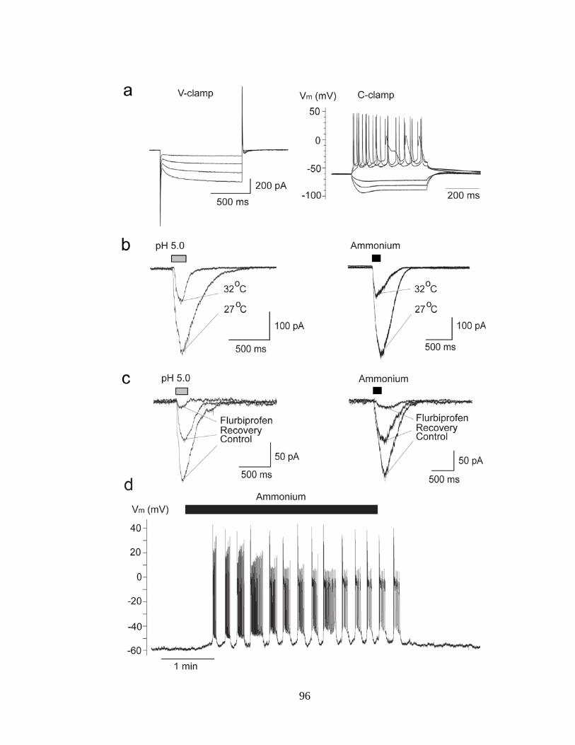
96

97
The effects of ASIC1a activation or blockade on spontaneous inhibitory activity
The presence of ASIC1a on interneurons - presumed to be GABAergic - predicts
that activation of these channels will increase GABAergic activity in the BLA network.
Indeed, bath application of 5 mM ammonium, in the presence of CNQX (10 μM), D-AP-
5 (50 μM), and SCH50911 (10 μM), increased the frequency of spontaneous inhibitory
postsynaptic currents (sIPSCs) recorded from principal neurons, from 25 ± 2 Hz to 36 ± 3
Hz (n = 11, P = 0.0006; Fig. 17a,b,c). Similarly, perfusion of the slices with acidified
ASCF (pH 6.6) increased the frequency of sIPSCs from 17 ± 2 Hz to 24 ± 1 Hz (n = 8, P
= 0.00008; Fig. 17d,e,f).
Figure 17. Activation of ASIC1a increases spontaneous inhibitory activity. Recordings were obtained from BLA principal cells in the presence of CNQX (10 μM), D-AP5 (50 μM), and SCH50911 (10 μM), at Vh = +30 mV. (a) Spontaneous inhibitory postsynaptic currents (sIPSCs) before, during, and after bath application of 5 mM ammonium. (b) Amplitude-frequency histogram of sIPSCs before and after bath application of 5 mM ammonium (n =11); bin width is 5 pA. (c) Group data of the frequency of sIPSCs in control medium, in 5 mM ammonium, and after washing out of ammonium (n = 11). (d) sIPSCs before, during, and after application of acidified ACSF. (e) Amplitude-frequency histogram of sIPSCs in control medium and in pH 6.65 (n = 8); bin width is 5 pA. (f) Group data of the frequency of sIPSCs in control medium, in low pH, and after return to control medium (n = 8). ***P < 0.001.

98

99
Since ASIC1a activation enhances sIPSCs, we asked if these channels are also
active in the basal state, contributing to the tonic level of inhibition (“background”
inhibition due to spontaneous GABA release) in the BLA. To answer this question, we
recorded sIPSCs from principal BLA neurons, and then applied either flurbiprofen, or the
tarantula venom, psalmotoxin (PcTx1), which specifically blocks homomeric ASIC1a
channels (27; 80; 85). Both compounds significantly decreased the frequency and
amplitude of sIPSCs; flurbiprofen decreased the frequency of sIPSCs from 26 ± 2 Hz to 7
± 2 Hz (n = 7, P = 0.00001; Fig. 18a,b,c), whereas PcTX1 decreased sIPSCs from 38 ± 2
Hz to 24 ± 3 Hz (n = 4, P = 0.005; Fig. 18d,e,f). To be certain that flurbiprofen or PcTX1
did not have a direct suppressive effect on GABAergic synaptic transmission, we also
evoked GABA currents by pressure application of GABA on the recorded principal
neurons. Neither flurbiprofen nor PcTX1 had any significant effect on the GABA-evoked
currents at the time that these drugs suppressed sIPSCs (Fig. 18a,d). These data suggest
that ASIC1a channels are active during basal synaptic activity and contribute
significantly to the generation of sIPSCs. We should note here that we also attempted to
antagonize the activity of ASIC1a with amiloride (298; 299) or nafamostat mesylate
(290), but found that these compounds directly affect GABAergic transmission; currents
evoked by pressure application of GABA were reduced by about 40% and 90% by
amiloride and nafamostat mesylate, respectively (see Supplementary Figure 1).
Figure 18. Antagonism of ASIC1a reduces spontaneous inhibitory activity. Recordings were obtained from BLA principal cells in the presence of CNQX (10 μM), D-AP5 (50 μM), and SCH50911 (10 μM), at Vh = +30 mV. (a) sIPSCs in control medium, in the presence of bath-applied flurbiprofen (2 mM), and after a 10 min-wash. Flurbiprofen suppressed sIPSCs, with no significant effect on the amplitude of GABAA-mediated currents evoked by pressure-applied GABA (arrowheads; 400 mM GABA, 200 ms). The lower traces show, in an expanded view, the last 5 s of the upper traces. (b) Amplitude-frequency histograms for

100
sIPSCs in control medium and in the presence of 2 mM flurbiprofen, for the cell shown in (a) (bin size, 10 pA). (c) Group data of the frequency of sIPSCs in control medium and in the presence of 2 mM flurbiprofen (n = 7, ***P < 0.001). (d) sIPSCs before, during, and after bath-applied PcTx1 venom (1:1000 dilution of the 100 μl lyophilized, milked venom). PcTx1 suppressed sIPSCs, with no significant effect on the amplitude of GABAA-mediated currents evoked by pressure-applied GABA. The lower traces show, in an expanded view, the last 5 s of the upper traces. (e) Amplitude-frequency histograms for sIPSCs in control medium and in the presence of PcTx1, for the cell shown in (d) (bin size, 10 pA). (f) Group data of the frequency of sIPSCs in control medium and in the presence of PcTx1 (n = 4, **P < 0.01).

101

102
Supplementary Figure 1: The non-specific ASIC1a antagonists Nafamostat mesylate
(NM) and amiloride reduce GABAA receptor-mediated currents. The recordings shown are from principal BLA neurons in the presence of 10 μM CNQX, 50 μM D-AP5, and 10 μM SCH50911 (Vh = +30 mV). (a) Currents evoked by pressure-applied GABA (400 mM, 200 ms) were blocked by bicuculline (40 μM). (b) NM (200 μM) nearly blocked GABA-evoked currents. (c) Amiloride also reduced the GABAA receptor-mediated currents.
ASIC1a activation increases glutamatergic excitation of interneurons

103
Considering that in addition to GABAergic cells, principal BLA neurons also
have functional ASIC1a channels, we examined if under acidic conditions, interneurons
are not depolarized only directly by activation of ASIC1a, but also due to glutamatergic
excitation secondarily to ASIC1a-mediated depolarization of principal cells. When we
recorded spontaneous excitatory postsynaptic currents (sEPSCs) from BLA interneurons,
application of acidified solution increased the frequency of sEPSCs from 19 ± 3 Hz to 25
± 3 Hz (n = 4, P = 0.01023, Fig. 19a). When we recorded currents evoked by pressure
application of acidified solution in the absence of CNQX, sEPSCs were present,
overlapping with the evoked currents (n = 3, Fig. 19b). The frequency of sEPSCs
recorded from interneurons was also increased by pressure application of ammonium (n =
4, Fig. 19c). To determine if in the basal state, ASIC1a activity on principal cells
contributes to glutamatergic excitation of interneurons, we recorded sEPSCs from
interneurons, and then applied flurbiprofen; the frequency of sEPSCs was reduced by
flurbiprofen from 20 ± 3 Hz to 5 ± 1 Hz (n = 4, P = 0.00528, Fig. 19d). Thus, activation
of ASIC1a channels depolarizes interneurons not only directly, but also indirectly due to
glutamate release from principal cells, and this mechanism is in effect also in the basal
state.

104
Figure 19. Activation of ASIC1a increases the excitatory drive of interneurons.
Recordings are from interneurons at Vh -58 mV and in the presence of D-AP5 (50 μM) and SCH50911 (10 μM). (a) Lowering the pH of the bath increased the frequency of sEPSCs. The lower current traces in (a) are from the same cell as in the upper trace, at an expanded view. CNQX (10 μM) blocked the recorded currents. (b) Currents evoked by pressure application (arrowhead, 200 ms) of acidified ACSF in the absence of CNQX, displayed “riding” EPSCs. (c)

105
Pressure application of ammonium (40 mM, 500 ms) increased the frequency of sEPSCs. (d) Bath application of flurbiprofen (1 mM) decreased the frequency of sEPSCs.
The net effect of ASIC1a activation on BLA excitability
The results described above demonstrate that activation of ASIC1a increases
inhibitory activity in the BLA due to direct and indirect (via synaptic glutamatergic
excitation) depolarization of interneurons. However, ASIC1a activation can be expected
to also increase excitatory activity, since principal neurons also carry these channels. The
question, therefore, arises as to what would be the net effect of ASIC1a activation on the
overall level of activity and excitability of the BLA. To answer this question, first we
examined the effect of ASIC1a activation on sIPSCs and sEPSCs recorded
simultaneously from principal cells. To quantify these effects we calculated the total
charge transferred. The charge, in pico Coulombs, was calculated as the area delimited by
the inhibitory or excitatory current and the baseline. Current areas were analyzed for a
time window of 5s. Bath application of ammonium (5mM) increased the charge
transferred by sIPSCs from 1.4 ± 0.2 pC to 38 ± 4 pC (n = 21, P = 0.0000008), while at
the same time the charge transferred by sEPSCs increased from 1.3 ± 0.1 pC to 9 ± 1 pC
(n = 21, P = 0.000003, Fig. 20a). The ammonium-induced increase of the charge
transferred by sIPSCs (5000 ± 1000 %) was significantly greater than that of the sEPSCs
(900 ± 200 %; P = 0.01318). Similar effects were observed during bath application of
ACSF of pH 6.6. The acidified solution increased the charge transferred by sIPSCs from
2.1 ± 0.6 pC to 19 ± 3 pC (n = 9, P = 0.0005), while at the same time the charge
transferred by sEPSCs increased from 2.0 ± 0.5 pC to 4.8 ± 0.6 pC (n = 9, P = 0.00003,
Fig. 20b). Again, the difference between the increase of sIPSCs (1400 ± 400 %) and the

106
increase of sEPSCs (290 ± 30 %) was statistically significant (P = 0.00544). Thus,
ASIC1a activation increases both GABAergic and glutamatergic spontaneous activity,
but the increase of GABAergic activity is significantly greater than the increase of
glutamatergic activity.
Next, we examined the effects of ASIC1a activation on BLA field potentials
evoked by stimulation of the external capsule. The field potentials evoked in the BLA are
not pure field EPSPs; they also contain population spiking activity and are under strong
GABAergic inhibition. For this reason, any manipulation that increases inhibitory activity
reduces the amplitude of the evoked field potential. Bath application of ammonium (8
mM) reduced the amplitude of the field potentials to 76.4 ± 4 % of the control values
(from 0.46 ± 0.02 mV to 0.36 ± 0.01 mV, n = 4, P = 0.008; not shown). The reduction of
the field potentials by ammonium suggested that ASIC1a activation suppresses
excitability in the BLA network. To obtain more clear evidence of such an effect, we first
induced epileptiform activity by increasing the concentration of K+ in the ACSF to 7 mM
and removing Mg++, and then applied ammonium. Bath application of 8 mM ammonium
reduced the evoked field potentials and eliminated the spontaneous epileptiform
discharges (n = 5, Fig. 20c). The same concentration of ammonium had no significant
effect on spontaneous epileptiform activity when flurbiprofen (2 mM) was present in the
slice medium (n = 4; not shown), which is consistent with the involvement of ASIC1a
channels in the suppressive effect of ammonium.

107
Figure 20. The net effect of ASIC1a activation is suppression of BLA excitability. (a) and
(b) Simultaneous recordings of sIPSCs (outward currents) and sEPSCs (inward currents) were obtained from principal cells at Vh -58 mV, and in the presence of D-AP5 (50 μM) and SCH50911 (10 μM). Bath application of 5 mM

108
ammonium (a), or acidified solution (b) increased the charge transferred by sIPSCs and sEPSCs; the increase in the charge transferred by sIPSCs was significantly greater than the increase in charge transferred by sEPSCs. Example traces are shown in the upper panels of (a) and (b), and group data are shown in the bar graphs; n = 21 in (a) and n = 9 in (b), ***P < 0.001. (c) Upper panel shows field potentials evoked in the BLA by single-pulse stimulation of the external capsule, and lower panel shows spontaneous field activity recorded in gap-free mode. Recordings are in medium containing 7 mM K+ and zero Mg++, which induced epileptiform activity. Bath application of 8 mM ammonium reduced the evoked field potentials and blocked epileptiform activity. Each of the three field potentials shown in the upper panel is an average of 10 sweeps; the stimulus artifacts have been truncated for clarity. The equidistant vertical lines in the traces of spontaneous activity are stimulus artifacts, as evoked field potentials were sampled during gap-free recordings, by stimulation applied every 20 sec.
The net effect of ASIC1a blockade on BLA excitability
To determine whether the activity of ASIC1a channels favors enhancement of
GABAergic inhibition over that of glutamatergic excitation also in the basal state, we
examined the effect of ASIC1a blockade by flurbiprofen on sIPSCs and sEPSCs recorded
simultaneously from principal cells (Fig. 21a). Flurbiprofen decreased the frequency of
sIPSCs from 15 ± 1 Hz to 5 ± 1 Hz (n = 9, P = 0.000004), and the frequency of sEPSCs
from 16 ± 1 Hz to 11 ± 1 Hz (n = 9, P = 0.0002). The reduction in the frequency of
sIPSCs (70 ± 4 %) was significantly greater than the reduction in the frequency of
sEPSCs (30 ± 4 %; P = 0.000002). We also tested the effects of flurbiprofen on evoked
field potentials, in normal ACSF. Bath application of 2 mM flurbirpofen increased the
amplitude of the field potentials from 0.61 ± 0.09 mV to 1.05 ± 0.19 mV (n = 5, P =
0.020, Fig. 21b). These results suggest that in the basal state the activity of ASIC1a in the
BLA favors enhancement of inhibition over excitation.

109
Figure 21. The net effect of ASIC1a antagonism is reduction of inhibition and increased
excitability. (a) Simultaneous recordings of sIPSCs (outward currents) and sEPSCs (inward currents) were obtained from principal cells at Vh -58 mV, and in the presence of D-AP5 (50 μM) and SCH50911 (10 μM). Bath application of 2 mM flurbiprofen decreased the frequency of sIPSCs to a greater extent than that of sEPSCs. An example is shown in the upper panel, and group data in the bar graphs (n = 9, ***P < 0.001). (b) Field potentials evoked in the BLA by stimulation of the external capsule. Bath application of 2 mM flurbiprofen reversibly increased the amplitude of the evoked responses. Each trace is an average of 10 sweeps.
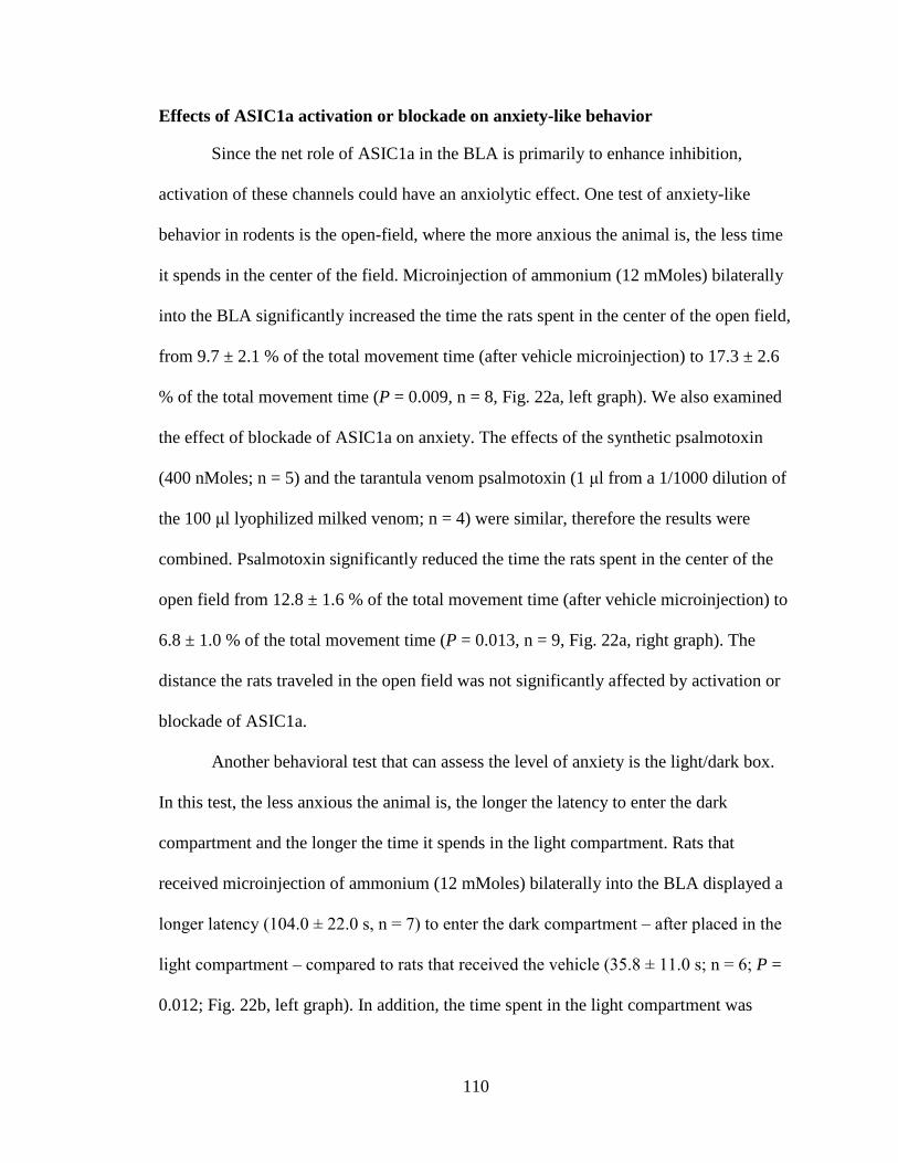
110
Effects of ASIC1a activation or blockade on anxiety-like behavior
Since the net role of ASIC1a in the BLA is primarily to enhance inhibition,
activation of these channels could have an anxiolytic effect. One test of anxiety-like
behavior in rodents is the open-field, where the more anxious the animal is, the less time
it spends in the center of the field. Microinjection of ammonium (12 mMoles) bilaterally
into the BLA significantly increased the time the rats spent in the center of the open field,
from 9.7 ± 2.1 % of the total movement time (after vehicle microinjection) to 17.3 ± 2.6
% of the total movement time (P = 0.009, n = 8, Fig. 22a, left graph). We also examined
the effect of blockade of ASIC1a on anxiety. The effects of the synthetic psalmotoxin
(400 nMoles; n = 5) and the tarantula venom psalmotoxin (1 μl from a 1/1000 dilution of
the 100 μl lyophilized milked venom; n = 4) were similar, therefore the results were
combined. Psalmotoxin significantly reduced the time the rats spent in the center of the
open field from 12.8 ± 1.6 % of the total movement time (after vehicle microinjection) to
6.8 ± 1.0 % of the total movement time (P = 0.013, n = 9, Fig. 22a, right graph). The
distance the rats traveled in the open field was not significantly affected by activation or
blockade of ASIC1a.
Another behavioral test that can assess the level of anxiety is the light/dark box.
In this test, the less anxious the animal is, the longer the latency to enter the dark
compartment and the longer the time it spends in the light compartment. Rats that
received microinjection of ammonium (12 mMoles) bilaterally into the BLA displayed a
longer latency (104.0 ± 22.0 s, n = 7) to enter the dark compartment ‒ after placed in the
light compartment ‒ compared to rats that received the vehicle (35.8 ± 11.0 s; n = 6; P =
0.012; Fig. 22b, left graph). In addition, the time spent in the light compartment was

111
significantly longer in rats administered ammonium (138.4 ± 12.5 s) compared to
vehicle-microinjected rats (48.2 ± 8.8 s; P = 0.00013; Fig. 22b, right graph).
Figure 22. In vivo activation of ASIC1a in the BLA suppresses anxiety-like behavior, while antagonism of ASIC1a increases anxiety. (a) In the open field test, the rats spent significantly more time in the center, after microinjection of ammonium bilaterally into the BLA (left graph), and significantly less time in the center, after microinjection of psalmotoxin into the BLA (right graph), compared to the time they spent in the center of the open field when injected with the vehicle. (b) In the light-dark box test, rats microinjected with ammonium bilaterally into the BLA took a significantly longer time to enter the dark compartment, and spent more total time in the light compartment compared to rats injected with the vehicle. *P < 0.05.

112

113
DISCUSSION
In the present study, we demonstrated that in the rat BLA, ASIC1a channels are
present on both interneurons and principal cells, and their activation by ammonium, or by
lowering extracellular pH, induces intense firing. When these channels are activated
throughout the BLA network, spontaneous GABAergic inhibitory activity is increased
significantly more than glutamatergic activity. Consistent with this finding, ammonium
blocked epileptiform discharges in vitro, and reduced anxiety-like behavior when
microinjected bilaterally into the BLA in vivo. Furthermore, ASIC1a channels are active
in the basal state, enhancing background GABAergic inhibition, not only by direct
depolarization of interneurons, but also by indirect excitation of interneurons secondary
to ASIC1a-mediated depolarization of principal cells. Accordingly, in vivo blockade of
ASIC1a in the BLA increased anxiety.
The BLA displays a markedly high expression of the ASIC1a subunit (73; 304),
but it also expresses the ASIC2a and ASIC2b subunits (34; 160). It is unknown if the
functional ASIC1a channels in the BLA are homomeric ASIC1a trimers, or if they co-
assemble with the ASIC2a or 2b. From the two mechanisms that we employed to activate
these channels, the low pH can activate any type of ASIC, while ammonium specifically
activates ASIC1a currents as demonstrated in isolated sensory neurons (220), as well as
in central dopaminergic neurons, hippocampal interneurons, and HEK 293 cells (223).
The effects we observed when ASICs were activated by either low pH or ammonium
application were blocked by flurbiprofen. In addition, both flurbiprofen and PcTX1
significantly reduced the frequency of sIPSCs in the BLA (Fig. 22). Flurbiprofen has
been shown to inhibit PcTX1-sensitive currents (298), and PcTX1 specifically blocks

114
currents mediated by homomeric ASIC1a (27; 85). Thus, at least a portion of the ASIC1a
channels in the BLA are probably homomeric trimers.
If we consider that ASIC1a channels are present on both interneurons and
principal neurons, and their activation strongly depolarizes both types of neurons (Figs.
15 and 16), but also that interneurons constitute only a small percentage of the total
neuronal population in the BLA (175; 241), then we have to ask why the predominant
effect of ASIC1a activation in the BLA network is an increase of GABAergic inhibition
rather than glutamatergic excitation. A possible explanation lies on the evidence that
much of the excitatory input within the BLA circuitry is directed onto interneurons, while
most of the inhibitory input is directed onto principal cells (154; 209; 226; 268). Our
observations are in accordance with these findings, as only a small percentage of the
interneurons we have recorded from have sIPSCs, while all of them display high activity
of single sEPSCs and, occasionally, bursts of sEPSCs. An additional factor that can make
BLA interneurons more excitable is that the GABAA receptor-mediated IPSPs recorded
from them reverse at more depolarized potentials than the GABAA IPSPs of projection
cells (170). Finally, the electrical coupling between BLA interneurons (191; 309) can
enhance inhibitory strength in the BLA network. As a result, when both interneurons and
principal cells are depolarized by ASIC1a activation, the inhibition of principal cells is
stronger than their excitation, probably because principal cells receive strong inhibitory
inputs, which are further reinforced due to concomitant strong excitation of interneurons
(see Fig. 19).
Both the electrophysiological and the behavioral results suggested that the
ASIC1a channels in the BLA are active in the basal state, preferentially increasing

115
background inhibition. Thus, flurbiprofen or PcTX1 reversibly decreased the frequency
and amplitude of isolated sIPSCs (Fig. 22), and when sIPSCs and sEPSCs were recorded
simultaneously, flurbiprofen preferentially reduced the sIPSCs (Fig. 21a). In addition,
flurbiprofen reduced the basal excitatory drive of interneurons (Fig. 19d). These results
are consistent with the increase of the evoked population response in the BLA by bath
application of flurbiprofen (Fig. 21b; increased population spiking due to reduced
inhibition), as well as with the increase in anxiety-like behavior in the open field, when
psalmotoxin was injected bilaterally in the BLA (Fig. 22a). How can ASIC1a channels be
active in the basal state? Extracellular pH can fluctuate during normal synaptic activity,
and the nature of these fluctuations (alkalinization or acidification, and their time course
relative to the intensity of synaptic activity) may differ in different brain regions (64;
313). The present data suggest that, in the BLA, pH shifts during spontaneous synaptic
activity, maintain a sufficient number of ASIC1a channels open to have a significant
effect on spontaneous inhibitory activity.
Previous studies have shown the involvement of ASIC1a activation in the
suppression of seizures in vivo, and the blockade of epileptiform activity in hippocampal
slices (321). A similar role of these channels was demonstrated in BLA slices, in the
present study, where bath application of ammonium suppressed epileptiform activity. In
agreement with the role of ASIC1a in suppressing excitatory activity in the BLA, we
found increased anxiety-like behavior when these channels were blocked, and
suppression of anxiety when they were activated. These results are in sharp contrast to
previous studies which have indicated that activation of ASIC1a is necessary for the
generation and expression of certain types of fear (320), and genetic elimination or

116
blockade of these channels have anxiolytic effects (73; 82). Species difference (mice
versus rats) in the function of ASIC1a in the BLA is possible. Such species difference
exists for the function of the GluK1 receptors in the BLA, the blockade of which is
anxiogenic in mice (311), but anxiolytic in rats (17). It should also be considered that the
strength and the temporal pattern of ASIC1a activation could play a determinant role in
the effect it will have on the network excitability ‒ and may have played a role in the
discrepant results ‒, as strong or slow acidification can lead to fast desensitization of
ASIC1a (38; 103), which can give way to other mechanisms that may produce
hyperexcitability.
The present study clearly demonstrated that in the rat amygdala the primary
function of ASIC1a channels is to increase inhibition, and, as a result, activation of these
channels has an anxiolytic effect. It is apparent, however, that we are still at the early
stages of understanding the roles that ASICa plays in the excitability of different brain
regions, what parameters affect these roles, and if there are species differences.
Delineating the functions of these channels can potentially have profound implications
for the treatment of several neurological and mental illnesses.
117
APPENDIX 3: THE LIMITATIONS OF DIAZEPAM AS A TREATMENT FOR NERVE AGENT-INDUCED SEIZURES AND NEUROPATHOLOGY IN RATS; COMPARISON WITH UBP302
James P. Apland, Vassiliki Aroniadou-Anderjaska, Taiza H. Figueiredo, Franco Rossetti,
Steven L. Miller, Maria F.M. Braga*
Neurotoxicology Branch, U.S. Army Medical Research Institute of Chemical Defense,
Aberdeen Proving Ground, Maryland, 21010 (J.P.A.); Department of Anatomy,
Physiology, and Genetics (V.A-A., T.H.F., F.R., S.L.M., M.F.M.B.) and Department of
Psychiatry (V.A-A., M.F.M.B.), F. Edward Hébert School of Medicine, Uniformed
Services University of the Health Sciences, Bethesda, Maryland, 20814
*Corresponding Author: Maria F.M. Braga, D.D.S., Ph.D., Department of Anatomy, Physiology, and Genetics, F. Edward Hébert School of Medicine, Uniformed Services University of the Health Sciences, 4301 Jones Bridge Road, Bethesda, MD 20814 Phone: (301) 295-3524 Fax: (301) 295-3566 Email: [email protected]
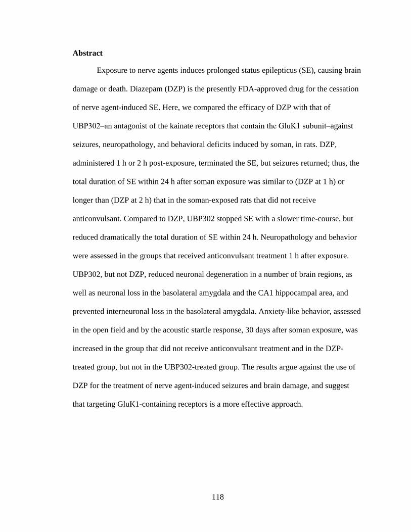
118
Abstract
Exposure to nerve agents induces prolonged status epilepticus (SE), causing brain
damage or death. Diazepam (DZP) is the presently FDA-approved drug for the cessation
of nerve agent-induced SE. Here, we compared the efficacy of DZP with that of
UBP302‒an antagonist of the kainate receptors that contain the GluK1 subunit‒against
seizures, neuropathology, and behavioral deficits induced by soman, in rats. DZP,
administered 1 h or 2 h post-exposure, terminated the SE, but seizures returned; thus, the
total duration of SE within 24 h after soman exposure was similar to (DZP at 1 h) or
longer than (DZP at 2 h) that in the soman-exposed rats that did not receive
anticonvulsant. Compared to DZP, UBP302 stopped SE with a slower time-course, but
reduced dramatically the total duration of SE within 24 h. Neuropathology and behavior
were assessed in the groups that received anticonvulsant treatment 1 h after exposure.
UBP302, but not DZP, reduced neuronal degeneration in a number of brain regions, as
well as neuronal loss in the basolateral amygdala and the CA1 hippocampal area, and
prevented interneuronal loss in the basolateral amygdala. Anxiety-like behavior, assessed
in the open field and by the acoustic startle response, 30 days after soman exposure, was
increased in the group that did not receive anticonvulsant treatment and in the DZP-
treated group, but not in the UBP302-treated group. The results argue against the use of
DZP for the treatment of nerve agent-induced seizures and brain damage, and suggest
that targeting GluK1-containing receptors is a more effective approach.

119
Introduction
The devastating effects of the sarin attack against civilians in Syria that the world
witnessed in August of 2013 (81) brought again to the forefront the question of readiness
and whether the existing medical countermeasures can save lives and protect against the
long-term health consequences of exposure. The primary action of nerve agents is the
inhibition of acetylcholinesterase (AChE). Without medical intervention, the ensuing
cholinergic crisis can culminate in the collapse of the cardiorespiratory system and death
(24). In addition to the peripheral effects, AChE inhibition in the brain produces
convulsive seizures and status epilepticus (SE), initiated by the excessive stimulation of
cholinergic receptors. If immediate death is prevented by adequate control of the
peripheral symptoms, but SE is not controlled effectively, lives may still be lost from the
prolonged SE, or severe brain damage can ensue with long-term behavioral
consequences. The lasting behavioral deficits that follow nerve agent exposure are well-
known from experimental studies in animals (74; 90; 155; 228), but also from studies in
the victims of the sarin attacks in Japan, who present neurological and neuropsychiatric
disturbances years after the exposure (118; 204; 315).
As the initiation of seizures after nerve agent exposure is due to excessive
elevation of acetylcholine, acting primarily on muscarinic receptors, administration of
muscarinic receptor antagonists can halt the development of SE, but only when
administered within a short period of time after exposure (153; 261; 266), suggesting that
seizures are sustained and reinforced by glutamatergic rather than cholinergic
mechanisms (181). One way to suppress glutamatergic hyperactivity is by enhancing
GABAergic inhibition. Benzodiazepines, which are positive allosteric modulators of
GABAA receptors (48; 96), have long been the first line of treatment for the cessation of
120
SE triggered by various etiologies (266). Accordingly, the benzodiazepine diazepam
(DZP) is also currently the only FDA-approved injectable drug for the control of seizures
caused by nerve agents.
A number of concerns, however, are associated with the use of DZP for the
cessation of nerve agent-induced SE. First, the efficacy of DZP decreases as the interval
between the initiation of SE and the administration of DZP increases (183; 261; 288); this
has also been documented in the lithium-pilocarpine model of SE (102; 130; 301), which
has many similarities to the nerve agent-induced SE, in both the mechanisms of seizure
initiation and the effects it produces (284). The development of resistance to DZP is
concerning because in a terrorist attack with nerve agents, it may not be possible for
medical assistance to arrive immediately; thus, when DZP is administered, the seizures
may have already become resistant to benzodiazepines. Second, benzodiazepines are
among the anticonvulsants with the most serious side effects (136; 185). Third, seizures
often recur after termination of the initial SE by benzodiazepines (174; 264), and at least
in the case of nerve agent-induced SE, it is unclear whether DZP compares favorably
with other anticonvulsant treatments in the duration of its anti-seizure effects. This is
particularly significant considering that the duration and intensity of seizures clearly
correlate with the severity of the resulting neuropathology (227; 258).
We recently demonstrated that nerve agent-induced seizures can be effectively
controlled by targeting the glutamatergic system. LY293558, which is an antagonist of
both the AMPA receptors and the kainate receptor subtype that contains the GluK1
subunit (GluK1Rs; formerly known as GluR5 kainate receptors or GLUK5 receptors; see
(66; 124)) was very efficacious in stopping seizures induced by the nerve agent soman

121
and protecting against neuronal damage (12; 89). In the present study, we used a rat
model of nerve agent exposure in which the anticonvulsant treatment was delayed to at
least 1 h, and compared the efficacy of DZP to that of another GluK1R antagonist, (S)-3-
(2-carboxybenzyl)willardiine (UBP302), which antagonizes selectively the GluK1Rs
(190), against soman-induced seizures, as well as acute and long-term neuropathology.
We also examined the efficacy of DZP and UBP302 in preventing the development of
anxiety, which is a prevalent behavioral deficit resulting from nerve agent-induced brain
damage, in both animals (74; 155; 228) and humans (118; 204).

122
Materials and Methods
Animals
Male, Sprague-Dawley rats (Charles River Laboratories, Wilmington, MA),
weighing 150-250 g (6 to 8-weeks-old) at the start of the experiments, were individually
housed in an environmentally controlled room (20–23°C, 12-h light/12-h dark cycle,
lights on 06:00 am), with food and water available ad libitum. The animal care and use
programs at the U.S. Army Medical Research Institute of Chemical Defense
(USAMRICD) and the Uniformed Services University of the Health Sciences (USUHS)
are accredited by the Association for Assessment and Accreditation of Laboratory
Animal Care International. All animal experiments were conducted following the Guide
for the Care and Use of Laboratory Animals by the Institute of Laboratory Animal
Resources, National Research Council and the Animal Welfare Act of 1966 (P.L. 89-
544), as amended, and were approved by the Institutional Animal Care and Use
Committees of the USAMRICD and the USUHS.
Soman administration and drug treatment
Soman (pinacolyl methylphosphonofluoridate) was obtained from the U.S. Army
Edgewood Chemical Biological Center (Aberdeen Proving Ground, MD). The agent was
diluted in cold saline, and administered via a single subcutaneous injection (154 µg/kg,
which is approximately 1.4 X LD50; (126)) to rats that were 7 to 8-weeks-old. To
increase survival rate, rats were administered HI-6 (1-(2-
hydroxyiminomethylpyridinium)-3-(4-carbamoylpyridinium)-2-oxapropane dichloride;
125 mg/kg, i.p.; Starks Associates, Buffalo, NY) 30 min prior to soman exposure. HI-6 is
a bispyridinium oxime that reactivates inhibited acetylcholinesterase, primarily in the

123
periphery (24). Within 1 min after soman exposure, rats also received an intramuscular
injection of atropine sulfate (2 mg/kg; Sigma- Aldrich, St. Louis, MO) to minimize
peripheral toxic effects. The soman-exposed rats were randomly divided into 3 groups:
Those that did not receive any further treatment (except for the oxime pretreatment and
the atropine; SOMAN group), those that received DZP (10 mg/kg, i.m.) at 1 h after
exposure to soman (SOMAN+DZP group), and those that received UBP302 (250 mg/kg,
i.p.) at 1 h after exposure to soman (SOMAN+UBP302 group). Some of the soman-
exposed rats had been implanted with electrodes for electroencephalographic (EEG)
monitoring (see next section for implantation procedure), 10 days before exposure. From
the implanted rats, some were administered DZP or UBP302 (doses same as above) at 1h
or 2 h after soman exposure; therefore, there were two SOMAN+DZP groups and two
SOMAN+UBP302 groups for the electrode-implanted rats (for the two time points of
anticonvulsant treatment; sample sizes are in the results section). DZP and UBP302 were
purchased from Hospira Inc. (Lake Forest, IL) and Tocris Bioscience (Bristol, UK),
respectively. Control animals received HI-6 and atropine, but were injected with saline
instead of soman (CONTROL group). For the SOMAN+UBP302 groups, we had to
decide on a dose based only on our own observations, as there are no previous studies in
which UBP302 has been injected systemically. First, we tested 100 mg/kg; this
concentration suppressed seizures, but with a very slow time course (it took more than 3
h to terminate seizure activity). We concluded with 250 mg/kg, after testing this
concentration also in control rats (rats not exposed to soman). Unlike DZP which
produces sedative effects even at 10 mg/kg, the 250 mg/kg of UBP302 administered to
control rats produced only a mild reduction in overall activity.

124
In the rats that were not implanted with EEG electrodes, the occurrence
and the progression of seizures were monitored behaviorally and classified according to
the Racine scale (232), with minor modifications: Stage 0, no behavioral response; Stage
1, behavioral arrest; Stage 2, oral/facial movements, chewing, head nodding; Stage 3,
unilateral/bilateral forelimb clonus without rearing, Straub tail, extended body posture;
Stage 4, bilateral forelimb clonus plus rearing; Stage 5, rearing and falling; and Stage 6,
full tonic-clonic seizures.
Electrode implantation and EEG recordings
Rats (6-week-old) were anesthetized with isoflurane using a gas anesthesia system
(Kent Scientific, Torrington, CT). Five stainless steel, cortical screw electrodes were
stereotaxically implanted, as described previously (89), using the following coordinates
(from (215)): two frontal electrodes, 2.0 mm posterior from bregma and 2.5 mm lateral
from the midline, and two parietal electrodes, 5.0 mm posterior from bregma and 2.5 mm
lateral from the midline; a cerebellar reference electrode was implanted 1.0 mm posterior
to lambda. Each screw electrode (Plastics One Inc., Roanoke, VA) was placed in a plastic
pedestal (Plastics One Inc.) and fixed to the skull with dental acrylic cement. For EEG
recordings (obtained 10 days after electrode implantation), rats were placed in the EEG
chamber and connected to the EEG system (Stellate, Montreal, Canada; 200 Hz sampling
rate). Video-EEG recordings were performed in the freely moving rats, as described
previously (89). EEG was continuously recorded for 24 hours after soman injection;
during that time the animals had free access to food and water. The termination of the
soman-induced SE was defined as the disappearance of large amplitude, repetitive
discharges (>1 Hz with at least double the amplitude of the background activity).
125
Fixation & tissue processing
Neuropathological analysis was performed in the amygdala, piriform cortex,
entorhinal cortex, hippocampus, and a neocortical region of rats that were not implanted
with EEG electrodes (the implantation procedure might cause some damage that could
interfere with the neuropathology results). One day, 7 days, and 30 days after soman
administration, rats were deeply anesthetized with pentobarbital (75-100 mg/kg, i.p.) and
transcardially perfused with PBS (100 ml) followed by 4% paraformaldehyde (200 ml).
The brains were removed and post-fixed overnight at 4º C, then transferred to a solution
of 30% sucrose in PBS for 72 hours, and frozen with dry ice before storage at -80º C until
sectioning. A 1-in-5 series of sections from the rostral extent of the amygdala to the
caudal extent of the entorhinal cortex was cut at 40 µm on a sliding microtome. One
series of sections was mounted on slides (Superfrost Plus, Daigger, Vernon Hills, IL) in
PBS for Nissl staining with cresyl violet. Two adjacent series of sections were mounted
on slides for Fluoro-Jade C (FJC) staining, or were stored at -20°C in a cryoprotectant
solution for GAD-67 immunohistochemistry. All neuropathological analysis was done in
a blind fashion.
Fluoro-Jade C staining and analysis
FJC (Histo-Chem, Jefferson, AR) was used to identify irreversibly degenerating neurons
in all the amygdalar nuclei and the piriform cortex (-2.04 mm to -3.36 mm from bregma),
a neocortical area (-2.04 and -6.36 mm from bregma), the entorhinal cortex and the CA1,
CA3, and hilar areas of the ventral hippocampus (-5.4 and -6.36 mm from bregma; all
coordinates from (215)); we studied the ventral hippocampus because it displays
significantly more severe neurodegeneration after soman exposure than the dorsal
126
hippocampus (14). Mounted sections were air-dried overnight and then immersed in a
solution of 1% sodium hydroxide in 80% ethanol for 5 min. The slides were then rinsed
for 2 min in 70% ethanol and 2 min in distilled water (dH20), and then incubated in
0.06% potassium permanganate solution for 10 min. After a 2 min rinse in dH20, the
slides were transferred to a 0.0001% solution of FJC dissolved in 0.1% acetic acid for 10
minutes. Following three 1-minute rinses in dH20, the slides were dried on a slide
warmer, cleared in xylene for at least 1 min, and coverslipped with DPX (Sigma-
Aldrich).
To assess the extent of neurodegeneration, we used a series of adjacent Nissl-
stained sections to trace the regions of interest. The tracings from the Nissl-stained
sections were superimposed on the FJC-stained sections, using the Stereo Investigator 9.0
(MicroBrightField, Williston, VT). The following rating system was used to score the
extent of neuronal degeneration in each structure and substructures: 0 = no damage, 1 =
minimal damage (1-10%), 2 = mild damage (11-25%), 3 = moderate damage (26-45%)
and 4 = severe damage (>45%). We have previously shown that qualitative assessment
using this scale produces results that are in agreement with quantitative assessments
(Qashu et al., 2010). The scores for neurodegeneration present on FJC-stained sections
were assessed considering the density of cells from Nissl-stained sections, along the
anterior to posterior extent, at 600 μm intervals.
Stereological quantification
Design-based stereology was used to quantify the total number of neurons in Nissl-
stained sections in the basolateral amygdala (BLA) and CA1 area. Sections were viewed
with a Zeiss Axioplan 2ie (Oberkochen, Germany) fluorescent microscope with a

127
motorized stage, interfaced with a computer running StereoInvestigator 9.0
(MicroBrightField). The BLA and CA1 regions were identified on slide-mounted
sections, and delineated for each slide of each animal, under a 2.5× objective, based on
the atlas of Paxinos and Watson (215). All sampling was done under a 63× oil immersion
objective. Nissl-stained neurons were distinguished from glial cells by their larger size
and pale nuclei surrounded by darkly-stained cytoplasm containing Nissl bodies. The
total number of Nissl-stained neurons was estimated using the optical fractionator probe,
and, along with the coefficient of error (CE), was calculated using Stereo Investigator 9.0
(MicroBrightField). The CE was calculated by the software according to Gundersen (m =
1; (105)) and Schmitz-Hof (2nd estimation; (246)) equations.
For Nissl-stained neurons in the BLA, a 1-in-5 series of sections was analyzed (8
sections on average). The counting frame was 35 × 35 µm, the counting grid was 190 ×
190 µm, and the dissector height was 12 µm. Nuclei were counted when the cell body
came into focus within the dissector which was placed 2 µm below the section surface.
Section thickness was measured at every counting site, and the average mounted section
thickness was 22.3 µm. An average of 365 neurons per rat was counted, and the average
CE was 0.045 for both the Gunderson and Schmitz-Hof equations. For Nissl-stained
neurons in the CA1 area, a 1-in-10 series of sections was analyzed (8 sections on
average). The counting frame was 20 × 20 µm, the counting grid was 250 × 250 µm, and
the dissector height was 10 µm. Nuclei were counted when the cell body came into focus
within the dissector which was placed 2 µm below the section surface. Section thickness
was measured at every counting site, and the average mounted section thickness was 18.3
µm. An average of 253 neurons per rat was counted, and the CE was 0.065 for
128
Gunderson (m = 1) and 0.060 for Schmitz-Hof (2nd estimation) equation. For
GABAergic interneurons immuno-labeled for GAD-67 in the BLA (see procedure
below), a 1-in-10 series of sections was analyzed (on average 6 sections). The counting
frame was 60 x 60 µm, the counting grid was 100 × 100 µm, and the dissector height was
20 µm. Nuclei were counted when the top of the nucleus came into focus within the
dissector which was placed 2 µm below the section surface. Section thickness was
measured at every 5th counting site, and the average mounted section thickness was 30
µm. An average of 260 neurons per rat was counted, and the average CE was 0.07 for the
Gunderson equation and 0.065 for Schmitz-Hof equation.
GAD67 immunohistochemistry
To label GAD-67 immunoreactive neurons, a l-in-5 series of free-floating sections
was collected from the cryoprotectant solution, washed three times for 5 min each in 0.1
M PBS, and then incubated in a blocking solution containing 10% normal goat serum
(NGS, Chemicon International, Temecula, CA) and 0.5% Triton X-100 in PBS for one
hour at room temperature. The sections were then incubated with mouse anti-GAD67
serum (1:1000, MAB5406; Chemicon), 5% NGS, 0.3% Triton X-100, and 1% bovine
serum albumin, overnight at 4º C. After rinsing three times for 10 min each in 0.1%
Triton X-100 in PBS, the sections were incubated with Cy3-conjugated goat anti-mouse
antibody (1:1000; Jackson ImmunoResearch, West Grove, PA) and 0.0001% DAPI
(Sigma-Aldrich, St. Louis, MO) in PBS for one hour at room temperature. After a final
rinse in PBS for 10 min, sections were mounted on slides, air dried for at least 30 min,
and coverslipped with ProLong Gold antifade reagent (Life Technologies, Grand Island,
NY).

129
Behavioral experiments
Animals from the SOMAN, SOMAN+DZP, SOMAN+UBP302 and CONTROL groups
were tested in the open field and the acoustic startle apparatus, 30 days after soman. In
the open field apparatus (40X40X30 cm clear Plexiglas arena), anxiety-like behavior was
assessed as described previously ((17; 228)), following the procedure used by Grunberg
and collaborators (87). One day prior to testing (on day 29 after soman exposure),
animals were acclimated to the apparatus for 20 min. On the test day, the rats were placed
in the center of the open field, and activity was measured and recorded for 20 min, using
an Accuscan Electronics infrared photocell system (Accuscan Instruments Inc.,
Columbus, OH). Data were automatically collected and transmitted to a computer
equipped with “Fusion” software (from Accuscan Electronics). Locomotion (distance
traveled in cm), total movement time, and time spent in the center of the open field were
analyzed. Anxiety behavior was measured as the ratio of the time spent in the center over
the total movement time, expressed as a percentage of the total movement time. Subjects
were exposed to an acclimation session on day 29 post-exposure, and followed next day
for a test session.
Acoustic startle response (ASR) testing was conducted with the use of the Med
Associates Acoustic Response Test System (Med Associates, Georgia, VT), which
consists of weight-sensitive platforms inside individual sound-attenuating chambers. A
ventilating fan built into the chamber provides background noise. Each rat was
individually placed in a ventilated holding cage. The holding cages are small enough to
restrict extensive locomotion, but large enough to allow the subject to turn around and
make other small movements. Each cage was placed on a weight-sensitive platform.
Subjects’ movements in response to stimuli were measured as a voltage change by a
130
strain gauge inside each platform. All animals were acclimated to the apparatus in two
sessions on post-soman days 28 and 29. Startle stimuli consisted of 120 or 110 dB sound
pressure level noise bursts of 20-ms duration. Each stimulus had a 2 ms rise and decay
time, such that the onset and offset were abrupt, which is a primary requirement for
startle. Each trial type (110 dB or 120 dB stimulus) was presented eight times. Trial types
were presented in random order to avoid effects and habituation, and inter-trial intervals
ranged randomly from 15 to 25 s. Responses were recorded by an interfaced Pentium
computer as the maximum response occurring during the no-stimulus periods and during
the startle period, and were assigned a value based on an arbitrary scale used by the
software of the test system.
Statistical analysis
Fisher exact test was used to compare the survival rate between the groups. Initial
SE duration, total SE duration, number of convulsive seizures recurring during the 24 h
period after termination of the initial SE, stereological estimations of the number of
neurons and interneurons, and results from behavioral tests were compared between the
SOMAN, the SOMAN+DZP and the SOMAN+UBP302 groups using analysis of
variance (ANOVA) followed by post-hoc tests as described in the figure legends. The
statistical values are presented as mean and standard error of the mean.
Neurodegeneration scores were compared between groups for each structure separately
using the Kruskal-Wallis test followed by Mann-Whitney U-test for comparisons
between pairs of groups. The statistical values are presented as median and the
interquartile range (IQR, the difference between the 75th and the 25th percentiles). For

131
all tests, differences were considered significant when P < 0.05. Sample sizes (n) refer to
the number of animals.
Results
Behavioral SE (stage 3 seizures progressing to higher stages) developed within 5 to 15
min after soman injection. Eleven out of 124 rats that were exposed to soman did not
develop seizures and were not included in the study. The survival rate for the animals that
were non-implanted with EEG electrodes (92 rats) was 63% (22 out of 34) for the
SOMAN group, which did not receive anticonvulsant treatment, 91% (21 out of 23) for
the SOMAN+DZP group, which received DZP at 1 h after soman challenge, and 96% (24
out of 25) for the SOMAN+UBP302 group, which received UBP302 at 1 h after soman
exposure. The higher survival rate of the DZP- and UBP302-treated rats, versus the
SOMAN group, was statistically significant (Fisher exact test, P = 0.018 and P = 0.003,
respectively).
For the rats that were implanted with EEG electrodes (n = 31), the survival rate
was 44% for the SOMAN group (4 out of 9) and 100% for the animals that were
administered DZP at 1 h (n = 6) or 2 h (n = 4) after exposure, as well as for the animals
administered UBP302 at 1 h (n = 8) or 2 h (n = 4) after exposure. In the SOMAN+DZP
group, the electrographic initial SE ‒the SE that started after soman injection and was
terminated, at least temporarily, after anticonvulsant administration (Fig. 23A)‒ lasted for
113.6 ± 10.6 min (n = 6), when DZP was administered 1 h after soman, and 133.7 ± 10.9
min (n = 4) when DZP was administered at 2 h after soman. In the SOMAN+UBP302
group, the electrographic initial SE (Fig. 23B) lasted for 189.9 ± 6.2 min (n = 8) when
UBP302 was administered 1 h after soman, and 236 ± 5.4 min (n = 4) when UBP302

132
was administered 2 h after soman. Compared to the duration of the SE in the SOMAN
group (609.4 ± 37.3 min, n = 4), the initial SE duration in the SOMAN+DZP and the
SOMAN+UBP302 groups was significantly lower (P < 0.001) whether the
anticonvulsants were administered at 1 h after soman exposure (Fig. 23C, left set of bars)
or at 2 h after soman exposure (Fig. 23D, left set of bars). The duration of the initial SE in
the SOMAN+UBP302 group was significantly greater than in the SOMAN+DZP group
(P < 0.05).
Seizures recurred in all of the rats that received DZP and in half of the rats that
received UBP302. The total duration of electrographic seizures within the 24 h period
after soman exposure (initial SE + recurring seizures) was significantly lower in rats
administered UBP302 at 1 h (203.6 ± 4.4 min, n = 8, P < 0.001; Fig. 23C, right set of
bars) or 2 h (237 ± 3.2 min, n = 4, P = 0.003; Fig. 23D, right set of bars) after exposure,
compared to the SOMAN group (655.2 ± 36.9 min, n = 4). In contrast, rats treated with
DZP at 1 h after exposure had similar total duration of SE (707.4 ± 62.5 min, n = 6) to the
untreated SOMAN rats (Fig. 23C, right set of bars), while in rats treated with DZP at 2 h
post-exposure, the total duration of SE (879 ± 59.2 min, n = 4) was significantly longer
than that in the SOMAN group (P < 0.05; Fig. 23D, right set of bars). The total duration
of SE in the SOMAN+UBP302 group was also significantly less than in the
SOMAN+DZP group (P < 0.01).

133
Figure 23. DZP terminates soman-induced SE, but does not reduce the total duration of
SE within the 24 h period after soman exposure, as seizures return; UBP302 reduces the total duration of SE within 24 h. (A) and (B) Example traces from EEG recordings showing that both DZP and UBP302–administered 1h after soman–terminated the SE induced by soman, but seizure activity returned after DZP administration. (C) Duration of initial SE and total duration of SE within 24 h after soman exposure, when DZP and UBP302 were administered at 1 h after soman injection. The three bars on the left show the duration of the initial SE (the SE that started 5 to 15 min after soman exposure and was terminated by DZP or UBP302, or spontaneously in the SOMAN group), while the three bars on the right show the total duration of SE. SOMAN, n = 4; SOMAN+DZP, n = 6; SOMAN+UBP302, n = 8. (D) Duration of initial SE and total duration of SE within 24 h after soman exposure, when DZP and UBP302 were administered at 2 h after soman injection. The three bars on the left show the duration of the initial SE, while the three bars to the right show the total duration of SE. SOMAN, n = 4; SOMAN+DZP, n = 4; SOMAN+UBP302, n = 4. *P < 0.05, **P < 0.01 and ***P < 0.001 in comparison to the SOMAN group (ANOVA followed by Bonferroni post-hoc test for the initial SE and ANOVA followed by Games-Howell post-hoc test for total SE). *P < 0.05, **P < 0.01 for the comparisons between the DZP-treated and the UBP302-treated groups (ANOVA followed by Fisher’s LSD test).

134
Using the video-EEG recording system, we also counted the number of
convulsive seizures that recurred after termination of the initial SE and within the
remaining time of the 24 h period after the exposure, in the SOMAN rats and the rats that
received anticonvulsant treatment at 1 h after soman exposure. After the prolonged SE in
the SOMAN group, the number of recurring convulsive seizures was very low (1.5 ±
0.86, n = 4; Fig. 24). The number of recurring convulsive seizures in the DZP-treated
group (27.2 ± 6.89, n = 6) was significantly greater than in the UBP302-treated group
(4.72 ± 2.10, n = 8, P < 0.01; Fig. 24).
Figure 24. The number of convulsive seizures that recurred in the DZP-treated rats after
termination of the initial SE was greater than in the UBP302-treated rats. (A) EEG baseline before soman exposure. (B) Representative recording of a convulsive seizure recurring after termination of the initial SE by DZP, and its correspondence with the behavioral seizure observations. (C) Number of convulsive seizures that occurred after cessation of the initial SE, within the remaining time of the 24 h period after soman exposure. SOMAN, n = 4; SOMAN+DZP, n = 6; SOMAN+UBP302, n = 8. **P < 0.01, significantly higher compared to the SOMAN+UBP302 group and the SOMAN group (ANOVA followed by Holm-Sidak post-hoc test).
Neuronal loss and degeneration, 1 day after soman administration
Neuropathological analysis was performed in the SOMAN group and in the groups that
received DZP or UBP302 at 1 h after soman exposure; neuronal loss was determined
based on comparisons with the CONTROL group. The neurodegeneration score for the
amygdala was moderate in the SOMAN group (median = 3, IQR = 3~4, n = 6), severe in

135
the SOMAN+DZP group (median = 4, IQR = 3~4, n = 6), and mild in the
SOMAN+UBP302 group (median = 2, IQR = 2~3, n = 6; Fig. 25). Neuronal
degeneration in the CA1, CA3 and hilar regions of the ventral hippocampus was severe in
the SOMAN group (CA1, median = 4, IQR = 3~4; CA3, median = 4, IQR = 3~4; hilus,
median = 4, IQR = 4~4), moderate in the SOMAN+DZP group (CA1, median = 3, IQR =
2.75~4; CA3, median = 3, IQR = 2.5~4; hilus, median = 3, IQR = 3~4), and mild to
moderate in the SOMAN+UBP302 group (CA1, median = 2, IQR = 2~3; CA3, median =
2.5, IQR = 2~3; hilus, median = 2.5, IQR = 2~3). The neurodegeneration score for the
neocortex was mild in the SOMAN group (median = 2.5, IQR = 2~3) and the
SOMAN+DZP group (median = 2, IQR = 1~2), and minimal to mild in the
SOMAN+UBP302 group (median=1.5, IQR = 1~2). The neuronal degeneration in the
amygdala, ventral hippocampus, and neocortex of the SOMAN+UBP302 group was
significantly less extensive compared to the SOMAN group (for the amygdala, P = 0.026;
for the CA1 area, P = 0.041; for the CA3 area, P = 0.015; for the hilus, P = 0.002; for the
cortex, P = 0.026; Fig. 25). In contrast, there were no significant differences between the
SOMAN+DZP group and the SOMAN group in the neurodegeneration scores of the
amygdala, hippocampus, and neocortex. In addition, neurodegeneration scores for the
piriform cortex and entorhinal cortex did not differ significantly among the SOMAN
group (piriform cortex, median = 3, IQR = 3~4; entorhinal cortex, median = 2, IQR =
2~2.25), the SOMAN+DZP group (piriform cortex, median = 4, IQR = 3~4; entorhinal
cortex, median = 2, IQR = 2~3), and the SOMAN+UBP302 group (piriform cortex,
median = 3, IQR = 3~3.25; entorhinal cortex, median = 2, IQR = 1.75~2). The control
group did not show any FJC-positive staining.
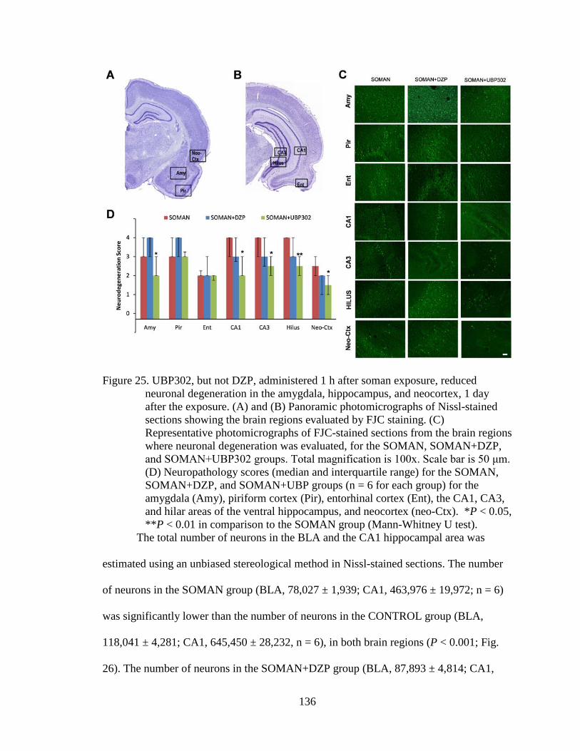
136
Figure 25. UBP302, but not DZP, administered 1 h after soman exposure, reduced neuronal degeneration in the amygdala, hippocampus, and neocortex, 1 day after the exposure. (A) and (B) Panoramic photomicrographs of Nissl-stained sections showing the brain regions evaluated by FJC staining. (C) Representative photomicrographs of FJC-stained sections from the brain regions where neuronal degeneration was evaluated, for the SOMAN, SOMAN+DZP, and SOMAN+UBP302 groups. Total magnification is 100x. Scale bar is 50 μm. (D) Neuropathology scores (median and interquartile range) for the SOMAN, SOMAN+DZP, and SOMAN+UBP groups (n = 6 for each group) for the amygdala (Amy), piriform cortex (Pir), entorhinal cortex (Ent), the CA1, CA3, and hilar areas of the ventral hippocampus, and neocortex (neo-Ctx). *P < 0.05, **P < 0.01 in comparison to the SOMAN group (Mann-Whitney U test).
The total number of neurons in the BLA and the CA1 hippocampal area was
estimated using an unbiased stereological method in Nissl-stained sections. The number
of neurons in the SOMAN group (BLA, 78,027 ± 1,939; CA1, 463,976 ± 19,972; n = 6)
was significantly lower than the number of neurons in the CONTROL group (BLA,
118,041 ± 4,281; CA1, 645,450 ± 28,232, n = 6), in both brain regions (P < 0.001; Fig.
26). The number of neurons in the SOMAN+DZP group (BLA, 87,893 ± 4,814; CA1,

137
439,186 ± 23,211; n = 6) did not differ significantly from that in the SOMAN group (P =
0.23 and P = 0.83 for the BLA and CA1, respectively; Fig. 26). In the SOMAN+UBP302
group, the number of neurons in the BLA (98,648 ± 4,513, n = 6) was significantly lower
than in the controls (P = 0.008), but also significantly higher than in the SOMAN group
(P = 0.005; Fig. 26C). The number of neurons in the CA1 area of the SOMAN+UBP302
group (588,100 ± 28,012, n = 6) did not differ from the control group (P = 0.276) and
was significantly higher than in the SOMAN group (P = 0.006; Fig. 26D).
Figure 26. UBP302, but not DZP, administered 1 h after soman exposure, reduced
neuronal loss in the BLA and the CA1 hippocampal area, 1 day after the exposure. (A) Panoramic photomicrographs of Nissl-stained half hemispheres outlining the amygdalar nucleus and the hippocampal subfield where stereological analysis was performed. (B) Representative photomicrographs of Nissl-stained sections showing BLA and CA1 cells from the CONTROL,

138
SOMAN, SOMAN+DZP and SOMAN+UBP302 groups. Total magnification is 630x and scale bar is 50 µm. (C) and (D) Group data (mean and standard error; n = 6 for each group) of stereological estimation of the total number of Nissl-stained neurons in the BLA (left) and CA1 area (right). **P < 0.01, ***P < 0.001 in comparison to CONTROL, ##P < 0.01 in comparison to the SOMAN group (ANOVA, Dunnett post-hoc test).
Neuronal loss and degeneration, 7 days after soman administration
The neurodegeneration results at 7 days after soman exposure are shown in Fig.
27. The SOMAN group (n = 6) had moderate to severe neurodegeneration in the
amygdala (median = 3, IQR = 2.75~4), piriform cortex (median = 3, IQR = 3~4 ),
entorhinal cortex (median = 3, IQR = 2~3), CA1 hippocampal area (median = 4, IQR = 3
~4), and CA3 hippocampal area (median = 3, IQR = 1~3.5), and mild to moderate in the
hilus (median = 2.5, IQR = 0~4) and neocortex (median = 2.5, IQR = 1~3). Similarly, in
the SOMAN+DZP group (n = 6), neurodegeneration was moderate to severe in the
amygdala (median = 3, IQR = 0.75~3.25), the piriform cortex (median = 3.5, IQR =
1.75~4), entorhinal cortex (median = 3, IQR = 1.5~3), CA1 hippocampal area (media =
3.5, IQR =2.5 ~4), and CA3 hippocampal area (median = 3, IQR = 0.75~3.25), and mild
to moderate in the hilus (median = 2.5, IQR = 1.75~3.25) and neocortex (median = 2,
IQR = 1~3). In the SOMAN+UBP302 group (n = 6), neurodegeneration was minimal to
mild in the entorhinal cortex (median = 1.5, IQR = 0.75~2.25), CA3 hippocampal area
(median = 0.5, IQR = 0~2), hilus (median = 1.5, IQR = 0~3), and neocortex (median = 1,
IQR = 0.75~2), mild to moderate in the amygdala (median = 2, IQR = 1.5~3) and CA1
area (median = 2, IQR =0 ~ 3), and moderate to severe in the piriform cortex (median =
3, IQR = 1.5~4). Compared to the SOMAN group, neurodegeneration in the
SOMAN+UBP302 group was significantly lower in the amygdala (P = 0.041), entorhinal
cortex (P = 0.041), CA1 (P = 0.009) and CA3 (P = 0.041) hippocampal areas.

139
Figure 27. UBP302, but not DZP, administered 1 h after soman exposure, reduced
neuronal degeneration in the amygdala, CA1 and CA3 dorsal hippocampal areas, and entorhinal cortex, 7 days after the exposure. (A) and (B) Panoramic photomicrographs of Nissl-stained sections showing the brain regions evaluated by FJC staining. (C) Representative photomicrographs of FJC-stained sections from the brain regions where neuronal degeneration was evaluated, for the SOMAN, SOMAN+DZP, and SOMAN+UBP302 groups. Total magnification is 100x. Scale bar is 50 μm. (D) Neuropathology scores (median and interquartile range) for the SOMAN, SOMAN+DZP, and SOMAN+UBP groups (n = 6 for each group), for the amygdala (Amy), piriform cortex (Pir), entorhinal cortex (Ent), the CA1, CA3 and hilar areas of the ventral hippocampus, and neocortex (neo-Ctx). *P < 0.05, **P < 0.01 in comparison to the SOMAN group (Mann-Whitney U test).
The total number of neurons in the BLA and the CA1 hippocampal area was
estimated 7 days after soman exposure; the results are shown in Fig. 28. The number of
neurons in the SOMAN group (BLA, 79,394 ± 3287; CA1, 440,698 ± 17,159; n = 6) was
again significantly lower than the number of neurons in the CONTROL group (BLA,
115,108 ± 2,673; CA1, 639,701 ± 29,350, n = 6), in both brain regions (P < 0.001). The

140
number of neurons in the SOMAN+DZP group (BLA, 87,729 ± 6,032; CA1, 463,196 ±
25,729; n = 6) did not differ significantly from those in the SOMAN group (P = 0.424
and P = 0.841 for the BLA and CA1, respectively). The number of neurons in the
SOMAN+UBP302 group (BLA, 99,678 ± 4,947; CA1, 568,098 ± 20,386; n = 6) was
significantly higher than in the SOMAN group (P = 0.011 for the BLA and P = 0.003 for
the CA1), but differed from the CONTROL group only in the BLA.
Figure 28. UBP302, but not DZP, administered 1 h after soman exposure, reduced
neuronal loss in the BLA and the CA1 hippocampal area, 7 days after the exposure. (A) Panoramic photomicrographs of Nissl-stained half hemispheres outlining the amygdalar nucleus and the hippocampal subfield where stereological analysis was performed. (B) Representative photomicrographs of Nissl-stained sections showing BLA and CA1 cells from the CONTROL, SOMAN, SOMAN+DZP and SOMAN+UBP302 groups. Total magnification is 630x and scale bar is 50 µm. (C) and (D) Group data (mean and standard error; n = 6 for each group) of stereological estimation of the total number of
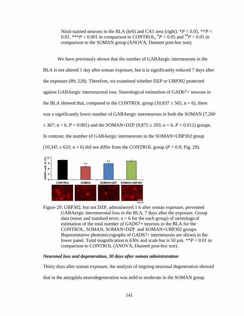
141
Nissl-stained neurons in the BLA (left) and CA1 area (right). *P < 0.05, **P < 0.01, ***P < 0.001 in comparison to CONTROL, #P < 0.05 and ##P < 0.01 in comparison to the SOMAN group (ANOVA, Dunnett post-hoc test).
We have previously shown that the number of GABAergic interneurons in the
BLA is not altered 1 day after soman exposure, but it is significantly reduced 7 days after
the exposure (89; 228). Therefore, we examined whether DZP or UBP302 protected
against GABAergic interneuronal loss. Stereological estimation of GAD67+ neurons in
the BLA showed that, compared to the CONTROL group (10,837 ± 565, n = 6), there
was a significantly lower number of GABAergic interneurons in both the SOMAN (7,260
± 367; n = 6, P = 0.001) and the SOMAN+DZP (8,875 ± 293; n = 6, P = 0.012) groups.
In contrast, the number of GABAergic interneurons in the SOMAN+UBP302 group
(10,345 ± 633; n = 6) did not differ from the CONTROL group (P = 0.9; Fig. 29).
Figure 29. UBP302, but not DZP, administered 1 h after soman exposure, prevented
GABAergic interneuronal loss in the BLA, 7 days after the exposure. Group data (mean and standard error; n = 6 for the each group) of stereological estimation of the total number of GAD67+ neurons in the BLA for the CONTROL, SOMAN, SOMAN+DZP and SOMAN+UBP302 groups. Representative photomicrographs of GAD67+ interneurons are shown in the lower panel. Total magnification is 630x and scale bar is 50 µm. **P < 0.01 in comparison to CONTROL (ANOVA, Dunnett post-hoc test).
Neuronal loss and degeneration, 30 days after soman administration
Thirty days after soman exposure, the analysis of ongoing neuronal degeneration showed
that in the amygdala neurodegeneration was mild to moderate in the SOMAN group
142
(median = 2.5, IQR = 2~3.75, n = 6) and the SOMAN+DZP group (median = 2, IQR =
2~3, n = 6), but only minimal in the SOMAN+UBP302 (median = 1, IQR = 0~2, n = 6; P
< 0.001 compared to SOMAN; Fig. 8). In the piriform cortex, neurodegeneration was
mild to moderate in the SOMAN group (median = 2, IQR = 2~3.75) and the
SOMAN+DZP group (median = 3, IQR = 2~4), but only minimal in the
SOMAN+UBP302 group (median = 1, IQR = 1~2; P = 0.002 compared to SOMAN; Fig.
30). In the CA1 hippocampal area, neurodegeneration was moderate in the SOMAN
group (median = 3, IQR = 3~4), and mild in the SOMAN+DZP group (median = 2, IQR
= 0~3; P = 0.002 compared to SOMAN) and the SOMAN+UBP302 groups (median = 2,
IQR = 0.5~3; P = 0.001). There were no significant differences between the three groups
in the neurodegeneration scores for the entorhinal cortex (SOMAN group: median = 1.5,
IQR = 1~2; SOMAN+DZP group: median = 2, IQR = 0~2; SOMAN+UBP302 group:
median = 1, IQR = 1~2), CA3 hippocampal area (SOMAN group: median = 3, IQR =
0~3; SOMAN+DZP group: median = 1, IQR = 1~3; SOMAN+UBP302 group: median =
2, IQR = ~3), the hilus (SOMAN and SOMAN+DZP groups: median = 1, IQR = 0~2;
SOMAN+UBP302 group: median = 0, IQR = 0~2), and the neocortex (SOMAN and
SOMAN+DZP groups: median = 1, IQR = 0~1; SOMAN+UBP302 group: median = 0,
IQR = 0~1).

143
Figure 30. UBP302, administered 1 h after soman exposure, reduces neuronal degeneration in the amygdala, piriform cortex, and CA1 hippocampal area, while DZP reduces neurodegeneration in the CA1 area, 30 days after the exposure. (A) and (B) Panoramic photomicrographs of Nissl-stained sections showing the brain regions evaluated by FJC staining. (C) Representative photomicrographs of FJC-stained sections from the brain regions where neuronal degeneration was evaluated, for the SOMAN, SOMAN+DZP, and SOMAN+UBP302 groups. Total magnification is 100x. Scale bar is 50 μm. (D) Neuropathology scores (median and interquartile range) for the SOMAN , SOMAN+DZP, and SOMAN+UBP groups (n = 6 for each group), for the amygdala (Amy), piriform cortex (Pir), entorhinal cortex (Ent), the CA1, CA3 and hilar areas of the dorsal hippocampus, and neocortex (neo-Ctx). *P < 0.05 in comparison to the SOMAN group (Mann-Whitney U test).
Thirty days after soman exposure, neuronal loss was still present in the SOMAN
group (n = 6), in which the total number of neurons (BLA, 82,119 ± 2099; CA1, 444,356
± 21,045) was significantly lower (P < 0.001) than in the CONTROL group (BLA,
119,860 ± 2898; CA1, 684,738 ± 15,340; n = 6; Fig. 31). In the SOMAN+UBP302 group

144
(n = 6), the total number of neurons (BLA, 101,412 ± 1535; CA1, 553,583 ± 27,489) was
significantly lower than in the CONTROL group, both in the BLA (P = 0.012; Fig. 31C)
and in the CA1 area (P = 0.006; Fig. 31D), but also significantly higher than in the
SOMAN group (P = 0.001). The number of neurons in the SOMAN+DZP group (BLA,
88,591 ± 7134; CA1, 463,211 ± 21,536; n = 6) was significantly lower than the
CONTROL group (P < 0.001), and did not differ from the number of neurons in the
SOMAN group (P = 0.55 and P = 0.34 for the BLA and the CA1 area, respectively; Fig.
31).
Figure 31. UBP302, but not DZP, administered 1 h after soman exposure, reduced
neuronal loss in the BLA and the CA1 hippocampal area, 30 days after the exposure. (A) Panoramic photomicrographs of Nissl-stained half hemispheres outlining the amygdalar nucleus and the hippocampal subfield where stereological analysis was performed. (B) Representative photomicrographs of

145
Nissl-stained sections showing BLA and CA1 cells from the CONTROL, SOMAN, SOMAN+DZP and SOMAN+UBP302 groups. Total magnification is 630x and scale bar is 50 µm. (C) and (D) Group data (mean and standard error; n = 6 for each group) of stereological estimation of the total number of Nissl-stained neurons in the BLA (left) and CA1 area (right). *P < 0.05, **P < 0.01, ***P < 0.001 in comparison to CONTROL; ##P < 0.01 in comparison to the SOMAN group (ANOVA, Dunnett post-hoc test).
Behavioral alterations 30 days after soman administration
To investigate whether the long-term neuropathology observed 30 days after
soman exposure had translated into behavioral deficits, we examined anxiety-like
behavior in all experimental groups, using the open field and the acoustic startle response
(ASR) tests. The distance traveled in the open field by the groups exposed to soman
(SOMAN: 2270 ± 265 cm, n = 10; SOMAN+DZP: 2290 ± 235 cm, n = 9;
SOMAN+UBP302: 1329 ± 279 cm, n = 12) was not significantly different from that in
the CONTROL group (1620 ± 219 cm, n = 8; Fig. 32A). However, both the SOMAN and
the SOMAN+DZP groups spent significantly less time in the center of the open field
(SOMAN group, 6.28 ± 1.56 % of the total movement time, n = 10, P = 0.034;
SOMAN+DZP group, 6.88 ± 1.29 % of the total movement time, n = 9, P = 0.027)
compared to the time spent by the CONTROL rats (15.22 ± 2.67 % of the total movement
time, n = 8). The SOMAN+UBP302 rats spent 17.61 ± 2.27 % (n = 12) of the total
movement time in the center of the open field, which was not different from the
CONTROL group (P = 0.73; Fig. 32B).
In the ASR test, there was a significant increase in startle amplitude in the
SOMAN (110 dB: 17.94 ± 0.83, n = 10, P < 0.001; 120 dB: 19.20 ± 0.35, P = 0.001) and
the SOMAN+DZP (110 dB: 17.38 ± 0.95, n = 9, P = 0.001; 120 dB: 18.85 ± 0.41, P =
0.003) groups compared to the CONTROL group (110 dB: 10.99 ± 1.35; 120 dB: 14.09 ±
1.54, n = 8; Fig. 10C). Startle response amplitude in the SOMAN+UBP302 group (110

146
dB: 13.95 ± 0.99; 120 dB: 16.03 ± 0.91, n =12) did not differ from the CONTROL group
(P = 0.123 for the 110 dB, and P = 0.291 for the 120 dB; Fig. 32C).
Figure 32. UBP302, but not DZP, administered 1 h after soman exposure, protected
against the development of anxiety, 30 days after the exposure. (A) Distance traveled (Mean ± SE) in the open field. (B) Percentage of time spent in the center of the Open Field. (C) Amplitude of startle responses to 120 dB and 110 dB acoustic stimuli. CONTROL, n = 8; SOMAN, n = 10; SOMAN+DZP, n = 9; SOMAN+UBP302, n = 12. *P < 0.05, **P < 0.01 and ***P < 0.001 (ANOVA, Dunnett post-hoc comparison to CONTROL).
Discussion
Seizures induced by exposure to nerve agents require medical intervention,
otherwise they can lead to severe brain damage or death. Administration of DZP is the
presently FDA-approved treatment for nerve agent-induced seizures. The present study
showed that if DZP is administered to soman-exposed rats at 1 hour post-exposure,

147
seizures are terminated effectively, but they soon return, resulting in a total duration of
SE within 24 h after exposure that is no different from the total SE duration in the soman-
exposed rats that do not receive anticonvulsant treatment. Moreover, if DZP is
administered at 2 h after soman exposure, the total duration of SE in the DZP-treated rats
is longer than in the rats that do not receive anticonvulsant treatment. The consequences
of the return of seizures after DZP treatment were evident in the neuropathology analysis
and the behavioral tests. Thus, DZP treatment provided no protection against neuronal
degeneration and death, except for a lower number of degenerating neurons in the CA1
hippocampal area, 30 days after the exposure. In contrast, treatment with the GluK1
antagonist UBP302, which reduced the total duration of SE within 24 hours post-
exposure, protected against neuronal damage in most of the brain regions examined. The
anxiety tests also revealed that UBP302, but not DZP treatment, prevented an increase in
anxiety-like behavior, 30 days after soman exposure.
The disadvantages of DZP treatment as an anticonvulsant
From the clinical experience and studies in human patients, as well as from experimental
studies in animals, the efficacy of benzodiazepines in terminating status epilepticus has
long been recognized to decrease as the latency between the initiation of SE and the time
point of drug administration increases (78; 102; 130; 174; 262; 301). This is also the case
with SE induced by nerve agents. Thus, in rats, Shih et al. (255) found that DZP was very
effective in stopping seizures when administered 5 min after the onset of soman-induced
SE, but was virtually ineffective when administration was delayed to 40 min after seizure
initiation. The mechanisms underlying the development of refractoriness to
benzodiazepines are not fully understood, but may involve internalization and

148
downregulation of GABAA receptors (78; 102; 199), or other dysfunctions in GABAergic
synaptic transmission that manifest as the seizures progress. At least in hippocampal
neurons, Goodkin et al. (102) have demonstrated that GABAergic inhibition displays a
high degree of plasticity, in that, during prolonged epileptiform bursting, the rate of
GABA receptor internalization increases rapidly, and the subunit composition of these
receptors swiftly changes (as suggested by the changes in the kinetics of miniature
IPSCs), perhaps due to movement of extrasynaptic GABAA receptors onto synaptic sites.
Therefore, when DZP is administered at delayed time points, the function of the
GABAergic system is already compromised, and, as a result, DZP may be ineffective.
Nevertheless, the available evidence suggests that the progressive refractoriness to
benzodiazepines is not an absolute phenomenon and can be partially overcome depending
on the animal model used to induce SE, the dose of DZP, or other factors. For example,
in a guinea pig model of nerve agent exposure, 10 mg/kg of DZP was effective against
soman-induced seizures in a sizeable proportion of the animals, even when administration
was delayed beyond 1 h after seizure onset, although its efficacy was still clearly reduced
as the latency of administration increased (McDonough et al., 2010). In addition, kainic
acid-induced SE can be stopped by 25 mg/kg DZP even when the drug is administered 3
h after the onset of seizures (231), and in the present study, 10 mg/kg DZP terminated SE
at either the 1 h or the 2 h time point of administration.
Considering the above, one would conclude that DZP remains an effective
anticonvulsant, but it is desirable to be used at early time points after seizure initiation,
for greater effectiveness. What, however, has not received enough attention is the
recurrence of intense seizures after cessation of the initial SE by DZP, and the impact that

149
such recurrence of SE has on the neuroprotective efficacy of DZP. Shih et al. (255)
reported that 25% of the animals receiving 9 mg/kg DZP at 5 min after the onset of
soman-induced seizures had seizures recurring within the 6 h monitoring period. In the
present study, seizures recurred in all of the soman-exposed rats that received DZP,
making the total duration of seizures within the 24 h period of video-EEG monitoring no
different from (Fig. 23C), or even longer than (Fig. 23D) in the soman-exposed rats that
did not receive anticonvulsant treatment. The mechanisms underlying the recurrence of
seizures after cessation of the initial SE by DZP probably involve a severely
compromised GABAergic system, in combination with the pharmacokinetics of the drug.
Thus, intense seizure activity impairs the function of GABAergic inhibition (78; 102;
199). Then, the brain is exposed to DZP, which, on one hand, enhances GABAA receptor
activity and thereby stops the SE temporarily, but on the other hand it may contribute to
more desensitization and downregulation of GABAA receptors (292; 296), further
impairing GABAergic synaptic transmission. As DZP is rapidly cleared from the
animal’s system (235), the brain remains with severely compromised GABAergic
inhibition, and therefore neuronal networks intermittently enter into intense seizure
activity, every time the unopposed glutamatergic activity intensifies.
Targeting the GluK1KRs to control seizures
While inhibition progressively weakens during SE, glutamatergic excitation is reinforced,
probably not only because of disinhibition; during SE, there is upregulation of AMPA
receptors (62; 234), as well as enhanced expression of the GluK1 subunit (291), which
could significantly contribute to worsening hyperexcitability and excitotoxicity.
Therefore, suppressing glutamatergic hyperactivity can be a more effective way to

150
control seizures, particularly if immediate treatment is not possible and the anticonvulsant
is administered with a delay after seizure onset. Indeed, we have shown previously that
the GluK1/AMPA receptor antagonist LY293558, administered 1 h after soman
exposure, stops seizures and fully protects against neuronal damage (12; 89). In the
present study, we tested UBP302, which is selective for the GluK1 receptors (190). It is
uncertain at what concentrations UBP302 reached the brain and whether other
glutamatergic receptors were also affected, however, this compound also suppresses
soman-induced seizure-like activity, in vitro, at relatively low concentrations that are
considered selective for GluK1 antagonism (11). UBP302 blocked the seizures induced
by soman, and protected against neuropathology and anxiety-related behavioral deficits.
The time-course of termination of the initial SE by UBP302 was slow in
comparison to that of DZP or LY293558. Although this may have to do with the type of
receptors being targeted and their involvement in seizure activity, it is also possible that it
relates to the pharmacokinetics of UBP302 which is not known. However, despite the
apparently slow time-course of action of UBP302, the total duration of SE within the 24 h
period after exposure was dramatically reduced in the UBP302-treated rats, compared to
either the soman-exposed rats that did not receive anticonvulsant treatment or the rats that
received DZP. The mechanisms by which a GluK1 antagonist may suppress
hyperexcitability and stop seizures have been discussed previously (17; 89). GluK1Rs
modulate both GABAergic and glutamatergic synaptic transmission, in a number of brain
regions (124). At least in the BLA and the hippocampus, the net effect of their activation
is excitatory. This is suggested by the findings that GluK1R antagonists block
epileptiform activity in hippocampal slices and limbic seizures in vivo (269). In the BLA,

151
GluK1Rs are present on postsynaptic (somatodendritic) sites of both principal cells
(Gryder and Rogawski, 2003) and interneurons (42), as well as on the presynaptic
terminals of both cell types, where they mediate facilitation of glutamate release (17) and
either facilitation or inhibition of GABA release, depending on the concentration of the
agonist (42). The net effect of their activation in the BLA network is an increased
excitation and excitability, as suggested by a greater enhancement of spontaneous EPSCs
versus IPSCs, the reduction of anxiety-like behavior when these receptors are blocked by
microinjection of UBP302 selectively into the BLA, and the increased anxiety or the
induction of seizures when a GluK1R agonist is injected into the BLA (17). Although it
remains to be determined what role GluK1Rs play in the overall excitation of other
neuronal networks, the fact that activation of these receptors increases the excitation state
of the BLA and the hippocampus, two highly seizurogenic brain regions, may explain
why blockade of these receptors can terminate seizures. It should also be noted that in
contrast to the downregulation and internalization of GABAA receptors after excessive
neuronal/seizure activity (78; 102; 199), which can render DZP ineffective, the
upregulation of the GluK1 subunit (291) may contribute to hyper-excitation, which
further explains the efficacy of GluK1R antagonists.
Neuropathology and associated behavioral deficits
Depending on the extent of brain damage after prolonged SE, behavioral deficits may
ensue. Increased anxiety is observed in animals that have been exposed to nerve agents
(74; 155; 228). Similarly, the human victims of the sarin terrorist attack in Japan report
enduring symptoms of anxiety disorders, long after the exposure (118; 204). The
amygdala plays a central role in emotional behavior, and dysfunction with increased

152
excitability of the BLA is associated with anxiety (101; 221; 253; 318). The hippocampus
is also significantly involved in the modulation of anxiety (84; 93). Both the amygdala
and the hippocampus are severely damaged by nerve agent-induced SE, as shown in the
present and previous studies (14; 15; 258). In the BLA, there is significant loss of
GABAergic neurons by day 7 after exposure to soman (88; 228), and at 14 and 30 days
after exposure the ratio of GABAergic interneurons to the total number of neurons in the
BLA is significantly decreased (228). Reduction of inhibitory activity in the BLA,
resulting from the interneuronal loss, will increase the excitability of the BLA network,
which can lead to the development of anxiety.
In the present study, treatment with UBP302 significantly reduced neuronal loss
and degeneration in a number of brain regions, including the hippocampus and the
amygdala, and prevented the loss of GABAergic interneurons in the BLA, as assessed 30
days after the exposure. The neuroprotection provided by UBP302, despite its being only
partial, was sufficient to prevent the development of anxiety-like behavior, as assessed in
the open field and the acoustic startle response tests. In contrast, DZP had no
neuroprotective effects, except for reduced neurodegeneration in the CA1 area at 30 days
after exposure, and did not prevent the development of anxiety. Since the extent of nerve
agent-induced neuropathology is solely or largely determined by the intensity and
duration of seizures (227; 258), the failure of DZP to protect the brain can be attributed to
the failure of this drug to reduce the total duration of seizures. Consistent with the present
findings revealing the inefficacy of DZP to reduce neuropathology and prevent
behavioral deficits, previous studies in which 10 mg/kg DZP was administered at 30 min

153
after soman-induced seizures show that this treatment did not prevent the development of
anxiety-like behavior (155) or epileptogenesis (76).
The present data clearly argue against the use of DZP as a medical
countermeasure for the treatment of SE induced by nerve agent exposure, at least when
anticonvulsant treatment is delayed. DZP is likely to substantially increase the survival
rate of exposed individuals, but because seizures recur, there is virtually no protection
against neuropathology and the resulting behavioral deficits. Administering DZP
repeatedly, every time intense seizures recur, not only may not be feasible, but may also
be detrimental to the victims’ health. The results suggest that targeting the glutamatergic
system is a more effective approach to controlling nerve agent-induced SE, and
antagonists of the GluK1Rs appear to be both safe and efficacious in this regard.
Authorship contributions
Participated in research design: Braga, Aroniadou-Anderjaska, Apland
Conducted experiments: Apland, Figueiredo, Rossetti, Miller
Performed data analysis: Figueiredo, Apland, Rossetti, Aroniadou-Anderjaska,
Braga
Wrote or contributed to the writing of the manuscript: Aroniadou-Anderjaska,
Figueiredo, Apland, Miller, Braga

154

155
APPENDIX 4: NEURO.TV: NEUROSCIENCE EDUCATION ON THE INTERNET
Xie Diana L1, Miller Steven L2, Boucher Leanne3, Kubie John L4, Gariépy Jean-Francois1*
1Duke Institute for Brain Sciences, Duke University, Box 91003, Levine Science Research Center, Room B107, 450 Research Drive, Durham, North Caroline 27708, United States of America 2Program in Neuroscience, Uniformed Services University of the Health Sciences, Bethesda, MD 20814, United States of America 3Farquhar Psychology Program, Nova Southeastern University, Davie-Fort Lauderdale, FL 33314, United States of America 4Department of Anatomy and Cell Biology, The Robert F. Furchgott Center for Neural and Behavioral Science, State University of New York Downstate Medical Center, Brooklyn, New York, USA
*Corresponding Author: Jean-Francois Gariepy, Ph.D. Duke Institute for Brain Sciences Duke University, Box 91003 Levine Science Research Center Room B107, 450 Research Drive Durham, North Carolina 27708, U.S.A. Telephone: 919-688-0333 Email: [email protected]

156
ABSTRACT
NEURO.tv is a new educational project that seeks to bring advanced concepts in
neuroscience to the general public. We film one-hour discussions with leading
neuroscientists, philosophers, and psychologists who have had significant impact on our
current understanding of brain function, and we publish these discussions on YouTube,
iTunes, and other social media outlets. Here, we explain the motivations behind this new
program.
AN ONLINE PROGRAM TO DISSEMINATE KNOWLEDGE ABOUT BRAIN AND MIND
The internet has an enormous impact on the way people learn. With the advent of
Massive Open Online Courses (MOOCs), academic blogs, and online education tools,
anyone can learn anything, from almost anywhere. While academics take part in this
online education movement, we have not yet fully benefited from the educational
opportunities that the internet can provide. Interactions between researchers in different
fields and the general public typically happen in universities or through the publishing of
textbooks and journal articles between academics are sometimes precluded by the
monetary and physical costs of conferences, traveling, and visits to universities. New
technologies offer an avenue for scientists to share their knowledge with colleagues and
members of the general public in a more efficient and open manner.
Despite the progress that the internet has made in leveling the barriers between
scientists and the general public, there exists a need for a forum in which advanced
subjects at the heart of current neuroscience research can be discussed openly, with
participation from the general public. A new internet-based program called NEURO.tv
addresses this need, by airing one-hour discussions with scientists who have made

157
significant discoveries in neuroscience. We believe this program provides a platform for
academics to present ideas and discoveries directly to the public in a setting that is not
rushed or overly technical, and that remains accessible to individuals of all backgrounds.
NEURO.tv’s format is not merely a repetition of presentation content by a speaker, but
rather a new type of interaction between scientists of different fields, discussing what
they really think about the scientific methods used in their research, advances in medical
treatment, and other important scientific problems that shape or limit current research.
Leading neuroscientists, philosophers, and psychologists have already
volunteered their time to appear on the show. Our success in having many scientists
commit to appearing on the program is due in part to the fact that filming an episode does
not require travel, since physical presence is not required; scientists can simply connect to
conversations with each other from their home or office computers. Within a one-hour
discussion, they are able to convey their thoughts on neuroscience-related topics to a
wide public on the scale of many thousands, which is advantageous compared to the
number of students they would reach in a traditional conference room or lecture hall. The
Internet-based, asynchronous delivery also provides broad and flexible access to these
fascinating discussions, years after the filming. Free access is a particularly crucial aspect
of NEURO.tv and may benefit developed as well as developing countries, which may
help reduce the gap in global access to advanced scientific knowledge (2). In principle,
there is no reason why individuals in one country ought to have access to discussions
with leading minds in neuroscientific research, while others cannot. As such, there
remains a major demand for higher level education to be made available to the public and
anyone interested in the subject. With the advent of crowd-funding platforms such as

158
Kickstarter, we have seen that independently operated programs like NEURO.tv can be
entirely funded by the individuals who are interested in such educational initiatives (312).
NEURO.tv is not just targeted at educating the public; those involved in its
production learn from their experience in the program as well. The program trains young
scientists in scientific communication by placing them in a panelist or co-host position, a
role that requires several weeks of preparation leading up to the filming. This preparation
includes reading scientific articles, formulating topics to discuss, and preparing questions
that will allow anyone with minimal understanding of biology to understand the show. It
is a rare, novel, firsthand learning experience in public science communication and
education.
Public science education has experienced a remarkable transformation as the
internet has grown, with many high quality podcasts, videos, and blogs now being
available for free to anyone interested in learning about science. However, a specialised
niche has not yet been filled, one in which listeners could learn about how research is
conducted, with an in-depth discussion on the details of a scientific discovery along with
the implications of a result or cutting-edge technique. NEURO.tv fills this niche, with
content that does not avoid these details, but rather embraces them, producing episodes
that do not only focus on the product of research, but also delve deeper into the thought
process behind it and why scientists care about the questions being covered.
The internet has also created opportunities to improve transparency and honesty
in academia. In an academic presentation setting, there is rarely room to discuss any of
the doubts scientists naturally harbor about their research or techniques, or the works of
others. Neuroscience is a work-in-progress, and as with all science, relies on the best

159
available techniques at the time the research is conducted, rather than the perfect ones.
Discussions on NEURO.tv take place in an informal setting, which allows scientists to
freely express their ideas and opinions about research and methods, without fearing being
dismissed. Open conversations like these can only serve to better the field and improve
the confidence of the general public in the scientific process and in the ability of
scientists to question themselves.
With the advent of the internet, there has been a movement towards increasing the
accessibility of scientific knowledge. We believe that programs such as NEURO.tv allow
a novel interface between scientists and the general public. With this new experiment in
scientific communication, we hope that highly-motivated individuals in the general
public will develop a taste for a deeper understanding of neuroscience, and that
neuroscientists will embrace the idea that part of the work lies on their shoulders, and that
simplifying the explanation of their research will benefit the general public, the field, and
themselves.

160
REFERENCES
1. 2000. Chemical-biological terrorism and its impact on children: a subject review. American Academy of Pediatrics. Committee on Environmental Health and Committee on Infectious Diseases. Pediatrics 105:662-70
2. 2003. Worldwide neuroscience. Nature neuroscience 6:901 3. 2014. In situ Hybridization White Paper. http://www.brain-map.org 4. Abdel-Rahman A, Shetty AK, Abou-Donia MB. 2002. Acute exposure to sarin
increases blood brain barrier permeability and induces neuropathological changes in the rat brain: dose-response relationships. Neuroscience 113:721-41
5. Adhikari A. 2014. Distributed circuits underlying anxiety. Frontiers in behavioral neuroscience 8:112
6. Alderson RF, Alterman AL, Barde YA, Lindsay RM. 1990. Brain-derived neurotrophic factor increases survival and differentiated functions of rat septal cholinergic neurons in culture. Neuron 5:297-306
7. Amitai Y, Almog S, Singer R, Hammer R, Bentur Y, Danon YL. 1992. Atropine poisoning in children during the Persian Gulf crisis. A national survey in Israel. Jama 268:630-2
8. Andersen SL. 2003. Trajectories of brain development: point of vulnerability or window of opportunity? Neuroscience and biobehavioral reviews 27:3-18
9. Anderson DR, Harris LW, Chang FC, Baze WB, Capacio BR, et al. 1997. Antagonism of soman-induced convulsions by midazolam, diazepam and scopolamine. Drug and chemical toxicology 20:115-31
10. Andreollo NA, Santos EF, Araujo MR, Lopes LR. 2012. Rat's age versus human's age: what is the relationship? Arquivos brasileiros de cirurgia digestiva : ABCD = Brazilian archives of digestive surgery 25:49-51
11. Apland JP, Aroniadou-Anderjaska V, Braga MF. 2009. Soman induces ictogenesis in the amygdala and interictal activity in the hippocampus that are blocked by a GluR5 kainate receptor antagonist in vitro. Neuroscience 159:380-9
12. Apland JP, Aroniadou-Anderjaska V, Figueiredo TH, Green CE, Swezey R, et al. 2013. Efficacy of the GluK1/AMPA receptor antagonist LY293558 against seizures and neuropathology in a soman-exposure model without pretreatment and its pharmacokinetics after intramuscular administration. The Journal of pharmacology and experimental therapeutics 344:133-40
13. Apland JP, Aroniadou-Anderjaska V, Figueiredo TH, Rossetti F, Miller SL, Braga MF. 2014. The limitations of diazepam as a treatment for nerve agent-induced seizures and neuropathology in rats: comparison with UBP302. The Journal of pharmacology and experimental therapeutics 351:359-72
14. Apland JP, Figueiredo TH, Qashu F, Aroniadou-Anderjaska V, Souza AP, Braga MF. 2010. Higher susceptibility of the ventral versus the dorsal hippocampus and the posteroventral versus anterodorsal amygdala to soman-induced neuropathology. Neurotoxicology 31:485-92
15. Aroniadou-Anderjaska V, Figueiredo TH, Apland JP, Qashu F, Braga MF. 2009. Primary brain targets of nerve agents: the role of the amygdala in comparison to the hippocampus. Neurotoxicology 30:772-6

161
16. Aroniadou-Anderjaska V, Fritsch B, Qashu F, Braga MF. 2008. Pathology and pathophysiology of the amygdala in epileptogenesis and epilepsy. Epilepsy research 78:102-16
17. Aroniadou-Anderjaska V, Pidoplichko VI, Figueiredo TH, Almeida-Suhett CP, Prager EM, Braga MF. 2012. Presynaptic facilitation of glutamate release in the basolateral amygdala: a mechanism for the anxiogenic and seizurogenic function of GluK1 receptors. Neuroscience 221:157-69
18. Aroniadou-Anderjaska V, Qashu F, Braga MF. 2007. Mechanisms regulating GABAergic inhibitory transmission in the basolateral amygdala: implications for epilepsy and anxiety disorders. Amino acids 32:305-15
19. Ashani Y, Catravas GN. 1981. Seizure-induced changes in the permeability of the blood-brain barrier following administration of anticholinesterase drugs to rats. Biochemical pharmacology 30:2593-601
20. Askwith CC, Benson CJ, Welsh MJ, Snyder PM. 2001. DEG/ENaC ion channels involved in sensory transduction are modulated by cold temperature. Proceedings of the National Academy of Sciences of the United States of America 98:6459-63
21. Atlas ADMB. 2009. Allen Developing Mouse Brain Atlas [Internet]. Allen Institute for Brain Science, Seattle (WA)(Available from: http://www. developingmouse. brain-map. org)
22. Bahn S, Volk B, Wisden W. 1994. Kainate receptor gene expression in the developing rat brain. The Journal of neuroscience : the official journal of the Society for Neuroscience 14:5525-47
23. Baille V, Clarke PG, Brochier G, Dorandeu F, Verna JM, et al. 2005. Soman-induced convulsions: the neuropathology revisited. Toxicology 215:1-24
24. Bajgar J. 2005. Complex view on poisoning with nerve agents and organophosphates. Acta medica 48:3-21
25. Bajgar J. 2010. Optimal choice of acetylcholinesterase reactivators for antidotal treatment of nerve agent intoxication. Acta medica 53:207-11
26. Balali-Mood M, Saber H. 2012. Recent advances in the treatment of organophosphorous poisonings. Iranian journal of medical sciences 37:74-91
27. Baron A, Waldmann R, Lazdunski M. 2002. ASIC-like, proton-activated currents in rat hippocampal neurons. The Journal of physiology 539:485-94
28. Bassler EL, Ngo-Anh TJ, Geisler HS, Ruppersberg JP, Grunder S. 2001. Molecular and functional characterization of acid-sensing ion channel (ASIC) 1b. The Journal of biological chemistry 276:33782-7
29. Beck KD, Knusel B, Hefti F. 1993. The nature of the trophic action of brain-derived neurotrophic factor, des(1-3)-insulin-like growth factor-1, and basic fibroblast growth factor on mesencephalic dopaminergic neurons developing in culture. Neuroscience 52:855-66
30. Ben-Ari Y, Holmes GL. 2006. Effects of seizures on developmental processes in the immature brain. The Lancet. Neurology 5:1055-63
31. Ben-Ari Y, Zigmond RE, Shute CC, Lewis PR. 1977. Regional distribution of choline acetyltransferase and acetylcholinesterase within the amygdaloid complex and stria terminalis system. Brain research 120:435-44
32. Bernasconi N, Natsume J, Bernasconi A. 2005. Progression in temporal lobe epilepsy: differential atrophy in mesial temporal structures. Neurology 65:223-8
162
33. Betz AL, Goldstein GW. 1981. Developmental changes in metabolism and transport properties of capillaries isolated from rat brain. The Journal of physiology 312:365-76
34. Biagini G, Babinski K, Avoli M, Marcinkiewicz M, Seguela P. 2001. Regional and subunit-specific downregulation of acid-sensing ion channels in the pilocarpine model of epilepsy. Neurobiology of disease 8:45-58
35. Bickler PE, Gallego SM, Hansen BM. 1993. Developmental changes in intracellular calcium regulation in rat cerebral cortex during hypoxia. Journal of cerebral blood flow and metabolism : official journal of the International Society of Cerebral Blood Flow and Metabolism 13:811-9
36. Bleakman R, Schoepp DD, Ballyk B, Bufton H, Sharpe EF, et al. 1996. Pharmacological discrimination of GluR5 and GluR6 kainate receptor subtypes by (3S,4aR,6R,8aR)-6-[2-(1(2)H-tetrazole-5-yl)ethyl]decahyd roisdoquinoline-3 carboxylic-acid. Molecular pharmacology 49:581-5
37. Bokonjic D, Rosic N. 1991. Anticonvulsive and protective effects of diazepam and midazolam in rats poisoned by highly toxic organophosphorus compounds. Arhiv za higijenu rada i toksikologiju 42:359-65
38. Bolshakov KV, Essin KV, Buldakova SL, Dorofeeva NA, Skatchkov SN, et al. 2002. Characterization of acid-sensitive ion channels in freshly isolated rat brain neurons. Neuroscience 110:723-30
39. Boskovic B. 1981. The treatment of Soman poisoning and its perspectives. Fundamental and applied toxicology : official journal of the Society of Toxicology 1:203-13
40. Bradford MM. 1976. A rapid and sensitive method for the quantitation of microgram quantities of protein utilizing the principle of protein-dye binding. Analytical biochemistry 72:248-54
41. Braga MF, Aroniadou-Anderjaska V, Li H, Rogawski MA. 2009. Topiramate reduces excitability in the basolateral amygdala by selectively inhibiting GluK1 (GluR5) kainate receptors on interneurons and positively modulating GABAA receptors on principal neurons. The Journal of pharmacology and experimental therapeutics 330:558-66
42. Braga MF, Aroniadou-Anderjaska V, Xie J, Li H. 2003. Bidirectional modulation of GABA release by presynaptic glutamate receptor 5 kainate receptors in the basolateral amygdala. The Journal of neuroscience : the official journal of the Society for Neuroscience 23:442-52
43. Braitman DJ, Sparenborg S. 1989. MK-801 protects against seizures induced by the cholinesterase inhibitor soman. Brain research bulletin 23:145-8
44. Buckley NJ, Bonner TI, Brann MR. 1988. Localization of a family of muscarinic receptor mRNAs in rat brain. The Journal of neuroscience : the official journal of the Society for Neuroscience 8:4646-52
45. Bunemann M, Hosey MM. 2001. Novel signalling events mediated by muscarinic receptor subtypes. Life sciences 68:2525-33
46. Burback BL, Cabell LA, Johnson JD, McDonough JA, Osheroff MR. 2013. International Journal of Toxicology special edition volume of MMB4 DMS. International journal of toxicology 32:3S-4S

163
47. Bymaster FP, McKinzie DL, Felder CC, Wess J. 2003. Use of M1-M5 muscarinic receptor knockout mice as novel tools to delineate the physiological roles of the muscarinic cholinergic system. Neurochemical research 28:437-42
48. Campo-Soria C, Chang Y, Weiss DS. 2006. Mechanism of action of benzodiazepines on GABAA receptors. British journal of pharmacology 148:984-90
49. Capacio BR, Byers CE, Merk KA, Smith JR, McDonough JH. 2004. Pharmacokinetic studies of intramuscular midazolam in guinea pigs challenged with soman. Drug and chemical toxicology 27:95-110
50. Capacio BR, Shih TM. 1991. Anticonvulsant actions of anticholinergic drugs in soman poisoning. Epilepsia 32:604-15
51. Carlsen J, Heimer L. 1986. A correlated light and electron microscopic immunocytochemical study of cholinergic terminals and neurons in the rat amygdaloid body with special emphasis on the basolateral amygdaloid nucleus. The Journal of comparative neurology 244:121-36
52. Carpentier P, Delamanche IS, Le Bert M, Blanchet G, Bouchaud C. 1990. Seizure-related opening of the blood-brain barrier induced by soman: possible correlation with the acute neuropathology observed in poisoned rats. Neurotoxicology 11:493-508
53. Carpentier P, Testylier G, Dorandeu F, Segebarth C, Montigon O, et al. 2008. Hyperosmolar treatment of soman-induced brain lesions in mice: evaluation of the effects through diffusion-weighted magnetic resonance imaging and through histology. Toxicology 253:97-103
54. Carta M, Fievre S, Gorlewicz A, Mulle C. 2014. Kainate receptors in the hippocampus. The European journal of neuroscience 39:1835-44
55. Cascino GD. 1995. Clinical correlations with hippocampal atrophy. Magnetic resonance imaging 13:1133-6
56. Cascino GD, Jack CR, Jr., Parisi JE, Sharbrough FW, Hirschorn KA, et al. 1991. Magnetic resonance imaging-based volume studies in temporal lobe epilepsy: pathological correlations. Annals of neurology 30:31-6
57. Cendes F, Andermann F, Dubeau F, Gloor P, Evans A, et al. 1993. Early childhood prolonged febrile convulsions, atrophy and sclerosis of mesial structures, and temporal lobe epilepsy: an MRI volumetric study. Neurology 43:1083-7
58. Cendes F, Andermann F, Gloor P, Gambardella A, Lopes-Cendes I, et al. 1994. Relationship between atrophy of the amygdala and ictal fear in temporal lobe epilepsy. Brain : a journal of neurology 117 ( Pt 4):739-46
59. Cendes F, Andermann F, Gloor P, Lopes-Cendes I, Andermann E, et al. 1993. Atrophy of mesial structures in patients with temporal lobe epilepsy: cause or consequence of repeated seizures? Annals of neurology 34:795-801
60. Chatterjee S, Riaz H. 2013. Death by insecticide. Bmj 346:f2029 61. Chen CC, England S, Akopian AN, Wood JN. 1998. A sensory neuron-specific,
proton-gated ion channel. Proceedings of the National Academy of Sciences of the United States of America 95:10240-5

164
62. Chen Y. 2012. Organophosphate-induced brain damage: mechanisms, neuropsychiatric and neurological consequences, and potential therapeutic strategies. Neurotoxicology 33:391-400
63. Cheng B, Mattson MP. 1994. NT-3 and BDNF protect CNS neurons against metabolic/excitotoxic insults. Brain research 640:56-67
64. Chesler M. 2003. Regulation and modulation of pH in the brain. Physiological reviews 83:1183-221
65. Clement JG, Broxup B. 1993. Efficacy of diazepam and avizafone against soman-induced neuropathology in brain of rats. Neurotoxicology 14:485-504
66. Collingridge GL, Olsen RW, Peters J, Spedding M. 2009. A nomenclature for ligand-gated ion channels. Neuropharmacology 56:2-5
67. Collombet JM. 2011. Nerve agent intoxication: recent neuropathophysiological findings and subsequent impact on medical management prospects. Toxicology and applied pharmacology 255:229-41
68. Collombet JM, Carpentier P, Baille V, Four E, Bernabe D, et al. 2006. Neuronal regeneration partially compensates the delayed neuronal cell death observed in the hippocampal CA1 field of soman-poisoned mice. Neurotoxicology 27:201-9
69. Collombet JM, Four E, Bernabe D, Masqueliez C, Burckhart MF, et al. 2005. Soman poisoning increases neural progenitor proliferation and induces long-term glial activation in mouse brain. Toxicology 208:319-34
70. Collombet JM, Pierard C, Beracochea D, Coubard S, Burckhart MF, et al. 2008. Long-term consequences of soman poisoning in mice Part 1. Neuropathology and neuronal regeneration in the amygdala. Behavioural brain research 191:88-94
71. Cortes R, Palacios JM. 1986. Muscarinic cholinergic receptor subtypes in the rat brain. I. Quantitative autoradiographic studies. Brain research 362:227-38
72. Coryell MW, Wunsch AM, Haenfler JM, Allen JE, Schnizler M, et al. 2009. Acid-sensing ion channel-1a in the amygdala, a novel therapeutic target in depression-related behavior. The Journal of neuroscience : the official journal of the Society for Neuroscience 29:5381-8
73. Coryell MW, Ziemann AE, Westmoreland PJ, Haenfler JM, Kurjakovic Z, et al. 2007. Targeting ASIC1a reduces innate fear and alters neuronal activity in the fear circuit. Biological psychiatry 62:1140-8
74. Coubard S, Beracochea D, Collombet JM, Philippin JN, Krazem A, et al. 2008. Long-term consequences of soman poisoning in mice: part 2. Emotional behavior. Behavioural brain research 191:95-103
75. Coyle JT, Yamamura HI. 1976. Neurochemical aspects of the ontogenesis of cholinergic neurons in the rat brain. Brain research 118:429-40
76. de Araujo Furtado M, Lumley LA, Robison C, Tong LC, Lichtenstein S, Yourick DL. 2010. Spontaneous recurrent seizures after status epilepticus induced by soman in Sprague-Dawley rats. Epilepsia 51:1503-10
77. de Araujo Furtado M, Rossetti F, Chanda S, Yourick D. 2012. Exposure to nerve agents: from status epilepticus to neuroinflammation, brain damage, neurogenesis and epilepsy. Neurotoxicology 33:1476-90
78. Deeb TZ, Maguire J, Moss SJ. 2012. Possible alterations in GABAA receptor signaling that underlie benzodiazepine-resistant seizures. Epilepsia 53 Suppl 9:79-88
165
79. Deshpande LS, Carter DS, Phillips KF, Blair RE, DeLorenzo RJ. 2014. Development of status epilepticus, sustained calcium elevations and neuronal injury in a rat survival model of lethal paraoxon intoxication. Neurotoxicology 44:17-26
80. Diochot S, Salinas M, Baron A, Escoubas P, Lazdunski M. 2007. Peptides inhibitors of acid-sensing ion channels. Toxicon : official journal of the International Society on Toxinology 49:271-84
81. Dolgin E. 2013. Syrian gas attack reinforces need for better anti-sarin drugs. Nature medicine 19:1194-5
82. Dwyer JM, Rizzo SJ, Neal SJ, Lin Q, Jow F, et al. 2009. Acid sensing ion channel (ASIC) inhibitors exhibit anxiolytic-like activity in preclinical pharmacological models. Psychopharmacology 203:41-52
83. Ellman GL, Courtney KD, Andres V, Jr., Feather-Stone RM. 1961. A new and rapid colorimetric determination of acetylcholinesterase activity. Biochemical pharmacology 7:88-95
84. Engin E, Treit D. 2007. The role of hippocampus in anxiety: intracerebral infusion studies. Behavioural pharmacology 18:365-74
85. Escoubas P, De Weille JR, Lecoq A, Diochot S, Waldmann R, et al. 2000. Isolation of a tarantula toxin specific for a class of proton-gated Na+ channels. The Journal of biological chemistry 275:25116-21
86. Etkin A, Wager TD. 2007. Functional neuroimaging of anxiety: a meta-analysis of emotional processing in PTSD, social anxiety disorder, and specific phobia. The American journal of psychiatry 164:1476-88
87. Faraday MM, Elliott BM, Grunberg NE. 2001. Adult vs. adolescent rats differ in biobehavioral responses to chronic nicotine administration. Pharmacology, biochemistry, and behavior 70:475-89
88. Figueiredo TH, Aroniadou-Anderjaska V, Qashu F, Apland JP, Pidoplichko V, et al. 2011. Neuroprotective efficacy of caramiphen against soman and mechanisms of its action. British journal of pharmacology 164:1495-505
89. Figueiredo TH, Qashu F, Apland JP, Aroniadou-Anderjaska V, Souza AP, Braga MF. 2011. The GluK1 (GluR5) Kainate/alpha-amino-3-hydroxy-5-methyl-4-isoxazolepropionic acid receptor antagonist LY293558 reduces soman-induced seizures and neuropathology. The Journal of pharmacology and experimental therapeutics 336:303-12
90. Filliat P, Coubard S, Pierard C, Liscia P, Beracochea D, et al. 2007. Long-term behavioral consequences of soman poisoning in mice. Neurotoxicology 28:508-19
91. Fisahn A, Yamada M, Duttaroy A, Gan JW, Deng CX, et al. 2002. Muscarinic induction of hippocampal gamma oscillations requires coupling of the M1 receptor to two mixed cation currents. Neuron 33:615-24
92. Foltin G, Tunik M, Curran J, Marshall L, Bove J, et al. 2006. Pediatric nerve agent poisoning: medical and operational considerations for emergency medical services in a large American city. Pediatric emergency care 22:239-44
93. Fournier NM, Duman RS. 2013. Illuminating hippocampal control of fear memory and anxiety. Neuron 77:803-6
94. Gale GD, Anagnostaras SG, Godsil BP, Mitchell S, Nozawa T, et al. 2004. Role of the basolateral amygdala in the storage of fear memories across the adult

166
lifetime of rats. The Journal of neuroscience : the official journal of the Society for Neuroscience 24:3810-5
95. Garcia SJ, Abu-Qare AW, Meeker-O'Connell WA, Borton AJ, Abou-Donia MB. 2003. Methyl parathion: a review of health effects. Journal of toxicology and environmental health. Part B, Critical reviews 6:185-210
96. Gielen MC, Lumb MJ, Smart TG. 2012. Benzodiazepines modulate GABAA receptors by regulating the preactivation step after GABA binding. The Journal of neuroscience : the official journal of the Society for Neuroscience 32:5707-15
97. Gilat E, Kadar T, Levy A, Rabinovitz I, Cohen G, et al. 2005. Anticonvulsant treatment of sarin-induced seizures with nasal midazolam: an electrographic, behavioral, and histological study in freely moving rats. Toxicology and applied pharmacology 209:74-85
98. Giniatullin R, Nistri A, Yakel JL. 2005. Desensitization of nicotinic ACh receptors: shaping cholinergic signaling. Trends in neurosciences 28:371-8
99. Gisabella B, Bolshakov VY, Benes FM. 2012. Kainate receptor-mediated modulation of hippocampal fast spiking interneurons in a rat model of schizophrenia. PloS one 7:e32483
100. Goel A, Aggarwal P. 2007. Pesticide poisoning. The National medical journal of India 20:182-91
101. Gonzalez LE, Andrews N, File SE. 1996. 5-HT1A and benzodiazepine receptors in the basolateral amygdala modulate anxiety in the social interaction test, but not in the elevated plus-maze. Brain research 732:145-53
102. Goodkin HP, Liu X, Holmes GL. 2003. Diazepam terminates brief but not prolonged seizures in young, naive rats. Epilepsia 44:1109-12
103. Grunder S, Chen X. 2010. Structure, function, and pharmacology of acid-sensing ion channels (ASICs): focus on ASIC1a. International journal of physiology, pathophysiology and pharmacology 2:73-94
104. Guerreiro C, Cendes F, Li LM, Jones-Gotman M, Andermann F, et al. 1999. Clinical patterns of patients with temporal lobe epilepsy and pure amygdalar atrophy. Epilepsia 40:453-61
105. Gundersen HJ, Jensen EB, Kieu K, Nielsen J. 1999. The efficiency of systematic sampling in stereology--reconsidered. Journal of microscopy 193:199-211
106. Gupta A, Agarwal R, Shukla GS. 1999. Functional impairment of blood-brain barrier following pesticide exposure during early development in rats. Human & experimental toxicology 18:174-9
107. Halley SA, Wrench JM, Reutens DC, Wilson SJ. 2010. The amygdala and anxiety after epilepsy surgery. Epilepsy & behavior : E&B 18:431-6
108. Hamilton MG, Posavad C. 1991. Alteration of calcium influx in rat cortical synaptosomes by soman. Neuroreport 2:273-6
109. Hamilton SE, Loose MD, Qi M, Levey AI, Hille B, et al. 1997. Disruption of the m1 receptor gene ablates muscarinic receptor-dependent M current regulation and seizure activity in mice. Proceedings of the National Academy of Sciences of the United States of America 94:13311-6
110. Harris LW, Gennings C, Carter WH, Anderson DR, Lennox WJ, et al. 1994. Efficacy comparison of scopolamine (SCP) and diazepam (DZ) against soman-induced lethality in guinea pigs. Drug and chemical toxicology 17:35-50

167
111. Harrison PK, Sheridan RD, Green AC, Scott IR, Tattersall JE. 2004. A guinea pig hippocampal slice model of organophosphate-induced seizure activity. The Journal of pharmacology and experimental therapeutics 310:678-86
112. Hauser WA. 1994. The prevalence and incidence of convulsive disorders in children. Epilepsia 35 Suppl 2:S1-6
113. Haut SR, Veliskova J, Moshe SL. 2004. Susceptibility of immature and adult brains to seizure effects. The Lancet. Neurology 3:608-17
114. Haut SR, Veliškova J, Moshé SL. 2004. Susceptibility of immature and adult brains to seizure effects. The Lancet Neurology 3:608-17
115. Hayward IJ, Wall HG, Jaax NK, Wade JV, Marlow DD, Nold JB. 1990. Decreased brain pathology in organophosphate-exposed rhesus monkeys following benzodiazepine therapy. Journal of the neurological sciences 98:99-106
116. Henretig FM, Cieslak TJ, Eitzen EM, Jr. 2002. Biological and chemical terrorism. The Journal of pediatrics 141:311-26
117. Hill JA, Jr., Zoli M, Bourgeois JP, Changeux JP. 1993. Immunocytochemical localization of a neuronal nicotinic receptor: the beta 2-subunit. The Journal of neuroscience : the official journal of the Society for Neuroscience 13:1551-68
118. Hoffman A, Eisenkraft A, Finkelstein A, Schein O, Rotman E, Dushnitsky T. 2007. A decade after the Tokyo sarin attack: a review of neurological follow-up of the victims. Military medicine 172:607-10
119. Holmes GL. 2005. Effects of seizures on brain development: lessons from the laboratory. Pediatric neurology 33:1-11
120. Holmes GL. 2009. The 2008 Judith Hoyer lecture: epilepsy in children: listening to mothers. Epilepsy & behavior : E&B 16:193-202
121. Holmes GL, Ben-Ari Y. 1998. Seizures in the developing brain: perhaps not so benign after all. Neuron 21:1231-4
122. Hyman C, Juhasz M, Jackson C, Wright P, Ip NY, Lindsay RM. 1994. Overlapping and distinct actions of the neurotrophins BDNF, NT-3, and NT-4/5 on cultured dopaminergic and GABAergic neurons of the ventral mesencephalon. The Journal of neuroscience : the official journal of the Society for Neuroscience 14:335-47
123. International Program on Chemical Safety. The WHO recommended classification of pesticides by hazard and guidelines to classification. p. v. Geneva: World Health Organization
124. Jane DE, Lodge D, Collingridge GL. 2009. Kainate receptors: pharmacology, function and therapeutic potential. Neuropharmacology 56:90-113
125. Jett DA. 2007. Neurological aspects of chemical terrorism. Annals of neurology 61:9-13
126. Jimmerson VR, Shih TM, Mailman RB. 1989. Variability in soman toxicity in the rat: correlation with biochemical and behavioral measures. Toxicology 57:241-54
127. Johansen JP, Cain CK, Ostroff LE, LeDoux JE. 2011. Molecular mechanisms of fear learning and memory. Cell 147:509-24
128. Johnson DD, Lowndes HE. 1974. Reduction by diazepam of repetitive electrical activity and toxicity resulting from soman. European journal of pharmacology 28:245-50

168
129. Johnson DD, Wilcox WC. 1975. Studies on the mechanism of the protective and antidotal actions of diazepam in organophosphate poisoning. European journal of pharmacology 34:127-32
130. Jones DM, Esmaeil N, Maren S, Macdonald RL. 2002. Characterization of pharmacoresistance to benzodiazepines in the rat Li-pilocarpine model of status epilepticus. Epilepsy research 50:301-12
131. Joosen MJ, van der Schans MJ, van Dijk CG, Kuijpers WC, Wortelboer HM, van Helden HP. 2011. Increasing oxime efficacy by blood-brain barrier modulation. Toxicology letters 206:67-71
132. Kadar T, Cohen G, Sahar R, Alkalai D, Shapira S. 1992. Long-term study of brain lesions following soman, in comparison to DFP and metrazol poisoning. Human & experimental toxicology 11:517-23
133. Kadar T, Shapira S, Cohen G, Sahar R, Alkalay D, Raveh L. 1995. Sarin-induced neuropathology in rats. Human & experimental toxicology 14:252-9
134. Kawana N, Ishimatsu S, Kanda K. 2001. Psycho-physiological effects of the terrorist sarin attack on the Tokyo subway system. Military medicine 166:23-6
135. Kegley S, Hill B, Orme S, Choi A. 2010. Parathion-Identification, toxicity, use, water pollution potential, ecological toxicity and regulatory information. Pesticide Action Network
136. Kellinghaus C, Stogbauer F. 2012. Treatment of status epilepticus in a large community hospital. Epilepsy & behavior : E&B 23:235-40
137. Kim JE, Kang TC. 2011. The P2X7 receptor-pannexin-1 complex decreases muscarinic acetylcholine receptor-mediated seizure susceptibility in mice. The Journal of clinical investigation 121:2037-47
138. Kim JJ, Fanselow MS. 1992. Modality-specific retrograde amnesia of fear. Science 256:675-7
139. King D, Spencer SS, McCarthy G, Spencer DD. 1997. Surface and depth EEG findings in patients with hippocampal atrophy. Neurology 48:1363-7
140. Koliatsos VE, Martin LJ, Walker LC, Richardson RT, DeLong MR, Price DL. 1988. Topographic, non-collateralized basal forebrain projections to amygdala, hippocampus, and anterior cingulate cortex in the rhesus monkey. Brain research 463:133-9
141. Kornblum HI, Sankar R, Shin DH, Wasterlain CG, Gall CM. 1997. Induction of brain derived neurotrophic factor mRNA by seizures in neonatal and juvenile rat brain. Molecular brain research 44:219-28
142. Kozer E, Mordel A, Haim SB, Bulkowstein M, Berkovitch M, Bentur Y. 2005. Pediatric poisoning from trimedoxime (TMB4) and atropine automatic injectors. The Journal of pediatrics 146:41-4
143. Kozhemyakin M, Rajasekaran K, Kapur J. 2010. Central cholinesterase inhibition enhances glutamatergic synaptic transmission. Journal of neurophysiology 103:1748-57
144. Krieger RI. 2001. Handbook of pesticide toxicology. San Diego: Academic Press 145. Krieger RI, Hayes WJ. 2010. Hayes' handbook of pesticide toxicology.
Amsterdam ; Boston: Elsevier : Academic Press 146. Krishtal OA, Pidoplichko VI. 1980. A receptor for protons in the nerve cell
membrane. Neuroscience 5:2325-7

169
147. Kruse AC, Kobilka BK, Gautam D, Sexton PM, Christopoulos A, Wess J. 2014. Muscarinic acetylcholine receptors: novel opportunities for drug development. Nature reviews. Drug discovery 13:549-60
148. Kubova H, Mares P. 2013. Are morphologic and functional consequences of status epilepticus in infant rats progressive? Neuroscience 235:232-49
149. Kwon OY, Park SP. 2014. Depression and anxiety in people with epilepsy. Journal of clinical neurology 10:175-88
150. Lallement G, Carpentier P, Collet A, Baubichon D, Pernot-Marino I, Blanchet G. 1992. Extracellular acetylcholine changes in rat limbic structures during soman-induced seizures. Neurotoxicology 13:557-67
151. Lallement G, Carpentier P, Collet A, Pernot-Marino I, Baubichon D, Blanchet G. 1991. Effects of soman-induced seizures on different extracellular amino acid levels and on glutamate uptake in rat hippocampus. Brain research 563:234-40
152. Lallement G, Clarencon D, Masqueliez C, Baubichon D, Galonnier M, et al. 1998. Nerve agent poisoning in primates: antilethal, anti-epileptic and neuroprotective effects of GK-11. Archives of toxicology 72:84-92
153. Lallement G, Dorandeu F, Filliat P, Carpentier P, Baille V, Blanchet G. 1998. Medical management of organophosphate-induced seizures. Journal of physiology, Paris 92:369-73
154. Lang EJ, Pare D. 1997. Similar inhibitory processes dominate the responses of cat lateral amygdaloid projection neurons to their various afferents. Journal of neurophysiology 77:341-52
155. Langston JL, Wright LK, Connis N, Lumley LA. 2012. Characterizing the behavioral effects of nerve agent-induced seizure activity in rats: increased startle reactivity and perseverative behavior. Pharmacology, biochemistry, and behavior 100:382-91
156. Lapchak PA, Araujo DM, Dugich-Djordjevic MM, Hefti F. 1992. The role of neurotrophins in the central nervous system: significance for the treatment of neurodegenerative diseases. In Treatment of Dementias:99-111: Springer. Number of 99-111 pp.
157. Lein ES, Hawrylycz MJ, Ao N, Ayres M, Bensinger A, et al. 2006. Genome-wide atlas of gene expression in the adult mouse brain. Nature 445:168-76
158. Lemercier G, Carpentier P, Sentenac-Roumanou H, Morelis P. 1983. Histological and histochemical changes in the central nervous system of the rat poisoned by an irreversible anticholinesterase organophosphorus compound. Acta neuropathologica 61:123-9
159. Liang LP, Ho YS, Patel M. 2000. Mitochondrial superoxide production in kainate-induced hippocampal damage. Neuroscience 101:563-70
160. Lingueglia E, de Weille JR, Bassilana F, Heurteaux C, Sakai H, et al. 1997. A modulatory subunit of acid sensing ion channels in brain and dorsal root ganglion cells. The Journal of biological chemistry 272:29778-83
161. Lipp JA. 1968. Cerebral electrical activity following soman administration. Archives internationales de pharmacodynamie et de therapie 175:161-9
162. Lipp JA. 1972. Effect of diazepam upon soman-induced seizure activity and convulsions. Electroencephalography and clinical neurophysiology 32:557-60

170
163. Lipp JA. 1973. Effect of benzodiazepine derivatives on soman-induced seizure activity and convulsions in the monkey. Archives internationales de pharmacodynamie et de therapie 202:244-51
164. Litchfield JT, Jr., Wilcoxon F. 1949. A simplified method of evaluating dose-effect experiments. The Journal of pharmacology and experimental therapeutics 96:99-113
165. Liu Z, Stafstrom CE, Sarkisian M, Tandon P, Yang Y, et al. 1996. Age-dependent effects of glutamate toxicity in the hippocampus. Brain research. Developmental brain research 97:178-84
166. Mamczarz J, Pereira EF, Aracava Y, Adler M, Albuquerque EX. 2010. An acute exposure to a sub-lethal dose of soman triggers anxiety-related behavior in guinea pigs: interactions with acute restraint. Neurotoxicology 31:77-84
167. Marks JD, Friedman JE, Haddad GG. 1996. Vulnerability of CA1 neurons to glutamate is developmentally regulated. Brain research. Developmental brain research 97:194-206
168. Marrs TC, Rice P, Vale JA. 2006. The role of oximes in the treatment of nerve agent poisoning in civilian casualties. Toxicological reviews 25:297-323
169. Martin LJ, Doebler JA, Shih TM, Anthony A. 1985. Protective effect of diazepam pretreatment on soman-induced brain lesion formation. Brain research 325:287-9
170. Martina M, Royer S, Pare D. 2001. Cell-type-specific GABA responses and chloride homeostasis in the cortex and amygdala. Journal of neurophysiology 86:2887-95
171. Mash DC, Potter LT. 1986. Autoradiographic localization of M1 and M2 muscarine receptors in the rat brain. Neuroscience 19:551-64
172. Mash DC, White WF, Mesulam MM. 1988. Distribution of muscarinic receptor subtypes within architectonic subregions of the primate cerebral cortex. The Journal of comparative neurology 278:265-74
173. Matsuoka Y, Okazaki M, Kitamura Y, Taniguchi T. 1999. Developmental expression of P-glycoprotein (multidrug resistance gene product) in the rat brain. Journal of neurobiology 39:383-92
174. Mayer SA, Claassen J, Lokin J, Mendelsohn F, Dennis LJ, Fitzsimmons BF. 2002. Refractory status epilepticus: frequency, risk factors, and impact on outcome. Archives of neurology 59:205-10
175. McDonald AJ, Mascagni F. 2002. Immunohistochemical characterization of somatostatin containing interneurons in the rat basolateral amygdala. Brain research 943:237-44
176. McDonough JH, Jr., Clark TR, Slone TW, Jr., Zoeffel D, Brown K, et al. 1998. Neural lesions in the rat and their relationship to EEG delta activity following seizures induced by the nerve agent soman. Neurotoxicology 19:381-91
177. McDonough JH, Jr., Dochterman LW, Smith CD, Shih TM. 1995. Protection against nerve agent-induced neuropathology, but not cardiac pathology, is associated with the anticonvulsant action of drug treatment. Neurotoxicology 16:123-32
178. McDonough JH, Jr., Jaax NK, Crowley RA, Mays MZ, Modrow HE. 1989. Atropine and/or diazepam therapy protects against soman-induced neural and
171
cardiac pathology. Fundamental and applied toxicology : official journal of the Society of Toxicology 13:256-76
179. McDonough JH, Jr., McLeod CG, Jr., Nipwoda MT. 1987. Direct microinjection of soman or VX into the amygdala produces repetitive limbic convulsions and neuropathology. Brain research 435:123-37
180. McDonough JH, Jr., Shih TM. 1993. Pharmacological modulation of soman-induced seizures. Neuroscience and biobehavioral reviews 17:203-15
181. McDonough JH, Jr., Shih TM. 1997. Neuropharmacological mechanisms of nerve agent-induced seizure and neuropathology. Neuroscience and biobehavioral reviews 21:559-79
182. McDonough JH, Jr., Zoeffel LD, McMonagle J, Copeland TL, Smith CD, Shih TM. 2000. Anticonvulsant treatment of nerve agent seizures: anticholinergics versus diazepam in soman-intoxicated guinea pigs. Epilepsy research 38:1-14
183. McDonough JH, McMonagle JD, Shih TM. 2010. Time-dependent reduction in the anticonvulsant effectiveness of diazepam against soman-induced seizures in guinea pigs. Drug and chemical toxicology 33:279-83
184. McDonough JH, Shih T-M. 2007. 14 ATROPINE AND OTHER ANTICHOLINERGIC DRUGS. Chemical warfare agents: toxicology and treatment:287
185. Mehta V, Singhi P, Singhi S. 2007. Intravenous sodium valproate versus diazepam infusion for the control of refractory status epilepticus in children: a randomized controlled trial. Journal of child neurology 22:1191-7
186. Mercey G, Verdelet T, Renou J, Kliachyna M, Baati R, et al. 2012. Reactivators of acetylcholinesterase inhibited by organophosphorus nerve agents. Accounts of chemical research 45:756-66
187. Mesulam MM, Mufson EJ, Levey AI, Wainer BH. 1983. Cholinergic innervation of cortex by the basal forebrain: cytochemistry and cortical connections of the septal area, diagonal band nuclei, nucleus basalis (substantia innominata), and hypothalamus in the rhesus monkey. The Journal of comparative neurology 214:170-97
188. Moffett MC, Schultz MK, Schwartz JE, Stone MF, Lumley LA. 2011. Impaired auditory and contextual fear conditioning in soman-exposed rats. Pharmacology, biochemistry, and behavior 98:120-9
189. Moorcraft PM, Peter. 2008. The Rhodesian War: A Military History. Pen & Sword. 208 pp.
190. More JC, Nistico R, Dolman NP, Clarke VR, Alt AJ, et al. 2004. Characterisation of UBP296: a novel, potent and selective kainate receptor antagonist. Neuropharmacology 47:46-64
191. Muller JF, Mascagni F, McDonald AJ. 2005. Coupled networks of parvalbumin-immunoreactive interneurons in the rat basolateral amygdala. The Journal of neuroscience : the official journal of the Society for Neuroscience 25:7366-76
192. Muller JF, Mascagni F, McDonald AJ. 2011. Cholinergic innervation of pyramidal cells and parvalbumin-immunoreactive interneurons in the rat basolateral amygdala. The Journal of comparative neurology 519:790-805
193. Muller JF, Mascagni F, Zaric V, McDonald AJ. 2013. Muscarinic cholinergic receptor M1 in the rat basolateral amygdala: ultrastructural localization and

172
synaptic relationships to cholinergic axons. The Journal of comparative neurology 521:1743-59
194. Murata K, Araki S, Yokoyama K, Okumura T, Ishimatsu S, et al. 1997. Asymptomatic sequelae to acute sarin poisoning in the central and autonomic nervous system 6 months after the Tokyo subway attack. Journal of neurology 244:601-6
195. Murphy MR, Blick DW, Dunn MA, Fanton JW, Hartgraves SL. 1993. Diazepam as a treatment for nerve agent poisoning in primates. Aviation, space, and environmental medicine 64:110-5
196. Myhrer T, Andersen JM, Nguyen NH, Aas P. 2005. Soman-induced convulsions in rats terminated with pharmacological agents after 45 min: neuropathology and cognitive performance. Neurotoxicology 26:39-48
197. Nagai T, Kimura H, Maeda T, McGeer PL, Peng F, McGeer EG. 1982. Cholinergic projections from the basal forebrain of rat to the amygdala. The Journal of neuroscience : the official journal of the Society for Neuroscience 2:513-20
198. Nakajima T, Ohta S, Fukushima Y, Yanagisawa N. 1999. Sequelae of sarin toxicity at one and three years after exposure in Matsumoto, Japan. Journal of epidemiology / Japan Epidemiological Association 9:337-43
199. Naylor DE, Liu H, Wasterlain CG. 2005. Trafficking of GABA(A) receptors, loss of inhibition, and a mechanism for pharmacoresistance in status epilepticus. The Journal of neuroscience : the official journal of the Society for Neuroscience 25:7724-33
200. Nishiwaki Y, Maekawa K, Ogawa Y, Asukai N, Minami M, et al. 2001. Effects of sarin on the nervous system in rescue team staff members and police officers 3 years after the Tokyo subway sarin attack. Environmental health perspectives 109:1169-73
201. Nitecka L, Frotscher M. 1989. Organization and synaptic interconnections of GABAergic and cholinergic elements in the rat amygdaloid nuclei: single- and double-immunolabeling studies. The Journal of comparative neurology 279:470-88
202. Nozaki H, Aikawa N, Shinozawa Y, Hori S, Fujishima S, et al. 1995. Sarin poisoning in Tokyo subway. Lancet 345:980-1
203. Ohbu S, Yamashina A, Takasu N, Yamaguchi T, Murai T, et al. 1997. Sarin poisoning on Tokyo subway. Southern medical journal 90:587-93
204. Ohtani T, Iwanami A, Kasai K, Yamasue H, Kato T, et al. 2004. Post-traumatic stress disorder symptoms in victims of Tokyo subway attack: a 5-year follow-up study. Psychiatry and clinical neurosciences 58:624-9
205. Okumura T, Takasu N, Ishimatsu S, Miyanoki S, Mitsuhashi A, et al. 1996. Report on 640 victims of the Tokyo subway sarin attack. Annals of emergency medicine 28:129-35
206. Organization WH. 1995. The International Programme on Chemical Safety. 207. Padilla S, Lassiter TL, Hunter D. 1999. Biochemical measurement of
cholinesterase activity. Methods in molecular medicine 22:237-45

173
208. Padou V, Boyet S, Nehlig A. 1995. Changes in transport of [14C] alpha-aminoisobutyric acid across the blood-brain barrier during pentylenetetrazol-induced status epilepticus in the immature rat. Epilepsy research 22:175-83
209. Pan BX, Dong Y, Ito W, Yanagawa Y, Shigemoto R, Morozov A. 2009. Selective gating of glutamatergic inputs to excitatory neurons of amygdala by presynaptic GABAb receptor. Neuron 61:917-29
210. Parent JM, Yu TW, Leibowitz RT, Geschwind DH, Sloviter RS, Lowenstein DH. 1997. Dentate granule cell neurogenesis is increased by seizures and contributes to aberrant network reorganization in the adult rat hippocampus. The Journal of neuroscience : the official journal of the Society for Neuroscience 17:3727-38
211. Parish C. 2013. Agency for toxic substances and disease registry. 212. Park JH, Cho H, Kim H, Kim K. 2006. Repeated brief epileptic seizures by
pentylenetetrazole cause neurodegeneration and promote neurogenesis in discrete brain regions of freely moving adult rats. Neuroscience 140:673-84
213. Park K, Lee S, Kang SJ, Choi S, Shin KS. 2007. Hyperpolarization-activated currents control the excitability of principal neurons in the basolateral amygdala. Biochemical and biophysical research communications 361:718-24
214. Patel M, Li QY. 2003. Age dependence of seizure-induced oxidative stress. Neuroscience 118:431-7
215. Paxinos G, Watson C. 2005. The rat brain in stereotaxic coordinates. Amsterdam ; Boston: Elsevier Academic Press
216. Petras JM. 1994. Neurology and neuropathology of Soman-induced brain injury: an overview. Journal of the experimental analysis of behavior 61:319-29
217. Phelps EA, LeDoux JE. 2005. Contributions of the amygdala to emotion processing: from animal models to human behavior. Neuron 48:175-87
218. Philippens IH, Melchers BP, de Groot DM, Wolthuis OL. 1992. Behavioral performance, brain histology, and EEG sequela after immediate combined atropine/diazepam treatment of soman-intoxicated rats. Pharmacology, biochemistry, and behavior 42:711-9
219. Phillips RG, LeDoux JE. 1992. Differential contribution of amygdala and hippocampus to cued and contextual fear conditioning. Behavioral neuroscience 106:274-85
220. Pidoplichko VI. 1992. Ammonia and proton gated channel populations in trigeminal ganglion neurons. General physiology and biophysics 11:39-48
221. Pidoplichko VI, Aroniadou-Anderjaska V, Prager EM, Figueiredo TH, Almeida-Suhett CP, et al. 2014. ASIC1a activation enhances inhibition in the basolateral amygdala and reduces anxiety. The Journal of neuroscience : the official journal of the Society for Neuroscience 34:3130-41
222. Pidoplichko VI, Dani JA. 2005. Applying small quantities of multiple compounds to defined locations of in vitro brain slices. Journal of neuroscience methods 142:55-66
223. Pidoplichko VI, Dani JA. 2006. Acid-sensitive ionic channels in midbrain dopamine neurons are sensitive to ammonium, which may contribute to hyperammonemia damage. Proceedings of the National Academy of Sciences of the United States of America 103:11376-80

174
224. Pidoplichko VI, Prager EM, Aroniadou-Anderjaska V, Braga MF. 2013. alpha7-Containing nicotinic acetylcholine receptors on interneurons of the basolateral amygdala and their role in the regulation of the network excitability. Journal of neurophysiology 110:2358-69
225. Pinheiro PS, Lanore F, Veran J, Artinian J, Blanchet C, et al. 2013. Selective block of postsynaptic kainate receptors reveals their function at hippocampal mossy fiber synapses. Cerebral cortex 23:323-31
226. Popescu AT, Pare D. 2011. Synaptic interactions underlying synchronized inhibition in the basal amygdala: evidence for existence of two types of projection cells. Journal of neurophysiology 105:687-96
227. Prager EM, Aroniadou-Anderjaska V, Almeida-Suhett CP, Figueiredo TH, Apland JP, Braga MF. 2013. Acetylcholinesterase inhibition in the basolateral amygdala plays a key role in the induction of status epilepticus after soman exposure. Neurotoxicology 38:84-90
228. Prager EM, Aroniadou-Anderjaska V, Almeida-Suhett CP, Figueiredo TH, Apland JP, et al. 2014. The recovery of acetylcholinesterase activity and the progression of neuropathological and pathophysiological alterations in the rat basolateral amygdala after soman-induced status epilepticus: relation to anxiety-like behavior. Neuropharmacology 81:64-74
229. Prager EM, Figueiredo TH, Long RP, 2nd, Aroniadou-Anderjaska V, Apland JP, Braga MF. 2014. LY293558 prevents soman-induced pathophysiological alterations in the basolateral amygdala and the development of anxiety. Neuropharmacology 89C:11-8
230. Pressler R, Auvin S. 2013. Comparison of Brain Maturation among Species: An Example in Translational Research Suggesting the Possible Use of Bumetanide in Newborn. Frontiers in neurology 4:36
231. Qashu F, Figueiredo TH, Aroniadou-Anderjaska V, Apland JP, Braga MF. 2010. Diazepam administration after prolonged status epilepticus reduces neurodegeneration in the amygdala but not in the hippocampus during epileptogenesis. Amino acids 38:189-97
232. Racine RJ. 1972. Modification of seizure activity by electrical stimulation. II. Motor seizure. Electroencephalography and clinical neurophysiology 32:281-94
233. Raffaele K, Hughey D, Wenk G, Olton D, Modrow H, McDonough J. 1987. Long-term behavioral changes in rats following organophosphonate exposure. Pharmacology, biochemistry, and behavior 27:407-12
234. Rajasekaran K, Joshi S, Kozhemyakin M, Todorovic MS, Kowalski S, et al. 2013. Receptor trafficking hypothesis revisited: plasticity of AMPA receptors during established status epilepticus. Epilepsia 54 Suppl 6:14-6
235. Ramsay RE, Hammond EJ, Perchalski RJ, Wilder BJ. 1979. Brain uptake of phenytoin, phenobarbital, and diazepam. Archives of neurology 36:535-9
236. Rauch SL, Whalen PJ, Shin LM, McInerney SC, Macklin ML, et al. 2000. Exaggerated amygdala response to masked facial stimuli in posttraumatic stress disorder: a functional MRI study. Biological psychiatry 47:769-76
237. Rogawski MA, Gryder D, Castaneda D, Yonekawa W, Banks MK, Lia H. 2003. GluR5 kainate receptors, seizures, and the amygdala. Annals of the New York Academy of Sciences 985:150-62

175
238. Rogers MA, Yamasue H, Abe O, Yamada H, Ohtani T, et al. 2009. Smaller amygdala volume and reduced anterior cingulate gray matter density associated with history of post-traumatic stress disorder. Psychiatry research 174:210-6
239. Rolls ET. 2015. Limbic systems for emotion and for memory, but no single limbic system. Cortex; a journal devoted to the study of the nervous system and behavior 62C:119-57
240. Romano JA, Lukey BJ, Salem H. 2008. Chemical warfare agents : chemistry, pharmacology, toxicology, and therapeutics. Boca Raton: CRC Press. xxv, 723 p. pp.
241. Sah P, Faber ES, Lopez De Armentia M, Power J. 2003. The amygdaloid complex: anatomy and physiology. Physiological reviews 83:803-34
242. Salmen B, Beed PS, Ozdogan T, Maier N, Johenning FW, et al. 2012. GluK1 inhibits calcium dependent and independent transmitter release at associational/commissural synapses in area CA3 of the hippocampus. Hippocampus 22:57-68
243. Saunders NR, Knott GW, Dziegielewska KM. 2000. Barriers in the immature brain. Cellular and molecular neurobiology 20:29-40
244. Saunders NR, Liddelow SA, Dziegielewska KM. 2012. Barrier mechanisms in the developing brain. Frontiers in pharmacology 3:46
245. Sax NI, Lewis RJ. 1992. Sax's dangerous properties of industrial materials. New York, N.Y.: Van Nostrand Reinhold
246. Schmitz C, Hof PR. 2000. Recommendations for straightforward and rigorous methods of counting neurons based on a computer simulation approach. Journal of chemical neuroanatomy 20:93-114
247. Schmued LC, Stowers CC, Scallet AC, Xu L. 2005. Fluoro-Jade C results in ultra high resolution and contrast labeling of degenerating neurons. Brain research 1035:24-31
248. Segal M, Dudai Y, Amsterdam A. 1978. Distribution of an alpha-bungarotoxin-binding cholinergic nicotinic receptor in rat brain. Brain research 148:105-19
249. Segerstrale M, Juuri J, Lanore F, Piepponen P, Lauri SE, et al. 2010. High firing rate of neonatal hippocampal interneurons is caused by attenuation of afterhyperpolarizing potassium currents by tonically active kainate receptors. The Journal of neuroscience : the official journal of the Society for Neuroscience 30:6507-14
250. Sengupta P. 2013. The Laboratory Rat: Relating Its Age With Human's. International journal of preventive medicine 4:624-30
251. Shafferman A, Ordentlich A, Barak D, Stein D, Ariel N, Velan B. 1996. Aging of phosphylated human acetylcholinesterase: catalytic processes mediated by aromatic and polar residues of the active centre. The Biochemical journal 318 ( Pt 3):833-40
252. Sheffler DJ, Williams R, Bridges TM, Xiang Z, Kane AS, et al. 2009. A novel selective muscarinic acetylcholine receptor subtype 1 antagonist reduces seizures without impairing hippocampus-dependent learning. Molecular pharmacology 76:356-68

176
253. Shekhar A, Sajdyk TJ, Gehlert DR, Rainnie DG. 2003. The amygdala, panic disorder, and cardiovascular responses. Annals of the New York Academy of Sciences 985:308-25
254. Sherwood TW, Lee KG, Gormley MG, Askwith CC. 2011. Heteromeric acid-sensing ion channels (ASICs) composed of ASIC2b and ASIC1a display novel channel properties and contribute to acidosis-induced neuronal death. The Journal of neuroscience : the official journal of the Society for Neuroscience 31:9723-34
255. Shih T, McDonough JH, Jr., Koplovitz I. 1999. Anticonvulsants for soman-induced seizure activity. Journal of biomedical science 6:86-96
256. Shih TM. 1990. Anticonvulsant effects of diazepam and MK-801 in soman poisoning. Epilepsy research 7:105-16
257. Shih TM. 1991. Cholinergic actions of diazepam and atropine sulfate in soman poisoning. Brain research bulletin 26:565-73
258. Shih TM, Duniho SM, McDonough JH. 2003. Control of nerve agent-induced seizures is critical for neuroprotection and survival. Toxicology and applied pharmacology 188:69-80
259. Shih TM, Koviak TA, Capacio BR. 1991. Anticonvulsants for poisoning by the organophosphorus compound soman: pharmacological mechanisms. Neuroscience and biobehavioral reviews 15:349-62
260. Shih TM, McDonough JH. 2000. Efficacy of biperiden and atropine as anticonvulsant treatment for organophosphorus nerve agent intoxication. Archives of toxicology 74:165-72
261. Shih TM, McDonough JH, Jr. 1999. Organophosphorus nerve agents-induced seizures and efficacy of atropine sulfate as anticonvulsant treatment. Pharmacology, biochemistry, and behavior 64:147-53
262. Shorvon S. 2001. The management of status epilepticus. Journal of neurology, neurosurgery, and psychiatry 70 Suppl 2:II22-7
263. Silman I, Sussman JL. 2008. Acetylcholinesterase: how is structure related to function? Chemico-biological interactions 175:3-10
264. Singhi S, Murthy A, Singhi P, Jayashree M. 2002. Continuous midazolam versus diazepam infusion for refractory convulsive status epilepticus. Journal of child neurology 17:106-10
265. Sirin GS, Zhou Y, Lior-Hoffmann L, Wang S, Zhang Y. 2012. Aging mechanism of soman inhibited acetylcholinesterase. The journal of physical chemistry. B 116:12199-207
266. Skovira JW, McDonough JH, Shih TM. 2010. Protection against sarin-induced seizures in rats by direct brain microinjection of scopolamine, midazolam or MK-801. Journal of molecular neuroscience : MN 40:56-62
267. Smith TD, Annis SJ, Ehlert FJ, Leslie FM. 1991. N-[3H]methylscopolamine labeling of non-M1, non-M2 muscarinic receptor binding sites in rat brain. The Journal of pharmacology and experimental therapeutics 256:1173-81
268. Smith Y, Pare JF, Pare D. 1998. Cat intraamygdaloid inhibitory network: ultrastructural organization of parvalbumin-immunoreactive elements. The Journal of comparative neurology 391:164-79

177
269. Smolders I, Bortolotto ZA, Clarke VR, Warre R, Khan GM, et al. 2002. Antagonists of GLU(K5)-containing kainate receptors prevent pilocarpine-induced limbic seizures. Nature neuroscience 5:796-804
270. Snoy P. 2010. Establishing Efficacy of Human Products Using Animals The US Food and Drug Administration’s “Animal Rule”. Veterinary Pathology Online 47:774-8
271. Song X, Pope C, Murthy R, Shaikh J, Lal B, Bressler JP. 2004. Interactive effects of paraoxon and pyridostigmine on blood-brain barrier integrity and cholinergic toxicity. Toxicological sciences : an official journal of the Society of Toxicology 78:241-7
272. Soreq H, Seidman S. 2001. Acetylcholinesterase--new roles for an old actor. Nature reviews. Neuroscience 2:294-302
273. Sosulina L, Graebenitz S, Pape HC. 2010. GABAergic interneurons in the mouse lateral amygdala: a classification study. Journal of neurophysiology 104:617-26
274. Spanedda F, Cendes F, Gotman J. 1997. Relations between EEG seizure morphology, interhemispheric spread, and mesial temporal atrophy in bitemporal epilepsy. Epilepsia 38:1300-14
275. Spencer DG, Jr., Horvath E, Traber J. 1986. Direct autoradiographic determination of M1 and M2 muscarinic acetylcholine receptor distribution in the rat brain: relation to cholinergic nuclei and projections. Brain research 380:59-68
276. Stafstrom CE. 2002. Assessing the behavioral and cognitive effects of seizures on the developing brain. Progress in brain research 135:377-90
277. Stafstrom CE, Holmes GL. 2002. Effects of uncontrolled seizures. Neural changes in animal models. Advances in experimental medicine and biology 497:171-94
278. Stein MB, Stein DJ. 2008. Social anxiety disorder. Lancet 371:1115-25 279. Sterri SH, Berge G, Fonnum F. 1985. Esterase activities and soman toxicity in
developing rat. Acta pharmacologica et toxicologica 57:136-40 280. Sugita S, Uchimura N, Jiang ZG, North RA. 1991. Distinct muscarinic receptors
inhibit release of gamma-aminobutyric acid and excitatory amino acids in mammalian brain. Proceedings of the National Academy of Sciences of the United States of America 88:2608-11
281. Swanson LW, Simmons DM, Whiting PJ, Lindstrom J. 1987. Immunohistochemical localization of neuronal nicotinic receptors in the rodent central nervous system. The Journal of neuroscience : the official journal of the Society for Neuroscience 7:3334-42
282. Talbot BG, Anderson DR, Harris LW, Yarbrough LW, Lennox WJ. 1988. A comparison of in vivo and in vitro rates of aging of soman-inhibited erythrocyte acetylcholinesterase in different animal species. Drug and chemical toxicology 11:289-305
283. Tandon P, Yang Y, Das K, Holmes G, Stafstrom C. 1999. Neuroprotective effects of brain-derived neurotrophic factor in seizures during development. Neuroscience 91:293-303
284. Tetz LM, Rezk PE, Ratcliffe RH, Gordon RK, Steele KE, Nambiar MP. 2006. Development of a rat pilocarpine model of seizure/status epilepticus that mimics chemical warfare nerve agent exposure. Toxicology and industrial health 22:255-66
178
285. Thiermann H, Worek F, Kehe K. 2013. Limitations and challenges in treatment of acute chemical warfare agent poisoning. Chemico-biological interactions 206:435-43
286. Timbrell J. 1999. Principles of biochemical toxicology. CRC Press 287. Timbrell J. 2001. Introduction to toxicology. CRC Press 288. Todorovic MS, Cowan ML, Balint CA, Sun C, Kapur J. 2012. Characterization of
status epilepticus induced by two organophosphates in rats. Epilepsy research 101:268-76
289. Tubbs RS, Miller JH, Cohen-Gadol AA, Spencer DD. 2010. Intraoperative anatomic landmarks for resection of the amygdala during medial temporal lobe surgery. Neurosurgery 66:974-7
290. Ugawa S, Ishida Y, Ueda T, Inoue K, Nagao M, Shimada S. 2007. Nafamostat mesilate reversibly blocks acid-sensing ion channel currents. Biochemical and biophysical research communications 363:203-8
291. Ullal G, Fahnestock M, Racine R. 2005. Time-dependent effect of kainate-induced seizures on glutamate receptor GluR5, GluR6, and GluR7 mRNA and Protein Expression in rat hippocampus. Epilepsia 46:616-23
292. Uusi-Oukari M, Korpi ER. 2010. Regulation of GABA(A) receptor subunit expression by pharmacological agents. Pharmacological reviews 62:97-135
293. van der Zee EA, Luiten PG. 1999. Muscarinic acetylcholine receptors in the hippocampus, neocortex and amygdala: a review of immunocytochemical localization in relation to learning and memory. Progress in neurobiology 58:409-71
294. Vannucci SJ, Simpson IA. 2003. Developmental switch in brain nutrient transporter expression in the rat. American journal of physiology. Endocrinology and metabolism 285:E1127-34
295. Vazdarjanova A, Cahill L, McGaugh JL. 2001. Disrupting basolateral amygdala function impairs unconditioned freezing and avoidance in rats. The European journal of neuroscience 14:709-18
296. Vinkers CH, Olivier B. 2012. Mechanisms Underlying Tolerance after Long-Term Benzodiazepine Use: A Future for Subtype-Selective GABA(A) Receptor Modulators? Advances in pharmacological sciences 2012:416864
297. Voicu VA, Bajgar J, Medvedovici A, Radulescu FS, Miron DS. 2010. Pharmacokinetics and pharmacodynamics of some oximes and associated therapeutic consequences: a critical review. Journal of applied toxicology : JAT 30:719-29
298. Voilley N, de Weille J, Mamet J, Lazdunski M. 2001. Nonsteroid anti-inflammatory drugs inhibit both the activity and the inflammation-induced expression of acid-sensing ion channels in nociceptors. The Journal of neuroscience : the official journal of the Society for Neuroscience 21:8026-33
299. Waldmann R, Champigny G, Bassilana F, Heurteaux C, Lazdunski M. 1997. A proton-gated cation channel involved in acid-sensing. Nature 386:173-7
300. Waldmann R, Lazdunski M. 1998. H(+)-gated cation channels: neuronal acid sensors in the NaC/DEG family of ion channels. Current opinion in neurobiology 8:418-24

179
301. Walton NY, Treiman DM. 1988. Response of status epilepticus induced by lithium and pilocarpine to treatment with diazepam. Experimental neurology 101:267-75
302. Wasterlain CG, Niquet J, Thompson KW, Baldwin R, Liu H, et al. 2002. Seizure-induced neuronal death in the immature brain. Progress in brain research 135:335-53
303. Weissman BA, Raveh L. 2008. Therapy against organophosphate poisoning: the importance of anticholinergic drugs with antiglutamatergic properties. Toxicology and applied pharmacology 232:351-8
304. Wemmie JA, Askwith CC, Lamani E, Cassell MD, Freeman JH, Jr., Welsh MJ. 2003. Acid-sensing ion channel 1 is localized in brain regions with high synaptic density and contributes to fear conditioning. The Journal of neuroscience : the official journal of the Society for Neuroscience 23:5496-502
305. Wess J. 2003. Novel insights into muscarinic acetylcholine receptor function using gene targeting technology. Trends in pharmacological sciences 24:414-20
306. Wolfensberger SP, Veltman DJ, Hoogendijk WJ, Boomsma DI, de Geus EJ. 2008. Amygdala responses to emotional faces in twins discordant or concordant for the risk for anxiety and depression. NeuroImage 41:544-52
307. Womble MD, Moises HC. 1992. Muscarinic inhibition of M-current and a potassium leak conductance in neurones of the rat basolateral amygdala. The Journal of physiology 457:93-114
308. Womble MD, Moises HC. 1993. Hyperpolarization-activated currents in neurons of the rat basolateral amygdala. Journal of neurophysiology 70:2056-65
309. Woodruff AR, Sah P. 2007. Networks of parvalbumin-positive interneurons in the basolateral amygdala. The Journal of neuroscience : the official journal of the Society for Neuroscience 27:553-63
310. World Health Organization IPoCS. 2010. The WHO recommended classification of pesticides by hazard and guidelines to classification 2009. p. 78: Geneva : World Health Organization
311. Wu LJ, Ko SW, Toyoda H, Zhao MG, Xu H, et al. 2007. Increased anxiety-like behavior and enhanced synaptic efficacy in the amygdala of GluR5 knockout mice. PloS one 2:e167
312. Xie DL. 2013. NEURO.tv - Discussion among scientists and philosophers. http://www.kickstarter.com/projects/410389794/neurotv-discussion-among-scientists-and-philosophe
313. Xiong ZQ, Stringer JL. 2000. Extracellular pH responses in CA1 and the dentate gyrus during electrical stimulation, seizure discharges, and spreading depression. Journal of neurophysiology 83:3519-24
314. Yamasue H, Abe O, Kasai K, Suga M, Iwanami A, et al. 2007. Human brain structural change related to acute single exposure to sarin. Annals of neurology 61:37-46
315. Yanagisawa N, Morita H, Nakajima T. 2006. Sarin experiences in Japan: acute toxicity and long-term effects. Journal of the neurological sciences 249:76-85
316. Yermolaieva O, Leonard AS, Schnizler MK, Abboud FM, Welsh MJ. 2004. Extracellular acidosis increases neuronal cell calcium by activating acid-sensing
180
ion channel 1a. Proceedings of the National Academy of Sciences of the United States of America 101:6752-7
317. Zhou J, Bradford HF, Stern GM. 1994. The stimulatory effect of brain-derived neurotrophic factor on dopaminergic phenotype expression of embryonic rat cortical neurons in vitro. Developmental brain research 81:318-24
318. Zhou R, Wang S, Zhu X. 2010. Prenatal ethanol exposure attenuates GABAergic inhibition in basolateral amygdala leading to neuronal hyperexcitability and anxiety-like behavior of adult rat offspring. Neuroscience 170:749-57
319. Zhu PJ, Stewart RR, McIntosh JM, Weight FF. 2005. Activation of nicotinic acetylcholine receptors increases the frequency of spontaneous GABAergic IPSCs in rat basolateral amygdala neurons. Journal of neurophysiology 94:3081-91
320. Ziemann AE, Allen JE, Dahdaleh NS, Drebot, II, Coryell MW, et al. 2009. The amygdala is a chemosensor that detects carbon dioxide and acidosis to elicit fear behavior. Cell 139:1012-21
321. Ziemann AE, Schnizler MK, Albert GW, Severson MA, Howard MA, 3rd, et al. 2008. Seizure termination by acidosis depends on ASIC1a. Nature neuroscience 11:816-22