Book Review By Tonya Moore PH 1232 PH Nutrition Practice Spring 2015.

The Effects of Water Flow, pH and Nutrition on
the Growth of the Native Aquatic Plant,
Aponogeton elongatus
Mark Norman Crossley B. Hort. Sc.
Massey University
A thesis submitted for the degree of Master of Philosophy at the University of Queensland, Gatton.
School of Agronomy and Horticulture
Date : 3/11/02

STATEMENT OF ORIGINALITY
“I hereby declare that the work presented in this thesis has not been previouslysubmitted for a degree at any university. To the best of my knowledge and belief,this thesis contains material that is original and has not been previously published or written by another author except where due reference is made.”
………………………………Mark Norman Crossley
i

ACKNOWLEDGMENTS
The author would like to thank his supervisors, Richard Williams, Bill Dennison and
Alan Wearing for their general assistance, encouragement, guidance, constructive
criticisms and editorial comment. Research grants from the Flora Foundation,
University of Queensland new staff scheme and Gatton College provided for much of
the infrastructure and some of the working expenses. Thanks to the Qld Dept of
Conservation and DPI forestry for the plant collection permits and access to the
collection site. Thanks also to Alan Lisle for his statistical help, David Pilon, who
designed and built the pH controller, provided the computer, wrote the software for
the controller and helped set up and debug the control system and Victor Robertson,
John Bertrum, Kath Raymont, and Robyn Collinge for their technical assistance.
Thanks also to Greg O’Sullivan who isolated the fungus Pythium from A. elongatus
leaf and stem lesions and to the Marine Botany Group postgraduates for their
feedback and assistance with the fluorescence measurements. Finally, the author
would like to thank his wife for her considerable patience and encouragement, and for
minor editing of thesis.
ii

LIST OF ABBREVIATIONS AND SYMBOLS
CH3COOH acetic acid
adapt adaptor
ADP adenosine diphosphate
ATP adenosine triphosphate
AC alternating current
NH4+ ammonium ion
A Amp
ANOVA analysis of variance
et. al. and others (Latin: et alabi)
AE anion exchange mechanism
approximate
aq. aqueous
B boron
BRI bridge rectifier
BSP British Standard Pipe
Ca calcium
CaCO3 calcium carbonate
CO2 carbon dioxide
H2CO3 carbonic acid
CA carbonic anhydrase
C capacitor
Cu copper
CAM crassulacean acid metabolism
DH degree of hardness (German)
oC degrees centigrade or Celsius
13C delta 13 carbon
D diode
DC or dc direct current
DIC dissolved inorganic carbon
dry wt. dry weight or biomass
iii

EC electrical conductivity
e- electron
ETR electron transport rates
equilibrium
Figs. figures
eg. for example (Latin: exempi gratia)
FAA formalin/acetic acid/alcohol
C-4 four carbon eg oxaloacetic acid
g gas or gram
GMA glycol methacrylate
> greater than
GND ground
h hours
H+ hydrogen ion
ICP inductively coupled plasma
I/O input/output board
ID internal diameter
Fe iron
K Potassium or kilo eg 1000 Ohm
LSD least significant difference
LSM least squares means
< less than
LED light emitting diode
r linear correlation coefficient
r2 linear correlation coefficient squared
L litre
Log logarithm
LS longitudinal section
Mg magnesium
MDH malate dehydrogenase
Mn manganese
m meter
µF microFarad
iv

µM microMolar
µmol micromoles
µS cm-1 microSiemens per centimetre
mL millilitre
mg milligram
mm millimetres
nF nano Farads
nm nanometres
NAD nicotinamide adenine dinucleotide
NADH nicotinamide adenine dinucleotide reduced
N nitrogen or Normality (chemistry: g equivalents L-1)
n number of samples
O/C open collector
O2 oxygen
ppm parts per million (mg/Kg)
% percentage
PC personal computer
PEP carboxylase phosphoenolpyruvate carboxylase
P phosphorus or probability
PAR photosynthetically active radiation
± plus or minus
pwr. power
pH power of hydrogen
PK pyruvate kinase
QLD Queensland
RLC rapid light curves
Eh redox potential
REG regulator
R relay
R or r resistor
RuBP ribulose 1, 5-bisphosphate
s second
Na sodium
v

cm2 square centimetres
SD standard deviation
SE standard error
SAM submerged aquatic macrophytes
S sulphur
ie. that is (Latin: id est)
C-3 three carbons eg phosphoglyceric acid
TDS total dissolved salts
TS Transverse section
Tr treatment
USA United States of America
V or v volts
Zn Zinc
vi

ABSTRACT
The Australian native freshwater macrophyte, Aponogeton elongatus, is a decorative
aquarium plant but little is known about the ecophysiology of its growth and
environment. Formerly, plants were collected from the wild to supply the
pet/aquarium retail industry in Australia and overseas. Conservation measures
introduced by Australian State Governments now restrict this practice. To achieve an
understanding of how environmental factors can be manipulated for optimum growth
in cultivation, the long-term effects of stirred water, nutrition and water pH on A.
elongatus growth were examined in two studies.
The plants were grown in sand filled pots in aquaria, under fluorescent lights (PAR;
100µmol m-2 s-1) in temperatures from 22-24°C. Slow release fertiliser (Osmocote
PlusR) placed in the sand substrate supplied nutrition. A four-fold increase in plant
growth in two months was found in stirred water with a pH level between 6.5 and 7.8
and fertile sediment. This macrophyte was also found to depend on its roots for
nutrient uptake and it was the combined effect of nutrient availability and stirring that
produced the strongest growth. The supply of dissolved inorganic carbon was the
main factor in the plant response to stirred water, presumably due to the reduction in
boundary layer resistance around the leaves. Dissolved free CO2 was more readily
assimilated than bicarbonate based on the strong growth response in water of pH 6.5
to 7.8 and strong correlation between the concentration of dissolved free CO2 and
plant growth.
It is apparent that the manipulation of nutrient level, dissolved free CO2 and pH
greatly influenced the growth of A elongatus in stirred water. These findings would
benefit the commercial cultivation of this plant and give some insight as to the extent
of A. elongatus adaptation to an aquatic environment.
vii

PUBLICATIONS
Crossley, M. N. and R. R. Williams. (2001). Cultivation of the freshwater aquatic
plant Aponogeton elongatus. (Poster and abstract) 20th International Eucarpia
Symposium, Section Ornamentals, Belgium, July 2001.
Crossley M. N., W C. Dennison, R. R. Williams and A. H. Wearing. (2002). The
interaction of water flow and nutrients on aquatic plant growth. Hydrobiologia
(Accepted).
viii

TABLE OF CONTENTS
Page
STATEMENT OF ORIGINALITY ........................................................................ i
ACKNOWLEDGMENTS ....................................................................................... ii
LIST OF ABBREVIATIONS AND SYMBOLS .................................................. iii
ABSTRACT ........................................................................................................... vii
PUBLICATIONS ................................................................................................ viii
TABLE OF CONTENTS ..................................................................................... ix
LIST OF APPENDICES ..................................................................................... xiii
LIST OF FIGURES ............................................................................................. xiv
LIST OF TABLES ............................................................................................ xviii
LIST OF PLATES ...............................................................................................xix
CHAPTER 1 GENERAL INTRODUCTION .............................................1
1.1 Background to Research ...............................................................................1
1.2 Significance of this Thesis ............................................................................7
1.3 General Objectives and Thesis Structure ......................................................8
CHAPTER 2 LITERATURE REVIEW ......................................................9
2.1 Introduction ...................................................................................................9
2.2 Aquatic Sediment ........................................................................................10
2.3 The Water Environment above the Sediment .............................................11
2.3.1 Light ..............................................................................................11
2.3.2 Irradiance (Intensity) ....................................................................11
2.3.3 Light Quality (Spectral Distribution) ...........................................11
2.3.4 Temperature .................................................................................12
2.4 Chemical Properties of Water .....................................................................13
2.4.1 pH .................................................................................................13
2.4.2 Alkalinity ......................................................................................14
2.4.3 Water Hardness ............................................................................14
ix

Page
2.4.4 Conductivity ..................................................................................16
2.4.5 Dissolved Gas Levels (Oxygen and Carbon Dioxide) ..................16
2.4.6 Plant Nutrient Levels ....................................................................18
2.5 Aquatic Plant Adaptations ..........................................................................18
2.5.1 Anoxia ...........................................................................................18
2.5.2 Anatomical Adaptations to Anoxia ................................................23
2.5.3 Photosynthetically Driven Oxidation ...........................................24
2.6 Photosynthesis .............................................................................................27
2.7 Dissolved Inorganic Carbon Uptake ...........................................................27
2.7.1 Water Flow ...................................................................................30
2.7.2 Waters with High DIC ..................................................................31
2.7.3 Substrate DIC ...............................................................................32
2.8 DIC Concentrating Mechanisms .................................................................32
2.8.1 Biophysical ...................................................................................33
2.8.2 Biochemical Concentrating Mechanisms .....................................36
2.8.2.1 C-4 photosynthesis .......................................................36
2.8.2.2 Crassulacean Acid Metabolism ....................................37
CHAPTER 3 THE INTERACTION OF WATER FLOW AND
NUTRIENTS ON AQUATIC PLANT GROWTH ...........39
3.1 Introduction .................................................................................................39
3.2 Materials and Methods ................................................................................40
3.2.1 Growth Conditions ......................................................................40
3.2.2 Experiment Design .......................................................................40
3.2.3 Preparation of Potted Plant Material ..........................................41
3.2.4 Plant Measurements .....................................................................42
3.2.5 Chlorophyll Fluorescence ............................................................42
3.2.6 Nutrient Levels in Water ...............................................................42
3.2.7 Dissolved CO2 Levels ...................................................................43
3.2.8 Monitoring ....................................................................................43
3.2.9 Statistical Analysis .......................................................................43
x

Page
3.3 Results .........................................................................................................44
3.3.1 General Observations ...................................................................44
3.3.2 Leaf Morphometrics .....................................................................44
3.3.3 Dry Weight ...................................................................................47
3.3.4 Chlorophyll Fluorescence ............................................................47
3.3.5 Water Nutrient Levels ...................................................................48
3.3.6 Dissolved CO2 Levels ....................................................................48
3.3.7 pH, Electrical Conductivity and Temperature .............................48
3.4 Discussion ...................................................................................................51
CHAPTER 4 DISSOLVED INORGANIC CARBON LIMITATION OF
AQUATIC MACROPHYTE GROWTH UNDER
CONTROLLED PH ............................................................56
4.1 Introduction .................................................................................................56
4.2 Materials and Methods ................................................................................58
4.2.1 Growth Conditions .......................................................................58
4.2.2 Experiment Design .......................................................................60
4.2.3 pH Control ....................................................................................61
4.2.4 Preparation of Potted Plant Material ..........................................61
4.2.5 Plant Measurements .....................................................................63
4.2.6 Chlorophyll Fluorescence ............................................................63
4.2.7 Carbon Isotopes ...........................................................................64
4.2.8 Nutrient Levels in Plants and Water ............................................64
4.2.9 Dissolved CO2 Levels ....................................................................64
4.2.10 Monitoring ....................................................................................64
4.2.11 Statistical Analysis .......................................................................65
4.3 Results .........................................................................................................65
4.3.1 General Observations ...................................................................65
4.3.2 Comparison of Runs 1, 2 and 3 ....................................................66
4.3.3 Leaf Morphometrics .....................................................................69
4.3.4 Biomass ........................................................................................69
4.3.5 Chlorophyll Fluorescence ............................................................76
xi

Page
4.3.6 Percentage Carbon and Carbon Isotope Ratios in Plant Tissues 77
4.3.7 Dissolved CO2 Levels ....................................................................79
4.3.8 Plant and Water Nutrient Levels ..................................................79
4.3.8 pH .................................................................................................87
4.3.9 Electrical Conductivity ................................................................88
4.4 Discussion ...................................................................................................89
4.4.1 DIC uptake ...................................................................................89
4.4.1.1 Effect of pH on CO2 Concentration ................................89
4.4.1.2 CO2 rather than HCO3 is the Major DIC Source ..........90
4.4.1.3 Threshold Effect ..............................................................92
4.4.2 Growth and Morphological Effects by pH ...................................92
4.4.3 Chemical Composition .................................................................94
4.4.3.1 Macronutrients and Micronutrients ................................94
4.4.3.2 Delta Carbon ..................................................................96
4.4.4 Photosynthesis ..............................................................................96
4.4.5 Conclusions .................................................................................97
CHAPTER 5 GENERAL DISCUSSION AND CONCLUSIONS ..........99
5.1 The Effects of Stirred Water, Nutrition and pH ..........................................99
5.2 Recommendations .....................................................................................101
APPENDICES .....................................................................................................103
BIBLIOGRAPHY ...............................................................................................138
xii

LIST OF APPENDICES
Page
APPENDIX 1 SOME ASPECTS OF APONOGETON ELONGATUS
ANATOMY .......................................................................103
1.1 Introduction .................................................................................................103
1.2 Method .........................................................................................................103
1.3 Results and Discussion ................................................................................104
APPENDIX 2 A LOW COST COMPUTER BASED ELECTRONIC
CONTROL SYSTEM FOR REGULATING PH LEVELS
IN SIX AQUARIA. ..........................................................117
2.1 Introduction .................................................................................................117
2.2 Materials and Methods ................................................................................118
2.2.1 Software ...........................................................................................121
2.3 Results and Discussion ................................................................................122
2.3.1 Performance ....................................................................................122
2.3.2 Cost ..................................................................................................122
APPENDIX 3 GRAPHS OF MINERAL NUTRIENTS IN PLANT
TISSUES AND WATER .................................................126
APPENDIX 4 DISEASE OBSERVATIONS ............................................134
APPENDIX 5 PEST OBSERVATIONS ...................................................136
xiii

LIST OF FIGURES
Page
Fig. 2.1 The effects of pH on percentage of H2CO3, HCO3-, and CO3
- to total DIC. ......18
Fig. 2.2 A possible metabolic separation between flood tolerant and intolerant
macrophytes (Crawford 1978)..................................................................20
Fig. 2.3 ‘The various means of proton disposal and range of end products in plants
capable of enduring prolonged periods of anoxia’ (Crawford 1978). ......21
Fig. 2.4 ‘A possible metabolic model that accounts for observed changes in
metabolite levels in eelgrass roots during anaerobic and aerobic treatments’
(Pregnall, et al. 1984). . ...........................................................................22
Fig. 2.5 A diagram showing the two concurrent mechanisms of bicarbonate use by
the macroalgae Ulva lactuca (Axelsson, et al. 1999)...............................35
Fig. 3.1 The response of potted A. elongatus plants to three fertiliser levels (1, 0.5,
0g pot-1) and stirred verses unstirred water. .............................................45
Fig. 3.2 Growth of A. elongatus after 22 weeks at a mean temperature of 24°C and
PAR of 102 µmol m-2 s-1...........................................................................46
Fig. 3.3 Rapid light curves derived from electron transport rate (ETR) (µmol electron m-2
s-1) verses photosynthetic photon flux density (PPFD) (µmol quanta m-2 s-1). ..47
Fig. 3.4 Comparison of stirred and unstirred water over time: (A) pH ; (B)
conductivity and (C) mean weekly water temperatures. ..........................50
Fig. 3.5 The interaction of ± stirring and fertiliser level on leaf dry weight per
plant in A. elongatus; stirred, unstirred water. .........................................55
Fig. 4.1 Diagram of experiment layout showing treatments (tanks) (Tr) in pairs
(blocks I to IV) where each treatment occurs with each other treatment
twice over the three runs ..........................................................................60
xiv

Page
Fig. 4.2 Acid/base dosing equipment comprised of a 1 litre schott bottle with a
(2.5mm ID) glass tube outlet (spigot) at the base, flexible smoothbore teflon
plastic tubing (8 and 6 mm ID), two 1/8” BSP stainless steel barbed hose
fittings, and one solenoid..........................................................................62
Fig. 4.3 The effect of four pH treatments on average area of an A. elongatus leaf for
each of the three runs (means ± SE). .......................................................67
Fig. 4.4 The effect of four pH treatments on A. elongatus dry weight for each of the
three runs (means ± SE). .........................................................................68
Fig. 4.5 The effects of pH on A. elongatus (A) plant leaf number and (B) average
leaf length, (C) width and (D) area...........................................................70
Fig. 4.6 The effect of pH on A. elongatus average leaf size and shape. ................71
Fig. 4.7 The effects of pH on leaf length to width ratio. ......................................72
Fig. 4.8 The effect of four levels of pH over three runs on initial plant fresh
biomass (wet weight) at the start of a run and on the net growth in fresh
biomass by the end of a run......................................................................72
Fig. 4.9 The effects of pH on (A) mean total biomass (grams dry weight per plant)
and biomass of (B) foliage, (C) tuber and (D) roots. ...............................73
Fig. 4.10 The effect of pH on the biomass (dry weight) of an average leaf. ..........74
Fig. 4.11 The effects of pH on biomass (% dry weight) partitioning into foliage,
tuber and root of an average plant. ...........................................................75
Fig. 4.12 The effect of pH over three runs on plant turnover time (defined as the
number of days to double the initial fresh biomass). ...............................75
Fig. 4.13 Specific leaf weight (mg of biomass per cm2 of leaf). ............................76
Fig. 4.14 ...The effect of four levels of pH on percentage carbon content in foliage,
tubers and roots of A. elongatus in run 1..................................................77
Fig. 4.15 The effect of pH on delta carbon (δ13C) content in foliage, tubers and roots
of A. elongatus in run 1 ...........................................................................78
xv

Page
Fig. 4.16 .........The relationship of ( ) delta carbon (δ13C) content of A. elongatus
(foliage, tubers and roots) to pH in run 1 .................................................78
Fig. 4.17 .The effects of pH on (A) total dissolved inorganic carbon (DIC) (B) free
CO2 (C) percentage of free CO2 to total DIC..........................................80
Fig. 4.18 The relationship of free CO2 ( ) to pH and predicted free CO2 ( ) to pH
for the three runs. .....................................................................................81
Fig. 4.19 The relationship of (A) average leaf, (B) tuber and (C) root biomass (mg or
g dry weight) to free CO2 (µM) for three runs. ........................................82
Fig. 4.20 The relationship of average EC (micro Siemens per centimetre) to sodium
(milligrams per litre) for three runs. .........................................................87
Fig. 4.21 The effects of pH on salinity (average electrical conductivity) (EC µS cm-
1) over three runs. .....................................................................................88
Fig. 4.22 Drawings of A. elongatus plants illustrating the threshold effect of pH 6.5
– 7.8 treatments verses pH 8.4 – 9.0 treatments. .....................................93
Appendices
Fig. A1.1 An interpretive drawing of plate1.2 showing the cellular structure of an A.
elongatus leaf transverse section..........................................................110
Fig. A2.1 Circuit diagram of acid/base relay system. ..........................................120
Fig. A2.2 Circuit diagram of power supply that converts 24 volt AC from a Dick
Smith mains power adaptor to 24 and 12 volt DC...............................120
Fig. A2.3 The effect of the controller on the pH of 6 tanks at the set pH levels of 6.5
( ), 7.8 ( ) and 9.0 ( ) (two independent tanks per pH level) in
run 3 over time (days). .........................................................................123
Fig. A3.1 The effect of pH on nitrogen uptake (A), the concentration of nitrogen (B)
and the nitrogen use efficiency (C) of foliage, tubers and roots of an
average A. elongatus plant in run 1......................................................127
xvi

Page
Fig. A3.2 The effect of pH on, phosphorus and potassium uptake (A & B), nutrient
concentration (C & D) and nutrient use efficiency (E & F) of foliage,
tubers and roots of an average A. elongatus plant in run 1. .................128
Fig. A3.3 The effect of pH on, calcium and magnesium uptake (A & B), nutrient
concentration (C & D) and nutrient use efficiency (E & F) of foliage,
tubers and roots of an average A. elongatus plant in run 1 ..................129
Fig. A3.4 The effect of pH on, sulphur and sodium uptake (A & B), nutrient
concentration (C & D) and nutrient use efficiency (E & F) of foliage,
tubers and roots of an average A. elongatus plant in run 1. .................130
Fig. A3.5 The effect of pH on (A) iron uptake and (B) the iron concentration of
foliage, tubers and roots of an average A. elongatus plant in run 1. ....131
Fig. A3.6 The effect of pH on, (A) boron and (B) manganese uptake and (C) boron
and (D) manganese concentration of foliage, tubers and roots of an
average A. elongatus plant in run 1......................................................132
Fig. A3.7 The effect of pH on, (A) copper and (B) zinc uptake and (C) copper and
(D) zinc concentration of foliage, tubers and roots of an average A.
elongatus plant in run 1........................................................................133
xvii

LIST OF TABLES Page
Table 2.1 A comparison of two commonly used hardness scales .......................15
Table 3.1 The mineral composition of the sediment (µg g-1) at the start of the
experiment. ..........................................................................................41
Table 3.2 Effects of stirred or unstirred water and three levels of fertiliser on A.
elongatus mean leaf number, length, width and area, after a 22 week
growth period with a mean temperature of 24°C and PAR 101.6 µmol
m-2 s-1 ...................................................................................................44
Table 3.3 Water nutrient concentrations, conductivity and temperature, 5 months
from the start of the experiment. .........................................................49
Table 4.1 The effects of different pH or the concentration of carbon species on the
rate of photosynthesis has been well demonstrated with the following
aquatic macrophytes. ...........................................................................57
Table 4.2 Environmental conditions of tanks for each of the three runs. ............59
Table 4.3 Effects of pH on A. elongatus leaf fluorescence as quantum yield and
electron transport rate (ETR). ..............................................................76
Table 4.4 Effects of pH on the primary and secondary nutrients contained in the
dry biomass of A. elongatus in run one and in water in runs 1 and 2. 85
Table 4.5 Effects of pH on the micronutrients contained in A. elongatus dry
biomass in run one and in water in runs 1 and 2. ...............................86
Appendices
Table A2.1 A comparison of the set pH levels with the average pH achieved by the
controller in each of six tanks over the time taken to complete each of
three runs. ..........................................................................................123
Table A2.2 A list of pH controller components and their cost compared to the cost
of purchasing the cheapest available commercial “off the shelf” pH
controllers at 4/11/99 .........................................................................124
xviii

LIST OF PLATES
Page
Plate 1.1 A medium sized ex-vitro specimen of A. elongatus, approx 6 months old,
growing in a 100 mm (420ml) plastic pot. .................................................2
Plate 1.2 An A. elongatus creek habitat with extensive riparian vegetation including
species of Melaleuca, Leptospermum and Syzygium. In the foreground A.
elongatus foliage intermingles with algae coated foliage of a Myriophyllum
species. Other aquatic macrophytes in the same habitat include Ottelia
ovalifolia, Potamogeton species and a narrow leaf form of Vallisneria
gigantea. .....................................................................................................3
Appendices
Plate A1.1 A transverse section of an A. elongatus leaf laminar ( 270). Tissue
collapse during the preparation of this specimen for permanent mounting
makes identifying the cellular components and their arrangement
unattainable. .........................................................................................109
Plate A1.2 A freshly cut transverse section of living A. elongatus leaf laminar. The
black zones in the middle of the leaf section are gas filled bubbles
occupying lacunae. ( 360). ...............................................................109
Plate A1.3 A second freshly cut transverse section of a living A. elongatus leaf
( 360). Chloroplasts are present in all the cells as well as black air filled
bubbles occupying lacunae in the middle layer of the leaf. ................110
Plate A1.4 A transverse section of an A. elongatus leaf midrib ( 50). The lacunae
and three vascular bundles can be clearly seen. ..................................111
Plate A1.5 A second transverse section of an A. elongatus leaf midrib ( 75). A
closer view of the extensive lacunae and other cellular components ..111
Plate A1.6 A transverse section of an A. elongatus leaf petiole ( 80). Similar to the
leaf midrib, the lacunae can be clearly seen.........................................112
xix

Page
Plate A1.7 A higher magnification of a transverse section of an A. elongatus leaf
petiole ( 140). The vascular bundles, some epidermis cells, and large
parenchyma cortex cells surrounding the lacunae can be seen despite
considerable cellular distortion. ........................................................112
Plate A1.8 A transverse section of an A. elongatus leaf/petiole primordia at the
apex of a tuber ( 95). The lacunae appear well developed at this stage
of development. .................................................................................113
Plate A1.9 A transverse section of a less developed A. elongatus leaf primordium in
the apex of a tuber ( 320). The lacunae are forming even at this early
stage. The more advanced leaf segment (X) shows that a leaf is only
two cells thick at leaf margin but increases to greater than four cells
closer to the midrib (not pictured). ....................................................113
Plate A1.10 A longitudinal section of an A. elongatus leaf petiole showing
epidermis, cortex and vascular system ( 85). The cortex contains
lacunae with diaphragm cells.............................................................114
Plate A1.11 A transverse section of an A. elongatus root ( 150). The vascular
cylinder is distinctive but lacunae are difficult to distinguish due to
extensive cellular collapse. ................................................................114
Plate A1.12 A transverse section of another A. elongatus root ( 170). The vascular
system is less distinctive but the lacunae although difficult to distinguish
are likely to be in the cortex .............................................................115
Plate A1.13 A closer look at part of an A. elongatus root transverse section (fig 12)
( 250). The vascular cylinder with two large metaxylem is quite
distinctive but the lacunae are difficult to distinguish from the collapsed
cells in the cortex and epidermis .......................................................115
Plate A1.14 Storage cells from an A. elongatus tuber. Cell nuclei and smaller
plastids are visible ( 110) ...............................................................116
xx

xxi
Page
Plate A1.15 A closer look at A. elongatus tuber storage cells with plastids ( 480).
In each plastid, a dark red hilum like centre is visible but typical starch
layering is not discernible. The plastids closely resemble
sphaerocrystals of the polysaccharide inulin (Fahn, 1969) ...............116
Plate A4.1 Photograph of foliage of Aponogeton elongatus showing circular leaf
lesions on an older leaf. These lesions eventually coalesced resulting in
leaf senescence .................................................................................134
Plate A5.1 Snails with conical shells on an A. elongatus leaf .............................136
Plate A5.2 Two types of snail on a leaf showing typical snail damage. The one at
the top has a conical shell. The one near the base has a semi flat spiral
shell....................................................................................................136

CHAPTER 1
GENERAL INTRODUCTION
1.1 Background to Research
Aponogeton elongatus F. Muell. ex Benth, is an Australian native aquatic
macrophyte, which belongs to the family Aponogetonaceae (van-Bruggen 1969;
Aston 1973). The family consists of one genus with species occurring in Africa,
Madagascar, India, South East Asia, New Guinea and Australia, all of which are
perennial, herbaceous aquatics. A. elongatus is recognised overseas and in Australia
as a decorative and hardy aquarium plant (Rataj and Horeman 1977; Elliot and Jones
1982). A number other species from Madagascar and tropical Asia are also popular
aquarium plants including, A. madagascariensis (Lace Leaf Plant), A. rididifolius, A.
ulvaceus, A. crispus, A. undulatus, A. echinatus, A. boivinanus and a range of hybrids
(Stodola 1967; Rataj and Horeman 1977).
A. elongatus produces a crown of leaves from a tuber buried in the sediment
(Plate1.1). Most of the leaves remain submerged but some floating leaves are
produced during flowering (Sainty and Jacobs 1981; Sainty and Jacobs 1994). The
submerged leaves are linear to narrow ellipsoid (2.5-55cm long x 0.5-5cm wide) with
a prominent mid rib and longitudinal veins (Aston 1973). Leaf colour ranges from
light green in low light to various shades of reddish to greenish brown in higher light
intensities. The leaf margins are often undulate and minute fragrant yellow flowers
are produced on an emergent spike.
This macrophyte grows predominantly submerged in still to flowing water in streams,
creeks and rivers (Aston, 1973; Sainty and Jacobs 1994). Found only in permanent
fresh water, rooted in mud or silt or in mixed sediment of silt, sand and gravel, it is
often found growing in water shaded by overhanging trees as well as in full sun
(Aston, 1973; Sainty and Jacobs 1981; Sainty and Jacobs 1994)(Plate1.2).
1

Plate 1.1. A medium sized ex-vitro specimen of A. elongatus, approx 6 months old,
growing in a 100 mm (420ml) plastic pot.
2

Plate 1.2 An A. elongatus creek habitat with extensive riparian vegetation including species of Melaleuca, Leptospermum and Syzygium. In the foreground A. elongatus
foliage intermingles with algae coated foliage of a Myriophyllum species. Other aquatic macrophytes in the same habitat include Ottelia ovalifolia, Potamogeton
species and a narrow leaf form of Vallisneria gigantea.
3

The distribution A. elongatus was considered to range from the Kimberley region of
Western Australia, the Northern Territory and on the East Coast of Australia, from
northern New South Wales to the coastal regions of Cape York (Aston, 1973; Sainty
and Jacobs 1994). However a recent study based on seed characteristics, has divided
A. elongatus into five separate species; A. kimberleyenis, A. lancesmithii, A.
euryspermus, A. vanbruggenii and A. elongatus (Hellquist and Jacobs 1998).
A. elongatus has also been split in to two sub species; subsp. elongatus which rarely
or never produces floating leaves and subsp. fluitans which commonly produces
floating leaves. The distribution of A. elongatus subsp. elongatus includes north
eastern New South Wales and the coastal regions of Queensland while A. elongatus
subsp. fluitans occurs in the Petrie area of SE Queensland (Hellquist and Jacobs
1998).
From personal observation, the difference between these two sub species is
questionable. Based on the location were the plants used in this study were collected,
they are likely to be subsp. elongatus. In addition, from personal observation, in their
natural habitat, these plants produce predominantly submerged leaves but in still
water in holding tanks in high light intensity they produce mostly floating leaves with
only a few submerged leaves. Hence, if the distribution of each subspecies given by
Hellquist and Jacobs (1998) were ignored, they would also fit the description of
subsp. fluitans. Perhaps the described differences between A. elongatus in the Petrie
area compared to other areas are phenotypic expressions rather than genetic.
Apart from some morphological and taxonomic studies (van-Bruggen 1969; Aston
1973; Hellquist and Jacobs 1998) even less is known about the physiology of A.
elongatus, particularly in relation to its growth habits, reproduction, responses to
environment, and methods of propagation and culture.
The only information available comes from a limited range of aquarium hobbyist
publications that are quite vague on this issue. Most recommend growing A.
elongatus in water temperatures ranging from 22-25°C but giving the plant a period of
rest during winter months in cooler water (15°C) (DeWit 1966; Stodola 1967; Rataj
and Horeman 1977). Other than this A. elongatus is said to require water hardness
4

from 3°- 9° DH (24-72 mg L-1 CaCO3), with a pH from 6.5 to 7.8 (DeWit 1966;
Stodola 1967).
Most Aponogeton species produce a tuber and many, including some Australian
species, go through an annual resting stage or dormancy period (Stodola, 1967; Aston,
1973; Sainty and Jacobs 1994). This appears to be an adaptation to climates with
seasonal wet and dry periods (Stodola 1967; Sainty and Jacobs 1994). Under
cultivation, in an aquarium, these plants often rot during the dormant period therefore
aquarists commonly recommend removing the dormant plants from the aquarium and
place them in moist media until growth recommences (Stodola 1967; Rataj and
Horeman 1977; Elliot and Jones 1982).
As A. elongatus has a tuber, it has been assumed that an annual resting stage or
dormancy period is a requirement. One Australian reference describes this as being a
significant factor in the culture of this species (Elliot and Jones 1982). However,
other references (DeWit 1966; van-Ramshorst 1991) claim that this species grows all
year round in an aquarium without a rest period. In this study, propagating and
growing A. elongatus in a range of experiments provided no evidence of dormancy or
a set rest period. One large plant, although not used in any experiment described in
this study, has been actively growing for three and a half years in a display tank under
24hr continuous light. The plants were also observed to have abundant healthy
foliage when collected from a creek in the middle of the winter. It appears that
provided conditions are suitable these plants are capable of growing continuously
without any ill effects. Seasonal dormancy as described in some literature may be a
form of plant quiescence in response to adverse environmental factors such as low
nutrition or nutrient unbalance, low temperatures, and drought but this has not been
tested.
Despite the recognition of A. elongatus as a decorative aquarium plant, in the past,
the Australian aquarium plant industry has shown little interest in propagating this
species because plants were obtained by exploiting natural sources. From personal
observation, A. elongatus is not common throughout its natural range and may already
be absent from some areas due to over exploitation. However, recent Queensland
5

State Government conservation laws offer some protection by restricting collection
from the wild. This includes restrictions on the sale of native Aponogeton species in
Queensland irrespective of the origin being intra or interstate (Queensland
Department of Environment 1996; Queensland Department of Environment 1998).
The sale of A. elongatus collected from the wild is prohibited however the sale of this
aquatic macrophyte is permitted under certain conditions. One of these conditions is
that the seller can prove that the plants have been propagated from cultivated mother
stock.
Most Australian species of Aponogeton reproduce entirely by seed, however a tiny
population of plants located in North Queensland are viviparous, that is they can
reproduce vegetatively by producing plantlets on their flowering spikes (van-Bruggen
1969). van-Bruggen, (1969) was unable to fully classify this plant but in a more
recent study by Hellquist and Jacobs (1998) it has been given the species name A.
proliferus.
Most of the plants used in the experiments described in this study were propagate by
tissue culture based on the techniques described by Polish researchers (Kukulezanka,
et al. 1979; Kukulezanka, et al. 1980). One medium sized A. elongatus tuber can be
cut up into about 50 tissue segments. Each explant (segment) is capable of producing
5-10 plantlets and with further subculturing, this number can be doubled. This means
a potential increase from one tuber of 250 to 1000 plantlets. However, this
multiplication rate has not been fully realised due to endogenous contamination
(mostly bacterial and one or two fungi) that caused losses as high as 50 to 70% of
explants. Repeated subculturing also proved to be of limited value as proliferation
decreased with each successive subculture and contamination continued to reduce the
number of cultures. The plantlets grow rapidly (three to four fold larger in two
months) when planted in pots filled with sterile washed river sand, fertilised with
OsmocoteR and placed in an aquarium.
Discussions with local aquatic plant nursery operators revealed they have had
difficulty in growing this plant from seedlings, however one person observed that A.
elongatus grows well in actively flowing creeks. Subsequent personal observations
with both tissue cultured explants and seedlings suggest that a major improvement in
6

growth could be obtained by growing them in artificially stirred water compared to
still or stagnant water.
Some initial questions arising from this observation lead to the formation of the
objectives for this study. Was stirred water the main factor, or was some other factor
contributing to this observed growth response. If stirred water was the main factor
what were the properties of stirring the water that contributed to A. elongatus growth.
1.2 Significance of this Thesis
(1) A. elongatus is recognised overseas and in Australia as an attractive aquarium
plant, which provides some opportunity for commercial production.
(2) It is an Australian native plant, which is considered endangered. Traditionally, all
the material offered for sale in the Australian and overseas aquarium trade has
been harvested from Australian rivers and creeks.
(3) This plant is not common throughout its natural range and may already be absent
from some areas due to over exploitation.
(4) Since the introduction of the Queensland State Government Nature Conservation
act, the Queensland commercial aquarium plant industry has shown considerable
interest in obtaining information on the culture of this plant. . .
(5) The results of this study could lead to the development of effective commercial
methods of culture that will reduce the danger of over exploitation of natural
populations, illegal or otherwise.
(6) This is the first study into specific aspects of A. elongatus anatomy and
physiology
7

8
1.3 Objectives and Thesis Structure
The development of effective methods of commercial cultivation depends on an
understanding of how selected environmental factors ie temperature, light, water flow,
nutrition, pH levels and dissolved inorganic carbon can be manipulated to obtain
optimum growth. The overall objective of this study was to examine the effect of
some of these factors on the growth of advanced ex vitro plantlets and seedlings of A.
elongatus. This study also gives some insight as to the extent of A. elongatus
adaptation to a range of limiting factors inherent to a natural aquatic environment.
Based on these objectives, Chapter 2 reviews the literature to identify the limiting
factors of an aquatic environment and the range of aquatic plant adaptations. Chapter
3 examines the interaction between water stirring (water flow) and nutrition on the
growth of A. elongatus. Chapter 4 examines the growth response of A. elongatus to
varying levels of dissolved inorganic carbon (DIC) uptake as influenced by different
pH and tests the hypothesis that the enhanced growth of A. elongatus in stirred water
is due to increased DIC. Chapter 5 discusses the overall significance of the results
and outlines future work.

CHAPTER 2
LITERATURE REVIEW
2.1 Introduction
This review is about the physiology and some morphology of aquatic plants in general.
There has been almost nothing in the scientific literature on the physiology or anatomy of
Aponogeton, particularly A. elongatus, but other aquatic plant (macrophyte) species have
been more widely researched. It is generally believed that most aquatic macrophytes have
terrestrial ancestors but they have become highly adapted to an aquatic environment
(Sculthorpe 1967). Some are so well adapted that they can grow fully submerged at a rate
equal to that of many terrestrial plants. This capacity for rapid luxuriant growth makes
some of these plants serious aquatic weeds (Reiskind, et al. 1997). Although possibly
originating from primitive terrestrial plants many aquatic macrophytes appear highly
advanced as they have developed very specialised morphological, anatomical structures
and physiological systems that enable them to flourish in an aquatic environment
(Sculthorpe 1967).
Most aquatic macrophytes, including A. elongatus, produce flowers of similar form and
structure to those of their terrestrial counterparts (Sculthorpe 1967). Often only the
flowers have maintained their terrestrial form and they are commonly emergent or aerial.
The majority of aquatic macrophytes still depend on either wind or insects for pollination.
However, some highly specialised aquatic macrophytes have fully submerged flowers and
are water pollinated (Sculthorpe 1967).
A range of environmental factors prevent terrestrial plants from surviving and growing
under water. The submerged sediment (soil) is usually water logged, anoxic and often
highly reduced (Gambrell and Patrick 1988). Saturation of foliage prevents transpiration,
and 104 times greater diffusion distances restrict the availability of carbon dioxide and
oxygen in water compared to air (Raven, et al. 1985; Keeley 1990)
9

2.2 Aquatic Sediment
One of the main reasons why water logged soils are devoid of oxygen is that the diffusion
rate of oxygen through water logged soil is 10-4 the rate of gaseous diffusion (Gambrell
and Patrick 1988). This means when soil becomes water logged, anoxic conditions
develop rapidly as plant roots and soil microbes use up oxygen. Only a mere 3 or 4 mm
of the sediment surface is oxygenated in permanently flooded sediments that are typically
found at the bottom of ponds, lakes and swamps (Gambrell and Patrick 1988). Beyond
this depth, the sediment is anoxic and often highly reduced. Bacterial facultative
anaerobes can use up oxygen more rapidly than it can be replaced by diffusion from the
sediment surface.
Waterlogged sediments are reduced because bacterial anaerobes and facultative anaerobes
that exist in waterlogged sediment obtain energy from the reduction of oxides, causing an
accumulation of soluble chemical reductants, some of which are highly phytotoxic
(Armstrong 1978). Nitrate, an important plant nutrient, is reduced to nitrogen gas and
rapidly lost to the atmosphere (denitrification), resulting in considerable losses of nitrogen
from anaerobic sediments (Gambrell and Patrick 1988). Gambrell and Patrick Jr (1988)
describe a simulated salt-water marsh losing the equivalent of 7.4kg nitrogen/ha/day due
to denitrification. Apart from a lack of nitrates, sulphur is reduced to phytotoxic sulphide,
and zinc is less available (Gambrell and Patrick 1988). Conversely, phosphorus, iron and
manganese are more available in reduced sediments. Phytotoxic volatile fatty acids and
hydrocarbons (methane) are also produced as a result of anaerobic respiration,
particularly in sediments high in organic matter (Penhale and Wetzel 1983).
It is this accumulation of reductants, coupled with the accumulation of organic matter that
can make some anoxic sediment strongly negatively aerated, providing a considerable
oxygen sink (Armstrong 1978). Therefore plant roots growing under waterlogged
conditions are subject to; a lack of oxygen, the presence of phytotoxins and a reduction in
the availability of some plant nutrients (Armstrong 1978).
10

2.3 The Water Environment above the Sediment
The above ground (above sediment) environment for aquatic plants is also radically
different from that experienced by terrestrial plants including aspects of light,
temperature, dissolved oxygen, carbon dioxide, chemical composition, pH, conductivity,
alkalinity and nutrient composition.
2.3.1 Light
Light is essential for both aquatic and terrestrial plants. Light can be broken into two
main variables, quantity (irradiance or light intensity), and quality (spectral distribution)
2.3.2 Irradiance
The level of irradiance received by terrestrial plants from the sun is dependent on a range
of factors including dust or smoke in the atmosphere, the amount of cloud, the time of
year, the time of day, latitude, altitude, and the amount of shading by other plants. These
factors also effect the level of irradiance received by submerged aquatic plants. However,
when light passes through water the level of irradiance is further reduced. Some of this
light is reflected at the water surface. Some is absorbed by various colloidal particles in
the water or scattered (reflected) from particle to particle. This limits light penetration
and causes it to follow a zigzag path in the water (Kirk 1994). Therefore, not all light
received by submerged aquatic plants comes from directly above; reflected light comes
from all directions.
Wave action can also effect light particularly in shallow clear water. Very bright bands of
light are formed by the convex part of a surface wave acting as a focusing lens.
Conversely the concave part of the wave de-focuses or scatters the light causing zones of
lower than normal light (Kirk 1994).
2.3.3 Spectral Distribution
The spectral distribution of light can change rapidly with water depth. In the sea, as
sunlight penetrates to the depths, red light is the first to be absorbed followed by orange,
yellow, green and finally blue. In relatively clear seawater at a depth of 15 meters most
of the light is at the blue green end of the spectrum, which can be utilised by many forms
of algae (Kirk 1994).
11

In fresh water, such as creeks, rivers and lakes, light behaves differently. Kirk (1994)
described inland lakes as having no blue light in as little depth as one meter due to the
presence of yellow materials such as clay colloids or organic material such as tannins or
humus. These materials are not common in seawater. The spectral distribution of light at
this depth consists of a broad band between red to green (750-550 nm) with a peak around
yellow (580 nm).
Aquatic macrophytes may be more able to adapt to the absence of blue light in lake or
river water at 1 m or more in depth than are algae. Chlorophyll a and b are the main
photosynthetic pigments found in most macrophytes (higher plants) (eg A. elongatus).
The absorption spectrum of chlorophyll a and b peaks around the red region (600-700nm)
with a much larger peak around the blue region (350-500nm). Most algae have
chlorophyll a and c1 or c2, which means they rely more heavily on getting their energy
from the blue end of the spectrum (Kirk 1994).
2.3.4 Temperature
All plant growth and development is influenced by temperature and plants vary regarding
the temperature range in which they can grow. For example the temperature range for
growth of A. elongatus is 15 - 30° C, A. crispus 22 - 30°C, A. undulatus 22-28°C, Egeria
densa 15 -25°C, Elodea canadensis 6.5-25°C (Sainty and Jacobs 1981) (Scheurmann
1993).
It is well known that diurnal temperature fluctuations in water can be much less than on
dry land but this is dependent on the volume of water. Generally the larger the water
volume the more even the temperature from night to day. Water tends to be slow in
absorbing heat and releasing it. The larger the volume the smaller the surface area
relative to the volume which reduces the rate of heat adsorption and release. Even
seasonal differences in temperature can be much less in large bodies of water such as
large lakes or oceans. In addition, the differences between maximum and minimum
temperatures are much greater near the surface or in the shallows than in deeper water.
The temperature range for different aquatic macrophytes tends to be narrower than for
terrestrial plants although many aquatic macrophytes can exist in shallow water of < 1m
12

where seasonal and diurnal temperature fluctuations are much greater (Sainty and Jacobs
1981; Bowes and Salvucci 1989).
2.4 Chemical Properties of Water
Some common parameters of water quality which effect water plants include; pH,
alkalinity, water hardness (temporary, permanent), conductivity, dissolved gases (oxygen
and carbon dioxide levels) and redox potential. Although these factors are treated
separately in this review, they are somewhat interrelated, as the level of one factor can
influence, or is linked to, the levels of the others.
2.4.1 pH
Natural sources of water vary greatly in pH. Aquatic macrophytes from different parts of
the world tend to reflect this in their tolerance to different pH. According to one
aquarium hobbyist publication, most cultivated aquatic macrophytes prefer pH in the
range 5 – 7.5 (Scheurmann 1993). Aquatic macrophytes can tolerate a range of pH levels
but they can vary in their tolerance of extremes.
A low pH effect on sediments for example can cause phytotoxic levels of some metals
(manganese, aluminium,) and reduce the availability of dissolved CO2 and other nutrients
(calcium, magnesium) while a high pH can reduce the availability of phosphate, sulphate,
iron, manganese (Gambrell and Patrick 1988; Jackson, et al. 1993; Handreck and Black
1994). The form of molecules present in solution can also be effected by pH. For
example at a low pH, ammonia (NH3) is ionised to ammonium (NH4+) due to a large
number of hydrogen ions, while in solutions with pH above 7.2, ammonium ions (NH4+)
are deionised back to ammonia (NH3) (Frith, et al. 1993). With pH levels above 8.5, most
of the dissolved carbon dioxide (CO2) is converted to bicarbonate (HCO3-) or carbonate
(CO3-2) (Sand-Jensen and Gordon 1984; Larkum, et al. 1989).
2.4.2 Alkalinity
Another water property is alkalinity. Alkalinity is a measure of the acid neutralising
capacity of a solution (Faust and Aly 1981). The main contributors to alkalinity in natural
13

water supplies are bicarbonate (HCO3-) and to a lessor extent carbonate (CO3
-2) and
hydroxide (OH-) ions, but may also include, borates (H4BO4-), phosphates (H2PO4
-2, PO4-
3) and silicates (H3SiO4-), usually in much lower proportions (Faust and Aly 1981). The
relative proportions of bicarbonate and carbonate ions is determined by an equilibrium
between atmospheric CO2, dissolved CO2, HCO3-, CO3
-2 and their interaction with pH and
other pH dependant elements (Stumm and Morgan 1970; Faust and Aly 1981; Sand-
Jensen and Gordon 1984).
Alkalinity measurement is of interest because it gives a measure of dissolved inorganic
carbon (DIC) and pH buffering capacity (Stumm and Morgan 1970). Ions such as
bicarbonate and carbonate increase the amount of acid that must be added to cause a unit
decrease in pH (Faust and Aly 1981). They combine with the hydrogen ions supplied by
the acid effectively removing them from solution, resulting in pH stability.
2.4.3 Water Hardness
Hardness is a commonly measured property of water. It is largely a measure of the
quantity of calcium and magnesium ions in water but in some waters, iron and manganese
can also contribute to hardness. Calcium ions are usually 3 to 10 times more common
than magnesium ions (Frith, et al. 1993).
Hardness can be further divided into permanent and temporary hardness. Temporary
hardness is due to the presence of calcium and magnesium bicarbonates in water. These
can be easily precipitated out of solution as calcium and magnesium carbonate.
Temporary hardness in water is formed when calcium or magnesium carbonate
(limestone) is dissolved by carbonic acid in the water, forming soluble calcium
bicarbonate. Carbonic acid is formed when carbon dioxide in the air dissolves in water.
The following reactions illustrates this:
CO2 + H2O H2CO3
H2CO3 + CaCO3 Ca(HCO3)2 2HCO3- + Ca2 +
Permanent hardness is due to the calcium and magnesium ions remaining after temporary
hardness has been removed. This has formed from soluble calcium and magnesium
14

compounds such as calcium chloride, calcium sulphate (relatively insoluble), magnesium
chloride or magnesium sulphate (Frith, et al. 1993).
There is similarity between water hardness and alkalinity particularly with temporary
hardness as the salts that contribute to hardness also contribute to alkalinity. The
relationship however is complex (Frith, et al. 1993).
Hardness is expressed in a number of different scales. Australia, like the USA, and Great
Britain, express hardness as mg L-1 of CaCO3 (table 2.1) (Frith, et al. 1993):
Table 2.1
A comparison of two commonly used hardness scales
Australian Hardness Scale German DH scale
mg L-1 CaCO3 Term DH Term
0 - 50 soft 0 to 5° very soft
50 - 100 moderately soft 5 to 10° soft
100- 200 slightly hard 10 to 20° medium hard
200- 300 moderately hard 20 to 30° hard
300- 450 hard over 30° very hard
over 450 very hard
Another scale commonly used by aquarium hobbyists is the German Degree of Hardness
(DH) (table 2.1). The Germans express water hardness in mg L-1 of calcium oxide (CaO).
One German degree of hardness (1° DH) equals 17.9 mg L-1 CaO (Sterba 1967).
Aquatic plants are reported to vary in their response to water hardness. For example, A.
elongatus prefers water hardness between 3 - 9° DH (24- 72 mg L-1 CaCO3) (DeWit
1966); A. crispus 2 - 15° DH (16- 120 mg L-1 CaCO3); A. undulatus 5 - 12°DH (40- 96
mg L-1 CaCO3); Egeria densa 8 -18°DH (64- 144 mg L-1 CaCO3) and Cabomba aquatica
2 - 8°DH (16- 64 mg L-1 CaCO3); (Scheurmann 1993).
15

An increase in water hardness also contributes to salinity due to the increase in calcium
and magnesium ions. However, unlike sodium, which is a major contributor to salinity,
both calcium and magnesium are desirable plant nutrients.
2.4.4 Conductivity
Conductivity is the electronic measure of salinity. Salinity is the sum of all ions in
solution. Salinity can be expressed in two ways; total dissolved salts (TDS) (mg L-1 or
ppm), or electrical conductivity (EC). EC units used include deciSiemens/meter (dS m-1),
milliSiemens/cm (mS cm-1) and microSiemens/cm (µS cm-1). The term commonly used
for measuring water is dS m-1.
There is little information in the literature regarding the extent of aquatic macrophyte
tolerance of salinity levels. However, aquatic macrophytes occur in a range of salinity
levels from fresh water (0.1 – 0.8 dS m-1), saline fresh water (0.9 to 2.4 dS m-1) to
brackish water (2.5 – 53 dS m-1), or seawater (54 dS m-1)(Sainty and Jacobs 1994). Some
occur only in fresh water (A, elongatus) while others appear to tolerate a wide range of
salinity from freshwater to seawater (Ruppia megacarpa).
EC can be used to monitor the overall concentration of nutrients in solution. This is
important when doing aquatic plant nutrition experiments as salinity levels can change
over time. Evaporation of water in tanks increases salinity as does regular additions of
water unless the water used is distilled water. EC also provides a quick but general
indication of plant nutrient uptake.
2.4.5 Dissolved Gas levels (oxygen and carbon dioxide)
Monitoring oxygen levels is of prime importance in aquaculture as fish or invertebrates
are users of oxygen. A range of factors determines the level of oxygen in water. These
include; temperature, salinity and altitude (Frith, et al. 1993). In aquariums and tanks,
high fish stocking rates and bacteria in the water commonly deplete the level of oxygen.
Algae and higher plants can also deplete oxygen during periods of dark (night).
16

Oxidation-reduction potential (Redox potential; ORP) is commonly used to indirectly
measure the amount of oxygen in water or submerged sediments. Theoretically,
oxidation-reduction processes should be in equilibrium but in natural waters (rivers and
lakes), many redox reactions are slow (Faust and Aly 1981). Also, there are differences
in large deep bodies of water such as lakes. The surface waters in contact with
atmospheric oxygen commonly have oxidation levels while deep in the lake, oxygen may
be absent resulting in reducing levels (Faust and Aly 1981). Faust and Aly (1981) also
suggest there may be intermediate levels in the lake where both oxidation and reduction
reactions occur out of equilibrium. Aquatic plants as well as other organisms can create
microenvironments, in which redox potentials differ from the surrounding macro
environment (Wium-Anderson and Anderson 1972).
A range of electronic meters are available for measuring redox potential (Eh). They
consist of a platinum electrode and a reference electrode from which the meter measures
potential difference, this potential difference is then displayed on a digital readout as Eh
values. Despite the fact that these meters are available to aquarium hobbyists as well as
researchers, Faust and Aly (1981) suggest that proper interpretation of observed values
can be very difficult. Reactions can occur on the platinum electrode that are non-
reversible, which can influence the observed redox value leading to misinterpretation.
More recently, highly sensitive microelectrodes have become available that accurately
measure oxygen and dissolved carbon dioxide levels and these are being used in aquatic
plant research (Pedersen, et al. 1995).
Carbon dioxide, like oxygen, is also found dissolved in water. However, CO2 reacts with
water to form carbonic acid, which dissociates into bicarbonate ions and further into
carbonate ions which inturn effects pH and alkalinity. This process is in equilibrium as
follows (Saruhashi 1955; Kirk 1994):
H2O + CO2 H2CO3 H+ + HCO3- 2H+ + CO3
2-
17

This equilibrium shifts to the right with increasing pH. At a pH between 6.7 to 8.8,
approximately 80% of the inorganic carbon is in the form of bicarbonate ions (Larkum, et
al. 1989; Kirk 1994). The distribution of CO2, H2CO3, and CO32- in pure water as a
function of pH is illustrated in Fig 2.1.
0
10
20
30
40
50
60
70
80
90
100
4.5 5.0 5.5 6.0 6.5 7.0 7.5 8.0 8.5 9.0 9.5 10.0
pH
% t
ota
l D
IC
Fig 2.1 The effects of pH on percentage of ( ) H2CO3*, ( ) HCO3-, and ( ) CO3
- to ( ) total DIC. Based on data calculated from formula and equilibrium constants provided by Stumm and Morgan (1970) for pure water at 25ºC open to the atmosphere. (H2CO3* = CO2 aq + H2CO3)
2.4.6 Plant Nutrient Levels
Much the same plant nutrients are required for aquatic plants as for terrestrial plants. The
main elements include nitrogen (in various forms ie ammonium, nitrate, nitrite ions)
phosphate, potassium, magnesium, calcium, sulphate, and trace elements (iron,
manganese, zinc etc). Aquatic macrophytes tend to take up more ammonium ions than
nitrate. Work with a marine macrophyte Ruppia maritima showed that ammonia is taken
up nine times faster than nitrate or nitrite (Thursby 1984).
2.5 Aquatic plant adaptations
2.5.1 Anoxia
Plants can be roughly grouped into the following categories according to their tolerance to
root anoxia; intolerant terrestrial plants, flood tolerant terrestrial plants, marsh plants and
submerged (and emergent) aquatic macrophytes. Plant tissues subject to a lack of oxygen
18

undergo anaerobic respiration. In aerobic respiration, pyruvate is broken down to Acetyl-
Co-A plus CO2. Acetyl-Co-A inturn is combined with oxyloacetate to form citrate within
the Kreb Cycle. In anaerobic respiration, pyruvate from glycolysis is broken down to
acetaldehyde, which in turn is broken down to ethanol. Intolerant terrestrial plants under
flooded soil conditions typically show an acceleration of glycolysis coupled with
increased production of alcohol dehydrogenase (Crawford 1978). The accumulation of
ethanol causes membrane disintegration and eventually cell breakdown. In addition, the
leakage of cell contents into the soil encourages the invasion of soil pathogens (Crawford
1978).
Flood tolerant terrestrial plants, marsh plants and submerged (and emergent) aquatic
macrophytes have all overcome anoxia somewhat by adapting their metabolic
mechanisms. This is achieved by one or more of the following; controlling the anaerobic
metabolic rate, diversification of the end products of glycolysis or the coupling of
metabolic pathways to avoid build up of toxic compounds (Crawford 1978) (Penhale and
Wetzel 1983).
Flood tolerant macrophytes under anoxic conditions typically do not show acceleration of
glycolysis or any change to alcohol dehydrogenase activity (Crawford 1978). Those
plants capable of tolerating only brief periods of anoxia have the ability to retard
anaerobic glycolysis and limit alcohol dehydrogenase activity. Adaptation to longer
periods of anoxia requires diversification of the end products of glycolysis. Ethanol is
replaced to a varying degree with less toxic products such as lactate found in some seeds,
malate in marsh macrophytes, shikimic acid in some aquatic macrophytes and glycerol in
Alnus incana (Crawford 1978).
Crawford (1978) suggests that in flood tolerant macrophytes the enzyme that converts
malate to pyruvate is inhibited (Fig 2.2). He also mentions evidence that some of these
plants produce oxalic acid, known to be a strong inhibitor of the malic enzyme. In this
way, there is a reduction in ethanol in favour of malate.
Anoxia tolerant plants have been found to produce a range of metabolites under
conditions of low oxygen (Crawford 1978). Crawford (1978) speculates that coupling of
19

metabolic pathways is beneficial in disposing of surplus protons and in increasing ATP
yield. He cites the example of the production of shikimic acid, which is coupled with
glycolysis and
Phospho-enolpyruvate
PEP CarboxylaseOxaloacetate
MDH
Malate
P K
PyruvateMalic enzyme
Acetaldehyde
Ethanol
NADH
NAD
NADH NAD
ADP
ATP
NADH
NAD
Flood tolerant plants inhibitthe malic enzyme duringflooding.
Fig 2.2 A possible metabolic separation between flood tolerant and intolerant
macrophytes (Crawford 1978).
amino acid synthesis. Fig 2.3 (Crawford, 1978) illustrates the various methods of proton
disposal and the range of end products produced by flood tolerant macrophytes resulting
from prolonged periods of anoxia.
A good example of the coupling of metabolic pathways can be found in a fully submerged
marine macrophyte, Zostera marina (eelgrass) (Pregnall, et al. 1984). This plant grows
prolifically in dense beds while rooted in anoxic and highly reduced sediments. Under
anoxic root conditions the amino acids alanine and γ-amino butyric acid rapidly
accumulated and glutamate and glutamine declined. There was little increase in ethanol
and malate remained static. Once the roots were placed under aerobic conditions both
alanine and γ-amino butyric acid declined but glutamate and glutamine increased.
20

Pregnall et al (1984) proposed a metabolic model to account for these and other observed
changes (fig 2.4). Pyruvate, instead of being converted to ethanol, is donated an amino
group (transaminated) by glutamine to form alanine. At the same time glutamine losses
that amino group to become glutamate which in turn is converted to γ-amino butyric acid
by a decarboxylation reaction releasing CO2 (Pregnall, et al. 1984).
It is interesting to note that the roots are normally adapted to periods of anoxia. If whole
aquatic macrophytes are subject to anoxia there is usually a rapid accumulation of ethanol
in the plant tissues (Crawford 1978) (Pregnall, et al. 1984). The foliage of most fully
emersed aquatic macrophytes is normally unlikely to experience anoxia, as there is
usually adequate supplies of dissolved oxygen in the surrounding water to sustain aerobic
respiration.
Erythrose4-Phosphate
SHIKIMATE LACTATEGLYCEROL
Glucose DHAP PEP Pyruvate Acetaldehyde ETHANOL
NADH2
NAD
NADH2 NADH2
NAD NAD
NADH2 NAD
Oxaloacetate MALATE
NADH2 NAD NADP
NADPH2
ALANINEASPARTATE
glutamate
α Ketoglutarate
NH4
NAD
NADH2 NADH2
NADglutamate
α KetoglutarateNH4
Flood intolerant species
Fig 2.3 ‘The various means of proton disposal and range of end products in plants
capable of enduring prolonged periods of anoxia’ (Crawford 1978). .
21

22

ANAEROBIC
Glucose Glucose
GLYCOLYSIS
NADH
Pyruvate
PENTOSE PHOSPHATE SHUNT
PhosphoglyceraldehydeNADPH
Alanine
Glutamine Glutamate γ-amino Butyrate
CO2
AEROBIC
GLYCOLYSIS Pyruvate Acetyl Co A
Glucose
CO2
NADH
KREB’S CYCLE
ATP
α KetoglutarateSuccinate
γ-amino Butyrate
CitrateOxaloacetate
Malate
α KetoglutarateSuccinate
GlutamateGlutamine
NH4+
Fig 2.4 ‘A possible metabolic model that accounts for observed changes in
metabolite levels in eelgrass roots during anaerobic and aerobic treatments’
(Pregnall, et al. 1984)
23

2.5.2 Anatomical adaptations to anoxia
Almost all aquatic and marsh macrophytes have anatomical adaptations that enable them
to grow in anoxic rooting substrate without dependence on metabolic processes. They
have a continuous lacunar (ventilation) system throughout their leaves, stems, and roots,
which generally provides more than adequate aeration of roots. Interestingly, gas spaces
can also be found in some flood tolerant terrestrial macrophytes but are not usually as
well developed as in marsh or true aquatic species (Sculthorpe 1967; Armstrong 1978;
Crawford 1978).
Gas spaces (lacunae) commonly develop from spaces (chinks) between young cells,
which enlarge by cell expansion, gaseous pressure and collapse of cells. They are often
further enlarged by division and expansion of cells surrounding the air spaces (Sculthorpe
1967). In some species, there is no loss of cells and the lacunae are more numerous but
not individually as large, forming a honey comb structure (Armstrong 1978). The site of
lacunae development is commonly cortex tissue in stems and roots and in the mesophyll
tissue of leaves (Sculthorpe 1967; Armstrong 1978). They can also develop in vascular
tissue as xylem lacunae but are much smaller in cross section (diameter) than those that
develop in cortex tissue (Sculthorpe 1967).
There are a number of advantages of root ventilation compared to metabolic methods of
overcoming sediment anoxia. Anoxia is avoided by maintaining a supply of oxygen for
root respiration, nutrient absorption and growth. In addition, some of the oxygen leaks
out into the rhizosphere, oxidises reduced elements, which in turn reduces toxicity and
improves nutrient availability (Armstrong 1978).
Work done with water lilies possibly illustrates how many marsh macrophytes and
emergent aquatics ventilate their roots, tubers or rhizomes (Dacey 1991). Water lilies,
like many other aquatic macrophytes, have a well-developed lacunar system. Dacey
(1991) discovered that there was significant inflow of oxygen and other gases from young
floating or emergent leaves, through these air spaces, to the under ground rhizomes and
roots and out again into the atmosphere through older leaves. This could not be explained
by simple diffusion. He found that heat from the sun caused evaporation of water within
the leaves. The resulting water vapour pressure with young leaves was found to exceed
24

external atmospheric pressure. Consequently, there is a mass flow of gases, down lacunae
to the roots. Apparently, the pores (stomata openings?) in young leaves are too small to
allow mass flow to the outside atmosphere to equalise the pressure. The pores are large
enough to allow diffusion of oxygen and carbon dioxide into the leaf. The oxygen
concentration within the leaf is lowered compared to the atmosphere because of mass
flow down the lacunae. This creates a diffusion gradient that allows more oxygen (as
well as other atmospheric gases such as carbon dioxide) to rapidly diffuse into the leaf.
Older leaves have much larger pores that allow mass flow out of leaves creating a ‘flow
through ventilation system’ or ‘biological steam engine’ (Dacey 1991).
2.5.3 Photosynthetically driven oxidation
A considerable number of aquatic macrophytes grow effectively and often rampantly with
predominantly submerged leaves. With submerged leaves, a flow through system for
ventilating the root system is not possible. Yet, most of these plants have a fully
functional lacunae system. Oxygen produced from photosynthesis is widely accepted as
the means of oxygenating the root system in many fully submerged aquatic macrophytes
(Wium-Anderson and Anderson 1972; Penhale and Wetzel 1983; Smith, et al. 1984;
Thursby 1984; Barko, et al. 1986; Sorrell and Dromgoole 1987; Dennison 1988;
Stevenson 1988; Larkum, et al. 1989; Sheppard and Evenden 1991; Pedersen, et al. 1995).
Oxygen can also diffuse into foliage from the surrounding water during periods of
darkness but this is considered only adequate to sustain plant organs that are above the
substrate. It has been found that root oxygen loss is considerably greater during the day
light period compared to the night period (Sand-Jensen, et al. 1982; Smith, et al. 1984;
Thursby 1984; Sorrell and Dromgoole 1987). This implies that most of the oxygen
transported to the roots is photosynthetic in origin. In fact, photosynthesis can produce
considerably more oxygen than that needed for root respiration, which can result in
extensive oxidation of the rhizosphere (Wium-Anderson and Anderson 1972; Pedersen, et
al. 1995). This means that aerobic respiration and normal function of roots is highly
dependent on the productivity of photosynthesis.
The mechanism of oxygen transport to the roots of fully submerged macrophytes is not
clearly understood (Smith, et al. 1984; Sorrell and Dromgoole 1987; Larkum, et al. 1989).
It has generally been considered to be simple gaseous diffusion but Smith et al (1984)
25

found for the marine macrophyte Zostera marina, that the measured O2 transport rates,
were 105 times faster than the known rates of gas phase diffusion. They based this on the
limitations of lacunae diameters and distances of transport but it is not clear if they
actually measured lacunae diameters or calculated likely diffusion rates based on
theoretical parameters. Sorrell and Dromgoole (1987) found this was not the case for the
freshwater macrophyte Egeria densa. They measured lacunar dimensions and calculated
the diffusion gradient of oxygen that was required to drive their observed transport rate.
They found that simple gas phase diffusion in the order of 5.49 X 10⁻² cm3 O2 cm-3 m-1
was sufficient to account for observed transport rates. However Larkum (1989)
speculates that as the lacunae are commonly pressurised to around 22kPa, mass flow may
also be involved under certain conditions but this is yet to be substantiated.
The level of organic matter in sediment may effect the ability of aquatic macrophytes to
oxygenate their rhizosphere (Barko and Smart 1983; Barko, et al. 1986; Barko and Smart
1986). Barko and Smart (1983) found that more than 5% (dry weight) organic matter in
sediment can inhibit the growth of the following species; Elodea canadensis, Hydrilla
verticillata, Myriophyllum spicatum, M. aquaticum, Potamogeton nodosus, and Sagittaria
latifolia. However they also found that the partly emergent species; M. aquaticum, P.
nodosus, and S. latifolia were less effected than the fully submerged species; E.
canadensis, H. verticillata, M. spicatum. The explanation for the effects of sediment
organic matter on plant growth is not clear. Barko and Smart (1983) suggest that growth
inhibition results from the accumulation of phytotoxic organic carbon compounds during
anaerobic decomposition. The reduced inhibition of emergent macrophytes compared to
submerged macrophytes may be due to the greater rhizosphere oxidising abilities of the
former. However in a later paper Barko and Smart (1986) found that sediment density
effects growth to a greater extent than organic matter content. They hypothesised that the
low density, high porosity associated with high organic matter content limits nutrient
uptake by having long diffusion distances. Organic matter has effectively diluted the
mineral nutrient concentration (Barko, et al. 1986).
It is worth remembering that, despite having photosynthetically driven oxidation of the
root system and rhizosphere, submerged aquatic macrophytes (SAM) experience differing
degrees of anoxia during the night when photosynthesis ceases (Pregnall, et al. 1984).
26

The severity and length of anoxia during the dark period depends on a range of factors.
These include how highly reduced the rooting substrate is, the amount of oxygen present
after photosynthesis ceases and to a limited degree, the amount of oxygen that can defuse
down to the roots from the surrounding water above the substrate. Pregnall et al. (1984)
found that a seagrass, Zostera marina, which grows in often highly reduced marine
sediments, quickly switches to an anaerobic metabolic process upon nightfall. However
Wium-Anderson (1972) found and Pedersen et al (1995) later confirmed that Lobelia
dortmanna growing in sandy lake sediment was able to oxygenate the rhizosphere to a
considerable depth (20cm). Littorella uniflora and Isoetes lacustris were also found to
behave in a similar way to Lobelia (Wium-Anderson and Anderson 1972). There was
sufficient oxygen produced in reserve for aerobic root respiration to occur over a
considerable part of the night period before anoxia occurred (Pedersen, et al. 1995). The
lake sediment where these plants were growing had low microbial oxygen consumption
rates (Pedersen, et al. 1995) and was possibly less reduced compared to the marine
sediments that Zostera marina commonly grows in. Penhale (1983) found that Z. marina
has increased lacunae space when growing in more anaerobic conditions compared to the
same species growing in less reduced sediment. They suggest that in more reduced
sediment oxygen release rates from roots are greater than in less reduced sediments.
Despite this, the effective depth of oxidation from the root surface may be less than in less
reduced sediments. Both Pregnall et al (1984) and Thursby et al (1984) found limited
penetration (0.75-1.25mm) of the oxidation zone around seagrass roots.
Other situations in which aquatic macrophytes may depend on anaerobic metabolic
processes to avoid anoxia include periods of overcast weather or when water conditions
are turbid, where light levels are insufficient for photosynthetically driven oxidation of
roots (Pregnall, et al. 1984).
Interestingly, it is claimed that some aquatic macrophytes that ventilate their root system,
are no more tolerant of anoxia than their terrestrial cousins should their ventilation system
fail (Armstrong 1978; Pregnall, et al. 1984).
Most aquatic macrophytes depend on anaerobic metabolic processes at some stage of their
life cycles to survive anoxic conditions. Anaerobic metabolic processes occur during the
27

germination of seeds such as Rorippa nasturtium-aquaticum (Sculthorpe 1967), Typha
latifolia (Sculthorpe 1967); (Frankland, et. al. 1987) Najas marina (Frankland, Bartley et
al. 1987) Najas flexilis (Wetzel and McGregor 1968). It is also possible that these
processes also occur in quiescent tubers or rhizomes buried in anoxic sediment, in aquatic
species that have a growing period followed by a leafless dormant period (Aponogeton,
Nymphaea species). These plants are often dependent on anaerobic metabolism until
leaves are produced and fully functional. Tubers and rhizomes of aquatic macrophytes
are often high in carbohydrates such as starch. An adequate supply of carbohydrate is
important for prolonged anaerobic respiration (Armstrong 1978), similar to aerobic
respiration.
2.6 Photosynthesis
Aquatic macrophytes show many similarities to their terrestrial cousins in regard to
photosynthesis. Nevertheless, there are also some distinct differences, particularly with
emersed aquatics, which is a strong reflection of their successful adaptation to an aquatic
environment.
Like some terrestrial plants that grow in deep shaded habitats, submerged aquatic
macrophyte leaves, petioles and stems commonly have very thin cuticles and chloroplasts
in the epidermis as well as in mesophyll cells (Sculthorpe 1967) (Larkum, et al. 1989).
The mesophyll is usually homogeneous with no division into palisade and spongy
mesophyll tissue. In some aquatic macrophytes ie, Elodea canadensis and Najas
graminea, the leaves lack mesophyll cells, consisting entirely of two layers of
photosynthetic epidermal cells (Sculthorpe 1967). Many SAM lack stomata or if they are
present, they are non-functional (Sculthorpe 1967; Bowes 1985). Chloroplasts of most
aquatic macrophytes, unlike the algae, contain chlorophyll a, b, and other pigments in
similar proportion to terrestrial macrophytes growing in shady habitats. In fact, most
SAM are considered to be shade plants (Bowes 1985).
Whether terrestrial or aquatic, the function of the photosynthesis is to capture light energy
in the form of NADPH and ATP (light reaction). During the light reaction, light excited
electrons from chloroplast pigments are transported to water molecules. The water
molecules are split to produce oxygen molecules and hydrogen ions. Electrons from this
28

reaction are used along with the hydrogen ions to produce NADPH from NADP+ while
some hydrogen ions are used to produce ATP from ADP (Salisbury and Ross 1992). The
oxygen produced is of course used by submerged aquatic macrophytes for root respiration
and oxidation of the rhizosphere.
NADPH and ATP from the light reaction are used in the dark reaction for reduction and
fixing of carbon dioxide (CO2) to produce sugars and starch. The process or pathway in
which CO2 is fixed to produce carbohydrates is varied. Terrestrial macrophytes as well as
most emergent aquatic and marsh macrophytes can be divided up into three main groups
in depending on their photosynthetic carbon fixation pathways (Hartmann, Kofranek et al.
1988; Keeley 1990; Salisbury and Ross 1992). These are C-3, C-4 and CAM plants, each
of which ‘can be distinguished by certain specific physiological, morphological and
biological characteristics’ (Hartmann, Kofranek et al. 1988).
2.7 Dissolved Inorganic Carbon Uptake
As stated earlier, to overcome the environmental constraints of their habitat, SAM have
developed a variety of different processes for collecting and fixing dissolved inorganic
carbon (DIC), compared to their terrestrial or emergent cousins. Some of these
adaptations involve modification of photosynthetic processes. Before discussing specific
processes, it is relevant to briefly describe the availability of different forms of DIC in an
aquatic habitat.
SAM occur in a variety of aquatic habits including rivers, streams, lakes, seasonal pools,
and in the margins of oceans (Bowes 1985). As mentioned earlier (Section 2.4.5), DIC in
water exists as dissolved CO2, bicarbonate ions (HCO3-) and carbonate ions (CO3
2-). The
concentration of dissolved CO2 in water, when in equilibrium with the atmosphere, is
approximately 0.01mM which is comparable with the level of CO2 that is available for
terrestrial plants (Sand-Jensen and Gordon 1984; Bowes 1985). However, in fresh water
dissolved CO2 levels vary considerably not only between locations but also over time.
Most freshwaters have pH levels that range from 7-8.5 with typical ratios of HCO3-: CO2
ranging from 4 to140 (Sand-Jensen and Gordon 1984). In contrast, seawater is generally
much less variable with higher and more stable HCO3- levels (2 mM) (Sand-Jensen and
29

Gordon 1984; Larkum, Roberts et al. 1989). Therefore, as bicarbonate occurs at much
higher levels than dissolved CO2, one could expect aquatic macrophytes to absorb
bicarbonate ions as the main source of carbon for photosynthesis.
Many SAM are able to utilise bicarbonate except those that are adapted to very soft water
(Raven, et al. 1985; Spence and Maberly 1985; Kirk 1994). Some aquatic mosses, for
example, lack the ability to use bicarbonate (Spence and Maberly 1985; Stevenson 1988;
Kirk 1994). Growth is more rapid if plants are able to utilise dissolved free CO2 (Sand-
Jensen and Gordon 1984; Kirk 1994). This includes well-known freshwater bicarbonate
users such as Elodea canadensis and Potamogeton crispus (Stevenson 1988; Kirk 1994).
However there are exceptions such as Hydrilla verticillata that have ten fold higher
photosynthetic rates at pH 9 where DIC is almost entirely bicarbonate (Bowes 1985).
One of the possible reasons why facultative freshwater bicarbonate users are more
common is that the uptake of bicarbonate requires expenditure of energy, which may not
be advantageous in all situations (Keeley 1990). In marine habitats, due to the high and
constant bicarbonate level, one would expect obligate bicarbonate users to be more
common amongst sea grass species. Marine aquatic macrophytes are reported to have a
greater affinity to bicarbonate than freshwater macrophytes (Stevenson 1988; Larkum, et
al. 1989). However, Stevenson claims it is not clearly known whether bicarbonate uptake
in marine aquatic macrophytes is influenced by the level of free CO2 present. Although it
is well recognised that many aquatic macrophytes can utilise bicarbonate, there is less
certainty as to whether some of these plants fix carbonate directly or first convert
bicarbonate to CO2 (Miller 1985; Raven, et al. 1985).
Although there is a considerable variety of physical environments, they have some
common factors. Submerged aquatic macrophytes are no different to their terrestrial or
emergent aquatic cousins in that CO2 has to be dissolved in water in order to enter
photosynthetic cells. However, much longer diffusion pathways are experienced by SAM
compared to terrestrial or emergent aquatic plants (Bowes 1985). At the same time, the
diffusion rate of carbon dioxide in water (and oxygen) is 10-4 times than in air (Bowes
1985; Dennison 1988; Stevenson 1988). The distance that DIC or O2 has to diffuse to
reach the epidermis of a leaf is commonly named the boundary layer or the unstirred layer
30
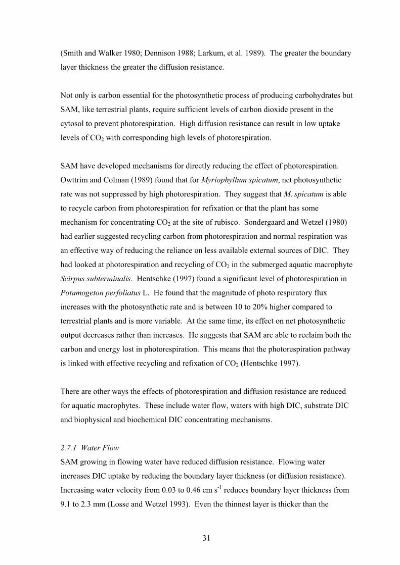
(Smith and Walker 1980; Dennison 1988; Larkum, et al. 1989). The greater the boundary
layer thickness the greater the diffusion resistance.
Not only is carbon essential for the photosynthetic process of producing carbohydrates but
SAM, like terrestrial plants, require sufficient levels of carbon dioxide present in the
cytosol to prevent photorespiration. High diffusion resistance can result in low uptake
levels of CO2 with corresponding high levels of photorespiration.
SAM have developed mechanisms for directly reducing the effect of photorespiration.
Owttrim and Colman (1989) found that for Myriophyllum spicatum, net photosynthetic
rate was not suppressed by high photorespiration. They suggest that M. spicatum is able
to recycle carbon from photorespiration for refixation or that the plant has some
mechanism for concentrating CO2 at the site of rubisco. Sondergaard and Wetzel (1980)
had earlier suggested recycling carbon from photorespiration and normal respiration was
an effective way of reducing the reliance on less available external sources of DIC. They
had looked at photorespiration and recycling of CO2 in the submerged aquatic macrophyte
Scirpus subterminalis. Hentschke (1997) found a significant level of photorespiration in
Potamogeton perfoliatus L. He found that the magnitude of photo respiratory flux
increases with the photosynthetic rate and is between 10 to 20% higher compared to
terrestrial plants and is more variable. At the same time, its effect on net photosynthetic
output decreases rather than increases. He suggests that SAM are able to reclaim both the
carbon and energy lost in photorespiration. This means that the photorespiration pathway
is linked with effective recycling and refixation of CO2 (Hentschke 1997).
There are other ways the effects of photorespiration and diffusion resistance are reduced
for aquatic macrophytes. These include water flow, waters with high DIC, substrate DIC
and biophysical and biochemical DIC concentrating mechanisms.
2.7.1 Water Flow
SAM growing in flowing water have reduced diffusion resistance. Flowing water
increases DIC uptake by reducing the boundary layer thickness (or diffusion resistance).
Increasing water velocity from 0.03 to 0.46 cm s-1 reduces boundary layer thickness from
9.1 to 2.3 mm (Losse and Wetzel 1993). Even the thinnest layer is thicker than the
31

submerged leaf of < 1mm (Losse and Wetzel 1993). Increasing water velocity, and
thereby reducing boundary layer thickness, gives a corresponding increase in net
photosynthetic productivity (Chambers, et al. 1991; Madsen, et al. 1993). However, there
is a limit to how much water velocity can increase and still give a net increase in
photosynthetic productivity.
Generally in water velocities greater than 1 cm s-1 net photosynthesis is reduced rather
than increased (Chambers, et al. 1991; Madsen, et al. 1993). Madsen et al (1993) reports
that Callitriche cophocarpa growth rate increases up to a water velocity of 1.5 cm s-1.
Above this rate, growth starts to decline. However, work they did with C. stagnalis
showed a net photosynthetic reduction of 25% when water velocity was increased from 1
to 4 cm s-1. Madsen et al (1993) also found that in velocities above 1 cm s-1, net
photosynthesis drops and respiration increases. They found that 16-67% of the reduction
in net photosynthesis was due to the increase in respiration. They conclude that this
reduction in net photosynthesis is mainly due to mechanical stress from agitation and
stretching of foliage.
Generally, once water velocities reach high levels ie >100 cm s-1, increasing biomass loss
is the main reason for a reduction in photosynthetic productivity (Chambers, et al. 1991).
However this is species dependant as Ranunculus aquatilis can grow in average water
velocities as high as 20 cm s-1, some marine SAM grow in waters as fast as 30 cm s-1, and
some marine macro-algae > 100 cm s-1 without detrimental effects (Schwenke 1971;
Boeger 1992).
Many SAM have foliage shapes, which help reduce boundary layer thickness in low water
velocities (Raven, et al. 1985). For example, leaves with undulations are commonly
found on a range of SAM including A. elongatus. Undulated margins of leaves are said to
help create turbulence therefore reducing boundary layer thickness.
2.7.2 Waters with High DIC
Another way SAM overcome diffusion resistance is to grow in aquatic environments
which support high natural respiration rates resulting in concentrations of DIC greater
than that fixed by photosynthesis and lost to the atmosphere (Raven, et al. 1985).
32

Producing foliage close to the water surface is an alternative way SAM reduce this
resistance (Raven, et al. 1985). This is a common growth habit of long stemmed SAM
such as Cabomba caroliniana, Ceratophyllum demersum, Egeria densa, Elodea
canadensis, Lagarosiphon major, Hydrilla verticillata and Potamogeton species. From
personal observation, these plants often produce dense canopies just below the water
surface.
2.7.3 Substrate DIC
Some SAM that grow in water deficient in DIC are able to supplement with DIC obtained
via the roots from the rooting substrate. Sediments of water bodies such as lakes, ponds
often contain accumulated remains of dead plant material. Raven et al (1985) claim that
these sediments can have high CO2 concentrations because of microbial activity.
Pedersen et al (1995) describes how CO2 is produced in otherwise anoxic substrate. They
found that the submerged aquatic macrophyte, Lobelia dortmanna as a result of
photosynthesis, released considerable quantities of O2 from its roots into the surround root
substrate during the day. During the night, there was enough accumulated O2 to enabled
aerobic microbial activity to produce CO2. This is supplemented by CO2 released from
the roots because of respiration. During the day, this plant is able to reabsorb this
accumulated CO2 for utilisation in photosynthesis (Pedersen, et al. 1995). This plant
commonly grows in sandy sediments in soft water of nutrient poor lakes. Other
macrophytes also grow in this habitat and derive most of their CO2 for photosynthesis
from sediments. They are collectively called isoetids and include Lobelia dortmanna,
Stylites andicola, Littorella uniflora, and Eriocaulon decangulare (Raven, et al. 1985;
Pedersen, et al. 1995).
2.8 DIC Concentrating Mechanisms
Biophysical or biochemical DIC concentrating mechanisms also overcome diffusion
resistance and minimise photorespiration in many SAM (Raven, et al. 1985). In some
cases these DIC concentrating mechanisms are only active when the concentration of
exogenous DIC levels are well below the threshold of that required for optimum
photosynthesis (Raven, et al. 1985; Keeley 1990).
33

2.8.1 Biophysical
SAM are considered to have biophysical concentrating mechanisms when they are found
to use only rubisco for DIC fixing, have a low CO2 compensation point and have a high
affinity for exogenous DIC (Raven, et al. 1985). Further evidence is the concentration of
DIC within photosynthetic cells exceeding that which can be expected based on normal
passive diffusion of exogenous DIC into cells. Raven et al (1985) suggests biophysical
concentrating mechanisms are common to many SAM as well as to many species of fresh
water and marine algae. It is not surprising that many SAM are able to utilise HCO3- as
the bicarbonate concentration in water is generally much higher than carbon dioxide
especially in high pH (8-9).
Evidence suggests various SAM use different kinds of DIC concentrating mechanisms.
Three types of DIC concentrating mechanisms have been suggested for HCO3-. These
are; HCO3- influx at the inner plastid envelope membrane, acid and alkaline zones, and/or
active HCO3- transport at the plasmalemma (Raven, et al. 1985). Active HCO3
- transport
requires the presence of carbonic anhydrase within the cell to catalyse the conversion of
bicarbonate to carbon dioxide for fixing by rubisco. This was confirmed by some
researchers with the unicellular algae Chlamydomonas reinharttii but later research by
others found that for SAM, carbonic anhydrase occurs mostly extracellularly (Raven, et
al. 1985; Bowes and Salvucci 1989; Husic 1991; Rybova, et al. 1991; Newman and Raven
1993; Beer 1998).
Some SAM such as Elodea nuttallii, Hydrilla verticillata and Potamogeton lucens,
produce acid and alkaline zones (pH polarity) on leaf surfaces (Prins and Helder 1985;
Raven, et al. 1985; Bowes and Salvucci 1989; Eighmy, et al. 1991; Reiskind, et al. 1997).
With Hydrilla verticillata the abaxial side of leaves has been found to have a pH of 4.0
and the adaxial side, pH 10.5 (Reiskind, et al. 1997). Raven et al (1985) suggests that
HCO3- passively diffuses in the acid zone where it is transformed to CO2. This in turn
diffuses into the photosynthetic cells. This acid zone could be either in the boundary
layer or within the cell wall (Larkum, et al. 1989). Prins and Helder (1985) found that for
Potamogeton lucens, bicarbonate uptake is a ‘chemiosmotically driven process’. They
conclude that the conversion of bicarbonate to CO2 is adjacent to the epidermis and is
reliant on light dependent H+ extrusion causing an electrical potential difference across
34

the leaf. While not excluding the possibility of the alternate hypothesis of
chemiosmotically driven H+ and HCO3- co-transport across the plasmalemma to the
interior of epidermal cells, they found no evidence to back this up. Instead, only CO2 is
thought to diffuse across the plasmalemma into the cytosol of leaf cells.
Whether this mechanism is theoretically able to supply enough CO2 to account for
measured uptake of HCO3- is considered contentious (Raven, et al. 1985). In fact,
Eighmy et al (1991) showed direct uptake of bicarbonate across the plasmalemma in
Elodea nuttallii, against the concentration gradient, in addition to leaf polarity. They
describe a bicarbonate cation symport mechanism within the plasmalemma, which greatly
surpassed the polarity mechanism in supplying DIC for photosynthesis. This means
direct bicarbonate uptake into a cell but the mechanism of conversion of bicarbonate to
CO2 for RUBISCO was less clear. Reiskind et al (1997) showed a similar mechanism in
Hydrilla verticillata and suggested a C-4 acid-based carbon concentrating mechanism in
the chloroplasts
Other SAM such as Myriophyllum and Vallisneria species show no evidence of leaf
polarity (Bowes and Salvucci 1989). Carbonic anhydrase (CA) located in the epidermal
cell wall or the plasmalemma is considered to play an important role in catalysing the
dehydration of bicarbonate to form CO2 for many submerged aquatic macrophytes and
unicellular algae (Bowes and Salvucci 1989; Husic 1991; Rybova, et al. 1991; Newman
and Raven 1993; Reiskind, et al. 1997; Beer 1998; Axelsson, et al. 1999; Newman and
Raven 1999; Rascio, et al. 1999). This appears to be an energy consuming process, as the
uptake of bicarbonate into the plasmalemma requires H+-ATPase (Rybova, et al. 1991).
However, even for plants using CA there are environmental conditions, such as high
temperatures and irradiance and very low CO2/HCO3- ratio, where direct uptake of
bicarbonate is an advantage (Beer 1998). Axelsson et al, (1999) found in the macro-algae
Ulva lactuca that direct bicarbonate uptake occurs across the cell plasmalemma via an
anion exchange mechanism (AE), in coexistence with the CA mechanism. They proposed
a simple model of the two mechanisms (Fig. 2.5).
35

Fig.2.5 A diagram showing the two concurrent mechanisms of bicarbonate use by
the macroalgae Ulva lactuca (Axelsson, et al. 1999).
Similar mechanisms have been found in the freshwater SAM Ranunculus trichophyllus.
Rascio et al (1999) observed that when environmental dissolved CO2 became low, there
was an associated increase in apoplastic CA activity. A direct bicarbonate uptake
mechanism was also proposed, based on evidence of photodependent H+-ATPase activity
in the transfer regions of the plasmalemma. However, in the large celled algae
Hydrodictyon reticulatum, H+-ATPase activity was found to be associated with CA
activity in the plasmalemma (Rybova, et al. 1991).
At about the same time, Newman and Raven (1999) found that CO2 was the main DIC
species used by the chloroplasts of Ranunculus penicillatus ssp. pseudofluitans. They
proposed that CO2 was the only form of DIC crossing the plasmalemma. Apoplastic CA
activity was confirmed but they did not test for H+-ATPase activity in the plasmalemma.
2.8.2 Biochemical Concentrating Mechanisms
Two types of biochemical DIC concentrating mechanisms have been found in SAM. Like
their terrestrial counterparts, some have been found to have either a type of C-4 carbon
fixing or crassulacean acid metabolism (CAM).
36

2.8.2.1 C-4 photosynthesis
Some SAM are able to fix carbon in a similar manner to terrestrial C-4 plants. In most
cases, it is a response to low ambient CO2 as they revert to C-3 carbon fixing when CO2
is not limiting (Raven, Osborne et al. 1985; Keeley 1990). A well-studied plant that
reflects this behaviour is Hydrilla verticillata. It reverts to C-4 acid synthesis in summer
under long days, high temperatures and low ambient CO2 levels (Bowes 1985; Raven, et
al. 1985; Keeley 1990). Keeley (1990) points out that C-4 carbon fixing occurs
simultaneously with C-3 carbon fixing within single cells for all SAM that have been
identified as having C-4 acid synthesis. Besides Hydrilla verticillata, these include,
Vallisneria spirilis, Eleocharis acicularis, E. baldwinii and Orcuttia californica (Keeley
1990; Uchino, et al. 1995).
Bowes (1985) and Keeley (1990) outline a model that describes C-4 fixation in SAM.
Both suggest that the two carboxylase enzymes, PEP carboxylase and rubisco are
separated into two locations within photosynthetic cells, rather than in separate cells as in
terrestrial C-4 plants. PEP carboxylase is located in the cytosol and rubisco in the
chloroplasts. CO2 defusing into a cell is ether fixed by PEP carboxylase or by rubisco.
Malate or other organic acids, products of C-4 synthesis, are transported from the cytosol
to the chloroplasts where they are decarboxylated to produce CO2 for refixing via rubisco
into 3-PGA as part of the Calvin cycle. This results in a concentration of CO2 at the site
of rubisco that reduces photorespiration.
Kranz anatomy (the bundle sheath cells that surround the vascular system), typical of C-4
terrestrial plants has not been found in C-4 SAM (Raven, Osborne et al. 1985; Keeley
1990; Uchino, Samejima et al. 1995). Keeley (1990) describes an amphibious aquatic C-
4 macrophyte Orcuttia californica that starts with submerged leaves followed by floating
and emergent leaves. Both the floating and emergent leaves have typical Kranz anatomy.
The juvenile submerged leaves lack this anatomy but were found to have similar turnover
of C-4 fixation products as terrestrial C-4 plants (Keeley 1990).
2.8.2.2 Crassulacean Acid Metabolism
37

When a process resembling CAM was first discovered in SAM, it was with some surprise
as CAM is usually associated with terrestrial succulents, adapted to an arid climate where
water conservation is a requirement (Salisbury and Ross 1992). Interestingly it has been
found in submerged plants of Crassula aquatica, but is also found in many Isoetids
(Isoetaceae) such as Littorella uniflora, Isoetes howelli and I. Lacustris (Raven, Osborne
et al. 1985). However not all Isoetids have CAM. Some aquatic species from other
families such as Poaceae, Cyperaceae and Plantaginaceae also have CAM-like
biochemical concentrating mechanisms (Keeley 1990; Uchino, et al. 1995).
In aquatic habitats, it is carbon rather than moisture limitation that has resulted in the
development of CAM in some aquatic macrophytes. Keeley (1990) described most CAM
aquatic macrophytes as occurring in habitats that are seasonally wet and then dry. During
the wet season, the plants grow submerged in pools. These pools are subject to large
diurnal fluctuations in DIC. During the day, dissolved CO2 is rapidly taken up by dense
aquatic vegetation so that CO2 is almost completely depleted by mid morning (Keeley
1990). CAM macrophytes are unable to utilise bicarbonate (Keeley 1990). During the
night, due to respiration from aquatic flora and fauna, dissolved CO2 levels increase back
to high levels. Therefore, in a similar manner to terrestrial CAM plants, aquatic CAM
plants fix CO2 during the night storing it as malate (Keeley 1990). Similarly, during the
day malate is decarboxylated to release CO2 within the cell where it is refixed via the C-3
Calvin Cycle (Keeley 1990). An adequate supply of CO2 is therefore maintained to
saturate photosynthesis and to prevent photorespiration.
Keeley (1990) points out that SAM either have no stomata or if stomata are present they
are non-functional. The fixing of CO2 between day and night is therefore dependent on
the exogenous concentration of CO2 rather than the opening and closing of stomata.
In conclusion SAM use a range of different mechanisms to overcome anoxic sediments
and limited DIC, making them distinctively different from terrestrial macrophytes. Which
or how many of these mechanisms any one individual species actually uses depends on
their genetic make up or their particular habitat and possibly is a measure of the species
adaptability to a range of aquatic environments.
38

39
A study of some of these mechanisms as used by A. elongatus grown in environmentally
controlled aquaria is provided in the following chapters

CHAPTER 3
THE INTERACTION OF WATER FLOW AND
NUTRIENTS ON AQUATIC PLANT GROWTH
3.1 Introduction
Aponogeton elongatus F. Muell. ex Benth, has been observed growing in slow to fast
flowing waters (Sainty & Jacobs, 1994; Hellquist & Jacobs, 1998). For other submerged
aquatic macrophytes (SAM), it is known that water flow from 1.5 -100 cm s-1, depending
on the species, increases net photosynthetic rate (Schwenke 1971; Carpenter et al., 1991;
Chambers et al., 1991; Losse & Wetzel 1993). However, many of these studies have been
based on short-term measures of photosynthetic rate or field measurements and
observations. The significance of this study is that it measures long term growth rates of
plants under controlled conditions in aquaria. Such long-term growth rates may reveal
differences that are difficult to detect with short-term measures of photosynthetic rate
(Titus et al., 1990).
While little is known about A. elongatus, for SAM in general, moderate water flow
improves leaf uptake of nutrients as well as dissolved inorganic carbon (DIC), and oxygen
(Smith & Walker, 1980; Larkum et al., 1989; Stevens & Hurd, 1997). It is also well
recognised that flowing water reduces the thickness of the unstirred (boundary) layer that
surrounds leaves of SAM (Schwenke, 1971; Carpenter et al., 1991; Chambers et al., 1991;
Losse & Wetzel 1993; Madsen et al., 1993). The thinner the boundary layer, the lower the
diffusion resistance to leaf uptake of nutrients and DIC (Smith & Walker 1980; Larkum et
al., 1989; Stevens & Hurd 1997). Given its observed habitat, it is likely that growth of A.
elongatus is greater in water of moderate flow compared to still water. This greater growth
may be due to the improved foliar uptake of nutrients and/or DIC. The aims of this study
were, to test the hypothesis that A. elongatus growth is increased by water flow (stirring),
and to determine the relationship between water flow and nutrients.
39

3.2 Materials and Methods
3.2.1 Growth Conditions
This experiment was conducted from 13 July to 22 December 1998, in a controlled
temperature room at the University of Queensland, Gatton. Air temperatures within the
room were maintained at 22.4 ± 1.0°C (± SD). The mean daily water temperature was 23.7
± 0.4°C (± SD). Light was provided by banks of 120cm long fluorescent tubes (40 watt
Grow Lux Wide Spectrum tubes) to give a mean photosynthetically active radiation
(PAR) level of 101 ± 3 (± SE) µmol m-2 s-1 measured 5 cm above the water surface. Light
was provided continuously throughout the experiment.
Twelve identical glass aquaria (tanks) were used, each 587 mm wide x 457 mm deep x 380
mm in height (100 L). Two tanks were placed under each light bank, partially surrounded
by black polyethylene sheeting to exclude the extraneous light from other light banks. A
1:1 mixture of tap water and deionised water was used to reduce the concentration of
nutrients, particularly phosphate, to minimise algal growth. Tanks were filled with this
water to a height of 340 mm (90 L) and were topped up with deionised water weekly to
replace evaporated water. The average electrical conductivity of the water was 237 ± 1µS
cm-1 over all tanks.
Where required, water was stirred using submersible (4.6 watt) power water filters (Rio-
PF400) installed in a back corner of each tank, providing a measured flow rate of 515 ± 5
L h-1 (± SE). The maximum water speed at pump outlets was approximately 182 cm s-1.
The swirling action created by each pump would have resulted in a wide range of reduced
water speeds across a tank.
3.2.2 Experiment design
The experiment was a two-factor, split plot design with two main plot treatments (stirred
and unstirred water) and three subplots (slow release fertiliser; 0 g, 0.5 g and 1 g). A pair
of tanks under a single light bank were allocated the main plot treatments, one tank stirred,
the other unstirred. Within each tank, individually potted plants received the sub plot
fertiliser treatments; three plants to each sub-plot treatment (the experimental unit), giving
nine plants per tank. Each of pairs of tanks under a single light bank represented one
complete block and there were six blocks.
40

In an attempt to reduce experimental error, plants were ranked by total leaf length then
allocated sequentially to the six blocks. Treatments were assigned randomly to the plots
(tanks) and subplots (plants).
3.2.3 Preparation of potted plant material
One hundred and eight 100mm diameter plastic pots (420 ml) were lined internally with
clear polyethylene bags, then 0.5g or 1g of controlled release fertiliser were placed in 72 of
the pots before filling with sediment. The fertiliser was 8-9 month Osmocote PlusR (16
N:3.5 P: 10 K:1.2 Mg plus trace elements). The approximate plant nutrient composition of
the sediment at the start of the experiment is shown in Table 3.1. Due to the slow release
nature of this fertiliser, these nutrients were not available to plant roots in the
concentrations outlined in the table. The sediment was equal parts of washed fine white
sand and coarse river sand (bulk density 1.43 g ml -1) mixed with 1.7g of powdered iron
(55.85% Fe) per pot.
Table 3.1.
The mineral composition of the sediment (µg g-1
) at the start of the
experiment. This is based on one gram of the controlled release fertiliser Osmocote PlusR per pot. (Calculated from data supplied by the fertiliser manufacturer).
Majorelements
µg g-1 Trace elements µg g-1
Nitrogen 265.7 Boron 0.3Phosphorus 58.1 Copper 0.8Potassium 166.0 Iron 6.6*
Sulphur 39.9 Manganese 1.4Magnesium 19.9 Molybdenum 0.5Calcium 0 Zinc 0.4
* Available iron levels may be much higher due to the addition of powered iron
A. elongatus plants, about 12 months old, were planted into the pots. The majority of
plants were originally propagated by tissue culture from parent stock collected from a
single site in southeast Queensland. However, 15 seedlings of similar size and maturity to
the micropropagated plants, were also used to make up the required numbers.
The sediment was packed into each pot firmly around the plant, to within 15 to 20 mm of
the rim of the pot. After this, granulated zeolite (2mm Horticulture Grade) was added to
the top of the sediment to a depth of 10-15 mm. The plastic liner was raised above the rim
41

of the pot by about 20-30 mm to create a zone of unstirred water (boundary layer) above
the potting mix. The placement of the fertiliser and inclusion of powdered iron in the
medium, plus the plastic liner and zeolite, were all designed to minimise the diffusion of
nutrients, particularly phosphate, from the potting mix into the surrounding water.
3.2.4 Plant measurements
Total leaf number, length (leaf blade base to leaf tip, ignoring the petiole) and blade width
(at the widest portion of each leaf blade) of each plant was measured at the beginning of
the experiment and in the 14th week. Leaf blades of non-senescent, fully developed leaves
were measured.
For dry weight measurements, one plant was randomly selected from each treatment in the
22nd week. After removing the potting mix, each plant was divided into foliage, tuber and
roots then fan forced oven dried at 60ºC for 5 days before being weighed.
3.2.5 Chlorophyll fluorescence
A portable submersible Diving-PAM (pulse amplitude modulated) Fluorometer (Walz,
Effeltrich, Germany) was used to measure leaf chlorophyll fluorescence in the form of
quantum (photochemical energy conversion) yield. These measurements were taken 11
weeks from the start of the experiment. For each measurement, electron transport rate
(ETR) was determined over nine actinic light levels (4, 31, 54, 84, 155, 259, 372, 594,
1013µmole quanta s-1 m-2) in the form of a rapid light curve (RLC). Each RLC was
produced over 90 seconds within which there were 10-second periods of actinic irradiance
and 1 second saturating pulses (Jones & Dennison, 1998). The youngest, non-shaded,
mature leaf was measured on each of three plants from each treatment over four replicates.
In addition, three floating leaves and three submerged leaves on outdoor plants of A.
elongatus growing under 60 – 80% shade were measured. All leaf measurements were
taken at one-quarter the length of the leaf blade from the leaf tip, avoiding the midrib and
any other major leaf veins.
3.2.6 Nutrient levels in water
A ‘Palintest Photometer 7000’(Palintest Ltd, Tyne & Wear, England) and reagents from
Palintest kits were used to measure nitrate nitrogen, ammonium nitrogen, phosphate,
42

potassium, magnesium and iron in the water of each of the tanks at the end of the
experiment following the instructions provided in the Palintest kits.
3.2.7 Dissolved CO2 levels
Total dissolved inorganic carbon (DIC) and dissolved free CO2 (dissolved CO2 and H2CO3)
were measured after 12 weeks using titration techniques described by Frith et al (1993).
One of the limitations with the dissolved free CO2 technique is that it could not measure
CO2 levels below 4.5 µM. One sample was tested for each of 12 tanks.
3.2.8 Monitoring
A waterproof, hand-held ‘TPS WP-80’ (TPS Pty Ltd, Brisbane, Australia) pH meter was
used to measure pH weekly and a waterproof, hand-held (TPS WP-84) electrical
conductivity meter was used to measure tank water conductivity every 2 weeks throughout
the experiment. Water temperature (in 4 tanks) and air temperature (2 locations) were
measured hourly using thermocouples connected to a data logger (‘Datataker 500’, Pacific
Data Systems Ltd, Brisbane, Australia)
3.2.9 Statistical Analysis
ANOVA were applied to the leaf area, dry weight and chlorophyll fluorescence data (SAS
release 6.12) and LSDs computed (MS Excel version 97). Leaf area data were transformed
to their square root before ANOVA. This was because the leaf area was estimated by
multiplying the length and width of a leaf, which tended to inflate the error mean square in
the ANOVA reducing the degree of precision in which the treatments were compared. The
inflated error term was reduced by square root transformation of the leaf area data. The
Dry weight data were transformed to their log to account for one outlier in each of the data
sets of leaf, tuber and root. F tests and t tests were applied to DIC, pH, conductivity and
temperature data using MS Excel. A significance level of P< 0.05 was adopted unless
otherwise indicated.
43

3.3 Results
3.3.1 General observations
By the end of the experiment, the fertilised plants appeared much larger than the
unfertilised plants (Fig 3.1). Plants fertilised with 1.0 g Osmocote were larger than those
with 0.5 g Osmocote, particularly in the stirred tanks. Plants in the unstirred tanks lost
more leaves and appeared smaller than those in the stirred tanks. The leaves of plants in
stirred tanks were dark red to purple green while those in unstirred tanks were a paler green
with little or no red or purple.
3.3.2 Leaf Morphometrics
The average leaf length and width, and hence leaf area, were greater on plants growing in
stirred water than in unstirred water (Table 2.1). Plants without fertiliser had fewer and
smaller leaves than those of the fertilised plants (P < 0.001). Leaf number and length were
not significantly different between the two levels of fertiliser (0.5g and 1g) but the leaves
were wider, and hence leaf area greater, on plants with 1g of fertiliser. There was no
significant interaction between fertiliser level and ± stirring for these parameters.
Table 3.2
Effects of stirred or unstirred water and three levels of fertiliser on A. elongatus
mean leaf number, length, width and area, after a 22 week growth period with a
mean temperature of 24°C and PAR 101.6 µmol m-2
s-1
. Leaf area data were transformed to the square root before ANOVA and LSD comparisons (All means in table are from original untransformed data). All data are an average of six blocks.Means within a column having a common letter are not significantly different at 5% level. There are no significant two-factor interactions.
TreatmentsLeaf Number
per plant Leaf Length
(cm) per plant Leaf Width
(cm) per plant Leaf Area
(cm2) per plant
Osmocote® 0g 22.6 a 221.4 a 35.2 a 400 a 0.5g 31.8 b 452.6 b 63.0 b 973 b 1.0g 33.9 b 533.6 b 75.7 c 1297 c
Stirred 31.5 a 484.5 b 67.4 b 1113 b Unstirred 25.3 a 320.3 a 48.6 a 667 a
Interactions (A × B)
Stirred 0g 22.9 255.1 40.8 4970.5g 33.3 543.6 72.5 12201.0g 38.3 654.9 89.0 1623
Unstirred 0g 20.4 187.7 29.7 3040.5g 28.2 361.0 53.5 7261.0g 27.4 412.3 62.5 971
44

1g 0.5g 0g
Stirred water
1g 0.5g 0g
Unstirred water
Fig. 3.1. The response of potted A. elongatus plants to three fertiliser levels(1, 0.5, 0g pot-1) and stirred verses unstirred water. The drawings represent the actual leaf numbers (Table 2), and leaf areas are scaled in proportion to the leaf area means ± 2%.
45

Unstirred
Stirred
0.0
0.5
1.0
1.5
2.0
0 0.5 1
Le
af
Dry
We
ight
(g)
Unstirred
Stirred
0.0
0.5
1.0
1.5
0 0.5 1
Tuber
Dry
Weig
ht (
g)
Unstirred
Stirred
0.0
0.2
0.4
0.6
0.8
1.0
0 0.5 1
Fertiliser (Osmocote) (g pot-1
)
Ro
otD
ry W
eig
ht
(g)
Unstirred
Stirred
0.0
0.5
1.0
1.5
2.0
2.5
3.0
3.5
4.0
0 0.5 1
To
tal P
lant D
ry W
eig
ht
(g)
Fig. 3.2 Growth of A. elongatus after 22 weeks at a mean temperature
of 24°C and PAR of 102 µmol m-2 s-1. The only significant interaction occurred between leaf dry weights. The differences between the
treatment means for roots are not significant. (Means ± 1 SE).
46

3.3.3 Dry Weight
Plants with the largest total dry weight were those growing in stirred water and fertilised
with either 0.5g or 1g of Osmocote (Fig 3.2). The difference between 0.5g or 1g of
Osmocote was not significant. Stirred water produced greater leaf dry weight than
unstirred water at the highest rate of fertiliser (1g) but there was no difference with 0 or
0.5g.
Fertiliser application had no significant effect on tuber dry weight but tubers in stirred
water had significantly greater dry weight than those in unstirred water (P < 0.01)(Fig 3.2).
In contrast, there were no differences in root dry weight between plants growing in either
stirred or unstirred water, or between different fertiliser levels (Fig 3.2).
3.3.4 Chlorophyll fluorescence
There was no significant difference between any treatments in maximum quantum yield or
maximum electron transfer rate (ETR). The maximum ETR value (5.9 ± 3.3 (±SE) µmol e-
m-2 s-1) of leaves growing in the tanks is similar to that for the rapid light curve
measurements taken from submerged leaves of plants growing outdoors under 60-80%
shade (9.0 ± 0.5 µmol e- m-2 s-1) (Fig 3). They fall well short of the maximum ETR of
floating leaves (53 ± 11 µmol e- m-2 s-1) on the same outdoor plants (Fig 3.3).
0
10
20
30
40
50
60
70
0 200 400 600 800 1000 1200 1400
PPFD (µmol quanta m-2
s-1
)
ET
R (
µm
ol
e-
m-2
s-1
)
Fig. 3.3 Rapid light curves derived from electron transport rate (ETR) (µmol electron m-
2 s-1) verses photosynthetic photon flux density (PPFD) (µmol quanta m-2 s-1). Floating
leaves (♦), submerged leaves ( ) of Aponogeton elongatus plants (mean of three) and mean RLC of three submerged leaves from three indoor tank grown plants from the
experiment (X). Data presented as the mean ± 1 SE.
47

3.3.5 Water nutrient levels
There was considerable variation in nutrient levels between tanks (Table 3.3) but most
nutrients except potassium (in some tanks) and magnesium were very low. The total
nitrogen in the water was no more than 1%, phosphorus 3% and potassium 36% of that
originally supplied as Osmocote. In contrast, magnesium was 45% higher than in
Osmocote because of the high level in the tap water used (Table 3.3).
A comparison of the means using a paired t test revealed no differences in any nutrients
between stirred and unstirred water tanks. However, nutrient levels (nitrogen, phosphorus
and potassium) were more variable in unstirred tanks than stirred tanks using Two-Sample
F-Test (P < 0.008). The exception was magnesium which was more variable in stirred
tanks (P = 0.008).
3.3.6 Dissolved CO2 levels
The dissolved inorganic carbon level (DIC) ranged from 941 – 1120 µM between tanks
while dissolved free CO2 ranged from 0 - 41 µM. For total DIC the variance in stirred
tanks was no different from the variance in unstirred tanks, while dissolved free CO2 in
unstirred tanks was much more variable according to a two-sample F-test (Table 3.3).
However, when the means were compared using a paired t test, Total DIC appeared higher
(P=0.054) in stirred tanks than in unstirred tanks while there was no difference in dissolved
free CO2..
3.3.7 pH, Electrical Conductivity and Temperature
There was a steady rise in mean pH from 8.1 in the first week to pH 8.58 in unstirred tanks
and pH 8.49 in stirred tanks after four weeks (Fig 3.4A). In week 10, the mean pH peaked
at 8.74 in unstirred tanks and 8.54 in stirred tanks. The pH remained higher in the
unstirred tanks than the stirred tanks through to the last measurement in week 20. The
maximum pH recorded was 9.14 in an unstirred tank compared to 8.59 in a stirred tank.
As indicated by the larger standard errors, the pH level varied much more between
unstirred tanks than between stirred tanks (Fig 3.4A).
Electrical conductivity increased slowly from a mean of 238 to 294µS cm-1 at the end of
the experiment. The differences between tanks became more pronounced towards the end
of the experiment as indicated by the error bars (Fig 3.4B).
48

Table 3.3
Water nutrient concentrations, conductivity and temperature, 5 months from the start of the experiment. Total dissolved inorganic carbon (DIC) (µmole CO2), dissolvedfree CO2 and pH, measured after 3 months. The ‘original water’ data are estimates of minerals in the tank water at the start of the experiment (tap water plus deionisedwater 1:1), based on measurements from the tap water. ‘Fertiliser per tank’ is an estimate of the amount of nutrients supplied in the Osmocote (calculated from datasupplied by the manufacturer).
Treatment N – NO3 N – NH4 Total N P K Mg Fe
Dissolved
Inorganic
Carbon
Dissolved
Free CO2 pH Conductivity Temperature
µM µM µM µM µM µM µM µM µM µS cm-1 °C
Mean Stirred 0.36 0.71 1.07 1.16 3.30 411 0.18 1048 22.7 8.41 303 23.7
Std Error 0.36 0.29 0.36 0.23 2.05 24 0.05 20 1.6 0.02 5 0.29
Mean Unstirred 0.71 3.57 4.14 0.84 46.3 420 0.18 986 18.2 8.58 285 23.2
Std Error 0.36 2.14 0.03 0.06 23.5 7 0.05 40 6.6 0.04 7 0.24
Original water 3.57 0 3.57 1.29 46.0 267 0.18
Fertiliser per tank 194 171 366 36.1 128 283 N. A.
49

C
21
22
23
24
25
26
1 3 5 7 9 11 13 15 17
Time (Weeks)
Te
mp
era
ture
ºC
A
7.8
8.0
8.2
8.4
8.6
8.8
9.0
1 3 5 7 9 11 13 15 17
pH
B
220
240
260
280
300
320
1 27 40 60 82 124 172
Co
nd
ucti
vit
y (
µS
)
Unstirred
Stirred
Unstirred
Stirred
Unstirred
Stirred
Fig. 3.4 Comparison of stirred and unstirred water over time: (A) pH
(based on 18 weekly measurements; mean ± 1 SE, n = 6); (B) conductivity
(based on seven measurement times; mean ± 1 SE, n = 6) and (C) meanweekly water temperatures.
50

However, there was no difference either in the variances or between the means of stirred
and unstirred tanks.
Mean daily water temperatures in stirred tanks were 0.4-0.7°C warmer than in unstirred
tanks (P < 0.01) (Fig 3.4 C). It is possible this temperature difference contributed to the
difference in plant growth between unstirred and stirred water, but since the temperature
difference is small, the effect is likely to be minimal.
3.4 Discussion
This study indicates that A. elongatus grows larger in flowing (stirred) water. Larger leaf
area and leaf dry weight per plant were the main responses to increasing nutrient level and
stirred water. Tuber weight was also greater in stirred water but was not significantly
affected by fertiliser level, which suggests that some factor other than mineral nutrition
limited growth in unstirred water.
A. elongatus and other species of Aponogeton in creeks and rivers, have longer leaves in
rapidly flowing water (Islam, 1996; Hellquist and Jacobs, 1998). In this study, the leaves
were both longer and wider but proportionally narrower in stirred water compared to
unstirred water (based on length breadth ratio). However, in many SAM, light intensity
and/or mineral nutrition may have a much greater influence on leaf shape compared to
water velocity (Kirk, 1994; Spence, 1995). A. elongatus has been observed to have much
thinner and broader leaves in dense shade caused by overhanging trees (Hellquist &
Jacobs, 1998). This is similar to the leaves of land plants adapted to dense shade
(Salisbury & Ross, 1992). In this study, the leaves are likely to be adapted to shade, as
the plants were grown with an irradiance level equal to approximately 5% sunlight.
Based on an approximate leaf thickness of 0.1mm, the plants had a leaf surface area to
leaf volume ratio of approximately 20:1. Leaf surface area to leaf volume ratios equal to
or greater than this are common in SAM (Madsen et al., 1993). Apart from more efficient
light interception, thin leaves may be more efficient in nutrient or DIC uptake
(Sculthorpe, 1967).
Roots of many aquatic macrophytes may have a primary role in nutrient uptake,
particularly nitrogen and phosphate (Bristow & Whitcombe, 1971; Barko et al., 1988;
51

Rattray et al., 1991; Robach et al., 1995; Best, et al., 1996; Wigand & Stevenson, 1997).
However, it has been shown that aquatic plant foliage can also take-up nutrients from the
water (Bristow & Whitcombe, 1971; Rattray et al., 1991; Robach et al., 1995; Best et al.,
1996). Whether nutrients such as phosphate are taken up by foliage or roots is dependent
on the nutrient concentration gradient between interstitial water and the water above the
sediment (Rattray et al., 1991; Best et al., 1996).
The present study indicates that A. elongatus obtained most of its nutrients from the
substrate via its roots. Those plants without the added fertiliser grew poorly and were
significantly smaller in leaf number, length and width, and leaf dry weight, even though
the unfertilised plants and fertilised plants were growing in the same tank of water. Water
flow (stirred water) is known to assist nutrient uptake via the foliage (Stevens & Hurd,
1997). Adding more nutrients directly to the water may improve nutrient uptake by the
plants, without fertiliser in the sediment, but a more likely consequence is an algae bloom.
A substantial quantity of iron was added to each pot in the form of fine metallic iron
particles. The aim was to restrict algal growth by reducing the solubility of phosphate.
The root substrate is likely to be largely anoxic and iron in the substrate is likely to be
present in reduced form (Gambrell and Patrick 1988). A narrow zone (≈2 mm) of aerobic
substrate occurs near and extending to the substrate surface (Armstrong 1978; Gambrell
and Patrick 1988; Caffrey and Kemp 1990). Any reduced soluble iron and phosphorus
ions that passes into this aerobic zone as a result of diffusion would oxidise, precipitating
out of solution as ferric phosphate (Gambrell and Patrick 1988).
High levels of iron are known to be toxic to aquatic plants (Barko et al. 1986). High iron
restricts the availability of phosphorus and disrupts sulphur metabolism (Barko et al.
1986). Therefore, it is possible that in the oxidated root rhizosphere (Wium Anderson and
Anderson 1972; Penhale and Wetzel 1983; Larkum et al. 1989; Pedersen et al. 1995;
Smith et al. 1984;) a high level of soluble iron could severely reduce phosphate uptake.
Iron powder was applied evenly to all treatments and plants grew well in the fertilised
treatments, particularly in stirred water, with no visual evidence of phosphate deficiency.
The unfertilised plants may have had phosphate deficiency among other nutrient
deficiencies but this can not be attributed to iron toxicity. For particles of metallic iron
(Fe) to get into solution, they first have to be oxidised (Fe2+ and Fe3+). A short time after
52

mixing the iron in the sand substrate the potted media was placed under water. Microbial
action would eventually render the substrate anoxic (Gambrell and Patrick 1988). Any
oxidised iron (Fe3+) would be reduced to a more soluble form (Fe2+) (Gambrell and
Patrick 1988) but it is possible only a tiny amount of the metallic iron particles was
oxidised before the media became anoxic. Therefore, the impact of soluble iron on
phosphate uptake could be minimal. It is also possible that phosphate uptake by the
leaves offset the affects of high iron on phosphate uptake by the roots. A more detailed
study is needed to fully test whether the iron level in the experiment reduced phosphate
uptake sufficiently to limit growth.
There was a leaf dry weight interaction between stirred water and 0.5g, and 1g of
OsmocoteR but not unstirred water. This suggests that fertiliser is not the limiting factor
in unstirred water. It is more likely DIC uptake is the primary reason for the greater
growth in stirred versus unstirred water. Perhaps the strongest indication that DIC is the
limiting factor is tuber weight. Tubers produced by these plants are primarily storage
organs rich in carbohydrates such as starch (based on starch iodine test), therefore tuber
weight must be strongly linked to photosynthetic productivity. It is likely that tubers in
stirred water are larger because the reduced boundary layer around leaves in stirred water
allowed greater DIC assimilation than in unstirred water (Smith & Walker, 1980; Larkum,
et al., 1989; Stevens & Hurd, 1997).
An interesting question is why pH was high overall. One unstirred water tank reached a
pH of 9.14. Some tanks, particularly the unstirred tanks, had a small amount of algal
growth, both benthic and pelagic. The uptake of dissolved free CO2 by both the algae and
A. elongatus could have contributed to the rise in pH (Kirk 1994). The uptake of
bicarbonate could also contribute to the high pH. Other aquatics increase the pH by
releasing OH¯ ions in exchange for bicarbonate ions, for example Elodea nuttallii and
blue green algae (Miller & Colman, 1980; Allen & Spence, 1981; Jahnke et al., 1991).
The slightly lower pH (difference 0.16) in stirred water could be due to the stirring action
of the water increasing the rate of CO2 replenishment from the surrounding air (Sand-
Jensen, 1983). Conversely, in unstirred tanks there may be a rise in pH because of plant
removal of CO2 coupled with slower replenishment from the air.
53

The dissolved free CO2 levels represents nearly double the air equilibrium value of 10µM
(Stumm & Morgan, 1970; Bowes & Salvucci, 1989). This level of dissolved free CO2
may still result in a deficit in stirred tanks, as boundary layer resistances although
reduced, are still high, (Bowes & Salvucci, 1989). This is likely to be more evident under
higher irradiance levels than were used in this study. The ability to use bicarbonate would
be an advantage as there was approximately 46 times as much bicarbonate present as
dissolved free CO2.
It is plausible that stirring the water increased DIC uptake irrespective of whether the
form used by the plant is bicarbonate or dissolved free carbon. Most fresh water
macrophytes have a higher affinity for dissolved free CO2 than bicarbonate (Allen &
Spence, 1981; Sand-Jensen, 1983). A. elongatus may be able to use bicarbonate but have
a preference for dissolved free CO2. Clearly, further investigation is required to confirm
whether this plant is able to use bicarbonate.
From personal observation, A. elongatus normally has predominantly submerged leaves
but under conditions of high irradiance in still (stagnant) water, it can switch to
predominantly floating leaves thus overcoming boundary layer limitations. Although this
macrophyte is found only in permanent water, its habitat is subject to variable flow rates
from stagnant water during droughts to rapid flow during high rainfall events. In our
laboratory, floating leaves were not a feature in the unstirred treatments possibly because
irradiance levels were low. Photosynthetic capacity (ETR) of outdoor floating leaves of
A. elongatus was approximately 6 times greater than submerged leaves on the same
plants and 9 times greater than either floating or submerged leaves in this study. The ETR
of the outdoor floating leaves are comparable with that recorded on seagrasses in clear
marine waters (Ralph et al., 1998). However, floating leaves are not as persistent as
submerged leaves, usually undergoing senescence within 7 to 12 days of reaching full
maturity compared to several months for submerged leaves. This continual replacement
of floating leaves may offset any advantage of accessing carbon directly from the
atmosphere. Further work is needed to determine why floating leaves are short lived and
whether the production of floating leaves in higher light intensities can reduce dependence
on flowing water for DIC uptake.
54

In conclusion, A. elongatus growth was limited by nutrient levels equal to or less than 0.5
g of fertiliser per pot. Above this nutrient level, the availability of DIC appears to be the
main factor limiting growth for Aponogeton normally dependent on submerged leaves
(Fig 3.5).
0.0
0.5
1.0
1.5
2.0
0 0.5
Nut
rition
Boundary Layers
Stirred water
Unstirred water
Pla
ntgro
wth
Sediment nutrient Concentration
1
Fig. 3.5 The interaction of ± stirring and fertiliser level on leaf dry weight per plant
in A. elongatus; (•) stirred, (♦) unstirred water. Below 0.5g of Osmocote, growth was limited by ‘nutrient’ supply. Above 0.5g plant growth in unstirred water was limited by the ‘boundary layer’.
Presumably, stirred water improves DIC uptake due to the decrease in diffusion boundary
layer resistance around the leaves (Larkum et al., 1989; Stevens & Hurd, 1997). The
resulting increase in photosynthetic productivity enabled greater growth of both tuber and
leaves. This provides some explanation for the observations of others that these
macrophytes flourish in creeks or river systems with permanently flowing water (Sainty &
Jacobs, 1994; Hellquist & Jacobs, 1998). Nutrient availability in different sediments is
known to have a marked effect on SAM growth (Barko & Smart, 1986; Stewart, 1991;
Best et al., 1996). This can be due to variation in nutrient concentration and/or sediment
density (Barko et al., 1988; Boeger, 1992). The availability of nutrients in different
sediments may limit growth and possibly the distribution of A. elongatus in rivers and
creeks. However, growth will be limited even in sediments containing abundant plant
nutrients without the effect of water flow on unstirred boundary layers.
55

Chapter 4
Dissolved Inorganic Carbon Limitation of Aquatic
Macrophyte Growth under Controlled pH
4.1 Introduction
The aquatic macrophyte Aponogeton elongatus F. Muell. ex Benth. was found to grow best in
stirred water, with an adequate supply of nutrients in the sediment (Chapter 3). Improved
dissolved inorganic carbon (DIC) uptake was hypothesised as the main reason for the
superior growth in stirred water.
Atmospheric carbon dioxide is absorbed by water until equilibrium is reached between the
concentration of atmospheric CO2 and dissolved CO2 in the water (Stumm and Morgan 1970).
In turn, this dissolved CO2 reacts with water to form carbonic acid, which dissociates into
bicarbonate ions and further into carbonate ions. This process is in equilibrium as follows
CO2 (g) CO2 (aq) + H2O H2CO3 HCO3- + H+ CO3
-2 + 2H+ (Saruhashi 1955).
These equilibrium reactions shift to the right with increasing pH (Kirk, 1994). Stumm and
Morgan (1970) proposed a simple model to show characteristics of the carbonate system in
pure water in equilibrium with the atmosphere at a set temperature of 25ºC. They proposed
that increasing the pH of water increases total DIC but the molar concentration of dissolved
free CO2 remains unaffected.
In natural waters, the concentration of dissolved free CO2 may vary considerably due to the
interaction of constantly changing factors such as pH, temperature, total DIC, alkalinity, ionic
strength, respiration and plant uptake for photosynthesis (Saruhashi, 1955; Stumm and
Morgan 1970; Frith, et al. 1993). Natural levels of bicarbonate range from 0.02 to > 2000
µM and dissolved free CO2 can range from 0 to 350 µM (Bowes and Salvucci 1989). Natural
water bodies often have levels of DIC well above equilibrium levels (Titus, et al. 1990). A
state of equilibrium between the concentration of atmospheric CO2 and dissolved free CO2
may be rare in biologically active natural waters (Kirk 1994).
56

Table 4.1 The effects of different pH or the concentration of carbon species on the rate of
photosynthesis has been well demonstrated with the following aquatic macrophytes.Note: Aquatic macrophytes with an asterisk (*) are seagrasses.
Aquatic macrophytes Researchers
Batrachium aquatile (L.) (Nielsen & Sand-Jensen 1989)
Berula erecta (Hudson) Coville (Nielsen & Sand-Jensen 1989)
Callitriche cophocarpa Sendtner (Nielsen & Sand-Jensen 1989)
Callitriche stagnalis Scop. (Sand-Jensen 1983)
Ceratophyllum demersum L. (Van, Haller et al. 1976)
Egeria densa Planch. (Browse 1979)
Elodea canadensis L. C. Rich. (Allen & Spence 1981; Elzenga & Prins 1988;Nielsen & Sand-Jensen 1989)
Elodea nutalli (Planch.)St.John (Elzenga & Prins 1988; Jahnke et al. 1991)
Eurhynchium rusciforme Milde (Allen & Spence 1981)
Fontinalis antipyretica L. (Allen & Spence 1981)
Halodule uninervis (Forssk.) Aschers * (Beer et al. 1977)
Halophila stipulacea (Forssk.) Aschers * (Beer et al. 1977)
Hippuris vulgaris L. (Allen & Spence 1981)
Hydrilla verticillata (L Fil.)Presl. (Van et al. 1976)
Littorella uniflora (L.) Ascherson (Nielsen & Sand-Jensen 1989)
Lobelia dortmanna L. (Nielsen & Sand-Jensen 1989)
Marine Macroalgae (Sand-Jensen & Gordon 1984)
Myosotis palustris L. (Nielsen & Sand-Jensen 1989)
Myriophyllum alterniflorum Macoun (Allen & Spence 1981)
Myriophyllum spicatum L. (Allen & Spence 1981; Titus & Stone 1982; Nielsen& Sand-Jensen 1989)
Nitella flexilis (L.) Ag. (Allen and Spence 1981)
Nuphar luteum (L.) Smith (Smits et al. 1988)
Nymphaea alba L. (Smits et al. 1988)
Nymphoides peltata (Gmelin) O. Kuntze (Smits et al. 1988)
Potamogeton crispus L. (Allen & Spence 1981; Sand-Jensen 1983; Nielsen &Sand-Jensen 1989)
Potamogeton densus L. (Nielsen & Sand-Jensen 1989)
Potamogeton lucens L. (Elzenga & Prins 1988)
Potamogeton natans L. (Bodner 1994)
Potamogeton panormitanus Biv. (Nielsen & Sand-Jensen 1989)
Potamogeton pectinalis L. (Sand-Jensen 1983; Nielsen & Sand-Jensen 1989)
Potamogeton polygonifolius Pourr. (Allen & Spence 1981)
Ranunculus aquatilis L. (Allen & Spence 1981)
Ranunculus fluitans Lam. (Bodner 1994)
Ruppia maritima L. * (Sand-Jensen & Gordon 1984)
Scirpus subterminalis Tor. (Beer & Wetzel 1981)
Sparganium emersum Rehman (Nielsen & Sand-Jensen 1989)
Sparganium erectum L. (Nielsen & Sand-Jensen 1989)
Sparganium simplex Huds. (Sand-Jensen 1983)
Syringodium isoetifolium (Aschers.)Dandy * (Beer et al. 1977)
Thalassodendron ciliatum (Forssk.) den Hartog * (Beer et al. 1977)
Vallisneria americana A. Michaux (Titus & Stone 1982; Titus et al. 1990)
Zostera marina L.* (Sand-Jensen & Gordon 1984)
57

The effects of pH or different concentrations of carbon species on the rate of photosynthesis
have been well demonstrated with other aquatic macrophytes (Table 4.1). This experiment
differs from most of these studies in that it concentrates on long term growth rates in an open
system as distinct from short term measures of photosynthetic rate in closed systems. Such
long-term growth rates may reveal differences that are difficult to detect with short-term
measures of photosynthetic rate (Titus, et al. 1990). A long-term growth experiment may not
show typical equilibrium CO2 levels as described by Stumm and Morgan (1970). It is
inherently closer to a natural water environment in that it is not as completely controlled as a
typical closed system experiment.
This experiment is possibly the first on the effects of different pH on A. elongatus. Little is
known about the optimum conditions to grow A. elongatus. Aquarium hobbyist publications
offer only vague comments on this topic. Elliot and Jones (1982) suggest that A. elongatus
requires hard, slightly alkaline water. Others recommend slightly acid to slightly alkaline
water with a pH between 6.5 - 7.8 (DeWit, 1966; Stodola, 1967).
The main aim of this experiment is to test the hypothesis that the enhanced growth of A.
elongatus in stirred water is due to increased DIC uptake. A second aim is to examine the
growth response of A. elongatus to varying levels of dissolved free CO2 as influenced by
different pH.
4.2 Materials and Methods
4.2.1 Growth Conditions
This experiment included three replications (runs) all implemented in the same controlled
temperature room at the University of Queensland, Gatton as in Chapter 3. The first run
commenced on 19 November 1999 and was completed in 89 days ending 16 February 2000.
The second run commenced on the 3 March and finished on 25 June 2000 (114 days). The
third and final run commenced on the 1 September 2000 and the last measurements were
taken on 9 November 2000 (69 days). During run two, there was a premature severe loss of
leaves in one treatment. Consequently, run two was extend well beyond the duration of run
58

one to allow time for the affected plants to recover. Run three was shorter than run one to
avoid a recurrence of what happened in run two (See results section for further details).
.
Within the room the mean air temperature was 22.9 ± 0.5 °C (± SD) and the water
temperature 23.7 ± 0.8 °C (± SD) over all three runs (Table 4.2). The mean difference in
temperature within blocks (pairs of tanks) was minimal for each of the three runs (Table 4.2).
Table 4.2
Environmental conditions of tanks for each of the three runs.
(Means ± standard deviation)
Runs
AirTemperature
°C
WaterTemperature
°C
Water temp.difference
(within blocks)
°C
PAR.µmol m-2 s-1
1 22.4 ± 0.4 23.5 ± 0.5 0.06 ± 0.04 91.4 ± 2.0
2 23.0 ± 1.1 24.6 ± 0.7 0.16 ± 0.13 114.6 ± 3.5
3 23.3 ± 1.2 23.1 ± 1.3 0.20 ± 0.09 93.6 ± 2.1
Light was provided by banks of 120cm-long fluorescent tubes of the same type and position
as described in chapter 3. Table 6 shows the mean photosynthetically active radiation (PAR)
level for each of the three runs, based on measurements taken 5 cm above the water surface.
PAR was measured using a quantum light meter and sensor (Licor, inc, Lincoln, Nebraska,
USA). Light was provided on a diel pattern of 14 h light followed by 10 h dark for all three
runs.
Nine identical glass aquaria (tanks) were used, as described in chapter 3. Two tanks were
placed under each light bank. Extraneous light from other light banks was excluded (chapter
3). All the tanks were filled with tap and deionised water to a height of 340mm, to give a
conductivity of about 260 µs cm-1over all tanks.
Submersible 4.6-watt power filters (Rio-PF400) stirred the water in all tanks. The tanks were
topped up to the 340mm mark with deionised water weekly to replace evaporated water. To
reduce excess algae growth the tanks sides were regularly scraped and the power filters
cleaned. Occasionally (every two weeks in the run 3) the tanks were partially emptied
59

(approx. one third) and refilled with fresh water (1:1 tap and deionised water). This helped
reduce plant nutrients in all tanks, as well as, sodium and algae in pH 9 tanks, and general
microbial growth in pH 6.5 and 7.8 tanks.
4.2.2 Experiment design
The experiment was based on a balanced incomplete block design. The treatments were:1. Ambient pH
2. Controlled pH 6.5
3. Controlled pH 7.8
4. Controlled pH 9.0.
The limited number of tank systems available prevented the experiment from being
completed in a single run; therefore, the experimental design was balanced over three runs.
All treatments were replicated twice in each run (fig.4.1).
Run 1
Block I II III IV
Tank 1 Tr2 Tr1 Tr1 Tr2
Tank 2 Tr3 Tr3 Tr4 Tr4
Run 2
Block I II III IV
Tank 1 Tr2 Tr1 Tr3 Tr1
Tank 2 Tr3 Tr2 Tr4 Tr4
Run 3
Block I II III IV
Tank 1 Tr1 Tr3 Tr2 Tr1
Tank 2 Tr3 Tr4 Tr4 Tr2
Fig. 4.1. Diagram of experiment layout showing treatments (tanks) (Tr) in pairs (blocks I to IV) where each treatment occurs with each other treatment twice over the three runs
60

Each treatment occurred twice with each other treatment over three runs. Each run contained
all the above treatments and their replicates in four blocks of two aquaria (tanks) under a light
box. Treatment pairs were randomly assigned to each of the four blocks. In addition, there
were two reference tanks in run 1, and one reference tank in run 2 and 3 that contained
ambient pH and stirred water without plants. These were not strictly part of the experiment
design
All developing floating leaves were removed on a weekly basis to reduce the direct uptake of
atmospheric carbon dioxide.
4.2.3 pH Control
A computer controlled digital /analog system was used to regulate pH of the water in each of
six tanks. The controller was set to maintain the pH at 6.5, 7.8 and 9.0 (two tanks for each
pH level). Refer to Appendix 2 for a description of this control system.
Glacial acetic acid (50%) and sodium hydroxide (1N) were used to adjust pH. There was one
acid and one base-dosing unit per tank. Each dosing unit was comprised of a 1 litre schott
bottle with a (2.5mm ID) spigot (glass tube outlet) at the base, flexible smoothbore teflon
plastic tubing (8 and 6 mm ID), two 1/8” BSP stainless steel barbed hose fittings, and one
solenoid (fig.4.2). Acid or base was gravity fed via the plastic tubing from the schott bottles
to the solenoids. When the solenoids were activated a small volume of acid or base was
released via tubing to a tank
The twelve 1/8” BSP (model 151112) (Goyen Distributors Ltd) valox plastic solenoids were
fitted with weatherproof 24volt DC coils. The solenoids were the smallest available with a
1.4mm orifice. These functioned well throughout the three runs but some leaked (externally)
after a period when used with sodium hydroxide. More chemically resistant solenoids were
available but were much larger and two to three times more costly.
4.2.4 Preparation of potted plant material
For each run, forty eight 100mm diameter plastic pots (420 mL) were lined with clear
polyethylene bags and 1g of 8-9 month controlled release Osmocote PlusR fertiliser (16 N:
3.5 P: 10 K: 1.2 Mg plus trace elements) was placed in the pots before filling with sediment.
.
61

Fig 4.2 Acid/base dosing equipment comprised of a 1 litre schott bottle
with a (2.5mm ID) glass tube outlet (spigot) at the base, flexible
smoothbore teflon plastic tubing (8 and 6 mm ID), two 1/8” BSP
stainless steel barbed hose fittings, and one solenoid
The approximate nutrient composition of the sediment at the start of each run is shown in
Table 2 in Chapter 3
Due to the slow release nature of this fertiliser, these nutrients are not immediately available
to plant roots in the concentrations outlined in the table. Independent measurements of the
release rate of similar 8-9 month OsmocoteR have shown that 49 to 52 % of the nutrients in
the fertiliser still remained after approx. 4½ months (133 days) of shaking the fertiliser in
water at temperatures between 21 to 25°C (Lamont, et al. 1987; Handreck and Black 1994).
The sediment that was used was similar to chapter 3. It was assumed that the natural plant
nutrient level in washed sand would be negligible compared to the amount of fertiliser
applied.
62

Approximately 6 month old seedlings of Aponogeton elongatus were used in the first run.
The second and third runs used 8 and 12 month old tissue cultured stock of Aponogeton
elongatus. The plants were potted in a similar procedure as that outlined in Chapter 3 except
the powered iron (55.85 % Fe) was mixed with granulated zeolite (2 mm Horticulture Grade)
and the mixture added to the top of the sediment to a depth of 10-15 mm at a rate of 0.2 g of
powdered iron per pot (266 mg L-1 of sediment). This was followed by a layer of fine gravel
(1mm to 8 mm in size).
Before potting, the plants were labelled, shaken to remove excess water and their weight
recorded. The plant material, as in the first experiment, was more variable than desired. In
an attempt to reduce variability in each run, all the available plants were ranked according to
their weight. After ranking and after discarding any outliers, 48 plants were selected for each
of the three runs and divided into 6 groups of 8 plants according to plant weight. Each group
of 8 plants was then randomly distributed to the 8 tanks until all the tanks contained six plants
(each tank was subsequently referred to as an experimental unit).
4.2.5 Plant measurements
For each plant, total leaf number, length and width, and plant dry weight were measured as
with Chapter 3. Dry weight was determined for all 48 plants from each run.
4.2.6 Chlorophyll fluorescence
As with Chapter 3, a portable submersible Diving-PAM Fluorometer, was used to measure
leaf chlorophyll fluorescence. Single quantum yield ([Fm’-F]/Fm’) measurements and rapid
light curves (RLC) of light adapted leaves and maximal yield (Fv/Fm) from dark-adapted (10
minutes) leaves were taken at the end of each run. For RLC measurements, electron transport
rate (ETR) was determined over nine actinic light levels (ie; 1, 7, 19, 28, 40, 70, 110, 180,
267 µmole quanta s-1 m-2). For single yield measurements of light adapted leaves, one leaf
was measured on each of six plants from each treatment over two replicates. For single
maximal yield measurements of dark adapted leaves and RLCs of light adapted leaves, one
leaf was measured on each of three plants from each treatment over two replicates. All leaf
measurements were taken at similar leaf positions as in Chapter 3.
63

4.2.7 Carbon Isotopes
An analysis of percentage carbon, delta carbon (δ13C) and nitrogen (δ15N) was carried out on
the dried plant material from run one. Dried tuber material from run two was also analysed
because of a shortage of material from run one in some treatments after inductively coupled
plasma (ICP) analyses. A steel ball grinder finely ground the plant material after which the
plant material was analysed by Analytical Services, Botany Department UQ using a Europa
Mass Spectrometer.
4.2.8 Nutrient Levels in Plants and Water
ICP measurements of major nutrients and selected micronutrients were carried out on the
plants and water in each tank in run 1. Water samples were taken at the beginning and at the
end of the first run. Plant samples were taken at the end of run one to measure dry weights
and for ICP analysis. The dried leaves tubers and roots from each experiment unit were first
ground to a coarse dust using an electric grinder (Culatti grinder, type 14-580S, Glen Creston
Ltd, Stanmore, England). Weighed samples (0.1 gram) were digested with nitric
acid/perchloric acid. The samples were then analysed by Analytical Services, School of
Agriculture and Horticulture, UQ, Gatton using standard ICP procedures with a flame
spectrophotometer. The following elements; P, K, Mg, Ca, Na, S, Fe, B, Mn, Cu, and Zn
were measured. Water from each of the pH treatment tanks and reference tanks in runs 1 and
2 were also analysed.
4.2.9 Dissolved CO2 Levels
Total dissolved inorganic carbon (DIC) and dissolved free CO2 (dissolved CO2 gas and
H2CO3) were measured using a titration technique described by Frith et al (1993). Three or
more samples were tested for each of nine tanks for each run. One of the limitations with this
technique is that it cannot measure free dissolved CO2 levels below 4.5µM. The technique is
also based on the assumption there is no free dissolved CO2 above pH 8.3 (Frith, et al. 1993).
4.2.10 Monitoring
A waterproof hand held pH meter ‘TPS WP-80’ (TPS Pty Ltd, Brisbane, Australia) and a
waterproof hand held electrical conductivity meter (TPS WP-84) were used to measure tank
64

water pH and conductivity once a week throughout the experiment. The pH meter was also
used to check the calibration of the pH controller.
Water temperature (in all tanks) and air temperature (1 location) were measured hourly using
thermocouples connected to a data logger (‘Datataker 500’, Pacific Data Systems Ltd,
Brisbane, Australia).
4.2.11 Statistical Analysis
ANOVA and least squares means (LSM) were applied to the leaf morphology, plant biomass,
chlorophyll fluorescence and DIC data (SAS release 6.12). Leaf area data was square root
transformed before ANOVA. Dry weight data, plant turnover time, and DIC data were log
transformed. This was to account for heterogenous variances where variances tended to be
proportional to the mean for each treatment in each of the data sets of leaf, tuber and root
(Gomez and Gomez 1984). Where data has been log transformed, equivalent (back
transformed)(antilog) means have been used in tables and graphs in preference to the original
estimated means. The original estimated means were invalid as they were based on data that
did not comply with one of the ANOVA assumptions (see above). Because the balanced
incomplete block design used in this experiment is also by definition a partially unbalanced
design the means have to be estimated to take into account the effects of blocks and runs (Ott,
1993). A significance level of P< 0.05 was adopted unless otherwise indicated.
4.3 Results
4.3.1 General Observations
Generally, the pH 6.5 to 7.8 treatments, in all three runs, produced the largest plants with a
relatively dense mass of foliage that filled the tanks. Towards the end of each run, some
mutual shading and reduced water flow was observed. The later is likely to have reduced
overall photosynthetic rate and subsequent growth. This stage was accompanied by an
excessive loss of leaves (Section 4. 3. 2).
The water became increasingly cloudy towards the end of each run in pH treatments 6.5 and
7.8, due to the growth of single-celled organisms. Microscopic examination revealed large
numbers of bacteria and protozoans with some algae. The micro-organisms also covered the
65

sediment, pots and leaves of the plants making them slimy to handle. The sliminess
increased with run duration and coincided with leaf loss, thus run three had the least build up
of micro-organisms and the least leaf loss. Micro-organism growth did not occur in the
higher pH treatments (ambient pH and pH 9.0) in all the runs.
In the ambient pH and pH 9.0 treatments plants grew much more slowly and did not reach a
stage of mutual shading. The plants in the ambient pH were under similar conditions as the
plants in the first experiment (stirred water with 1g of slow release fertiliser), however, they
produced only half the expected growth. Algae control was more difficult at these higher pH
treatment levels.
4.3.2 Comparison of runs 1, 2 and 3
A foliage disease resulted in substantial leaf loss in all three runs. A description of this
disease is in the Appendix 4. The first major incidence of this disease was in run one towards
the end of the plant establishment period but before pH control was established. Almost all
the plants rapidly developed foliage symptoms resulting in almost complete defoliation. All
plants recovered, replacing lost foliage to varying degrees once pH control commenced but
recovery was much slower in the ambient and pH 9.00 treatments. There was no major
foliage loss before commencement of pH control in runs 2 and 3.
In all three runs, there was a loss of leaves towards the end in pH 6.5 and to some extent in
pH 7.8. The symptoms towards the end of the run were different from those described in the
Appendix 4, in that the leaves first yellowed along the margins followed by dark brown
necrosis spreading inwards towards the leaf mid rib. Young, partially or fully developed
leaves were the first to succumb.
The worst affected plants were in pH 6.5, in run two, as the plants became almost fully
defoliated by 49 days after commencement of pH control and after a period of relatively
strong growth. Plants in the pH 7.8 maintained most of their foliage. By the end of the run,
the plants in pH 6.5 treatments had produced new leaves to replace those lost, but many of
the leaves were small and distorted. This is evident in the low average leaf area and dry leaf
biomass of plants in treatment 6.5 in run 2 compared to plants in treatment 7.8 (Fig. 4.3 &
4.4). The duration of run 2 was extended to give pH 6.5 treatment plants a chance to recover
some foliage however they failed to catch up with plants in treatment 7.8 despite an extra 66
days.
66

0
10
20
30
40
50
60
70
80
90
100
6.5 7.6 8.4 9.0 6.5 7.6 8.4 9.0 6.5 7.6 8.4 9.0
pH treatments
Av
era
ge
le
af
are
a (
cm
2)
Run 1 Run 2 Run 3
Fig. 4.3. The effect of four pH treatments on average area of an A. elongatus leaf for each of the three runs (means ± SE). The duration of run one was 89 days, run two 114 days and run three 69 days.
67

0
500
1000
1500
2000
2500
3000
3500
4000
6.5 7.8 8.4 9.0 6.5 7.8 8.4 9.0 6.5 7.8 8.4 9.0
pH treatments
Dry
weig
ht
(mg
pla
nt -
1 )
foliage
tuber
roots
Run 3 Run 2
Run 1
Fig. 4.4. The effect of four pH treatments on A. elongatus dry weight for each of the three runs (means ± SE). The duration of run one was 89 days, run two 114 days and run three 69 days.
68
The .dry weight data for each of the three runs clearly illustrates the effects of the pH
treatments (fig 4.4). In runs 1 and 3, the tuber biomass of plants in pH treatment 6.5 was
greater than for plants in pH 7.8. In run 2 there was no difference in tuber biomass between
pH 6.5 and 7.8, which is likely due to a reduction in photosynthetic output because of leaf
loss in pH 6.5. The differences in overall biomass between runs 2 and 3 are most likely due
to the different periods of growth (fig 4.4). The plants in run one would likely have had
greater overall biomass compared to run 3 if it was not for the complete defoliation that
occurred near the beginning of run one.
The loss in biomass from the plants in pH 6.5 compared to pH 7.8 starting midway through
run 2 markedly reduced the mean biomass across the three runs and therefore skewed the
results. Leaf loss is apparently treatment related in that it occurred to varying degree in all
three runs in treatment pH 6.5. The severity of leaf loss in run 2 suggests that A. elongatus
may be more susceptible to this disease at lower pH but it is assumed that this severe
defoliation effect in pH 6.5 in run 2 is a secondary, uncontrolled factor unrelated to plant
response to CO2. Therefore, the pH 6.5 treatment in run 2 was treated as missing data in all
ANOVA and mean comparisons of leaf morphometrics and biomass data.
4.3.3 Leaf Morphometrics
Plants growing in pH 6.5 and 7.8 produced greater foliage area and average leaf area
compared to plants in pH 8.4 (ambient) and pH 9.0 (Fig. 4.5). In the two lower pH levels the
leaves were not only longer but also proportionally narrower (ie greater leaf length: width
ratio) than plants in pH 8.4 and pH 9.0 (Fig. 4.6 & 4.7). The average leaf length was greater
on plants in pH 6.5 compared to pH 7.8 but average leaf width was similar. All leaf
dimensions were less in pH 8.4 and 9.0 with no significant difference between these two
treatments. Leaf number was not significantly different between all treatments (Fig. 4.5).
4.3.4 Biomass
The A. elongatus plants in pH treatments, 6.5 and 7.8 produced the most growth in wet
biomass (wet weight at end of each run less initial wet weight) (Fig 4.8) and higher total,
foliage, average leaf, tuber and root dry biomass (P<0.001) (Fig. 4.9 & 4.10). Plants
growing in pH 6.5 had significantly larger tuber dry biomass per plant than in pH 7.8 (Fig
4.9). Average leaf and root biomass showed a similar pattern of response to pH (not
significant).
69

B
cc
b
a
0
10
20
30
40
Av
era
ge
le
af
len
gth
(cm
)
C
bb
ab
a
0
1
2
3
4
Av
era
ge
le
af
wid
th
(cm
)
D
bb
a
a
0
20
40
60
80
100
6.5 7.8 8.4 9.0
pH treatments
Av
era
ge
le
af
are
a
(cm
2)
A
0
5
10
15
20
25
30
35
Le
av
es /p
lan
t
Fig 4.5 The effects of pH on A. elongatus (A) plant leaf number and (B) average leaf length, (C) width and (D) area. Treatments with a common letter or no letters (alphabetnotation above bars) are not significantly different at 5% level. Error bars are 95% confidence intervals. The leaf area data was square root transformed before ANOVA and LSM comparisons and the square of square root transformed data is presented. The
pH level 8.4 ± 0.2 (mean ± SD) indicates the ambient (control) pH treatment level and is based on data collected weekly with a pH meter.
70

6.5 7.8 8.4 9.0
pH treatments
5
10
15
20
25
30
35
Fig. 4.6 The effect of pH on A. elongatus average leaf size and shape. Leaf
drawings are scaled to average leaf length and width (means ± 6 %) (fig 4.5). The 8.4 indicates the mean ambient pH
71

a a
b b
0
2
4
6
8
10
12
14
16
6.5 7.8 8.4 9
pH treatments
Le
af
len
gth
/wid
th r
ati
o
Fig. 4.7 The effects of pH on leaf length to width ratio. Treatments with a common letter (alphabet notation above bars) are not significantly different at 5% level. Error bars
are 95% confidence intervals. 8.4 ± 0.2 (± SD) is the mean ambient pH
aa
b
b
0
10
20
30
40
50
60
6.5 7.8 8.4 9.0
pH treatments
Fre
sh
bio
ma
ss
(g w
et
wt.
pla
nt-1
)
net growth
initial wt.
Fig. 4.8 The effect of four levels of pH over three runs on initial plant fresh biomass (wet weight) at the start of a run and on the net growth in fresh biomass by the end of a run.Treatments with a common letter (above columns) are not significantly different at 5% level. Error bars are 95% confidence intervals. The 8.4 is the mean ambient pH
72

B
bb
a
a
0.0
0.5
1.0
1.5
2.0
2.5
Folia
ge b
iom
ass
(g d
ry w
t. p
lant-1
)
Ca
b
c c
0.0
0.2
0.4
0.6
0.8
1.0
1.2
Tuber
bio
mass
(g d
ry w
t. p
lant-1
)
D
cb
a
a
0.0
0.2
0.4
0.6
0.8
1.0
6.5 7.8 8.4 9.0
pH treatments
Root
bio
mass
(g d
ry w
tpla
nt-1
)
A
bb
a
a
0.0
1.0
2.0
3.0
4.0
Tota
l bio
mass
(g d
ry w
t. p
lant-1
)
Fig. 4.9 The effects of pH on (A) mean total biomass (grams dry weight per plant) and biomass of (B) foliage, (C) tuber and (D) roots. Treatments with a common letter (alphabet notation) are not significantly different at 5% level. Error bars are 95% confidence intervals. The original data was transformed before ANOVA and LSM comparisons. These bar charts are based on the equivalent (back transformed, antilog) means of log10 data. The 8.4 is the mean ambient pH.
73

a
bb
a
0
20
40
60
80
100
120
6.5 7.8 8.4 9.0
pH treatments
Av
era
ge
le
af
bio
ma
ss
(mg
dry
wt.
Le
af
-1)
Fig. 4.10 The effect of pH on the biomass (dry weight) of an average leaf. The original data was transformed before ANOVA and LSM comparisons. This bar chart is based on the antilog of log10 data. Treatments with a common letter (above columns) or no letter are not significantly different at 5% level. Error bars are 95% confidence intervals. The 8.4 is the mean ambient pH.
Plants growing in pH 8.4 had significantly larger root dry weights compared to pH 9.0 (Fig
4.9). There was no significant difference in wet biomass, total dry biomass and foliage dry
biomass, between pH 6.5 and 7.8 or between pH 8.4 and 9.0.
The relative proportions of foliage, tuber and roots of an average plant were similar for each
of the pH treatments (Fig 4.11). This suggests that overall plant size was the main difference
between the pH treatments
Plants in pH treatments 6.5 and 7.8 had the greatest turnover, taking significantly fewer days
to double initial wet biomass than plants in treatments 8.4 and 9.0 (Fig 4.12). There was no
significant difference in turnover between pH 6.5 and 7.8 or between pH 8.4 and 9.0.
Specific leaf weight (leaf dry weight per one square centimetre of leaf area) tended to be
higher at pH 6.5 or 7.8 but the differences were not significant because of considerable
variation within each treatment (Fig 4.13).
74

0
20
40
60
80
100
120
6.5 7.8 8.4 9.0
pH treatments
Pla
nt
bio
mass p
art
itio
nin
g
(% d
ry w
t.)
foliage
tuber
root
Fig. 4.11 The effects of pH on biomass (% dry weight) partitioning into foliage,tuber and root of an average plant. Treatments were not significantly different at 5% level. Error bars are 95% confidence intervals. The 8.4 is the mean ambient pH.
a a
b
b
0
100
200
300
400
500
600
700
800
900
6.5 7.8 8.4 9.0
pH treatments
Pla
nt
turn
ov
er
tim
e (
da
ys
)
Fig. 4.12 The effect of pH over three runs on plant turnover time (defined as the number of days to double the initial fresh biomass). The original data was transformedbefore ANOVA and LSM. This bar chart is based on the antilog of log10 data.Treatments with a common letter (above columns) are not significantly different at 5% level. Error bars are 95% confidence intervals. The 8.4 is the mean ambient pH.
75

0.0
0.2
0.4
0.6
0.8
1.0
1.2
1.4
1.6
6.5 7.8 8.4 9.0
pH treatments
Sp
ec
ific
le
af
we
igh
t
(mg
cm
- 2)
Fig. 4.13 Specific leaf weight (mg of biomass per cm2 of leaf). Treatments with a common letter (above columns) or no letter are not significantly different at 5% level.Error bars are 95% confidence intervals. The 8.4 is the mean ambient pH.
4.3.5 Chlorophyll fluorescence
Despite significant differences in leaf morphometrics, biomass, and turnover, the pH
treatments had no apparent effect on photosynthetic efficiency measured as leaf chlorophyll
fluorescence. There were no significant differences between pH treatments in dark adapted
and light adapted quantum yield and electron transport rates (ETR) (table 4.3).
Table 4.3
Effects of pH on A. elongatus leaf fluorescence as quantum yield and electron
transport rate (ETR).This table contains yield and ETR data from single measurements of dark adapted leaves (10 minutes) andlight adapted leaves and maximum yield and ETR data from rapid light curves (RLC) from light adaptedleaves. For the single yield measurements ETR was calculated using the formula ETR = quantum yield x PAR x ETR factor (Leaf absorbtance) x 0.5 (Ralph, et al. 1998). Mean ambient PAR near leaf surface was
20 µmol quanta m-2 s-1 and average leaf absorbtance 0.73. All data is an average of three runs. All Meansin each column are not significantly different at 5% level. Means ± SE.
pH Treatments 6.50 7.80 Ambient 9.00
Quantum Yield (µmol quanta m-2
s-1
)
Dark adapted (10 min) (Fv/Fm) 0.733 ± 0.006 0.742 ± 0.006 0.740 ± 0.006 0.728 ± 0.006
Light adapted (∆F/Fm’) 0.712 ± 0.011 0.688 ± 0.011 0.703 ± 0.011 0.669 ± 0.011
Maximum RLC (∆F/Fm’) 0.721 ± 0.011 0.723 ± 0.011 0.724 ± 0.011 0.708 ± 0.011
ETR (µmol quanta e- m
-2 s
-1)
Dark adapted (10 min) 17.4 ± 0.1 17.6 ± 0.1 17.6 ± 0.1 17.3 ± 0.1
Light adapted 16.6 ± 0.6 15.7 ± 0.6 16.5 ± 0.6 16.3 ± 0.6
Maximum RLC 14.7 ± 1.0 13.8 ± 1.0 12.2 ± 1.0 11.6 ± 1.0
76

4.3.6 Percentage Carbon and Carbon Isotope Ratios in Plant Tissues
There was no significant difference in percentage carbon in A. elongatus plants between
treatments (Fig.4.14). Interestingly this data is consistent with there being large amounts of
polysaccharide in the tubers. Iodine solution applied to a tuber showed the presence of starch
but microscope examination of tuber cell contents showed the presence of another
polysaccharide, inulin in numerous plastids (Appendix 1). Inulin (mostly fructose), like
starch (mostly glucose), is about 40% carbon and the tubers contain on average 40% approx
carbon. The lower percentage carbon in the roots is possibly due to the relatively high
mineral levels (Table 4.4).
15
20
25
30
35
40
45
50
6.5 7.8 8.4 9.0
pH treatments
% c
arb
on
Foliage
tuber
tuber run 2
roots
Fig. 4.14 The effect of four levels of pH on percentage carbon content in foliage, tubers and roots of A. elongatus in run 1 (Due to insufficient tuber material in treatments pH 8.4 and 9.0, tubers in run two were also analysed). Based on means± SD (n = 2). The 8.4 treatment indicates the mean ambient pH (control)(Fig 4.2).
The delta carbon data shows a trend of decreasing discrimination against carbon13 with
increasing pH (Fig. 4.15). Plotting the combined delta carbon data from roots, tuber and
foliage against pH revealed a strong positive correlation (r = 0.8, r2 = 0.61, P < 0.01, n = 15).
Regression analysis on the same data revealed a highly significant linear increase in plant
delta carbon 13 content with pH in this experiment (Fig 4.16).
77

-45
-40
-35
-30
-25
-20
6.5 7.8 8.4 9.0
pH treatments13C
foliage
tuber
tuber run2
roots
Fig. 4.15 The effect of pH on delta carbon (δ13C) content in foliage, tubers and roots of A.
elongatus in run 1 (Due to insufficient tuber material in treatment pH 8.4 and 9.0, tubers in
run two were also analysed). The 8.4 is the mean ambient pH (control). δ13C data are means± SD (n = 2)
-45
-40
-35
-30
-25
-20
6.0 7.0 8.0 9.0 10.0
pH treatments
13C
Predicted δ13C (Y) = -66.14 + 4.49pH (X) (r2 = 0.61**)
Fig. 4.16 The relationship of ( ) delta carbon (δ13C) content of A. elongatus
(foliage, tubers and roots) to pH in run 1 (Includes tuber δ13C data from run 2). A line fit plot based on linear regression of 15 means (n = 2).
78

4.3.7 Dissolved CO2 Levels
Total dissolved carbon (DIC) and dissolved free carbon were found significantly different
between treatments (Fig. 4.17). Because the variance was heterogenous, the data was log
transformed before ANOVA.
The highest total DIC occurred in the pH 9.0 treatment (Fig 4.17). The second highest DIC
level was in pH 7.8 treatment, but this level was not significantly different from the ambient
pH (8.4) treatment. The lowest DIC occurred in pH 6.5, which was significantly lower than
pH 7.8 but was not significantly different from pH 8.4. A reference tank without plants
(mean pH 8.05 ± 0.17)(not included in the ANOVA) had similar DIC levels to the pH 6.5 and
8.4 pH treatments.
Both free dissolved CO2 and free CO2 as a percentage of total DIC decreased with increasing
pH (Fig 4.17 graphs B & C,). There was a significant negative linear relationship (R2 = 0.95)
between pH and free CO2 (Fig 4.18). There was also significant linear relationship between
free dissolved CO2 (µM) available in each pH treatment and root, tuber and average leaf
biomass. The strongest linear relationship was between the free dissolved CO2 and tuber dry
biomass (grams) (r2 = 0.9993, P < 0.01, n = 4) (Fig 4.19B). There was also a significant
positive linear relationship between free dissolved CO2 levels and average leaf dry biomass
(r2 = 0.95, n = 4) and root dry biomass (r2 = 0.93, n = 4) (Fig 4.19A & C). The intercept
values from regression analysis for average leaf and root were not significantly different (P >
0.05) from zero grams dry weight.
There was no significant linear relationship between free dissolved CO2 levels and total plant
dry biomass. Total foliage biomass per plant was not significantly different in treatment pH
6.5 compared to pH 7.8 despite the average leaf on plants in pH 6.5 having greater biomass
and length. This suggests that plants in pH 6.5 had fewer leaves. Indeed, mean leaf number
per plant is less in pH 6.5 compared to plants in pH 7.8 (not significant). This may be a
reflection of the disease effect, despite the elimination of the worst effected pH 6.5 treatment
from run two data. There is also no linear relationship between total DIC and plant biomass.
79

A
a
b
ab
c
0
500
1000
1500
2000
2500
3000
3500
4000
4500
DIC
(C
O2)
µM
B
cc
b
a
-10
10
30
50
70
90
110
130
150
170
190
Fre
e C
O2
µM
Ca
b
cc
-1
1
3
5
7
9
11
13
15
6.5 7.8 8.4 9.0 reference
pH treatments
% F
ree
CO
2to
to
tal
DIC
Fig 4.17 The effects of pH on (A) total dissolved inorganic carbon (DIC) as µM of CO2, (B) free CO2 as µM and (C) percentage of free CO2 to total DIC. Treatmentswith a common letter (alphabet notation above bars) are not significantly different at 5% level. Except for the reference, error bars are 95% confidence intervals. The original data was transformed before ANOVA and LSM comparisons. These bar charts are based on the antilog of log10 data. The 8.4 is the mean ambient pH. A reference tank without plants although not part of the original analysis was included in each run (mean and STD Error (n = 6)).
80

Note that the dissolved free CO2 levels in the pH 9.0 treatments were below measurable
levels and treated as zero data. The dissolved free CO2 levels in the pH 9.0 treatment given
in fig 4.17 were estimated means of this unknown data produced by SAS ANOVA according
to the Incomplete Block Design.
-20
0
20
40
60
80
100
120
140
6.0 7.0 8.0 9.0 10.0
pH
Fre
e C
O2 (
µM
)
Predicted free CO2 (Y) = 467 - 53 pH (X) (r2 = 0.97*)
Fig. 4.18 The relationship of free CO2 ( ) to pH and predicted free CO2 ( ) topH for the three runs. The fitted line is based on linear regression.
4.3.8 Plant and Water Nutrient Levels
Only plants in run one were measured for plant nutrients, which limits any statistical analysis
of the data. The nutrient content of the water above the sediment was based on two runs,
which allowed statistical analysis (Tables 4.4 and 4.5, Appendix 3, Figs A3.1 to A3.7).
There were some differences in plant and water nutrient levels between pH treatments. For
leaves the highest percentage nitrogen, potassium, calcium, boron, copper and zinc occur in
the pH 8.4/9.0 treatments and the lowest in pH 6.5/7.8 treatments (Tables 4.4 and 4.5,
Appendix 3 Figs A3.1 to A3.7). Potassium in the foliage was much lower in pH 6.5
compared to the other treatments (Table 4.4, Appendix 3 Fig A3.2). Potassium and boron
were also much higher in the leaves compared to tubers and roots in pH 8.4/9.0.
81

A
0
10
20
30
40
50
60
70
80
90
Av
era
ge
Le
af
bio
ma
ss
(mg
dry
wt.
)
Predicted free CO2 (Y) = 17.5ns
+ 0.5* pH (X) (r2 = 0.94*)
B
0.0
0.1
0.2
0.3
0.4
0.5
0.6
0.7
0.8
Tu
be
r b
iom
as
s
(g d
ry w
t.)
Predicted free CO2 (Y) = 0.085** + 0.005** pH (X) (r2 = 0.9993**)
C
0.0
0.1
0.2
0.3
0.4
0.5
0.6
0.7
0.8
0 20 40 60 80 100 120 140
Free CO2 (µM)
Ro
ot
bio
ma
ss
(g d
ry w
t.)
Predicted free CO2 (Y) = 0.142ns
+ 0.004* pH (X) (r2 = 0.93*)
Fig. 4.19 The relationship of (A) average leaf, (B) tuber and (C) root biomass (mg or g dry weight) to free CO2 (µM) for three runs. The symbols ( ) indicate the biomasspoints and the line ( ) the predicted biomass. The fitted line is based on linearregression. The intercept and slope for tuber are highly significant (**)(P < 0.01). The slope for leaf and root are significant (*)(P < 0.05). The intercepts for average leaf and root are not significant (ns)(P > 0.05).
82

Sodium was an exception to the above trends in that the percentage in foliage is much higher
in pH 6.5/7.8 treatments than in pH 8.4/9.0 treatments (Table 4.4, Appendix 3 Fig A3.4). It
was also much higher in foliage compared to roots and tubers particularly in the pH 6.5/7.8
treatments. Like boron, these sodium levels appear to have no clear relationship to the levels
in the water in the different pH treatments. Magnesium, iron and manganese were also
marginally higher in leaves in the 6.5 treatment compared to the other treatments (Table 4.4,
Appendix 3 Figs A3.3, A3.5 and A3.6). This may reflect a pH effect on nutrient availability,
as these nutrients are known to be more available at lower pH. With the possible exception
of magnesium, this was not the case in tubers and roots where levels were similar between
treatments. Iron levels in the water were also much higher in the pH 6.5 treatment.
Conversely magnesium levels in pH 6.5 were no different to the levels in pH 8.4 while
manganese levels were similar across all treatments (P > 0.05).
For tubers, pH 8.4/9.0 treatments generally had a higher level of all the nutrients tested
compared to the pH 6.5/7.8 treatments. Nutrient levels in roots were more varied between
treatments. Nitrogen, sulphur, sodium, manganese, iron, copper and boron levels were
similar in all treatments. Calcium and zinc in roots tended to increase with pH with the
highest levels in pH 9.0 (Table 4.4 and 4.5, Appendix 3, Figs A3.3 and A3.7). In
comparison, percentage phosphorus and potassium in roots have tended to increase with
decreasing pH with the highest levels in pH 6.5 (Appendix 3 Fig A3.2). Similarly,
magnesium was higher in pH 6.5/7.8 than in pH 8.4/9.0 (Appendix 3 Fig A3.3). Iron in the
roots was substantially higher than in the foliage and tubers over all treatments (Appendix 3
Fig A3.5).
The nutrients were variable in the water surrounding the plants. However, phosphorus,
boron, manganese, copper and zinc were not significantly different between treatments (P >
0.05). The concentrations of these nutrients were higher in all four treatments compared to
control tanks without plants and to tanks with pots but without plants. The pots without
plants were filled with the same sediment as in the pH treatments but fertiliser was not added.
There was a general reduction in the level of potassium with decreasing pH, but there was not
a significant difference between pH treatments 6.5 and 7.8 pH (Appendix 3 Fig A3.2). The
levels of potassium in the water in the pH 6.5 and 7.8 treatments were lower than the levels in
83

the control tanks with no plants. This suggests the water in the lower pH treatments was
depleted of potassium due to foliar or root uptake. There is a weak linear correlation between
foliar potassium content and water content (r = -0.92, r2 = 0.84, n = 4). The lack of strong
correlation was most likely due to potassium uptake via the roots from the fertiliser supplied
in the sediment. It is possible that foliar uptake was supplementary to root uptake. There
was possibly a negative linear correlation between potassium levels in the water and foliage
dry weight (r = -0.95, r2 = 0.90, P = 0.051, n = 4)(based on logged data). In comparison, the
boron levels in the leaves appear to have no relationship with the levels in the water.
Calcium levels in water were highest at pH 6.5 and 8.4 treatments and lowest at pH 9.0.
There was a similar pattern with magnesium although there is no significant linear
relationship between them. There was also no relationship between calcium, magnesium in
the water and plant levels of these nutrients. The level of calcium at pH 9.0 was lower than
in the reference tanks without plants and the reference tanks containing pots with sediment
but no plants. Like potassium, this suggests depletion of calcium. There was a significant
negative linear correlation between calcium levels in water and sodium levels (r = -0.97, r2 =
0.95, n = 4) or total dissolved inorganic carbon (DIC) (r = -0.98, r2 = 0.96, n = 4).
Levels of sulphur in the water are significantly lower in the pH 6.5 and 7.8 treatments
compared to the pH 8.4 and 9.0 treatments. This is not strongly reflected in plant sulphur
levels except for tubers, which appear significantly lower in pH 6.5 and 7.8 treatments.
Foliage levels may also be marginally lower in the pH 6.5 and 7.8 treatments. The sulphur
levels in water appeared depleted in the pH 6.5 and 7.8 treatments, as sulphur was higher in
the control tanks without plants or control tanks containing pots with sediment without plants.
This may be due to plant uptake as there was a near significant negative linear correlation
between the levels of sulphur in the water and foliage (and total plant) dry weight (r = -0.94,
r2 = 0.88, P = 0.056, n = 4).
Sodium levels in the water were significantly lower in the pH 6.5 and 8.4 treatments
compared to the pH 7.8 and 9.0 treatments. This pattern was similar to boron, DIC and EC
levels. There was a significant linear relationship between sodium and EC (r = 0.955, r2 =
0.91, n = 4) (Fig 4.20). The pH controller tended to oscillate between additions of sodium
hydroxide and acetic acid to maintain the set pH in the water of pH 7.8 treatment tanks.
Additions to the pH 9.0 treatment tanks were almost entirely sodium hydroxide and to pH 6.5
treatment tanks were almost entirely acetic acid. This means that pH 7.8 and 9.0 treatments
84

Table 4.4
Effects of pH on the primary and secondary nutrients contained in the dry biomass of A. elongatus in run one and in water in runs 1 and 2.
Plant nutrient data is mean ± standard deviation (SD) (n = 2). Water data is an average of two runs from ANOVA and LSD comparisons. Means in columns with a common letter are not significantly different at 5% level. Additional data is provided from the start of run one before pH treatments were started (run 1) (n =1), and fromtwo reference tanks. One reference tank contained pots filled with sediment but with no plants (run 1)(n =1) and means ± SD and other reference tanks contained waterwithout pots or plants (run 1 and 2) (n = 2). All additional data is from water with ambient pH. Nitrogen was not measured in water.
Treatment N P K Ca Mg S Na
pH g/100g g/100g g/100g g/100g g/100g g/100g g/100g
Foliage 6.5 2.91 ± 0.02 0.33 ± 0.01 2.38 ± 0.15 0.73 ± 0.01 0.34 ± 0.02 0.30 ± 0.02 2.00 ± 0.157.8 3.42 ± 0.07 0.35 ± 0.05 4.15 ± 0.58 0.82 ± 0.03 0.27 ± 0.01 0.32 ± 0.04 2.44 ± 0.898.4 (Ambient) 4.41 ± 0.57 0.41 ± 0.04 4.99 ± 0.16 0.97 ± 0.05 0.27 ± 0.01 0.37 ± 0.04 0.87 ± 0.059.0 4.33 ± 0.04 0.43 ± 0.06 4.69 ± 0.01 1.05 ± 0.05 0.28 ± 0.00 0.39 ± 0.00 1.29 ± 0.03
Tuber 6.5 0.36 ± 0.16 0.14 ± 0.01 0.59 ± 0.06 0.18 ± 0.09 0.07 ± 0.01 0.12 ± 0.00 0.10 ± 0.037.8 0.34 ± 0.01 0.19 ± 0.03 0.94 ± 0.27 0.39 ± 0.20 0.12 ± 0.04 0.19 ± 0.05 0.17 ± 0.108.4 (Ambient) 2.24 ± 1.86 0.52 ± 0.10 1.48 ± 0.04 1.07 ± 0.13 0.18 ± 0.00 0.37 ± 0.02 0.38 ± 0.079.0 2.94 ± 0.08 0.46 ± 0.17 1.09 ± 0.16 0.77 ± 0.16 0.16 ± 0.01 0.39 ± 0.05 0.39 ± 0.02
Roots 6.5 2.34 ± 0.89 0.20 ± 0.02 3.67 ± 0.50 0.70 ± 0.02 0.21 ± 0.01 0.51 ± 0.19 0.43 ± 0.107.8 2.57 ± 0.15 0.20 ± 0.01 3.50 ± 0.73 0.91 ± 0.11 0.22 ± 0.04 0.43 ± 0.02 0.51 ± 0.028.4 (Ambient) 2.79 ± 0.01 0.16 ± 0.01 1.29 ± 0.17 1.40 ± 0.04 0.16 ± 0.02 0.38 ± 0.01 0.45 ± 0.059.0 2.57 ± 0.39 0.17 ± 0.01 0.91 ± 0.20 1.69 ± 0.03 0.13 ± 0.00 2.37 ± 0.01 0.43 ± 0.01
N P K Ca Mg S Na
mg/L mg/L mg/L mg/L mg/L mg/L mg/L
Water Start of run 1 - 0.010 2.63 16.5 6.4 4.3 17.46.5 - 0.016 a 0.44 a 22.0 a 8.0 b 1.9 a 21.0 a 7.8 - 0.018 a 0.60 a 16.5 b 6.7 a 2.0 a 66.8 b8.4 (Ambient) - 0.015 a 1.47 b 20.2 ab 8.8 b 3.8 b 37.8 a 9.0 - 0.014 a 3.27 c 9.9 c 6.8 ab 3.3 b 90.4 bPots only - 0.005 2.30 16.3 6.0 2.7 23.9no pots - 0.005 ± 0.007 4.40 ± 0.57 22.9 ± 4.7 9.1 ± 1.8 5.0 ± 0.9 29.4 ± 5.9
85

Table 4.5
Effects of pH on the micronutrients contained in A. elongatus dry biomass in run one and in water in runs 1 and 2.
Plant micronutrient data is mean ± standard deviation (n = 2). Water micronutrient data is an average of two runs from ANOVA and LSD comparisons. Therewas no significantly different between treatments at 5% level. Additional data is provided from the start of run one before pH treatments were started (run 1) (n=1), and from two reference tanks. One reference tank contained pots filled with sediment but with no plants (run 1)(n =1) and other reference tanks containedwater without pots or plants (run 1 and 2) (n = 2) (means ± SD). All additional data is from water with ambient pH.
Treatment Fe B Mn Cu Zn
pH mg/kg mg/Kg mg/Kg mg/Kg mg/Kg
Foliage 6.5 3100 ± 100 15.5 ± 21.9 174.8 ± 46.1 43.9 ± 1.7 240.9 ± 89.27.8 1500 ± 800 37.7 ± 49.8 103.0 ± 2.4 63.2 ± 26.0 308.6 ± 90.7
Ambient (8.4) 1500 ± 800 115.4 ± 33.9 81.0 ± 21.4 94.6 ± 5.3 677.0 ± 187.89.0 1900 ± 200 156.2 ± 9.4 90.5 ± 6.6 86.9 ± 12.3 907.1 ± 0.0
Tuber 6.5 1000 ± 200 0.0 ± 0.0 114.8 ± 11.2 018.6 ± 1.7 0220.9 ± 137.17.8 3100 ± 2600 0.3 ± 0.4 270.0 ± 204.2 037.0 ± 1.6 0425.3 ± 215.0
Ambient (8.4) 4300 ± 0 31.7 ± 43.7 482.1 ± 146.0 113.7 ± 22.4 1460.6 ± 225.39.0 5500 ± 1400 35.5 ± 17.5 592.4 ± 164.4 88.4 ± 13.0 0957.4 ± 466.5
Roots 6.5 14600 ± 3500 8.4 ± 9.7 347.0 ± 15.3 130.0 ± 10.1 0326.3 ± 108.57.8 12200 ± 4600 18.9 ± 25.2 344.0 ± 53.7 129.0 ± 38.1 0626.3 ± 240.0
Ambient (8.4) 11100 ± 900 34.2 ± 30.2 342.6 ± 25.2 183.3 ± 6.0 1520.7 ± 234.49.0 22500 ± 2300 38.6 ± 24.7 366.2 ± 52.0. 127.2 ± 37.8 2167.2 ± 135.9
Fe B Mn Cu Zn
mg/L mg/L mg/L mg/L mg/L
Water Start 0.002 0.022 0.004 0.013 0.0016.5 0.242 ± 0.264 0.053 ± 0.014 0.083 ± 0.025 0.014 ± 0.002 0.017 ± 0.0037.8 0.002 ± 0.000 0.079 ± 0.014 0.001 ± 0.025 0.017 ± 0.002 0.017 ± 0.003
Ambient (8.4) 0.002 ± 0.000 0.045 ± 0.014 0.004 ± 0.025 0.018 ± 0.002 0.016 ± 0.0039.0 0.002 ± 0.000 0.093 ± 0.014 0.017 ± 0.025 0.020 ± 0.002 0.019 ± 0.003
Pots only 0.002 0.030 0.003 0.015 0.001No pots 0.002 ± 0.000 0.043 ± 0.010 0.0031 ± 0.0001 0.018 ± 0.004 0.002 ± 0.000
86

tended to accumulate sodium ions. There was also a significant linear relationship between
sodium water levels and total DIC (r = 0.91, r2 = 0.83, n = 5) and boron water levels (r = 0.93,
r2 = 0.87, n = 5).
Iron levels in the water were generally very low in runs one and two, except in pH 6.5
treatment. In run one, only one of the two pH 6.5 treatment tanks had high iron levels. In run
two, both pH 6.5 treatment tanks had high iron levels in the water and the mean iron level
was 34 times higher than the mean iron content in run one.
pH
The pH controller consistently maintained the set pH levels, 6.5, 7.8 and 9.0 throughout all
three runs, though some initial set up adjustments at the beginning of each run and the
occasional calibration adjustment was required (See Appendix 2, Fig A2.3). The ambient
pH, not regulated by the controller, varied more between the three runs. Before plants were
placed in the tanks for each run, the mean pH of the water was 7.8 ± 0.3 (± SD). After plants
were placed in the tanks, the pH in the ambient tanks steadily rose, peaking as high as pH 8.9
in the third run.
200
220
240
260
280
300
320
340
360
380
400
0 20 40 60 80 1
Sodium (mg L-1
)
me
an
EC
( µ
S c
m-1
)
00
Fig. 4.20 The relationship of average EC (micro Siemens per centimetre) to sodium(milligrams per litre) for three runs. The symbols ( ) indicate the mean EC points and the line ( ) the predicted EC. A line fit plot based on linear regression.
87

4.3.9 Electrical Conductivity
A combination of evaporation and the regular topping up of water resulted in a small increase
in electrical conductivity (EC) for the pH 6.5 and ambient treatments (control), and a
reference tank (without plants in each run) from a mean starting level of 262.3 ± 2.7µS cm-1
(Fig. 4.21).
The maximum EC in all treatments depended largely on the duration of each run. The pH 7.8
and 9.0 treatments gained much higher EC largely due to the regular additions of sodium
hydroxide by the pH controller. Approximately 6 times as much sodium hydroxide was
applied to the pH 9.0 tanks compared to the pH 7.8 tanks. Maximum levels of EC in run
three were more consistent between treatments due partly to the shorter duration of the run
and to more frequent partial water changes. The average EC levels of all three runs show a
similar trend to the maximum levels in each run.
a
bc
ab
c
200
250
300
350
400
450
Start 6.5 7.8 Control 9.0 Reference
pH treatments
EC
µ
S c
m-1
Fig. 4.21 The effects of pH on salinity (average electrical conductivity) (EC µS cm-
1) over three runs. Treatments with a common letter (alphabet notation above bars) are not significantly different at 5% level. Error bars above the four treatments are 95% confidence intervals. A reference tank without plants in each run and the average EC at the start of each run, although not part of the original analysis, has
been included (means ± Std error)(start; n = 24, reference; n = 39).
88

4.4 Discussion
An exceptional experimental system open to the atmosphere was used for this study. This
long-term growth study, using a unique experimental system to control pH (See appendix 2),
has identified major effects of pH on levels of DIC, and plant growth, morphology and
composition. There was also a major shift in plant 13C composition and changes in plant
nutrient balances.
4.4.1 DIC uptake
4.4.1.1 Effect of pH on CO2 concentration
Most experiments on the effect of pH on aquatic plants and the supply of free dissolved CO2,
have been in systems closed to the atmosphere (Nielsen 1960; Van, et al. 1976; Browse 1979;
Beer and Wetzel 1981; Titus and Stone 1982; Beer and Eshel 1983; Sand-Jensen 1983; Sand-
Jensen and Gordon 1984; Miller 1985; Elzenga and Prins 1988; Smits, De Lyon et al. 1988;
Nielsen and Sand-Jensen 1989; Jahnke, et al. 1991; Bodner 1994). Open system experiments
have been avoided because it was believed that at air equilibrium, free dissolved CO2 will
remain the same irrespective of pH and total DIC (Stumm and Morgan 1970; Beer and Eshel
1983; Sand-Jensen 1983; Titus, et al. 1990). Given that the atmospheric CO2 concentration is
0.035%, the corresponding aqueous phase free CO2 at air equilibrium (25°C) is approx 10µM
(Stumm and Morgan 1970; Bowes and Salvucci 1989). However, in this experiment
continuous regulation of pH resulted in changes in free dissolved CO2 in the water. Thus the
dissolved free CO2, values in this study do not reflect this theory of an equilibrium with the
air. At pH 6.5 the dissolved free CO2 was approximately 13 times the theoretical value while
pH 7.8 was 6 times. The control treatment with the ambient pH of 8.4 was the closest with
0.82, while pH 9.0 was equal to or less than 0.16 times the theoretical value.
There was approximately 5 times as much dissolved CO2 in the control treatment (pH 8.4)
compared to pH 9.0, however, the difference was non-significant (based on ANOVA and
LSD comparisons of means) due to high levels of variance. Since the technique used to
measure dissolved free CO2 was not sensitive enough to measure values below 4.5 µM, all
the measurements of dissolved CO2 from the pH 9.0 treatment that were less than 4.5 µM
were given zero values. Zero values were also given to three out of five measurements of the
ambient pH treatments. It was difficult therefore to show that there was a difference in
dissolved free CO2 levels between the control treatment (pH 8.4) and pH 9.0. In any case the
89

leaf morphometric and plant biomass results with the exception of roots, are consistent with
there being no difference in dissolved free CO2 levels. The roots had greater biomass in the
control treatment compared to pH 9.0. Whether this can be associated with a difference in
dissolved CO2 or another pH effect is not clear.
Nevertheless, the plants in the control treatment may have had access to more dissolved free
CO2 than is possible in the fixed pH 9.0 tanks. The actual pH in the control treatments
fluctuated by 0.2 (SD) over time around the mean of pH 8.4. This is based on the assumption
that dissolved free CO2 was essentially zero above pH 8.3 (Frith, et al. 1993) and that pH
would be below 8.3 some of the time in the control tank. This is reflected in run 2 where the
plants in the control had accumulated 23% more biomass than the plants in pH 9.0. The
duration of this run was 22% longer than the other two runs, which may give enough time for
the difference to become more evident.
It is worth noting that the reference tanks (with out plants) had a average pH of 8.05 and a
dissolved free CO2 concentration more than 3 times the theoretical air equilibrium value
given by Bowes (1989). The CO2 levels in the water appear to have more resemblance to
natural water bodies than to Stumm’s (1970) pure water equilibrium with atmospheric CO2
model.
4.4.1.2 CO2 rather than HCO3 is the major DIC source
In this study, plant growth closely paralleled the concentration of dissolved free CO2. There
is no correlation between plant growth and the level of total DIC but there is almost perfect
correlation between tuber dry weight and dissolved free CO2. The carbon content of the
tubers is largely stored carbohydrate. However, as the data used was gross tuber dry biomass,
the net growth in dry biomass (value unknown) (gross dry weight less dry weight at start) is
likely to have a value of less than or equal to zero at the zero CO2 level (intercept). This
could mean that that A. elongatus is dependent on dissolved free CO2. However, as there is
less certainty about the dissolved free CO2 concentration above approximately pH 8.2, A.
elongatus may still be able to use bicarbonate when there is a deficiency of dissolved CO2.
Eighty percent of aquatic macrophytes supplement free dissolved CO2 with bicarbonate
(Spence and Maberly 1985) however, they are more readily able to assimilate dissolved CO2.
Photosynthetic rates have been shown to be greatly increased by lowering pH or directly
90

increasing free dissolved CO2 (Nielsen 1960; Sand-Jensen 1983; Nielsen and Sand-Jensen
1989). Browse (1979) showed that total resistance to bicarbonate uptake is 8 to 12 times’
that of free dissolved CO2 for Egeria densa. Similarly marine seagrass species that are
known to use bicarbonate still have six times greater photosynthetic rates when free dissolved
CO2 levels in seawater are increased (Beer 1998). For the plants in this study, bicarbonate
uptake in pH of 8.4 or greater may have partially compensated for the deficiency of free
dissolved CO2. However, the higher uptake resistance of bicarbonate may still have result in
reduced plant growth compared to plants growing with abundant free dissolved CO2.
Plants in pH 6.5 (run 3) in this study had approximately five times greater biomass and
gained 18 times more carbon than plants in pH 9.0 where they had greater than 80 times more
dissolved free CO2. The decrease in dissolved free CO2 with high pH did not give a
proportional decrease in growth which suggests that the decline in free CO2 from pH 6.2 to
9.0 was partly overcome by bicarbonate utilisation (Titus and Stone 1982). Conversely it
could be equally argued that the reduction in growth in pH 9.0 is proportional to the reduction
in availability of dissolved free CO2 and that the increase in growth at low pH was not
proportional to the increased availability of dissolved free CO2. A. elongatus may be able to
obtain enough dissolved free CO2 at pH 9 to provide the growth found at this pH, without
using bicarbonate, despite very low dissolved free CO2 levels. Sand-Jensen (1987) suggests
that any CO2 assimilated by the plant is soon replenished by more CO2 from bicarbonate due
to the equilibrium between dissolved free CO2 and bicarbonate ions in solution.
It is difficult to know how low the dissolved free CO2 levels were in the pH 9 (or pH 8.4)
because they were below measurable levels. Chloroplasts in A. elongatus leaf epidermis cells
have direct contact (via diffusion boundary resistance layer) with the external DIC pool as
well as to the internal recycled CO2 pool in the lacunae (Appendix 1). Even in well-stirred
conditions, boundary layer resistance to DIC uptake is very high (Bowes and Salvucci 1989;
Larkum, et al. 1989). If the movement of CO2 into a plant is by simple diffusion (Bowes and
Salvucci 1989; Kirk 1994) then the photosynthetic epidermal cells will have to be strong
sinks for the low dissolved free CO2 found in pH of 9.0 to enter the cells (Raven, et al. 1985).
A facilitated diffusion process eg carbonic anhydrase or an active DIC uptake process
involving bicarbonate (Larkum, et al. 1989) seems more likely.
91

4.4.1.3 Threshold effect
Most of the leaf morphology, plant biomass, growth rate, turnover time, and specific leaf
biomass data differed abruptly between pH 7.8 and 8.4, indicating a threshold effect in plant
response (Fig 4.22). Dissolved CO2 is the most likely environmental factor (other than pH)
changing in concentration from 2.5 mg L-1 at pH 7.8 to < 0.2 mg L-1 above pH 8.4. However
the presence of acetic acid in the two lower pH treatments( pH 6.5, 7.8) and the absence of
acetic acid in the control and high pH treatments (pH 8.4, 9.0) may also be a factor. It is
possible that microbial action broke down the acetic acid in the two lowest pH treatments 6.5
and 7.8, with respired CO2 adding to the dissolved free CO2 (Hite and Cheng 1996). As the
application of acetic acid was a continual process, this could have maintained elevated free
CO2 levels. Approximately three times as much acetic acid was applied to the pH 6.5
treatments compared to the pH 7.8 treatments, which may explain the difference in CO2
levels between these two treatments. The observed bloom of microorganisms in the pH 6.5
and 7.8 treatments towards the end of each run adds further weight to this possibility.
Acetic acid can also react with carbonate minerals to form bicarbonate ions, ie CH3COOH +
Ca CO3 = Ca+2 + HCO3- + CH3COO- (Hite and Cheng 1996). However, there was no clear
indication of higher Ca levels in the water of pH 6.5 and 7.8 compared to the control
treatments where no acetic acid was added. Organic acids such acetic acid can also react
with ferric iron compounds to produce more soluble ferrous ions and bicarbonate in acidic
anaerobic (reduced) conditions eg CH3COOH + Fe(OH)3 + 14H+ = 8Fe+2 + 2HCO3- + 20H2O
(Hite and Cheng 1996). This may provide an explanation for the high iron levels in the water
in the pH 6.5 treatments.
Acetic acid may have had a direct beneficial effect on growth, however, this seems less likely
given there is no evidence to show that acetic acid has a regulatory effect on plant growth.
Nevertheless, the possibility that acetic acid contributed to the level of dissolved CO2 in the
two lower pH treatments cannot be ignored.
4.4.2 Growth and Morphological effects by pH
Plants in the lower pH treatments not only were substantially larger in leaf, tuber and root
biomass but the shape of the leaves were also different compared to the higher pH treatments.
Increased nutrient supply is unlikely to be the causal factor since all treatments received the
92

Acetic acidNo acetic acid
Tuber
Roots
Foliage
Higher
water
Fe
Lower
Leaf
Na, Mn
9.1 to < 4.5 M dissolved Cµ O2
Sediment
Higher water S, K.
Fig. 4.22 Drawings of A. elongatus plants illustrating the threshold effect of pH 6.5 – 7.8 treatments verses pH 8.4 – 9.0 treatments. Drawing is scaled (± 5%) to tuber and root dry weight, leaf number and average leaf areas.
same level of fertiliser and there was no clear pattern of increased nutrient concentrations in
the water. Similarly, the response to stirred water verses unstirred water, particularly tuber
growth, was independent of the levels of minerals supplied (Chapter 3)
For A. elongatus, it appears that adequate nutrition is important to ensure maximum leaf
number but ultimate leaf size and shape is also influenced by dissolved free CO2 level. The
irradiance level and nutrition are widely recognised as having a major influence on leaf shape
eg (Kirk 1994; Spence 1995; Hellquist and Jacobs 1998). Rattray (1991) has demonstrated
that DIC is more important than nutrients on growth rate, however, there is no recognition of
the effect of DIC on leaf size and shape.
93

4.4.3 Chemical composition
4.4.3.1 Macronutrients and Micronutrients
Nutrient levels in plants and water were measured to see if there were any direct effects of pH
on nutrient availability. It was expected that the availability of nitrogen, calcium,
magnesium, iron, manganese, boron, copper and zinc would be greatly reduced at pH 9.0
based on the effect of pH on the nutrient availability in most terrestrial soils and growing
media (Handreck and Black 1994). Aquatic sediments are similar in that low pH coupled
with mild redox increases the availability of microelements ie, Al, Cu, Fe, Mn and Zn
(Gambrell and Patrick 1988; Jackson, et al. 1993). However, a number of nutrients (N, P, K,
Ca, Na, B, Cu, Zn) tend to be higher in plant tissues in the pH 8.4/9.0 treatments in
comparison to the pH 6.5 and 7.8 treatments. The comparatively large amount of fertiliser
present may have offset any pH effect on nutrient availability (Handreck and Black 1994). In
addition, pH effects on the sediment would be minimal as sand generally has little cation
exchange ability.
Since these are the only known data on the plant nutrients in A. elongatus, establishing
whether the nutrient levels are typical for the species is difficult. Based on leaf nutrient
analysis of Aponogeton natans and many cultivated terrestrial plants, most plant nutrient
levels across all treatments in run one are well above typical deficiency levels (Dorofaeff
1974; Shardendu and Ambasht 1991; Reuter and Robinson 1997). This supports the
suggestion from Chapter 3 that the fertiliser level of one gram per plant was more than
adequate to maintain growth for the duration of each run.
It is likely that growth was limited in the high pH levels due to low levels of dissolved CO2.
Growth dilution may explain the lower nutrient levels in the lower pH treatments (Titus, et al.
1990). Some micronutrients could be approaching toxic levels although it is apparently
normal for higher levels to occur in aquatic plants compared to terrestrial plants, as
micronutrients are more available in anoxic sediments (Barko, et al. 1986). In general, plants
are also capable of absorbing nutrients in excess of immediate requirements and holding
these nutrients in various sites within the plant (Reuter and Robinson 1997). These nutrients
are remobilised only when required.
94

Iron levels in the leaves in all treatments (0.146 to 0.314 g 100g-1) may not be exceptionally
high compared to the range of leaf levels found in other aquatic macrophytes. Natural iron
levels in the leaves of other Aponogeton species are not known but leaf levels ranging from
0.0265 to 0.320 g 100g-1 have been found in Vallisneria americana, Hydrilla verticillata and
Nymphoides peltata (Titus, et al. 1990; Stewart 1991; Smolders, et al. 1994). Smolders
(1994) describes iron deficiency symptoms with leaf levels of 0.0118 g 100g-1 in Nymphoides
peltata. Iron levels may vary widely in freshwater aquatic macrophytes. The iron levels are
much higher in the roots but this could be due to deposits of insoluble ferric iron (iron oxide
sheath) on the outer surfaces of roots (rhizosphere) as a result of oxidation of soluble ferrous
iron (Sheppard and Evenden 1991; Wigand and Stevenson 1997). The formation of this iron
oxide sheath is said to restrict phosphorus uptake due to iron phosphate precipitation (Jones
1975; Barko, et al. 1986; Wigand and Stevenson 1997). However, there is no clear evidence
of high phosphate levels on the roots that can be related to iron phosphate precipitation.
Natural levels of phosphorus range from approx. 0.08 to 0.12 % in the leaves of A. natans
(Shardendu and Ambasht 1991). The plant levels of phosphate appear more than adequate in
this experiment, which confirms the speculation in chapter 3 that the fertiliser supplied a level
of phosphorus sufficient to offset any restriction due to high iron levels.
Sodium and calcium cations may play a role in balancing the uptake of bicarbonate, as they
are usually abundant in leaves of submerged aquatic macrophytes and (Prins and Helder
1985; Barko, et al. 1986). In this experiment, both calcium and sodium were much higher in
leaves compared to the surrounding water in all treatments. The highest calcium (1.4 times
the level in pH 6.0) occurs in leaves in the pH 9.0 treatment with the lowest calcium level in
the water. This suggests that the plants in pH 9.0 were taking up calcium to facilitate the
uptake of bicarbonate. However, pH 9.0 also had the second to lowest sodium in leaves (0.6
times the level in pH 6.0) and the highest sodium level in the water (4.3 times the level in pH
6.0). It is difficult to relate this directly to bicarbonate uptake but this plant may have an in-
built mechanism that limits sodium uptake in more saline waters. It is also possible that
sodium is excreted from leaves to facilitate the uptake of other more essential cations, ie
potassium or calcium (Sculthorpe 1967). The depletion of calcium in the water at pH 9.0
may also be due to a reaction with carbonate resulting in precipitation and deposition of
calcium carbonate (limestone) on the glass sides of the tanks (Beer, et al. 1977). These
deposits are most noticeable on the sides of pH 9.0 tanks making cleaning difficult. Higher
95

levels of carbonate are normally present at pH 9 (Stumm and Morgan 1970; Beer, et al. 1977;
Beer and Eshel 1983).
4.4.3.2 Delta carbon
There are two stable carbon isotopes 12C and 13C, of which 12C is 100 times more common
than 13C (Raven, et al. 1987). In brief, photosynthesis discriminates against the heaver
isotope and delta 13C ( 13C) is a measure of this discrimination. The more positive the 13C
value the less photosynthetic discrimination against 13C. The 13C values in this experiment
suggest bicarbonate use, as there is less plant discrimination against 13C with increasing pH.
Smith and Walker (1980) suggest the closer the plant 13C is to the 13C of bicarbonate in the
water (eg 0 ‰) the more the plant is dependent on bicarbonate as a source of carbon for
photosynthesis. Alternatively, the decreasing plant discrimination with increasing pH may be
the result of the corresponding depletion in CO2 and internal carbon recycling (Abel 1984).
4.4.4 Photosynthesis
Photosynthetic activity as measured by PAM fluorometer (quantum yield and ETR) was
similar across all treatments. Quantum yield measurements of dark-adapted leaves give a
measure of photosystem II photochemical efficiency (Ralph, et al. 1998) which appears to be
the same across all treatments despite the differences in pH, dissolved CO2 and plant growth.
Quantum yield measurements may not discriminate between external dissolved carbon
dioxide fixation in the low pH treatments and efficient recycling of internal carbon and /or
external bicarbonate use in the higher pH treatments (Hentschke 1997). The use of a carbon
concentrating mechanism that uses bicarbonate either directly or indirectly is considered a
more energy consuming process compared to the uptake of dissolved CO2 (Owttrim and
Colman 1989; Eighmy, et al. 1991). Photosystem II photochemical energy may be partly
consumed in alternate chemical pathways to the Calvin cycle, assisting in the utilisation of
bicarbonate but resulting in less photosynthetic productivity over time compared to the lower
treatments (pH 6.5 and 7.8) where dissolved CO2 is more abundant.
It is not currently known whether A. elongatus has the ability to internally recycle CO2 from
respiration, including photorespiration, back into photosynthesis but it is well documented in
other aquatic macrophytes. Sondergaard and Sand-Jensen (1979) suggest that Littorella
uniflora (a plant with a similar growth habit to A. elongatus) does not have efficient recycling
because the lacunae are only in the major leaf veins. A. elongatus is similar to L. uniflora in
96

that the majority of the leaf laminar is only three or four cells thick, however microscopic
studies have revealed that unlike L. uniflora there are interconnecting gas filled lacunae
between the upper and lower epidermal cells that are linked to extensive lacunae in the leaf
veins (Appendix 1). It appears therefore that A. elongatus has the apparatus to efficiently
recycle internal CO2, which would partially compensate for the lack of sufficient external
dissolved free CO2 at pH 9.0.
Alternatively, as the duration of each quantum yield and ETR measurement is a few minutes,
it is possible there were some small undetectable differences between treatments that only
become evident after a much longer period (weeks) as accumulated plant biomass.
4.4.5 Conclusions
The results of this experiment are consistent with the hypothesis that growth of A. elongatus
is limited by the supply of dissolved inorganic carbon (DIC) rather than nutrients in the
water. Furthermore, these results support the earlier argument (Chapter 3) that the increase in
growth in stirred water was due to enhanced supply of DIC rather than increased nutrient
uptake.
The effect of pH on plant nutrient availability was minimal with no obvious detrimental
effects on plants in pH 9.0 other than less dissolved free CO2. Fertiliser levels were
apparently more than adequate for growth in all treatments. Growth dilution was the main
reason for lower plant levels of some nutrients in the lower pH treatments (pH 6.5-7.8). The
high iron levels had only a minimal effect on plant phosphate levels.
A question that arose in the previous experiment (Chapter 3) was why the pH was so high
overall. One still water tank had reached pH 9.14. Was this purely the result of passive plant
uptake of dissolved carbon dioxide (CO2 aq) or were the plants utilising a DIC concentrating
mechanism which elevates pH and involves the use of bicarbonate (Chapter 2).
In this study (current chapter) the growth of plants in the pH 8.4 and 9.0 treatment in run 2,
the major shift in delta 13C, and calcium/sodium levels in the leaves do not preclude the
likelihood that a DIC concentrating mechanism is involved. Alternate chemical pathways
may explain why photochemical efficiency was the same across all treatments despite
97

98
differences found in growth. However, the evidence is not strong enough to exclude the
possibility that these plants were only able to use dissolved free CO2.
Nonetheless, there was a large response in plant growth and morphology to dissolved free
CO2 particularly in the lower pH treatments, with a clear threshold effect between the low pH
(6.5-7.8) treatments and high pH (8.4-9.0) (Fig 4.22). This suggests A. elongatus is highly
dependent on dissolved free CO2 but may have some ability to concentrate DIC when
dissolved free CO2 is low. If such ability exists, it is likely to be an energy consuming
process, which results in much reduced growth compared to plants growing in moderate to
high dissolved free CO2.
The likely dependence on free dissolved CO2 possibly reflects the plants habitat and
distribution ie creeks and river systems with permanently flowing water (Chapter 3)(Sainty
and Jacobs 1994; Hellquist and Jacobs 1998). Flowing waters of wetter climatic zones with
extensive riparian vegetation tend to be more acidic (pH 6.0 to 7.0) (Rataj and Horeman
1977) and have inherently less bicarbonate than more alkaline waters (Stumm and Morgan
1970).

Chapter 5
General Discussion and Conclusions
5.1 The effects of stirred water, nutrition and pH
Aponogeton elongatus is a predominantly submerged aquatic macrophyte. It is
widely grown as an aquarium plant but little is known about the ecophysiology of its
growth and development. This thesis investigates aspects of the interaction between
A. elongatus and its aquatic environment, particularly the acquisition of mineral
nutrients and dissolved inorganic carbon (DIC).
The growth of all plants is dependent on acquisition of nutrients and CO2. The
aquatic environment differs from the terrestrial environment in that, on the one hand,
the surrounding water is potentially a ready source of soluble nutrients whilst on the
other hand, the water limits the supply of gaseous CO2. The experiments reported in
chapters 3 and 4 sought to determine the primary sources of nutrients and DIC for A.
elongatus, and how their supply is affected by two environmental parameters, water
flow and pH.
The growth of A. elongatus was greater when the water in the aquaria was stirred
rather than still (Chapter 3). This was not due to enhanced supply of nutrients since
the primary source of nutrients was shown to be from the substrate via the roots.
Plant growth was dependent on the level of minerals provided in the substrate, and
there was a significant increase in plant growth where the supply of minerals to
individual plants was increased even though they shared the same water environment
(Section 3.4).
An alternative explanation for the response to stirring of the water is that it increased
the availability of dissolved inorganic carbon (DIC) by reducing boundary layer
resistance around the leaves (Section 3.4).
This DIC can be in the form of bicarbonate or dissolved free CO2 the balance
depending on the pH of the water. When the pH of the water was regulated at set
99

points between 6.5 and 9.4 plant growth was greatest between 6.5 and 7.8 (Chapter 4).
Given that pivotal point in the growth response to pH coincides with the theoretical
transition in the equilibrium between free CO2 and bicarbonate as pH approaches 8.2,
it clearly suggests that A. elongatus more readily assimilates dissolved free CO2 than
bicarbonate. This conclusion is supported by the strong relationship between the level
of dissolved free CO2 and plant growth (Chapter 4).
Under the range of conditions tested, the growth of A. elongatus was greatest when
the plants were grown in stirred water at a pH of 6.5 or 7.7, with adequate minerals
supplied in the substrate. These conditions gave an approximately four-fold increase
in the growth of A. elongatus over a two month period compared to the control
conditions.
The clear preference for dissolved free CO2 does not preclude the possibility that
these plants may be able to concentrate DIC when dissolved free CO2 is low (Chapter
4). There is certainly some advantage in being able to directly use bicarbonate as a
source of carbon, as the concentration is generally 10 fold higher than dissolved free
CO2. A carbon concentrating mechanism may be involved as indicated by some of
the data in this study (chapter 4). This includes the shift in plant delta carbon and the
shifts in potassium, calcium and sodium levels in leaves and in solution with
increasing pH. A. elongatus also appears over time to become more efficient in
obtaining DIC in the ambient and pH 9.0 treatments. The longer duration of run two
resulted in much greater plant growth in the ambient pH and pH 9.0 treatments
compared to the other two shorter runs (runs 1 & 3) extrapolated to a similar duration
(chapter 4). Perhaps this plant requires time to adjust to low dissolved free CO2
levels. This is certainly true of another aquatic macrophyte, Hydrilla verticillium.
This aquatic macrophyte can be induced over a period of 10 days to change from C-3
to a separate C-4 photosynthetic state in conditions of low dissolved free CO2
concentrations (Bowes and Salvucci 1989; Reiskind, et al. 1997). A prolonged period
of conditioning (approx. two to three months) in low dissolved free CO2 levels may
be required before assessing A. elongatus for its ability to concentrate DIC.
100

5.2 Recommendations
Further work is needed to establish whether A. elongatus is able to concentrate DIC
and to determine the mechanism. This could include determining CO2 compensation
and oxygen inhibition points of leaves from plants grown with low free CO2
conditions compared to plants with high CO2. A comparatively low photorespiratory
state with low free CO2 would add further weight to the existence of a DIC
concentrating mechanism (Reiskind, et al. 1997). Analysing leaves for the presence
or absence of external and internal levels of carbonic anhydrase and for the presence
of pH polarity on leaf surfaces may also give some insight as to possible DIC
concentrating mechanisms.
A. elongatus, from observation, produces more floating leaves in higher light
intensities, which is likely to be a response to a diminished supply of dissolved free
CO2. Further work could be done to clarify that a restriction in dissolved free CO2 is
the factor that causes the plant to produce floating leaves and whether floating leaves
are more productive as in terms of tuber dry weight gain than submerged leaves
(Chapter 3).
For optimum growth and cultivation, the following factors need further investigation:
(1) The effects of different water flow rates on plant growth to determine optimum
water speeds.
(2) The effect of applying CO2 to the water at a rate that keeps the pH between 6.5
to 7.8 could also be investigated. The longer-term problem of excess
microorganisms apparently associated with using acetic acid could be avoided
using this method (Chapter 4).
(3) Inorganic acids such as 70% sulphuric acid with 30% nitric acid could also be
used to keep the pH below pH 7.8 (Titus, et al. 1990). However, there may not
be the same level of dissolved free CO2 using inorganic acids as was found in
this study (chapter 4) using acetic acid. It is generally accepted that the level of
dissolved free CO2 is not effected by changes in pH in water exposed to the air
(Stumm and Morgan 1970; Beer and Eshel 1983; Sand-Jensen 1983; Titus, et al.
1990). To fully test this, perhaps the second experiment (Chapter 4) is worth
101

102
repeating using inorganic acids instead of acetic acid.
(4) A closer look at plant nutrition may also be advantageous for commercial
production, especially nitrogen, phosphorus and potassium (N: P: K) and
perhaps calcium and magnesium. Slow release fertilisers of different N, P and
K ratios could be evaluated for more optimum nutrition.
(5) Isolation of the pathogen that caused plant disease in both experiments and the
development of control measures would also be useful (Appendix 4)
(6) Finally, some aspects of propagation also need further work. Tissue culture was
sufficiently successful to provide most of the plant material required for the
experiments described in this thesis. However as mentioned in the introduction
(chapter 1), endogenous contamination was a problem and proliferation declines
with repeated subculturing. This creates a demand for fresh plant material for
tissue culture. If the method of tissue culture used to produce the plants for
experiments 1 and 2 (Chapters 3 and 4), were adopted by commercial aquatic
plant producers, without eliminating the problems, the requirement to harvest
plant material from the wild would be reduced but may not be eliminated.
(7) Further work is also needed to develop techniques that enable rapid
establishment of seedlings in large numbers. Seedlings could be a useful means
of commercial propagation as these plants produce large amounts of seed that
germinates very easily. Yet, turning these freshly germinated seedlings into
large numbers of fully established young plants is more difficult. This may not
be necessary, however, as even small numbers of established seedlings could
prove to be the means of maintaining sufficient stock for tissue culture. Once
the seedlings are established, tuber growth will be rapid using stirred water,
ample nutrition and a plentiful supply of dissolved free CO2.

BIBLIOGRAPHY
Abel, K. M. (1984). Inorganic carbon source for photosynthesis in the seagrass
Thalassia hemprichii (Ehrenb.) Aschers. Plant Physiology 76: 776-781.
Abercrombie, M., C. J. Hickman and M. L. Johnson (1961). Dictionary of Biology,
Penguin Books Ltd, Middlesex, England.
Allen, E. D. and D. H. N. Spence (1981). The differential ability of aquatic plants to
utilise the inorganic carbon supply in fresh waters. New Phytologist 87(2): 269-
283.
Armstrong, W. (1978). Root aeration in the wetland condition. Plant Life in
Anaerobic Environments. D. D. Hook and R. M. M. Crawford, Ann Arbor
Science Publishers, Inc: 269-297.
Aston, H. I. (1973). Aquatic plants of Australia. Melbourne, University Press.
Axelsson, L., C. Larsson and H. Ryberg (1999). Affinity, capacity and oxygen
sensitivity of two different mechanisms for bicarbonate utilization in Ulva
Lactuca L. Plant, Cell and Environment 22(8): 969-978.
Barko, J. W., M. S. Adams and N. L. Clesceri (1986). Environmental factors and their
consideration in the management of submersed aquatic vegetation: A review.
Aquatic Plant Management 24: 1-10.
Barko, J. W. and R. M. Smart (1983). Effects of organic matter additions to sediment
on the growth of aquatic plants. Journal of Ecology 71: 161-175.
Barko, J. W. and R. M. Smart (1986). Sediment related mechanisms of growth
limitation in submersed macrophytes. Ecology 67: 1328-1340.
Barko, J. W., R. M. Smart, D. G. McFarland and R. L. Chen (1988). Interrelationships
between the growth of Hydrilla verticillata (L.f.) Royle and sediment nutrient
availability. Aquatic Botany 32: 205-216.
Beer, S. (1998). The acquisition of inorganic carbon in marine macrophytes. Israel
Journal of Plant Sciences 46(2): 83-87.
Beer, S. and A. Eshel (1983). Photosynthesis of Ulva sp. II. Utilisation of CO2 and
HCO3- when submerged. Journal of experimental marine biology and ecology
70: 99-106.
138

Beer, S., A. Eshel and Y. Waisel (1977). Carbon metabolism in seagrasses. Journal of
Experimental Botany 223(106): 1180-1189.
Beer, S. and R. G. Wetzel (1981). Photosynthetic carbon metabolism in the
submerged aquatic angiosperm Scripus subterminalis. Plant Science Letters 21:
199-207.
Best, E. P. H., H. Woltman and F. H. Jacobs (1996). Sediment-related growth
limitation of Elodea nuttallii as indicated by a fertilization experiment.
Freshwater Biology 36(1): 33-44.
Bodner, M. (1994). Inorganic carbon source for photosynthesis in the aquatic
macrophytes Potamogeton natans and Ranunculus fluitans. Aquatic Botany
48(2): 109-120.
Boeger, R., T. (1992). The influence of substratum and water velocity on growth of
Ranunculus aquatilis L. (Ranunculaceae). Aquatic Botany(42): 351-359.
Bowes, G. (1985). Pathways of CO2 fixation by aquatic organisms. Inorganic carbon
uptake by aquatic photosynthetic organisms. W. J. Lucus and J. A. Berry, the
American Society of Plant Physiologists: 187-210.
Bowes, G. and M. E. Salvucci (1989). Plasticity in the photosynthesis carbon
metabolism of submersed aquatic macrophytes. Aquatic Botany 34: 233-266.
Bristow, J. M. and M. Whitcombe (1971). The role of roots in the nutrition of aquatic
vascular plants. American Journal of Botany 58(1): 8-13.
Browse, J. A. (1979). An open-circuit infrared gas analysis system for measuring
aquatic plant photosynthesis at physiological pH hydrogen-ion-concentration.
Australian Journal of Plant Physiology 6: 493-498.
Browse, J. A., J. M., F. I. Dromgoole and J. M. A. Brown (1979). Photosynthesis in
the aquatic macrophyte Egeria densa. III. Gas exchange studies. Australian
Journal of Plant Physiology 6: 499-512.
Caffrey, J. M. and W. M. Kemp (1990). Nitrogen cycling in sediments with estuarine
populations of Potamogeton perfoliatus and Zostera marina. Marine Ecology
Progress Series 66: 147-160.
Carpenter, R. C., J. M. Hackney and W. H. Adey (1991). Measurements of primary
productivity and nitrogenase activity of coral reef algae in a chamber
incorporating oscillatory flow. Limnology and Oceanography 36(1): 40-49.
139

Chambers, P. A., E. E. Prepas, H. R. Hamilton and M. L. Bothwell (1991). Current
velocity and its effect on aquatic macrophytes in flowing water. Ecological
Applications 1(3): 249-257.
Crawford, R. M. M. (1978). Metabolic adaptations to anoxia. Plant life in anaerobic
environments. D. D. Hook and C. R. M. M., Ann Arbor Science Publishers,
Michigan: 119-136.
Dacey, J. (1991). Ventilation in water lilies: A biological steam engine. Plant
Physiology. F. Salisbury, B. and C. Ross, W., Wadsworth Publishing Company,
Belmont, California: 90-92.
Dennison, W. C. (1988). Does dissolved inorganic carbon availability limit seagrass
productivity (unpublished)
DeWit, H. C. D. (1966). Aquarienpflanzen, Verlag Eugen Ulmer Stuttgard.
Dick-Smith-Electronics Discovery series K-2805 parallel port interface assembly
manual. North Ryde, NSW, Dick Smith Electronics.
Dorofaeff, F. D. (1974). Plant Analysis, a supplement to soil analysis and their
interpretation in horticulture. Wellington, Ministry of Agriculture and Fisheries,
NZ.
Eighmy, T. T., L. S. Jahnke and W. R. Fagerberg (1991). Studies of Elodea nuttallii
grown under photorespiratory conditions. II. Evidence for bicarbonate active
transport. Plant, Cell and Environment 14(2): 157-165.
Elliot, R. W. and D. L. Jones (1982). Encyclopedia of Australian Plants Suitable for
Cultivation. Melbourne, Lothian Publishing Company Pty Ltd.
Elzenga, J. T. M. and H. B. A. Prins (1988). Adaptation of Elodea and Potamogeton
to different inorganic carbon levels and the mechanism for photosynthetic
bicarbonate utilization. Australian Journal of Plant Physiology 15(6): 727-735.
Fahn, A. (1969). Plant Anatomy, Pergamon Press Ltd, Oxford.
Faust, S. D. and O. M. Aly (1981). Chemistry of Natural Waters, Ann Arbor Science
Publishers Inc, The Butterworth Group, 230 Collingwood, P.O Box 1425, Ann
Arbor, Michigan 48106.
Frankland, B., M. R. Bartley, D. H. N. Spence and R. M. M. Crawford (1987).
Germination under Water. Plant Life in Aquatic and Amphibious Habitats,
Blackwell scientific Publications; Oxford; UK.: 167-177.
140

Frith, M., N. Forteath and L. Wee (1993). Appendix II. Chemical determination of
selected water quality parameters. Aquaculture Source Book : Recirculation
Systems, Design and Management. P. Hart and D. O'Sullivan, Turtle Press,
Launceston, Tasmania: 111-127.
Gambrell, R. P. and W. H. J. Patrick (1988). Chemical and microbiological properties
of anaerobic soils and sediments. Plant life in anaerobic environments. D. D.
Hook and R. M. M. Crawford, Ann Arbor Science Publishers Inc: 375-423.
Gomez, K. A. and A. A. Gomez (1984). Statistical procedures for agricultural
research. New York, John Wiley and Sons, Inc, New York 10158.
Handreck, K. A. and N. D. Black (1994). Growing media for ornamental plants and
turf. Sydney 2052 Australia, University of New South Wales Press.
Hartmann, H. T., A. M. Kofranek, V. E. Rubatzky and W. J. Flocker (1988). Plant
Science: growth, development and utilization of cultivated plants. Englewood
Cliffs, Prentice Hall Inc. A division of Simon & Schuster, Englewood Cliffs,
New Jersey 07632.
Hellquist, C. B. and S. W. L. Jacobs (1998). Aponogetonaceae of Australia, with
descriptions of six new taxa. Telopea 8(1): 7-19.
Hentschke, G. P. (1997). A fluorescence based method for determining true gross
photosynthesis reveals a significant photorespiratory flux in Potamogeton
perfoliatus L. Marine Estuarine and Environmental Sciences Program,
University of Maryland at College Park: 76.
Hite, C. D. and S. Cheng (1996). Spatial characterisation of hydrogeochemistry within
a constructed fen, Greene County, Ohio. Ground-water. Columbus, Ohio :
Ground Water Pub. Co 34(3): 415-424.
Husic, H. D. (1991). Extracellular carbonic anhydrase of Chlamydomonas reinhardtii:
localization, structural properties, and catalytic properties. Canadian Journal of
Botany 69(5): 1079-1087.
Islam, Q. R. (1996). Morphology and nutritional value of Aponogeton undulatus
Roxb. growing in deeply flooded areas in Bangladesh. Hydrobiologia 340(1-3):
317-321.
Jackson, L. J., J. Kalff and J. B. Ramussen. (1993). Sediment pH and redox potential
affect the bioavailability of Al, Cu, Fe, Mn, and Zn to rooted aquatic
macrophytes. Canadian Journal of Fisheries and Aquatic Sciences 50(1): 143-
148.
141

Jahnke, L. S., T. T. Eighmy and W. R. Fagerberg (1991). Studies of Elodea nuttallii
grown under photorespiratory conditions: I. Photosynthetic characteristics.
Plant, Cell and Environment 14(2): 147-156.
Jones, A. B. and W. C. Dennison (1998). Photosynthetic capacity in coral reef
systems: investigations for the underwater PAM Fluorometer. Proceedings of
the Australian Corel Reef Society, 75th Anniversary Conference., Heron Island,
School of Marine Science, The University of Queensland.
Jones, R. (1975). Comparative studies of plant growth and distribution in relation to
waterlogging: VIII. The uptake of phosphorus by dune and dune slack plants.
Journal of Ecology 63(1): 109-116.
Keeley, J. E. (1990). Photosynthetic pathways in fresh water aquatic plants. Trends in
Ecology and Evolution 5(10): 330-333.
Kirk, J. T. O. (1994). Light and photosynthesis in aquatic ecosystems. Cambridge,
CB2 1RP, Cambridge University Press.
Kukulezanka, K., K. Klimaszewska and S. Slawinski (1979). Aponogetons in vitro-
cultuur. Het Aquarium 49(11): 293-295.
Kukulezanka, K., K. Klimaszewska and S. Slawinski (1980). In vitro culture of
Aponogeton. Tropical Fish hobbyist 295(4): 36-39.
Lamont, G. P., R. J. Worrall and M. A. O'Connell (1987). The effects of temperature
and time on the solubility of resin-coated controlled-release fertilisers under
laboratory and field conditions. Scientia Horticulturae 32: 265-273.
Larkum, A. W. D., G. Roberts, J. Kuo and S. Strother (1989). Gaseous movement in
seagrasses. Biology of seagrasses. A. W. D. Larkum, A. J. McComb and S. A.
Shepherd. Amsterdam, New York, Elsevier: 686-722.
Losse, R. F. and R. G. Wetzel (1993). Littoral flow rates within and around
submerged macrophyte communities. Freshwater Biology(29): 7-17.
Madsen, T. V., H. O. Enevoldsen and T. B. Jorgensen (1993). Effects of water
velocity on photosynthesis and dark respiration in submerged stream
macrophytes. Plant, Cell and Environment(16): 317-322.
Miller, A. G. (1985). Study of inorganic carbon transport: the kinetic reaction
approach. Inorganic carbon uptake by aquatic photosynthetic organisms. W. J.
Lucus and J. A. Berry, the American Society of Plant Physiologists: 17-29.
Miller, A. G. and B. Colman (1980). Evidence of HCO3 transport by the blue green
alga (Cyanobacterium) Coccochloris peniocystis. Plant Physiology 65: 397-402.
142

Mooney, H. A. (1972). The carbon balance of plants. Annual review of ecology and
systematics 3: 315-346.
Newman, J. A. and J. R. Raven (1999). CO2 is the main inorganic C species entering
photosynthetically active leaf protoplasts of the freshwater macrophyte
Ranunculus penicillatus ssp. pseudofluitans. Plant, Cell and Environment 22:
1019-1026.
Newman, J. R. and J. A. Raven (1993). Carbonic anhydrase in Ranunculus
penicillatus ssp. pseudofluitans: Activity, location and implications for carbon
assimilation. Plant, Cell and Environment. 16(5): 491-500.
Nielsen, S. E. (1960). Uptake of CO2 by the plant. Encyclopedia of Plant Physiology.
W. Ruhland. Berlin, Springer Verlag. 5/1: 70-84.
Nielsen, S. L. and K. Sand-Jensen (1989). Regulation of photosynthetic rates of
submerged rooted macrophytes. Oecologia (Berlin) 81(3): 364-368.
O'Brian, T. P. and M. E. McCully (1981). The study of plant structure, principles and
selected methods. Ternarcarphi Pty Ltd, Melbourne, Australia
Ott, R. L. (1993). An introduction to statistical methods and data analysis. Belmont,
California, Wadsworth Publishing Company.
Owttrim, G. W. and B. Colman (1989). Measurement of the photorespiratory activity
of the submerged aquatic plant Myriophyllum spicatum L. Plant, Cell and
Environment 12(8): 805-811.
Paliwal, S. C. (1976). Epidermal structure and distribution of stomata in Aponogeton
natans (L.) Engl. and Krause. Current Science 45(10): 386-387.
Pedersen, O., K. Sand-Jensen and N. P. Revsbech (1995). Diel pulses of O2 and CO2
in sandy lake sediments inhabited by Lobelia dortmanna. Ecology 76(5): 1536-
1545.
Penhale, P. A. and R. G. Wetzel (1983). Structure and functional adaptations of
eelgrass (Zostera marina L.) to the anaerobic sediment environment. Canadian
Journal of Botany 61: 1421-1428.
Pregnall, A. M., T. A. Kursar, R. D. Smith and R. S. Alberte (1984). Metabolic
adaptation of Zostera marina (eelgrass) to methods of root anoxia. Marine
Biology 83: 141-147.
Prins, H. B. A. and R. J. Helder (1985). HCO3- assimilation by Potamogeton lucens:
polar cation transport and the role of H+ extrusion. Inorganic carbon uptake by
143

aquatic photosynthetic organisms. W. J. Lucus and J. A. Berry, the American
Society of Plant Physiologists: 271-286.
Queensland Department of Environment (1996). About the native Plants Legislation,
Information for nursery owners., Queensland State Government.
Queensland Department of Environment (1998). Native plants subject to the nature
conservation legislation, Queensland State Government.
Ralph, P. J., R. Gademann and W. C. Dennison (1998). In situ seagrass
photosynthesis measured using a submersible, pulse-amplitude modulated
fluorometer. Marine Botany 132: 367-373.
Rascio, N., F. Cuccato, F. Dalla Vecchia, N. La Rocca and W. Larcher (1999).
Structural and functional features of the leaves of Ranunculus trichophyllus
Chaix., a freshwater submerged macrophophyte. Plant, Cell and Environment
22(2): 205-212.
Rataj, K. and T. J. Horeman (1977). Aquarium plants, their identification, cultivation
and ecology, T. F. H. Publications, Inc. Ltd.
Rattray, M. R., C. Howard-Williams and J. M. A. Brown (1991). Sediment and water
as sources of nitrogen and phosphorus for submerged rooted aquatic
macrophytes. Aquatic Botany 40(3): 225-238.
Raven, J. A., J. J. Macfarlane and H. Griffiths (1987). The application of carbon
isotope discrimination techniques. Plant Life in Aquatic and Amphibious
Habitats. R. M. M. Crawford, Blackwell scientific Publications; Oxford; UK.:
129-149.
Raven, J. A., B. A. Osborne and A. M. Johnson (1985). Uptake of CO2 by aquatic
vegetation. Plant, Cell and Environment 8: 417-425.
Reiskind, J. B., T. V. Madsen, L. C. Van Ginkel and G. Bowes (1997). Evidence that
inducible C4-type photosynthesis is a chloroplastic CO2-concentrating
mechanism in Hydrilla, a submersed monocot. Plant, Cell and Environment
20(2): 211-220.
Reuter, D. J. and J. B. Robinson (1997). Plant analysis an interpretation manual.
Collingwood, Victoria, Australia, CSIRO Publishing.
Robach, F., I. Hajnsek, I. Eglin and M. Tremolieres (1995). Phosphorus sources for
aquatic macrophytes in running waters: Water or sediment? Acta Botanica
Gallica 142(6): 719-731.
144

Rybova, R., L. Nespurkova and K. Janacek (1991). Adaptive mechanisms of
inorganic carbon transport and metabolism in Hydrodictyon reticulatum.
Aquatic Botany 42(1): 31-40.
Sainty, G. R. and S. W. L. Jacobs (1981). Water Plants of New South Wales, Water
Resources Commission, New South Wales.
Sainty, G. R. and S. W. L. Jacobs (1994). Water Plants of Australia: A Field Guide,
Saintly and Associates, 2/1B Darley st, Darlinghurst 2010.
Salisbury, F. B. and C. W. Ross (1992). Plant physiology. Belmont, Wadsworth
Publishing Company, Belmont, California 94002.
Sand-Jensen, K. (1983). Photosynthetic carbon sources of stream macrophytes
Callitriche stagnatis, Potamogeton crispus, Potamogeton pedenatus,
Sparganium simplex. Journal of Experimental Botany 34(139): 198-210.
Sand-Jensen, K. (1987). Environmental control of bicarbonate use among freshwater
and marine macrophytes. Plant life in aquatic and amphibious habitats. R. M.
M. Crawford, Blackwell Scientific Publications: 99-112.
Sand-Jensen, K. and D. M. Gordon (1984). Differential ability of marine and
freshwater macrophytes to utilize HCO3- and CO2. Marine Biology 80: 247-
253.
Sand-Jensen, K., C. Prahl and H. Stokohlm (1982). Oxygen release from roots of
submerged aquatic macrophytes. Oikos 38: 349-354.
Saruhashi, K. (1955). On the equilibrium concentration ratio of carbonic acid
substances dissolved in natural water - a study on the metabolism in natural
waters (II) -. Papers in Meteorology and Geophysics 6(1): 38-55.
Scheurmann, I. (1993). Aquarium Plants Manual, Barron's Educational Series, Inc.
250 Wireless Boulevard, Hauppauge, New York 11788.
Schwenke, H. (1971). Water Movement. Marine Ecology, A comprehensive,
integrated treatise on life in oceans and coastal waters, environmental factors,
Part 2. O. Kinne, Wiley-Interscience, a division of John Wiley and Sons Ltd. 1:
1091-1121.
Sculthorpe, D. C. (1967). The Biology of Aquatic Vascular Plants. London, Edward
Arnold (Publishers) Ltd. London.
145

Shardendu, B. and R. S. Ambasht (1991). Relationship of nutrients in water with
biomass and nutrient accumulation of submerged macrophytes of a tropical
wetland. New Phytologist 117: 493-500.
Sheppard, S. C. and W. G. Evenden (1991). Can aquatic macrophytes mobilize
technetium by oxidizing their rhizosphere. Journal of Environmental Quality.
20(4): 738-744.
Smith, F. A. and N. A. Walker (1980). Photosynthesis by aquatic plants: effects of
unstirred layers in relation to assimilation of CO2 and HCO3- and to carbon
isotopic discrimination. The New Phytologist 86: 245-259.
Smith, R. D., W. C. Dennison and R. S. Alberte (1984). Role of seagrass
photosynthesis in root aerobic processes. Plant physiology 74: 1055-1058.
Smits, A. J. M., M. J. H. De Lyon, G. Van Der Velde, P. L. M. Steentjes and J. G. M.
Roelofs (1988). Distribution of three nymphaeid macrophytes (Nymphaea alba
L., Nuphar lutea (L.) Sm. and Nymphoides peltata (Gmel.) O. Kuntze) in
relation to alkalinity and uptake of inorganic carbon. Aquatic Botany 32: 45-62.
Smolders, A., J. G. M. Roelofs and G. Van Der Velde (1994). Iron deficiency in
Nymphoides peltata owing to the exhaustion of dissolved iron in anaerobic
sediments. Aquatic Botany 47(3-4): 349-353.
Sondergaard, M. and K. Sand-Jensen (1979). Carbon uptake by leaves and roots of
Littorella uniflora (L.) Aschers. Aquatic Botany 6(1): 1-12.
Sondergaard, M. and R. G. Wetzel (1980). Photorespiration and internal recycling of
CO2 [carbon dioxide] in the submersed angiosperm Scirpus subterminalis.
Canadian Journal of Botany 58(6): 591-598.
Sorrell, B. K. and F. I. Dromgoole (1987). Oxygen transport in the submerged
freshwater macrophyte Egeria densa Planch. I. Oxygen production, storage and
release. Aquatic Botany 28: 63-80.
Spence, D. H. N. (1995). Light and plant response in fresh water. Light as an
ecological factor: II. G. C. Evans, R. Bainbridge and O. Rackham. Oxford,
Blackwell Sci. Publ.
Spence, D. H. N. and S. C. Maberly (1985). Occurrence and ecological importance of
HCO3- use among aquatic higher plants. Inorganic carbon uptake by aquatic
photosynthetic organisms. W. J. Lucus and J. A. Berry, the American Society of
Plant Physiologists: 125-143.
146

Sterba, G. (1967). Aquarium Care, A Comprehensive Guide, Studio Vista, Blue Star
House, Highgate Hill, London N19.
Stevens, C. L. and C. L. Hurd (1997). Boundary-layers around bladed aquatic
macrophytes. Hydrobiologia 346(0): 119-128.
Stevenson, J. C. (1988). Comparative ecology of submerged grass beds in freshwater,
estuarine and marine environments. Limnology and Oceanography 33: 867-893.
Stewart, K. K. (1991). Competitive interaction between monoecious Hydrilla and
American eelgrass on sediments of various fertility. Florida Scientist 54(3-4):
135-147.
Stodola, J. (1967). Encyclopedia of water plants, T. F .H. publications, Inc. Ltd.
Stumm, W. and J. J. Morgan (1970). Aquatic Chemistry: An introduction emphasizing
chemical equilibria in natural waters. New York, Wiley-Interscience, New
York.
Thursby, G. B. (1984). Root exuded oxygen in the aquatic angiosperm Ruppia
maritima. Marine Ecology Progress Series 16: 303-305.
Titus, J. E., R. S. Feldman and D. Grise (1990). Submersed macrophyte growth at low
pH. I. CO2 enrichment effects with fertile sediment. Oecologia 84(3): 307-313.
Titus, J. E. and W. H. Stone (1982). Photosynthetic response of two submersed
macrophytes to dissolved inorganic carbon concentration and pH Myriophyllum
spicatum, Vallisneria americana. Limnology and Oceanography 27(1): 151-
160.
Tootill, E., Ed. (1984). Dictionary of Botany, Penguin Books Ltd, London.
Uchino, A., M. Samejima, R. Ishii and O. Ueno (1995). Photosynthetic carbon
metabolism in an amphibious sedge, Eleocharis baldwinii (Torr.) Chapman:
modified expression of C4 characteristics under submerged aquatic conditions.
Plant and Cell Physiology 36(2): 229-238.
Van, T. K., W. T. Haller and G. Bowes (1976). Comparison of the photosynthetic
characteristics of three submersed aquatic plants. Plant-Physiology 58: 761-768.
van-Bruggen, H. W. E. (1969). Revision of the genus Aponogeton (Aponogetonaceae)
III. The species of Australia. Blumea 17: 122-133.
van-Ramshorst, J. D., Ed. (1991). The complete aquarium encyclopedia of tropical
fresh water fish, The Promotional reprint company limited, UK.
Wetzel, R. G. and D. L. McGregor (1968). Axenic Culture and Nutritional studies of
aquatic macrophytes. The American Midland Naturalist 80(1): 52-64.
147

148
Wigand, C. and C. J. Stevenson (1997). Facilitation of phosphate assimilation by
aquatic mycorrhizae of Vallisneria americana Michx. Hydrobiologia 342/343:
35-41.
Wium-Anderson, S. and J. M. Anderson (1972). The influence of vegetation on the
redox profile of the sediment of Grane Langso, a Danish Lobelia lake.
Limnology and Oceanography 17: 948-952.

Appendix 1
Some Aspects of Aponogeton elongatus Anatomy
1.1 Introduction
Almost nothing is known about the anatomy of A. elongatus. As an aquatic
macrophyte with a majority of fully submerged leaves and a few floating leaves, it
should contain anatomical modifications that would demonstrate successful
adaptation to a submerged aquatic environment.
Modifications common to many submerged aquatic macrophytes but not all, include
schizogenous development of an air filled lacunae system that extends from the leaves
to the roots often with cellular diaphragms subdividing the lacunae (Sculthorpe 1967;
Larkum, et al. 1989). Lacunae commonly occur in the cortex tissues of stems and
roots and in the mesophyll of leaves (Sculthorpe 1967). The main function of lacunae
in aquatic macrophytes is to allow the passage of gases such as oxygen and carbon
dioxide between the leaves and the roots.
Lignified cell walls are uncommon. Stomata in submerged leaves, if present, are
usually non-functional and the cuticle is thin or absent (Sculthorpe 1967; Bowes
1985). Leaves are often much thinner than those of terrestrial plants and may be as
little as three cells thick (Sondergaard and Sand-Jensen 1979; Hentschke 1997).
Chloroplasts often occur in the epidermal cells of leaves.
The main aim of this study was to determine the anatomical features of A. elongatus
cells and tissues that show adaptation to an aquatic environment.
1. 2 Method
A whole plant that had the sediment washed from the roots was given to a Histologist
in the Botany Department, UQ for sectioning and preparation of permanent mounts of
root, tuber, petiole, and leaf tissues for light microscopy.
103

In brief the method was:
The selected small pieces of plant tissues were first fixed in 50% FAA
(formalin/acetic acid/alcohol). This was followed by dehydration with a graded series
of ethanol and tertiary butyl alcohol solutions of rising concentration. After this, with
a series of standard procedures, the plant tissues were infiltrated with paraffin wax in
a 60°C oven for approx 24 hrs. The wax/tissue blocks were then sectioned on an A.
O. Spencer 810 Rotary Microtome at a thickness of 7µm. The sections were then
mounted on glass slides and stained with 1% aqueous Safranin Orange and 1%
ethanol/clove oil based green stains in separate procedures before mounting a cover
slip. The staining colours nuclei and lignified cell walls red, cytoplasm, and non-
lignified cell walls green.
At a later time, after receiving the results of the above, some fresh leaf TS sections
were prepared and cut (approx thickness 200µm) by hand using a razor blade and
mounted in water for microscopic examination.
Selected sections were photographed using a microscope-mounted camera containing
slide film. The developed slides where then scanned into a computer.
1.3 Results and Discussion
Microscopic examination of the permanently mounted microtome sections proved
disappointing due to extensive tissue collapse which occurred during preparation.
The transverse sections of leaf were the worst affected, with almost all the cellular
tissue having folded up into a crumpled mess (Plate A1.1). Thicker tissues faired
better as areas of leaf such as the midrib and other major leaf veins suffered less
damaged than the thinner parts of the leaf laminar (Plates A1.4 & A1.5). The tissue
sections of petiole and roots suffered this damage, to a varying degree, mainly in the
outer cortex tissue and epidermal cells (Plates A1.6, A1.7, A1.10, A1.11, A1.12 &
A1.13.). Similar to leaves, the smaller the root sections the greater the damage. The
fragile nature of the thin cellulose cell walls was the likely cause of the tissue damage
based on discussion with the histologist who prepared the tissues. He suggested the
delicate nature of the cell walls, coupled with the effects of alcohol and molten wax,
resulted in irreversible tissue collapse.
104

Although the fresh samples of leaf tissues were difficult to cut very thinly,
microscopic examination, of the tissue, revealed considerable detail about the cellular
structure of the leaf laminar (Fig A1.1, Plates A1.2, & A1.3). The cells in the leaf
laminar have very thin cell walls, which possibly explains why obtaining sufficiently
thin sections of intact cells was difficult. Chloroplasts are present in both the
mesophile and epidermal cells. The black zones in the middle of the leaf laminar are
air bubbles, which are trapped within lacunae. The lacunae are between sparsely
packed mesophile parenchyma cells in the middle of the leaf laminar sandwiched
between the upper and lower epidermis cells and one possibly two layers of more
tightly packed mesophile cells (Fig A1.1 and plate A1.3). A continuous air filled
lacuna apparently exists due to the gaps within the layer of sparsely packed mesophile
parenchyma cells, based on microscopic examination.
The number of layers of cells that make up a transverse section of an A. elongatus leaf
depends on the position on a leaf from which the section was taken. From
observation, the leaf laminar is thicker near the midrib and substantially thinner at the
leaf margins. Microscopic examination of transverse sections of young immature
leaf, in the tuber apex, has revealed only two cell layers, the upper and lower
epidermis in the leaf margins but up to 4 or 5 layers closer to the midrib (Plate A1.9).
No lacunae are present where there is only two cell layers. However, based on other
aquatic macrophytes, it is possible that when the leaf matures and become
photosynthetically active, the resulting O2 gas pressure forces the epidermis layers
apart resulting in the development of lacunae (Sculthorpe 1967). This occurs because
oxygen diffuses slowly in water and tends to accumulate in intercellular spaces when
photosynthetic oxygen output exceeds the rate of diffusion into the surrounding water
(Sculthorpe 1967; Gambrell and Patrick 1988).
Transverse sections of leaf midrib show that a large percentage of the midrib cellular
volume is occupied by lacunae (Plates A1.4 & A1.5). The remaining volume is taken
up by at least three vascular bundles and large parenchyma cells, which surround the
lacunae. The presence of lacunae in the midrib and laminar of a leaf perhaps explains
why the leaves are buoyant in the water and float upwards when cut from the rest of
the plant. Damaged, diseased or old leaves can sometimes develop a water soaked
105

appearance and loose their buoyancy possibly because lacunae in the leaf laminar
have become deflated due to a reduction in photosynthetic output, perhaps allowing
water to flood in.
The transverse and longitudinal sections of petiole also indicate extensive lacunae
(Plates A1.6, A1.7, A1.10) which most likely extend from the leaf laminar and midrib
to the tuber. It is worth noting that the cuticle if present on leaf and petiole epidermis,
is very thin. No distinctive stomata structures were observed but stomata have been
found on submerged leaves of another species A. natans from India (Paliwal 1976).
These stomata were smaller and fewer in number than those on the upper surface of
floating leaves of this species and were considered non-functional and a relic from a
more terrestrial ancestor. A thin permeable cuticle therefore is an advantage in an
aquatic environment to reduce resistance to the exchange of dissolved inorganic
carbon, oxygen and nutrients between the water and the plant cells of a submerged
leaf (Sculthorpe 1967).
The lucunae in the midrib and petiole form early in leaf development (Plates A1.8,
A1.9). Even quiet small leaf primordia show the presence of lacunae amongst
developing cortex cells and vascular bundles (Plate A1.9). The longitudinal sections
of the petioles show the presence of diaphragms of small cells that subdivide the
lucunae (Plate A1.10). Cellular diaphragms are thought to prevent extensive flooding
of the lucunae from wounds and to provide lateral support (Sculthorpe 1967). It is
known in other aquatic macrophytes that these diaphragms have pores that are thought
to behave like stomata in terrestrial plants in regulating the flow of gases (Larkum, et
al. 1989). Although these diaphragms could restrict the flow of oxygen or other
gases, calculations on other aquatic macrophytes have shown that diaphragms
contribute less than 2.5% of the total resistance to gaseous diffusion within lucunae
(Sorrell and Dromgoole 1987; Larkum, et al. 1989).
The root transverse sections are badly effected by cellular collapse and distortion
especially the epidermis, exodermis and outer cortex (Plates A1.11,A1.12,A1.13).
The inner cortex, endodermis and the vascular cylinder were least effected in Plates
A1.11 but even this is damaged in Plate 1.12. The root section depicted in Plate
106

A1.12 suffered more damage possibly because it was smaller. However, the
arrangement of the damaged cortex tissue indicates the presence of lacunae.
The vascular cylinder of the root is dominated by two adjacent metaxylem with
comparatively thick cell walls. There is some reddish stain in these cell walls, which
indicates they are partially lignified (Plate A1.13). Adjacent to them is some much
smaller protoxylem also with lignified cell walls and the thinner blue walled cells
surrounding them are possibly phloem and associate parenchyma tissue. Around this
inner core is a ring of thin walled cells which is possibly the pericycle followed by
several incomplete rings of cells with moderately thick walls of which the inner most
is the endodermis. The cells in the major break in the rings are likely to be passage
cells (Plate A1.13).
The stem of A. elongatus is a swollen tuber mostly composed of parenchyma storage
cells (Plates A1.14 & A1.15). Microscopic examination of transverse sections of
tuber has revealed the storage cells contain numerous plastids. In each plastid, a dark
red hilum like centre is visible but typical starch layering is not discernible. The
contents of the plastids are very similar to sphaerocrystals of inulin found in dahlia
tubers (Fahn 1969). Inulin is another polysaccharide similar to starch but composed
mainly of fructose molecules (Abercrombie, et al. 1961; Tootill 1984).
If the thin cell walls had been less fragile, more information could have been obtained
about the anatomy of A. elongatus, especially the shape and structure of leaf and root
tissue. However, tissue collapse may be avoided by trying other techniques for
preparing permanent mounts for light microscopy. The tissue damage may have
occurred during either the dehydration and infiltration stages (O'Brian and McCully
1981). Assuming the cellular damage was due to exposure to paraffin wax, one
technique worth trying is to substitute paraffin wax, a non-polar hydrophobic
material, for a polar embedding agent such as glycol methacrylate (GMA)(O'Brian
and McCully 1981). Another technique worth trying has been successfully used on
the leaves of the submerged aquatic macrophyte Ranunculus trichophyllus (Rascio, et
al. 1999). In brief, Rascio, et al.(1999) fixed sections of R. trichophyllus leaf in
glutaraldehyde with sodium cacodylate buffer followed by osmium tetroxide in the
107

108
same buffer. The tissue was then dehydrated in solutions of ethanol and propylene
oxide and embedded in araldite before sectioning with an ultramicrotome.
This study has confirmed the existence of thin cell walls that generally lack
lignification, the extensive lucunae in leaves, petioles and roots, the presence of
diaphragms and leaves reduced to a minimum of two cells thick at leaf margins, all of
which indicate that A. elongatus is well adapted to an aquatic environment. Internal
recycling of CO2 seems more likely (Chapter 3 and 4) given the presence of extensive
lacunae. The carbohydrate storage cells that occupy most of the tuber provide further
evidence that an A. elongatus tuber can be used as a measure of photosynthetic
performance and DIC acquisition (Chapter 3 and 4).

Plate A1.1
A transverse section of an A. elongatus leaf laminar ( 270). Tissue collapse during the preparation of this specimen for permanent mounting makes identifying the cellular components and their arrangement unattainable.
Plate A1.2
A freshly cut transverse section of living A. elongatus leaf laminar. The black zones in the middle of the leaf section are gas filled bubbles occupying lacunae. ( 360).
109

Chloroplasts
Upper epidermis
Lower epidermis
Lacunae
Epiphytic
algae
Fig. A1.1
An interpretive drawing of plate1.2 showing the cellular structure of an A. elongatus
leaf transverse section. (Approx. actual size 380).
Plate A1.3
A second freshly cut transverse section of a living A. elongatus leaf ( 360).Chloroplasts are present in all the cells as well as black air filled bubbles occupying lacunae in the middle layer of the leaf.
110

Plate A1.4
A transverse section of an A. elongatus leaf midrib ( 50). The lacunae and three vascular bundles can be clearly seen.
Vascular
bundles
EpidermisLacunae
Cortex tissue
Vascular
bundlesParenchyma
Plate A1.5
A second transverse section of an A. elongatus leaf midrib ( 75). A closer view of the extensive lacunae and other cellular components.
111

Plate A1.6
A transverse section of an A. elongatus leaf petiole ( 80). Similar to the leaf midrib,the lacunae can be clearly seen.
Plate A1.7
A higher magnification of a transverse section of an A. elongatus leaf petiole ( 140).The vascular bundles, some epidermis cells, and large parenchyma cortex cells surrounding the lacunae can be seen despite considerable cellular distortion.
112

Plate A1.8
A transverse section of an A. elongatus leaf/petiole primordia at the apex of a tuber ( 95). The lacunae appear well developed at this stage of development.
X
.Plate A1.9
A transverse section of a less developed A. elongatus leaf primordium in the apex of a tuber ( 320). The lacunae are forming even at this early stage. The more advanced leaf segment (X) shows that a leaf is only two cells thick at leaf margin but increasesto greater than four cells closer to the midrib (not pictured).
113

Diaphragms
Lacunae
Epidermis
Cortex tissue
Vascular tissue
Diaphragms
Lacunae
Cuticle
Plate A1.10
A longitudinal section of an A. elongatus leaf petiole showing epidermis, cortex and vascular system ( 85). The cortex contains lacunae with diaphragm cells.
Plate A1.11
A transverse section of an A. elongatus root ( 150). The vascular cylinder is distinctive but lacunae are difficult to distinguish due to extensive cellular collapse.
114

Plate A1.12
A transverse section of another A. elongatus root ( 170). The vascular system is less distinctive but the lacunae although difficult to distinguish are likely to be in the cortex.
Phloem
Protoxylem
Metaxylem
Pericycle
Endodermis
Plate A1.13
A closer look at part of an A. elongatus root transverse section (fig 12)( 250). The vascular cylinder with two large metaxylem is quite distinctive but the lacunae are difficult to distinguish from the collapsed cells in the cortex and epidermis.
115

Plate A1.14
Storage cells from an A. elongatus tuber. Cell nuclei and smaller plastids are visible( 110).
Plate A1.15
A closer look at A. elongatus tuber storage cells with plastids ( 480). In each plastid, a dark red hilum like centre is visible but typical starch layering is not discernible.The plastids closely resemble sphaerocrystals of the polysaccharide inulin (Fahn, 1969).
116

Appendix 2
A low cost computer based electronic control system
for regulating pH levels in six aquaria.
2.1 Introduction
The long term effects of different water pH levels on the growth of the submerged
aquatic macrophyte Aponogeton elongatus are described in chapter 4. Over 15 papers
have been written on the effects of pH on other aquatic macrophytes, mainly on
photosynthetic rate rather than on plant growth (eg Sand-Jensen 1983; Nielsen and
Sand-Jensen 1989). Some of these experiments have pH treatments consisting of
small volumes of solution (1 to 5 ml) that are buffered to reduce pH change over
relatively short periods (usually minutes)(Titus and Stone 1982; Owttrim and Colman
1989; Jahnke, et al. 1991). Long term growth experiments such as that described in
chapter 4 use much larger volumes of water (100 litres plus) and may last several
months (Titus, et al. 1990). The option of using buffered solutions to maintain a
range of set pH levels may still be possible but would be more complex and difficult.
A simpler method is to adjust pH around a set pH level with acid and base. This
could be done manually but the need to make frequent adjustments to avoid large
fluctuations around a set pH value and the need to make adjustments to a range of
fixed pH values in different tanks makes some form of automation desirable.
An electronic controller can make adjustments more frequently and potentially more
precisely than is possible with manual adjustment. There are a number of
commercially available pH controllers but most of them can only deal with one
station. This means multiple equipment is needed to enable monitoring and control of
a number of different independent solutions all with different fixed pH levels.
Multiple controllers of this type were used by Titus et al (1990) who successfully
carried out a long term growth experiment on Vallisneria americana. Such pH
control systems range from (in Australian dollars) $460 - $1700 each, excluding
dosing equipment. More sophisticated multi-station controllers are also available but
are considerably more expensive.
117

An alternative was to develop and build a relatively inexpensive computer based
multi-station control system out of readily available electronic components, with the
ability to control different pH levels in a range of tanks.
2.2 Materials and Methods
This controller was built in mid September 1999 at the University of Queensland,
Gatton as part of an experiment looking at the effect of pH on the growth of the
Australian native aquatic macrophyte Aponogeton elongatus (Chapter 4). The
experiment was concluded by the 9th October 2000.
The pH control system was based on a HP 486SX personal computer (PC) connected
to a 8 bit parallel port interface card that can convert analog signals from sensors to
digital signal and vis versa. This enabled computer control of a set of solenoids or
motors, using appropriate MS DOS based software, with feedback from relevant
environment sensors.
The parallel port interface card (Digital /Analogue)(input/output board) was a Dick
Smith Electronics Discovery Series kit (cat: K-2805). This kit was comprised of
unattached electronic components that had to be soldered to a circuit board (card)
before the I/O card was operational. The circuit board and instruction manual were
provided in the kit and there was some limited interface software (QBASIC) programs
printed in the manual, that can be expanded to suit specific applications. The card has
a maximum of 10 analog inputs and 8 digital outputs (the operating characteristics are
outlined in the manual). A 25-pin extension cable was used to connect this card to the
computer parallel port. The card was housed in a plastic case (Dick Smith Electronics
‘Zippy Box’; cat: H-2851).
The analogue input side of the card was wired, using ribbon cable, to a 12 way
terminal block attached to the top of the zippy box lid. The terminal block was used
to receive the wires from six pH and four temperature sensors. A resistor (100 K) was
attached to the input side of the I/O board for each of the thermistors (temperature
sensors) to convert electrical resistance to voltage (voltage divider).
118

The output side of the I/O board was wired with a 500mm length of 12 way ribbon
cable to a series of double pole, double throw 12 volt relays housed in another Zippy
box. A circuit diagram of the relays is shown in Fig. A2.1. Although not essential,
light emitting diodes (LEDS) were attached to the input side of each relay to visually
indicate when a specific acid or base solenoid was being activated by the controller.
The output sides of the relays were wired to two further terminal blocks screwed to
the top of the lid. These were wired to the solenoids using lightweight ‘fig8’ two core
electrical cable.
A Dick Smith mains power adaptor supplied 24 volts AC (1.5 amps) which was
converted to 24 volts and 12 volts DC by a bridge rectifier and two regulators (power
supply) housed in the jiffy box containing the I/O board (Fig A2.2). This powered the
I/O board and the relays.
The experiment in which the controller was used required six pH controlled aquaria
(tanks). Each tank had 2 solenoids, one pH probe and ± one temperature probe. For
each tank one solenoid was used for base and one solenoid used for acid.
In total, 6 pH probes, 4 temperature sensors and 12 solenoids. There were 3 set pH
levels; 6.5, 7.7 and 9.0 in the experiment. Two tanks were allocated to each of these
set pHs. A more complete description of the acid and base dosing equipment that
includes the solenoids is outlined in Chapter 4.
The temperature sensors were made by soldering thermi-resisters (thermistors) on to
lightweight two core electrical lead. To fully insulate and waterproof, each thermistor
was plunged into a small glass test tube filled with epoxy resin. As controller inputs
were limited only 4 temperature sensors were used. These were placed to take account
of any temperature differences between the tanks.
The six pH sensors proved to be most expensive items of the controller system. The
pH probes were Australian made, plastic bodied ionodeR pH electrodes, model PBFC.
They were attached via BNC connecters to single core microphone cable to a terminal
block on the zippy box. The connectors allowed easy detachment for maintenance but
the trade off was a small increase in electronic noise.
119

To O/C outputson I/O board
Base pwrrelay
Acid pwrrelay
R7 R8
0V
Commto acid
Commto base
D7 D1 D8 D2 D9 D3 D10 D4 D11 D5 D12 D6
R1 R2 R3 R4 R5 R6
24Vdc
12Vdc
Tank 1 Tank 2 Tank 3 Tank 4 Tank 5 Tank 6
LED 1
1K
+12V
Fig A2.1. Circuit diagram of acid/base relay system. Consists of double pole/double throw 5 amp relays (R1-8), diodes (1N4007) (D1-12), eight resisters (1K) and light emitting diodes (LEDS 1-8). One LED and resistor was connected to each of the eight relay input wires from the open collector (O/C) outputs on the input output (I/O) board although only one LED and resistor is drawn on the diagram. The LEDS provided a visual indication of power supply to individual acid or base solenoids in each of the six tanks.
0vC1
1000uf 50v
Vin +24v Vin
REG2MC7824CT
REG1LM7812CT
1
2 2
13 3 12vBR1
24v
+12v
C21000uf 50v
Fig A2.2 Circuit diagram of power supply that converts 24 volt AC from a Dick Smith mains power adaptor to 24 and 12 volt DC. Consist of a bridge rectifier (BR1), three capacitors (C1-C3) and two regulators (12 volt REG 1 and 24 volt REG 2)
120

Once the controller system was up and running there was a need to clean and check
the calibration of the pH probes at least one every week against an accurate portable
pH meter (TPS WP-80). The controller was periodically recalibrated for each of the
pH probes.
2.2.1 Software
The MS Dos based control program was written in Turbo Pascal. It is able to monitor
and maintain pH around the preset limits of a set pH for each of the six tanks
sequentially and independently. For example if the pH of a tank is outside the preset
limits the program modifies the pH by switching on the tank’s acid or base solenoid to
add preset amounts at preset intervals until pH reaches the preset limit. The program
would activate either a base or acid solenoid if the preset upper and lower set limits of
pH were exceeded (eg for pH 6.5, pH 6.40 – 6.60). The length of time a solenoid is
activated determines the amount acid or base applied (adjustable) (eg 20 – 30
milliseconds to apply one ml of 1M NaOH). This time was related to the maximum
flow rates of the acid or base dosing equipment (solenoids).
Due to the likely delay in pH change after the addition of acid or base (time for
mixing), there was a preset minimum time between each application (eg 1-5 minutes).
For pH monitoring, data from each of the pH probes was slope adjusted, based on the
average of the four thermistors (temperature probes). Data was also automatically
adjusted for asymmetry and slope variances for each pH probe. In addition, the
program was designed for manual calibration of the pH and temperature probes.
The program was set up so the computer screen displayed tank number, date, time, set
pH, actual pH and temperature conditions of each of the tanks, which was updated at
regular intervals.
This data was recorded on the PC hard drive at regular time intervals and when pH
adjustment occurred. The time intervals are adjustable from every 10 minutes to once
every 24 hours. There was also provision for transferring this data from hard drive to
floppy disk as a text (*.csv) file.
121

To allow for the occasional power interruptions the AUTOEXEC.BAT file was set up
so that when the computer reboots the controller program automatically reloads. A
copy of the controller program is available from our Internet site ie
http:/www.aghort.uq.edu.au/staff/dpilon/misccode. You are welcome to further
modify or refine it (provided it remains freeware).
2.3 Results and Discussion
2.3.1 Performance
There was excellent separation of the different pH treatments in all three runs as is
shown for run 2 in Fig.A2.3. The controller also maintained consistent pH in all set
pH treatments. Although the average recorded pH does not exactly match the set pH
points they fit within a pH range allocated to each set point (table A2.1). It may be
possible to set narrower limits around each pH set point with some refinements to the
system. In particular a 10 to 16 bit Digital /Analogue card would have been better
than the 8-bit card that was used in this system. A reduction in electronic noise may
be possible with improved shielding between the pH probes and the controller.
Although the solenoids proved adequate for the experiment, some improvement in pH
control may have been possible if they had been more resistant to the effects of acid
and base (acetic acid and sodium hydroxide) (Chapter 4). It is also important to
ensure that the temperature sensors (thermistors) are electrically insulated from the
water when submerged, as any leakage will effect the performance of the pH probes.
2.3.2 Cost
Table A2.2 shows the potential savings that were made in constructing a controller
out of electronic components compared to purchasing the cheapest available ‘off the
shelf ‘ pH controller. The cost of the acid and base dosing equipment (solenoids) was
not included, as this cost would occur irrespective of the type of controller that was
used. However, the opportunity costs of the time involved in purchasing the different
components, construction of the controller system and ironing out of bugs were not
calculated, which may prevent a fair cost comparison with the cheapest available
(PinpointR) pH controller.
122

5.5
6.0
6.5
7.0
7.5
8.0
8.5
9.0
9.5
10.0
0 10 20 30 40 50 60 70 80 9
Time (Days)
pH
0
Fig A2.3 The effect of the controller on the pH of 6 tanks at the set pH levels of 6.5 ( ), 7.8 ( ) and 9.0 ( ) (two independent tanks per pH level) in run 3 over time (days)(means ± SD)(n = 48). The plants were removed after the 69th day but the controller continued operating until switched off on the 89 day.
Table A2.1
A comparison of the set pH levels with the average pH achieved by the
controller in each of six tanks over the time taken to complete each of
three runs. These means ± STD are based on pH data measured and recorded by the controller every 30 minutes. (Run 1, n = 4319, run 2, n = 5685, run 3, n = 4000).
Average pH (Means ± STD)
Tank no Set pH Run 1 Run 2 Run 3
1 6.50 6.55 ± 0.09 6.59 ± 0.08 6.58 ± 0.08
2 6.50 6.59 ± 0.07 6.59 ± 0.08 6.62 ± 0.06
3 7.80 7.83 ± 0.11 7.88 ± 0.09 7.82 ± 0.09
4 7.80 7.84 ± 0.10 7.84 ± 0.08 7.84 ± 0.09
5 9.00 8.95 ± 0.11 9.04 ± 0.11 8.99 ± 0.05
6 9.00 8.87 ± 0.07 8.96 ± 0.05 8.97 ± 0.06
123

Table A2.2
A list of pH controller components and their cost (Total 1) compared to the cost of
purchasing the cheapest available commercial “off the shelf” pH controllers
(Total 2) at 4/11/99. The electronic components in the interface kit (I/O board) havenot been listed in this table.
Item description No Cost ($)
486 SX 33 Personal Computer 1 0.00pH electrodes plastic bodied ionodeR, model PBFC 6 594.00Parallel port interface kit (analog/digital) (Cat. no. K 2805) 1 42.50Power adaptor 240/24volt AC (Cat. no. M 9635) 1 39.95Relays double pole/double throw, 12 volt, 5 amp 8 38.96Plastic terminal blocks, 12 way, 10 amp 3 4.50Resistors 1 K ohms 8 0.40
100 K 4 0.20Thermistors 100 K 4 7.00Capacitors 220 nF 16V 1 0.85
1000 uS 50V 2 1.70Regulator 12 volts LM7812CT 1 3.00Regulator 24 volts MC7824CT 1 4.00LEDS 8 1.80Diodes (1N4007) 12 1.20Heat sink (clip on type) 1 2.00Case mounted socket 1 7.00Printer port extension cable (25 pin) 1 14.80Ribbon cable (12 way) 1 m 2.01Microphone cable (single core) 25 m 15.25Electrical cable lightweight 2 core 20 m 20.00“Zippy” box (Cat. no. H 2851) 2 11.30Connectors (female) BNC 6 43.50Solder
Total 1 855.60
pH controllers (PinpointR) Aquasonic Ltd 6 2784.00
Power adaptors 240/24 volt AC 12 479.40Plus cost of Data logging equipment
including sensors
Total 2 3264 +
The PinpointR pH controller has pH/temp probes included in the price (Table 2.2).
This controller is only able to monitor and maintain one fixed pH. Apart from the
cost of purchase, this PinpointR controller comes with 240-volt AC outlets that require
124

125
conversion to a safer 24 volts DC using power packs (12 of them), an additional cost.
This ‘off the shelf ‘ controller also has no data logging function and lacks flexibility.
In contrast, the computer based pH control system that was used in this experiment is
adaptable to a range of other uses that require simple multiple independent controls.
This includes operating solenoids, electric motors and switches, based on physical
environmental variables such as temperature, humidity, irradiance and oxygen/carbon
dioxide level (Dick-Smith-Electronics). All that is needed is some reprogramming
and different environmental sensors. Building this system is easy, provided some
expertise in writing and editing software and basic knowledge of electronics is
available.

Appendix 3
Graphs of mineral nutrients in plant
tissues and water
The following graphs contain nutrient data of A. elongatus tissues (leaves, tubers and
roots) and of the surrounding water in which these plants were grown. The data used to
produce these graphs is also displayed in tables 8 and 9 in Chapter 4. The main aim for
measuring plant and water nutrient levels was to assess the effects of pH on nutrient
uptake. These effects are discussed in Chapter 4.
All of the nutrients were measured by ICP analysis as described in Chapter 4, except
nitrogen, which can not be measured using this method. The concentration of nitrogen in
plant tissue was obtained from the carbon and nitrogen isotope analysis (Chapter 4). The
nitrogen concentration in the water was not measured (Fig A3.1). All the plant nutrient
data was based on run one only (Chapter 4). The water nutrient data was based on runs 1
and 2, which allowed statistical analysis.
For the macronutrients, N, P, K, Ca, Mg, S, Na, the nutrient data is expressed in three
forms, plant uptake (mg plant-1), nutrient concentration (%) and nutrient efficiency (g
plant dry weight mg-1 of nutrient). For microelements (Fe, B, Mn, Cu, and Zn), nutrient
data was expressed as plant uptake (mg plant-1) and nutrient concentration (ppm or
mg kg-1) but nutrient efficiency was excluded.
It is probable that this is the first nutrient analysis ever taken on A. elongatus tissues
(chapter 4). The only other nutrient analysis of Aponogeton is some work done by
Shardendu and Ambasht (1991). They analysed the tissues of A. natans (L.) Engle and
Krause as well as other aquatic macrophytes growing in a natural lentic wet land in India.
They measured N, P, K, Ca, Mg and Na of which N, P, K, and Na were between 10% to
60% lower than in A. elongatus. Apart from the fact that they are separate species and
likely to have different nutrient requirements, A. elongatus was cultivated with sufficient
fertiliser to insure mineral nutrition was not the limiting factor for growth (Chapter 4).
This is less likely for any aquatic macrophyte growing in a more natural situation. This
data should provide a useful point of reference for any further work that is done on the
nutrition of A. elongatus either in the laboratory or in the field.
126

B
0
1
2
3
4
5
6
6.5 7.8 8.4 9.0
Co
nce
ntr
ati
on
(%
)
C
0.0
0.1
0.2
0.3
0.4
0.5
6.5 7.8 8.4 9.0
pH treatments
Nu
trie
nt
Eff
icie
ncy
(g d
ry w
t m
g-1
)
A
0
5
10
15
20
25
30
35
6.5 7.8 8.4 9.0
Up
take
(m
g p
lan
t-1)
Foliage
Tuber
Roots
Nitrogen
Fig A3.1 The effect of pH on nitrogen uptake (milligrams nutrient per average dryweight)(A) , the concentration of nitrogen (% of nutrient to dry weight)(B) and the nitrogen use efficiency (grams dry weight per milligram of nutrient)(C) of foliage,
tubers and roots of an average A. elongatus plant in run 1. A mean pH level of 8.4 ± 0.2
(± SD) is the ambient pH (control). Based on means ± SD (n = 2)
127

C
0.0
0.1
0.2
0.3
0.4
0.5
0.6
6.5 7.8 8.4 9.0
Co
nce
ntr
ati
on
(%
)
G
aa
a a
0.000
0.005
0.010
0.015
0.020
0.025
0.030
start 6.5 7.8 8.4 9 pots
only
no
potspH treatments
Wa
ter
(mg
/L)
H
a a
b
c
0
1
2
3
4
5
start 6.5 7.8 8.4 9 pots
only
no
potspH treatments
E
0
0.2
0.4
0.6
0.8
6.5 7.8 8.4 9.0
Nu
trie
nt
eff
icie
ncy
(g d
ry w
t m
g-1
)
F
0.00
0.05
0.10
0.15
0.20
6.5 7.8 8.4 9.0
B
0
10
20
30
40
6.5 7.8 8.4 9.0
leaves
tuber
root
PotassiumA
0.0
0.5
1.0
1.5
2.0
2.5
3.0
3.5
6.5 7.8 8.4 9.0
Up
take
( m
g p
lan
t-1)
Phosphorus
D
0
1
2
3
4
5
6
6.5 7.8 8.4 9.0
Fig A3.2 The effect of pH on, phosphorus and potassium uptake (milligrams nutrient per average dry weight)(A & B), nutrient concentration (% of nutrient to dry weight)(C& D) and nutrient use efficiency (grams dry weight per milligram of nutrient)(E & F)of foliage, tubers and roots of an average A. elongatus plant in run 1. Based on means ± SD (n = 2). Graphs (G) and (H) show the mean effect of pH on the nutrient content inthe water (mg per litre) of two runs. Treatments with a common letter are not
significantly different at 5% level. A mean pH level of 8.4 ± 0.2 (± SD) is the ambientpH (control).
128

G
a
bab
c
0
5
10
15
20
25
30
start 6.5 7.8 8.4 9 pots
only
no
potspH treatments
Wa
ter
(mg
L-1
)
H
ba
b
ab
0
2
4
6
8
10
12
start 6.5 7.8 8.4 9 pots
only
no
potspH treatments
E
0.0
0.2
0.4
0.6
0.8
1.0
6.5 7.8 8.4 9.0
Nu
trie
nt
eff
icie
ncy
(g d
ry w
t m
g-1
)
F
0.0
0.5
1.0
1.5
2.0
6.5 7.8 8.4 9.0
B
0
1
2
3
4
6.5 7.8 8.4 9.0
leaves
tuber
root
Magnesium
A
0
2
4
6
8
6.5 7.8 8.4 9.0
up
take
(m
g p
lan
t-1)
Calcium
C
0.0
0.4
0.8
1.2
1.6
2.0
6.5 7.8 8.4 9.0
Co
nce
ntr
ati
on
(%
)
D
0.0
0.1
0.2
0.3
0.4
6.5 7.8 8.4 9.0
Fig A3.3 The effect of pH on, calcium and magnesium uptake (milligrams nutrient per average dry weight)(A & B), nutrient concentration (% of nutrient to dry weight)(C &
D) and nutrient use efficiency (grams dry weight per milligram of nutrient)(E & F) of foliage, tubers and roots of an average A. elongatus plant in run 1. Based on means ± SD (n = 2). Graphs (G) and (H) show the mean effect of pH on the nutrient content inthe water (mg per litre) of two runs. Treatments with a common letter are not
significantly different at 5% level. A mean pH level of 8.4 ± 0.2 (± SD) is the ambientpH (control).
129

C
0.0
0.2
0.4
0.6
0.8
6.50 7.80 8.40 9.00
Co
nce
ntr
ati
on
(%
)
D
0.0
0.5
1.0
1.5
2.0
2.5
3.0
3.5
6.50 7.80 8.40 9.00
G
a a
bb
0
1
2
3
4
5
start 6.5 7.8 8.4 9 pots
only
no
potspH treatments
Wa
ter
(mg
/L)
H
a
b
a
b
0
20
40
60
80
100
start 6.5 7.8 8.4 9 pots
only
no
potspH treatments
E
0.0
0.2
0.4
0.6
0.8
1.0
6.50 7.80 8.40 9.00
Nu
trie
nt
eff
icie
ncy
(g d
ry w
t m
g-1
)
F
0.0
0.4
0.8
1.2
1.6
6.50 7.80 8.40 9.00
B
0
5
10
15
20
25
30
35
6.50 7.80 8.40 9.00
leaves
tuber
root
Sodium
A
0.0
0.5
1.0
1.5
2.0
2.5
3.0
3.5
6.50 7.80 8.40 9.00
Up
take
(mg
pla
nt-1
)
Sulphur
Fig A3.4 The effect of pH on, sulphur and sodium uptake (milligrams nutrient per average dry weight)(A & B), nutrient concentration (% of nutrient to dry weight)(C &
D) and nutrient use efficiency (grams dry weight per milligram of nutrient)(E & F) of foliage, tubers and roots of an average A. elongatus plant in run 1. Based on means ± SD (n = 2). Graphs (G) and (H) show the mean effect of pH on the nutrient content inthe water (mg per litre) of two runs. Treatments with a common letter are not
significantly different at 5% level. A mean pH level of 8.4 ± 0.2 (± SD) is the ambientpH (control).
130

B
0.0
0.5
1.0
1.5
2.0
2.5
3.0
3.5
4.0
6.5 7.8 8.4 9.0
Co
nce
ntr
ati
on
(%
)
C
0.000
0.005
0.010
0.015
0.020
0.025
0.030
0.035
start 6.5 7.8 control 9 pots no pots
Wa
ter
(mg
/L)
Ru
n 1
D
0.0
0.1
0.2
0.3
0.4
0.5
6.5 7.8 control 9 no pots
pH treatments
Wa
ter
(mg
/L)
run
2
A
0
2
4
6
8
10
12
6.5 7.8 8.4 9.0
Up
take
(m
g p
lan
t-1)
leaves
tuber
root
Iron
Fig A3.5 The effect of pH on (A) iron uptake (milligrams micronutrient per averagedry weight) and (B) the iron concentration (% of micronutrient to dry weight) of foliage, tubers and roots of an average A. elongatus plant in run 1. Based on means ± SD (n = 2). Graphs (C) and (D) show the effect of pH on the iron content in the water (mg per litre) of run one (89 days) and run two (114 days). Based on means ± SD (n =
2). A mean pH level of 8.4 ± 0.2 (± SD) indicates the ambient pH treatment (control)
131

C
0
20
40
60
80
100
120
140
160
180
6.5 7.8 8.4 9.0
Co
nce
ntr
ati
on
(p
pm
)
E
a
a
a
a
0.00
0.02
0.04
0.06
0.08
0.10
0.12
0.14
start 6.5 7.8 8.4 9 pots
only
no
potspH treatments
Wa
ter
(mg
L-1
)
D
0
100
200
300
400
500
600
700
800
6.5 7.8 8.4 9.0
F
aa a a
0.0
0.2
0.4
0.6
0.8
1.0
1.2
1.4
start 6.5 7.8 8.4 9 pots
only
no
potspH treatments
A
0.00
0.01
0.02
0.03
0.04
0.05
6.5 7.8 8.4 9.0
Up
take
(m
g p
lan
t-1)
Boron
B
0.0
0.5
1.0
1.5
2.0
2.5
3.0
3.5
4.0
6.5 7.8 8.4 9.0
leaves
tuber
root
Manganese
Fig A3.6 The effect of pH on, (A) boron and (B) manganese uptake (milligramsmicronutrient per average dry weight) and (C) boron and (D) manganese concentration (ppm of micronutrient to dry weight) of foliage, tubers and roots of an average A.
elongatus plant in run 1. Based on means ± SD (n = 2). Graphs (E) and (F) show the mean effect of pH on boron and manganese content in the water (mg micronutrient per litre) of two runs. Treatments with a common letter are not significantly different at
5% level. A mean pH level of 8.4 ± 0.2 (± SD) is the ambient pH (control).
132

C
0
40
80
120
160
200
6.5 7.8 8.4 9.0
Co
nce
ntr
ati
on
(p
pm
)
E
a
aa
a
0.000
0.005
0.010
0.015
0.020
0.025
start 6.5 7.8 8.4 9 pots
only
no
potspH treatments
Wa
ter
(mg
/L)
D
0
500
1000
1500
2000
2500
6.5 7.8 8.4 9.0
F
a a aa
0.000
0.005
0.010
0.015
0.020
0.025
start 6.5 7.8 8.4 9 pots
only
no
potspH treatments
B
0.00
0.02
0.04
0.06
0.08
0.10
6.5 7.8 8.4 9.0
leaves
tuber
root
Zinc
A
0.00
0.02
0.04
0.06
0.08
0.10
6.5 7.8 8.4 9.0
Up
take
(m
g p
lan
t-1)
Copper
Fig A3.7 The effect of pH on, (A) copper and (B) zinc uptake (milligrams micronutrientper average dry weight) and (C) copper and (D) zinc concentration (ppm of micronutrient to dry weight) of foliage, tubers and roots of an average A. elongatus plantin run 1. Based on means ± SD (n = 2). Graphs (E) and (F) show the mean effect of pH on copper and zinc content in the water (mg per litre) of two runs. Treatments with a
common letter are not significantly different at 5% level. A mean pH level of 8.4 ± 0.2
(± SD) is the ambient pH (control).
133

Appendix 4
Disease Observations
A foliage disease resulted in defoliation and stunting of effected plants in all three
runs (chapter 4). This foliage disease was noticed in the first experiment (Chapter 3),
but only a few plants developed severe symptoms. In Chapter 4 (experiment 2), this
disease was more widespread and had a major effect on the results.
Milder symptoms consisted of black sunken lesions on young undeveloped leaves
(leaf spikes) followed by leaf distortion during subsequent leaf development. More
severe symptoms consisted of basal severing of leaf spikes as the lesions completely
ringed them. Circular lesions also develop on older leaf blades, commonly on leaf
margins (Plate A4.1) which eventually cause premature leaf senescence. This disease
mainly affected foliage as tuber and roots were a normal healthy colour.
Plate A4.1 Photograph of foliage of Aponogeton elongatus showing circular leaf lesions on an older leaf. These lesions eventually coalesced resulting in leaf senescence.
134

135
Samples of infected foliage were plated out onto selective PAR (corn meal agar with
antibiotics; Penicillin, Ampicillium and Rifampicin) growth media. A fungus found
growing on this media was isolated and grown on PDA (potato dextrose agar) media
A fast growing fungal species of Pythium (species unknown) was identified.
Immature leaf parts of the Qld Umbrella Tree (Schefflera actinophylla), a known bait
plant for Pythium and Phytophthora pathogens, were floated on top of the water in the
infected tanks and uninfected tanks. Black lesions that developed on some of these
leaf parts in infected tanks were also plated out and were found to contain the same
species of Pythium. S. actinophylla leaves in uninfected tanks did not develop
lesions. Although a number of species of this fungus are known plant pathogens,
other species are solely saprophytic. Further study is needed to confirm whether this
species of Pythium is able to cause disease on Aponogeton elongatus and to determine
the degree of pathogenicity.
Acknowledgment
Many thanks to Greg O’Sullivan who did the work of isolating the fungus Pythium
from A. elongatus leaf and stem lesions.

Appendix 5
Pest Observations
Leaf damage by water snails was another factor that contributed to experimental
variability in experiments one and two. Although there was an attempt to avoid
introducing snails to the experiment, a few tiny snails or snail’s eggs were overlooked
on some of the plants utilised in the experiment. Snail numbers had multiplied in
some tanks by the end of the trial despite an attempt to remove snails during regular
tank maintenance. This resulted in a low percentage of perforated leaves in some
tanks. A number of different snail species appear to eat the leaves of A. elongatus
although they may prefer selected types of alga (Plate A5.1 and A5.2). The snail
species were not identified.
Plate A5.1 Snails with conical shells on an A. elongatus leaf.
Plate A5.2 Two types of snail on a leaf showing typical snail damage. The one at the top has a conical shell. The one near the base has a semi flat spiral shell.
136

137
Several insect pests have also been observed. An unidentified green aphid has been
observed infesting the upper surfaces of floating leaves of mother plants grown in
outdoor tanks. This apparently contributes to the premature senescence of the
effected leaves.
Caterpillars of an unidentified moth species have also caused severe damage to
floating leaves. When small these semi aquatic green-brown caterpillars fold over
small sections of the leaf margins or, when larger, web together two overlapping
floating leaves to protect themselves from predators.
Lastly the larvae of a Caddis Fly (?) species had totally defoliated a large A.
elongatus, within a week. This plant had been flourishing in a display tank for three
years. These larvae severed the leaves just above the tuber to provide short pieces (4
to 8 mm) of petiole to inhabit. None of these insects had a direct effect on the
experiments outlined in this thesis.