THE EFFECTS OF TOPICAL CALCIPOTRIOL ON SYSTEMIC CALCIUM ... · SYSTEMIC CALCIUM HOMEOSTASIS IN...
218
THE EFFECTS OF TOPICAL CALCIPOTRIOL ON SYSTEMIC CALCIUM HOMEOSTASIS IN PATIENTS WITH PSORIASIS Thesis submitted for the degree of Doctor of Medicine University of Leicester by Dr John F Bourke MB, MRCP(lre)
-
Upload
truonghanh -
Category
Documents
-
view
215 -
download
0
Transcript of THE EFFECTS OF TOPICAL CALCIPOTRIOL ON SYSTEMIC CALCIUM ... · SYSTEMIC CALCIUM HOMEOSTASIS IN...

THE EFFECTS OF TOPICAL CALCIPOTRIOL ON
SYSTEMIC CALCIUM HOMEOSTASIS IN
PATIENTS WITH PSORIASIS
Thesis submitted for the degree
of
Doctor of Medicine
University of Leicester
by
Dr John F Bourke MB, M RCP(lre)

UMI Number: U 529438
All rights reserved
INFORMATION TO ALL USERS The quality of this reproduction is dependent upon the quality of the copy submitted.
In the unlikely event that the author did not send a com plete manuscript and there are missing pages, th ese will be noted. Also, if material had to be removed,
a note will indicate the deletion.
Disscrrlation Publishing
UMI U 529438Published by ProQuest LLC 2015. Copyright in the Dissertation held by the Author.
Microform Edition © ProQuest LLC.All rights reserved. This work is protected against
unauthorized copying under Title 17, United States Code.
ProQuest LLC 789 East Eisenhower Parkway
P.O. Box 1346 Ann Arbor, Ml 48106-1346


ABSTRACT
Calcipotriol is a new and eflFective topical treatment for chronic plaque psoriasis
vulgaris. It is an analogue o f caldtriol (1,25 dihydroxyvitamin D - the active metabolite o f
vitamin D), and so has the potential to affect systemic calcium metabolism even vAen used
topically. Animal studies indicate that parenteral caldpotiiol has a weaker effect on
systemic calcium homeostaâs than calcitriol. Extensive clinical trials using relatively small
amounts o f caldpotriol (50pg/g) ointment (on average 30-40g/wk) detected no effect on
serum calcium in vivo provided that the recommended maximum weekly dose o f lOOg was
not exceeded. Cases o f hypercalcaemia from calcipotiiol ointment have been reported,
both after excessive use and in relation to manufacturers recommendations.
Short wave ultraviolet l i ^ t (UVB) is commonly used to treat psoriasis in
combination with topical agents such as dithranol and tar. The use o f calcipotiiol in
combination with UVB is becoming common practice. UVB initiates synthesis o f vitamin
D in the skin and therefore might enhance the calciotropic effects o f caldpotriol whm
used in combination.
The aims o f this study were to detect any alteration o f calcium homeostasis in
patients treated with topical caldpotriol, to identify tiie mechanisn^s) o f any detected
effects and to determine vÆether the addition o f UVB would enhance those effects.
In summary, we have confirmed that topical calcipotiiol does have an effect on
systemic calcium homeostads. Intestinal absorption of calcium, and probably phosphate, is
increased, when large doses are applied (up to 360g of the 50pg/g ointmmt per week).
Serum calcium and phosphate rise vdnle serum PTH and 1,25 dihydroxyvitamin D3 levels
fell Urinary excretion of calcium and phophate are increased. The suppressive effects on
PTH and endogenous 1,25 dihydroxyvitamin D3 levels may be due to direct inhibition by
calcpotriol as well as indirect effects o f rising serum calcium and phophate.
At the upper limit o f the recommended dose (lOOg/wk), caldpotriol has a small
but measurable effect on systemic calcium homeostasis as manifested by a small rise in 24h
urine calcium and serum ionized calcium These changes are probably not o f clinical
significance. The addition o f short wave ultraviolet light has no additive effect on systemic
calcium homeostasis at recommended doses.

ACKNOWLEDGEMENTS
I am grateful to Paul Whittaker for his help with the biochemical assays; to Dr. LW
LeVan who performed the serum calcpotriol and some o f the 1,25 vit D assays; to Naomi
Farmer and Amanda Trevellyan, who supervised the dietary apects o f the studies. I am
grateful to Leo Laboratories Ltd vsdio supported much o f this work financially and
provided supplies o f calcpotriol ointment. I am grateful to Dr RAC Graham-Brown and
Dr J Berth-Jones whose encouragement persuaded me to embark on and persist with this
project. Finally I am gratefiil to Dr. SJ Iqbal and Dr. PE Hutchinson for their support and
guidance in the completion of this work.

E t h ic a l A pp r o v a l
Ethical approval was obtained for all o f the studies reported herein from the
Leicestershire Ethics Committee.

ABBREVIATIONS
l,25vitD = lo,25(OH)2D3 =
Calcitriol
la,25dihydroxyvitamiii D 3
ALP = Alkaline phophatase
CaE = Calcium excretion index
GMCSF = Granulocyte Macrophage Colony Stimulating Factor
GFR = Glomerular Filtration Rate
HLA-DR = Class n human leukocyte antigen
HPLC = High performance liquid chromatography
IFNa = Merferon Alpha
msiY = Interferon Gamma
IL-1 = Interleukin 1
DL-2 = hiterleukin 2
IL-4 = Interleukin 4
IL- 6 = Interleukin 6
BL- 8 = interleukin 8
MED = Minimal erythema dose
mRNA = Messenger ribonucleic acid
PASI = Psoriasis Area and Severity Index
PTH = Parathyroid hormone
PUVA , = Psoralen phis UVA (photochemotherapy for psoriasis)
SER = Standard error o f the mean
Tmpo4/GFR = Rm al threshold phophate concentration
UVA = Long wave ultraviolet light (320-400mn)
UVB = Short wave ultraviolet light (290-320mn)
Vitamin D2 = Calciferol

CONTENTS
Page
A bstract
p3 Acknowledgements
* Ethical Approval
p j Abbreviations
Contents
► CHAPTER 1 I ntroduction
1.1 Vitamin D
1.1.1. Synthesis and metabolism
1.1.1.1 Synthesis o f vitamin D in the skin
k pJ4 1.1.1.2 Synthesis and metabolism o f calcitriol
p l6 1.1.2 Mode o f action
1.1.3 Effects on systemic calcium homeostasis
1.1.4 Effects on cell proliferation and differentiation
1
/ , 2 0 1.1.5 Modulation o f inflammation
1.2 Psoriasis
f 2 2 1 .2 . 1 Pathophysiology
1 .2 .2 . Treatment
1.3 Vitamin D and Psoriasis►
p25 1.3.1 Vitamin D and its metabolites
1.3.2 Tacaldtol

p2P
1.4 Calcipotriol
1.4.1 Introduction
1.4.2 Absorption and metabolism of topical calcipotriol
1.4.3 Formulation
1.4.4 Clinical efficacy in psoriasis
p36 1.4.5 Adverse effects of topical calcpotriol
1.4.6
(excluding effects on systemic calcium homeostasis)
Adverse effects o f topical calcpotriol
1.4.6.1
on systemic calcium homeostasis
Licensed usage
1.4.6.2 Unlicensed usage
1.5 Phototherapy for psoriasis
1.6 An overview of systemic calcium homeostasis and its assessment
1.6.1 Serum
p42 1.6.1.1 Serum calcium
p44 1.6.1.2 Serum phophate
p45 1.6.1.3 Serum alkaline phophatase
p46 1.6.1.4 Serum vitamin D3
;^f7 1.6.1.5 Serum PTH
1.6.1.6 Serum osteocalcin
p48 1.6.1.7 Serum C-terminal propptide of collagen type 1
1.6.2 Urine
p49 1 .6 .2 . 1 Urine biochemistry
1.6 .2.2 Urine excretion indices
;?50 1.6.2.3 Urine hydroxyproline
p jy 1.6.2.4 Urine concentration of deoxypyridinoline cross-links
;,J2 1.6.3 Intestinal absoption

1.7 Plan of Investpation
1.7.1 Sunmiaiy
p54 1.7.2 Aims of this project
CHAPTER 2 Methods
2.1 Patients
2 .1.1 Groups
p56 2.1.2 Inclusion and exclusion criteria
2.1.3 In-patients
2.1.4 Out-patients
2.1.5 Application o f ointment
2 . 1 . 6 Sample collection
2.2 Psoriasis area
2.3 Studies
I and severity index (PASI)
p60 2.3.1 Pilot Study
p62 2.3.2 Ifigh dose study
p64 2.3.3 Recommended doses
2.3.3.1 lOOg
2.S.3.2 90g
2.3.3.3 Single 6 g dose
p66 2.3.S.4 Occlusion phis calcpotriol
p67 2.3.3.S Long term calcipotriol
2.3.4 Addition o f UVB
p69 2.3.5 Unstable pustular psoriasis

2.4 Brief outline of methods of investigation of the mechanism(s) of
calcipotriol’s effect on systemic calcium homeostasis.
1
2.4.1
2.4.2
Assessment o f the effects on intestinal absorption
Assessment o f the effects on parathyroid secretion and
fimction
2.4.3 Assessment o f the effects on bone
2.4.4 Assessment o f any effects on renal function
2.5 Analytical methods
f7 2 2.5.1 H ôpital laboratory
p74 2.5.2
2.5.2.1
Additional methods
Serum ionized calcium
f 7 J 2.5.2.2 Serum bone pecific alkaline phophatase
p76 2.5.2.S Serum 1,25 dihydroxyvitannn Dg
;,7g 2.5.24 Serum PTH
2.5.2.5 Serum osteocalcin
2.52.6 Serum C-terminal propptide o f collagen type 1
2.5.2.7 Tdopcm/GFR, Tmca, Cag
>2.52.8 Hydroxyproline
2.5.2.9 Urine concentration o f deoxypyridinoline cross-links
p86 2.5.2.10 Strontium absoption test
pg7 2.5.2.11 Serum levels o f calcpotriol and 1,25
dihydroxyvitamm D3 by HPLC
Î 2.6 Statistical analysis

CHAPTER 3 RESULTS
3.1 Performance and quality control of the assays
3.1.1 SMAC/DAX Tedmicon analyzer
3.1.2 25 hydroxyvitamin D3
3.1.3 Serum ionized calcium
3.1.4 Serum bone pecific alkaline phophatase
3.1.5 Serum 1,25 dihydroxyvitamin D3
3.1.6 Serum PTH
p96 3.1.7 Serum oaeocalcin
3.1.8 Serum C terminal propptide of type 1 collagen
;?P7 3.1.9 Urine hydroxyproline
3.1.10 Urine deoxypyridinoline cross-links
;?Pg 3.1.11 Stable strontium absoption test
3.2 Section I Pilot study
;,PP 3.2.1 Results
3.2.2 Discussion
3.3 Section I High dose study
;?y0 2 3.3.1 Results
p i 06 3.3.2 Discussion
10

3.4 Section H I Recommended doses
3.4.1 Results
3.4.1.1 lOOg
3.4.1.2 90g
3.4.1.3 Single 6 g dose
3.4.1.4 Calcipotriol plus occlusion
3.4.1.5 Long term calcipotriol
3.4.2 Discussion
3.5 Section IV Addition of UVB
3.5.1 Results
3.5.2 Discussion
3.6 Section V Unstable generalized pustular psoriasis
3.6.1 Remits
3.6.2 Discussion
CHAPTER 4 Conclusions
p l2 0 4 . 1 Mechanisms of the effect of calcipotriol
on systemic calcium homeostasis
p l2 2 4.2 Effects of recommended doses of topical calcipotriol
p l23 4.3 Effecte of the combination of UVB and calcipotriol
4.4 Effects of calcipotriol on unstable pustular psoriasis
p i 24 4.5 Implications of these findings for the management of psoriasis with
calcipotriol
p i 27 4.6 M onitoring for toxicity
p i 28 4.7 Summary
11

p l31
12

CHAPTER 1 INTRODUCTION
1.1 Vitamin D
1.1.1 Synthesis and metabolism
1.1.1.1 Synthesis o f vitamin D in the skin
The ddn is the prmcpal source of vitamin D in man. UVB photolyses 7-
dehydrocholesterol in the skin to previtamin D 3 . Vitamin D3 (cholecalciferol) is formed
from previtamin D3, over a period o f a few hours/ by a time- and tenperature-dependent
isomerization process.^ Further ejqposure to UVB results in the formation of biologically
inert products; previtamin D3 is photoisomeiized to lumisterol and tachysterof and
vitamin D3 is photodegraded to 5,6-trans-vitamin D3 and suprasterols 1 and 2 (Fig. 1).
Melanin, by absorbing UV radiation, may also partly limit formation of previtamin 1 ) 3 .
Thus, sunlight regulates the production o f previtamin D3 and limits the systemic
availability o f vitamin D 3
13

IgQ .
I(D
IO
“D
I3
P
Ocro '
<8
3(D3-
' a
I
" n
( 5 "cm
CO
!( / )
a
I3D
CDCDz :3Ô)Q -CD
"Di&a
8
I?IIIO
<
:30g-3 'Q.3 ‘
CÛ■D
13
< 00
ICO
3"
CD3
CO
:CD0

1.1.1.2 Synthesis and metabolism o f calcitriol
Although, the skin is the more in^ortant source of vitamin D in man, dietary intake
o f both vitamin Dg and vitamin D3 also contribute to vitamin D status. This dietary intake
may be critical when exposure to sunhght is lacking. Vitamin Dz and D 3 are inactive and
must be hydroxylated to form the active metabolite, calcitriol (1,25 dihydroxyvitamin D).
Vitamin D binds to serum vitamin D binding protein (Fig. 2) and, together with dietary
vitamin Dz and D 3 (incorporated into chylomicrons), is transported to the liver where it
undergoes 25-hydroxylation to 25-hydroxyvitamin D (a cytochrome P-450 dependent
reaction). Plasma concentrations of 25-hydroxyvitatain D reflect vitamin D input fio m .
endogenous and exogenous sources.
25-hydroxyvitamin D is then carried to the proximal tubule o f the kidney where it
undergoes la-hydroxylation to calcitriol (1,25 dihydroxyvitamin D - the active metabohte
of vitamin D). la-hydroxylation is delicately controlled by a negative feedback system®
which involves parathyroid hormone, serum calcium (indirectly) and serum phosphate, and
also serum levels o f calcitriol itself (Fig. 2). There is httle regulation of hepatic 25
hydroxylation.’
Calcitriol is metabolized to 1,24,25 trihydroxyvitanhn D by 24R-hydroxylase. This
enzyme also converts 25-hydroxyvitamin D 3 to 24,25 dihydroxyvitamin D (Fig. 2). These
two metabohtes are less actwe than calcitriol and are themselves metabolized by other side
chain hydroxylases, ultimately resulting in the production of calcitroic acid which is water
soluble and inactive, hr general when la-hydroxylase is suppressed, 24R-hydroxylase is
stimulated thus increasing inactivation o f calcitriol and 25-hydroxyvitamin D.
14

P T 1
O to
Inhibits '1 «hydroxylase g
$i f 9i
O 03"D 0 )03 0
® CD5S to2 CO® CD
q> CO2 CD
iili00 CO
i l liii>
I3o

Extra-renal production of calcitriol has also been demonstrated in vitro in several
tissue types, including the placenta,® bone,^ mahgnant melanoma^® and kératinocytes.“
Under normal circumstances, extrarenal production of calcitriol contributes little to
systemic calcium homeostasis. However, in certain pathological conditions, such as
sarcoidosis,^^ extra-renal production can cause significant disturbance of calcium
homeostasis.
15

1,1.2 Mode of action
Calcitriol exerts its effects through the vitamin D receptor which is a
nuclear/cytosol protein of approximately 50kDa. The vitamin D receptor has a wide tissue
distribution having been located in intestinal epithehal cells, parathyroid chief cells,
lyn^hocytes, kératinocytes and many other tissues/® It has also been identified in
mahgnant tumour cell lines/'* The vitamin D receptor bears structural homology to the
superfamily o f steroid hormone receptors, which includes the glucocorticoid, oestrogen,
progesterone, androgen, thyroid and retinoic acid receptors. These receptors each have a
domain which binds to the appropriate steroid, a domain which interacts with other
members o f the nuclear receptor superfamüy and a DNA binding domain. Calcitriol bmds
to the vitanhn D receptor and the resulting complex then forms a dimer with a retmoid
receptor, the retmoid X receptor (RXR).*® Interaction of this dimer with the vitamm D
response element mduces gene transcription and protein synthesis. Vitamin D response
elements are present m the promoter region of target genes with which calcitriol interacts.
Some o f the proteins known to be produced as a result include the calcium binding
proteins, calbindin Dgg m d calbindin D 2 sk/^ and the extracellular bone matrix protein,
osteoca lc in .T he calcium binding proteins are involved in transmembrane transport of
calcium in the intestine. Osteocalcin binds calcium in the bone matrix.
16

Recently, evidence has also emerged for effects of calcitriol which are independent
o f the vitamin D receptor. Rapid vitamin-D-dependent absorption of calcium from the
intestine has been demonstrated in vitro}^ Rapid increases in intracellular calcium in
kératinocytes and osteoblasts have also been detected.*®’® The significance o f these
alternative pathways in vivo is uncertain.
1.1.3 Effects on systemic calcium homeostasis
Amongst the most important actions of calcitriol which are known to be of
physiological significance in man are those on the intestine, bone, parathyroid gland and
kidney.
Calcitriol increases intestinal calcium absorption by stimulating the production of
calcium binding proteins.®* Intestinal phosphate absorption is also increased.®®
Calcitriol has a dual effect on bone. Bone mineralization is increased, indirectly,
through increased availabihty of calcium and phosphorus. It also interacts, via its receptor,
with the osteoblast lineage of cells to produce a variety of proteins. Paradoxically,
calcitriol also initiates increased reabsorption of bone by stimulating osteoblasts to
produce resorption factors that stimulate osteoclastic bone resorption.®® It also influences
haemopoetic\macrophage precursors to differentiate towards osteoclasts.
17

Calcitriol inhibits parathyroid hormone (PTH) production.®'* PTH increases serum
calcium levels by stimulating resorption of bone and inhibiting urinary excretion of
calcium PTH synthesis is stimulated by falling serum calcium levels. Synthesis is inhibited
serum calcium levels rise®® and also when serum calcitriol levels rise (Fig. 2).
In the kidney, calcitriol suppresses activation of vitamin D by inhibiting l a
hydroxylase and stimulatiug 24 hydroxylase.
As outlined above, and illustrated in figure 2, calcitriol synthesis is dehcately
regulated by intricate links and feedback mechanisms. In the situation o f hypercalcaemia,
there is compensation for moderate changes in serum calcium, but this may result in
precipitation of calcium in urine and formation of renal calculi. In more severe toxicity,
once the standard homeostatic mechanisms are overcome, ectopic calcification and
demineralization o f bone may occur with potentially serious effects such as pancreatitis
and renal failure.® Deficiency o f vitamin D, on the other hand, may lead to osteomalacia.
18

1,1,4 Effects on cell proliferation and differentiation
In addition to a pivotal role in systemic calcium homeostasis, calcitriol has
immunosuppressive and anti-proliferative effects. In vitro enhancement o f maturation of
monocytes/macrophages®® has been demonstrated. Calcitriol inhibits cell proliferation and
promotes differentiation in mouse®® and human keratinocyte cultures.®®’®® Comified
envelope formation and transcription o f transglutaminase are enhanced by
c a l c i t r i o l . H u m a n kératinocytes grown on dermis stripped o f its epidermis are
able to reconstruct a morphologically normal stratified and keratinizing epidermis®® and, in
air liquid interface culture, calcitriol increases the number of stratum comeum layers and
reduces water permeation.®®
Calcitriol also inhibits proliferation and promotes differentiation of many benign
and malignant cell hnes hrcluding skin fibroblasts,®®’®'* breast cancer cells,*® and leukaemia
cells.®®’® Several vitamin D analogues®®’®®’®® includmg calcipotriol'*®’'** also demonstrate
these actions. These properties have pronrpted the investigation of the use of calcitriol and
its analogues in the treatment o f haematological malignancies'*® and also hr breast
cancer.'*® '*'* Inhibition o f proliferation and enhanced differentiation o f kératinocytes is one
possible explanation for the effect o f calcitriol in psoriasis.
19

1.1.5 M odulation of inflammation
Lymphocytes
Calcitriol and its analogues aie also potent modulators of inflammation. Calcitriol
inhibits interleukin-1 (IL-1) induced T-cell proliferation and the production o f cytokines
such as IL-2 and IL - 6 by these cells.'*®’*®’'*®’'*® and inhibits accumulation of mKNA for IL-2,
IFNy and GMCSF. CD8 + activity is promoted and generation of cytotoxic and natural
IdHer cells is inhibited.'*®
Macrophages
Calcitriol increases cytotoxicity of macrophages.®® Interferon alpha (IFNa), IFNy
and calcitriol have synergistic effects on the proliferation o f monocytes-macrophages (U-
937 leukaemia cell line). IFNa increases the expression of the vitamin D receptor.®*
Interaction between IFNy and calcitriol is less clear. Calcitriol has been shown both to
enhance®® and to antagonize®®’®'* IFNy induction of class II HLA antigens hi tumour cell
lines. In human peripheral blood monocytes, calcitriol has been shown to down-regulate
HLA-DR expression.®® Calcitriol promotes increased expression o f CD 14 but decreased
expression o f CD23.
Myeloid cells
Calcitriol inhibits platelet-activating factor receptor gene expression in HL-60
cells, thus interfering with a potent lipid mediator o f inflammation®® Calcitriol inhibits
proliferation and enhances differentiation of the HL-60 line.®®
20

Polymorphs
The release of arachidonic acid by polymorphonuclear leukocytes is inhibited by
calcitriol,®® which also inhibits migration o f these cells.®®
Putative mechanisms o f effect in psoriasis
The effects calcitriol and its analogues on keratinocyte proliferation and
differentiation as well as their immunosuppressive and anti-hrflammatory actions may
explain their efficacy in the treatment o f psoriasis. As outlined below, while keratinocyte
hyperprohferation and de-differentiation are hallmarks of the disease, the importance of
the lymphocytic infiltrate in psoriasis has been realized over the past 15 years. Thus these
agents may have a dual effect in psoriasis therapy.
21

I 1.2 Psoriasis
1.2.1 Pathophysiology
Psoriasis is a common skin disorder affecting approximately 2% of the population,
presenting most commonly in the teens although it may develop at any age. It is
I characterized by erythematous scaly plaques of variable size which are found most
commonly on the elbows and knees but any skin site may be affected. Histopathologically
the disease is characterized by hyperproliferation of the epidermis, producing characteristic
papillomatosis, accumulation of inflammatory cells particularly T lymphocytes, monocytes
and neutrophils and elongation and increased tortuosity o f the dermal papillary blood
vessels. The pathogenesis o f psoriasis is incompletely understood. There appears to be a
genetic predisposition but the mode of inheritance is imcertain. Both autosomal
dominant®” and multifactorial inheritance®^ have been described. Thirty-six percent of
psoriatics have a first degree relative with psoriasis and twin studies reveal a 72%
concordance between monozygotic twins. ® Psoriasis is also associated with the presence
o f HLA A l, B13, B17, B37, DR7 and HLA-Cw6 . Factors other than inheritance also
I' come into play. Streptococcal irrfections are a well recognized trigger factor and it has
been proposed that this is due to a streptococcal superantigen with structural homology to
keratins in the epidermis.®^ The presence o f such a superantigen could lead to
autoimmunity and the development o f chronic disease. In support o f this is the finding of
restricted T cell clones m chronic plaque and guttate psoriasis.®"^
22

The fact that there is an early accumulation of predominantly memory T cells in the
dermis o f evolving psoriatic plaques lends support to an autoimmune basis as does the
association between psoriasis and certain major histocompatibility antigens. The evidence
in favour of psoriasis being a T cell mediated disease is strengthened by the fact that
cyclosporin, a selective inhibitor o f T lymphocyte function, is a highly effective treatment
for psoriasis.
The hallmark of psoriasis is hyperproHferation of kératinocytes®® which leads to a
thickened epidermis and a raised scaly plaque. There is shortening of the epidermal
germmatrve cell cycle, an increase in the number o f cells hr the proliferative pool and
marked shortening of the epidermal turnover time m psoriatic lesions.®® As well as being
hyperproliferative, kerathiocyte differentiation is also abnormal. Cytokeratm expression is
altered and hrvolucrin and membrane-bound transglutaminase appear prematurely.®^
While, in the past, these changes were thought to be of primary importance hr the
pathophysiology o f psoriasis, it is now felt that the dermal T cell inflammatory infiltrate
drives the keratinocyte proliferation and de-differentiation via cytokines such as IFN-y.
23
1.2.2 Treatment
Treatments for psoriasis can be divided into topical and systemic. Topical
treatments include emollients, tar, salicylic acid, dithranol, vitamin D and corticosteroids.
These may be used alone in mild to moderate disease and are commonly used hr
combination with systemic therapy in more severe disease. The efficacy o f some topical
therapies, particularly corticosteroids and emollients, may be helped by occlusion with
polythene or similar dressings. Occlusion increases absorption into and th ro u ^ the
epidermis by increasing temperature and hydration of the skin.®* Occlusion also appears to
have an intrinsic therapeutic effect of its own in psoriasis, the mechanism of which is
unknown.®”
Systemic therapy for psoriasis includes the use of phototherapy (UVB and PUVA),
antimetaboHtes such as methotrexate, hydroxyurea and thioguanine, the retinoids acitretin,
etretinate and isotretinoin, and the hnmunosupressants azathioprine and cycloq)orin. The
mode o f action of many o f these treatments is now considered to be either anti
proliferative; inhibiting keratinocyte turnover, or immunosuppressive; inhibiting the dermal
lymphocyte infiltrate which drives the inflammatory process, or a combination o f both.
Calcitriol, the active metabolite o f vitamin D3 , being both a potent immunosuppressive and
anti-proliferative agent, would appear to be an ideal treatment for psoriasis.
24

1.3 Vitamm B and psoriasis
1.3.1 Vitamin D and its metabolites
Vitamin D, in some form, has been used to treat a variety of sldn disorders
including psoriasis from the early part of this century/” It was claimed to be effective for
the treatment o f pemphigus vulgaris,^^ scleroderma/^ tuberculosis,’* and dermatitis/'^
Several open studies were published in the 1930's and 1940's demonstrating the efficacy of
vitamin D in the treatment of psoriasis/’’’®’’® However, interest in vitamin D waned m the
50's with the advent o f topical steroids, dithranol and oral methotrexate.
The serendipitous observation of Morimoto et a/” that l a hydroxycholecalciferol
( 1 -alpha), given to a patient to treat osteoporosis, improved coexistent psoriasis,
reawakened interest in vitamin D. The authors subsequently studied a total of 52 patients
with psoriasis and reported responses to oral 1 -alpha-hydroxyvitamin D3 (Ipg daily), oral
calcitriol (0.5pg daily) and topical calcitriol at concentrations ranging from O.lpg/g to
O.Spg/g.’*’’” Two small open studies*”’* also demonstrated a beneficial effect of oral and
topical calcitriol and placebo-controUed studies*’’®* confirmed these findings.
25

HoHick and coworkers*'^ first investigated oral calcitriol and demonstrated a
significant improvement in psoriasis but at some cost with regard to toxicity. Up to 2|rg of
calcitriol was sufficient to induce a 26% remission rate and a mean 60% improvement in
chronic plaque psoriasis but resulted in transient hypercalciuria in 40% of patients. By
giving the oral calcitriol at night, toxicity was reduced. HoBick et o l.^ then investigated
topical calcitriol. They found that calcitriol ISpg/g ointment was an effective treatment for
chronic plaque psoriasis and detected no effect on systemic calcium homeostasis in
patients who used up to 60(j,g/d.
The majority o f other studies also found that topical calcitriol was safe. However
Langner et a/.**'*® found that patients using larger amounts o f calcitriol to treat areas
greater than 600cm’ developed hypercalciuria and some of the patients even developed
asymptomatic hypercalcaemia. Recently Sipps et a/*® investigated the use of a mean of 50g
of topical calcitriol (3|rg/g) per week in 9 patients over a period of 6 weeks. They found
no effect on serum calcium, PTH, calcitriol, osteocalcin, alkaline phosphatase, 24h urine
calcium or gastrointestinal absorption of calcium as estimated by the stable strontium
absorption test.
26

Concerns about possible toxicity of calcitriol prompted a search for naturally
occurring or synthetic analogues of calcitriol with similar (or greater) efficacy but without
the calciotropic effects. la,24dihydroxycholecalciferol (tacalcitol, TV-02) and la,24-ene-
25 cyclopropyl cholecalciferol (calcipotriol) are the first agents to be developed and
licensed for the treatment o f psoriasis (see below). A number of other analogues are in
various stages of production and one in particular - 1(S), 3(R)-dihydroxy-20(R)-4*-
hydroxy-41 *-ethyl-1 -hexyloxy-9, 10-secopregna 5 (Z), 10(lg)-triene also known as KH
1060 - appears to be a very promising agent althou^ clinical studies have not yet been
carried out.
27

1.3.2 la,24-dihydroxycholecalciferol (Tacalcitol)
This synthetic analogue o f vitamin D has been used in a number of small studies to
treat patients with psoriasis. It does not require activation and therefore bypasses initial
control mechanisms. Metabolism and receptor-binding capacity have yet to be determined.
Although it is said to be less toxic than la-hydroxycholecalciferol, Mortensen et al *’ have
recently found that the effects on systemic calcium homeostasis when applied topically to
female Lewis rats, were similar to calcitriol. In fairly limited open clinical trials, no toxicity
has yet been demonstrated. Kato et al ** evaluated 11 patients using tacalcitol at
concentrations o f between 1 and 4 p-g/g in petrolatum. Occlusion was used initially and
the response was good in ah patients. In 8 patients the omtment applied twice daily
without occlusion and 5 o f those patients improved. No adverse effect on serum calcium
or phosphate was noted. Two further open Japanese studies*”’”” have found tacalcitol to be
effective and safe, although the numbers of patients were again small. More recently,
Gerritsen et a f^ performed a double bhnd placebo controUed, right/left, withm patient
study with tacalcitol 4p,g/g ointment. There was a significant clinical improvement with
tacalcitol which was greater than placebo and this was reflected in significant reduction
histologically in inflammation and keratinocyte proliferation. There were no alterations in
serum calcium or phosphate.
28

I 1.4 Calcipotriol
1.4.1 Introduction
Calcipotriol (known as calcipotriene in the USA) was first synthesised by
Calveriey”’ in 1987, while Leo laboratories were searching for a suitable alternative to
calcitriol with less effect on systemic calcium homeostasis. It is a synthetic analogue of
vitamin D characterized by a terminal cyclopropyl ring and a 24- rather than a 25-
hydroxyl group (Figure 3). The mode o f action o f calcipotriol appears to be identical to
that o f 1,25 dihydroxyvitamin D3 . It combines with the vitamin D receptor in the same
(i manner as calcitriol (outlined above) and binds as avidly as calcitriol in vriro.”* It has also
been shown in vitro, to be equipotent to calcitriol in many respects. It is a potent inhibitor
of proliferation and promoter o f differentiation o f U937 lymphoma cell.”'*’' ” Calcipotriol
has also been shown to promote differentiation and inhibit proliferation of mouse and
human kératinocytes.'^^’”® Calcipotriol has been found to inhibit osteoblast-like cell
proliferation,”® increase osteocalcin and alkaline phosphatase activity,”’ and stimulate
osteoclast-like cell formation in human bone marrow cultures.”* These in vitro similarities
I have been confirmed in vivo in the treatment of psoriasis. Holland et a f^ demonstrated a
reduction in expression of cytokeratin K16, which is a marker o f hypeiproliferation, in
psoriasis, following treatment with calcipotriol. Cytokeratin K2, a marker of
differentiation, increased to a level h i^ e r than normal. These changes may, however, be
secondary to alterations in the dermal inflammatory infiltrate as De Jong et al, found
that the first change in psoriatic plaques treated with calcipotriol was a marked reduction
29

(DT3c
f53
8am1-4-CD
Il o92. m
«II 8
SQ .
■Oo3
CQ
§ d ;o . CL 0) ^no CDno a>
â 53 - 3 CD ^
f SQ L
m 3 :
«Ii f(Q 5 -
g P? o '
Ë . o

in intensity of the polymorph infiltrate in the epidermis. This was followed by a reduction
hi epidermal proliferation and subsequent alterations in cytokeratin expression.
Calcipotriol has also been riiown to reduce expression o f IL 6 and BL8 in psoriatic
plaques.*”*’ ”’ These two cytokines are thought to be important in the pathophysiology of
psoriasis.
While calcipotriol and calcitriol have shmlar potencies in most respects there are
two areas where they appear to differ. Firstly, hi anhnal studies, calcipotriol has been
shown to have a much weaker effect on systemic calcium homeostasis. It is this function
which makes calcipotriol such an attractive agent for the treatment of psoriasis. In rats
calcipotriol is metabolized much more rapidly than calcitriol. Consequently, it has a much
weaker effect on systemic calcium homeostasis, in these anhnals. Oral, intravenous and
intraperitoneal injection of calcipotriol has a 1 0 0 - to 2 0 0 -fold weaker effect on systemic
calcium homeostasis than calcitriol.'*” Similar findings have been reported when
calcipotriol was administered topically to female Lewis rats.*’ Bouillon et al, *”® found that
calcipotriol did not bind as avidly to serum vitamin D binding protein as calcitriol. This has
been proposed as a probable explanation for the more rapid metabolism of calcipotriol.
However a subsequent study involving a number o f vitamin D analogues failed to
demonstrate a relationship between T2 and afBnity for serum vitamin D binding protein.*”®
Two principal metabolites o f calcipotriol, MC 1080 and MC 1046, have been identified.
Calcipotriol is metabolized to these two inactive products by rat and human hepatocytes in
vitro by oxidation at the 24 position.*”’’*”*
30

The second area in which calcipotriol appears to differ from calcitriol is in its
effects on receptor-independent pathways. Norman et n / /”” found that calcipotriol had a
much weaker effect on acute gastrointestinal calcium absorption in vitro.*^ The
significance of this finding is uncertain.
Recently, Naveh-Many et a/.**” reported a differential suppressive effect of
calcipotriol on PTH mRNA production in vitro by bovine parathyroid cells. They found
that calcipotriol was less effective by a factor o f approximately 1 0 as compared to both
1,25 dihydroxyvitamin D 3 and oxacalcitriol. The clinical significance o f this finding is
uncertain.
* Calcipotriol is referred to as BT in this paper.
31

1.4.2 Absorption and metabolism o f topical calcipotriol in man
There have been two studies investigating the metabolism of calcipotriol. In the
first, carried out in healthy human volunteers, approximately 1 % of radio-labehed topical
calcipotriol was absorbed and aU of the calcipotriol was excreted within 48hrs, as judged
by radioactivity levels.*** In the second, 3 groups were investigated; healthy human
volunteers, psoriatics after a single dose and psoriatics after treatment for 2 weeks.**’
Between 2.6 and 12% of the administered dose was absorbed. The results of these studies
must be interpreted with caution as direct measurement o f calcipotriol levels was not
performed. Radiolabelled calcipotriol (*H-calcipotriol) was used and urine, faeces and
serum measured at frequent intervals for 21 days for radioactivity and tritiated water. A
sensitive assay of calcipotriol has recently been developed and it should now be possible to
study the metabohsm o f calcipotriol in vivo hr patients using it topically.***
32

1.4.3 Formulation and guidelines fo r use
Calcipotriol is poorly absorbed orally and has therefore been developed as a topical
treatment for psoriasis. Until very recently, calcipotriol was available only as a 50p,g/g
ointment in 30g and lOOg tubes. It is now also available as a 50|rg/g cream and scalp
application. The lOOg tube has been replaced by a 120g tube and a new 60g tube is also
available. Most o f the literature relates to use of calcipotriol (50p,g/g) ointment. It is
applied twice daily and the manufacturers recommend that no more than lOOg be used per
week. The studies described in chapters 2-4 relate to the 50|rg/g ointment as the other
preparations were not available when this work was carried out.
Topical calcipotriol is recommended for the treatment of mild to moderate chronic
plaque psoriasis affecting up to 40% body surface area. Initially, its use was restricted to a
period o f 6 weeks but that restriction has now been removed. The maximum
recommended dose is lOOg o f ointment or cream per week.
33
1.4.4 Clinical efficacy in psoriasis
Initial pilot studies, carried out by Kragballe et a/,**'*’**® confirmed the efficacy of
topical calcipotriol in the treatment o f chronic plaque psoriasis and suggested that the
optimal concentration was 50p,g/g. In the first, Kragballe et al studied 27 patients in
double-blind fariiion. Calcipotriol cream, at concentrations o f 10, 33 and 100p,g/g, apphed
over a period o f 6 weeks, was significantly better than placebo. In the second study, 25,
50 and 100u(x/g were compared to placebo in a double-blind right-left within-patient
placebo-controlled study involving 50 patients. 50jxg/g was found to be the optimum
concentration. Several subsequent large multicentre studies have confirmed the efficacy of
topical calcipotriol in the treatment o f chronic plaque psoriasis over periods of 6 - 8 weeks.
Kragballe et a/**® compared calcipotriol in another double-blind right-left within-patient
study with betamethasone and found calcipotriol to be superior in 347 patients. Cunlifte et
a/**’ subsequently found calcipotriol to be equivalent to betamethasone in a randomized
double-blind parallel-group study invoking 409 patients. Berth-Jones et a/*** found
calcipotriol to be superior to dithranol in a randomized parallel-group study involving 478
patients. These studies confirmed the rirort term benefits o f calcipotriol in the treatment of
psoriasis. A number of studies have also examined long term use although not in a
controlled fashion. Kragballe et al reported sustained benefit hr 15 patients over periods of
up to 6 months.**” Ramsay et al also found sustained beneficial effects over periods of up
to 1 year hr 161 patients.*’”
34

Calcipotriol has been used as adjunctive therapy in the treatment of more severe
psoriasis. In combination with UVB, it has been shown to be no more effective than UVB
alone.*’* However the number o f patients studied was small (20). One subsequent, as yet
unpublished study involving 101 patients demonstrated enhanced efficacy of UVB plus
calcipotriol over calcipotriol alone (personal communication Leo laboratories). This does
not answer the question of whether the addition of calcipotriol to UVB has any benefit
over UVB alone.
In a randomized double-blind parallel group study involving 107 patients,
calcipotriol was found to enhance the effect o f PUVA and reduce the total cumulative
dose of UVA given.*” The use of calcipotriol in combination with cyclosporin has also
proved beneficial. In a randomized double-blind parallel group study involving 69 patients,
the use o f calcipotriol was shown to enhance the efficacy of cyclosporin such that very
low doses could be used to clear extensive psoriasis.*’*
35
1.4.5 Adverse effects o f calcipotriol (excluding effects on systemic calcium
homeostasis)
Lesional and perilesional irritation are the principal adverse effects of calcipotriol
and occur in up to 20% of patients.*’'* Facial (4%)*** and flexural irritation are also a
problem. Facial dermatitis may develop even in patients who are not applying calcipotriol
directly to the face,**® presumably due to inadvertent transfer from the hands after
application to other body sites. Contact allergic dermatitis has been reported rarely*’® and
recently photosensitive dermatitis has also been reported*’® although this is probably very
rare.
1.4.6 Adverse effects o f calcipotriol on systemic calcium homeostasis
It is important to consider the evidence for calcipotriol toxicity in licensed and
unlicensed usage separately. Calcipotriol is Hcensed for use in mild to moderately severe
chronic plaque psoriasis provided that no more than lOOg of the 50p,g/g ointment are used
per week. Calcipotriol is not licensed for use in severe or unstable psoriasis or in other
unrelated disorders.
36

1.4.6.1 LICENSED USAGE
Mild/moderate chronic plaque psoriasis f40-50s of calcipotriol 50fig/g ointment per
week)
Published safety data are available on approximately 1000 patients with mhd to moderate
chronic plaque psoriasis requiring less than lOOg of calcipotriol (50pg/g) ointment per
week. In these studies, mostly multicentre, no short term (6 - 8 weeks) effect on serum
calcium could be demonstrated.**®'* More sensitive parameters o f calcium homeostasis
were only measured hi a small number of patients hi 2 studies. No short term effects on
serum alkaline phosphatase, osteocalcin, PTH, 25-vitD, 1,25-vitD, 24h urine calcium,
urine calcium/creatinine ratio or tubular reabsorption of phosphate and calcium could be
demonstrated in patients ushig on average 25g and 40g o f the ointment per week.*” '*’*
Long teim toxicity has also been examhied. No effect on serum calcium was demonstrated
in 15 patients using calcipotriol for 6 months**” and, hi a separate study, hi 161 patients
using calcipotriol for one year.*’” There has been no investigation o f the effect of long
term calcipotriol on more sensitive parameters o f calcium homeostasis.
Moderatelv extensive chronic plaque psoriasis trequiring annroximatelv 10 Og per week)
There is one report describmg 2 patients, using 70-80g of calcipotriol 50pg/g
ointment, who developed hypercalcaemia. *’” However, in one case, the level was
measured 2 weeks after stopping calcipotriol and was only just above the normal limit In
the other, hypercalcaemia developed after prolonged use of calcipotriol (11 weeks). It is
possible that these patients represent a small subgroup of patients particularly susceptible
to calcipotriol toxicity.
37

Gumowski-Stmek et ûf//*” studied 10 patients using a mean o f approximately 90g
o f calcipotriol ointment per week and found no significant alteration in a wide variety of
parameters of systemic calcium homeostasis including an oral calcium tolerance test.
1.4.6.2 UNLICENSED USAGE
Extensive psoriasis (requiring greater than 100s per week)
There is one reported case*** and three unpuhhshed reports (Personal
communication Leo Laboratories Ltd) o f hypercalcaemia in patients treating extensive
chronic plaque psoriasis with excessive amounts of calcipotriol (200-700g per week).
There is also a report of a patient who developed hypercalciuria, without any change in
serum calcium, while using 150g per week.***
Unstable or pustular psoriasis
There have been 3 reports o f hypercalcaemia in patients with unstable psoriasis, 2
published,**’’*** and one unpublished (Personal communication, Leo Laboratories Ltd.). In
2 of these patients, less than lOOg per week were used. It is likely that excessive
absorption of calcipotriol through inflamed skm was the cause of toxicity in these patients.
38

Non-psoriatic patients
Because of enhancement of cell differentiation, there has been considerable interest
in the effects o f vitamin D and its analogues on cancer cells. Bower et a/**'* studied the
effects o f topical calcipotriol in patients with metastatic breast cancer. Of nineteen patients
who used Ig o f calcipotriol lOOpg/g ointment under occlusion, 2 developed
hypercalcaemia after 2 and 8 days of therapy. One patient required rehydration and a
biphosphonate as well as withdrawal o f calcipotriol. Whether this was a genuine effect of
such a small dose o f calcipotriol and how much o f the hypercalcaemia was related to bony
métastasés are difficult questions to answer. Patients with bony métastasés may possibly
be hypersensitive to vitamin D and/or its analogues.
There has also been some interest m the treatment o f congenital ichthyosis with
topical calcipotriol.**® One patient treated with 80-100g/d o f calcipotriol (50pg/g)
omtment for 1 week developed vomiting and w e i^ t loss and a serum calcium
3.55mmol/L.**® Levels returned to normal 8 days after withdrawal of calcipotriol.
39

1.5 Phototherapy for psoriasis
Phototherapy has been in use for the treatment of psoriasis since the early part o f
this century.**’ The mechanism o f action is uncertain, although immunosuppressive
activity has been demonstrated. ***
Two types o f phototherapy are used to treat psoriasis; short wave ultraviolet hght
(UVB) and a combination of long-wave ultraviolet light and 8 -methoxypsoralen (PUVA).
The use of UVB is o f relevance to this study as this is the wavelength which
stimulates synthesis o f cholecalciferol in the sldn (Figure 2). UVB may be of benefit in
combination with calcipotriol (personal communication Leo laboratories), and this
combination is likely to become common practice in dermatology units.
UVB is most commonly produced by mercury vapour lanq>s with alkaline earth
phosphor coatings to convert the wavelength to broad band UVB (290 - 320). The
Phillips TL12 lamp, which is probably the most common lamç) in use, also emits small
amounts o f UVA and UVC. More recently, a narrow-hand UVB lamp has been developed
(Phillips; TL-01) which has a peak emission at 3 Hum with most o f the emission (83%)
occurring between 309 and 313nm. This has been shown to be highly effective in the
treatment o f psoriasis. **”
40

Ultraviolet l i ^ t is administered either by an overhead set o f lanq)S with the patient
lying on a bed (usually broad-band UVB), analogous to the modem sun-bed, or a cabinet
with lamps on 4 sides in which the patient stands. Patients are initially tested with different
doses o f UVB on selected areas on the back and asked to return to the clinic 24hrs later.
The minimum dose which causes erythema is noted (niinimal erythema dose; MED) and
the patient is then given whole body treatment with 70% of the MED at the initial visit.
The dose of UVB administered is increased at each visit by 20 - 40% depending on the
patient’s sensitivity. I f erythema develops, the dose is not increased and a dose may be
omitted depending on the severity o f the bum. Treatment is usually carried out three times
a week untü there has been 90% clearance of psoriasis or failme to respond. The usual
treatment period is 6 - 8 weeks although longer periods are sometimes necessary.
UVB is usually combined with topical therapy. The initial regime described by
Goeckerman involved tar. *'*” Dithranol is also commonly used. The use o f calcipotriol in
combination has been reported*’* and no adverse effect on serum calcium has been found.
More detailed assessment o f systemic calcium homeostasis in such patients has not been
carried out.
41

1.6 Am overview of systemic calcium homeostasis and its assessment.
1.6.1 Serum
1.6.1.1 Serum calcium
Calcium is essential for the function of many cells, particularly skeletal and
cardiac muscle and nervous tissue. Maintenance of serum calcium within normal limits is
central to systemic calcium homeostasis. Calcium exists in extracehulai fluid in three
states. Approximately 40% is bound to plasma proteins particularly albumin;
approximately 1 0 % freely diffuses through the capillary membrane but is complexed with
other anions such as citrate and phosphate; the remaining 50% is free ionized calcium.
Ionized calcium is the physiologically active fraction and variations levels, if sustained,
may lead to serious complications. Hypocalcaemia can lead to tetany and death.
Hypercalcaemia may lead to dehydration, coma and death. Serum calcium is maintained by
a combination of bone mineralization and resorption, renal excretion and intestinal
absorption. Serum ionized calcium is also dependent on pH. As pH increases, for exanple
in hyperventilation, binding of calcium to albumen is increased, this leads to a reduced
serum ionized calcium and may result in synptomatic hypercalcaemia in the face of normal
serum total calcium
42

Nittety-nine percent o f body calcium is in bone where there is a readily
exchangeable reservoir and a much larger pool of stable calcium that is only slowly
exchangeable. This is under the control of vitamin D, PTH and calcitonin as well as being
directly affected by serum concentrations of calcium and phosphate. In normal healthy
adults, about 500mmol of ionized calcium per day moves into and out of the readily
exchangeable pool in bone while only 7.5mmol/day is interchanged between plasma and
the stable pool.
A large amount o f calcium is filtered daily in the kidneys but 98-99% is reabsorbed
initially in the proximal tubules (60%) and later in the distal tubule (20-25%) and the loop
of Henle (10%). Distal tubular reabsorption is under the control o f PTH.
Intestinal absorption is principally under the influence of 1,25 dihydroxyvitamin D 3
although some passive absorption also takes place.
In common clinical practice, total serum calcium is used as an index of calcium
status. As the concentration of total calcium, in serum, is principally affected by albumin
concentration, it is usually adjusted for serum albumin concentration. Normal serum
albumin level is taken to be 40g/L and therfore serum total adjusted calcium is calculated
for an alteration fi:om this norm using the formula:
Adjusted Total Serum Calcium = Total Serum Calcium - [ (Serum Albumin - 40) x 0.025]
43

This value is known as the serum total adjusted calcium and is an indirect indicator
o f ionized calcium *” It is more accurate to measure serum total ionized calcium directly
but this is a more difficult assay to perform The patient must be resting and the sanple
taken without application o f a tourniquet. The sample must then be analyzed immediately
under anaerobic conditions. The resulting value is also adjusted for pH.
1.6.1.2 Serum phosphate
As with calcium, most body phosphorus is contained in bone (85-90%) and
exchange between plasma and bone is controlled simultaneously with calcium 85-90% of
phosphorus filtered in the kidney is actively reabsorbed in the proximal tubule. This
process is powerfully inhibited by parathyroid hormone. Phosphate is absorbed from the
intestine partially under the control of 1,25 dihydroxyvitamin D.
Eighty-five percent o f serum inorganic phosphate is in the free ionized state,
approximately 85% of this being H PO / ' and NaHPO/, and 15% as HgPOf . 15% of
inorganic phosphate in serum is protein bound. In common clinical practice, serum total
inorganic phosphate is measured. A lthou^ serum phosphate is important for a wide range
of structural and metabolic functions such as bone mineralization; is a component of
phospholipids, for cell membranes; and as a component o f ATP for energy transfer, it is
not as finely regulated as serum calcium

Seram phosphate is a useM paiameter for the assessment o f calcium homeostasis
when used in conjunction with serum calcium. In many disorders o f calcium homeostasis,
serum phosphate changes reciprocally with serum calcium The reverse is true of disorders
o f parathyroid function where a fall in serum phosphate in the presence o f a rise in serum
calcium, for example, usually indicates primary hyperparathyroidism
1.6.1.3 Serum allcaline phosphatase
Serum alkaline phosphatase activity reflects a group o f isoenzymes derived
principally from liver and hone. It is also released into serum from the mtestine, in
approximately 2 0 % of the population, hr pregnancy, significant amounts are released from
the placenta to be detectable in serum A lthou^ not specific, serum total alkaline
phosphatase is measured, in common clinical practice, to screen for diseases of bone and
liver. It is possible to measure the individual isoenzyme derived from bone, bone specific
alkaline phosphatase, which is derived mainly from osteoblasts. Osteoblasts are important
in the growth and remodelling o f bone. The exact function of alkaline phosphatase is
uncertain. It may serve to hydrolyze pyrophosphate which is a potent inhibitor of
minerahzation. Serum alkaline phoq)hatase is normally elevated in children and
adolescents. In adults, serum alkaline phosphatase is elevated in bone disorders resulting
from vitamin D deficiency, Paget's disease o f bone and primary or secondary m a lig n a n c y
involving bone.
45

1.6.1.4 Serum Vitamin D
25-liydroxyvitamiii D is the most readily available measure o f vitamin. D.
This is a reasonably accurate reflection o f supply of vitamin D. Measurement of serum 25
hydroxyvitamin D 3 is o f use in the diagnosis o f privational vitamin D deficiency, for
example m Asian or elderly patients. It is also useful in assessing adequacy o f replacement
therapy. It may be misleading in the diagnosis o f osteomalacia.
Measurement 1,25 drhydroxyvitanhn D3 should be a more accurate indication of
vitamin D activity. However serum 1,25 dihydroxyvitamin D3 is much more difi&cult to
measure and reports firom the literature have given conflicting results. For example, high
normal, normal and low levels o f 1,25 dihydroxyvitamin D3 have all been recorded in
chronic renal failure. Poor analytical performance at the lower end of the reference
range may have been responsible for some o f the variability. A number of assays are
available for the measurement o f 1,25 dihydroxyvitamin D3. The standard method relies on
high performance hquid chromatography (HPLC) and is laborious and time consuming. A
new assay using a monoclonal antibody and immunoseparation is now available (IDS
immunodiagnostics Ltd.) which should overcome some of these difficulties. Serum 1,25
dihydroxyvitamin D 3 is useful in more complicated causes o f disordered calcium
homeostasis, for example sarcoidosis and vitamin D resistant hypophosphataemic rickets.
46

1.6.1.5 Serum PTH
Parathyroid hormone is an imqportant regulator o f systemic calcium
homeostasis. It inhibits tubular reabsorption o f phosphate and stimulates reabsoiption of
calcium in the kidney. It also stimulates l a hydroxylase which is the ultimate enzyme
involved in the activation of vitamin D. PTH production is in turn inhibited by 1,25
dihydroxyvitamin D3 and secretion is inhibited by serum ionized calcium. PTH is
responsible for the mobilization o f calcium from bone. It exerts this effect by activation of
adenyl cyclase which results in increased osteoclastic activity and triggering the formation
o f more osteoclasts. PTH undergoes diumal variation with peak serum levels at 4am and
trough levels at 9am. Until recently, direct measurement o f PTH was carried out by
radioimmunoassay which was not clinically helpful, immunoreactive PTH includes several
fragments o f PTH and most o f the immunological activity measured is not the active
hormone. Secondly, different antibodies recognize different parts o f the PTH peptide,
which causes particular problems in renal failure where C-terminal fragments rise to
abnormally high levels. Monoclonal antibodies which measure intact PTH are now
available and are much more clinically useful.
IMAM Serum Osteocalcin
Osteocalcin is a protein synthesized by osteoblasts and released into
circulation. The concentration in serum is elevated in states of high osteoblast activity and
decreased in states o f diminished bone synthesis.
47

1.6.1.7 Serum C~terminal propeptide o f collagen type I
ProcoUagen is the precursor o f collagen which is cleaved to form collagen.
The C-terminal propeptide of procollagen is cleaved and released in to circulation where it
is readily measurable hi serum. C-termmal propeptide of collagen type I is synthesized by
osteoblasts, type I collagen being the mam constituent o f the organic matrix of bone. Like
osteocalcin, it is an indicator o f osteoblastic activity.

1.6.2
1.6.2.1 Urine Biochemistry
Urinary calcium output is dependent on many factors which regulate
systemic calcium homeostasis. Changes in urinary calcium excretion are often an eaily
indicator o f changes in systemic calcium homeostasis. A fasting calcium/creatinine ratio
has also been used as a measure of bone reabsorption. Urinary phosphate is largely
determined by dietary intake.
1.6.2.2 Urine excretion indices
A number o f indices may be calculated from the serum and urine calcium,
phosphate and creatinine concentrations.
Tubular excretion o f calcium (Cag) may be calculated using the method of Peacock
and Nordin/'*® by the formula: Cag = (Uca x Pcr) / Ucr. Where Uca is the urine
concentration of calcium, Pcr is the serum concentration of creatinine and Ucr is the urine
concentration o f creatinine.
Renal threshold phosphate concentration (TmpoVGER) may be calculated using the
method described by Walter and Bijvoet.^'*’ This method involves the use of mine
phosphate concentration (Upo4 ), urine creatinine concentration (Ucr), serum phosphate
concentration ([PO4 ]) and serum creatinine concentration ([Cr]) and the formula;
Cpo4 Upo4 X [Cr]
Ccr Ucr X [PO4 ]
49

A nomogram is then used to calculate the renal threshold phosphate concentration.
This gives a more accurate indication of renal handling of phosphate than other
measurements such as phosphate clearance, phosphate/creatinine ratio, phosphate
excretion index or functional reabsorption of phosphate in that it distinguishes between
tubular reabsoiption o f phosphate and glomerular filtration rate and the net inflow of
phosphate into the extracellular space fiom bone gut and soft tissues.
1.6o2<,3 Urine hydroxyproline
Hydroxyprohne, glycine and proline make up most of the collagen
molecule. Hydroxyproline is found in no other mammalian protein with the exception of
elastin where it is found only in small amounts. Total urinary excretion of hydroxyproline
therefore is a good indicator o f collagen turnover and, as most o f the turnover o f collagen
in normal circumstances occurs in bone, it is a reflection of bone turnover o f collagen.
50

1.6.2.4 Urine concentration o f deoxypyidinoline cross-linJcs
Pyridimum crosslinks are products of a unique series o f reactions during
the maturation o f collagen fibrils that lead to the formation of pyridinoline and
deoxypyridinoline. Bone collagen contains both pyridinoline and deoxypyridinoline and
release o f these components fiom bone undergoing resorption constitutes the main source
o f both crosslinks in urine/'^^ Measurement o f both types o f cross-links or individual
cross-link measurements give an accurate of collagen turnover in bone. Deoxypyridinoline
has a slightly more restricted tissue distribution than pyridinoline which is also found in
cartillage and tendon. Excretion of crosslinks is greatly increased in conditions of
increased bone turnover such as Paget's disease and primary hyperparathyroidism
51

1.6.3 Intestinal absorption
Intestmal absoiption of calcium is under the direct control o f 1,25
dihydroxyvitamin D3 . Intestinal absorption is stimulated both acutely by non-genomic
mechanisms (transcalctachia) and over a period of hours by genomic mechanisms. The
majority o f calcium absorption takes place in the duodenum and jejunum. Assessment of
intestinal absorption is difficult and uniehable. Two established methods are available;
radioisotope studies using ' ^Ca and the stable strontium absorption test.^^° Both of these
tests are relatively insensitive and there is considerable inter- and intra-patient variability.
151,152,153 Q£ two tests, the strontium absorption test has the advantages of being
cheaper, easier to administer and safer for the patient in that no radioactive isotopes are
used.
52

1.7 Flam of imvestigation
1.7.1 SUMMARY
1) There has been little detailed investigation of systemic calcium homeostasis in
patients applying topical calcipotriol.
2) There have been no dose ranging studies.
3) There have been no detailed long term studies.
4) It is uncertain how the current maximum recommended dose (lOOg per week)
was arrived at. There appears to be no information to support it.
5) Toxic effects on systemic calcium homeostasis have been reported in psoriatic
patients even within the recommended guidelines.
6 ) Now that calcipotriol is in world-wide use and regular use in general practice in
this country, it is essential that its effects on systemic calcium homeostasis are
quantified.
7) Long term use of calcipotriol must be assessed in detail now that the 6 week
restriction has been lifted.
53

8 ) It is also inperative that the mechanism of those effects is elucidated, ff
calcipotriol were to have a differential effect in vivo on any aspect o f systemic
calcium homeostasis, it could have serious consequences and could result in a state
o f relative vitamm D deficiency or toxicity. As outlined above, there is some in
vitro evidence that calcipotriol has a weaker effect than calcitriol on intestinal
absorption. Even small imbalances in systemic calcium homeostasis could well be
important now that calcipotriol is being used on a long term basis.
1.7.2 AIMS OF THIS PROJECT
1) To confiim that topical calcipotriol has an effect on lystemic calcium
homeostasis when used topically to treat patients with psoriasis.
2) To elucidate the mechanisms of that effect.
3) To determine whether topical calcipotriol has any effect on systemic
calcium homeostasis when used within the manufacturers guidelines.
4) To determine whether the use of recommended doses of calcipotriol and
short-wave ultraviolet hght (narrow-band UVB; TL-01) in combination has
a measurable effect on systemic calcium homeostasis.

CHAPTER 2 METHODS
2.1 Padents
2.1.1 Groups
A series of studies were carried out. These studies can be divided into four groups.
1) High doses of calcipotriol were apphed to patients with extensive chronic
plaque psoriasis to confirm that topical calcipotriol could affect systemic calcium
homeostasis and to elucidate the mechanisms of that effect.
2) Patients with mild to moderately extensive disease applied doses up the
recommended maximum (lOOg per week) to determine whether there was any effect on
systemic calcium homeostasis at those doses.
3) The addition o f UVB was assessed in patients with moderately extensive disease
applying the maximum recommended dose of calcipotriol (lOOg/wk).
4) Finally, patients with pustular psoriasis were given as much calcipotriol as
necessary to treat their disease.
55

2.1.2 Inclusion and exclusion criteria
Male and female patients over the age of 18 years were recruited. Females who
were pregnant or breast-feeding were excluded. Patients with renal impairment and those
with a histoiy of renal calculi were excluded. Patients with abnormal calcium homeostasis
and patients receiving vitamin D, calcium supplements or thiazide diuretics were also
excluded. Patients receiving systemic therapy or UV radiotherapy were excluded except in
the final study when the effects o f UVB were being specifically assessed.
2.1.3 In-patients
Two of the studies involved the administration of large doses o f topical calcipotriol
which were potentially toxic. These patients were, therefore, admitted to hospital for the
duration of the study. While in hospital, patients were kept mobile in order to prevent
hypercalciuria due to immobility. They were not allowed outside in order to exclude any
exogenous effect on vitamin D synthesis fi:om natural sunlight. An exception was made in
patients with unstable psoriasis, described in the final study, who were kept on bed rest as
is standard practice.
2.1.4 Out-patients
The remaining studies were canied out in the outpatient department. Patients
attended fasting on the morning of each visit.
56

2.1.5 Application o f ointment
Calcipotriol 50p,g/g ointment, as the commercially available product, was used in
all studies. It was applied twice daily except in one study where the effect o f a single dose
was specifically studied. Patients were carefidly instructed, at the beginning o f the study,
as to how to apply the ointment and how much to apply per day and per week. They were
asked not to apply any other topical preparations or take any systemic anti-psoriatic
medication throughout the study.
57

2 .1 . 6 Sample collection
Whole blood and spot urine sanqiles were taken in the morning with the patients in
a fasting state. Blood samples were centrifiiged immediately with a Capricorn 'Bench Top'
centrifuge (model No CEP 289, Capricorn Laboratory Equipment, Christchurch Rd,
Ringwood, Hants, BH24 3BB) at 4000 rpm (1800 x g) for 15 minutes. Serum was drawn
off and firozen immediately, initially in hquid nitrogen and later in a -70°C freezer. Blood
was also taken using a heparinized syringe and transported immediately to the laboratory
for assessment o f ionized calcium Whole blood samples for peak PTH levels were taken
at night, at 4am Samples were centrifuged and the serum was frozen in s im ila r manner to
the morning samples. Twenty-four hour urine samples were cohected the day before each
visit. Patients were instructed carefidly at the beginning of the study, how to cohect these
samples. Instructions were repeated regularly throughout the study to ensure comphance.
The initial morning sample on day 1 was discarded. Patients then cohected ah urine passed
throughout the remainder o f that day and the first sample on day 2 to complete the 24
hour sample.
58

2,2 Assessment of psoriasis severity
Severity o f psoriasis was assessed using the psoriasis area and severity index
(PASI)/^^ This involves scoring 4 defined areas (head and neck, upper limbs, trunk, lower
limbs) for erythema, induration and scale on a 5-point scale (0 = none, 1 = mild, 2 =
moderate, 3 = severe, 4 = very severe). Area affected is scored on a 7 point scale ( 0 = 0 %,
1 = >0<10%, 2 = >10%<30%, 3 = >30%<50%, 4 = >50%<70%, 5 = >70%<90%, 6 =
>90%<100%). In each area the sum of the scores for erythema, scale and induration are
multiplied by the area score and then multiplied by 0 . 1 for the head and neck, 0 . 2 for the
upper limbs, 0.3 for the trunk and 0.4 for the lower limbs. The PASI equals the sum of the
scores for each of the 4 areas. The calculation is described by the formula:
PASI = [A x 0.1 X (E+I+S)Head/neck ] + [A X 0.2 X (E+I+S)u limbs ] +
[A X 0.3 X (E+I+S)Trank ] + [A X 0.4 X (E+I+S)l limbs]
Where E = Erythema score (0-4)
I = Induration score (0-4)
S = Scale score (0-4)
59

2.3 Studies
2.3.1 Pilot Study
The purpose of this study was to determine whether calcipotriol, applied
topically in h i ^ doses, would affect systemic calcium homeostasis.
Patients were admitted to hospital for a period of 3 weeks. They were kept mobile
but not allowed to expose themselves to sunlight during their stay. Patients applied white
soft paraffin for 2 days to enable baseline measurements to be taken. This was followed by
calcipotriol (SOpg/g) ointment for 2 weeks and by 2% crude coal tar for a final week,
which acted as a biochemical washout period. Two hundred grams of the calcipotriol
ointment were applied over the first week and, if there were no clinically significant effects
on systemic calcium homeostasis, 300g were apphed over the second week. In the initial
stages, this regime was chosen because the safety of large doses of calcipotriol was
uncertain. Serum total adjusted calcium was measured at baseline and three times a week
throughout the study. 24h urine calcium was measured at baseline and twice weekly
during the study. Serum PTH was measured at 4am (peak) and at 9am (trough) at
basehne, at the end of the treatment period (2 wks) and at the end o f the washout period
(3wks). Serum 1,25 dihydroxyvitamin D3 levels and serum calcipotriol levels were
measured at basehne, at the end o f weeks 2 and 3.
60

Given that an effect on systemic calcium homeostasis was demonstrated in the
pilot study, two questions would then need to be answered. Firstly, what was the
mechanism of this effect and secondly, would there be any significant effects at
recommended doses of calcipotriol.
Two types of study were designed. In the first, variably high doses of calcipotriol
were administered in hospital and detailed assessment of systemic calcium homeostasis
was made with particular reference to intestinal absorption o f calcium and bone
metabohsru In the second, outpatients were treated with recommended doses of
calcipotriol under various circumstances to detect any alteration o f systemic calcium
homeostasis.
61

2.3.2 H i ^ Dose Study
The purpose o f this study was to elucidate the mechanism(s) o f the effect
o f calcipotriol on systemic calcium homeostasis. The principal areas of interest were
intestinal absorption of calcium and bone metabolism
Patients were started on a fixed calcium intake diet o f lOOOmg per day, three
weeks prior to admission and were maintained on that diet while in hospital. The patients
were assessed by a dietitian and the diet explained in detail. They were given a sheet
(Appendix 1) explaining what the diet involved.
When admitted to hospital, they were kept mobile but not allowed to expose
themselves to sunlight during their stay. Patients apphed white soft paraffin for 2 days to
enable basehne measurements to be taken. This was fohowed by calcipotriol (50|iig/g)
ointment for 2 weeks. The amount of calcipotriol apphed was m proportion to the extent
of psoriasis up to a maximum of 360g per week. The initial intention had been to use the
fohowing nomogram: 15-20% of sldn surface area affected - 150g/wk, 20-25% -
200g/wk, 25-30% - 250g/wk, 30-35% - 300g/wk, >35% - 350g/wk. However as
calcipotriol ointment comes pre-packed in 30g tubes, it was more practical to use units of
30g. The amounts apphed were as fohows; 15-20% of skin surface area affected -
150g/wk, 20-25% - 210g/wk, 25-30% - 240g/wk, 30-35% - 300g/wk, >35% - 360g/wk.
62

Patients' height and weight were recorded on admission. For the purposes of
analysis, the dose of calcipotriol was then calculated in grams of 50p,g/g ointment per
kilogram body w e i^ t per week and changes in systemic calcium homeostasis were
plotted against this figure.
Intestinal absorption of calcium was assessed using the strontium absorption test,
as described below. This test was carried out at the beginning of the study and after 2
weeks. The effects of calcipotriol on bone metabolism were assessed by measuring serum
osteocalcin, serum C-terroinal propeptide of type I coUagen, serum bone specific alkaline
phosphatase, and fasting (am) urine hydroxyproline and deoxypytidinoHne cross-links at
baseline and after 2 weeks.
As in the pilot study, serum total adjusted calcium was measured at baseline and
three times a week throughout the study, 24h urine calcium at baseline and twice weekly
during the study and serum 4am (peak) and 9am (trough) PTH, serum 1,25
dihydroxyvitamin D 3 levels, and calcipotriol levels at baseline and after 2 weeks' treatment.
In addition measurements were made of serum phosphate, serum creatinine, serum urea
and serum total alkaline phosphatase at baseline and three times a week during the study,
24h urine phosphate and creatinine at baseline and twice weekly during the study and
serum ionized calcium, fasting (am) urine calcium, creatinine, and phosphate at baseline
and at the end o f the treatment period (2 wks)
Urine excretion indices for phosphate and calcium and glomerular filtration rate
(GFR) were calculated on the basis of the 24h and fasting am mine levels.
63

2.3.3 Recoromended doses
A total o f five studies were carried out at recommended doses. Three of
these involved in-depth assessment of systemic calcium homeostasis after fixed doses of
calcipotriol within the manufacturer's guidelines.
2.3o3ol lOOg o f calcipotriol (50pg/g) ointment per week - maximum recommended
dose
In the first o f these three, patients were instructed to apply exactly lOOg of
calcipotriol (50fxg/g) ointment per week for 4 weeks. Serum total adjusted calcium, serum
phosphate, serum alkaline phosphatase, serum 9am PTH and 24h urine calcium were
measured at baseline, 2 and 4 weeks. Serum osteocalcin, c-terminal propeptide of type I
collagen and serum bone specific alkaline phosphatase were measured in selected patients
at baseline and after 2 weeks.
64

Z.3.3.2 90g o f calcipotriol (50ug/g) ointment per week
In the second, patients were randomly assigned, in double-blind fadiion, to apply
exactly 90g per week of either calcitriol (3p,g/g) ointment or calcipotriol (50p,g/g)
ointment over 8 weeks. Serum total adjusted calcium, serum ionized calcium, serum
phosphate, serum alkaline phosphatase, seinm osteocalcin, serum 25 hydroxyvitainia D3 ,
serum 9am PTH and 24h urine calcium, phosphate, creatinine and hydroxyproline were
measmed at baseline, 1 , 2, 4, 6 and 8 weeks. This study was carried out in parallel with a
multicentre study designed to compare the efficacy and tolerability o f calcipotriol (50p,g/g)
and calcitriol (3|rg/g) ointments. The study was sponsored by Duphar Laboratories Ltd.
2.3.3,3 A single 6g dose o f calcipotriol (50ug/g) ointment
In the thir d study, patients were randomly assigned to apply either a single 6 g dose
of calcipotriol (50pg/g) ointment or placebo (white soft paraffin) on 2 separate occasions,
3 weeks apart. Patients attended fasting and had baseline sanq)les taken for serum total
adjusted calcium, serum ionized calcium, serum pho^hate, serum alkaline phosphatase,
serum 9am PTH and spot urine calcium, phoqrhate and c re a t in in e. These parameters were
repeated at 1, 2, 4, 6 , 8 and 9 hours after application of the ointment under controlled
dietary conditions. Having attended fasting, the patients were given a fixed breakfast and
fixed lunch at each visit. They also consumed one pint of milk, half with breakfast and half
at 1 1 0 0 am
65

The two remaining studies involved the assessment o f patients using small amounts
o f calcipotriol (50p.g/g) ointment, which would be the norm for most patients in clinical
practice.
2o3o3o4 Occlusion plus calcipotriol
In this study, patients applied calcipotriol (50pg/g) ointment twice daily while
occluding the limbs with polythene film at n i ^ t (a commonly enqiloyed method of
enhancing efficacy of topical preparations in the treatment o f psoriasis) for a period of 8
weeks. The patients were randomized, in single blind fashion, to one of two groups.
Group A applied calcipotriol to aU affected areas twice daily and at n i ^ t they wrapped the
limbs on one side with polythene film. The patients in group B apphed calcipotriol twice
daily to the limbs on one side and apphed placebo (white soft paraffin) to the opposite
side. Both sides were occluded at night with polythene film. The study was conducted in
single blind fashion, sealed envelopes being handed to the patients at the beginning o f the
study with instructions o f which the investigator was unaware.
Serum total adjusted calcium, serum phosphate and 24h urine calcium and
phosphate were measured at basehne and every 2 weeks thereafter. As individual limbs
were being assessed for psoriasis severity, an adapted PASI was used where the scores for
individual limbs were considered separately and not added together.
66

2o3.3,5 Long term calcipotriol (lyr)
In the jSnal study, patients apphed calcipotriol (SOpg/g) ointment twice daily over a
period of one year. Serum total adjusted calcium and 24h urine calcium were measured at
basehne and monthly thereafter.
2.3.4 Calcipotriol plus short wave ultraviolet hght (UVB)
Male and female patients, between the ages o f 18 and 75yrs, with
moderately extensive chronic plaque psoriasis were asked to take part in the study.
Patients with renal impairment, a history o f renal calculi, taking thiazide diuretics and
pregnant or lactatmg females were excluded from the study. Patients recehdng other
systemic anti-psoriatic therapy and patients with a history of photosensitivity were also
excluded.
67

Patients were assigned to one o f three treatment groups; lOOg of calcipotriol
(50pg/g) ointment per week, UVB alone, or UVB plus lOOg of calcipotriol (50pg/g)
ointment per week. The groups were matched for age and sex. UVB therapy was given 3
times per week in incremental doses as described by Green et al}^^ In brief, all patients
were tested on selected areas of the back to determine the minimum dose of UVB which
resulted in erythema (Minimal Erythema Dose - MED). The patients were given 70% of
the MED at the first treatment and the dose was increased by 40% thereafter unless they
developed erythema. In cases o f mild erythema, the dose was increased by 20%. In cases
of moderate erythema, the dose was left unchanged. In cases of marked erythema,
treatment was withheld and restarted when erythema had settled at 70% of the previous
dose. All patients received narrow-band UVB in a DixweU EMLY UVA + UVB cabinet
(Dixwell, Zone Industrielle du Pontet, 69360 St Symphorien d'Ozon, Lyon Sud, France)
using Phillips TL-01 tubes. Serum total adjusted calcium, serum ionized calcium, serum
phosphate, serum alkaline phosphatase, serum 9am PTH and 24h urine calcium were
measured at baseline and at 2 -weeldy intervals over a six week period.
68

2.3.5 Unstable pustular psoriasis
Patients admitted to hospital for treatment o f severe unstable psoriasis
were assessed for suitability for treatment with topical calcipotriol. The standard treatment
for these patients in the initial period is bed rest and emollients. If that proved
unsuccessfrd, and if there were no contraindications, a trial of treatment with topical
calcipotriol was begun. Because of the nature of this condition, a strict study protocol was
not devised for these patients and monitoring for toxicity from calcipotriol consisted of
measurement o f serum total adjusted calcium. Calcipotriol was apphed in amounts
sufficient to cover the area of sldn affected.
69

2.4 Brief oiitlme of methods used to determine the mechanism of action of
calcipotriol on systemic calcium homeostasis
2.4.1 Assessment o f the effects on intestinal absorption o f calcium
The effect on the intestine was measured using the stable non-isotopic
strontium absorption test. This test was chosen in preference to radioisotope studies
because it is safer and easier to perform. It has been shown to be as effective as
radioisotope studies in the assessment of intestinal absoiption o f calcium. The
induction of transcaltachia^^ (acute gastro-intestinal absorption o f calcium) was studied by
examining the acute effects o f a single topical dose of calcipotriol on serum and urine over
a nine hour period.
2.4.2 Assessment o f the effects on parathyroid hormone secretion andfunction
The effect o f calcipotriol on parathyroid hormone levels was studied by
measuring peak (4am) and trough (9am) serum levels of the intact hormone. Tmpo4/GFR
was also measured as a fiirther indicator of parathyroid hormone bioactivity.
70

2 A 3 Assessment o f the effects on bone
Because we were uncertain what the effects on bone might be and because
different bone markers show different patterns o f alteration in given disease states, we
chose a broad battery o f markers o f bone formation (serum bone specific alkaline
pho^hatase, osteocalcin, and c-terminal propeptide of type I collagen) and bone
resorption (Urine hydroxyproline and deoxypyiidinohne X-hnks).
2.4.4 Assessment o f any effects on renalfunction
Any direct/indirect effect on the kidney was determined usmg
measurements o f serum and urine calcium and creatmine and the appropriate nomogram
(seepage
71

2.5 Analytical methods
2.5.1 Carried out by hospital laboratory
The following parameters were measured routinely by the hospital laboratory
initially using the SMAC II Technicon Analyzer (1991-1993) and subsequently on the
DAX Technicon Analyzer (1993-1995).
Serum
Urea, creatinine, total calcium, phosphate, albumin, total alkaline phosphatase.
Serum total adjusted calcium was calculated using the formula:
Adjusted total serum calcium = Serum calcium + (40 - serum albumin)/40
Serum 25 hydroxyvitamin Ds levels were measured by radioimmunoassay
(INCSTAR 25-OH-D assay, MCSTAR Corporation, Stillwater, Minnesota, USA).
Urine
24 hour calcium, 24 hour Phosphate, 24 hour creatinine. Fasting (am) urine
calcium, phosphate, and creatinine.
72

Normal Ranges:
Serum urea 2.5 - 6.5 mmol/L
Serum creatiuine 60 - 120 pmol/L
Serum total calcium 2.1 - 2.6 mmol/L
Serum total adjusted calcium 2.1 - 2 . 6 mmol/L
Serum phosphate 0 .8- 1.4 mmol/L
Serum alkaline phosphatase 40 - 130 lU/L
24h urine calcium 2.5 - 7.5 mmoh24h
24h urine phosphate 15 - 50mmoL24h
Serum 25 hydroxyvitamin Dg
March 19.6 - 33.8ng/ml
June 16.0 - 38.6ng/ml
September 27.2 - 45.4ng/ml
December 10.7 - 26.5ng/ml
73

2.5.2 Additional measurements
2.Sc2ol Serum ionized calcium
Principle
This was measured using a Ciba Coming Ca 634 ISE Ca++/pH analyzer (Ciba Coming
Diagnostics Ltd, Halstead, Essex, UK). The calcium ion selective electrode consists of a
neutral carrier based calcium sensor immobilized in PVC. The sensor is in contact with the
sample on one side and with the electrode fill solution on the other. Electrical connection
is via Ag/AgCl wire. The pH selective electrode consists of a glass capillary which is
selective to hydrogen ions. The sample passes th ro u ^ the capillary with the outer surface
in contact with the electrode fill solution. Electrical connection is via Ag/AgCl wire. The
ionized calcium was measured at the actual and corrected (7.40) pH.
Method
Samples were taken in the rested state in the morning, without a cuf^ ensuring that
the patient was firee of anxiety, pain and not hyperventilating. Heparinized syringes were
used and the samples were taken immediately to the laboratory for analysis.
Normal Range: 1.0 - 1.25
74
2oSo2.2 Bone specific Allcaline Phosphatase
Total serum alkaline phosphatase was measured using standard laboratory
methodology on the SMAC D/DAX Technicon analyzer.
Bone specific alkahne phosphatase was measured using the Alkphase-B kit (Metra
Biosystems Inc. Mountain View, California, CA 94043, USA).
Principle
Alkaline phosphatase is measured by enzyme linked immunosorbant assay
(ELISA). Alkphase-B is an immunoassay in a microtitre plate format utilizing a
monoclonal anti-bone specific alkaline phoqrhatase antibody coated on the plate to
capture bone specific alkaline phosphatase in the sample. The enzyme activity of the
captured bone specific alkaline phosphatase is detected with a pNPF substrate.
Method
Serum samples, stored at -70°C, were thawed out and b ro u ^ t to room
temperature. 12SpL o f assay buffer was added to each well. 25(aL o f each sample and
25pL o f standards (0, 2, 20, 50, 80, 140 U/L) and h i ^ and low controls were added to
individual weUs and incubated for 3 hours at room temperature. Wells were washed 4
times in IX wash buffer. 150pL of substrate solution was then added to each well and
incubated at room tenq)erature for 30 minutes. Finally, lOOpL o f IN NaOH was added
and the optical density was read at 405nm using a plate reader.
Normal Range: F - 11.6 - 30.6 U/L
M - 15.0-41.3 U/L
75

2.So2o3 Serum 1,25 dihydroxyvitamin Ds levels
1,25 dihydroxyvitamm D3 levels in serum were measured using the gamma-B 1,25-
dihydxoxyvitamin D 3 Mt (IDS iramunodiagnostic systems Ltd., Boldon, Tyne and Wear,
NE35 9PD, UK).
Principle
The method involves the purification of 1,25 dihydroxyvitamin D 3 in serum by
hnmunoextraction followed by quantitation by radioimmunoassay (RIA). Samples are
dehpidated and dihydroxyvitamin D3 extracted from potential cross-reactants by
incubation for 3 hours with a h i ^ y specific solid phase monoclonal anti-D antibody. The
hnmunoextraction gel is then washed and purified 1,25 vit D eluted into glass tubes.
Reconstituted eluates and calibrators are incubated overnight with a highly specific 1,25
vitD sheep anti-l,25dihydroxyvitamin D 3 antibody. ^^-1,25 dihydroxyvitamin D3 is added
and incubated for 2 hours. Separation o f bound from free is achieved by a short incubation
with Sac-Cel® followed by centrifugation, décantation and counting. Bound radioactivity
is inversely proportional to the concentration of dihydroxyvitamin D3
76

Method
500pL of each sample was added to individual tubes. 50pL o f delipidation reagent
was added to each tube. Tubes were centrifuged at 2000 x g for 30 rninutes. lOOpL of
dehpidated sample was added to each immunocapsule which contains monoclonal
antibody to 1,25 dihydroxyvitamin D 3 . Capsules were rotated end over end for 3 hours
and then centrifuged for 1 minute at 500-750 x g to remove the sample. Each
immunocapsule was washed twice. Elution reagent was added and then centrifuged for 1
minute at 500-750 x g to collect the eluate. This step was repeated twice. Eluate was then
evaporated in a water bath for 30 minutes. lOOpL of eluate was decanted into individual
tubes. 200pL o f primary antibody was added to each tube and incubated overnight. 200pL
of ^^1-1,25 dihydroxyvitamin D3 is added to each tube and incubated for 2 hours.
lOOpL o f Sac-Cel® was added to each tube and incubated for 30 minutes. Samples
were then washed and counted in a gamma counter.
Normal Range: 4 8 - 1 lOpmoEL
77

2 3 3 A Serum Parathyroid hormone
The intact serum parathyroid hormone was measured initially by the SAS laboratory
(University College Hospital, CardiS^ Wales) using the Ciba Coming Magic light Method
(Ciba Coming Diagnostics Ltd., Halstead, Essex, UK.). After the initial pilot study, aU
PTH assays were carried out using the Gamma-BCT Intact PTH assay (IDS
immunodiagnostic systems Ltd., Boldon, Tyne and Wear, NE35 9PD, UK).
Principle
An N-terminal ^ecific monoclonal antibody (anti-PTH 1-34) is coated onto the
inner surface of a polystyrene tube (the sohd-phase or capture antibody). Samples are
incubated in antibody coated tubes for 4 hours at 2°C. Tubes are then decanted and
washed to remove unbound sanq)le. They are then incubated at 4°C overnight with affinity
purified goat anti-PTH (39-84), decanted and washed twice before counting. The
concentration o f PTH in the patient's sample is calculated from the concentration of bound
reactivity. The use o f N-terminal capture antibody with a spht incubation format avoids C-
terminal fragment interference.
78

Method
200pL o f each of each sample, standards and controls were added to appropriately
labeled tubes. The samples were incubated for 4 hours at 2°C. 2mL o f diluted wash
solution was added to aU tubes, which were then decanted and drained briefly on a pad of
absorbent paper. 200pL of '^^I-anti-PTH was then added to each tube and two additional
tubes set aside as total counts. The tubes were covered and incubated overnight. The
tubes were decanted and drained thoroughly on absorbent paper. 2mL of diluted wash
solution was added and the tubes were left to stand for 5 minutes. This step was repeated,
the tubes inverted and drained. Counting was carried out with a gamma counter. All
samples were measured in duplicate.
Normal Range:
18 - 60yrs 0.8 - 5.2pmol/L 61 - 88yrs 1.5 - 6.4pmol/L
79

2oSo2oS Serum Osteocalcin
This was measured by enzyme-linked immunosorbant assay (ELISA) using the
NOVOCALCIN Mt (Metra Biosystemc Inc. Mountain View, California, CA 94043,
USA).
Principle
The assay is a competitive, enzyme-linked immunosorbant assay. It uses
osteocalcin coated strips, a monoclonal anti-osteocalcin antibody and an anti-mouse
alkaline pbophatase conjugate to quantify osteocalcin in serum The osteocalcin hr the
sample competes with the osteocalcin on the strip well for the monoclonal antibody. A
second antibody-enzyme conjugate (anti-mouse IgG Alkaline phosphatase conjugate) is
then added to the strip well to bind to the anti-osteocalcin monoclonal antibody. A
substrate, para-nitrophenyl phosphate (pNPP), is added to produce a yellow colour wMch
is read at 405mm
80

Serum samples, stored at -70°C, were thawed out and brought to room
temperature. 25 pL of each sample was added to individual wells. 25 pL of Standards (0, 2,
4, 8, 16, 32ng/mL) and high and low controls were added to individual wells. 125pL of
anti-osteocalcin antibody was added to each well and incubated for 2 hours at room
temperature. Wells were washed 3 times in IX wash buffer. 150pL of enzyme conjugate
was added to each well, incubated at room temperature for 60 minutes and then washed as
before. 150pL of working substrate solution was then added to each well and incubated at
room temperature for 30 minutes. Finally, 50pL of 3N NaOH was added and the optical
density was read at 405nm using a plate reader.
Norm al Rang®: Females 3.7 - 10.0 ng/nd
Males 3.4 - 9.1 ng/ml
81

2o5o2.6 Serum C-terminal propeptide o f type I collagen assay
Serum C-terminal propeptide of type I coUagen (C l CP) levels were measmed by
ELISA using the PROCOLLAGEN-C Idt (Metra Biosystemc Inc. Mountain View,
California, CA 94043, USA).
Principle
This assay is a sandwich immunoassay in a microtitre plate format which uses a
strip-well module coated with a monoclonal anti-ClCP antibody, a rabbit anti-ClCP anti
serum, a goat anti-rabbit alkaline phosphatase conjugate, and a pNPP substrate to quantify
C lC P in human serum
Method
Serum samples, stored at -70°C, were thawed out, brought to room temperature
and diluted 1:12. lOOpL of diluted samples, standards (0, 1, 2, 5, 20, 80 ng/mL) and high
and low controls were added to individual wells and incubated at room temperature for 2
hours.. lOOpL of rabbit anti-ClCP antibody were added to each well and incubated for 45
minutes at room temperature. Wells were washed 3 times in IX wash buffer. lOOpL of
enzyme conjugate was added to each well, incubated at room temperature for 45 minutes
and then washed as before. lOOpL of substrate solution was then added to each well and
incubated at room temperature for 30 minutes. Finally, 50pL of 3N NaOH was added and
the optical density was read at 405nm using a plate reader.
Normal Range: Females 69 - 147 ng/ml
Males 76 - 163 ng/ml
82

2„So2o7 Tubular maximum reabsorption o f phosphate and calcium (TmpofGFR,
Tmc^ and Calcium excretion index (Ca^).
TrotpoVGER was measured using the nomogram derived by Walton and Bijvoet '^
and described above. Cag was measured using the method described by Peacock and
Nordin '* as described above (see page 50)
2.S.2.8 Hydroxyproline
Urine hydroxyproline concentration was measured using the Hypronosticon^ Idt
(Organon Tekaika B.V., Boxtel, Holland).
Principle
Hydroxyproline occurs in the urine mainly in a peptide-bound form It is bound to
a strong acid cation exchange resin. The resin is washed with distilled water to eliminate
interference. The residual peptides are then hydrolyzed by heatiug to 100°C for 16 hours.
Hydroxyproline is eluted from the resin and oxidized to a pynole derivative which is
coloured with an Ehrhch's reagent and quantitatively determined photometrically.
83

Method
One resin tablet was added to each tube. 0.5ml of each sample was added to the
corresponding tube. 1ml o f standard solution was then added to each tube and distilled
water added up to the 10ml mark. Tubes were Aaken vigorously and then centrifuged at
1500 X g, after which the supernatant was decanted. Tubes were then incubated overnight
for 16 hours at 102°C. 1 drop of 1% phenolphthalein in ethanol was added to each tube
followed by 2N NaOH unth the indicator changed. Tubes were centrifuged for 5 minutes
at 1500 X g, filled with distilled water to 2.5ml and centrifuged again. The supernatant was
pipetted fiom the hydrolysis tubes and 1 drop of 2N HCL added. 1ml of isopropanol
followed by 0.5ml of oxidant in buffer was then added. After standing for 4 minutes, the
colour reagent was added and absorbance measured at 560nm with a spectrophotometer.
Normal Range: 22 - 65yrs 25 - 80mg hydroxyproline/24h/m^
84

2,5o2o9 Urine concentration o f deoxypyidinoline cross-lin/cs
Urine levels o f deoxypyridinoline were measured using the PYMLINKS-D kit
(Metra Biosystemc Inc. Mountain View, California, CA 94043, USA).
Principle
The Pyrilinks-D assay is a competitive enzyme immunoassay in a microtitre plate
format utilizing a monoclonal anti-deoxypyridinohne antibody coated on the plate to
capture deoxypyridinoline. Deoxypyridinoline in the sanple conpetes with conjugated
deoxypyridinoline-alkaline phosphatase for the anti-body and the reaction is detected with
a paranitrophenol (pNPP) substrate.
Method
First morning urine samples, stored at -70°C, were thawed out and brought to
room temperature. Samples, standards (0, 3, 10, 30, 100, 300 nM) and high and low
controls were diluted 1:10 and 50:1 added to individual wells. lOOpL o f cold enzyme
conjugate were added to each well, incubated at room temperature for 2 hours minutes
and then washed 3 times in IX wash buffer. 150pL of substrate solution was then added
to each well and incubated at room temperature for 60 minutes. Finally, lOOpL of IN
NaOH was added and the optical density was read at 405nm using a plate reader.
Normal Range: Females 2.5 - 6.5 nmol deoxypyridinohne/mmol creatinine
Males 2.5 - 5.5 nmol deoxypyridinoline/mmol creatinine
85

2.5.2.10 Non-isotopic Strontium absorption test
The modified method described by Milsom S et was used to measme intestinal
calcium absorption.
Principle
Serum strontium was measmed on a Hitachi Z-8200 graphite furnace analyzer.
Samples were diluted to between 1:10 and 1:50 in O.IM HNO3. The result was compared
to a standard curve o f aqueous strontium solution.
Method
Non-radioactrve stable strontium chloride (2.5mmol) was given with a standard breakfast
which consisted o f lOOg of peaches, 50ml o f peach juice, 2 shces o f white bread, lOg of
butter, lOg of honey (48mg/1.2mmol of elemental calcium), followed later by 250ml of
orange juice and 2 plain cracker biscuits. 10ml of venous blood was taken before ingestion
of the strontium and after 1, 2, and 4 horns. Blood was spun as outlined above and serum
was pipetted off for assay o f serum strontium concentration.

2.5.2.11 Analysis o f calcipotriol and 1,25-dihydroxyvitamin Ds in serum (Pilot study)
These assays were carried out simultaneously on selected patients by Dr. LW
Le Van from LUNAR Corporation, Madison, WI, USA.
Principle
The serum was analyzed for calcipotriol and for 1,25-dihydroxyvitarüin Ds using a
specific hi^-performance liquid chromatography (HPLC)/radioreceptor assay, which is
summarized as follows.
Method
To each plasma sample (0.75ml) was added 50pl of ethanol containing
radiolabeled internal standard (approximately 1300 cpm (^H)-l,25-(OH)2Ds). After
mixing, each sample was extracted by adding 1.0 ml of acetonitrile and mixing thoroughly
followed by centrifugation. The supernatant was mixed with l.OmL of 0.4M phosphate
buffer (pH 10.5), centrifuged, and applied to a Bond-Elut® sohd-phase extraction
cartridge. The Bond-Elut® cartridge was eluted sequentially as follows: water, 2ml;
methanohwater (70:30), 5ml; hexane:dichloromethane (9:1), 5mL; hexane:isopropanol
(99:1), 3mL; and hexane:isopropanol (95:5), 5mL. The latter fraction, containing
calcipotriol and l,25-(OH)2Ds, was collected, concentrated, and apphed to a HPLC
system consisting of a Zorbax®-SIL column (4.6mm x 25cm) eluted with 4.2%
isopropanol in hexane:dichloromethane (60:40) at 2.0mL/minute. Fractions corresponding
to the elution times observed for calcipotriol and l,25-(OH)2Ds standards were collected
and evaporated to dryness, then reconstituted in absolute ethanol (50 to 150pL).
87

Aliquots of the reconstituted HPLC fractions containing the conponnds of interest
were quantified using a non-equilibrium competitive radioreceptor assay based on
diplacement of (^H)-1,25-(OH)2Ds) from the bovine thymus 1,25-dihydroxyvitamin D
receptor. This assay has been described previously by Reiohardt et al}^^ Concentrations of
calcipotriol and l,25-(OH)2Ds were determined individually usmg separate standard
curves (1 to 25pg/tube) prepared from each of the repective compounds. Results were
calculated using the SPLINE curve fit (RIASMART®) software package, Packard
Instrument Company). A known portion of the l,25-(OH)2D3 fraction from each sample
was taken for liquid scintillation counting to determine the method recovery of the
radiolabeled internal standard. After allowance for sample volume (0.75mL) and method
recovery (74%), the standard curves correpond to assay worldng ranges of approximately
2 to 45 pg/mL.

2.6 Statistical analysis
In all studies, the (two tailed) paired t test was used to compare the baseline and
end o f study values for biochemistry. The (two tailed) Wilcoxon signed rank test was used
to compare baseline and end o f study values of PASI. hi the occlusion and UVB studies,
the Mann Whitney U test was used to compare the PASI in each group.
In the high dose study, ordinary least squares analysis was used to correlate
changes in systemic calcium homeostasis with dose o f calcipotriol apphed.

3 RESULTS
3.1 Performance and quality control of the assays
3.1.1 SMAC/BAX technicon analyzer
Ihtemal quality control samples of h i ^ and low values for each biochemical
parameter were measured with each run of a given assay. External quality control samples
were also assessed on a regular basis. All samples were within acceptable limits. The
results of the assays are presented separately for each study below.
3.1.2 Serum 25 hydroxyvitamin ®3
Internai quahty control samples of high and low values for each biochemical
parameter were measured with each run of a given assay. External quality control samples
were also assessed on a regular basis. All samples were within acceptable limits. The
results of the assays are presented separately for each study below.
90

3.1.3 Ionized calcinm
All samples were measured in duplicate to ensure precision of the assay.
Coefficient of variation over the period o f the study was 1%. Accuracy of the machine
was assessed by the use of internal control samples which were measured in the morning
before starting a series. The quahty control samples were either Hi, Med or Lo, indicating
levels for pH and ionized calcium at the upper, middle or lower range o f expected sample
values. The ranges for acceptable limits for each of these is given below as weh as the
individual measurements th io u ^o u t the study.
Acceptable limits for quahty control samples:
Lo C a++(0.69-0.83) pH (7.11-7.17).
Med Ca-H-(1.12-1.26) pH (7.31-7.37)
m Ca-H-(1.64-1.78) pH (7.68-7.72)
91

Date Lo pH Med pH Hi pH
18/8/94 0.64 7.16 1.08 7.3522/2/94 0.75 7.2 1.2 7.3828/2/94 0.69 7.18 1.17 7.387/3/94 0.69 7.13 1.17 7.3414/3/94 0.7 7.18 1.19 7.3421/3/94 0.7 7.18 1.17 7.3423/3/94 0.7 7.0 1.28 7.3128/3/94 0.7 7.18 1.17 7.346/4/94 0.71 7.218/4/94 0.75 7.1925/4/94 1.7 7.723/5/94 0.67 7.167/5/94 1.69 7.7116/5/94 0.71 7.1823/5/94 1.66 7.713/6/94 0.76 7.1927/6/94 1.71 7.714/7/94 1.1 7.3711/7/94 0.72 7.1818/7/94 1.16 7.3825/7/94 1.7 7.7111/8/94 1.21 7.3823/8/94 0.76 7.1724/8/94 0.72 7.1512/9/94 0.74 7.1719/9/94 1.67 7.723/10/94 0.78 7.1614/10/94 1.64 7.717/10/94 1.19 7.3410/11/94 0.72 7.1922/11/94 1.68 7.730/11/94 0.76 7.2229/12/94 0.78 7.183/1/95 1.58 7.719/1/95 0.74 7.1816/1/95 1.67 7.7118/1/95 0.72 7.18 1.14 7.3523/1/95 0.75 7.1830/1/95 1.66 7.723/2/95 0.69 7.1928/2/95 0.75 7.164/3/95 0.73 7.1913/3/95 1.7 7.727/3/95 0.75 7.18 1.19 7.353/4/95 0.78 7.17
92

Date Lo pH Med pH Hi pH
13/4/95 0.74 7.2119/4/95 0.74 7.181/5/95 0.71 7.1815/5/95 0.75 7.16 1.15 7.34 1.62 7.7122/5/95 0.75 7.17 1.2 7.32 1.69 7.7131/5/95 0.78 7.186/6/95 1.67 7.7214/6/95 0.7 7.1810/7/95 1.7 7.7120/7/95 0.73 7.1831/7/95 1.22 7.337/8/95 1.62 7.71/9/95 0.78 7.114/9/95 0.79 7.1513/9/95 1.74 7.691/10/95 0.78 7.11 1.18 7.32 1.7 7.656/11/95 0.83 7.11
93

Stability of samples stored in a fridge at 4®C
An assessment of the stability o f ionized calcium when samples were stored in a
fridge at 4®C was also carried out. This was done because, although the samples were
brought to the machine immediately, occasionally the machine malfunctioned or had to be
cahbrated. In that event, samples were stored in the fridge until the machine was
functional up to a maximum o f half an hour. The samples were found to be stable for up to
45mins (table 1).
Results
The results o f the assay are presented separately for each study below.
3.1.4 Rome specific alkaline phosphatase
The assay ran well, without problems. The within run coefficient o f variation is
between 3.9% and 5.8%. Internal quality control Hi and Lo samples were measured at the
beginning o f the assay. The results were within the acceptable limits and are presented
below. The acceptable ranges have been mislaid.
m 70 U/L
Lo 14 U/L
Results
The results o f the assay are presented separately for each study below.
94

Base 5 mins 10 mins 25 mins 45 mins 60 mins
Patient 1 1.2 1.18 1.11
Patient 2 1.16 1.16 1.11
Patient 3 1.28 1.36 1.34 1.31 1.26 1.26
Patient 4 1.14 1.17 1.14
Patient 5 1.22 1.23 1.2 1.18 1.20
Patient 6 1.16 1.16 1.15 1.15 1.15 1.15
Patient 7 1.23 1.25 1.12
Patient 8 1.24 1.26 1.19 1.19
Serum adjusted ionized calcium (mmol/L) on selected samples repeated over a period of up to one hour. Samples stored in fridge at 4°C. After 60 minutes, 2 of 8 samples were unstable.

3.1.5 Serum 1^5 diîsydroxyvitamm B 3
The assay ran. weU without problems. The within run coefficient o f variation is
between 5% and 8 %. Internal quality control (Hi and Lo) samples were measured at the
beginning of the assay. The results were within the acceptable limits and are presented
below.
Hi 122 pmol/L (acceptable range; 113 - 174 pmol/L)
Lo 43 pmol/L (acceptable range: 38-63 pmol/L)
Results
The results o f the assay are presented separately for each study below.
3.1.6 Serum Parathyroid hormone level
The assay ran well without problems. Within run coefficient o f variation is between
2.5% and 6 %. Internal quality control (Hi and Lo) saiuples were measmed at the
begmning o f the run. The results were within the acceptable limits and are presented
below.
Hi 19.0 pmol/L (acceptable range: 16-21 pmol/L)
Lo 4.0 pmol/L (acceptable range: 2.9 - 4.8 pmol/L)
Results
The results are presented for each individual study below.
95

3.1.7 Serum osteocalcin
The assay ran well. Within run coefficient o f variation is between 4.8% and 10%.
Internal (Hi and Lo) quality control samples were measured before and after the assay.
The results were within the acceptable limits and are presented below. The acceptable
limits have been mislaid.
Hi 21 ng/ml 17 ng/ml
Lo 6 ng/ml 4 ng/ml
Results
The results o f the assay are presented for each individual study below.
3.1.8 Serum C-terminal propeptide off type I collagen
The assay ran well, without problems. The within run coefficient o f variation is
between 5.5% and 6.8%. Ihtemal quality control (Hi and Lo) samples were measured at
the beghmiug and end o f the assay, were within the acceptable limits and are presented
below. The acceptable ranges have been mislaid.
Hi 341 ng/ml 275 ng/ml
Lo 55 ng/ml 55 ng/ml
Results
The results are presented for each individual study below.
96

3.1.9 Urine hydrosyproline
The assay ran well. Ihtemal quality control samples (Hi and Lo) were measured at
the beghming o f the assay and were withm the acceptable limits. The results are presented
below.
Hi 79pmol/L (acceptable range: 71 - 107p,moTL)
Lo 13 pmol/L (acceptable range: 13 - 19pmol/L)
Results
The results o f the assay are presented for each individual study below.
3.1.10 Urine deoxypyridinoline cross-links
The assay peiformed satisfactorily. The within run coefficient o f variation is
between 3.6% and 9.5%. Ihtemal quality control samples (Hi and Lo) were measured at
the begmning o f the assay and were within the acceptable limits. The results are presented
below. The acceptable ranges have been mislaid.
Hi 92 nmol/L
Lo 16 nmol/L
Results
The results o f the assay are presented for each individual study below.
97

3.1,11 Noffl-isotopk strontium absorption test
The assay peiformed satisfactorily. Full precision and recovery experiments were
performed. Coefficient of variation over the working range is between 5 and 8%.
Duplicate samples from 5 patients were measured by Dr JP Day, Dept of Chemistry,
University of Manchester, for method validation (table 4).
3.2 SECTEON I Pilot Study
3.2,1 Results
Fourteen patients, 4 male and 1 0 female, were recruited. Mean age was
48 years with a range of 21 to 79 years. The regime was very effective therapeutically
(Figure 4). Mean PASI at baseline was 18.9 (range 7.2 to 53.7) and fell to 6.7 (1.2 to
32.9) after 2 weeks' treatment with calcipotriol. Mean PASI fell somewhat further to 6.2
(1.2 to 24.8) after a final week of treatment with crude coal tar (Table 2).
Mean serum calcium rose Jftom 2.26mmol/L to 2.35mmol/L after 2 weeks
(P<0.001), and fell in week 3 to 2.29mmoFL (Fig. 5). Mean 24h urine calcium rose firom
4.49mmol/24h to 6.74mmol/24h after 2 weeks (P<0.0001) and fell in week 3 to
5.1mmol/24h (Fig. 6 ). Serum 4am(peak) and 9am(trough) PTH levels were measured in
seven patients (Fig. 7). Mean 4am PTH fell from 5.1pmol/L to 2.5pmol/L and mean 9am
PTH fell firom 4. IpmoFL to 2.0pmol/L after 2 weeks (P<0.01). Mean 4am PTH rose to
3.75pmol/L and mean 9am PTH rose to 3.2pmol/L iu week 3. Mean serum 1,25
dihydroxyvitamin D 3 levels were measured by HPLC in four patients (Fig. 8 ) in whom
they fell from 23.2 to 10.8pg/ml after 2 wks (P<0.05) rising in week 3 to 22. Ipg/nd.
Serum calcipotriol levels were measured in the same four patients (Fig. 9). Mean serum
calcipotriol concentration rose from undetectable levels to a mean of 11.3pg/ml falling in
week 3 to 4.05pg/ml. hi one patient, treatment with calcipotriol had iuadvertently been
started prior to baseline measurements being taken and so calcipotriol was just detectable
in serum
99

Figure 4. Patient with extensive chronic plaque psoriasis before and after treatment with high dose
topical calcipotriol (pilot study).

Table 2
Base Wk2 Wk3
Patient 1 18.9 4.6 3.2
Patient 2 14.7 2.2 4.9
Patient 3 7.2 2.8 2.2
Patient 4 32.4 12.1 11.8
Patient 5 22.4 3.4 1.7
Patient 6 12.9 5.3 4.9
Patient 7 22.2 5.9 8.2
Patient 8 9.6 1.2 1.2
Patient 9 8.2 2.2 2.8
Patient 10 11.6 6.7 6.7
Patient 11 53.7 32.9 24.8
Patient 12 28 6.1 *
Patient 13 13.4 5.6 5.6
Patient 14 9 2.5 2.2
Mean 19 6.7 6.2
SEM 2.9 1.2 1.2
Individual PASI in pilot high dose study before treatment (Base) after 2 weeks treatment with calcipotriol (Wk2) and one week after withdrawal while treating psoriasis with tar (Wk3). *Result not available.

i ll îi lCD CQ CD ^
o
ilIIi ï
Ç l
I Iœ Ci3 -
Qî
3CQcsaif 00
iiQ . =œ 3 m (D
CDC3Ic
IsQ.c
SCDm
CD3
&CT
I3
3 - m c o ^
IIm$I
CD
CDm
CD3
I
X "
w
w
l ok )
IN5w
lO rob t
hob >
Z
g *
ofi}
II
IIIo
7i Z
P(D Qj
s3
g »
IIgIgOE '3

i - 3
m
^ % g '3
(D3I—I-
i . Io 3
(D3
CQ
I .IICL
CDCD
IV) Q 01 m 00
mm
(D
N >
W
CD
gO
II
I&2.
iQ
| i<0 fi)
Po c
^ O m

■D3o
m
ai o>DOm
o
3O
Q .
Q .
=T
fi® 0)

f
S ’01(D
3(D
0101
IV)O
•a«9. W3 Ü1
<_
IN) D01
X"K )
1
II
I I
I3.(D
Io

T3croi Mo O1
f-l-
g " & z m , w
g â $ % 2
CLCL
ÛLe nmS
Q.
Û.
=r ■O
I I I
Q .
o_
Q ."t">
—K
W5
(D
3.2.2 Discussion
The results o f the pilot study revealed that topical calcipotriol could affect
systemic calcium homeostasis, if used m high doses. Serum total calcium and 24h urine
calcium rose significantly while serum PTH and 1,25 dihydroxyvitamin Dg fell during
treatment with calcipotriol. The most likely explanation for these changes was a rise in
intestinal absorption of calcium leading to a rise in serum calcium with subsequent
inhibition of PTH production. The fall in PTH levels would then reduce activation of 25
dihydroxyvitamin Dg to 1,25 dihydroxyvitamin Dg. Both the rise in serum calcium and the
faU in serum PTH would result in a rise in 24h urine calcium Other possible explanations
for the findings included increased release of calcium firom bone and a direct effect on
PTH production by calcipotriol. Parallel effects on serum phosphate might also be
inçortant. Vitamin D enhances the intestinal absorption o f phosphate and, therefore,
calcipotriol would be expected to have a similar effect. A rise in serum phosphate would
have inhibitory effects on 1 alpha hydroxylase thus inhibiting activation o f vitamin Dg. One
or more o f these mechanisms could give rise to the changes observed. The effects
appeared to be dose dependent in that serum and urine calcium rose initially on 200g per
week and rose fiuther on SOOg per week. The effects were readily reversible returning
towards normal 1 week after withdrawal o f calcipotriol.
100

However, the parameters measured had not returned to normal in aU
patients and calcipotriol was stdl detectable iu serum 1 week after stopping treatment.
This was surpiisiug given the rapid metabolism of calcipotriol in animals and suggested
either a saturation effect o f the enzymes metabolising calcipotriol or that calcipotriol was
being stored perhaps in body fat and later released as serum levels began to fall.
Thus calcipotriol, at high doses could cause hypercalciuria and a significant rise in
serum calcium Apart firom the concern about possible hypercalcaemia, hypercalciuria
could lead to the development o f renal calculi particularly with long term use of
calcipotriol. The possibility o f an adverse effect on bone mineralization had also to be
considered.
Two possible confoundiag factors arose following this study. Change hr diet and
immobilisation in hospital could, at least in part, explain some of the effects on systemic
calcium homeostasis. It was felt that these factors were unlikely to have greatly
contributed to the alterations observed as the patients were actively encouraged to
mobilize while in hospital and the diet in hospital is not high in calcium (approx. 800mg/d).
Furthermore, the rapid return o f systemic calcium homeostasis towards normal after
withdrawal o f calcipotriol indicated a true effect of the drug. Nonetheless, it was felt that
mobility and diet would have to be carefully controlled in firrther in-patient studies.
101

3,3 SECTION n Eûgh Dose Study
3,3,1 Results
16 patients, 9 male and 7 female were recruited. Mean age of the patients
was 44 years with a range from 18 to 83 years. This regime was also highly effective
therapeutically (Figures 10a and 10b). Mean PASI at baseline was 25.0 (range 13.3 to
41.3) and this fell to 7.9 (3.3 to 17.4) after 2 weeks’ treatment with calcipotriol (Table 3).
Assessment o f intestinal absorption revealed a dose dependent increase iu
strontium absorption at the end o f the study. Although there was no significant alteration
in strontium levels in the group as a whole at the end of the study, change in strontium
levels correlated directly (P < 0.05; r = 0.7) with dose of calcipotriol in grams of
calcipotriol (50pg/g) ointment per kg body weight per week (Fig 11, Table 4). Five of the
patients developed hypercalcaemia (Fig 12, Table 5) and in that group, there was a
significant rise in strontium levels (from 2305 to 3501pg/L, P = 0.015). There was
considerable variation both within patients and between different patients which raised
questions about the reliability of the test. The full list of results is tabulated in Table 4.
Samples from 5 patients were assessed by Dr J Day, Manchester University for the
purposes o f quality control.
102

Figure lOa. Patient with extensive chronic plaque psoriasis before treatment with high dose topical
calcipotriol.

Figure 10b. Patient with extensive chronic plaque psoriasis after treatment with high dose topical
calcipotriol.
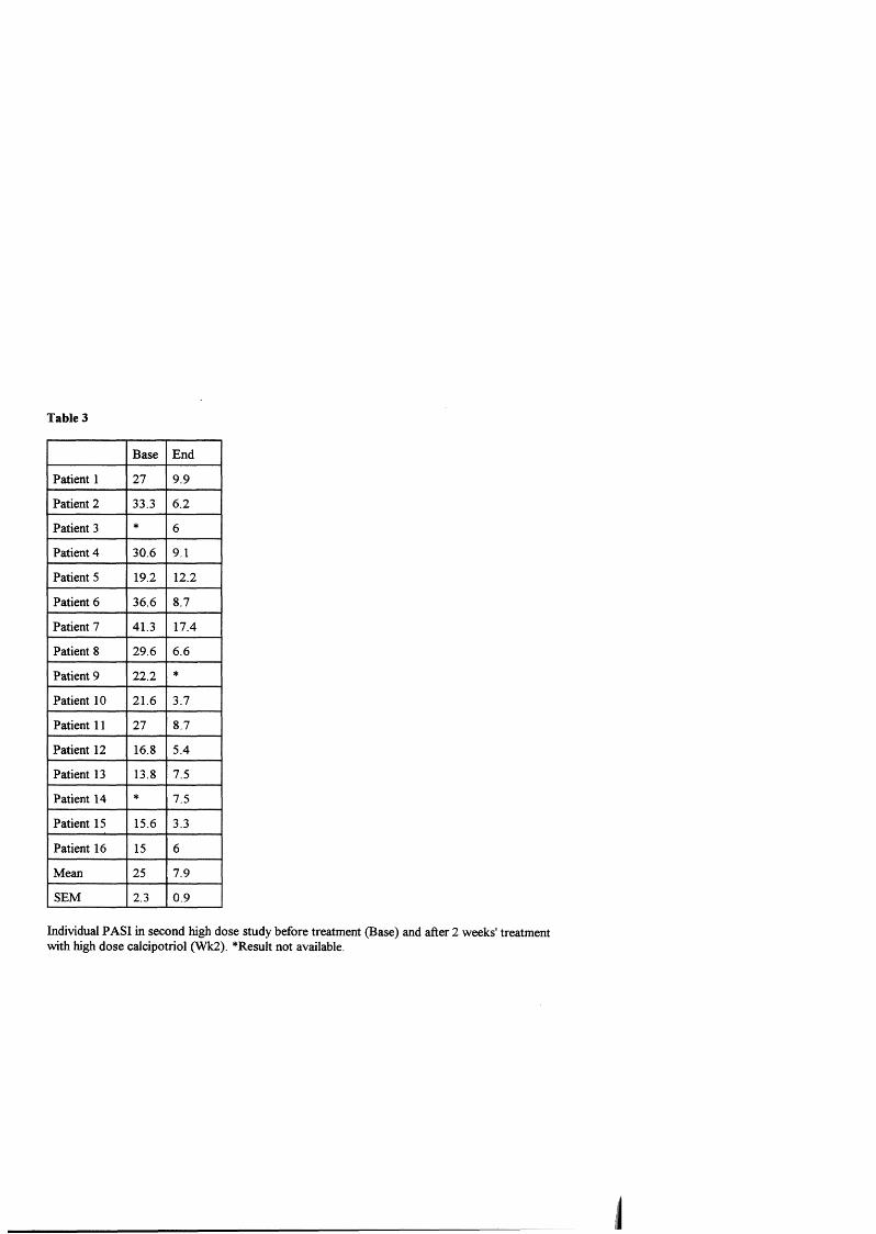
Table 3
Base End
Patient 1 27 9.9
Patient 2 33.3 6.2
Patient 3 * 6
Patient 4 30.6 9.1
Patient 5 19.2 12.2
Patient 6 36.6 8.7
Patient 7 41.3 17.4
Patient 8 29.6 6.6
Patient 9 22.2 *
Patient 10 21.6 3.7
Patient 11 27 8.7
Patient 12 16.8 5.4
Patient 13 13.8 7.5
Patient 14 * 7.5
Patient 15 15.6 3.3
Patient 16 15 6
Mean 25 7.9
SEM 2.3 0.9
Individual PASI in second high dose study before treatment (Base) and after 2 weeks' treatment with high dose calcipotriol (Wk2). *Result not available.

3
9 ( g
U 3 .
9? ® 2.
i l li ;I ?? î Î
i t. S S '
Ho
■g o'D) CJi
arv>
s0-o'
1oo
30
a
18 . I
ë I5 - 3Cf>
I(/>
I o i ig (Qo—&
lO
w
=r
o>
-1. P o Ü1 "Ni b3*
00
(O

Table 4
Leicester
Pre Rx 1 Post Rx
Manchester
Pre Rx 1 Post Rx
Base 4hrs Base 4hrs Base 4hrs Base 4hrs
Patient 1 24 1635 45 1840 11 2399 13 2653
Patient 2 37 824 31 1833 * * * *
Patient 3 0 1005 0 1415 * * * *
Patient 4 * * * * * * * *
Patient 5 0 705 0 414 * * * *
Patient 6 0 2280 0 808 * * * *
Patient 7 36 2780 272 3555 10 3719 169 5190
Patient 8 0 2403 0 3534 * * * *
Patient 9 0 3214 41 5084 * * * *
Patient 10 31 2021 76 1210 * * * *
Patient 11 43 698 37 1510 13.2 791 23 2119
Patient 12 19 968 26 1029 * * * *
Patient 13 10 1500 10 1950 10 2291 10 2776
Patient 14 0 2952 0 935 * * * *
Patient 15 46 2945 100 1936 41 4961 86 2952
Patioent 16 28 1099 39 881 * * * *
Mean 18 1802 45.1 1862 17 2832 60.2 3138
SEM 4.6 234 17.9 328 6.02 706 30.5 532
Serum strontium (pg/L) before (Base) and 4hrs after (4hrs) ingestion of 2.5mmol of stable strontium before OPre Rx) and after (Post Rx) treatment with high dose topical calcipotriol. Results from Manchester on 5 patients for quality control.

z "T#
û . Ci o
s g
’Il^ 3
l |. 5 - 3
II
,î ICL CL
III3CD
OCD
â
6N )
oro P
wok
penoG)
II: iN g
3CD3
ro
gQ
f
— en
I(O
So>0
1î -
(Q
I0 0
(O
z ^Il " A
p o 0 > 00 o

Table 5
Sea Base Base W kl Wkl Wkl Wk2 Wk2 Wk2 Ica Base End
Patient 1 2.3 2.3 2.4 2.4 2.5 2.4 2.4 2.34 1.3 1.31
Patient 2 2.3 2.1 2.4 2.5 2.5 2.8 2.8 * 1.11 1.5
Patient 3 2.5 2.4 2.4 2.5 * 2.5 2.5 2.52 1.22 1.31
Patient 4 2.3 2.2 2.4 2.6 2.9 * * * 1.14 1.2
Patient 5 2.4 * 2.3 2.3 2.3 2.4 2.4 2.41 1.35 1.57
Patient 6 2.3 * 2.2 2.2 2.3 2.4 2.3 2.29 1.21 1.21
Patient 7 2.4 2.4 2.5 2.6 2.8 2.7 * * 1.21 1.38
Patient 8 2.3 * 2.5 * 2.9 * * * 1.22 1.49
Patient 9 2.4 2.3 2.5 2.5 2.5 2.7 2.6 2.69 1.32 1.42
Patient 10 2.4 * 2.3 2.3 2.5 2.5 2.5 2.48 1.22 1.39
Patient 11 2.3 2.3 2.3 2.4 2.4 2.4 2.4 2.32 1.25 1.3
Patient 12 2.2 * 2.4 2.5 2.3 2.3 2.3 2.37 1.27 1.29
Patient 13
Patient 14
2.2
2.4
2.2
*
2.2
2.5
2.2
2.4
2.2
2.4
2.2
2.4
2.2
2.3
2.24
2.33
1.24
1.27
1.27
1.27
Patient 15 2.2 2.4 2.2 * 2.3 2.4 2.2 2.05 1.2 1.21
Patient 16 2.2 * 2.4 2.2 2.3 2.2 2.3 2.27 * 1.24
Mean 2.3 2.3 2.4 2.4 2.5 2.4 2.4 2.36 1.2 1.33
SEM 0.02 0.02 0.03 0.03 0.06 0.04 0.04 0.04 0.02 0.03
Serum total adjusted calcium (Sea) in mmol/L before treatment (Base) and during treatment (Wkl and Wk2). Serum ionized calcium (Ica) in mmol/L before treatment (Base) and at the end of treatment (End) in patients applying up to 360g of ointment per week. *Result not available. Patients in italics were Asian.

Assessment of bone turnover revealed no significant alteration at the end of the
study. Although there was a significant downward trend (Fig 13, Table 6) in serum total
alkaline phosphatase (fiom 88.8 to 79.4; P < 0.05), there was no significant change in any
of the ^ecific parameters of bone turnover which were measured. Serum bone specific
alkaline phosphatase, serum P I CP, serum osteocalcin, urine hydroxyproline and urine
concentration of deoxypyridinoline cross-links remained unchanged (Tables 7,8). The
reduction in serum total alkaline phosphatase may be explained by a reduction in hepatic
alkaline phosphatase refiecting the fact that psoriatics are known to have a higher intake of
alcohol than the general population. A period of abstention fiom alcohol while in hospital
could result in a fall in hepatic alkaline phosphatase.
Other parameters o f systemic calcium homeostasis were affected as in the pilot
study. Mean serum calcium rose fiom 2.30 to 2.47mmol/L, five patients becoming
hypercalcaemic (Fig. 12, Table 5). Four o f these (patients 2, 4, 7 and 8)were withdrawn
before they had received a full 2 weeks o f treatment. Mean serum ionized calcium rose
fiom 1.23 to 1.34mmol/L (Fig. 14, Table 5), mean 24h urine calcium fiom 4.29 to
6.45mmol/24h (Fig. 15, Table 9), 5 patients becoming hypercalciuric. Mean serum
phosphate rose fiom 1.17 to 1.46mmol/L (Fig. 16, Table 10), 11 patients becoming
hypeiphosphataemic, and mean fasting urine calcium rose fiom 1.89 to 2.91mmol/L
(Table 11). Mean 4am and 9am PTH fell fiom 3.7pmol/L and 3.2pmoFL to 1.3pmol/L and
1.6pmol/L, 6 patients becoming biochemically hypoparathyroid (Fig. 17, Table 12).
103

-■ i| ( Û ^
3 -o_
i. t
àQ.
E
(û
(D
W
9m
(gCD
g
T3
IQ .
( g
I1a
2 Q.E
■D.
KCQcrR
Iagï '
T 33"
8■ R
ICD
c r
I3
CD
CDCD
âQ
CDm
IIi■wllo■ ■ Ii
O o o o oo o
o 10o
w
U1
a>
00
(O
CD3

Table 6
Base Base Wkl Wkl Wkl Wk2 Wk2 Wk2
Patient 1 61 49 55 55 54 52 63 58
Patient 2 200 147 154 138 112 120 122 *
Patient 3 121 * 120 110 115 113 114 122
Patient 4 61 58 55 57 59 * * *
Patient 5 84 * 82 91 82 81 83 79
Patient 6 77 * 80 80 80 78 80 80
Patient 7 92 * 90 87 82 37 * *
Patient 8 70 * 70 * 60 * * *
Patient 9 57 58 61 59 56 59 52 43
Patient 10 121 * 113 132 120 122 112 99
Patient 11 69 69 68 62 63 66 66 66
Patient 12 91 * 95 87 94 92 87 81
Patient 13 61 64 52 63 62 55 53 51
Patient 14 106 * 106 103 106 105 108 110
Patient 15 106 110 122 * 115 130 132 126
Patient 16 73 * 78 82 80 77 83 77
Mean 91 79 88 86 84 85 89 82.7
SEM 9.3 15 7.3 7.2 5.9 7.9 7.4 8.11
Serum total alkaline phosphatase in patients applying high dose topical calcipotriol before (Base) and during (W kl, Wk2) treatment. *Result not available.

Table 7
PICP Osteocalcin BS-ALP Hyprol
Base End Base End Base End Base End
Patient 1 110 88 13 15 16 13 * *
Patient 2 198 154 5 13 22 16 * *
Patient 3 88 55 17 20 22 23 186 83 6
Patient 4 88 132 13 10 12 12 :%76 *
Patient 5 99 77 6 10 26 34 1296 363
Patient 6 77 66 13 10 18 15 1826 896
Patient 7 110 88 18 23 14 16 * *
Patient 8 99 66 7 13 9 12 * *
Patient 9 121 143 19 38 18 13 34R8 3248
Patient 10 66 99 9 6 21 16 3749 868
Patient 11 143 143 5 7 17 19 * *
Patient 12 143 198 13 17 37 26 30^9 *
Patient 13 550 583 14 14 16 16 * *
Patient 14 77 88 10 14 20 21 53 94.4
Patient 15 * 88 17 6 21 20 378 158 1
Patient 16 66 * 8 17 18 18 33 8 *
Mean 136 138 12 15 19 18 189 125
SEM 31 33 1.2 2 1.6 1.5 402 317
Serum procollagen C peptide (PICP; ng/ml), serum osteocalcin (ng/ml), serun bone specific alkaline phosphatase (UÂ.) and fasting (am) urine hydroxyproline (Hyprol; pmol/L) in patients applying high dose topical calcipotriol before (Base) and after (End) treatment. Several of the urine samples were lost or not analyzed for all parameters. *Result not available

Table 8
X-Links (Base) X-Links (End) Base/Creat End/Creat
Patient 1 41 36 5.9 7.3
Patient 2 * * * *
Patient 3 110 28 8.9 7.3
Patient 4 * * * *
Patient 5 35 25 7.5 7.6
Patient 6 90 25 5.4 4.5
Patient 7 * * * *
Patient 8 * * * *
Patient 9 74 120 6.8 103
Patient 10 * * * *
Patient 11 * * * *
Patient 12 * * * *
Patient 13 * 220 * 124
Patient 14 40 24 7.5 4.1
Patient 15 17 58 6.3 6.2
Patient 16 20 19 7.7 6.3
Mean 53 4 617 7.0 7.3
SEM 113 22.4 0.4 0.9
Fasting (am) urine deoxypyridinoiine cross links (X-Links; nmol/L) and deoxypyridinoline/creatinine ratio (nmol/mmol) before treatment (Base) and after 2 weeks’ treatment with calcipotriol (End). Several o f the morning urine samples were lost or not analysed for cross-links * Result not available

& 2! S <Û
» I -
O OWO
^ O
lO
w
o _
—ha i
E o- «=■ S s o>
l l h■ D CD g f
(O
I3
Ii
gc q ’i r
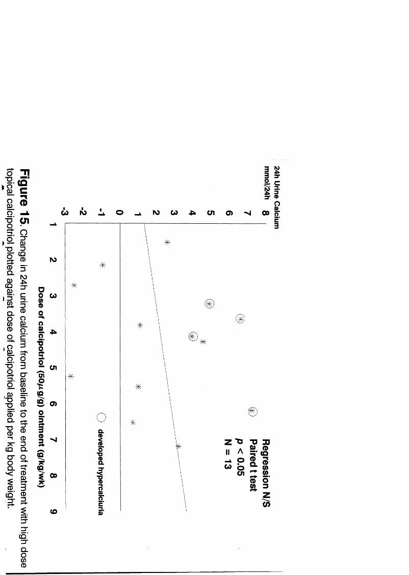
(D
Ol
ICQCD
g
3
Ig
(DCD3Q .
ar*4*®
I®3
3"g
c q '3"Q .
i
W IÔ IN> W O l o>
HIf» S
!s .
s
J
I(QS01IBs
lO
w
Ü1
o>
g 00 7T
(O
Q.
1{
IIw
s
33
i lZ0 3

Table 9
Base Base W kl Wkl Wk2 Wk2
Patient 1 3.2 * 9.4 127 726 8.9
Patient 2 * * 116 * * 112
Patient 3 .6 * 1.3 2.3 1.9 1.3
Patient 4 4.6 2.2 * * * *
Patient 5 1.9 * 3.1 1.5 3 3
Patient 6 3.4 * 6.8 10.7 3.6 111
Patient 7 2.9 * 3.6 * * *
Patient 8 3.1 4.9 10.2 10 5 112 *
Patient 9 3.4 * * 4.8 * 6.6
Patient 10 * * 1.4 * 1.6 *
Patient 11 2 * 4.1 5.1 7.3 5.8
Patient 12 5.9 6.9 14 5 8.5 7.5 13
Patient 13 9 * 3.2 6.1 5.2 7.4
Patient 14 14 * 8.9 18.2 129 10 1
Patient 15 5.3 3 * 2.6 3.2 *
Patient 16 1.7 * 3.9 4.3 * *
Mean 4.4 4.3 6.3 7.3 5.9 784
SEM 0.9 1 1.2 1.4 1.1 1.2
24h Urine calcium (mmol/24h) before treatment (Base) and during treatment (Wkl and Wk2) in patients applying up to 360g of ointment per week. Several samples were incomplete collections and were therefore unsuitable for analysis o f 24h urine calcium. *Result not available
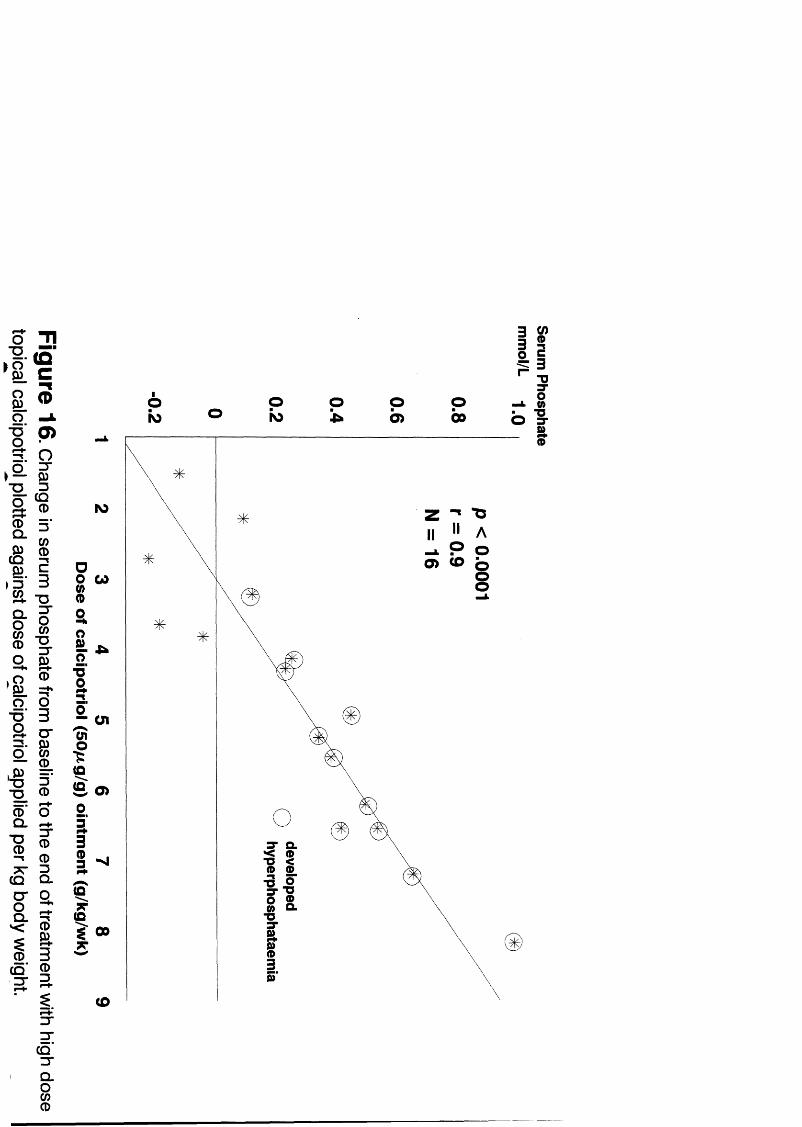
( Ûc
01 &
s
1 1
ICD
(Dm
I£
I(û
S
IIo po> p
00o o 3-
p0> (O
w
ai
o>
î i
00
(O
CQ
Q .
i

Table 10
Base Base W kl Wkl Wkl Wk2 Wk2 Wk2
Patient 1 1.3 1.2 1.1 1.5 1.4 1.4 1.3 1.5
Patient 2 0.9 0.9 1.2 1.4 * 2 175 *
Patient 3 1.3 1.1 1.3 1.5 * 1.4 172 151
Patient 4 1.1 0.9 1.2 1.6 1.4 * * *
Patient 5 1.3 * 1.1 1.1 1 1.3 117 126
Patient 6 1 * 1.5 1.1 0.9 1.1 131 147
Patient 7 1.1 * 1.1 1.2 1.2 1.6 * *
Patient 8 1.4 * 1.6 * 1.9 * * *
Patient 9 1.4 1.3 1.6 1.9 1.7 1.7 209 2 13
Patient 10 0.9 * 0.9 1.3 1.3 1.5 1.44 1J3
Patient 11 1.1 1.6 1.1 1.4 1.1 1.6 141 172
Patient 12 1.5 * 1.5 1.4 1.3 1.3 1.4 119
Patient 13 1 0.9 1.3 1.3 1.5 1.5 107 I j #
Patient 14 1.3 * 1.3 1 1.4 1.1 0.99 1.2
Patient 15 0.7 1.1 0.9 0.8 0.8 * 087 105
Patient 16 1.3 * 1.2 1.2 1.1 1 112 1.24
Mean 1.2 1.1 1.3 1.3 1.3 1.4 136 1.40
SEM 0.1 0.1 0.1 0.1 0.1 0.1 009 0.09
Serum Phosphate (mmol/L) in patients applying high dose topical calcipotriol before (Base) and during (W kl, Wk2) treatment. Patients applied up to 360g of ointment per week for a maximum of 2 weeks. Patients 2, 4, 7 and 8 stopped treatment early due to the development of hypercalcaemia. *Result not available

Table 11
Fasting (am) Urine
Calcium Creatinine Phosphate Calcium Ex Index
Ca/CrtRatio
Base End Base End Base End Base End Base End
Patient 1 L29 3 36 6.9 4.9 4.8 1622 .19 .69 16.2 624
Patient 2 .5 5.5 4.7 * 4.5 143 .11 * 6.9 *
Patient 3 .77 1.5 12.4 3.84 3 71 20 .06 .39 7.1 44.1
Patient 4 * * 7.2 * * * * * * *
Patient 5 124 1.21 469 3.3 166 951 .26 .37 225 308
Patient 6 1.15 3 31 166 5.6 169 9 13 .07 .59 6.4 567
Patient 7 2.4 4 476 18.2 288 4.9 .05 .22 4.1 176
Patient 8 Z27 3.3 * 4 7 15 7.3 * .83 * 693
Patient 9 3 89 5^1 109 116 907 315 .36 .48 264 46.6
Patient 10 298 289 163 6.2 563 253 .18 .47 * 489
Patient 11 * * * * * * * * * *
Patient 12 405 * 13 3 * 24 * .3 * 25 9 *
Patient 13 6 17 641 12.9 177 21 459 .48 .36 352 264
Patient 14 243 263 5.3 5.9 8.1 18 .46 .45 403 419
Patient 15 42 .6 2.7 9.3 737 1945 .16 .06 14.2 5.8
Patient 16 .3 .6 2.6 3 3.6 5.1 .12 .2 143 222
Mean 2.1 3.1 12 7.8 11 15 7 0.2 0.4 18 394
SEM 0.5 0.5 3 1.5 2.2 336 003 003 3.4 5.5
Fasting (am) urine calcium (mmol/L), creatinine (gmol/L), phosphate (mmol/L), calcium excretion index (pmol/L), and calcium:creatine index (mmol/pmol) in patients applying high dose topical calcipotriol before (Base) and after (End) treatment. Patients applied up to 360g of ointment per week for a maximum of 2 weeks. Patient 11 was unable to provide fasting am urine samples. *Result not available.

i l l$
§ O g m
f -o 5 -0
-§ II I S ^
I I
II<g §m. m
i l
I -0 0
a §o 0
II13 3 “D 0
o °0 _ I—»
^ ?03
CD CL
IS .
I1ICQ
S0
1I(QI
W t b
ir(£)o I
IS>
w
cn
o>
1 100
CO

Table 12
PTH Base4am
Base9am
End4am
End9am
Tnipcw/GFR
Base End
Patient 1 1.9 1.5 0.5 0.5 1.5 122
Patient 2 4.8 2.4 0.5 0.5 * *
Patient 3 7.5 5.3 2.7 2.1 1.46 186
Patient 4 3.6 3.8 * * * *
Patient 5 2.2 1.6 * * 1.04 1.02
Patient 6 4 2.7 0.9 1 106 142
Patient 7 4.5 4.7 0.9 0.8 1.42
Patient 8 2 1.9 0.5 0.5 * *
Patient 9 3.6 2.8 1.1 1.1 158 2
Patient 10 8.5 7 * 4.1 1.15 1.6
Patient 11 2.3 3.5 0.8 0.8 » *
Patient 12 2.5 2.4 0.8 0.8 * *
Patient 13 1.1 2.2 * 0.7 085 112
Patient 14 2.7 2.5 2.2 3.3 1J2 0.9
Patient 15 4.4 4.2 4.7 4.3 062 086
Patient 16 3.8 2 0.5 1.5 111 108
Mean 3.7 3.2 1.3 157 1.18 132
SEM 0.5 0.4 0.4 036 009 O il
Serum PTH (pmol/L) andTmpg^/GFR in patients applying high dose topical calcipotriol before treatment (Base) and after treatment (End). Patients applied up to 360g of ointment per week for a maximum of 2 weeks. *Result not available

The changes in these parameters, with the exception of 24h urine calcium,
correlated with dose of calcipotriol applied.
Urine indices o f renal function were also affected. Mean calcium: creatinine ratio
(Fig. 18, Table 11) rose from 216 to 356mmol/pmol and mean calcium excretion index
rose from 18.7 to 29.5 (Table 11). Mean TmpoVGFR rose from 1.17 to 1.31 (Fig 19,
Table 12).
There was also rise in mean mine phosphate excretion - mean 24h mine phosphate
rose from 13.5 to 20.2mmoF24h (Fig 20, Tablel3); and mean frsting urine phosphate
(Table 11) rose from 11.5 to 15.7mmol/L. However, this was inconsistent and did not
correlate well with dose of calcipotriol applied.
There was some concern about a rise iu mean serum creatinine (from 8 8 . 6 to
93.1 pmol/L), which was dose dependent (Fig 21, Table 14). However, glomerular
filtration as judged by creatinine clearance was not altered (Fig 22, Table 14).
This study revealed one other interesting finding. 1,25 dihydroxyvitamin D3 levels,
as measmed by the gamma-B 1,25-dihydroxyvitamin D Mt (IDS immunodiagnostic
systems Ltd., Boldon, Tyne and Wear, NE35 9PD, UK), rose markedly (Fig 23, Table 15)
from a mean of 55.8 to 368.2pmol/L (10 o f 11 patients above the normal range). This
conflicted with the findings of om initial pilot study and it became apparent that
calcipotriol in serum was interfering with the assay.
104

We therefore sent duplicate samples to Dr LeVan for further assessment. His
HPLC method confirmed that 1,25 dihydroxyvitamin D3 levels had actually fallen while
calcipotriol levels had risen (Fig 23, Table 15). Mean serum 1,25 dihydroxyvitamin D 3
levels fell fi-om 30.7 (SER=3.1) to 12.0pg/ml (2.0) while calcipotriol levels rose from
undetectable or barely detectable in 4 patients who had received calcipotriol prior to
baseline saitples being taken to a mean of 25.7pg/ml (3.5).
105
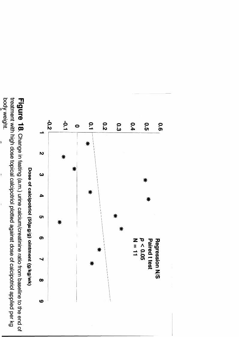
2!
i !CmmAœ
Q _ CQ
8 ® .CD 3
! lIf"D
OU
(Dm
g
i |D) 3
l ï
f ii i«T 3
3(D
Ô" OCL e
1 3 CD ® 0
(Q CL
IS
I
In
£2.
IISs
6k ) s g : O
Û1Ob>
w
Ü1
O)Z t ï :□ DO
o
00
(D

i sPS ."S.oQ . S
to93
(QCD
(B9?.3CD
$o
% "T | O 3 0
l |
} |T 3 CD
IICD
( S ' â
§■ 2 ,o _
* < CD
I. ICQ CD 3 - 3
3 -3 -
c o '3 -Q .O
$
#S
Ô 6 6 pOl w ro t O
o w
P o G> b
CO

s -3 “Q.i0
1E
- go
3( Oc3N )0
s1(D—•
zr
3- o3"
8 "D
i
(B9>.8
mg
"O"Q
miB:ECQO"R'<(D
c q '3
N)
08 “
a
1 * fi «a i
CD
IgCD3
f«os O)0
13 -U
O IQ
E IBQ.
ai3
$ 00 %"
CD
$
i 12. cIV 3.3* (D
■0S’
O 0>"O3PRm
z "0 TB 30Il A 2 |g-L O° g $I§
ZO)

Table 13
Base Base W kl Wkl Wk2 Wk2
Patient 1 5.9 * 40 39.9 36.5 38.8
Patient 2 * * 22.3 * 28.2 *
Patient 3 * * 11 26.5 42.1 9.27
Patient 4 * * * * * *
Patient 5 10.2 * 24.7 16.3 18.2 22.8
Patient 6 * * * * * *
Patient 7 7.29 * 10.3 * * *
Patient 8 8.3 7.7 17.1 16.8 26 *
Patient 9 18.8 * * 15.4 * 15.5
Patient 10 * * 1 * 3.9 *
Patient 11 * * * * * *
Patient 12 * * * * * *
Patient 13 16.4 * 13.8 25.5 27 26.1
Patient 14 * * * * * *
Patient 15 15.4 11 * 14 * *
Patient 16 39.5 16.7 39.9 27.8 * *
Mean 15 12 20 23 26 22.5
SEM 2.7 2.6 4.7 3.2 4.7 5.0
24h Urine phosphate (mmol/24h) in patients applying high dose topical calcipotriol before (Base) and during (W kl, Wk2) treatment. Patients applied up to 360g of ointment per week for a maximum of 2 weeks. Urine phosphate concentration was not measured in many patients as regularly as 24h urine calcium. *Result not available.

0T315Q .
10
"O
gQ .
(SË .36Q .1a
SQ .
"OQ
"g_ô 'Q_
■g
7TCQ
ICD
CQ"3"
(QC0
N>
93
CQCD
se
II3s
î i "
I I3"® (O
II(O
CDCD
âO
s Sm
g
3"g
CQ'3"Q .
i
N)
W
O§ a
S
f .U1
0>
00
co
i

Table 14
Creatinine Base Base Wkl Wkl Wkl Wk2 Wk2 Wk2 GFRBase
GFREnd
Patient 1 91 82 93 87 95 89 90 91 104 90
Patient 2 72 58 72 69 74 74 78 * * 66
Patient 3 116 112 94 114 * 114 138 113 38.8 25.9
Patient 4 83 78 72 98 100 » * * 50.8 *
Patient 5 85 * 92 88 93 90 86 84 45.3 69.1
Patient 6 92 * 113 104 98 105 87 96 105 124
Patient 7 81 * 97 99 80 * * * 44.7 43.5
Patient 8 84 * * * * 84 * * 46.8 84.9
Patient 9 72 76 83 89 97 97 90 98 82.4 137
Patient 10 * * 105 * * * 106 105 * 35.2
Patient 11 107 103 97 100 93 105 98 105 59.5 *
Patient 12 85 * 104 97 105 96 92 87 * *
Patient 13 77 70 75 71 77 72 72 73 145 139
Patient 14 88 * 89 87 85 87 90 94 * *
Patient 15 91 91 * * * * 93 90 62.7 *
Patient 16 124 * 109 125 124 112 119 111 90.2 79.4
Mean 90 84 93 94 93 94 95 96 72.8 81.3
SEM 3.9 6.2 3.7 4.5 4.1 4.1 4.8 3.2 9.8 12.4
Serum creatinine (pmol/L) and GFR (ml/min) in patients applying high dose topical calcipotriol before (Base), during (Wkl, Wk2) and after (End) treatment. Patients applied up to 360g of ointment per week for a maximum of 2 weeks. *Result not available.

Ë_Q .
T3Q
■D
I<gs.3
Q .
ii.Q.
"Oo
"O
s:T3CD
g
§ ■
3( Oc0
KICD
5 ‘Q5iI3CD
g "CD
â
CDm
CD3
3"3Tco'3"Q .iO
T3S
I
O o s s
ro
o8 “(D
m
I5
Im
£2.3
IB
s
ai
O)
(O
e
Z ZIl 0

eg
•Io_iO 3
? Z !
I"| 3
l O o W
i
■g■g_g;T3(D
CQ
Ii
I T
I f5' (DCD
O W
i |CD ^a a0 9.
31 g
Q .
g
g 5
( i i=T ^
o II Ie . 5 -
S P
f î- ?
IlQ _
Ia
IO
I(Q
SI(Q
II
ro so oo
w
U1
o>
Ü100
(Q—% 8(O
o p
1“oo

Table 15
25 vitD 1,25 vitD Calcipotriol 1,25 vitD(HPLC)
Base End Base End Base End Base End
Patient 1 19 17 37 500 0 39.3 17.3 10.6
Patient 2 13 7.2 52 * 0 22.3 27.7 8.3
Patient 3 8.2 6.9 46 187 5.8 27.1 44.4 6.7
Patient 4 * * * * * * * *
Patient 5 11 11 51 287 0 8.6 29 12.5
Patient 6 19 10 * * 0 13.6 30.7 6.1
Patient 7 13 13 51 500 0 42 37.7 9.6
Patient 8 7.7 10 24 395 4.7 23.1 19.1 7.4
Patient 9 7.5 12 51 500 4.7 37.7 39.1 6
Patient 10 5 5 134 * 14.4 38.2 22 14.3
Patient 11 17 16 46 500 0 47.4 26.5 7.6
Patient 12 21 16 64 500 5.1 15.3 28.5 6.8
Patient 13 7.9 9.6 62 * 0 19.4 42 21.1
Patient 14 25 10 53 300 0 * 57.3 30.3
Patient 15 11 17 S3 87 0 9.6 29.8 26.5
Patient 16 22 16 57 294 6.8 16.1 10.1 6.1
Mean 14 12 56 368 2.8 26 31 12
SEM 6.4 1 6.6 42 1.1 3.5 3.1 2.0
Serum 25 hydroxyvitamin D (25 vitD; ng/ml), serum 1,25 dihydroxyvitamin D by radioimmunoassay (1,25 vitD; pmol/L), serum calcipotriol (pg/mg) and 1,25 dihydroxyvitamin D by high performance liquid chromatography (1,25 vit D; pg/mg) in patients applying high dose topical calcipotriol before (Base) and after (End) treatment. Patients applied up to 360g of ointment per week for a maximum of 2 weeks. Calcipotriol was detectable at baseline in a number of patients who had inadvertently started treatment with the ointment before the samples were taken. Calcipotriol interfered with the RIA assay giving a falsely high values for 1,25 dihydroxyvitamin D^.^Result not available.

3.3.2 ©isctassion
In this study we confirmed that calcipotriol in large amounts increases
serum total adjusted calcium and 24h urine calcium, suppresses PTH and 1,25
dihydroxyvitaniin D 3 . We also demonstrated a rise in serum ionized calcium, serum
phosphate and urine phosphate. No effect on bone metabolism was demonstrated althou^
the study period was very short and the numbers of patients studied small so that an effect
on bone turnover cannot be excluded. Also, many of the patients did not have urine
deoxypyridinoiine cross-links measured which is felt, by many, to be the most sensitive
index o f bone turnover.
We have also demonstrated increased intestinal absorption o f calcium in those
patients receiving the h ip e s t doses of calcipotriol, particularly those who developed
hypercalcaemia. That the strontium absorption test did not detect increased absorption in
those applying lower doses is probably a reflection o f the insensitivity o f the test. There
was marked inter and intra-individual variation in strontium levels after oral administration
of a fixed dose under carefidly controlled conditions. The change in levels from baseline to
the end o f the study varied from a two-fold increase to a 50% reduction. Most studies
using this method in the past to estimate intestinal calcium absorption have relied on single
measurements in each patient. Reynolds et found marked variation in levels on
different days in the same individuals and concluded that it was a very insensitive test.
Unfortunately, there is no evidence that any other methods of evaluating intestinal calcium
absorption are better that the strontium absoiption test.
106

Renal indices measured in these patients confirmed the dominant effect on PTH
secretion, with calcium excretion index, calcium creatinine ratio and TmpoVGFR all rising.
107

' There were a number o f other interesting jSndings in this study. Firstly, the changes
in systemic calcium homeostasis correlated very closely with dose o f calcipotriol apphed
per unit body w e i^ t. This is not surprising, but it has not previously been documented.
This finding may explain the development of hypercalcaemia in patients using
recommended amounts o f calcipotriol while some studies using maximum or almost
maximum recommended doses faded to demonstrate any effect whatsoever. Smaller
patients are more likely to develop side effects fiom calcipotriol at lower doses.
The second interesting finding was the fact that calcipotriol interfered with the
1,25 dihydroxyvitamin Dg assay Idt. On reflection, this is not surprising, given that it is an
antibody based test and that calcipotriol binds to the vitamin D antibody as avidly as 1,25
dihydroxyvitamin D3 in vitro. This has important potential implications for patients using
topical calcipotriol. Measurement o f 1,25 dihydroxyvitamin D3 using this type o f assay is
not reliable in such patients and HPLC measurement is necessary.
108

Finally, it is noteworthy that 3 o f the patients who developed hypercalcaemia were
Asian patients. Two out o f four non-Asian patients applying greater than 5g per kg per
week developed hypercalcaemia, whereas all three Asian patients applying those doseS
developed hypercalcaemia. The Asian patients developed hypercalcaemia earher than the
non-Asian, all developing hypercalcaemia within the first week (Table 5, Asian patients in
itahcs). The Asian patients recorded higher serum total adjusted calcium levels than the
two non-Asians who developed hypercalcaemia despite the fact that the two non-Asians
were applying larger doses o f calcipotriol. This may be of relevance, in that one of the two
patients who was reported to have developed hypercalcaemia while using recommended
amounts o f calcipotriol, was Asian. However, the number of patients studied in this
project was very small and these findings may well be a chance occurrence. Furthermore,
we can suggest no explanation as to why these patients should be particularly susceptible
to the toxic effects o f calcipotriol.
109

3.4 SECTION m Recommended doses
3.4.1 Results
3.4.1.1 100g o f calcîpotriol (50pg/g) ointment per week
21 patients, 10 male and 11 female, were recruited. Mean age o f the patients was
44yrs with a range from 19 to 74yrs. Mean PASI at baseline was 15.8 (range 38.5 to 7.2)
and feh to 5.8 (11.7 to 0.9) after 4 weeks' therapy (Table 16).
Mean 24hr urine calcium rose significantly (Fig 24) from 4.71mmoF24h to
5.07mmol/24h (P<0.05). Six patients developed hypercalciuria. Almost all had abnormal
baseline urine calcium levels and only one patient with a normal value at baseline
developed an abnormally high level during treatment - 8.5mmol/24h. This level was not
sustained and the patient's urine calcium returned to normal while stül applying
calcipotiiol. Mean serum phosphate also rose (Fig 25, Table 17) from 0,97mmoFL to
1.12mmol/L (P<0.005). There was no significant change in serum total adjusted calcium
(Fig. 24), serum 9am PTH, serum osteocalcin, c-terminal propeptide o f type I collagen
and serum bone specific alkaline phosphatase (Tables 17,18).
110

Table 16
Base Wk2 Wk4
Patient 1 * * *
Patient 2 * * *
Patient 3 * * *
Patient 4 35 * 5.4
Patient 5 * * *
Patient 6 24 * 8.8
Patient 7 18 * 9.7
Patient 8 12 7.7 5.2
Patient 9 9.6 * 5.4
Patient 10 14 13 3.8
Patient 11 19 8.8 6
Patient 12 8.4 * 8.2
Patient 13 12 7.6 5.2
Patient 14 7.8 2.6 1.3
Patient 15 9.9 2.8 3.6
Patient 16 7.2 4.9 4.6
Patient 17 11 7.4 105
Patient 18 39 27 11.7
Patient 19 14 6 2
Patient 20 11 2.8 0.9
Patient 21 * * *
Mean 16 8.2 5.8
SEM 2.3 2 0.8
Individual PASI in patients applying 100g o f ointment per week before treatment (Base) after 2 and 4 weeks o f treatment (Wk2 and Wk4). *Result not available.

§$
CQ >Êr.
IIC
IITC
5
• 1-(D"8"O
• Î
cn 0) 00
g0 )(D
3<D
I<D? T
W
IO
NO
zCO 1
IN)ÎO
roCO
ISO ISO01
ISOÔ )
II
âa
IO
CD
§
Po cs =SÔ
m
A iIIÎ3
II3g2.c
lOk l
o&}

. ^ - n• B ( q -
g . Ç
q ® i IN) 3. ui
► ® sCD
II11
I s ■§-
I I
•II$■
» -B
. II
DOm
o
o
I(D
K >
I"OQ
o(0
00 o(D ro w
00 p
Q .
Q .
3 S’
11 r - 0
' 8
I
S
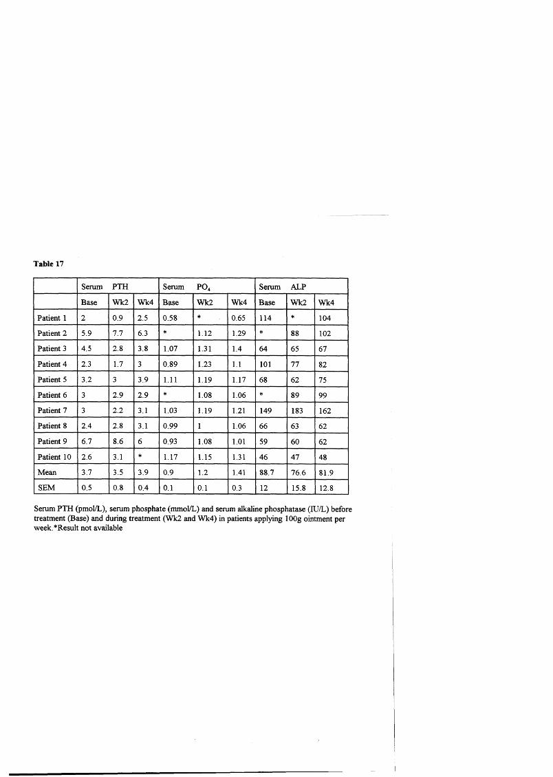
Table 17
Serum PTH Serum PO^ Serum ALP
Base Wk2 Wk4 Base Wk2 Wk4 Base Wk2 Wk4
Patient 1 2 0.9 2.5 058 * 065 114 * 104
Patient 2 5.9 7.7 6.3 * 1.12 129 * 88 102
Patient 3 4.5 2.8 3.8 1.07 1.31 1.4 64 65 67
Patient 4 2.3 1.7 3 089 1.23 1.1 101 77 82
Patient 5 3.2 3 3.9 111 1.19 1.17 68 62 75
Patient 6 3 2.9 2.9 * 108 1.06 * 89 99
Patient 7 3 2.2 3.1 1.03 119 121 149 183 162
Patient 8 2.4 2.8 3.1 099 1 1.06 66 63 62
Patient 9 6.7 8.6 6 093 l^K 1.01 59 60 62
Patient 10 2.6 3.1 * 117 115 131 46 47 48
Mean 3.7 3.5 3.9 0.9 1.2 1.41 887 7 6 6 819
SEM 0.5 0.8 0.4 0.1 0.1 0.3 12 15 8 12 8
Serum PTH (pmol/L), serum phosphate (mmol/L) and serum alkaline phosphatase (lU/L) before treatment (Base) and during treatment (Wk2 and Wk4) in patients applying lOOg ointment per week.*Result not available

Table 18
Serum PICP Osteocalcin BS-ALP
Base End Base End Base End
Patient 1 66 55 6 65 10 10
Patient 2 132 154 7 14 10 16
Patient 3 77 110 17 13 18 19
Patient 4 55 77 7 7 12 11
Patient 5 143 88 13 11 16 17
Patient 6 66 66 29 21 24 23
Patient 7 143 88 6 6 11 11
Patient 8 66 77 5 6 10 15
Patient 9 77 99 12 13 17 16
Mean 917 90 113 17.3 142 15 3
SEM 122 9.7 2.6 6.2 1.6 1.4
Serum Procollagen peptide (PlCP;ng/ml), Osteocalcin (ng/ml) and Bone specific alkaline phosphatase (BS-ALP;U/L) before and after 4 weeks' treatment in patients applying lOOg ointment per week.*Result not available

3.4.1.2 90g o f calcipotriol (50fjg/g) versus 90g o f calcitriol (3fj.g/g) ointment per
week
Twenty-four patients, 14 female and 10 male, were recruited. Mean age was 42
years with a range o f 18 to 80 years. 12 were assigned to the calcipotriol group, 12 to the
calcitriol group. Four patients in the calcipotriol group failed to complete the study. One
of these was withdrawn after 6 weeks because of lack of efficacy and one, after 4 weeks,
because of the possibility of pregnancy. The other 2 failed to attend after 2, and 4 week's
treatment. Four patients in the calcitriol group failed to complete the study. Two were
withdrawn after 6 weeks because of lack of efficacy, one patient went abroad on holiday
after 1 week of treatment and one patient failed after 1 week o f treatment. Baseline data
were available on all patients, data from at least 4 visits on 21 patients and 'on-treatment'
data on all but one patient.
There was no significant difference ia the mean PASI (Table 19,20) of the two
groups at baseline; calcipotriol group - 14.9 (range 3.7 to 38.2), calcitriol group - 13 (7.6
to 19.2). Mean PASI in patients applying calcitriol fell by 32% from 13 to 8.8 (P = 0.013)
and in patients applying calcipotriol by 68% from 14.9 to 4.7 (P < 0.005). The reduction
was significantly greater in the calcipotriol-treated group (P = 0.014).
There was a small but significant increase in serum ionized calcium (Fig 26) in the
calcipotriol-treated group (from 1.21mmol/L to 1.25mmol/L, P = 0.01) but no significant
changes in any o f the other parameters o f calcium homeostasis measured. There were no
significant changes in any o f the parameters measured in the calcitriol-treated group .
I l l

Table 19
Calcipotriol Base Wkl Wk2 Wk4 Wk6 Wk8
Patient 1 11 2.3 3.1 0 0 0
Patient 2 3.7 5.2 2.8 1.7 1.3 *
Patient 3 IS 7.4 3.2 1.8 1.6 0.9
Patient 4 11 7.8 4.7 4.4 3.7 3.7
Patient 5 38 16 6.2 8 * *
Patient 6 19 9.8 7.6 9.6 * 7.3
Patient 7 12 4.6 3.8 * 1.6 1.4
Patient 8 6.2 3.8 4.2 * * *
Patient 9 16 8.1 3.2 3 0.5 2.2
Patient 10 16 * 6.4 3.8 2.7 1.7
Patient 11 16 14 13 15 16 *
Patient 12 14 10 9 9 * *
Mean 15 8.1 5.6 5.6 3.5 2.5
SEM 2.5 1.3 0.9 1.5 1.9 0.9
Individual PASI in patients applying 90g o f calcipotriol ointment per week for up to 8 weeks. *Result not available.

Table 20
1,25 vitD
Patient 1 11 7 12 6.7 4.5 6.5
Patient 2 19 15 3.1 6.5 7.4 7.3
Patient 3 16 13 * 9.2 1 1 9.2
Patient 4 12 5.2 5.8 7.6 4.8 6.3
Patient 5 7.6 4.6 4.2 5.4 8.4 6.2
Patient 6 7.8 2 2.4 2 2.4 *
Patient 7 15 14 1 1* 5 8
Patient 8 15 14 9 * 14 18 8
Patient 9 14 1 1* 9.2 9.8 *
Patient 10 15 14 * * * *
Patient 11 11 9.8 7.5 5.9 7.4 7.8
Patient 12 * * * * * *
Mean 13 9.9 6.9 6.6 7.5 8.8
SEM 1 . 1 1.4 1.3 0.8 1 . 1 1.5
Individual PASI in patients applying 90g o f 1,25 dihydroxyvitamin D3 ointment. *Result not available

!?
' <
CO ®
f p
•ifQ . 3Î s
I ICQC
IQ . c
N> toto
toW
to•Cü
to01
Q .R
■I
I a? § I S'f \ \ Q .
“D- g
3 'CQ
7T
mA.
SI fIi
g
§
Is?nc
to3
w &

3.4.1.3 A single 6g dose o f calcipotriol (50jug/g) ointment compared to placebo
10 patients, 3 male and 7 female, were recruited. Mean age was 48yrs, range 23 to
70yrs. There was no significant difference between the biochemical profiles over a nine
hour period in each patient when calcipotriol was applied as compared to placebo (Figs
27-9).
3.4.1.4 Calcipotriol (50pg/g) ointment plus occlusion
48 patients, 22 male,and 26 female, were recruited. Mean age was 43yrs, range 18
to 70yrs. Patients, on average, used 30-40g of the ointment per week. Seven patients
failed to complete the study; 2 were withdrawn because of initant reactions, 2 because of
lack of efficacy and 3 were lost to follow-up. Data were available for analysis on 41
patients who corqpleted the study. 19 were assigned to group A (calcipotriol Vs
calcipotriol plus occlusion) and 22 to group B (calcipotriol plus occlusion Vs placebo plus
occlusion). In group A, occlusion plus calcipotriol (mean severity score fell from 7.9 to
3.0, Table 21) was significantly more effective than calcipotriol alone (mean score feU
from 7.6 to 4.6; Table 22), P < 0.005. In group B, occlusion plus white soft paraffin
(Table 23) had no significant effect on severity of psoriasis while the response to
calcipotriol (Table 24) plus occlusion was similar to group A (7.8 to 2.2, P < 0.005).
There was no significant change in mean serum total adjusted calcium, phosphate,
alkaline phosphatase or mean 24h urine calcium and phosphate (Fig 30).
112

"O
s 3 3%(O(Drr 3 c
P CDa -%CD0) N)Q_•Ni
0 13 33'(D
Q)3Q.O 0)C m3 S0) MA? T3(D O-t fHh0) C"O-o 3(Dâ So O3 ca 3O) 0)Cû 3o Q.
■Qo 3"0) OQ. U)“O"O 3*a a3. (Do Q)3Q.01 Iso-3
g
0 - n 1 3
1
ow
oô>
oloo Q■n
IO -o
(O
S= ros
"D Cg 2 .IO
S o"O3"
o.Ol
II0>
S- XN
Sai ai roo roai wai | i '
£ »i

C oo ro G ) 00
■f
H "I
Q .
"O
lligZ Z
O
C Q .
Q .
=T
PO
ob i o en
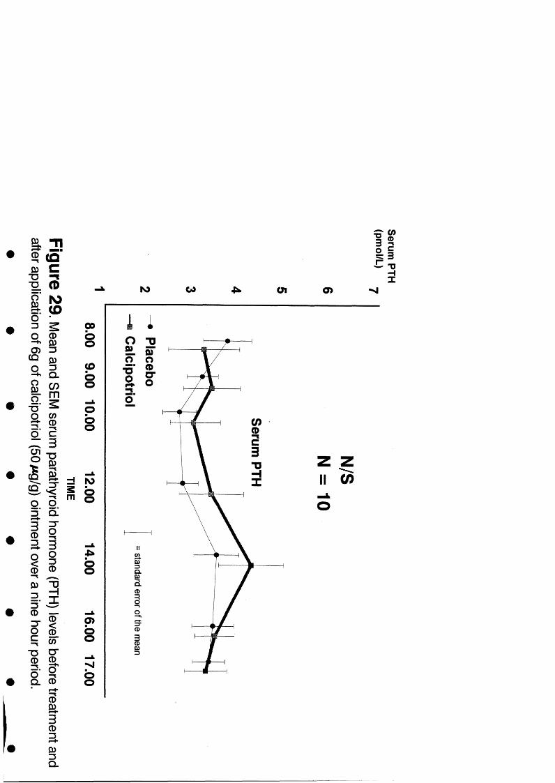
. I€- g
I• â
Cû
I?
T 1
( ÛC- ImIO(D
IsQ .0 5m
$o c
gBQ.3
CDa
i
1Q-
iiCD
D)3
CD —
I III■ s
0)
îl% 3
- sK ) W en o>
00sc os§
mIOs
s
p>ss
s . F
II
Q .
Q .
z zIl 0 )
(D3-§Q .

Table 21
Base Wk2 Wk4 Wk6 Wk8
Patient 1 5 1 4 1 2
Patient 2 8 8 8 8 6
Patient 3 9 * 6 * 5
Patient 4 8 4 3 6 3
Patient 5 7 3 2 2 2
Patient 6 8 6 5 5 6
Patient 7 11 6 4 2 1
Patient 8 6 6 5 4 8
Patient 9 9 9 9 8 8
Patient 10 8 7 6 3 3
Patient 11 7 4 6 5 3
Patient 12 7 3 3 2 *
Patient 13 9 3 3 2 2
Patient 14 9 4 2 1 1
Patient 15 8 5 3 2 0
Patient 16 9 3 5 1 0
Patient 17 7 4 3 1 1
Patient 18 8 2 2 1 1
Patient 19 7 4 4 3 3
Mean 7.9 4.6 4.4 3.2 3.1
SEM 0.3 0.5 0.5 0.5 0.6
Severity scores for the limb(s) assigned to be treated with calcipotriol plus occlusion in group A. *Result not available.

Table 22
Base Wk2 Wk4 Wk6 Wk8
Patient 1 3 1 3 2 2
Patient 2 8 6 6 8 8
Patient 3 9 * 7 * 6
Patient 4 8 6 4 1 4
Patient 5 6 4 2 2 3
Patient 6 8 6 6 8 7
Patient 7 11 5 5 2 1
Patient 8 6 6 5 5 6
Patient 9 9 9 9 9 8
Patient 10 6 6 5 4 3
Patient 11 7 6 3 5 5
Patient 12 7 3 2 2 *
Patient 13 9 7 8 7 6
Patient 14 9 6 6 6 7
Patient 15 8 7 4 6 3
Patient 16 9 7 4 3 4
Patient 17 7 6 3 3 1
Patient 18 7 8 7 7 6
Patient 19 7 5 2 3 3
Mean 7.6 5.8 4.8 4.9 4.6
SEM 0.4 0.4 0.5 0.5 0.5
Severity scores for the limb(s) assigned to be treated with calcipotriol alone in group A. *Result not available.

Table 23
Base Wk2 Wk4 Wk6 Wk8
Patient 1 3 3 3 6 7
Patient 2 8 6 4 3 3
Patient 3 9 7 8 7 3
Patient 4 9 7 9 10 10
Patient 5 5 5 5 6 *
Patient 6 9 9 7 6 6
Patient 7 8 11 7 5 5
Patient 8 6 8 7 8 6
Patient 9 6 7 7 8 8
Patient 10 8 8 6 7 6
Patient 11 8 8 8 8 8
Patient 12 8 8 8 8 8
Patient 13 8 7 8 * *
Patient 14 10 7 8 8 10
Patient 15 9 9 6 7 8
Patient 16 9 * * 12 *
Patient 17 7 6 6 6 6
Patient 18 6 6 6 6 6
Patient 19 8 8 8 * *
Patient 20 6 6 5 * *
Patient 21 6 6 6 6 6
Patient 22 7 6 5 5 6
Mean 7.4 7 6.5 6.9 6.6
SEM 0.4 0.4 0.3 0.4 0.4
Severity scores for the limb(s) assigned to be treated with placebo plus occlusion in group B. *Result not available.

Table 24
Base Wk2 Wk4 Wk6 Wk8
Patient 1 3 2 2 1 1
Patient 2 9 4 3 3 2
Patient 3 9 6 6 6 3
Patient 4 9 4 4 3 2
Patient 5 5 2 3 4 *
Patient 6 9 9 6 4 3
Patient 7 8 11 4 4 1
Patient 8 9 5 5 5 4
Patient 9 6 2 1 0 0
Patient 10 8 7 6 5 1
Patient 11 8 3 3 3 1
Patient 12 8 9 4 3 3
Patient 13 8 4 4 * *
Patient 14 10 4 4 4 5
Patient 15 9 9 3 2 1
Patient 16 9 * * 12 *
Patient 17 7 7 6 6 4
Patient 18 6 6 4 1 1
Patient 19 9 2 3 * *
Patient 20 7 3 4 * *
Patient 21 6 7 5 4 5
Patient 22 9 3 1 1 0
Mean 7.8 5.2 3.9 3.7 2.2
SEM 0.4 0.6 0.3 0.6 0.3
Severity scores for the limb(s) assigned to be treated with calcipotriol plus occlusion in group B *Result not available.

cr Tlm — :I ICD (D
3 WQ. p
l lIII IIII I 11
(D Q-
So c "
g
II
§Q_
I T
5
SQ .c3%
lO lOro
lOw
ro toÜ1
3O I
Q.O.
II
II
4:^
-j. ro w o i
3 K
ii~ 9

3.4.1.5 Long term use o f calcipotriol (50pg/g) ointment
Twenty patients, 7 male and 13 female, were recruited. Mean age was 43 yrs,
range from 37 to SOyrs. Mean PASI fell firom 6.48 (range 0.9 to 10.8) to 3.35 (0 to 5.4).
All 20 patients completed 12 months' treatment with calcipotriol. Patients used on average
30-40g per week with a range of 20=76g. There was no significant change in serum total
adjusted calcium or 24h urine calcium throughout the study period (Fig 31).
113
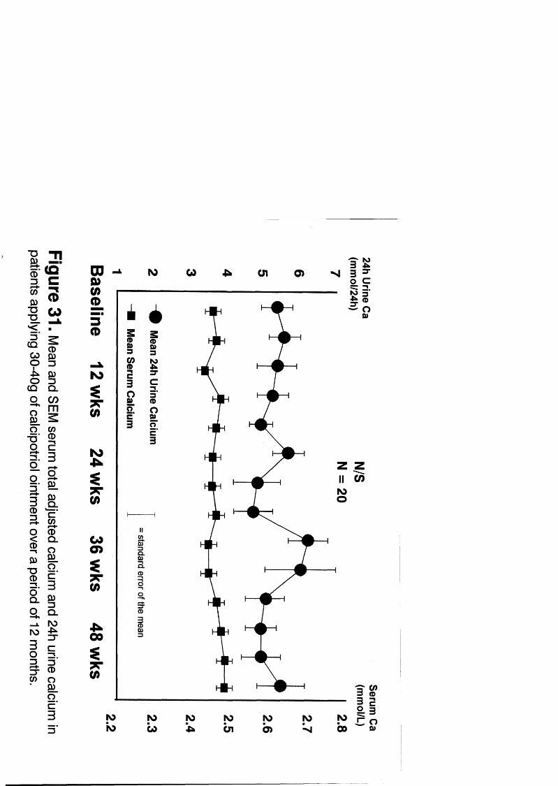
CD
"O T i
I fI ? I fO 3
CQ ~ICOm
Q CO 1 '
oo
CD53
I1Q .
2 D .
a Ia o ._ L no
-p^
CD
iD)
no ^
11 !" a
o .
3
o cII3CD
ro
O
Ql
Q.
3-
no now
no

3.4.2 Bîscifflssion
These studies demonstrate that conventional doses o f calcipotriol have no
measurable effect on systemic calcium homeostasis. However, at doses approaching the
maximum recommended (lOOg/wk), there is a small but significant effect. Serum
phosphate, serum ionized calcium and 24h urine calcium appear to be more sensitive
indices o f that effect.
There appears to be no acute effect on gastro-intestinal absorption of calcium at
these doses. This is in keeping with the in vitro work of Norman et who found that
calcipotriol had a much weaker effect on acute gastrointestinal absorption o f calcium than
calcitriol.
The clinical significance o f these findings is uncertain but probably minor. The only
possible significant consequence would be an increased risk of renal calculi due to a rise
in urine calcium. The rise in urine calcium, however, was small and in most cases
hypercalciuria did not develop. The risk o f renal calculi rises exponentially with rise in
mine calcium (Figme 32) but the curve is fairly flat until a level o f 10mmol/24h is
reached. The mean rise in urine calcium in patients using the maximum recommended
amount o f calcipotriol was only 7.5% therefore the increased risk of renal calculi on
average will be small.
114

g 3(D_ tnc
m-ë w(D N)CL=r 30o 0)3 7T
z a1 CL
s3 CDCD O
T3SL 3'
(D
1
2 2
3"
3T
lO o >
I
» I
gs?Ï3
or
O l
&
3(D
8Q .c3

The only exception to this would be in people who aheady have a high urine
calcium at baseline, because the risks of renal calculi rise exponentially above levels of
10mmol/24h. It would be wise to measure urine calcium at baseline in any patient with
moderately extensive psoriasis and monitor those patients who have a h i ^ or high-normal
urine calcium
From a clinical efficacy point of view, it is surprising that occlusion alone had no
effect on psoriasis in our study over an e i ^ t period. Given that there is extensive hterature
indicating a therapeutic effect o f occlusion alone^^ some effect would have been expected.
One possoble explanation is that our study involved the use of occlusion overnight only
and polythene film rather than one of the more adherent occlusive dressings which have
been previously studied.
115

3.5 SECTION IV Calcipotriol plus short wave ultraviolet hght (UVB)
3.5.1 Results
All three treatments were effective. Mean PASI fell in the UVB group from
12.6 to 7.6 (P < 0.005). Mean PASI fell from 11.7 to 6.3 in the calcipotriol group (P <
0.01) and from 14 to 3.8 (P < 0.005) in the combined therapy group (Table 25). The
combination of UVB and calcipotriol was sli^tly more effective than either agent alone
although the difference was not statistically significant.
Mean serum phosphate rose (P < 0.05) in the group receiving therapy with
UVB plus calciptriol from 0.92 (SER=.05) to 1.22(.09)mmol/L. However, the baseline
serum phosphate in this group was significantly lower than either of the other groups and
there was no difference between any of the groups in terms of mean serum phosphate at
the end o f the study (Figure 33). There was a small rise in serum phosphate in both of the
other groups which fell short o f statistical significance. There was no significant change in
any o f the other parameters o f systemic calcium homeostasis measured in any of the
groups (Tables 26-31).
116

sQ .IÊo
CD3
III3 .0
g.13r*#-
0
1I
"T1
( QC(5
ïèCDsIO )m
SE3
1 3
III.CDiII
V .
I
p00 S lO w
DDm(mm
3(D
fN )
I(D
I gSSL ■5'g I» K
O b
(Q-%
C
3 ^ œ 3 CD
IIc-o: i— 3"

Table 25
UVB + Calcipotriol Calcipotriol UVB
Base Wk2 Wk4 Base Wk2 Wk4 Base Wk2 Wk4
Patient 1 12 6.4 1.8 * * 13.2 6.4 * 3.8
Patient 2 30 21 * 114 8.4 7 9 9 5.5
Patient 3 16 5.7 1.3 119 4.2 3.7 294 266 315
Patient 4 22 15 8.3 9.8 4.4 3 162 9.4 6.4
Patient 5 12 9.9 6.1 108 6.6 13 8 10 6.9 3
Patient 6 5.2 2.8 1.8 7.1 2.9 3.8 9.1 6.4 1.8
Patient 7 6 * * 122 3.8 1.6 16.2 14.4 9.8
Patient 8 16 2.7 6 122 5 4.1 11.4 10.7 8.9
Patient 9 13 8.2 2.7 19 10.8 6.1 * 6.7 3.2
Patient 10 2.6 3.9 2.1 111 6.6 6.6 5.8 3.2 2
Mean 14 8.4 3.8 117 5.9 6.3 126 104 7.6
SEM 2.6 1.9 0.8 1.0 0.8 1.3 2.3 2.2 2.8
Individual PASI in patients being treated with UVB plus lOOg of calcipotriol per week, lOOg of calcipotriol ointment alone and UVB alone. *Result not available.

Table 26
Serum Calcium Ionized calcium Serum phosphate
Base Wk2 Wk4 Base Wk2 Wk4 Base Wk2 Wk4
Patient 1 2 18 2.21 226 131 126 128 113 132 125
Patient 2 2 18 228 * 126 1.31 * .72 101 *
Patient 3 225 227 221 119 125 127 .88 121 1J5
Patient 4 2J3 233 229 127 1.3 126 .98 .8 1.04
Patient 5 241 2.3 231 126 125 126 .94 .9 .94
Patient 6 185 201 2 14 111 1 121 .74 123 166
Patient 7 * * * 118 * * * * *
Patient 8 2 0 7 2 11 226 1.23 1.31 132 .94 .99 132
Patient 9 233 238 231 139 141 1.36 .8 .96 .95
Patient 10 2.1 2.2 2.08 124 126 * 119 105 123
Mean 2.2 2.2 2.2 1.2 1.3 1.3 0.9 1.1 122
SEM 0.1 003 003 003 003 002 0.1 0.1 008
Serum total adjusted calcium (mmol/L), serum ionized calcium (mmol/L) and serum inorganic phosphate (mmol/L) in patients receiving UVB plus 100g of calcipotriol ointment per week. *Result not available.

Table 27
PTH 1 24h Urine calcium
Base Wk2 Wk4 Base Wk2 Wk4
Patient 1 1.9 1.1 2.6 4.2 4.3 5.3
Patient 2 3.2 2.1 * 1.6 * *
Patient 3 1.5 1.6 2.1 2.4 6.9 13 5
Patient 4 4.7 4.3 3.9 .6 1.4 .8
Patient 5 5 3.9 4.3 3.5 5 15 1.5
Patient 6 26 8.5 5.6 .6 .5 .3
Patient 7 * * * * * »
Patient 8 4 3.4 .7 111 3 69 3.4
Patient 9 2 2 2.6 2.2 3.1 5.3
Patient 10 1.2 1.9 * 2.6 3.9 2.1
Mean 5.5 3.2 3.11 3.2 3 62 403
SEM 2 6 0 076 061 113 071 1.51
Serum parathyroid hormone (pmol/L) and 24h urine calcium (mmol/24h) in patients receiving UVB plus lOOg o f calcipotriol ointment per week. *Result not available.

Table 28
Serum calcium Ionized calcium Serum phosphate
Base Wk2 Wk4 Base Wk2 Wk4 Base Wk2 Wk4
Patient 1 2 15 2^2 2 18 1.25 I j^ 1.26 .9 1 98
Patient 2 2J2 2.17 * 125 * * .9 123 *
Patient 3 2J4 223 2 19 129 132 * .97 1.3 107
Patient 4 208 223 2 14 1.26 * * 113 1.02 1.32
Patient 5 2.21 221 * 1.3 129 1.28 1.13 1.41 *
Patient 6 225 2.18 227 132 1.29 1.22 1J4 1.49 1.61
Patient 7 2 19 2.28 205 128 * 129 109 .98 .9
Patient 8 229 2J3 224 127 126 121 124 124 Ij^
Patient 9 225 2 17 2.3 1.2 14^ 129 1 .99 1.06
Patient 10 2J3 229 23 4 1.17 1.24 128 136 105 I j^
Mean 2.2 2.2 2.2 1.3 1.3 1.3 1.1 1.2 1.18
SEM 002 002 003 002 0.01 0.01 0.06 0.06 076
Serum total adjusted calcium (mmol/L), serum ionized calcium (mmol/L) and serum inorganic phosphate (mmol/L) in patients applying 100g of calcipotriol ointment per week. *Result not available.

Table 29
PTH 1 24h Urine calcium
Base Wk2 Wk4 Base Wk2 Wk4
Patient 1 6.1 5.9 6.3 3.3 2.5 3.4
Patient 2 .7 1.4 * 5.4 2.7 2.5
Patient 3 1.1 2.2 1.4 5.3 8.7 7.3
Patient 4 3 1.5 2.2 9.7 6.9 8.5
Patient 5 1.6 2.4 1.1 1.8 2.1 .9
Patient 6 * 2.1 .8 6.2 5.9 5.6
Patient 7 2.2 1.6 2.2 3.7 4.4 6.5
Patient 8 3.5 1.3 1.8 114 2.9 1.2
Patient 9 3.8 3.7 2.9 2.1 2.7 3.2
Patient 10 2.7 1.8 .5 1.5 3.4 3.1
Mean 2.7 2.4 2.1 4 4.2 422
SEM 0.5 0.5 0.6 0.9 0.7 087
Serum parathyroid hormone (pmol/L) and 24h urine calcium (mmol/24h) in patients applying lOOg of calcipotriol ointment per week. *Result not available.

Table 30
Serum Calcium Ionized calcium Serum phosphate
Base Wk2 Wk4 Base Wk2 Wk4 Base Wk2 Wk4
Patient 1 2 2 7 233 222 * * 1.44 .99 124 119
Patient 2 2 13 2.3 228 1J5 129 1.3 LOI 118 .97
Patient 3 2 2 6 224 221 IG? 128 125 116 135 .9
Patient 4 2.18 2 12 222 123 126 129 1.3 134 143
Patient 5 2U5 2 14 2.06 121 126 125 109 116 1.26
Patient 6 2.17 228 227 118 127 134 1.04 128 115
Patient 7 2.2 2 0 9 221 1J3 129 124 .86 1 117
Patient 8 22 8 221 2 14 1G2 1.28 127 .94 116 .9
Patient 9 2.24 226 226 125 129 124 126 1.2 113
Patient 10 2 2 7 222 227 123 127 * 1.15 1 1.06
Mean 2.2 2.2 2.2 1.2 1.3 1.3 1.1 1.2 1.11
SEM 00 2 002 O^G &01 .004 001 0.04 0.04 045
Serum total adjusted calcium (mmol/L), serum ionized calcium (mmol/L) and serum phosphate (mmol/L) in patients receiving UVB alone. *Result not available.
morgamc

Table 31
PTH 1 24h Urine calcium
Base Wk2 Wk4 Base Wk2 Wk4
Patient 1 1.7 1.8 1.8 5.1 3.1 3.6
Patient 2 1.1 1.6 * 3.6 1.9 6.3
Patient 3 1.9 1 1.9 * 3.1 *
Patient 4 6.4 3.8 4 1 1.1 1.3
Patient 5 1.4 2.6 0.7 3.8 5 9.5
Patient 6 2.3 2.2 1.7 6.3 8.1 8.5
Patient 7 3.1 1.6 1.5 3.3 3.8 6.8
Patient 8 2 3 3 5.9 8.4 5.1
Patient 9 1.4 * 1.6 5.5 8 5.1
Patient 10 3 6 1.3 2.5 6.7 7.4 6.5
Mean 2.5 2.1 2.1 4.6 5 5 86
SEM 0.5 0.3 0.3 0.6 0.9 082
Serum parathyroid hormone (pmol/L) and 24h urine calcium (mmol/24h) in patients receiving UVB alone. *Result not available.

3 .5 .2 Biscussioim
This study detected no clinically significant additive or synergistic effect on
systemic calcium homeostasis when the combination of calcipotriol and UVB were used to
treat chronic plaque psoriasis. The small effect on serum phosphate suggests that UVB
plus calcipotriol may have a slightly greater effect than either agent alone but this is
unlikely to be of clinical in^ortance. It is perhaps surprising that there was no
demonstrable effect on serum ionized calcium or 24h urine calcium. This may be due to
the smaller number of patients in each limb as compared to our earlier studies looking at
90 and lOOg per week.
hr summary, although both calcipotriol and UVB are effective treatments
for psoriasis, the combination of the two has not been found in this study to be superior to
either agent alone. The combination of UVB plus calcipotriol not appear to be associated
with clinically significant adverse effects on systemic calcium homeostasis.
117
3.6 SECTION V Unstable pustular psoriasis
3.6.1 Résulta
3 patients with generalised pustular psoriasis were treated with topical
calcipotriol.
Case 1
A 68-year-old woman was admitted with pustular psoriasis covering 45%
o f her body. Calcipotriol ointment was applied twice daily and she improved rapidly,
pustulation disappearing within 24 hours. She was treated in hospital for 3 weeks at a dose
o f lOOg o f calcipotriol (50ug/g) per week and then discharged on the same treatment.
There was no change in serum calcium during that time period.
Case 2
A 76-year-old woman with long-standing unstable psoriasis who had 2
previous admissions with severe pustular psoriasis but been controlled with acitretin 25mg
daily for the previous 2 years, was admitted with sudden deterioration. Ninety percent of
her skin was covered with pustules. On admission, there was biochemical evidence of both
renal and hepatic inqrahment and we were reluctant to institute systemic therapy. She was
treated initially with bed rest emollients, and intravenous fluids. However, after 3 days she
had deteriorated further and treatment with topical calcipotriol was started. Her psoriasis
inqjroved rapidly, pustulation settling within 24hrs.
118
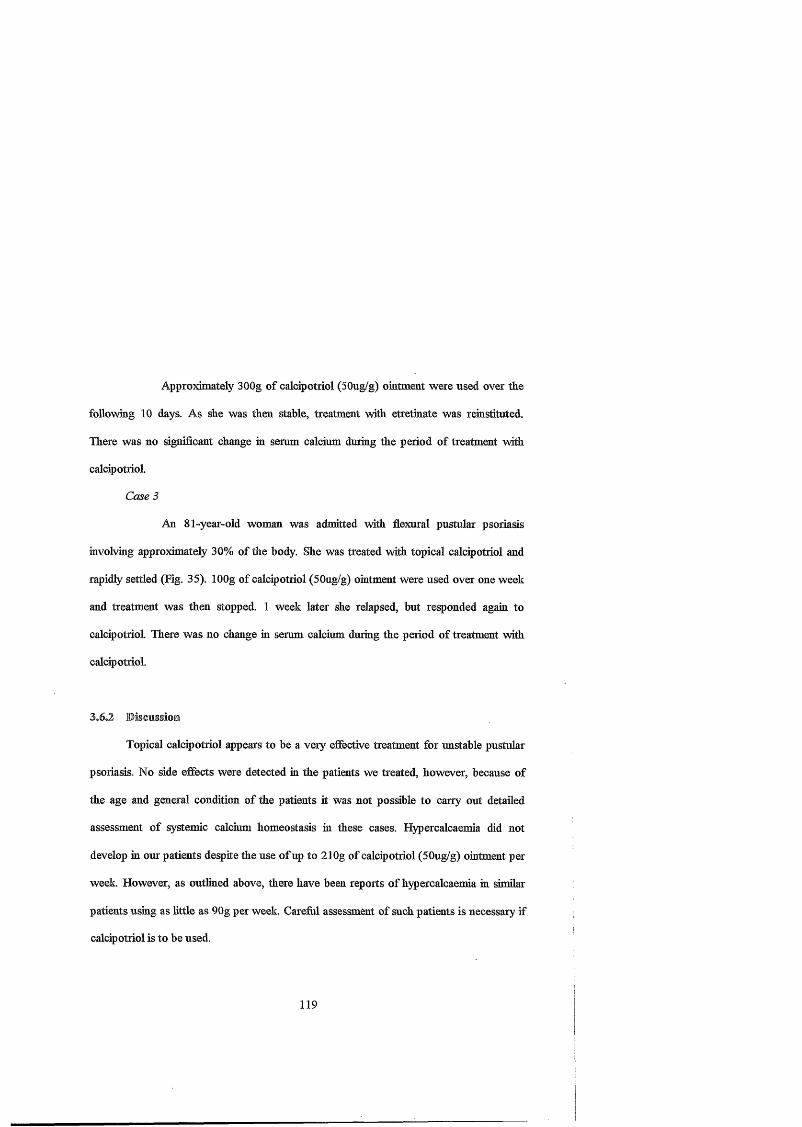
Approximately 300g of calcipotriol (50ug/g) ointment were used over the
following 10 days. As she was then stable, treatment with etretinate was reinstituted.
There was no significant change in serum calcium during the period of treatment with
calcipotriol.
Case 3
An 81-year-old woman was admitted with flexural pustular psoriasis
involving approximately 30% of the body. She was treated with topical calcipotriol and
rapidly settled (Fig. 35). lOOg of calcipotriol (50ug/g) ointment were used over one week
and treatment was then stopped. 1 week later she relapsed, but responded again to
calcipotriol. There was no change in serum calcium during the period of treatment with
calcipotriol.
3.62 Discussion
Topical calcipotriol appears to be a very effective treatment for unstable pustular
psoriasis. No side effects were detected in the patients we treated, however, because of
the age and general condition of the patients it was not possible to carry out detailed
assessment o f systemic calcium homeostasis in these cases. Hypercalcaemia did not
develop in our patients despite the use o f up to 210g of calcipotriol (50ug/g) ointment per
week. However, as outlined above, there have been reports o f hypercalcaemia in s im ila r
patients using as little as 90g per week. Careful assessment of such patients is necessary if
calcipotriol is to be used.
119

Figure 35. Patient with pustular psoriasis before and after treatment with topical calcipotriol.

We have demonstrated that, despite calcipotriol's apparent reduced toxicity in
animal studies, it does affect systemic calcium homeostasis when used topically to treat
patients with chronic plaque psoriasis. The effect is detectable even at recommended
doses.
4.1 Mechanisms off the effects off topical calcipotriol on systemic calcium
homeostasis in patients with psoriasis
These results indicate that calcipotriol has similar effects on systemic
calcium homeostasis to 1,25 dihydroxyvitamin D3 . When used topically, in high doses, it is
absorbed percutaneously and may be detected in serum using the method described by
LeVan.^®° Calcipotriol enhances gastrointestinal absorption o f calcium and probably also
phosphate. Serum total calcium and phosphate and serum ionized calcium both rise as a
result. Serum levels o f parathyroid hormone are depressed due to the rise in serum calcium
and possibly a direct inhibitory effect of calcipotriol. Urine excretion o f calcium and
phosphate are both increased. Urinary calcium excretion is increased directly due to higher
serum concentrations and also indirectly as a result o f lower PTH levels. Serum 1,25
dihydroxyvitamin D3 levels are suppressed due to inhibition of l a hydroxylase, l a
hydroxylase is inhibited by reduced serum PTH, possibly directly by calcipotriol itself and
also by rising serum phosphate levels.
120

There is no evidence of mobilization of calcium, or phoq)hate, fiom bone.
However, the effects on bone markers were only studied at higher doses for very short
periods. An effect o f prolonged use cannot therefore be excluded.
Calcipotriol may differ in one respect from 1,25 vit Dg. We could detect no acute
effect o f calcipotriol on intestinal absorption o f calcium at doses where chronic (8 weeks)
usage did result in a rise in serum ionized calcium This is in keeping with the findings of
Norman et who noted reduced induction of transcaltachia by calcipotriol in vitro as
compared to 1,25 vit Dg It is unlikely that this is of clinical significance as contmued
application o f calcipotriol does result in an increase in serum calcium levels even at
recommended doses.
121

4.2 Effects off recommended doses
Recommended doses o f calcipotriol do appear to have a measurable effect
on systemic calcium homeostasis which is small and only detectable when the m a x im u m
recommended dose is approached. At doses of 90g of the 50jxg/g ointment per week,
serum ionized calcium rose slightly but there were no changes in any of the other
parameters measured. At lOOg per week a rise in serum phosphate and 24h urine calcium
excretion was detectable. The rise in urine calcium excretion has the potential for serious
adverse effects in that it could result in or predispose to renal calcuH, in susceptible
individuals. However the mean rise in 24h urine calcium was only 0.36mmol/24h.
Furthermore the risk of renal calculi relative to the general population only rises
appreciably when the 24h urine calcium is greater than 10mmoF24h.^^^ Although a number
of patients using lOOg per week did have levels this high, they had abnormal values prior
to starting the study and only one patient with a normal value at baseline developed an
abnormally high level during treatment - 8.5mmoF24h. This level was not sustained and
the patient's mine calcium returned to normal while stiU applying calcipotriol.
Most patients using calcipotriol only use 30 - 40g per week and this amount
appears to be safe. The addition o f occlusion with polythene film does not appear to
adversely affect systemic calcium homeostasis with these standard doses of calcipotriol.
Long term use of standard amounts of calcipotriol also appears to be safe hr this regard.
122

4.3 Effects off calcipotriol plus
No clinically significant enhancement of the effect o f topical calcipotriol on
systemic calcium homeostasis has been demonstrated when it is used in combination with
UVB. We have not investigated the use o f high dose topical calcipotriol plus UVB,
although the additive effect o f UVB is likely to be minimal
4.4 Effects off calcipotriol in unstable psoriasis
In our hmited experience with the use of topical calcipotriol in unstable
pustular psoriasis, the use o f up to 21 Og per week did not cause hypercalcaemia over short
periods. However, the experience of others^^^’ ^ would indicate that hypercalcaemia may
occur in these patients even with recommended doses. More detailed assessment o f such
patients would probably reveal some alteration m systemic calcium homeostasis’ however
that was not deemed possible in our particular patients.
123

4.5 Implications of these findings for the treatment of psoriatic patients with
topical calcipotriol
These results indicate that topical calciptiiol, even at recommended doses,
may affect systemic calcium homeostasis.
At high doses, there is considerable risk o f toxicity. The dose per kg body w ei^ t
is the important measmement to take into consideration. Five of nine patients receiving
360g o f 50ug/g ointment per week developed hypercalcaemia. Five out o f five patients
applymg greater than 5.5g per kg o f the ointment per week developed hypercalcaemia.
The significance o f hypercalcaemia in 2 o f 4 Asian patients is uncertain. Given the
small numbers, it may be a chance occurrence. This is an issue which probably merits
further assessment with greater numbers o f patients.
Our high dose studies have not revealed any effect of calcipotriol which differs
fiom 1,25 dihydroxyvitamin Ds and therefore, it is unlikely that a situation of relative
vitamin D deficiency would arise fiom long term use of this agent. The possible differential
effect on acute intestinal calcium absorption which is supported by the results o f our single
dose study, is probably not of clinical significance as intestinal absorption does appear to
be increased by long term use of calcipotriol. We cannot exclude a differential effect on
bone at high doses over prolonged periods. This may also merit further investigation.
124

Patients with unstable pustular psoriasis are probably also at risk of toxicity fiom
calcipotriol. A ltiiou^ we found in our three patients that it was effective and safe at doses
as high as 2 1 0 g per week, there are reports in the literature o f hypercalcaemia even with
doses less than lOOg per week.
The principal concern for patients using recommended doses is the development of
renal calculi after long-term use. The effect on urine calcium is only detectable when the
maximum recommended dose of lOOg of the 50ug/g ointment is approached and hr most
patients the change is so small as to be inconsequential. However, patients with pre
existing hypercalciuria are an exception in that as a small rise in urine calcium hr these
patients wiU increase the risk of developing renal calculi.
For most patients with mild to moderate chronic plaque psoriasis, who use in the
region o f 30 - 40g o f calcipotriol 50pg/g ointment per week, there is no evidence of any
toxic effect on systemic calcium homeostasis. Both long-term use and the addition of
occlusion with polythene film at night to selected areas appears to be safe in these patients.
Finally, calcipotriol interferes with antibody-based kits used to measure
l,25dihydroxyvitarnitt D 3 . I f it is necessary to determine the level in patients applying
calcipotriol, either the drug should be stopped for 2-3 weeks, or HPLC should be used to
ensure accuracy of the results.
125

Our findings are in stark contrast to those of previous toxicity studies, where no
effect o f calcipotriol was demonstrated. We feel that there are two principle reasons
for this discrepancy. Firstly, ours were the first studies designed to investigate fixed doses
o f the drug. All previous studies had investigated patients using a wide variety of doses
and any effect o f those using higher doses would have been diluted by the rest o f the
group. Secondly, there have been very few published studies^^ '^° which investigated doses
approaching the manufacturers guidelines and none which investigated patients using
potentially toxic doses.
We are aware of one, as yet unpublished study, which assessed the use of
higher doses of calcipotriol in some detail. This has been presented in poster form but has
not been subjected to peer review, as far as we are aware.
Our findings are in keeping with the growing number of case reports of toxicity at
h i^ e r doses“ ’ and the occasional report at recommended doses. Given that
hypercalcaemia can develop at doses o f 2 1 0 g o f ointment per week, it would be very
surprising if no effect could be detected whatsoever at lOOg or 90g per week. We feel that
our findings are more consistent with post marketing surveillance reports than previous
toxicity studies.
We feel that our findings also have implications for future assessment of topical
agents prior to licensing. Topical drugs should be assessed, as any oral formulation would,
at fixed doses to accurately determine the maximum recommended dose and detect any
significant side effects.
126

4.6 MonMoi’ing for toxicity
We have found serum phosphate, 24 hour urine calcium and serum ionized calcium
to be more sensitive indicators o f calcipotriol toxicity. Serum ionized calcium is not widely
available and must be taken under controlled conditions and assayed immediately. It is
unlikely to be useful for monitoring outside specialist units.
24 hour urine calcium is a relatively simple assay to carry out althou^ it does
require the full co-operation o f the patient. This assay also has important clinical
hrgplications, in that a high normal value at baseline is a contra-hidication to treatment with
calcipotriol because o f the risk o f developmg renal calculi.
Serum phosphate is also a relatively simple assay to carry out and could be used as
a screening test a lthou^ in itself it is probably not clinically relevant.
127

4.7 Summary
Topical calcipotriol is a safe and effective treatment for mild to moderate chronic
plaque psoriasis. In patients with more extensive psoriasis who use amounts approaching
the maximum recommended dose o f calcipotriol, serum total adjusted calcium, serum
pho^hate and 24 hour urine calcium should be measured regularly. Serum ionized
calcium, if available, can also be used in the early detection of toxicity.
In patients with extensive chronic plaque psoriasis, high dose calcipotriol is an
effective treatment but should only be administered in hospital with the patients being
carefidly monitored. Doses greater than 5.5g of the 50pg/g ointment per kg body weight
week should not be used as this is likely to cause hypercalcaemia.
A lthou^ high dose calcipotriol is also effective in the treatment o f patients with
unstable psoriasis, they are at particular risk because of increased absorption of
calcipotriol and should be closely monitored.
The combination o f UVB and calcipotriol does not appear to confer any additional toxicity
risk although the clinical benefit o f this combination remains to be proven.
128

D m T A R v im R STABnvTKifNC n m ir
As part o f this present trial we wiE be providing you with a fixed, calcium intake in
hôpital. To allow the body to adjust, for 3 weeks prior to hospital admission, it would be
helpful if you fohowed these dietary guidelines:
We would like you to maintain your calcium intake at approximately lOOOg per day. This
will meet all the body's requirements.
The main sources of calcium in the diet are milk, yogurt, cheese and white bread.
To help keep your intake constant you need to eat daily:
5 calcium exchanges (in any combination)
One calcium exchange (200mg) is equal to:
1/3 pint o f milk
OR 1 carton of yogurt
OR 150g of ice-cream
OR 2 0 0 g o f milk pudding
OR lOOg of milk chocolate
129

Preferably you should eat wholemeal bread, or no more than 4 slices o f white bread a day
If your calcium intake is normally very low you will be provided with calcium supplements
Otherwise no calcium supplements should be taken
Large amounts o f salt in the diet can cause you to excrete calcium It is therefore advisable
to avoid adding salt to your food at the table and avoid very salty foods
The list below will help you to do this
AVOID:
tmned/processed meats, eg bacon, salami
cheese (unsalted cream cheese and cottage cheese are allowed)
salted nuts, crisps, savoury snacks
stock cubes, gravy powders, granules
(gravy browning can be used as an alternative)
tinned and packet soup
Oxo, Bovril, Marmite
A salt substitute may be used, eg Ruthamal or Seloral
130

PRESENTATIONS/POSTERS
Im m ediate and long term effects of topical calcipotriol on calcium homeostasis
during treatm ent off psoriasis. Berth-Jones J, Bourke JF, Elouzi H, Iqbal SJ, Hutchinson
PE. presented at the BAD, July 1992.
S years experience with vitamin D and its analogues in the treatment of psoriasis.
Berth-Jones J, Bourke JF, Hutchinson PE. Poster at the AAD, Dec 1992 (SILVER
MEDAL AWARD).
An assessment of the effects of topical calcipotriol on systemic calcium metabolism.
Bourke JF, Berth-Jones J, Hutchinson PE. Poster at the EADV, Sept 1993.
Calcipotriol in children. Presented at the BSPD annual meeting, Leicester Oct 1993.
Occlusion enhances the efffcacy of topical calcipotriene in the treatment of chronic
plaque psoriasis. Bourke JF,Berth-Jones J,Iqbal SJ,Hutchinson PE.Poster at the AAD,
Dec 1993.
The effects of topical calcipotriol on systemic calcium homeostasis. Bourke JF, Berth-
Jones J, Wong M, Holland S, Iqbal SJ, Hutchinson PE. Poster at Ninth workshop on
Vitamin D, Orlando, USA 1994.
Seven years of clinical experience with topical calcipotriene in psoriasis. Berth-Jones
J, Bourke J, Hutchinson PE. Poster at the ninth workshop on vitamin D. Orlando 1994.
High dose calcipotriol consistently suppresses serum parathyroid hormone levels.
Bourke JF, Berth-Jones J, Iqbal SJ, Hutchinson PE. Presented at BAD, London 1994.
131

9. A double blind study comparing the efficacy of calcitriol and calcipotriol in the
treatment of chronic plaque psoriasis. Bourke JF, Featherstone S, Iqbal SJ, Hutchinson
PE. Presented at BAD, Glasgow 1995.
10. The effects of up to lOOg of topical calcipotriol (50pg/g) on systemic calcium
homeostasis. Bourke JF, Beith-Jones J, Wong M, Elouzi H, Iqbal SJ, Hutchinson PE.
Presented at Vitamin D: Actions and applications in Dermatology. Aarhus 1995.
11. The effects of high dose topical calcipotriol (SOpg/g) on systemic calcium
homeostasis, Bourke JF, Mumford R, Holland S, Vasanji K, Farmer N, Iqbal SJ, Le Van
LW, Hutchinson PE. Poster at Vitamin D: Actions and applications in Dermatology.
Aarhus 1995.
132

PUBLICATIONS
1. Generalised pustular psoriasis: response to calcipotrioL Berth-Jones J, Bonrke J F,
Baüey K, Graham-Brown R A C , Hutchinson P E H . B r Med J 1992; 305: 868-9.
2. High dose topical calcipotriol for extensive psoriasis vulgaris. Bourke J F, Berth-Jones
J, Iqbal S J, Hutchinson P E. Br JDeimatol 1993; 129: 74-6.
3. Urine calcium excretion during treatment of psoriasis with topical calcipotriol
Berth-Jones J, Bourke J F, Iqbal S J, Hutchinson PE . Br J Dermatol 1993; 129: 411-4.
4. Occlusion enhances the efficacy of topical calcipotriol in the treatment of chronic
plaque psoriasis vulgaris. Bourke J F, Berth-Jones J, Hutchinson P E. Clin E>q)
Dermatol 1993; 18: 504-6.
So High dose topical calcipotriol consistently suppresses serum parathyroid hormone
levels. Bourke JF, Beith-Jones J, Iqbal SJ, Hutchinson PE. Clinical Endocrinology 1994;
41:295-7.
6. Vitamin D analogues in psoriasis - effects on systemic calcium homeostasis. (Review)
Bourke JF, Iqbal SJ, Hutchinson PE. Br J Derm 1996; 135; 347-54.
7. A Randomized Double-Blind Comparison of the Effects on Systemic calcium
Homeostasis of Topical Calcitriol (3pg/g) And Calcipotriol (SOpg/g) in the
Treatment of Chronic Plaque Psoriasis Vulgaris.
Bourke JF, Iqbal SJ, Hutchinson PE. Acta Dermatovenereol/n Press.
133

PAPERS SUBMITTED
1. Observations on the effects of high dose topical calcipotriol on systemic calcium
homeostasis in patients with extensive chronic plaque psoriasis. Bourke JF, Mumford
R, Iqbal SJ, Hutchinson PE. Submitted to the Journal o f Investigative Dermatology.
2o A comparison of the effects of ultraviolet light and calcipotriol on systemic calcium
homeostasis in patients with chronic plaque psoriasis. Bourke JF, Iqbal SJ, Hutchinson
PE. Submitted to Clinical and Experimental Dermatology.
UETTERS
1. Hypercalcaemia with topical calcipotriol. Bourke JF, Berth-Jones J, Hutchinson PE. Br
Med J 1993; 306: 1344-5.
2. Possible interference with calcipotriol in the new gamma-B-125 dihydroxyvitamin D
IDS assay. Iqbal SJ, Whittaker P, Bourke JF, Mumford R, Hutchinson PE, Le Van LW.
Clinical Chemistry In Press.
3. A single topical dose of 6g of calcipotriol (SOug/g) ointment has no acute effect on
systemic calcium homeostasis. Bourke JF, Berth-Jones J, Elouzi H, Wong M,
Hutchinson PE, Iqbal SJ. Clin Exp Dermatol In Press.
134

PUBLICATIONS (Cont)
ABSTRACTS
1. Immediate and long term effects of topical calcipotriol on calcium homeostasis
during treatment off psoriasis. Berth-Jones J, Bourke J F, Elouzi H, Iqbal S J,
Hutchinson P E. Br J Dermatol 1992; 127(suppl 40): 17-8.
2. An assessment of the effects of topical calcipotriol on systemic calcium metabolism.
Bourke JF, Berth-Jones J, Iqbal SJ, Hutchinson PE. Journal of the European Academy of
Dermatovenereology book of abstracts(JemecGBE,ed.) Kandxup. Copenhagen 1993; 380.
3. High dose topical calcipotriol suppresses serum parathyroid hormone levels. Bourke
JF, Berth-Jones J, Iqbal SJ, Hutchinson PE. Br J Dermatol 1994; 131 (suppl 44): 17.
4. The effects of topical calcipotriol on systemic calcium homeostasis. Bourke JF, Berth-
Jones J, Wong M, Holland S, Iqbal SJ, Hutchinson PE. Proceedings of the ninth workshop
on vitamin D (Norman AW, Bouillon R,Thomasset M, eds.) de Gruyter. New York; 1995:
587-8.
5. Seven years clinical experience with topical calcipotriene in psoriasis. Berth-Jones J,
Bourke J, Hutchinson PE. Proceedings o f the ninth workshop on vitamin D.(Norman AW,
Bouillon RjThomasset M, eds.) de Gruyter. New York; 1995: 586-7.
6. A double blind comparison of calcitriol (3ug/g) and calcipotriol (50ug/g) in the
treatment of chronic plaque psoriasis, Bourke JF, Iqbal SJ, Hutchinson PE.
Br J Dermatol 1995; 133(suppl45): 17.
135

7. The effects off up to lOOg of topical calcipotriol (SOpg/g) on systemic calcium
homeostasis. Bourke JF, Berth-Jones J, Wong M, Elouzi H, Iqhal SJ, Hutchinson PE.
Presented at Vitamin D: Actions and applications in Dermatology. J Invest Dermatol in
Press.
8. The effects of high dose topical calcipotriol (SOpg/g) on systemic calcium
homeostasis, Bourke JF, Mumford R, Holland S, Vasanji K, Farmer N, Iqbal SJ, Le Van
LW, Hutchinson PE. Poster at Vitamin D: Actions and applications in Dermatology. J
Invest Dermatol in Press.
9. The effects of UV® plus calcipotriol on systemic calcium homeostasis in patients
w ith chronic plaque psoriasis. JF Bourke, SJ Iqbal, PE Hutchinson. Br J Dermatol 1996;
(suppl 47): 31.
10. High dose topical calcipotriol enhances intestinal absorption but does not affect
bone. Bourke JF,Mumford R,Trevellyan A, Whittaker P, Iqbal SJ, Hutchinson PE. Br J
Dermatol 1996; (suppl 47): 31.
11. High dose calcipotriol suppresses l^Sdihydroxyvitamin Ds and interferes with
measurement of serum 1,2Sdihdyi'oxyvitamm Ds levels in patients with chronic
plaque psoriasis. JF Bourke, P Whittaker, R Mumford, SJ Iqbal, PE Hutchinson. Br J
Dermatol 1996; 134: 583.
136

1. Tim XQ, Chen TC, Matsucka LY et al. Kinetic m d thennodynamic studies o f the conversion o f previtamin D3 to vitamin D3 in humm skin. J Biol Chem 1993; 268; 14888-92.
2. MacLaughlin JA, Anderson RR, Holick MF. Spectral character o f sunlight modulates photosynthesis of previtamin D m d its photoisomers in humm skin. Science 1982; 216; 1001-3.
3. Holick MF, MacLaughlin JA, Doppelt SA. Regulation of cutmeous previtamin D3 photosynthesis in mm; skin pigment is not m essential regulator. Science 1981; 211; 590-3.
4. Webb AR, De Costa B, Holick MF. Effect o f winter solar hradiation on the photochemistry of 7-dehydrocholesterol, its photoisomers, m d vitamin D. Photochem photobiol 1986; 116s; 43s.
5. Mawer B. The sunshine vitamin. Biol Sci Rev 1992; 3; 2-7.
6 . Holick MF, Adams JS. Vitamin D metabolism m d biological function. In: Metabolic bone diseases and clinically related disorders (Avioli LV, Krme SM, eds), Philadelphia, London: WB Saimders, 1990:155-95.
7. Battachaija MM, DeLuca HE. The regulation of rat liver calciferol-25-hydroxylase. JB io l Chew 1973; 248: 2963-73.
8 . Gray TK, Lester GE, Loreno RS. Evidence for extrarenal l a hydroxylation of 25 hydroxyvitamin D3 in pregnmcy. Science 1979; 204; 1311-13.
9. Howard GA, Turner RT, Sherrard DJ, Bayhnk DJ. Humm bone cells in culture metabolize 25 hydroxyvitamin D 3 to 1,25 dihydroxyvitamin D3 m d 24,25 dihydroxyvitamin D3. JB io l Chem 1981; 256; 7738-40.
10. Frankel TL, Mason RS, Hersey P, Murray E, Posen S. The synthesis o f vitamin D metabolites by humm melmoma cells. J Clin Endocrinol Metab 1983; 57; 627-31.
11. Bikle DD, Nemanic MK, Whitney JO, Elias PW. Neonatal humm foreskin kératinocytes produce 1,25 dihydroxyvitamin D 3 . Biochemistry 1986; 25; 1545-8.
12. Adams S, Sharma OR, Gacad MA, Singer FR. Metabohsm o f 25-hydroxyvitamin D3 by cultured pulmonary macrophages in sarcoidosis. J Clin Invest 1983; 72: 1856-60.
137

13. Brumbaugh BF, Haussier MR. Nuclear and cytoplasmic components for vitamin D metabolites. Life Sci 1975; 16: 353-62.
14. Abe J, Nakano T, Nishii Y et al. A novel vitamin Ds analogue, 22-oxa- la , 25 dihydroxyvitamin D 3 , inhibits the growth o f human breast cancer without causing hypercalcaemia. In Proceedings o f the eighth worlcshop on vitamin D (Norman AW, Bouillon R, Thomasset M eds). Berlin: Walter de Gruyter and Co. 1991: 449-50.
15. MacDonald PN, Dowd DR, Nakajima S et al. Retinoid X receptors stimulate and 9-cis retinoic acid inhibits l,25-dihydroxyvitaminD3-activated expression of the rat osteocalcin gene. Molec and Cell Biol 1993; 13; 5907-17.
16. Wasserman RH. Vitamin D-induced calcium-binding proteins: an overview. In: Calcium binding proteins: structure andfunction (Siegel FL, Carafoh E, Kretsinger RM, McLennan DH, Wasserman RH eds), Amsterdam, Elsevier 1980: 357.
17. Morrison NA, Shine J, Fragonas JC, Verkest V, McMenemy LM, Eisman JA. 1,25 dihydroxyvitamin D3 responsive element and glucocorticoid repression in the human osteocalcin gene. Science 1989; 246; 1148-61.
18. Nemere I, Norman A W. The rapid, hormonally-stimulated tran^o rt o f calcium (transcaltachia). J Bone Min Res 1987; 2; 167-9.
19. Cafl&ey JM, Farach-Carson MC. Vitamin D3 metabohtes modulate dihydropyridine- sensitive calcium currents in clonal rat osteosarcoma cells. JB io l Chem 1989; 264; 20265-74.
20. Bittiner B, Bleehan SS, MacNeh S. la , 25(OH)2vitamin D3 increases intracellular calcium in human kératinocytes. Br J Dermatol 1991; 124; 230-35.
21. Dupret JM, Brun P, Perret C et al. Transcriptional and post-transcriptional regulation of vitamin D-dependent calcium-binding protein in rat duodenum by 1,25 dihydroxycholecalciferol. JB io l Chem 1987; 262; 16553-7.
22. DeLuca HF. Vitamin D: the vitamin and hormone. FedProc 1974; 33; 2211-9.
23. Chambers TJ. The role of 1,25 dihydroxyvitamin D3 in the generation of osteoclasts. In: Vitamin D mollecular, cellular and clinical endocrinology (Norman AW, Schaefer K, Grigoleit HGG, Henath DV, eds), Berlin: Walter de Gruyter, 1988: 581-90.
24. Slatopolsky E, Weerts C, Thielan J et al. Marked suppression o f secondary hyperparathyroidism by intravenous administration of 1,25 dihydroxycholecalciferol in uraemic patients. J Clin Invest 1984; 74; 2136-43.
138

25. Nemeth. EF, Scarpa A. Receptor-dependent mobilization of cellular Ca++ and the regulation of hormone secretion in parathyroid cells. In: Calcium regulation and bone metabolism (Cohn DV, Martin TJ, Munier PJ, eds), Basel: Elsevier Science PubHshers B.V., 1987: 167-71.
26. Abe B, Miyaura C, Sakagami H et al. Differentiation of mouse myeloid leukaemia cells induced by la ,25 dihydroxyvitamin D3. Proc Natl Acad Sci USA 1981; 78: 4990-4.
27. Hosomi J, osoi J, Abe E et al. Regulation of terminal differentiation of cultured mouse epidermal cells by 1 alpha,25-dihydroxyvitamin D 3 . Endocrinology 1983; 113; 1950-7.
28. Smith EL, Pincus SH, Donovan L, Hohck MF. A novel approach for the treatment of psoriasis. J Am Acad Dermatol 1988; 19; 516-28.
29. Smith EL, Walworth NC, Holick MF. Effect of 1 a,25-dihydroxyvitamin D3 on the morphologic and biochemical differentiation of cultured human epidermal keratinocutes grown in serum-free conditions. J Invest Dermatol 1986; 86; 709-14.
30. Araujo OE, Flowers FP, Brown KD. Vitamin D therapy in psoriasis. DICP 1991; 25: 835-9.
31. Desescluse C, Pavlovitch HJ, Balsans S et al. Vitamin D3 influences DNA synthesis by cultured kératinocytes. J Invest Dermatol 1981; 76: 313 (abstr).
32. Regnier M, Darmon M. 1,25-dihydroxyvitamin D3 stimulates tspecifically the last steps of epidermal differentiation o f cultured human kératinocytes. Differentiation 1991; 47; 173- 88.
33. Mak VHW, Cump stone MB, Kennedy AH et al. Barrier function of human keratinocyte cultures grown at the air-liquid interface. J Invest Dermatol 1991; 96: 323-7.
34. Clement TL, Adams JS, Horiuchi N et al. Interaction of 1,25-dihydroxyvitamin D3 with kératinocytes and fibroblasts from skin o f normal subjects and a subject with vitamin-D- dependent rickets, type H: a model for the study o f the mode of action of 1,25- dihydroxyvitanrin D3. J C/m E'Kûfocrmo/Me/ûrô 1983; 56: 826-30.
35. Tanaka H, Abe E, Miyaura C et al. 1,25-dihydroxycholecalciferol and a human myeloid leukaemia cell line(HL-60): the presence of a cytosol receptor and induction of differentiation. Biochem J 1982; 204; 713-9.
139

36. Mangelsdorf DJ, Koeffler HC, Donaldson CA et al. 1,25-diliydroxyvitaniiQ Ds-induced differentiation in a human promyelocytic leukaeemia cell line (HL-60): receptor-mediated maturation to macrophage-like cells. J Cell Biol 1984; 98: 391-8.
37. Calverley MJ, Binderup E Binderup L. The 20-epi modification in the vitamin D series: selective enhancement of'non-clasical' receptor-mediated effects, hr: Proceedings o f the eighth worl(shop on vitamin D. (Paris 1991)(Nomian AW, Bouillon R, Thomasset M, eds). Berlin: Walter de Gruyter & Co. 163-4.
38. Hansen K, Calverley MJ, Binderup L. Synthesis and biological activity of 22-oxa vitamin D analogues. In: Proceedings o f the eighth worlcshop on vitamin D (Paris 1991) (Norman AW, Bouillon R, Thomasset M, eds). Berlin: Walter de Gruyter & Co. 161-2.
39. Binderup E, Calverley MJ, binderup L. Synthesis and biological activity of 1-alpha- hydroxylated vitamin D analogues with polyunsaturated side chains. In: Proceedings o f the eighth worlcshop on vitamin D (Paris 1991) (Norman AW, Bouillon R, Thomasset M, eds). Berlin: Walter de Gruyter & Co. 159-60.
40. Binderup L, Bramm E. Effects of a novel vitamin D analogue MC903 on cell proliferation and differentiation in vitro and on calcium metabolism in vivo. Biochem Pharmacol 1988: 37: 889-95.
41. Kragballe K, Wildfang EL. Calcipotriol (MC903), a novel vitamin D3 analogue stimulates terminal differentiation and inhibits proliferation of cultured human kératinocytes. Arch Dermatol Res 1990; 282: 164-7.
42. EHas L. The basis for treatment of myelodysplastic syndrome and acute nonlynq)hoblastic leukemia with biologic agents. Leulcemia 1988; 2: 400-2.
43. Bower M, Colston KW, Stein RC et al. Topical calcipotriol treatment in advanced breast cancer. 1991; 337; 701-2.
44. Elstner E, de Vos S, Pakkala S, Hebber D, Binderup L, Uskovic M, Umiel T, Koefider HP. In vitro effects o f potent vitamin D3 analogues on proliferation and differentiation of seven human breast cancer cell lines, fir: Proceedings o f the ninth worlcshop on vitamin D$ (Orlando 1994) (Norman AW, Bouillon R, Thomasset M, eds). Berlin: w i te r de Gruyter & Co. 441-8.
45. Tsoukas CD, Prowedini DM, Manolagas SC. 1,25 dihydroxyvitamin D3: a novel immunoregulatory hormone. Science 1984; 224; 1438-40.
140

46. Hutsmyer FG, Beimiger L, Manolagas SC. Conparison of the effects o f 22-oxa- 1,25(0H)2D3 and MC903 on the production of EL-6 , (-ÎFN and T lynphocyte proliferation in peripheral blood mononuclear cells. JBone Miner Res 1991; 6: S292 (Abstr.)
47. Tsoukas CD, Prowedini DM, Manolagas SC. 1,25 dihydroxyvitamin D3 : a novel immunoregulatory hormone. Science 1984; 224: 1438-40.
48. Rigby WF, Stacy T, Fanger MW. Inhibition o f T lymphocyte mitogenesis by 1,25 dihydroxyvitamin D 3 (calcitriol). J Clin Invest 1984; 74: 1451-5.
49. Lemire JM. Immunomodulatory role o f 1,25-dihydroxyvitamin D3. J Cell Biochem 1992; 49: 26-31.
50. Lemann J, Gray RW. Calcitriol, calcium and granulomatous disease. N Eng J M ed 1984; 311:1115-17.
51. Petrini M, Dastoli G, Valentini P et al. Synergistic effects o f alpha interferon and 1,25 dihydroxyvitamin D3: preliminary evidence suggesting that interferon induces expression o f the vitamin receptor. Haematologica 1991; 76: 467-71.
52. Morel PA, Manolagas SC, Prowedini DM, Wegmann DJ, Chiller JM. Interferon y- induced lA expression o f WEHI-3 cells is enhanced by the presence o f 1,25 dihydroxyvitamin D3. J Immunol 1986; 136: 2181-5.
53. Tokuda N, Mano T, Levy RB. 1,25 dihydroxyvitamin D3 antagonizes interferon-gamma- induced expression o f class II major histocompatibility antigens on epithehal and interstitial cells, endocrinology 1990; 127: 1419-24.
54 Carrington MN, Thqaip-Hiltbold B, Knoth J, Ward J. 1,25 dihydroxyvitamin D 3 decreases expresion o f HLA class II molecules in a melanoma cell line. J Immunol 1988; 140: 401-5.
55. Tokuda N, Mizuki N, Kasahara M, Levy RB. 1,25 dihydroxyvitamin D 3 down-regulation o f HLA-DR on human peripheral blood monocytes. Immunology 1992; 75: 349-54.
56. Muller E, Dupuis G, Turcotte S, Rola-Pleszczynski M. Human PAF receptor gene expression: induction during HL-60 cell differentiation. Biochem Biophys Res Commun 1991; 181: 1580-6.
57. Abe J, Nakano T, NiAii Y et al. A novel vitamin D3 analogue, 22-oxa-1,25 dihydroxyvitamin D3, inhibits the growth o f human breast cancer in vitro and in vivo without causing hypercalcaemia. Endocrinology 1991; 129: 823-7.
141

58. Tanaka Y, Klauck T M , Jubiz W. In vivo effects o f 1,25 dihydroxyvitamin D 3 and minerals on the synthesis o f lenkotriene B4 and 5-hydroxyeicosatetraenoic acid by rat peritoneal neutrophils. Prostaglandins Leulcot Essent Fatty Acids 1988; 32: 9-14.
59. Zhou JY, Norman AW, Lubbert M et al. Novel vitamin D analogues that modulate leukaemic cell growth and differentiation with Uttle effect on either intestinal calcium absorption or bone mobilization. Blood 1989; 74: 82-93.
60. Tiflkainen A, Lassus A, Karvonen le t al. Poshasis and HLA-Cw6 . Br J Dermatol 1980; 102: 179-84.
61. Watson W, Cann HM, Farber EM et al. The genetics of psoriasis. Arch Dermatol 1972; 105: 197-207.
62. Brandrup F, Hauge M, Hermigsen K, Eriksen B. Psoriasis hr an unselected series o f twins. Arch Dermatol 1978; 114: 874-8.
63. Swerlick RA, Cunningham MW, Hall NK. Monoclonal antibodies cross-reactive with group A streptococci and normal and psoriatic human skin. J Invest Dermatol 1986; 87: 367-71.
64. Lewis HM, Baker BS, Bokth S, Powles AV, Garioch JJ, Valdimarsson-H, Fry-L. Restricted T-cell receptor V beta gene usage in the skin of patients with guttate and chronic plaque psoriasis. Br J Dermatol 1993 ; 129: 514-20.
65. Van Scott EJ, Ekel TM. Kinetics of hyperplasia hr psoriasis. Arch Dermatol 1963; 88: 373-81.
6 6 . Bauer F. Cell kinetics. In: Mier PD, Van de Kerkhof PCM eds. Textbook o f Psoriasis. Edinburgh: Churchill Livingstone, 1986: 100-12.
67. Barker JNWN. The pathophysiology of psoriasis. Lancet 1991; 338: 227-30.
6 8 . Fritsch WC, Stoughton RB. The effect o f terrperature and humidity on the penetration of" C acetylsalicylic acid in excised human sldn. J Invest Dermatol 1963; 41: 307-11.
69. Gottlieb AB, Cohen SR, Carter DM. Efficacy of actiderm in the treatment of psoriasis. InRyan TJ, ed. Beyond Occlusion: Dermatology Proceedings. Royal Society o f Medicine Services International Congress and Symposium Series No. 137. London: Royal Society o f Medicine Services Ltd, 1988: 25-34.
70. Kra&a J. Simple treatment for psoriasis. JL ab Clin M ed 1936; 21: 1147-8.
142

71. King H, Hamilton CM. Pemphigus controlled by vitamin D. Arch Dermatol Syph 1939; 39: 515-17.
72. Norman CF. Vitamin D therapy in the treatment o f scleroderma and alHed conditions: three cases. Geriatrics 1946; 2s 24-33.
73. Dowling GB, Thomas EWP. Treatment o f lupus vulgaris with calciferol. Lancet 1946; Is 919-22.
74. McLaughlin R R Vitamins A and D topical applications. Medical Times 1939; 303-11.
75. Thacker EA. The treatment o f psoriasis with various vitamin D preparations. Ill M ed J 1940; 352-60.
76. Ceder ET, Zon L. The treatment of psoriasis with massive doses o f crystalline vitamhre D and irradiated ergosterol. Preliminary report. Pub Health Rep 1937; 52s 1580.
77. Morimoto S, Kumahara Y. A patient with psoriasis cured by 1-alpha hydroxyvitanhn Dg. MedJOsalca University 1985; 35s 51.
78. Morimoto S, Qnishi T, Imanaka S et al. Topical administration o f 1,25-dUiydroxyvitamin- Ds for psoriasis: report of five cases. CalcifTiss hrt 1986; 38s 119-22.
79. Morimoto S, Yoshikawa K, Kozuka T et al. An open study o f vitamin Da treatment in psoriasis vulgaris. BrJD erm atol 1986; 115s 421-9.
80. Holland DB, Wood EJ, Roberts SG et al. Epidermal keratin levels during oral 1-alpha hydroxy vitamin Ds treatment for psoriasis. Skin Pharmacol 1989; 2 s 68-76.
81. Long CC, Marks R Calcipotriol and betamethasone valerate for psoriasis. Lancet 1991 ; 337: 921-2.
82. Okano M. 1 alpha, 25-(OH)2Ds use on psoriasis and ichthyosis. IntJD erm atol 1991; 30:62-4.
83. Langner A, Verjans H, Stapor V et al. Treatment o f chronic plaque psoriasis by la,25- hydroxyvitamin Ds (l,25(OH)2Ds) ointment, hr: Proceedings o f the eighth worlcshop on vitamin D (Norman AW, Bouillon R, Thomasset M, eds). Berlin: Walter de Gruyter &Co. 1991; 430-1.
84. Holick MF, Pochi P, Bhawan J. Topically apphed and orally adrniriistered 1,25 dihydroxyvitainitt Ds is a novel, safe, and effective therpay for the treatment o f psoriasis: a three year experience with histologic analysis. J Invest Dermatol 1989; 92s 446.
143

85. Langner A, Vegans H, Stapor V et al. Topical calcitriol in the treatment o f chronic plaque psoriasis: a double-blind study. B r J Dermatol 1993; 128: 566-71.
86. Sipps AJA, Netelenbos JC, Barto R et al. Topically applied low-dose calcitriol has no calciotropic effect in patients with stable plaque psoriasis. J Am Acad Dermatol 1994; 30: 966-9.
87. Mortensen IT, Lichtenberg, Binderup L. Toxicity of l,25(OH)2Dg, tacalditol and calcipotriol after dermal treatment o f rats. J Invest Dermatol (Abstr) In Press.
88. Kato T, Rokugo M, Terui T, Tagami H. Successfirl treatment of psoriasis with topical application o f active vitamin Ds analogue, la ,24 dihydroxycholecalciferol. Br J Dermatol 1986; 115: 431-3.
89. Nakamizo K. Investigation o f the clinical effects o f TV-02 ointment in the treatment of psoriasis. The Nishi Nihon Journal o f Dermatology 1989; 51: 970-9.
90. Nagae Y, Hori Y. Clinical studies on TV-02 ( l a , 24R-(OH)2Ds) ointment against psoriasis and keratosis. The Nishi Nihon Journal o f Dermatology 1990; 52: 97-101.
91. Gerxitsen-MJ; Boezeman-JB; van-Vlijmen-Willems-IM; Van-de-Kerkhof-PC. The effect oftacalcitol (l,24(OH)2D3) on cutaneous inflammation, epidermal proliferation and keratinization in psoriasis: a placebo-controlled, double-blind study. Br J Dermatol 1994 131: 57-63.
92. Calverley MJ, Synthesis of MC903 a biologically active vitamin D analogue. Tetrahedron 1988; 43: 4609-19.
93. Mezzetti G, Bagnara M, Monti M G et al. lalpha,25-dihydroxycholecalciferol and human histiocyte lymphoma cell line (U-937): the presence of receptor and inhibition of proliferation. Life Sci 1984; 34: 2185-91.
94. Binderup L, Bramm E. Effects o f a novel vitamin D analogue MC903 on cell proliferation and differentiation hr vitro and on calcium metabohsm hr vivo. Biochem Pharmacol 1988; 37: 589-95.
95. Kragballe K, Wildfang IL. Calcipotriol (MC903), a novel vitamin Ds analogue, stimulates terminal differentiation and inhibits proliferation o f hrrman kératinocytes. Arch Derm Res 1990; 282: 164-7.
96. Evans DB, Thavarajah M, Binderup L, Kanis JA. The regulatory actions o f MC903, a novel vitamin Ds analogue, on human osteoblast-like cells hr culture. Bone 1990; 11: 223.
144

97. Marie PJ, Connes D, Hott M, Miravet L. Comparative effects o f a novel vitamin D anaolgue MC903 and 1,25-dihydroxyvitarnin Ds on alkaline phosphatase activity, osteocalcin and DNA sunthesis by human osteoblastic cells in culture. Bone 1990; 11: 171-9.
98. Thavarajah M, Evans DB, Binderup L, Kanis JA. l,25(OH)2Ds and calcipotriol (MC903) have similar effects on the induction o f osteoclast-like cell formation in human bone marrow cultures. Biochem Biophys Res Comm 1990; 171: 1056-63.
99. Holland DB, Roberts SG, Russell A et a l Changes in epidermal keratin levels during treatment o f psoriasis with the topical vitamin Ds analogue MC903. B r J Dermatol 1990; 122: 284.
100. De Jong EMGJ, Van de Kerkhof PCM. Simultaneous assesssment o f inflammation and epidermal proliferation in psoriatic plaques during long-term treatment with the vitamin Ds analogue MC903; modulations and interrelations. Br J Dermatol 1991; 124: 221-4.
101. Oxhokn A, Oxhohn P, Staberg B. Expression of mterleukin-6 -like molecules and tumour necrosis factor after topical treatment o f psoriasis with a new vitamin D analogue (MC903). Acta Derm Venereol (Stoclch) 1989; 69: 385-90.
102. Kemeny L, KenderessyAS, Olasz E, Michel G, Ruzicka T, Farkas B, Dobozy A. The interleukin- 8 receptor: a potential target for antipsoriatic therapy? Eur J Pharmacol 1994; 258: 269-72.
103. Ameglio F, Bonifati C, Pietravalle M, Fazio M. Interleukin- 6 and tumour necrosis factor levels decrease in the suction blister fluids o f psoriatic patients during effective therapy. Dermatology 1994; 189: 359-63
104. Lemster BH, Carroll PB, Rüo HR, Johnson N, Nikaein A, Thomson AW. IL-8 /IL- 8
receptor expression in psoriasis and the response to systemic tacrolimus (FK506) therapy. Clin-Exp-Immunol 1995; 99: 148-54.
105. Bouillon R, AUewaert K, Xiang DZ et al. Vitamin D analogues with low affinity for the vitamin D binding protein: enhanced in vitro and decreased in vivo activity. JBone Min Res 1991; 6: 1051-7.
106. Kissmeyer A-M, Mathiasen IS. The affinity o f vitamin D analogues for the vitamin D binding protein correlated with the serum level and the serum half-life (T2 ). In: Proceedings o f the ninth worlcshop on vitamin D (Norman AW, Bouillon R, Thomasset M, eds), Berlin: Walter de Gruyter and Co. 1994 in Press.
145

107. Sorensen H, Binderup L, Calverley MJ et al. In vitro metabolism of calcipotriol (MC 903), a vitamin D analogue. Biochem Pharmacol 1990; 39: 391-3.
108. Kissmeyer AM, Binderup L. Calcipotriol (MC903); pharmacokinetics in rats and biological activities o f metabohtes. Biochem Pharmacol 1991; 41: 1601-6.
109. Norman AW, Nemere I, Zhou LX et al. l,25(OH)2-vitamin D3 , a steroid hormone that produces biologic effects via both genomic and nongenomic pathways. J Steroid Biochem Molec Biol 1992; 41: 231-40.
110. Naveh-Many T, Silver J. The effects o f calcitriol, 22-oxacalcitiiol and calcipotriol on seram calcium and parathyroid hormone gene expression. Endocrinology 1993; 133: 2724-8.
111. Leo Laboratories. Dovonex: Investigators handbook.
1 12. Delaney C, Liao W, Cohen M et al. Percutaneous absorption of calcipotriene (calcipotriol) in normal volunteers and psoriatic patients after a single dose of H- calcipotriene and in psoriatic patients after a sin^e dose of H-calcipotriene following multiple dose treatment with non-radiolabeled calcipotriene 0.005% ointment. J Invest Dermatol (Abstr) In Press.
.113. Le Van LW, Knutson JC, Valhere CR, Bishop CW. A sensitive and specific assay forcalcipotriol (MC903) in seinm or plasma. In: Proceedings o f the ninth worlcshop on vitamin D (Norman AW, Bouillon R, Thomasset M eds), Berlin: Walter de Gruyter 1995; in press.
114. KragbaUe K, Beck ffl, Soggard H. Improvement o f psoriasis by a topical vitamin D analogue (MC903) in a double-bhnd study. B r J Dermatol 1988; 119: 223-30.
115. Kragballe K Treatment of psoriasis by the topical apphcation of the novel cholecalciferol analogue calcipotriol (MC903). Arch Dermatol 1989; 125: 1647-52.
116. Kragballe K, Qertsen BT, de Hoop D et al. Double-blind, right/left comparison o f calcipotriol and betamethasone valerate in treatment of psoriasis vulgaris. Lancet 199L 337: 193-6.
117. Cunliffe WJ, Berth-Jones J, Claudy A et al. Comparative study of calcipotriol (MC903) ointment and betamethasone 17-valerate ointment in patients with psoriasis vulgaris. J Am Acad Dermatol 1991; 26: 736-43.
146
118. Berth-Jones J, Chu AC, Dodd WAH et al. A multicentre, parallel-group comparison of calcipotriol and short-contact dithranol therapy in chronic plaque psoriasis. Br J Dermatol 1992; 127: 266-71.
119. Kragballe K, F o ^ K, Sogaard H. Long-term efficacy and tolerability of topical calcipotriol in psoriasis. AcraDerw Venereol (Stoclch) 1991; 71: 475-8.
120. Ramsay CA, Berth-Jones J, Brundin G et al. Long-term use o f topical calcipotriol in chronic plaque psoriasis. Dermatology 1994; 189: 260-4.
121. Kragballe K. Combination of topical calcipotriol (MC903) and UVB radiation for psoriasis vulgaris. Dermatologica 1990; 181: 211-4.
1 2 2 . Frappaz A, Thivolet J. Calcipotriol in cobination with PUVA: a randomised double-blind placebo-controlled study in severe psoriasis. Eur J Dermatol 1993; 3: 364-7.
123. Grossman RM, Thivolet J, Claudy A et al. A novel therapeutic approach to psoriasis with combination calcipotriol ointment and very low-dose cyclosporine: results o f a multicenter placebo-controlled study. J Am Acad Dermatol 1994; 31: 68-74.
124. Murdoch D, Clissold SP. Calcipotriol: a review o f its pharmacological properties and therapeutic use in psoriasis vulgaris. Drugs 1992; 43: 415-29.
125. Yip J, Goodfield M. Contact Dermatiits from MC903, a topical vitamin Dg analogue. Contact Dermatitis 1991; 25: 139-40.
126. McKenna K, Stem R. Photosensitivity associated with combined UVB and calcipotriol therapy. B r J Dermatol 1995; 133(suppl 45): 63.
127. Mortensen L, Kragballe K, Wegmann E et al. Treatment o f psoriasis vulgaris with topical calcipotriol has no short-term effect on calcium or bone metabolism A randomised, double-blind, placebo-controlled study. Acta Derm Venereol (Stoclch) 1993; 73: 300-4.
128. Gumowski-Sunek D, Rizzoli R, Saurat JH. Effects of topical calcipotriol on calcium metabolism in psoriatic patients: comparison with oral calcitriol. Dermatologica 1991; 183: 275-9.
129. Hardman KA, Heath DA, Nelson HM. Hypercalcaemia associated with calcipotriol (Dovonex) treatment. B rM e d J l9 9 2 \ 306: 896.
130. Gumowski-Sunek D, Rizzoli R, Saurat J-H. Oral calcium tolerance test in extensive psoriasis treated with topical calcipotriol. Dermatology 1995; 190: 43-7.
147

131. McKenna KE, Burrows D. Hypercalciuiia and topical calcipotriol therapy. Br J Dermatol 1994; 131: 588-9.
132. Dwyer C, Chapman RS. Calcipotriol and hypercalcaemia. Lancet 1991; 338: 764-765.
133. Russell S, Young MJ. Hypercalcaemia during treatment of psoriasis with calcipotriol. Br J Dermatol 1994; 130: 795-6.
134. Bower M, Colston KW, Stein RC et al. Topical calcipotriol in advanced breast cancer. Twicg/1991; 357: 701-2.
135. Tucker GPH, Van de Kerkhof PCM, Van Dijk MR, Steijlen PM. Effect o f topical calcipotriol on congenital ichthyoses. B r J Dermatol 1994; 131: 546-50.
136. Hoeck HC, Laurberg G, Laurberg P. Hypercalcaemic crisis after excessive use of a vitamin D derivative. JIn tM ed 1994; 235: 281-2.
137. Goeckerman WH. Treatment of psoriasis. Arch Dermatol Syphilol 1931; 24: 46-50.
138. Kitajima T, Imamura S. Roles of CD4+ and CD8+ cells in UVB-induced suppression of splenic alloantigen-presenting function. Exp Dermatol 1993; 2: 154-6.
139. Green C, Ferguson J, Lakshmipathi T, Johnson BE. 31 Inm UVB phototherapy - an effective treatment for psoriasis. B r J Dermatol 1988; 119: 691-6.
140. Goeckerman WH. Treatment o f psoriasis. Arch Dermatol Syphilol 1931 ; 24: 446-50.
141. Ingram JT. The significance and management o f psoriasis. Br M ed J 1953; 2: 830-8.
142. Editorial. Correcting the calcium. Br M ed J 1977; I: 598.
143. Eastwood JB, Nisbet JA, Colston JW, Ang L, Flanagan AM, Chambers TJ, Maxwell JD. Plasma 250H Vit D and PTH in detection o f osteomalacia in Asian outpatients. Clin Sci 1990; 78: 383-9.
144. Slatopolsky E, Gray R, Adams ND, Adams J, Lewis K et al. Low serum levels o f 1,25 dihydroxyvitamin D are not responsible for the development of secondary hyperparathyroidism in early renal failure. Kidney Int 1978; 14: 73-8.
145. Ritz E. Vitamin D therapy in renal failure - challenges and problems for the future. In : Norman AW, Schaefer Grigoleit HCG, Herrath DV eds. Vitamin D: molecular, cellular and clinical endocrinology. Berlin: Walter de Gruyter, 1988: 1027-30.
148

146. Peacock M, Nordin BE. Tubular reabsorption of calcium in normal and hypercalciuric subjects. J Clin Path 1968; 21: 353-8.
147. Walton RJ, Bijovet OEM. Normogram for derivation of renal threshold phosphate concentration. Lancet 1975; 2: 309-10.
148. Prockop DJ, lüvirikko KI. Relationship o f hydroxyproline excretion in urine to collagen metabolism Ann Intern M ed 1967; 66: 2530-5.
149. Editorial. Pyridinium crosslinks as markers o f bone resorption. Lancet 1992; 340: 278-9.
150. Mhsom S, Ibbertson K, Hannan S, Shaw D, Pybus J. Simple test o f intestinal calcium absorption measured by stable strontium Br M ed J 1987; 295: 231-4.
151. Tellez M, Reeve J, Royston JP et al. The reproducibihty o f double-isotope deconvolution mesurements of intestinal calcium absorption. Clin Sci 1980; 59: 169-72.
152. Reynolds TM, Smith SC. Poor reproducibility o f strontium absorption test. Clinical Chemistry 1994; 40: 1789-90.
153. Sips AJAM, Van der Vijgh WJF, Barto R, Coen Netelenbos J. Reply to; Poor reproducibihty of strontium absorption test. Clinical Chemistry 1994; 40: 1790.
154. Frederiksson T, Pettersson U. Severe psoriasis - oral therapy with a new retinoid. Dermatologica 1978; 157: 238-44.
155. Green C, Ferguson J, Lakshmipathi T, Johnson BE. 311 nm UVB phototherapy—an effective treatment for psoriasis. B r J Dermatol 1988; 119: 691-6
156. Sips AJAM, Netelenboes JC, Barto R, Lips P, Van der Vijgh WJF. One-hour test for estimating intestinal absorption o f calcium by using stable strontium as a marker. Clin Chew 1994; 40: 257-9.
157. Reinhardt TA, Horst RL, Orf JW, HoUis BW. A microassay for 1,25-dihydroxyvitamin D not requiring high performance hquid chromatography: apphcation to clinical studies. J Clin EndocrinolM etab\9M\ 58: 91-8.
158. Reynolds TM. Smith SC. Poor reproducibihty o f strontium absorption test. Clinical Chemistry 1994; 40: 1789-90.
159. Calcium stone formation. In: Metabolic bone and stone disease. (Nordin BEC, Need AG, Morris HA eds.) Edinburgh; Churchih Livingstone Ltd., 297-324.
149

160. Le Van LW, Knutson JC, Valliere CR, Bidiop CW. A sensitive and specific assay for calcipotriol (MC903) in serum or plasma. In; Proceedings o f the ninth worlcshop on vitamin D (Norman AW, Bouillon R, Thomasset M eds), Berlin: Walter de Gruyter & Co. 1994 In Press.
161. Wehr R, Gibson J, Chestnut C et al. The safety o f topically applied calcipotriene 0.005% ointment (a vitamin Ds analogue) as measured by blood, urine and bone density analysis in psoriatic patients. Poster No 188. American Academy Annual Meeting, San Francisco 1992.
150