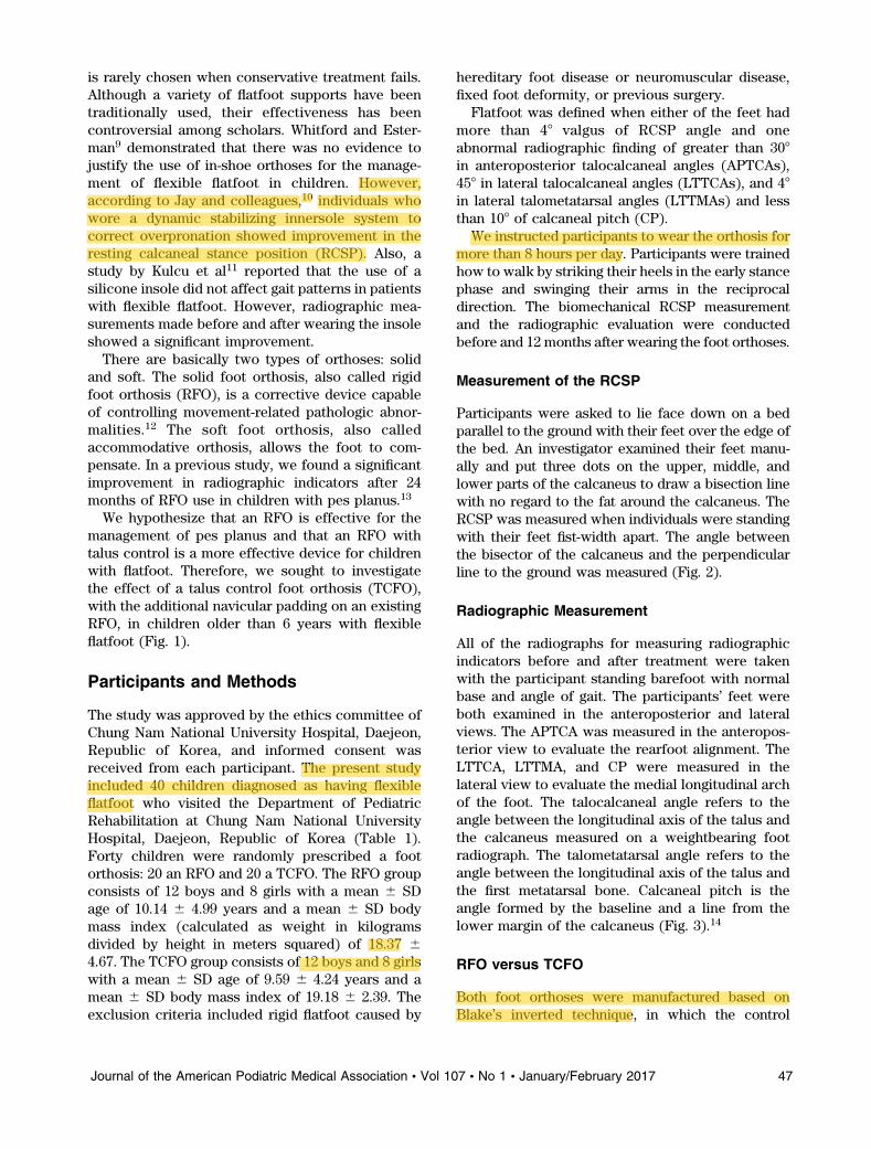
The Effects of Talus Control Foot Orthoses in Children ... · The Effects of Talus Control Foot...
8
ORIGINAL ARTICLES The Effects of Talus Control Foot Orthoses in Children with Flexible Flatfoot So Young Ahn, MD, MS* Soo Kyung Bok, MD, PhD* Bong Ok Kim, MD, PhD* In Sik Park, MS, BS(Pod, CPed)† Background: A talus control foot orthosis (TCFO) combines an inverted rigid foot orthosis (RFO) with a broad upright portion that rises well above the navicular to cover and protect the talonavicular joint. We sought to identify the therapeutic effect of TCFOs in children with flexible flatfoot. Methods: Flexible flatfoot was diagnosed in 40 children when either of the feet had greater than 48 valgus of resting calcaneal stance position (RCSP) angle and one of the radiographic indicators was greater than 308 in anteroposterior talocalcaneal angles, 458 in lateral talocalcaneal angles, and 48 in lateral talometatarsal angles and less than 108 of calcaneal pitch in barefoot radiographs. Of 40 children with flexible flatfoot, 20 were fitted with a pair of RFOs and 20 with TCFOs, randomly. Follow-up clinical and radiographic measurements were completed 12 months later. Results: All of the radiographic indicators changed toward the corrective direction in both groups. There were significant improvements in calcaneal pitch and RCSP in both groups (P , .05). In the TCFO group, the anteroposterior talocalcaneal angle and the RCSP showed statistically significant improvement compared with the RFO group. Conclusions: In this study, the TCFO was more effective than the RFO at treating children with flexible flatfoot. (J Am Podiatr Med Assoc 107(1): 46-53, 2017) Flatfoot is one of the most common diseases treated at a pediatric rehabilitation department. 1 It refers to the loss of the medial longitudinal arch of the foot as a result of complex action between the forefoot, mid- foot, and hindfoot. 2 The incidence of flatfoot varies widely by age. According to Volpon, 3 rapid progression of medial longitudinal arch development was observed between 2 and 6 years of age, and the incidence rate drops sharply at age 6 years. However, some children remain flatfooted because their medial longitudinal arch does not develop normally for some reason. Flatfoot can be divided into flexible type and rigid type. With flexible flatfoot, there is loss of arch height in the weightbearing position (closed kinetic chain). With rigid flatfoot, the arch is not present, whether bearing weight or not (closed and open kinetic chain). 4,5 Rigid flatfoot is most often found in people who have congenital coalition, arthritis, vertical talus, or structural defects that require surgical treatment. 6 In most children, flexible flatfoot deformity does not cause any clinical symptoms. However, if the child has symptomatic, rigid, or progressive flatfoot, he or she needs to be closely examined and managed appropriately. 7 The symptoms include lower-limb pain, increased lower-limb fatigue, Achilles tendin- opathy, and abnormal gait patterns. If children are left untreated, the symptoms may further aggravate, resulting in secondary deformities, such as bunions, hallux rigidus, and pelvic malalignment. Factors predisposing to flatfoot can usually be categorized. Markers of increased susceptibility to physiologic flatfoot include age, sex (male), weight (obesity), and joint hypermobility. Conversely, non- physiologic flatfoot markers include neuromuscular disorders (cerebral palsy or myopathy), hereditary disorders (Down’s syndrome or Marfan syndrome), collagen disease (ligament laxity), external injury, and skeletal abnormalities. 4,8 Conservative and surgical interventions are both used to treat flatfoot. However, surgical intervention *Department of Rehabilitation Medicine, Chung Nam National University Hospital, Daejeon, Republic of Korea. †Korean Pedorthic Institute, Goyang, Republic of Korea. Corresponding author: So Young Ahn, MD, MS, Depart- ment of Rehabilitation Medicine, Chung Nam National University Hospital, 266 Munhwa-ro, Jung-gu, Daejeon, 301- 747, Republic of Korea. (E-mail: [email protected]) 46 January/February 2017 Vol 107 No 1 Journal of the American Podiatric Medical Association
Transcript of The Effects of Talus Control Foot Orthoses in Children ... · The Effects of Talus Control Foot...

ORIGINAL ARTICLES
The Effects of Talus Control Foot Orthoses in Children withFlexible Flatfoot
So Young Ahn, MD, MS*Soo Kyung Bok, MD, PhD*
Bong Ok Kim, MD, PhD*In Sik Park, MS, BS(Pod, CPed)†
Background: A talus control foot orthosis (TCFO) combines an inverted rigid footorthosis (RFO) with a broad upright portion that rises well above the navicular to coverand protect the talonavicular joint. We sought to identify the therapeutic effect of TCFOsin children with flexible flatfoot.
Methods: Flexible flatfoot was diagnosed in 40 children when either of the feet hadgreater than 48 valgus of resting calcaneal stance position (RCSP) angle and one of theradiographic indicators was greater than 308 in anteroposterior talocalcaneal angles, 458
in lateral talocalcaneal angles, and 48 in lateral talometatarsal angles and less than 108
of calcaneal pitch in barefoot radiographs. Of 40 children with flexible flatfoot, 20 werefitted with a pair of RFOs and 20 with TCFOs, randomly. Follow-up clinical andradiographic measurements were completed 12 months later.
Results: All of the radiographic indicators changed toward the corrective direction inboth groups. There were significant improvements in calcaneal pitch and RCSP in bothgroups (P , .05). In the TCFO group, the anteroposterior talocalcaneal angle and theRCSP showed statistically significant improvement compared with the RFO group.
Conclusions: In this study, the TCFO was more effective than the RFO at treatingchildren with flexible flatfoot. (J Am Podiatr Med Assoc 107(1): 46-53, 2017)
Flatfoot is one of the most common diseases treated ata pediatric rehabilitation department.1 It refers to the
loss of the medial longitudinal arch of the foot as aresult of complex action between the forefoot, mid-foot, and hindfoot.2 The incidence of flatfoot varies
widely by age. According toVolpon,3 rapidprogressionofmedial longitudinalarch development was observedbetween 2 and 6 years of age, and the incidence ratedrops sharply at age 6 years. However, some children
remain flatfooted because their medial longitudinalarch does not develop normally for some reason.
Flatfoot can be divided into flexible type and rigidtype. With flexible flatfoot, there is loss of arch heightin the weightbearing position (closed kinetic chain).
With rigid flatfoot, the arch is not present, whetherbearing weight or not (closed and open kineticchain).4,5 Rigid flatfoot is most often found in people
who have congenital coalition, arthritis, vertical talus,
or structural defects that require surgical treatment.6
In most children, flexible flatfoot deformity does not
cause any clinical symptoms. However, if the child
has symptomatic, rigid, or progressive flatfoot, he or
she needs to be closely examined and managed
appropriately.7 The symptoms include lower-limb
pain, increased lower-limb fatigue, Achilles tendin-
opathy, and abnormal gait patterns. If children are left
untreated, the symptoms may further aggravate,
resulting in secondary deformities, such as bunions,
hallux rigidus, and pelvic malalignment.
Factors predisposing to flatfoot can usually be
categorized. Markers of increased susceptibility to
physiologic flatfoot include age, sex (male), weight
(obesity), and joint hypermobility. Conversely, non-
physiologic flatfoot markers include neuromuscular
disorders (cerebral palsy or myopathy), hereditary
disorders (Down’s syndrome or Marfan syndrome),
collagen disease (ligament laxity), external injury,
and skeletal abnormalities.4,8
Conservative and surgical interventions are both
used to treat flatfoot. However, surgical intervention
*Department of Rehabilitation Medicine, Chung Nam
National University Hospital, Daejeon, Republic of Korea.†Korean Pedorthic Institute, Goyang, Republic of Korea.Corresponding author: So Young Ahn, MD, MS, Depart-
ment of Rehabilitation Medicine, Chung Nam National
University Hospital, 266 Munhwa-ro, Jung-gu, Daejeon, 301-
747, Republic of Korea. (E-mail: [email protected])
46 January/February 2017 � Vol 107 � No 1 � Journal of the American Podiatric Medical Association
meh_rehab
Highlight
meh_rehab
Highlight
meh_rehab
Highlight
meh_rehab
Highlight
meh_rehab
Highlight
is rarely chosen when conservative treatment fails.Although a variety of flatfoot supports have beentraditionally used, their effectiveness has beencontroversial among scholars. Whitford and Ester-man9 demonstrated that there was no evidence tojustify the use of in-shoe orthoses for the manage-ment of flexible flatfoot in children. However,according to Jay and colleagues,10 individuals whowore a dynamic stabilizing innersole system tocorrect overpronation showed improvement in theresting calcaneal stance position (RCSP). Also, astudy by Kulcu et al11 reported that the use of asilicone insole did not affect gait patterns in patientswith flexible flatfoot. However, radiographic mea-surements made before and after wearing the insoleshowed a significant improvement.
There are basically two types of orthoses: solidand soft. The solid foot orthosis, also called rigidfoot orthosis (RFO), is a corrective device capableof controlling movement-related pathologic abnor-malities.12 The soft foot orthosis, also calledaccommodative orthosis, allows the foot to com-pensate. In a previous study, we found a significantimprovement in radiographic indicators after 24months of RFO use in children with pes planus.13
We hypothesize that an RFO is effective for themanagement of pes planus and that an RFO withtalus control is a more effective device for childrenwith flatfoot. Therefore, we sought to investigatethe effect of a talus control foot orthosis (TCFO),with the additional navicular padding on an existingRFO, in children older than 6 years with flexibleflatfoot (Fig. 1).
Participants and Methods
The study was approved by the ethics committee ofChung Nam National University Hospital, Daejeon,Republic of Korea, and informed consent wasreceived from each participant. The present studyincluded 40 children diagnosed as having flexibleflatfoot who visited the Department of PediatricRehabilitation at Chung Nam National UniversityHospital, Daejeon, Republic of Korea (Table 1).Forty children were randomly prescribed a footorthosis: 20 an RFO and 20 a TCFO. The RFO groupconsists of 12 boys and 8 girls with a mean 6 SDage of 10.14 6 4.99 years and a mean 6 SD bodymass index (calculated as weight in kilogramsdivided by height in meters squared) of 18.37 6
4.67. The TCFO group consists of 12 boys and 8 girlswith a mean 6 SD age of 9.59 6 4.24 years and amean 6 SD body mass index of 19.18 6 2.39. Theexclusion criteria included rigid flatfoot caused by
hereditary foot disease or neuromuscular disease,fixed foot deformity, or previous surgery.
Flatfoot was defined when either of the feet had
more than 48 valgus of RCSP angle and oneabnormal radiographic finding of greater than 308
in anteroposterior talocalcaneal angles (APTCAs),458 in lateral talocalcaneal angles (LTTCAs), and 48
in lateral talometatarsal angles (LTTMAs) and lessthan 108 of calcaneal pitch (CP).
We instructed participants to wear the orthosis formore than 8 hours per day. Participants were trainedhow to walk by striking their heels in the early stancephase and swinging their arms in the reciprocaldirection. The biomechanical RCSP measurementand the radiographic evaluation were conductedbefore and 12 months after wearing the foot orthoses.
Measurement of the RCSP
Participants were asked to lie face down on a bedparallel to the ground with their feet over the edge ofthe bed. An investigator examined their feet manu-ally and put three dots on the upper, middle, andlower parts of the calcaneus to draw a bisection linewith no regard to the fat around the calcaneus. TheRCSP was measured when individuals were standing
with their feet fist-width apart. The angle betweenthe bisector of the calcaneus and the perpendicularline to the ground was measured (Fig. 2).
Radiographic Measurement
All of the radiographs for measuring radiographicindicators before and after treatment were takenwith the participant standing barefoot with normalbase and angle of gait. The participants’ feet wereboth examined in the anteroposterior and lateralviews. The APTCA was measured in the anteropos-terior view to evaluate the rearfoot alignment. TheLTTCA, LTTMA, and CP were measured in thelateral view to evaluate the medial longitudinal archof the foot. The talocalcaneal angle refers to theangle between the longitudinal axis of the talus andthe calcaneus measured on a weightbearing foot
radiograph. The talometatarsal angle refers to theangle between the longitudinal axis of the talus andthe first metatarsal bone. Calcaneal pitch is theangle formed by the baseline and a line from thelower margin of the calcaneus (Fig. 3).14
RFO versus TCFO
Both foot orthoses were manufactured based onBlake’s inverted technique, in which the control
Journal of the American Podiatric Medical Association � Vol 107 � No 1 � January/February 2017 47
meh_rehab
Highlight
meh_rehab
Highlight
meh_rehab
Highlight
meh_rehab
Highlight
meh_rehab
Highlight
meh_rehab
Highlight

target is the medial side of the calcaneus. When
patients were prescribed an inverted orthosis man-
ufactured based on Blake’s technique, the ratio of the
correction angle of the RCSP and the pouring angle
of the negative mold was 5:1. The initial correction
angle is usually two-thirds of the total correction
angle. For example, if the RCSP is �68 inverted, the
correction angle of the RCSP would be 68 and the
pouring angle would be 308 (68 x 5). Because the
initial correction angle starts with two-thirds of the
total correction angle, it should be inverted 208. The
TCFO is a modified Blake’s inverted orthosis, having
a medial flange brought up around the medial side of
the navicular. When manufacturing the TCFO, the
medial portion of the positive mold is adjusted for
navicular support (Fig. 4).
Each custom-made foot orthosis was fabricated by
Biomechanics Co, Goyang, Republic of Korea. For
capturing the negative foot impression accurately, the
neutral position of weightbearing plaster cast tech-
nique, which is one of the most popular and reliable
measurements of forefoot/rearfoot relationships, was
performed.15 The rigid orthotic shell is made of
polypropylene with a thickness of 5 mm, and ethylene
vinyl acetate was used for heel posting (Fig. 1).
Statistical Analysis
Statistical analysis was performed using a statistical
software program (IBM SPSS Statistics for Windows,
version 19.0; IBM Corp, Armonk, New York). To
minimize the error range, two physiatrists measured
the radiographic parameters independently. All of the
values were averaged with a standard deviation. The
paired-samples t test was used to compare the RCSP
and the radiographic measurements before and after
wearing foot orthoses. Also, an independent-samples
t test was used to perform a comparative analysis of
the effect of the two orthoses. A P , .05 was
considered statistically significant.
Results
The RCSP
The mean 6 SD value of the RCSP changed from
–7.558 6 4.078 to –3.558 6 3.218 in the RFO group
Figure 1. A, Talus control foot orthosis. B, Rigid foot orthosis.
Table 1. Characteristics of the 40 Study Participants
CharacteristicRFO Group
(n ¼ 20)TCFO Group
(n ¼ 20)
Age (years) 10.14 6 4.99 9.59 6 4.24
Sex, M:F (No.) 12:8 12:8
Height (cm) 138.23 6 10.17 139.28 6 12.78
Weight (kg) 35.13 6 16.93 37.41 6 11.33
BMI 18.37 6 4.67 19.18 6 2.39
Note: Values are given as mean 6 SD except where
indicated otherwise.
Abbreviations: BMI, body mass index (calculated as weight
in kilograms divided by height in meters squared); RFO, rigid
foot orthosis; TCFO, talus control foot orthosis.
48 January/February 2017 � Vol 107 � No 1 � Journal of the American Podiatric Medical Association
meh_rehab
Highlight
meh_rehab
Highlight
meh_rehab
Highlight
meh_rehab
Highlight

and from –9.285 6 2.958 to –2.058 6 1.938 in the
TCFO group (Figs. 5 and 6 and Table 2).
Radiographic Measurements
The mean 6 SD value of the APTCA changed from
35.758 6 8.468 to 33.42 6 7.428 in the RFO group and
from 36.918 6 7.378 to 33.088 6 7.058 in the TCFO
group (Fig. 5 and Table 2). The mean 6 SD value of
the LTTCA changed from 43.678 6 11.898 to 47.178
6 6.788 in the RFO group and from 46.508 6 9.018
to 48.878 6 7.568 in the TCFO group. The mean 6
SD value of the LTTMA changed from 20.298 6
12.948 to 21.548 6 13.858 in the RFO group and from
18.278 6 7.838 to 18.518 6 6.848 in the TCFO group.The mean 6 SD value of CP changed from 8.338 6
6.048 to 13.318 6 5.668 in the RFO group and from8.948 6 4.348 to 15.058 6 4.848 in the TCFO group.
Discussion
Flexible flatfoot is one of the most common footdeformities in children whose medial longitudinalarch disappears during weightbearing. Subtalaroverpronation is one of the characteristics offlatfoot. The subtalar joint normally pronatesapproximately 48 during the first 25% of the stancephase. In the case of flatfoot, the subtalar joint
Figure 2. Calcaneal bisection and measurement of the resting calcaneal stance position.
Figure 3. Anteroposterior and lateral views of both feet parameters: 1* anteroposterior talocalcaneal angle, 2*lateral talocalcaneal angle, 3* lateral talometatarsal angle, and 4* calcaneal pitch.
Journal of the American Podiatric Medical Association � Vol 107 � No 1 � January/February 2017 49

pronates more than 48 during the early stance phase
or remains pronated after the early stance phase.
Thus, the medial longitudinal ligaments have to
resist the excessive and prolonged force by abnor-
mal range of pronation.14 Symptoms of flatfoot are
usually accompanied by anatomical abnormalities
such as subtalar subluxation, medial protrusion, and
collapse of the medial longitudinal arch caused by
talar adduction and plantarflexion, tibial internal
rotation, calcaneal eversion, and plantarflexion at
the subtalar joint. Also, forefoot abduction occurs at
the midtarsal joint, which results in more relative
supination in the forefoot compared with the
hindfoot.16
Treatment of flexible flatfoot has been the subject
of controversy among scholars, especially nonsur-
gical treatment using foot orthoses. The effective-
ness of a variety of foot orthoses and shoes has
been examined in many studies, with diverse
opinions on their efficacy.9,10,12,17-19
In a study by Wenger et al,17 children with flexible
flatfoot aged 1 to 6 years wore orthopedic shoes,
heel cups, and custom-molded plastic inserts
(University of California Biomechanics Laboratory,
San Francisco, California); however, no significant
difference was found between the control and
treatment groups. However, in that study, old-style
foot orthoses were used, and they enrolled children
with flatfoot younger than 6 years, who were still
developing the medial longitudinal arch. Because
the medial longitudinal arch develops on its own by
age 6 years, it is not appropriate to conclude that
the foot orthosis used in the present study produced
meaningful improvements in the RCSP and the
radiographic measurements without comparison
with the control group. Therefore, we included
children older than 6 years and measured both
RCSP and radiographic measurements to enhance
the reliability of the results.
In the previously mentioned study by Whitford and
Esterman,9 children aged 7 to 11 years who wore
foot orthoses for 12 months showed no improvement
in gross motor proficiency, self-perception, exercise
efficiency, and pain. It is believed that the effect of
controlling the movement-related pathology was
decreased compared with an RFO because the type
of orthosis used in that study was semirigid. In the
present study, we used an RFO to obtain maximal
correctional efficacy.
In the present study, both foot orthoses were
manufactured based on Blake’s inverted technique.
It is a technique developed by Richard L. Blake to
treat children with severe flatfoot whose RCSP is
less than�58. The medial side of the calcaneus was
the basic point for the correction. The twisting
around the oblique axis of the subtalar joint and the
long axis of the midtarsal joint induces calcaneal
inversion and forefoot eversion. So that it can
contribute to the rearfoot supination and talar
dorsiflexion, abduction, consequently to form the
medial longitudinal arch. The TCFO has an addi-
tional broad upright portion rising well above the
navicular bone that affects the location and move-
ment of the navicular and the talonavicular joint,
resulting in effective correction of plantarflexion
and medial deviation of the talus (Fig. 7).20,21
Figure 4. A, Talus control foot orthosis. The arrow indicates the broad upright portion rising above thenavicular bone. B, Rigid foot orthosis.
50 January/February 2017 � Vol 107 � No 1 � Journal of the American Podiatric Medical Association
meh_rehab
Highlight
meh_rehab
Highlight
meh_rehab
Highlight

Figure 6. Improvement of calcaneal pitch (CP) (A) and resting calcaneal stance position (RCSP) (B) in therigid foot orthosis (RFO) and talus control foot orthosis (TCFO) groups. *P , .05.
Figure 5. Mean 6 SD changes before and after treatment in the anteroposterior talocalcaneal angle (APTCA)(A), lateral talocalcaneal angle (LTTCA) (B), lateral talometatarsal angle (LTTMA) (C), calcaneal pitch (CP)(D), and resting calcaneal stance position (RCSP) (E) in the rigid foot orthosis (RFO) and talus control footorthosis (TCFO) groups. *P , .05.
Journal of the American Podiatric Medical Association � Vol 107 � No 1 � January/February 2017 51

We measured four radiographic indicators for the
evaluation of midfoot and hindfoot alignment. For
hindfoot alignment, the lateral and anterior talocal-
caneal angles and CP were measured. For midfoot
alignment, the talometatarsal angle was used. By
measuring these various angles, we tried to increase
the reliability of the results and evaluate the
relationship between each part of the foot.
In the present study, the RCSP and all of the
radiographic indicators changed toward the correc-
tive direction in both groups 12 months after
wearing foot orthoses compared with those before
treatment. However, both groups showed significant
improvements in their RCSP and CP (P , .05).
Positional changes in the location of the sagittal and
coronal planes of the calcaneus are expected, which
means changes in the rearfoot in the early phase
because Blake’s technique intends to correct the
medial side of the calcaneus. In both groups, the
LTTCA and the LTTMA did not show significant
improvement 12 months after wearing the orthosis.
In previous studies with an RFO applied to flexible
flatfoot, the APTCA and the LTTMA showed
significant improvement, and the LTTCA showed a
tendency toward improving after 24 months of RFO
application.13 Changes in the sagittal and transverse
planes of the talus are expected after wearing foot
orthoses longer than 24 months. Future research
including more long-term follow-up is needed.
The TCFO produced considerable improvements
in the APTCA and the RCSP compared with the
RFO. It is assumed that because medial flange
pressure, which supports the navicular bone, was
loaded from medial to lateral it produced changes in
Table 2. Changes in Radiographic Indicators and RCSP
Indicator
RFO Group TCFO Group
Initial After 12 mo Initial After 12 mo
APTCA (8) 35.75 6 8.46 33.42 6 7.42 36.91 6 7.37 33.08 6 7.05a
LTTCA (8) 43.67 6 11.89 47.17 6 6.78 46.50 6 9.01 48.87 6 7.56
LTTMA (8) 20.29 6 12.94 21.54 6 13.85 18.27 6 7.83 18.51 6 6.84
CP (8) 8.33 6 6.04 13.31 6 5.66a 8.94 6 4.34 15.05 6 4.84a
RCSP (8) –7.55 6 4.07 –3.55 6 3.21a –9.25 6 2.95 –2.05 6 1.93a
Note: Values are given as mean 6 SD.
Abbreviations: APTCA, anteroposterior talocalcaneal angle; CP, calcaneal pitch; LTTCA, lateral talocalcaneal angle; LTTMA,
lateral talometatarsal angle; RCSP, resting calcaneal stance position; RFO, rigid foot orthosis; TCFO, talus control foot orthosis.aP , .05 compared with initial evaluation by paired t test.
Figure 7. Mechanical forces of the rigid foot orthosis (RFO) and the talus control foot orthosis (TCFO) at the 1*talus, 2* talonavicular joint, 3* metatarsal, 4* inferior calcaneus, 5* medial calcaneus, and 6* navicular.
52 January/February 2017 � Vol 107 � No 1 � Journal of the American Podiatric Medical Association

the location of the transverse plane of the talus andthe coronal plane of the calcaneus rather than
producing changes in the location of the sagittalplane of the subtalar joint in the rearfoot.
This study is based on the study by Volpon3 thatshowed complete normal foot development in 6
years. Having a control group that did not wear foot
orthoses is important for improving the complete-ness of this study. To minimize errors, participants
were limited to children older than 6 years. In lightof the authors’ experience of managing a ‘‘shoe
clinic’’ for more than 10 years, we verified theclinical effects of the foot orthosis. Although the
normal foot alignment group showed some im-
provement after 6 years, we are convinced that archdevelopment will be faster with foot orthoses,
which also corresponds with the study by Gouldet al.22 Also, ethical problems occurred in enrolling
a control group that had no intervention despite
severe flexible flatfoot because flatfoot is one of themost common foot problems in childhood and can
affect the walking pattern and lead to fatigue andfoot deformity.18 Future research with a larger
sample size and a control group is warranted toquantify and generalize the effects of the foot
orthosis.
Conclusions
The present results suggest that the use of footorthoses in children with flexible flatfoot produced
improvement in clinical and radiographic measure-ments in both the RFO and TCFO groups. However,
much more improvement was seen in the APTCA
and the RCSP in the TCFO group. The resultssuggest that the TCFO is more effective than the
RFO for the treatment of flexible flatfoot in children,indicating its usefulness in the future treatment of
children diagnosed as having serious flatfoot.
Financial Disclosure: None reported.
Conflict of Interest: None reported.
References
1. LUHMANN SJ, RICH MM, SCHOENECKER PL: Painful idiopathic
rigid flatfoot in children and adolescents. Foot Ankle Int
21: 59, 2000.
2. ANANTHKRISHNAN D, CHING R, TENCER A, ET AL: Subluxation
of talocalcaneal joint in adults who have symptomatic
flatfoot. J Bone Joint Surg Am 81: 1147, 1999.
3. VOLPON JB: Footprint analysis during growth period. J
Pediatr Orthop 14: 83, 1994.
4. HARRIS EJ, VANORE JV, THOMAS JL ET AL: Diagnosis and
treatment of pediatric flatfoot. J Foot Angle Surg 43:
341, 2004.
5. NAPOLITANO C, WALSH S, MAHONEY L, ET AL: Risk factors that
adversely modify the natural history of the pediatric
pronated foot. Clin Podiatr Med Surg 17: 397, 2000.
6. FERRI M, SCHRAFENBERGER AV, GOPLEN G, ET AL: Weight-
bearing CT scan of severe flexible pes planus deformi-
ties. Foot Ankle Int 29: 199, 2008.
7. GARCIA-RODRIGUEZ A, MARTIN-JIMENEZ F, CARNERO-VARO M,
ET AL: Flexible flat feet in children: a real problem?
Pediatrics 103: e84, 1999.
8. HARRIS EJ: The natural history and pathophysiology of
flexible flatfoot. Clin Podiatr Med Surg 27: 1, 2010.
9. WHITFORD D, ESTERMAN AA: Randomized controlled trial of
two types of in-shoe orthoses in children with flexible
excess pronation of the feet. Foot Ankle Int 28: 715, 2007.
10. JAY RM, SCHOENHAUS HD, SEYMOUR C, ET AL: The dynamic
stabilizing innersole system (DSIS): the management of
hyperpronation in children. J Foot Ankle Surg 34: 124,
1995.
11. KULCU DG, YAVUZER G, SARMER S, ET AL: Immediate effects
of silicone insoles on gait patterns in patients with
flexible flat foot. Foot Ankle Int 28: 1053, 2007.
12. LEUNG AL, MAK AT, EVANS JH: Biomechanical gait
evaluation of the immediate effect of orthotic treatment
for flexible flat foot. Prosthet Orthot Int 22: 25, 1998.
13. BOK SK, KIM BO, LIM JH, ET AL: Effects of custom-made
rigid foot orthosis on pes planus in children over 6 years
old. Ann Rehabil Med 38: 369, 2014.
14. DAVIDS JR, GIBSON TW, PUGH LI: Quantitative segmental
analysis of weight-bearing radiographs of the foot and
ankle for children: normal alignment. J Pediatr Orthop
25: 769, 2005.
15. LAUGHTON C, MCCLAY DAVIS I, WILLIAMS DS: A comparison
of four methods of obtaining a negative impression of
the foot. JAPMA 92: 261, 2002.
16. KIM SB, YOON KS, PARK HS, ET AL: Radiologic measure-
ment of flatfoot. J Korean Acad Rehab Med 24: 995,
2000.
17. WENGER DR, MAULDIN D, SPECK G, ET AL: Corrective shoes
and inserts as treatment for flexible flatfoot in infants
and children. J Bone Joint Surg Am 71: 800, 1989.
18. BORDELON RL: Correction of hypermobile flatfoot in
children by molded insert. Foot Ankle 1: 143, 1980.
19. CAPASSO G: Dynamic varus heel cup: a new orthosis for
treating pes planovalgus. Ital J Orthop Traumatol 19:
113, 1993.
20. BLAKE RL, FERGUSON H: Foot orthosis for the severe
flatfoot in sports. JAPMA 81: 549, 1991.
21. BLAKE RL: Inverted functional orthosis. JAPMA 76: 275,
1986.
22. GOULD N, MORELAND M, ALVAREZ R, ET AL: Development of
the child’s arch. Foot Ankle 9: 241, 1989.
Journal of the American Podiatric Medical Association � Vol 107 � No 1 � January/February 2017 53
meh_rehab
Highlight