THE EFFECTS OF RESPIRATORY MUSCLE TRAINING ON …
177
1 THE EFFECTS OF RESPIRATORY MUSCLE TRAINING ON STRENGTH AND PERFORMANCE IN COLLEGIATE SWIMMERS By VIPA BERNHARDT A DISSERTATION PRESENTED TO THE GRADUATE SCHOOL OF THE UNIVERSITY OF FLORIDA IN PARTIAL FULFILLMENT OF THE REQUIREMENTS FOR THE DEGREE OF DOCTOR OF PHILOSOPHY UNIVERSITY OF FLORIDA
Transcript of THE EFFECTS OF RESPIRATORY MUSCLE TRAINING ON …

1
THE EFFECTS OF RESPIRATORY MUSCLE TRAINING ON STRENGTH AND PERFORMANCE IN COLLEGIATE SWIMMERS
By
VIPA BERNHARDT
A DISSERTATION PRESENTED TO THE GRADUATE SCHOOL OF THE UNIVERSITY OF FLORIDA IN PARTIAL FULFILLMENT
OF THE REQUIREMENTS FOR THE DEGREE OF DOCTOR OF PHILOSOPHY
UNIVERSITY OF FLORIDA

2
Vipa Bernhardt

3

4
ACKNOWLEDGMENTS
First and foremost, I would like to thanks my parents, Chamaipon and Helge, for
their endless support and for always being there for me, even though the Atlantic Ocean
separates us. Skype has been one of the best technical advances for our widely
spread-out family to be able to talk and see each other. To my brother, Jens, whose
footsteps I followed and that led me to pursue a career in science.
Special thanks go to my graduate mentor, Dr. Paul Davenport, for his teaching,
guidance, and encouragement. Thanks to my supervisory committee: Dr. David Fuller,
Dr. Nancy Denslow, Dr. Danny Martin, Dr. Linda Hayward, and Dr. Roger Reep for the
support and comments throughout my dissertation process.
I am grateful for having had the opportunity to share my experience with my fellow
labmates, past and present: Drs. Pei-Ying Sarah Chan, Kate Pate, Karen Hegland,
Andrea Vovk and soon-to-be Drs. Mark Hotchkiss, Irene Tsai, Poonam Jaiswal. As well
as the other scientists in and around B3-16: Drs. Teresa Pitts, Barbara Smith, and soon-
to-be Dr. Ana Bassit. I would like to thank Stacey Nedrud for her help and friendship in
and out of the lab, Will Walters for critical comments, and Mandy Huff for continual
moral support. I would also like to acknowledge Dr. Natalia Garcia-Reyero for her help
with the microarrays.
Last but not least, I would like to thank the UF swim team: the coaches Gregg
Troy, Martyn Wilby, and Pete Knox for their support to carry out my project and allowing
me to interfere somewhat with swim practices and the swimmers who did the extra work
for my research.

5
TABLE OF CONTENTS
page
ACKNOWLEDGMENTS.............................................................................................................. 4
LIST OF TABLES ......................................................................................................................... 8
LIST OF FIGURES....................................................................................................................... 9
ABSTRACT ................................................................................................................................. 11
CHAPTER
1 INTRODUCTION................................................................................................................. 13
Exercise and Respiration ................................................................................................... 13 Intermittent Transient Tracheal Occlusions (ITTO) in Rats as a Model for
Respiratory Muscle Training .......................................................................................... 16
Respiratory Load Compensation Response ................................................................... 17 Sensory Gating .................................................................................................................... 19
Specific Aims ....................................................................................................................... 22 Specific Aim 1: To Investigate the Effects of Expiratory Muscle Strength
Training in Collegiate Swimmers ........................................................................... 22
Specific Aim 2: To Investigate the Changes in the Gene Expression Profile of the Medial Thalamus following ITTO in Anesthetized Rats .......................... 23
Specific Aim 3: To Investigate the Changes in the Gene Expression Profile of the Medial Thalamus following 10 Days of Repeated ITTO in Conscious Rats......................................................................................................... 24
2 THE EFFECTS OF EXPIRATORY MUSCLE STRENGTH TRAINING IN COLLEGE SWIMMERS ..................................................................................................... 28
Introduction .......................................................................................................................... 28 Materials and Methods ....................................................................................................... 31
Subjects ......................................................................................................................... 31
Experimental Design ................................................................................................... 31 Procedures.................................................................................................................... 32
Pulmonary function assessment ........................................................................ 32 Maximum expiratory pressures .......................................................................... 32 Expiratory muscle strength training (EMST)..................................................... 32
Airflow training (AFT)............................................................................................ 33 O2max test on swim bench ............................................................................... 33
Timed interval swim tests .................................................................................... 34 Statistical Analysis ....................................................................................................... 35
Results .................................................................................................................................. 35 Demographics .............................................................................................................. 35
Pulmonary Functions and MEP ................................................................................. 36

6
O2 Test ........................................................................................................................ 36
Swimming Performance.............................................................................................. 37 Discussion ............................................................................................................................ 38
Pulmonary Functions and MEP ................................................................................. 38 Oxygen Consumption ( O2) ....................................................................................... 39
Functional Significance of Improvements in Swim Performance Times ............. 41 Properties of Respiratory Muscles ............................................................................ 42
Pacing Strategies ......................................................................................................... 46 Potential Mechanisms ................................................................................................. 47 Perception of Breathlessness .................................................................................... 48
Conclusions .................................................................................................................. 51
3 TRACHEAL OCCLUSION IN ANESTHETIZED RATS MODULATES GENE
EXPRESSION PROFILE OF MEDIAL THALAMUS...................................................... 63
Introduction .......................................................................................................................... 63 Materials and Methods ....................................................................................................... 67
Animals .......................................................................................................................... 67 Surgical Procedures .................................................................................................... 67 Experimental Protocol ................................................................................................. 68
Physiological Data Analysis ....................................................................................... 69 Total RNA Isolation...................................................................................................... 70
RNA Amplification and Microarray Analysis ............................................................ 70 Gene Ontology and Pathway Analysis ..................................................................... 71
Results .................................................................................................................................. 72
Physiological Responses to ITTO ............................................................................. 72 Modulation of Gene Expression Profile Following ITTO........................................ 72
Discussion ............................................................................................................................ 73 Airway Occlusions Elicit the Load Compensation Response ............................... 73 Airway Obstruction in Disease and Association with Anxiety and Depression .. 74
Airway Occlusions Induce Serotonin Receptor HTR2A and Reduce Dopamine Receptor DRD1 ..................................................................................... 75
Airway Occlusions Alter Genes Involved in Anti-Apoptosis .................................. 77 Functional Analysis...................................................................................................... 78 Conclusions .................................................................................................................. 79
4 TRACHEAL OCCLUSION CONDITIONING IN CONSCIOUS RATS MODULATES GENE EXPRESSION PROFILE OF MEDIAL THALAMUS ............... 88
Introduction .......................................................................................................................... 88 Materials and Methods ....................................................................................................... 91
Animals .......................................................................................................................... 91
Surgical Procedures .................................................................................................... 91 Placement of tracheal occluder .......................................................................... 91
Analgesia and postoperative care...................................................................... 92 Experimental Protocol ................................................................................................. 92 Microarray Analysis ..................................................................................................... 93

7
Results .................................................................................................................................. 93 Modulation of Gene Expression Profile Following ITTO........................................ 93
Gene Ontology and Pathway Analysis ..................................................................... 93 Discussion ............................................................................................................................ 95
Thalamic Firing Mode and Sensory Gating ............................................................. 95 Chronic Exposure to ITTO Modulates Genes Involved in Stress, Anxiety,
and Depression......................................................................................................... 99
Chronic Exposure to ITTO Modulates SHOX2 ..................................................... 101 Chronic Exposure to ITTO Modulates Pathways Involved in Learning and
Memory, Cell Processes, and Cell Signaling ..................................................... 102 Conclusions ................................................................................................................ 103
5 SUMMARIES AND CONCLUSIONS ............................................................................. 110
Summary of Study Findings ............................................................................................ 110 Study #1 Summary .................................................................................................... 110
Study #2 Summary .................................................................................................... 111 Study #3 Summary .................................................................................................... 112
Discussion .......................................................................................................................... 113
The Role of Serotonin in Response to Respiratory Stimuli ................................. 113 The Effects of Respiratory Training ........................................................................ 114
Methodological Considerations and Directions for Future Studies ........................... 115 Fatigue due to Regular Training .............................................................................. 116 Respiratory Training Stress Stimulus ..................................................................... 116 O2max Testing ......................................................................................................... 117
Prevalence of Respiratory Disease in Swimmers ................................................ 118 Specificity of Medial Thalamic Nuclei ..................................................................... 118 Genomics Versus Proteomics ................................................................................. 119
Conclusions........................................................................................................................ 119
APPENDIX
A RATING SCALES ............................................................................................................. 121
B LIST OF MODULATED GENES FOLLOWING ACUTE ITTO ................................... 123
C LIST OF MODULATED GENES FOLLOWING REPEATED ITTO ........................... 138
D LIST OF MODULATED GENE ONTOLOGY BIOLOGICAL PROCESSES
FOLLOWING REPEATED ITTO .................................................................................... 157
LIST OF REFERENCES ......................................................................................................... 160
BIOGRAPHICAL SKETCH ..................................................................................................... 177

8
LIST OF TABLES
Table page
1-1 Summary of EMST studies with reported MEP and functional changes............... 26
2-1 Anthropometric data participants ................................................................................. 53
2-2 Pulmonary function test data ........................................................................................ 53
2-3 Maximum expiratory pressure test data. .................................................................... 54
3-1 Control breath comparisons of breath timing, Pes, and ΔEMGdia between experimental and control animals ................................................................................ 83
3-2 Comparisons between control, occlusion, and recovery breaths in the experimental animals ..................................................................................................... 83
3-3 Comparisons between control, occlusion, and recovery breaths in the
experimental animals with combined values from O1 and O2................................ 83
3-4 Genes of interest that were significantly regulated following ITTO. ....................... 85
3-5 Highly regulated biological processes following ITTO were found with gene ontology analysis. ........................................................................................................... 86
4-1 Candidate genes significantly differentially regulated following chronic ITTO. .. 106
4-2 Significantly modulated Gene Ontology Biological Processes. ............................ 106
B-1 List of modulated genes following acute ITTO ........................................................ 123
C-1 List of modulated genes following repeated ITTO .................................................. 138
D-1 List of modulated GO groups following repeated ITTO.......................................... 157

9
LIST OF FIGURES
Figure page
1-1 Model of respiratory information processing including sensory signal
transduction, subcortical, cortical, and perceptual processing ............................... 27
2-1 Components of the training devices for A) EMST, B) AFT. ..................................... 52
2-2 Percent change in MEP for each individual ............................................................... 54
2-3 The EMST group significantly increased MEP post-training compared to pre-training. ............................................................................................................................ 54
2-4 Pre- and post-performance times for 6 x 100 m freestyle test for both EMST and AFT groups .............................................................................................................. 55
2-5 Percent change in 100 m time for each individual. ................................................... 56
2-6 Percent change of mean swim time during the 6 x 100 m freestyle test for EMST and AFT groups.................................................................................................. 56
2-7 Correlation of % time change during 6 x 100 m test and change in MEP pre- to post-training for each individual............................................................................... 57
2-8 Percent time change of the first 100 m interval ......................................................... 58
2-9 Percent time change of the last 100 m interval ......................................................... 58
2-10 Heart rate before and during the 6x100 m freestyle test. ........................................ 59
2-11 Changes in Ratings of Breathlessness between first and last rating within one 6x100 m test ............................................................................................................ 60
2-12 Changes in Ratings of Perceived Exertion between first and last rating within
one 6x100 m test ............................................................................................................ 60
2-13 Functional significance of % improvements in swim time pre- and post-
training compared to the 2008 Olympic Games Men’s 100 m freestyle preliminary races ............................................................................................................ 61
2-14 Possible mechanisms leading to the sensation of breathlessness. ....................... 62
3-1 Diagram of surgical preparation including placement of tracheal occluder, esophageal pressure tube, and diaphragm electrodes............................................ 80
3-2 Location of collected thalamic tissue sample............................................................. 81
3-3 Physiological changes as a result of ITTO ................................................................. 82

10
3-4 Comparisons between control, occlusion, and recovery breaths in the experimental animals. A) Ti, B) Te, C) Ttot, D) Pes, E) EMGdia............................... 84
3-5 Interaction between dopamine (DRD1) and serotonin receptors (HTR2A) under the control of MAPK1 ......................................................................................... 87
4-1 Schematic of the experimental protocol for repeated ITTO. ................................. 104
4-2 Representative plethysmograph pressure traces for one occlusion trial on day 10............................................................................................................................. 105
4-3 Pathway analysis of transcripts (p < 0.05) involved in the biological processes of learning and/or memory. ..................................................................... 107
4-4 Pathway analysis of transcripts (p < 0.05) involved in cell processes................. 108
4-5 Pathway analysis of transcripts (p < 0.05) involved in cell signaling. .................. 109
5-1 Model for possible effects of respiratory muscle training on perception. ............ 120

11
Abstract of Dissertation Presented to the Graduate School of the University of Florida in Partial Fulfillment of the
Requirements for the Degree of Doctor of Philosophy
THE EFFECTS OF RESPIRATORY MUSCLE TRAINING ON STRENGTH AND PERFORMANCE IN COLLEGIATE SWIMMERS AND ON THALAMIC GENE
EXPRESSION IN A RAT MODEL
By
Vipa Bernhardt
December 2010
Chair: Paul W. Davenport Major: Medical Sciences
Exercise performance in highly trained athletes may be limited by the respiratory
system, possibly due to respiratory muscle fatigue and/or feeling of breathlessness. The
feeling of breathlessness may be mediated by the medial thalamus, the proposed
region responsible for gating of respiratory sensory feedback to reach consciousness.
The present studies were undertaken to evaluate the effects of an expiratory muscle
strength training (EMST) in collegiate swimmers and to determine the role of the medial
thalamus in a rat model of intrinsic, transient tracheal occlusions (ITTO).
Study #1 tested the hypothesis that EMST could increase the maximum expiratory
pressure (MEP)-generating capacity, decrease the perception of effort and
breathlessness, and improve swimming performance in highly trained swimmers. The
results demonstrated that EMST significantly increased MEP. Swimming performance,
as measured by a 6 x 100 m freestyle test, showed a trend for improvement in swim
time, however, the change was not statistically significant. Ratings of breathlessness
and effort, which were assessed during the swim test, were also not statistically

12
significant but showed a trend for improvement. The results suggest that EMST could
be used as an adjunct for swimmers’ athletic conditioning.
Study #2 tested the hypothesis that ITTO in anesthetized animals would induce
immediate gene expression changes in the medial thalamus. Analysis showed an up-
regulation of genes involved in the stress response. Results from this study supported
the role of the medial thalamus as a component in the gating of respiratory stimuli,
specifically the stressful stimuli of tracheal occlusions.
Study #3 tested the hypothesis that 10 days of repeated exposure to ITTO in
conscious animals would induce gene expression state changes in the medial thalamus.
Results from this study demonstrated that repeated ITTO elicited state changes in the
expression of genes involved in neuronal firing mode, suggesting a modulation of gating
of respiratory information.
Together, these three experiments suggest that EMST could be beneficial in highly
trained swimmers and that this positive effect may be due to an increase in expiratory
muscle strength and a change in gating of respiratory feedback through the medial
thalamus, which could delay the feelings of breathlessness.

13
CHAPTER 1 INTRODUCTION
Exercise and Respiration
Historically, exercise training has primarily focused on improving the
cardiovascular system and strengthening the locomotor musculature. Very little
attention has been given to the respiratory system because it was thought this system
was “over-built” compared to the rest of the oxygen transport system, because the
capacity of the pulmonary system is usually much greater than the demand placed on it
during exercise in the healthy, young adult (Dempsey et al., 1990). It was not until the
1980’s that this view was challenged by studies performed by Dempsey and colleagues
(Dempsey, 1986). Dempsey hypothesized that in highly trained athletes the respiratory
system might become the limiting factor during exercise, ultimately causing the
individual to terminate the exercise. This hypothesis was based on the idea that with
training, the capabilities of the cardiovascular and musculo-skeletal systems exceed
that of the respiratory system. Indeed, a number of studies since then have
demonstrated that the respiratory system does not adapt to the same extent as the
cardiovascular system and the locomotor muscles in response to whole body training
(Dempsey, 2006), rendering it the weak link in the oxygen transport chain. Specifically,
the lung’s diffusion capacity and pulmonary capillary blood volume does not change in
the highly trained, while maximum pulmonary blood flow increases linearly with the
enhanced maximal oxygen consumption ( O2max) (Dempsey et al., 1990). Also,
although ventilatory demands increase markedly, the capability of the airways to
produce higher flow rates or of the lung parenchyma to stretch to higher tidal volumes
remains unaltered.

14
The cascade of events leading to exercise termination, as proposed by
Dempsey, includes physiological factors such as respiratory muscle fatigue, as well as
neural factors such as the metaboreflex and the sensation of breathlessness (Dempsey
et al., 2008). Early work by Loke et al. (Loke et al., 1982) showed that maximal
inspiratory and expiratory pressures (MIP and MEP, respectively) measured after a
marathon was significantly lower than pre-marathon values, suggesting potential global
respiratory muscle fatigue following exercise. Studies using a more direct technique of
transcutaneous bilateral phrenic nerve stimulation have demonstrated inspiratory
muscle (diaphragm) fatigue during heavy endurance exercise (Babcock et al., 1995;
Johnson et al., 1993; Mador et al., 1993). Expiratory (abdominal) muscle fatigue
following exercise has been demonstrated using surface electromyography (Fuller et al.,
1996) and magnetic stimulation of the nerve roots supplying the abdominal muscles
(Taylor et al., 2006; Verges et al., 2006). Both inspiratory (Mador and Acevedo, 1991)
and expiratory (Taylor and Romer, 2008; Verges et al., 2007) muscle fatigue impairs
exercise performance. The metaboreflex is activated when blood flow and oxygen
delivery to contracting muscles is insufficient for the rate of metabolism, so that
chemical products of muscle metabolism accumulate within the muscle. This
accumulation would stimulate group III and IV afferents which, in turn, elicit a reflex
increase in sympathetic activity to the heart and vasculature which increases heart rate
and blood pressure (Mitchell, 1990). Simultaneously, the increased sympathetic outflow
causes vasoconstriction and thus reduced oxygen transport to the exercising limb
muscles, possibly leading to peripheral limb fatigue (Dempsey et al., 2006). The most
recent evidence for the existence of a respiratory muscle metaboreflex comes from a

15
study using inspiratory pressure threshold loading in rowers, in which a load of 60% MIP
elicited a sustained increase in heart rate, mean arterial pressure, diastolic and systolic
blood pressure (McConnell and Griffiths, 2010). Increased respiratory and peripheral
locomotor muscle fatigue is thought to activate higher brain centers for the conscious
awareness of breathlessness and exertion (Dempsey et al., 2008). Thus, the extremely
uncomfortable sensation of breathlessness may be the deciding factor for exercise
termination.
Since respiratory muscle fatigue is one of the first steps in the cascade,
interventions using respiratory muscle strength training (RMST) could potentially delay
or eliminate fatigue during high-intensity exercise. RMST was first performed, and is still
being used in the clinical setting to improve cough and swallow function, and alleviate
dyspnea in pathological conditions such as Chronic Obstructive Pulmonary Disease
(COPD) (Harver et al., 1989; Lisboa et al., 1997; Weiner et al., 2003a), Multiple
Sclerosis (MS) (Chiara et al., 2006), and Parkinson’s Disease (PD) (Pitts et al., 2008).
Exercise physiologists soon followed to examine the potential ergogenic effects of
RMST in the athletic population. Most studies have focused on training the inspiratory
muscles, while only very few studies have examined the effects of expiratory muscle
training on exercise performance in athletes. Table 1-1 summarizes the studies using
expiratory muscle strength training including protocols and results. Comparisons
between previous studies of RMST are difficult because of the implementation of
diverse implementation of training regimens, different laboratory tests of exercise
performance, and the diverse subject population ranging from athletes to lung disease
patients. Limitations of many of these studies include the lack of control or sham

16
groups. Chapter 1 examines the effects of a specific expiratory muscle strength training
(EMST) protocol on maximal expiratory pressure generating capacity and exercise
performance in highly trained collegiate swimmers by comparing an EMST group with a
placebo air-flow training (AFT) group.
Intermittent Transient Tracheal Occlusions (ITTO) in Rats as a Model for
Respiratory Muscle Training
Respiratory muscle training in humans is a voluntary process that requires the
participant’s motivation, attention, and ability to follow directions; qualities that animals
do not fulfill. One way to elicit respiratory muscle training in animals is to perform
tracheal occlusions. Our laboratory has developed a rat model of ITTO that promotes a
better understanding of the effects of training on muscle tissue, neural activation and
changes in gene expression profiles via invasive techniques. In our model of ITTO,
occlusions are administered by inflating a rubber cuff that is secured around the
extrathoracic trachea. Inflating the cuff closes off the lumen of the trachea. An occlusion
of a complete breath (during one inspiration and expiration cycle) represents an infinite
inspiratory and expiratory resistive load. In order to maintain alveolar ventilation during
resistive loading, an animal’s respiratory muscles must work harder. The first
experiments were carried out while the animals were under urethane anesthesia,
allowing for the examination of the respiratory load compensation reflex and the
immediate effects of ITTO on the respiratory neural network (Chapter 3). Anesthesia
suppresses breathing and the modulatory involvement of higher brain centers. Thus, the
next experiments focused on ITTO in conscious, behaving rats. In these studies the
animals were presented with repeated ITTO for 10 days to allow for conditioning and
learning effects (Chapter 4).

17
Using the ITTO model, remodeling of diaphragm and intercostal muscles has been
found in the form of significant increases in cross-sectional areas of fast-twitch type IIx/b
fibers, suggesting respiratory muscle hypertrophy (Smith et al., 2010). Experiments
using c-Fos, an indirect protein marker of neuronal activity that is expressed when
neurons fire action potentials (Bullitt, 1990), and cytochrome oxidase, a mitochondrial
enzyme marker for neuronal functional activity, have shown extensive modulation of the
neural network including brainstem respiratory nuclei as well as supra-pontine nuclei
involved in discriminative and affective neural pathways (Pate et al., 2008; Pate et al.,
2010). Of particular interest was the finding of a significant increase in c-Fos activation
in the medial thalamus following tracheal occlusions (Vovk et al., 2006), because it is
thought that the thalamus acts as a gatekeeper for sensory information to higher brain
centers. In addition, ITTO conditioning has been shown to produce anxiety-like
behaviors (Pate et al., 2010).
These experiments clearly demonstrated that tracheal occlusions modulate the
respiratory neural network, not just in the brainstem where the subconscious respiratory
rhythm is generated, but also in higher brain centers involved in the perception and
sensation of breathing. The ITTO model could thus be used to examine the molecular
changes that occur with this type of respiratory muscle training and could potentially
elucidate the role of the perception of breathlessness on limiting exercise performance.
Respiratory Load Compensation Response
The respiratory system is continually active, and any prolonged interruption is a
threat to an animal’s survival. Thus, it is of critical importance to maintain ventilation in
the face of a variety of stimuli by adjusting the breathing pattern. Breathing frequency
(fb) and tidal volume (Vt) are the two components contributing to ventilation ( E=Vt * fb).

18
Clark and von Euler (Clark and von Euler, 1972) described the relationship between
volume and timing in anesthetized cats. They demonstrated that inspiratory time (Ti)
depends on inspiratory volume and that the subsequent expiratory time (Te) depends
on the preceding Ti. This volume-timing relationship was abolished after vagotomy,
indicating that intact vagi were required to evoke the volume-timing response. A similar
volume-timing relationship was found when a mechanical stimulus in the form of an
external resistive load was applied. Loading of the inspiratory (Zechman et al., 1976) or
expiratory (Koehler and Bishop, 1979) phase caused a decrease in volume and an
increase in the duration of the respective loaded breath phase. The response to the
added loads was called the respiratory load compensation reflex. Load compensation is
a sensory-motor response utilized to maintain appropriate alveolar ventilation.
Vagotomy prevents afferent feedback and leads to prolonged breath phases that are
terminated by an inherent brainstem pattern generator (Zechman et al., 1976). As with
vagotomy, complete tracheal occlusions prevent changes in lung volume, decreasing
vagal afferent activity. The increased breath durations seen in response to occlusion
approach values similar to those seen with vagotomy, confirming that vagal feedback
from the respiratory pump is an important component in activating the load
compensation response (Phillipson, 1974; Zechman et al., 1976).
A respiratory load of sufficient magnitude can be consciously perceived by the
human and elicit uncomfortable sensations proportional to the size of the load (Killian et
al., 1981). The reflexive load compensation response is activated by resistive loads and
occlusions, but breathing is also behaviorally modulated in conscious animals and
humans (Davenport and Vovk, 2009).

19
Sensory Gating
Gating of incoming sensory information is a way to control what and how much
information will be received by higher brain centers. Gating is thought to be a protective
mechanism for humans and animals to prevent the conscious perception of
unnecessary stimuli and instead attend only to the meaningful ones. Sensory
information from the periphery travels via spinal afferents to subcortical structures,
where the stimuli are filtered and selected to either be relayed on to the cognitive
centers or discarded. One of the proposed brain areas functioning as a gate for
respiratory stimuli is the thalamus (Chan and Davenport, 2008). Malfunction of the
thalamic gate or any interference with neurotransmitter modulatory systems have been
associated with states of psychosis and delirium (Gaudreau and Gagnon, 2005) and
disorders such as schizophrenia, post traumatic stress disorder, psychotic mania, and
obsessive compulsive disorder (Javanbakht, 2006). Specifically, modulations to the
serotonergic and dopaminergic systems appear to be important in gating processes.
Activation of the serotonin receptor subtype 2A (HTR2A) reduces sensory gating so that
more sensory information reaches cortical areas and thus consciousness, which in turn
can lead to the pathology of anxiety disorders (Javanbakht, 2006). In addition, Carlsson
and colleagues (Carlsson et al., 1999) have postulated that hyperactivity of dopamine
reduces the protective influence of the inhibitory action of striatothalamic GABAergic
neurons onto thalamocortical glutamatergic neurons, which can then lead to sensory
overload and hyperarousal, confusion, or psychosis. Indeed, it has been shown that
neuroleptic drugs that block receptors in the brain’s dopaminergic pathways improve
sensory functioning and gating in schizophrenic patients (Freedman et al., 1987).

20
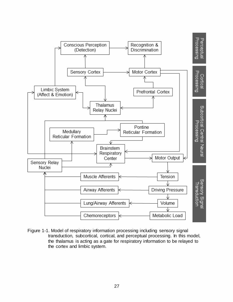
Gating of respiratory activity is important for an animal’s well-being. Eupneic
breathing is usually not consciously perceived, meaning that respiratory afferents during
normal breathing are gated out and do not reach higher brain centers. However, if
ventilation changes sufficiently or breathing is attended to, the sensation is gated in and
the animal becomes aware of its breathing (Chan and Davenport, 2008). This
awareness is usually associated with distressing emotion (O'Donnell et al., 2007).
Figure 1-1 shows the proposed schematic model of respiratory somatosensation and
gating. The respiratory control center located in the brainstem provides ventilatory motor
drive via descending bulbospinal projections that synapse with anterior horn cells in the
cervical and thoracic spinal cord which in turn project to the respiratory muscles (Guz,
1997). Voluntary control of respiration is processed from the motor cortex through
corticospinal connections to the respiratory muscles. Ventilation, including changes in
volume, pressure, oxygen (O2), and carbon dioxide (CO2), is monitored by sensory
feedback receptors positioned in the respiratory muscles, airways, lung, and
chemoreception centers. These afferents connect back to the brainstem respiratory
control center for automatic, subconscious control of breathing. In addition, respiratory
afferents have been shown to reach the cerebral cortex. Specifically, electrical
stimulation of the phrenic nerve resulted in the activation of neurons in the primary
somatosensory cortex in the cat (Davenport et al., 2010). Breathing occurs mostly
without conscious awareness, suggesting that a gate probably exists between the
brainstem respiratory centers and higher brain regions.
The thalamus may be involved in respiratory gating based on evidence from
several studies. Chen et al. (Chen et al., 1992) showed that when respiratory drive was

21
stimulated as measured by increased phrenic nerve activity, previously tonically active
thalamic single units switched to rhythmic increases in firing that was associated with
each respiration. Retrograde tracing experiments in cats indicated that phrenic afferents
activate thalamocortical projections (Yates et al., 1994). Also, Zhang and Davenport
(Zhang and Davenport, 2003) showed that inspiratory occlusions activated thalamic
neurons in cats and rats. Positron emission tomography (PET) studies in humans
exposed to hypercapnia identified neuronal activation extending from the upper
brainstem, up through the midbrain, hypothalamus, and thalamus (Corfield et al., 1995).
Other PET and functional magnetic resonance imaging studies in humans have shown
that voluntary hyperpnea, or the behavioral modulation of breathing, activates distinct
cortical (primary sensorimotor cortices, supplementary motor, and premotor cortex) as
well as subcortical (thalamus, globus pallidum, caudate, and cerebellum) structures
(McKay et al., 2003).
Respiratory information relayed through the thalamus reaches cortical areas for
recognition and discrimination, as well as the limbic system for emotional processing
(Figure 1-1) (Davenport and Vovk, 2009). It is the interplay between these brain areas
that are responsible for the generation of the perception of breathlessness. This feeling
of breathlessness, known clinically as the symptom dyspnea, is an aversive sensation.
Animals and humans alike modify their behavior to avoid feeling breathless, such as
terminating exercise as soon as the sensation becomes overwhelmingly uncomfortable.
This situation is especially detrimental for respiratory disease patients who avoid
exercise and maintain an inactive lifestyle, creating other serious impacts on health.
22
Perception of breathlessness also negatively affects the competitive athlete and it could
be the limiting factor of performance.
Specific Aims
Specific Aim 1: To Investigate the Effects of Expiratory Muscle Strength Training in Collegiate Swimmers
Rationale: The hydrostatic pressure of water challenges the body’s respiratory
and cardiovascular systems. An early study of the mechanics of respiration during
submersion in water found that vital capacity and expiratory reserve volume decreased
and the total work of breathing increased significantly (Hong et al., 1969). Competitive
swimmers are unique in that ventilation is limited to their stroke cycle; inspiration
occurring only when the swimmer’s face is out of the water, and expiration taking place
under water (exception: backstroke). Thus while under water, the swimmer has to
exhale against the pressure of the water, essentially inducing expiratory flow limitation
(EFL). EFL during cycling exercise has been shown to limit exercise performance to
about 65% of the individuals maximal work rate (Iandelli et al., 2002). A significant
decrease in quadriceps muscle blood flow and increase in intercostals muscle blood
flow suggest a redistribution of the cardiac output away from the locomotor and toward
the respiratory muscles (Athanasopoulos et al., 2010). Furthermore, perception of
dyspnea and leg muscle fatigue was significantly greater with EFL compared to
unloaded exercise (Athanasopoulos et al., 2010; Iandelli et al., 2002; Kayser et al.,
1997).
The respiratory muscles are skeletal muscles and thus adapt to a training
stimulus in the same way as other skeletal muscles. In limb muscles, increases in
strength have been shown to occur very rapidly in the early phase of a training protocol

23
(Hakkinen and Komi, 1983; Moritani and deVries, 1979). Recent studies in normal
healthy people and respiratory disease patients have shown that expiratory muscle
strength training (EMST) can specifically strengthen the muscles involved in expiratory
air movement to generate higher positive pressures (Baker et al., 2005; Chiara et al.,
2006; Gosselink et al., 2000; Griffiths and McConnell, 2007; Saleem et al., 2005;
Sapienza et al., 2002; Sasaki et al., 2005; Smeltzer et al., 1996; Suzuki et al., 1995;
Weiner et al., 2003b). However, there are only few and inconclusive studies on the
effects of expiratory muscle training on performance in highly trained athletes such as
college varsity swimmers. An increased ability to generate expiratory pressure could
facilitate the swimmer’s respiration. Presumably, the athlete will need to produce less
effort during the breathing cycle and the feeling of breathlessness will be reduced.
Conversely, for the same effort, the athlete can increase performance because
respiration will become less of a limiting factor. A reduced perception of breathlessness,
or a reduced work of breathing, would require a lower fraction of the cardiac output, so
that more can be diverted to the locomotor muscles, thus decreasing the perception of
exertion.
Hypotheses: Compared to air-flow training, four weeks of EMST will:
Increase the maximum expiratory pressure generating capacity
Decrease the perception of breathlessness and exertion
Improve swim performance
Specific Aim 2: To Investigate the Changes in the Gene Expression Profile of the
Medial Thalamus following ITTO in Anesthetized Rats
Rationale: Complete tracheal occlusion places an maximally obstructive load on
the airway and should elicit a load compensation response, with modulation of breath
timing and esophageal pressure. Airway occlusion is a stressful stimulus (Pate et al.,

24
2010). Respiratory diseases involving acute or chronic exposures to airway occlusion,
such as asthma and chronic obstructive pulmonary disease (COPD) are associated with
significantly higher rates of anxiety and depression compared to the general population
(Moussas et al., 2008).
The thalamus is the proposed brain structure responsible for the gating of
respiratory sensory information to the cortex. Information is continuously sent to this
region from where it is either relayed to higher brain centers or suppressed. Thus, even
in an anesthetized state, modulation of the gene expression profile of the thalamus can
elucidate the immediate changes occurring with airway occlusion.
Hypotheses: ITTO in anesthetized rats will induce:
A load compensation response with changes in breath timing and esophageal pressure
Gene expression changes in the medial thalamus, specifically, modulation of neurotransmitter receptor genes involved in stress and anxiety pathways
Specific Aim 3: To Investigate the Changes in the Gene Expression Profile of the Medial Thalamus following 10 Days of Repeated ITTO in Conscious Rats
Rationale: Respiratory muscle weakness and fatigue have been implicated in an
increased perception of breathlessness (1999; Gandevia et al., 1981; Kayser et al.,
1997; Mador and Acevedo, 1991) and studies have shown that strength or endurance
training of these muscles can improve this condition (Romer et al., 2002; Suzuki et al.,
1995; Verges et al., 2008a; Verges et al., 2008b). However, the underlying mechanisms
of the reduced perception following respiratory muscle training are not clear. Strength
training causes adaptive changes within the nervous system that allow for full activation
of a muscle in specific movements and better coordination of the activation of the
relevant muscles, thereby effecting a greater net force (Sale, 1988). It is possible that

25
the increased capacity of the muscles leads to an increased threshold for load
detection, potentially resulting in decreased feedback from the muscles. The reduced
feedback could result in less activation of neurons in the thalamic gate, reducing the
degree of information relayed to the cortex for load perception. Additionally, challenging
the respiratory system with added external loads could change the threshold of the
thalamic gate itself, such that an increase in sensory threshold would result in less
gating-in of aversive respiratory feedback.
Since chronic airway obstructions in human patients are associated with increased
risk of depression, repeated ITTO could trigger the molecular cascade leading to this
detrimental condition.
Hypotheses: Repeated ITTO in conscious rats induce gene expression changes
in the medial thalamus, specifically:
Modulation of genes involved in sensory gating
Modulation of genes involved in anxiety and depression

26
Table 1-1. Summary of EMST studies with reported MEP and functional changes. Study Participants EMST protocol MEP changes Functional changes
(Suzuki et al., 1995) Healthy 30% MEP, 15min, 2x/d, 4wk EXP: ↑ 25.4% Progressive treadmill exercise test:
↓ dyspnea
(Smeltzer et al., 1996) MS 3x15reps, 2x/d, 7d/wk, 3mo EXP: ↑ 36%, CONT: ↓ 1.9%
Not studied
(Gosselink et al., 2000) Severe MS 60% MEP, 3x15reps, 2x/d, 3mo ↑ 35%, (N.S. compared to CONT)
Improved cough measures
(Hoffman-Ruddy, 2001) Professional Singers
80% MEP, 4x6reps, 4 wk EXP: ↑ 84%, CONT: ↑ 1.4%
EXP: ↓dyspnea during singing, ↑ relative energy, phase duration
(Sapienza et al., 2002) High School Band Players
75% MEP, 4x6reps, 5d/wk, 2wk ↑ 48% Not studied
(Weiner et al., 2003b) COPD 15-60%, 30min/d, 6d/wk, 3mo EXP: ↑ 21% 6min walk test: ↑ distance 19%,
↔ dyspnea daily activities
(Sasaki et al., 2005) Healthy Women
30% MEP, 2x15min/d, 2wk ↑ 10.3% Progressive walking treadmill test:
↓ O2 /kg and RPE
(Saleem et al., 2005) 1 PD Patient 75% MEP, 5x5reps 4wks: ↑ 55% 20wks: ↑158%
Improved UPDRS III scores
(Baker et al., 2005) Healthy 75% MEP, 5x5reps, 5d/wk, 4 or 8 wk
4wks: ↑ 41%, 8wks: ↑ 51%
Not studied
(Chiara et al., 2006) MS 40-80% MEP, 4x6reps, 8 wk ↑ both MS and CONT Improved max voluntary cough values in mild MS
(Kim et al., 2009) Sedentary Elderly
75% MEP, 5x5reps, 4 wk ↑ 44% Improved cough measures
(Griffiths and McConnell, 2007)
Club Rowers 50% MEP, 30reps, 2x/d 4wks: ↑ 3.5% (N.S.) 6wks combined
IMST/EMST: ↑31%
6 min all-out rowing test: N.S.
(Mota et al., 2007) COPD 50% MEP, 30min/d, 3d/wk, 5 wk EXP: ↑ 19% 6min walk test: ↑ distance 13%, Improved dyspnea at rest and QoL
(Kroff, 2008) Women Field Hockey Players
Combined IMST/EMST, 30 breaths, 2x/d, 12 wk
EXP: ↑ 9% CONT: ↑ 4%
20 m shuttle run test: ↑ number of shuttles in EXP and CONT
Studies are listed in chronological order.
EXP = experimental group; CONT = control group; MS = Multiple Sclerosis; N.S. = not statistically significant (p > 0.05); COP D = Chronic
Obstructive Pulmonary Disease; PD = Parkinson’s Disease; O2 = oxygen consumption; RPE = ratings of perceived exertion; UPDRS III = Unified Parkinson’s Disease Rating Scale; QoL = Quality of Life.
27
Figure 1-1. Model of respiratory information processing including sensory signal transduction, subcortical, cortical, and perceptual processing. In this model,
the thalamus is acting as a gate for respiratory information to be relayed to the cortex and limbic system.

28
CHAPTER 2 THE EFFECTS OF EXPIRATORY MUSCLE STRENGTH TRAINING IN COLLEGE
SWIMMERS
Introduction
Competitive swimmers control their breathing pattern to match the stroke cycle. In
comparison to land-based sports, water exercise places additional challenges on the
athlete’s body due to the hydrostatic pressure of the aqueous environment, especially
the respiratory system. During swimming, inspiration occurs only when the swimmer’s
face is out of the water, with expiration taking place under water. Thus, while under
water, the swimmer has to exhale against the pressure column of the water which
requires additional expiratory muscle force. Thus, swim performance may be directly
affected by the strength of expiratory muscle pressure generating capacity.
Recent evidence has shown that expiratory muscle training can specifically
strengthen the muscles involved in expiratory air movement to generate higher positive
pressures (Baker et al., 2005; Chiara et al., 2006; Gosselink et al., 2000; Griffiths and
McConnell, 2007; Saleem et al., 2005; Sapienza et al., 2002; Sasaki et al., 2005;
Smeltzer et al., 1996; Suzuki et al., 1995; Weiner et al., 2003b). Expiratory muscle
strength training (EMST) increases maximum expiratory pressures (MEP) in almost all
study populations, from normal healthy individuals (Baker et al., 2005; Sapienza et al.,
2002; Sasaki et al., 2005; Suzuki et al., 1995), to athletes (Amonette and Dupler, 2002;
Griffiths and McConnell, 2007), to patients suffering from COPD (Weiner et al., 2003b),
MS (Chiara et al., 2006; Gosselink et al., 2000; Smeltzer et al., 1996), or PD (Saleem et
al., 2005). The highest increases in MEP could be seen with a strength training protocol
of short expiratory burst of greater than 60% MEP. Sapienza and colleagues (Sapienza
et al., 2002) trained high school band players at 75% MEP with 25 breaths per day for 5

29
days per week and found increases of 47 and 48% in MEP in boys and girls,
respectively, after 4 weeks of training. Using the same protocol, in a case study of early
idiomatic PD, Saleem et al. (Saleem et al., 2005) reported a 55% increase in MEP after
4 weeks and 158% after 20 weeks. Chiara et al. (Chiara et al., 2006) also found a
significant increase in MEP in patients with MS.
Only a few studies have examined the effects of EMST on exercise performance.
Suzuki et al. (Suzuki et al., 1995) trained healthy subjects at 30% MEP for 15 minutes
twice daily for 4 weeks. During an incremental submaximal running test, the subjects
exhibited increased tidal volume, and decreased minute ventilation, breathing
frequency, and ratings of dyspnea. Sasaki (Sasaki et al., 2005), using the same training
paradigm, showed that O2/kg body weight and ratings of dyspnea decreased. The only
study that examined EMST on performance in athletes reported that training at 50%
MEP for 4 weeks increased MEP, however, no significant difference in performance
could be found during an incremental rowing test (Griffiths and McConnell, 2007).
Traditionally, the respiratory system, specifically, the lung, airways, and respiratory
muscles, have been viewed as being structurally overbuilt and usually well adapted for
normal every-day use and moderate exercise in healthy people with respect to
maximum metabolic requirements for gas transport (Aliverti, 2008; Dempsey, 1986;
Maglischo, 2003). The large diffusion surface area and the short distance between the
alveolar membrane and capillary ensure that ventilation is usually not a limiting factor
for exercise metabolism. However, studies have shown that in highly trained athletes,
the respiratory system may in fact impose a limiting factor to exercise performance
(Boutellier et al., 1992; Dempsey, 2006; Harms et al., 2000). Indeed, with the increased
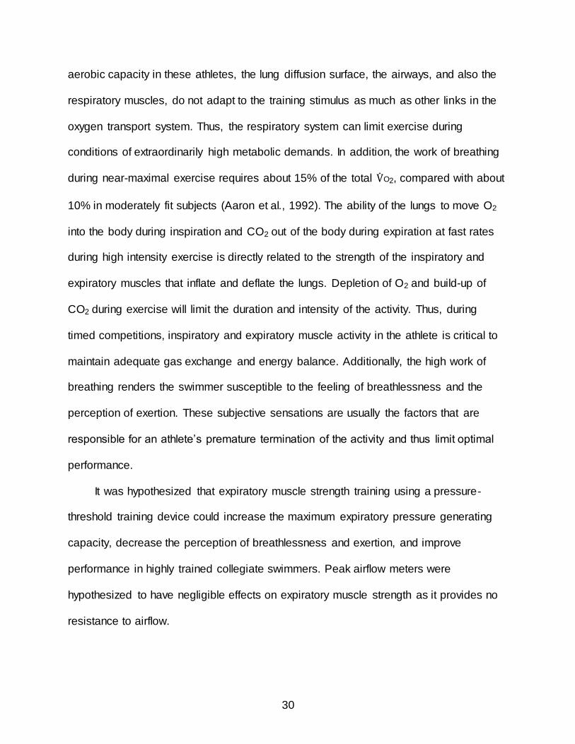
30
aerobic capacity in these athletes, the lung diffusion surface, the airways, and also the
respiratory muscles, do not adapt to the training stimulus as much as other links in the
oxygen transport system. Thus, the respiratory system can limit exercise during
conditions of extraordinarily high metabolic demands. In addition, the work of breathing
during near-maximal exercise requires about 15% of the total O2, compared with about
10% in moderately fit subjects (Aaron et al., 1992). The ability of the lungs to move O2
into the body during inspiration and CO2 out of the body during expiration at fast rates
during high intensity exercise is directly related to the strength of the inspiratory and
expiratory muscles that inflate and deflate the lungs. Depletion of O2 and build-up of
CO2 during exercise will limit the duration and intensity of the activity. Thus, during
timed competitions, inspiratory and expiratory muscle activity in the athlete is critical to
maintain adequate gas exchange and energy balance. Additionally, the high work of
breathing renders the swimmer susceptible to the feeling of breathlessness and the
perception of exertion. These subjective sensations are usually the factors that are
responsible for an athlete’s premature termination of the activity and thus limit optimal
performance.
It was hypothesized that expiratory muscle strength training using a pressure-
threshold training device could increase the maximum expiratory pressure generating
capacity, decrease the perception of breathlessness and exertion, and improve
performance in highly trained collegiate swimmers. Peak airflow meters were
hypothesized to have negligible effects on expiratory muscle strength as it provides no
resistance to airflow.

31
Materials and Methods
Subjects
Seventeen University of Florida (Division I) varsity swimmers (fifteen males, two
females) participated in this study. All procedures were approved by the University of
Florida Institutional Review Board. All participants were classified as normal on the
basis of habitual good health and no evidence of respiratory restriction or obstruction.
All participants consented to the study requirements in writing.
Inclusion criteria consisted of:
Participation at every scheduled swim practice session
No history of cardiorespiratory disease
No history of smoking
No evidence of current major or minor illness
No prior participation in expiratory muscle strength training
Exclusion criteria consisted of:
FEV1.0 of less than 80% of predicted
Regular episodes of bronchoconstriction
Taking medication for respiratory disease
Positive pregnancy test (females) Experimental Design
The study followed a single-blind, placebo-controlled pre-training/post-training
repeated measures design. Participants were randomly assigned to an EMST or an
airflow training (AFT) group. Both groups performed training 5 times per week for 4
weeks. Pulmonary functions, MEP, O2 during an incremental upper body performance
test, and performance measures during a 6 x 100 m freestyle swim test were assessed.

32
Procedures
Pulmonary function assessment
Lung functions for all subjects were determined before and after EMST or AFT
training. Instructions for spirometry testing were based on the American Thoracic
Society Standard (1995). Following a few normal breaths, the subject inhaled deeply
and then provided a forced expiration. Forced expiratory volume in one second (FEV1.0)
and forced vital capacity (FVC) were recorded (Jaeger Toennies) and the ratio
FEV1.0/FVC calculated. Resting respiratory resistance was measured by the forced
oscillation method (Jaeger Toennies).
Maximum expiratory pressures
MEP was measured before and after training. MEP was defined as the greatest
positive pressure obtained at the mouth sustained for at least 1 s while performing a
maximal expiratory effort from total lung capacity (TLC). MEP was assessed using a
portable pressure manometer with a 16 gauge controlled leak in the exhaust port to
prevent generation of high pressures by the buccal muscles and maintain an open
glottis during the measurements. The participants stood upright with their nose closed
by a nose-clip. They were instructed to make maximal forceful expiratory efforts.
Repeated measurements were taken, with a 1 to 2 min rest between trials, until 3
measurements within 10% variability were obtained. The best MEP measurement was
recorded for data analysis.
Expiratory muscle strength training (EMST)
Each participant in the EMST group was assigned an expiratory pressure-
threshold trainer. Trainers were handed out before each training session and collected
at the end to ensure compliance. The training device consisted of a mouthpiece and a

33
one-way spring-loaded valve (Figure 2-1A). The participants were instructed to take a
deep breath in, put their mouth around the mouthpiece, and exhale as hard as possible.
Expiratory airflow was blocked by the valve until a sufficient pressure was produced to
overcome the spring force. The threshold load was set initially at 75-80% of the
participants’ pre-training MEP and increased weekly by 15%. EMST was performed at
the same time of day, five days per week for four weeks. Each training session
consisted of five sets of five-breath repetitions with 1-2 min rest between sets.
Airflow training (AFT)
AFT was used as a placebo control. AFT was conducted with peak airflow meters
(Figure 2-1B). The participants were told that the purpose of the study was to compare
the EMST with the AFT device. As with EMST, the training devices were handed out
only for the training sessions. The participants of the AFT group were instructed to
target a specific expiratory airflow rate that was 75-80% of the pre-training maximal
peak expiratory airflow rate. The participants were instructed to take a deep breath in
and then exhale with as much force as needed to reach the desired airflow rate. The
participants were able to see their effort on the device and could adjust the airflow rate
of their next breath. The targeted airflow rate was increased each week by 15%. As for
the EMST group, training was performed at the same time of day, five days per week for
four weeks. Each training session consisted of five sets of five-breath repetitions with 1-
2 min rest between sets.
O2max test on swim bench
O2max tests were performed before and after the training period. All participants
had prior experience with the Vasa Swim Ergometer (Vasa, Essex, Vermont). This
ergometer featured a flywheel drive system with variable wind resistance depending on

34
the pulling power of the subject. Wind resistance was adjusted by changing the opening
of the damper door. An attached electronic monitor measured time and stroke rate. The
participants lay in a prone position on the bench of the ergometer. The bench distance
to the flywheel was fixed and the legs were supported for comfort and to reduce lower
body movement. The participant used the paddles of the ergometer and mimicked the
upper body movement of the butterfly stroke. A complete test consisted of 10 levels of
90 s each. The resistance of the ergometer was incrementally increased by using the
seven levels of the damper door with a constant stroke rate (35 strokes per min); during
the last three levels the stroke was increased by 2 strokes per min each time. Stroke
rate was maintained by using a metronome. The test was terminated when the
participant voluntarily stopped or after the 10 levels were completed. Throughout the
test the participants were verbally motivated. The participants breathed through a
mouthpiece that was connected to a metabolic cart (ParvoMedics, Sandy, UT). A nose
clip ensured that the participant only breathed through their mouth. The rate of oxygen
uptake, O2, and the rate of carbon dioxide output, CO2, and minute ventilation ( E)
were measured via a sampling tube connected to the mouthpiece. Ratings of
breathlessness (RB) and perceived exertion (RPE) were collected alternately every
45 sec. A modified Borg category-ratio scale ranging from zero to ten was used to
assess the participants’ subjective ratings throughout the test (Appendix A) (Borg,
2008).
Timed interval swim tests
Participants performed a timed interval swim test during the weeks before and
after EMST or AFT training. This test consisted of all-out 6 x 100 m freestyle on an

35
interval of 2:30 min. The test was conducted in a 50-m outdoor swimming pool at the
same time of day during a regular swim training session. A standard warm-up of 2000 m
preceded the test. Time and alternately RB and RPE (Appendix A) were collected after
each 100 m interval. Heart rates were collected after warm-up immediately before the
test start, after interval #1, and after interval #6. Heart rate was measured using a chest
belt with a transmitter that measures the electrocardiogram and sends the heart rate
information to a wrist watch monitor (Polar Electro Inc., Lake Success, NY).
Statistical Analysis
All values were reported as mean ± SD unless stated otherwise. Baseline
differences in anthropometrical data between subjects were compared using an
independent Student’s t-test. For pulmonary function (FEV1.0, FVC, and FEV1.0/FVC),
pre- and post-training comparisons were analyzed using one-way ANOVA with repeated
measures. MEP values, as well as average time, times of first and last 100 m during the
6 x 100 m swim test were compared using two-way repeated measures ANOVA
(Subject, Factor 1 = Group EMST/AFT, Factor 2 = Time of measurements pre/post).
Pearson’s correlation was used to measure of dependence between changes in MEP
and swim time.
Results
Demographics
Twenty-two swimmers originally volunteered and completed pre-testing. Five
withdrew for reasons unrelated to this study. Seventeen (77%) underwent the complete
training period and post-testing. Characteristics of these 17 are presented in Table 2-1.
There were no significant differences in any of these data between the groups.
Participants were randomly divided into either EMST or AFT groups. The EMST group

36
consisted of 3 sprint, 9 middle-distance swimmers; the AFT group included 3 sprint, 3
middle-distance, and 2 distance swimmers.
Pulmonary Functions and MEP
Pulmonary function values are shown in Table 2-2. PFT values in all subjects were
greater than predicted (FEV1.0: 113.67 ± 13.06% in EMST group and 118.63 ± 11.25%
in AFT group; FVC: 118.67 ±12.97 in EMST group and 125.38 ± 12.08% in AFT group).
There were no significant differences between groups and none of the values changed
significantly with either EMST or AFT. FEV1.0 and FVC values were greater than healthy
non-athletes (% predicted) as well as land-based athletes and swimmers (Armour et al.,
1993; Doherty and Dimitriou, 1997; Holmberg et al., 2007; Rong et al., 2008; Sonetti et
al., 2001; Wells et al., 2005), and similar to national-level male swimmers (Armour et al.,
1993).
All individuals in the EMST group showed significant increases in MEP, while
those in the AFT exhibited non-significant small increases or decreases (Figure 2-2).
There was a significant Time effect (pre/post) (p = 0.001) and interaction between
Group and Time (p = 0.049). Post-hoc analysis showed that post-training values were
significantly increased compared to the pre-training in the EMST group (p = 0.004), but
not in the AFT group (p = 0.236) (Table 2-3 and Figure 2-3).
O2 Test
Of the 17 participants, 2 did not perform the post-training O2 test due to
shoulder/wrist injuries unrelated to this study. The incremental swim ergometer O2 test
was a submaximal exercise for most swimmers. Of the 30 pre- and post-training tests,
23 were fully completed, while 7 were terminated by the participant either because of

37
maximum rating of breathlessness (2 individuals) or maximum rating of perceived
exertion (5 individuals). The swim ergometer’s computer reported the work expended
for every stroke, which allowed for analysis of the correlation between the work rate and
O2. Both of these values increased throughout the test, consistent with the
incrementally increasing resistance of the test protocol. There was no significant
difference between the EMST and the AFT group, and neither pre- and post-training.
There were no significant differences in minute ventilation, breathing frequency, and
respiratory exchange ratio. Ratings of perceived exertion and breathlessness increased
in all participants with no significant differences between groups or pre- and post-
training.
Swimming Performance
Average swim time in the 6 x 100 m freestyle test decreased post-training by -1.0
± 1.7 sec (p = 0.084) in the EMST group and -0.2 ± 1.6 sec in the AFT group (p = 0.378)
(Figure 2-4). Post-hoc analysis revealed a significant difference between groups (p =
0.036) that was not dependent on pre- or post-values. Expressed in percent the
improvement in time was -1.44 ± 2.73% in the EMST group and -0.29 ± 2.66% in the
AFT (Figure 2-5). There was no significant difference in the time change between the
groups (p = 0.204). There was no significant correlation between the individual changes
in MEP and swimming performance between pre- and post-training (r = 0.382, p =
0.160) (Figure 2-6).
The first 100 m interval of the pre- and post- 6 x 100 m test was non-significantly
slower in both groups (EMST: 1.21% ± 1.14, AFT: 1.53% ± 1.59) and the last 100 m

38
interval of each test was non-significantly faster in both groups (EMST: 1.56% ± 2.01,
AFT: 0.6% ± 1.25) (Figures 2-7 and 2-8).
Heart rates increased significantly (p < 0.001) in all participants throughout the test
compared to baseline. There was no significant difference between groups or pre- and
post-training (Figure 2-9).
Ratings of perceived exertion and breathlessness increased significantly
(p < 0.001) in all participants throughout the test. There were no significant differences
between groups or pre- and post-training (Figures 2-9 and 2-10).
Discussion
Pulmonary Functions and MEP
Intensive swim training leads to increases in pulmonary functions (Clanton et al.,
1987). In the present study we show that high level competitive swimmers exhibit larger
than predicted FVC and FEV1.0 values, consistent with previous studies (Doherty and
Dimitriou, 1997; Stuart and Collings, 1959). Other studies have also demonstrated
above average vital capacities, residual lung volumes, functional residual capacities,
and total lung capacities in swimmers compared to non-water athletes and non-athletes
(Cordain et al., 1990; Magel and Faulkner, 1967; McKay et al., 1983). The respiratory
training in our study did not further increase lung function, suggesting that no structural
changes in the anatomy of the respiratory system occurred with training.
MEP increased significantly in the EMST group, but not the AFT group. Since
there was no change in pulmonary functions, the change in MEP cannot be explained
based on lung mechanics. The ability to generate higher expiratory pressures is directly
related to the strength of the expiratory muscles. Increased expiratory pressure
generating capacity is particularly important during swimming since exhalation occurs

39
when the head is under water with a water pressure of at least 20 cmH2O. AFT was not
effective in increasing MEP; thus pressure-threshold strength training is a better method
to increase expiratory muscle pressure generating capacity. Pre-training MEP values
were similar to healthy non-athletes (Baker et al., 2005; Sapienza et al., 2002) and club-
level rowers (Griffiths and McConnell, 2007), suggesting that the overall fitness and
highly conditioned abdominal muscles of swimmers does not correlate with a higher
expiratory pressure-generating capacity. The increase in MEP with EMST shows that
respiratory training can be aimed specifically at targeted expiratory muscles.
Oxygen Consumption ( O2)
Exercise performance heavily depends on the interplay between the respiratory
and cardiovascular systems. There are several steps in the oxygen consumption chain
including cardiac output, inspired fraction of oxygen (FIO2), alveolar ventilation, lung and
muscle diffusion capacities, hemoglobin concentration, and mitochondrial rate of O2
consumption (Wagner, 1996). It is believed that maximal O2 transport from the lungs to
the working locomotor muscles and diffusion from the muscle capillaries to the
mitochondria are the major determinants of O2max (Saltin and Calbet, 2006; Wagner,
2006). O2max is defined as the maximum amount of oxygen a person can take up
during one minute of exercise and is directly related to a person’s ability to supply
energy for muscular contraction through aerobic metabolism.
Traditionally, O2max tests are administered on a treadmill or stationary bicycle
ergometer. However, specificity of the test in terms of the muscles used and the
movements during the particular sport is critical to ensure that the measured O2 relates
to the actual O2 the athlete experiences during training or competition. A trained

40
swimmer’s body reacts very differently than that of a runner during various exercise
tests (Armstrong and Davies, 1981; Corry and Powers, 1982; Holmer et al., 1974;
Magel and Faulkner, 1967). Corry and Powers (Corry and Powers, 1982) showed that
runners could reach only 53% of their running O2max in an arm cranking exercise,
while swimmers reached 79%. Other studies also have illustrated that elite swimmers
had lower O2max, heart rate, E, and ventilatory coefficient during maximum swimming
than during maximum running or cycling (Holmer, 1972; Holmer et al., 1974; Magel and
Faulkner, 1967). The swim ergometer used in the present study exhibits several
advantages over other ergometers when measuring the respiratory responses of
swimmers. First, the test was performed in a prone position imitating the swimmer’s
horizontal body position. Body position plays an important role in cardiopulmonary
control due to gravity (Rowland et al., 2008). During heavy exercise, O2 kinetics is
slower in a horizontal than in an upright position (Koga et al., 1999) and venous return is
greater reducing blood hydrostatic pressure in the legs (Holmer et al., 1974). Second,
the swim ergometer simulates closely the actual stroke pattern of movement used by
the swimmer in the water. Third, the flywheel drive system of the ergometer, which is
the same as used in rowing ergometers, offers variable wind resistance depending on
the pulling power of the test participant. This simulates the resistance of water in that
the harder the participant pulls, the more resistance the flywheel provides. To our
knowledge, our study is the first to use this particular swim ergometer to measure the
respiratory responses of swimmers during an incremental exercise test. Other
incremental O2max studies have been conducted using a variety of models and

41
protocols (Armstrong and Davies, 1981; Konstantaki et al., 1998; Potts et al., 2002;
Rowland et al., 2008; Swaine and Zanker, 1996).
The effects of respiratory muscle training on oxygen consumption are unclear,
some studies showing improvements in O2 (Holm et al., 2004; Sasaki et al., 2005),
while others report no changes (Downey et al., 2007; Fairbarn et al., 1991; Gething et
al., 2004; Romer et al., 2002; Sonetti et al., 2001; Sperlich et al., 2009). In the present
study, there were no significant changes in O2 or other measurements during the
incremental exercise test after respiratory training. There is a limit on the O2max an
athlete can achieve that is set by genetic factors. Training can only improve O2max
until that limit is reached. Thus, the lack of improvement could be due to the already
maxed out oxygen consumption chain in these athletes.
Functional Significance of Improvements in Swim Performance Times
Small, but noteworthy changes were detected in swimming performance. The
observed 1.15% time difference between the two groups post-training represents the
difference between first and twelfth place at the 2008 Olympic Games Men’s 100 m
freestyle preliminary races (Figure 2-11).
A recent study found remarkably similar swim performance time improvements
after inspiratory muscle training (IMT) (Kilding et al., 2010). The IMT group performed
30 inspiratory efforts at 50% maximal inspiratory pressure (MIP) twice daily for six
weeks, while the control group did a sham training consisting of 60 breaths at 15% MIP
once daily. Swim performance was measured in time trials pre- and post-training.
Significant improvements were found during a 100 m (-1.7 ± 1.4%) and 200 m (-1.5 ±
1.0%), but not a 400 m (0.6 ± 1.2%) time trial. The training paradigm in the present

42
study differed in that it was a lower-repetition, higher-intensity expiratory muscle
strength training for a shorter period of time (four weeks). The swim performance test
used here was an endurance sprint test rather than a time trial. The average 100 m
freestyle times during the 6 x 100 m test achieved by the collegiate swimmers in our
study were almost identical to the 1 x 100 m time trial in Kilding et al.’s study, indicating
that our athletes performed at a higher level. The higher the level of performance is, the
smaller and rarer the improvements are. Thus, an improvement of 1.15% can prove to
be very beneficial for high-level athletes.
Properties of Respiratory Muscles
The respiratory system consists of inspiratory, expiratory, and accessory muscles,
which may either separately, or in concert, limit exercise performance. Differences
between the inspiratory and expiratory muscles include (a) muscle activity, (b) muscle
fiber type, and (c) predisposition to fatigue. The muscle activity pattern differs greatly
between inspiratory and expiratory muscles. The diaphragm, the most important
inspiratory muscle, is continuously engaged in rhythmic activity to create a sub-
atmospheric pressure driving air into the lungs. This muscle cannot pause to rest under
any circumstance, so it must be very resistant to fatigue. Indeed, Johnson and
colleagues (Johnson et al., 1993) have shown that the diaphragm only fatigues with
prolonged constant-load exercise of high intensity levels greater than 85% of O2max.
Diaphragm fatigue did not occur following a maximal incremental exercise cycling
performance in moderately fit subjects (Romer et al., 2007). However, Lomax and
McConnell (Lomax and McConnell, 2003) measured maximal inspiratory pressures
(MIP) before and after a single 200 m freestyle swim at 90-95% of race pace in

43
competitive swimmers. After the swim the average MIP decreased from ~112 to
~80 cm H2O, suggesting that inspiratory muscle fatigue could be induced in this
population in less than 2.7 min. In contrast to inspiration, expiration is usually a passive
process during normal breathing due to the elastic recoil of the respiratory system that
pushes air out without the need to contract the expiratory muscles. However, during
exercise, the elastic recoil pressure of the lungs is not sufficient to keep up with the
increased demand, so the expiratory muscles actively contract to force air out of the
lungs. As ventilatory demand increases during constant work heavy exercise expiratory
muscle pressures increase more than inspiratory muscle pressures so that the
expiratory muscles take on a greater proportion of the total respiratory muscle work
(Krishnan et al., 2000), thus relieving the inspiratory muscles. Inspiratory muscle
pressures plateau while expiratory muscle pressures continue to increase throughout
heavy exercise (Kearon et al., 1991). Expiratory muscles may facilitate inspiration by
lowering the end-expiratory lung volume, thus providing passive elastic recoil of the
inspiratory muscles during the initial portion of inspiration (Henke et al., 1988).
Muscle fiber morphology also varies between inspiratory and expiratory muscles.
Histochemical profiling of human respiratory muscles have shown a fiber type
distribution of about 50% slow twitch, 25% fast twitch (FTa) and 25% FTb fibers in the
costal diaphragm, 62/20/18% in inspiratory intercostals, and 64/35/1% in expiratory
intercostals (Mizuno, 1991). The cross-sectional area of the expiratory intercostals was
found to be ~50% larger than inspiratory intercostals (Mizuno and Secher, 1989). The
larger the muscle fiber, the stronger its contractile properties, and the faster its fatigue
characteristics (Sieck and Prakash, 1997). According to these characteristics, the

44
expiratory muscles are able to contract with great force early on but are unable to
sustain the high intensity so that force production decreases quickly. Furthermore, the
high amount of large capillary-rich FTa and the lack of FTb fibers in the expiratory
intercostals suggest that these muscles are used during repeated dynamic ventilatory
actions requiring relatively large force (Mizuno and Secher, 1989). Our respiratory
training in rats using tracheal occlusions showed significant increases in cross-sectional
area of the fast twitch type IIx/b fibers in the diaphragm and intercostals muscles (Smith
et al., 2010).
It has been shown that expiratory loading imposes a much higher oxygen
consumption than inspiratory loading, suggesting that the efficiency of the expiratory
muscles to overcome respiratory loads is much lower than that of the inspiratory
muscles (Dodd et al., 1988). The larger fiber size of the expiratory muscles combined
with their sporadic activation with increasing work of breathing renders them especially
susceptible to fatigue. High-intensity, exhaustive exercise leads to a decline in
expiratory muscle endurance (Fuller et al., 1996). Taylor et al. (Taylor et al., 2006) and
Verges et al. (Verges et al., 2006) have shown that cycling exercise elicits abdominal
muscle fatigue, as measured by magnetic stimulation. Cycling is traditionally viewed as
a lower limb muscle exercise, yet these results show that the expiratory muscles
perform a considerable amount of work as well. The observation that the abdominal
muscles fatigued in this experimental setting argues that during swimming these
muscles may fatigue even quicker because they play a dual role in stabilizing and
rotating the body in the water. In these two studies the researchers recruited healthy
male subjects of a broad range of fitness levels and demonstrated that there was no

45
significant correlation between the subjects’ O2max and their abdominal muscle
contractility (Taylor et al., 2006; Verges et al., 2006). This suggests that even highly
trained athletes are not immune to respiratory muscle fatigue. Furthermore, Kyroussis et
al. (Kyroussis et al., 1996) reported that the abdominal muscles fatigued after only two
minutes of maximal isocapnic ventilation, as measured by twitch gastric pressure
elicited by magnetic stimulation. Resistive loads added to inspiration or expiration
induces muscle fatigue, which can be attributed directly to the high work of breathing
against the load (Suzuki et al., 1991). Expiratory muscle fatigue can significantly impair
exercise performance (Mador and Acevedo, 1991; Verges et al., 2006). Prior induction
of expiratory muscle fatigue by resistive breathing resulted in a significant decrease in
performance as measured by distance covered and speed during a 12 min running test,
(Verges et al., 2007) as well as exercise time to exhaustion in a cycle ergometer test at
90% of peak power (Taylor and Romer, 2008).
The structural and functional properties of the respiratory muscle fibers can be
modified in response to physiological and pathological conditions, such as training,
aging, and respiratory diseases (Polla et al., 2004). Volianitis et al. (Volianitis et al.,
2001) have shown that respiratory muscle fatigue in male rowers can be alleviated with
respiratory muscle training. Thus strengthening the expiratory muscles could lead to a
higher resistance to fatigue and in turn improve performance.
Interestingly, concurrent inspiratory and expiratory muscle training had little effect
on performance parameters. Amonette and Dupler (Amonette and Dupler, 2002)
reported that maximal E increased but O2max did not change in trained triathletes
after 4 weeks of combined IMT/EMT of 30 breaths twice daily. The same training device

46
was employed by Wells et al. (Wells et al., 2005) in adolescent swimmers. Twelve
weeks of concurrent IMT/EMT with increasing resistance (50-80% MIP/MEP) resulted in
significant increases in all pulmonary function and respiratory muscle strength
parameters. However, peak velocity during a swim test did not change and critical
speed tended to improve but was not significant at 12 weeks. As Wylegala et al.
(Wylegala et al., 2007) pointed out, the high intensities of training in the later weeks
could have led to chronic respiratory muscle fatigue and may have blunted potential
performance improvements especially if the recovery time was not sufficient before
testing. Griffiths and McConnell (Griffiths and McConnell, 2007) trained two groups of
competitive rowers for 4 weeks on either inspiratory or expiratory trainers at 50% MIP or
MEP and then combined the groups for another 6 weeks of concurrent IMT/EMT
training. During the last 6 weeks, MEP only increased significantly in the group that
performed EMT in the first 4 weeks (31%). However, there were no significant changes
in this group in the rowing test. All of these studies used a training protocol of 30
breaths at a relatively low intensity. Thus, this endurance training may not be as
effective as a strength training protocol with high-intensity loading.
Pacing Strategies
The finding of increased time of the first 100 m interval and the decreased time of
the last 100 m interval during the 6 x 100 m swim test can be attributed to the use of
different pacing strategies by the swimmers. Pacing and the perception of effort are
closely intertwined. When the perception of effort becomes too large, pace will decline.
It has been suggested that a brain area exists that controls pace by incorporating
knowledge of the endpoint, memory of prior events of similar distance or duration, and
knowledge of external and internal conditions (St Clair Gibson et al., 2006). In the

47
present study, probably the most important factor that changed was the memory of the
pre-training test. Before the tests, the swimmers were told to give maximal effort on all
six intervals. During the post-training test, the swimmers remembered the previous test
and the associated high perception of effort. This memory most likely resulted in a slow-
start pacing strategy (St Clair Gibson et al., 2006), which is characterized by a
submaximal pace at the beginning, in order to minimize possible system failure.
Potential Mechanisms
One potential mechanism by which the respiratory system influences exercise
performance has been brought forth by Dempsey (Dempsey et al., 2008; Dempsey et
al., 2006). According to this hypothesis the increased respiratory muscle work during
heavy exercise leads to respiratory and limb muscle fatigue and, in turn, reduced
exercise performance. In detail, Aaron et al. (Aaron et al., 1992) estimated that the
respiratory muscles require 10-16% of total O2 and cardiac output due to
hyperventilation during heavy exercise. This competition for cardiac output between the
respiratory muscles and the exercising limb muscles would lead to respiratory muscle
fatigue. Diaphragm fatigue has been shown to induce a metaboreflex via small diameter
group IV fibers (Hill, 2000) causing increased sympathetic activation of vasoconstriction
and decreased oxygen transport to limb musculature. The decreased cardiac output to
the limb muscles could result in peripheral muscle fatigue and increased perception of
effort, eventually leading to decreased exercise performance. Evidence that respiratory
muscle work affects peripheral fatigue comes from a study by Romer et al. (Romer et
al., 2006), which demonstrated that unloading of the respiratory muscles using a

48
proportional assist ventilator decreased exercise-induced peripheral muscle fatigue by
30-35%.
Strengthening of the respiratory muscles makes them more resistant to fatigue. A
delay in respiratory muscle fatigue attenuates the metaboreflex vasoconstriction of limb
muscles. In addition, more efficient respiratory muscles require less of the cardiac
output. These two factors could result in delayed limb muscle fatigue, decreased
perception of effort and breathlessness, and ultimately, increase exercise tolerance.
Perception of Breathlessness
The final factor limiting exercise is exercise related perception which is sensory
and behavioral in nature (Killian and Campbell, 1995). These sensations include
perception of general exertion, effort, and the feeling of breathlessness. When the
discomfort associated with any of these factors becomes intolerable the subject will
slow down or terminate the exercise. During a maximal effort exercise, the subject is
aware of both the increasing central motor command and the decline in power output
(Killian and Gandevia, 1996); the awareness of these two factors gives rise to the
sensation of fatigue (Jones and Killian, 2000). Breathlessness, either in combination
with peripheral muscle fatigue or by itself, is the most common factor limiting
performance (Killian and Campbell, 1995). The sensation of breathing likely depends on
several different mechanisms that are involved in the regulation of breathing including
feed-forward and feedback mechanisms (Figure 2-12) (Chonan et al., 1990b).
Perception of exertion and breathlessness develops when there is a mismatch between
the central respiratory motor command and afferent information. When exercise
intensity increases, the central motor command increases. Corollary discharges from
descending motor commands to the sensory cortex increases and thus perception of

49
exertion and breathlessness increases. Also, when exercise intensity increases,
increased inputs from a variety of receptors send their feedback to the sensory cortex,
including receptors in the respiratory muscles, the chest walls, the lungs, the lower and
upper airways, as well as chemoreceptors (Killian and Gandevia, 1996). Furthermore,
with increasing muscle fatigue or weakness, the respiratory motor drive increases to
achieve a given muscle tension, which in turn will increase the sense of effort (Homma
and Masaoka, 1999). Subjects with weakened (Gandevia et al., 1981) or partially
paralyzed (curarized) inspiratory muscles (Campbell et al., 1980) show higher
magnitude estimations of respiratory loads and inspiratory muscle training significantly
decreased this perception for small loads (Kellerman et al., 2000). Inspiratory muscle
training could have decreased the motor drive in proportion to the increased respiratory
muscle capacity or the decrease in muscle work for the task might have reduced the
activation of mecahnoreceptors, thus decreasing sensory feedback (Kellerman et al.,
2000)
The sensation of breathlessness is further augmented by increases in expiratory
motor output induced by external resistive loads (Chonan et al., 1990a). With EFL
during exercise in normal subjects, end-expiratory lung volume could not be reduced, so
that end-inspiration occurred at an ever higher lung volume, leading to dynamic
hyperinflation (Kayser et al., 1997). The work of the inspiratory muscles increased
because of the increased elastic loads and the evermore disadvantageous part of the
length-tension relationship. Severe dyspnea in the flow-limited subjects resulted in
termination of the exercise (Kayser et al., 1997). Iandelli et al. (Iandelli et al., 2002) also
demonstrated that performance during airflow-limited exercise was seriously impaired
50
mainly because of increased pressures developed by the expiratory muscles. In this
study the control subjects rated their breathing sensation during the exercise a value of
4 on the Borg scale (0-10) at the maximum power output (Wmax), while airflow-limited
subjects defined their dyspnea rankings as 9.3 and they were only able to exercise to
65% of control Wmax. Because expiration in swimming occurs under water, expiratory
airflow is restricted by the pressure column of the water. This water column is a function
of the depth of the swimmer’s mouth from the surface of the water, about 20 cm H2O.
Thus, with the expiratory effort to overcome the water column pressure threshold load,
swimmers may experience feelings of breathlessness sooner than non-water athletes,
hence pressure threshold expiratory muscle strength training may be especially
effective in compensating for the under-water exhalation load and reducing
breathlessness. In the present study, there was a trend toward increased ratings of
breathlessness post-training in the AFT group, while the perception in the EMST group
did not change. Ratings of perceived exertion followed a similar trend in that the AFT
group rated higher post-training and the EMST group lower. Pressure-threshold training
has the potential to positively influence the conscious sensations of breathlessness and
exertion as perceived through ventilatory factors, while air flow training did not. These
observations are even more interesting given that the EMST group improved in
swimming time more than the AFT group. It is possible that the lower perception of
breathlessness and exertion in the EMST group caused the improvements in swim
performance, which is consistent with the hypothesis of a complex brain control model
of fatigue (Edwards and Walker, 2009).

51
Conclusions
Four weeks of a high-intensity, low-repetition expiratory muscle strength training
paradigm significantly increased the maximal expiratory pressure generating capacity in
highly trained collegiate swimmers compared to a non-resistance peak airflow training.
Swim performance times tended to improve (not statistically significant) and there was a
trend towards lower ratings of breathlessness and perceived exertion. These promising
results merit further investigations into the ergogenic effects of EMST in athletes. EMST
could be used as an adjunct for athletic performance to improve expiratory muscle
strength.

52
Figure 2-1. Components of the training devices for A) EMST, B) AFT.

53
Table 2-1. Anthropometric data participants. Values are presented as mean ± SD.
EMST (n=9) AFT (n=8) p-value
Age (years) 19.6 ± 1.1 19.1 ± 1.0 0.420
Height (cm) 185.9 ± 9.8 182.9 ± 5.5 0.454
Body mass (kg) 82.4 ± 9.6 77.0 ± 4.7 0.169
Competitive training history (years) 10.6 ± 4.8 10.5 ± 4.6 0.981
Table 2-2. Pulmonary function test data. Values are presented as mean ± SD.
Pre-Training Post-Training % Δ p-value
Value % predicted Value % predicted
EMST
FEV1.0 (L) 5.23 ± 1.04 113.67 ± 13.06 5.17 ± 1.14 112.44 ±16.82 -1.15 0.508
FVC (L) 6.48 ± 1.14 118.67 ± 12.97 6.47 ± 1.22 118.33 ± 15.91 -0.19 0.508
FEV1.0/FVC 80.65 ± 7.48
79.69 ± 7.74
-1.18 0.156
AFT
FEV1.0 (L) 5.46 ± 0.58 118.63 ± 11.25 5.32 ± 0.62 115.63 ± 12.51 -2.54 0.113
FVC (L) 6.90 ± 0.75 125.38 ± 12.08 6.83 ± 0.71 118.29 ± 9.92 -0.92 0.523
FEV1.0/FVC 79.56 ± 8.24
78.16 ± 8.30
-1.77 0.202

54
Figure 2-2. Percent change in MEP for each individual. All participants in the EMST
group increased their maximal expiratory pressure generating capacity, while individuals in the AFT group showed only modest increases or decreases in MEP.
Table 2-3. Maximum expiratory pressure test data. Values are presented as mean ±
SD.
Pre-Training (in cmH2O) Post-Training (in cmH2O) % Δ p-value
EMST 122.11 ± 21.22 157.67 ± 23.63 29.86 <0.0001
AFT 131.50 ± 30.42 142.00 ± 39.62 7.85 0.236
Figure 2-3. The EMST group significantly increased MEP post-training compared to pre-
training. MEP changes in EMST group were significantly greater than in AFT group. Values expressed as mean ± SEM. * p < 0.0001.
0
20
40
60
80
100
120
140
160
180
200
EMST AFT
ME
P (
cm
H2O
)
Pre
Post
*

55
Figure 2-4. Pre- and post-performance times for 6 x 100 m freestyle test for both EMST and AFT groups. Values are mean ± SEM. * p < 0.05.
55.0
60.0
65.0
70.0
Group
Me
an
Tim
e (
se
c)
EMST AFT
PrePost
*

56
Figure 2-5. Percent change in 100 m time for each individual.
Figure 2-6 . Percent change of mean swim time during the 6 x 100 m freestyle test for
EMST and AFT groups. Values are mean ± SEM.
-6
-4
-2
0
2
4
Pre Post
Tim
e ch
an
ge
(%
)
EMST
-6
-4
-2
0
2
4
Pre Post
Tim
e c
ha
ng
e (%
)
AFT
-3
-2
-1
0
EMST AFT
% T
ime
ch
an
ge

57
Figure 2-7. Correlation of % time change during 6 x 100 m test and change in MEP pre-
to post-training for each individual.
y = 0.0392x - 1.8757 R² = 0.1459
-8
-6
-4
-2
0
2
4
-30 20 70
% T
ime
ch
an
ge
MEP change (Δ cm H2O)

58
Figure 2-8. Percent time change of the first 100 m interval. Values are mean ± SEM.
Figure 2-9. Percent time change of the last 100 m interval. Values are mean ± SEM.
0
1
2
3
4
EMST AFT
% T
ime
ch
an
ge
-4
-3
-2
-1
0
EMST AFT
% T
ime
ch
an
ge

59
Figure 2-10. Heart rate before and during the 6x100 m freestyle test.

60
Figure 2-11. Changes in Ratings of Breathlessness between first and last rating within
one 6x100 m test. Values are mean ± SEM.
Figure 2-12. Changes in Ratings of Perceived Exertion between first and last rating
within one 6x100 m test. Values are mean ± SEM.
-1.5
-1
-0.5
0
0.5
1
1.5Δ
Ra
ting
s o
f P
erc
eiv
ed
Exe
rtio
n
EMST AFT
# 1
# 3
# 5
-0.5
0
0.5
1
1.5
2
Δ R
ating
s o
f B
rea
thle
ssne
ss
EMST AFT
# 2
# 4
# 6

61
Figure 2-13. Functional significance of % improvements in swim time pre- and post-
training compared to the 2008 Olympic Games Men’s 100 m freestyle preliminary races.

62
Figure 2-14. Possible mechanisms leading to the sensation of breathlessness.

63
CHAPTER 3 TRACHEAL OCCLUSION IN ANESTHETIZED RATS MODULATES GENE
EXPRESSION PROFILE OF MEDIAL THALAMUS
Introduction
The sensation of dyspnea, or breathlessness, is one of the primary symptoms in
pulmonary and cardiovascular diseases (Manning and Schwartzstein, 1995; O'Donnell
et al., 2007). It is one of the symptoms that most limits patients with obstructive
pulmonary diseases, and it can also be considered one of the most important factors in
determining the severity of the disease and the health-related quality of life (Carvalho et
al., 2007). Sensory information is continuously sent to the respiratory centers in the
brainstem; minor changes in breathing are controlled by these centers without the
activation of higher brain centers. When changes in respiratory information reach a
certain threshold, then a gating-in process occurs resulting in the cognitive awareness
of breathing (1999; O'Donnell et al., 2007). The conscious awareness of breathing as in
dyspnea requires the activation of higher brain centers (O'Donnell et al., 2007).
The neural control pathway to the higher centers is thought to be a gated process
(Chan and Davenport, 2008). Only information that is selectively attended to or is above
a certain threshold would be able to pass through to the cortex. The brain structure
involved in the gating of various sensory afferents, such as somatosensory, auditory,
and visual information to the cortex is the thalamus (Kimble & Kaufman, 2004).
Furthermore, our laboratory has found increased c-Fos expression in response to
tracheal occlusions in the medial thalamus, indicating activity of neurons in this brain
area (Pate et al., 2008; Vovk et al., 2006). Thus, it was hypothesized that respiratory
sensory information may also be processed and relayed to higher brain centers through
the thalamus.
64
The thalamus is the largest structure in the diencephalon, located centrally in the
brain. It is ideally situated in a position to receive incoming sensory information and
send nerve fibers out to the cerebral cortex in several directions. Functionally, the
thalamus is believed to serve as the processing and relay station that most sensory
information (one known exception is olfaction) must pass before reaching the cortex
(Kimble and Kaufman, 2004; McCormick and Bal, 1994; Sherman and Guillery, 2002).
For the well-studied visual, auditory, and somatosensory systems, different thalamic
relay neurons are responsible for relaying specific information. Visual stimuli pass
through the lateral geniculate nucleus, auditory information through the medial
geniculate body, and somatosensory stimuli are processed by the ventrobasal complex
(Alitto and Usrey, 2003). In the present study, it was hypothesized that respiratory
afferents carry lung and airway information to the thalamus, specifically the medial
thalamus as based on the c-Fos data, where it is processed and potentially relayed to
the cortex.
Midline thalamic nuclei receive projections from areas such as the periaqueductal
gray (Krout and Loewy, 2000b), the parabrachial nucleus (Krout and Loewy, 2000a), the
superior colliculus (Krout et al., 2001), and the brainstem (Krout et al., 2002). The
thalamus integrates many bidirectional connections with several regions of the cortex,
most importantly the recurrent loop to and from various cortical areas, but also to the
amygdala (possibly for emotional processing), the hippocampus (for learning and
memory), and the limbic system (Newman, 1995, Kimble & Kaufman, 2004). A single
thalamic nucleus can send afferents to multiple cortical areas (Herrero et al., 2002).
Intralaminar thalamic nuclei have diffuse projections to the cerebral cortex (Jones and

65
Leavitt, 1974). Reciprocal corticothalamic neurons connect back to the thalamic relay
and interneurons (McCormick and Bal, 1994). Anatomical studies have shown that
about 50% of thalamic connections are coming from the cortex. It is believed that this
corticothalamic feedback functions in “egocentric selection”, which refers to the ability of
cortical neurons to analyze thalamic input, select certain sensory features, and then
amplify the transmission of these features by feedback to the thalamus (Suga et al.,
2002). This suggests that cortical areas can either enhance or suppress particular
information. Other top-down connections such as from the cingulated gyrus and
prefrontal cortex feeding back onto the thalamic neurons help in selecting stimuli that
are relevant, salient, and novel (Kimble and Kaufman, 2004). In some instances, the
thalamus may impair rather than facilitate the processing of environmental stimuli, such
as during trauma (Kimble and Kaufman, 2004).
Thalamic relay neurons receive both excitatory (glutamatergic) and inhibitory
(GABAergic) transmission and the balance/ratio between these signals is what
eventually determines the response (McCormick and Bal, 1994). Other
neurotransmitters and neuromodulators also influence thalamic neurons, most notably
serotonin from the raphe nucleus (McCormick and Bal, 1994). Modulations of any
component in the neural transmission pathway (such as transmitters, receptors,
transporters) alter the signal processing of the thalamic relay neurons and thus change
sensory information gating.
Genomic high-throughput technologies, such as DNA microarrays, serve as a
powerful tool for identifying gene expression profiles in response to a specific stimulus.
The development of microarrays has provided the opportunity to compare and analyze

66
gene expression differences of thousands of genes simultaneously, and characterize
the biological processes occurring as a result of the impact. Oligonucleotide
microarrays, as used in this study, are systematically prepared based on known gene
sequence information. Probes on the arrays are 60 nucleotide in length, which has
shown higher specificity and sensitivity compared to shorter oligos (Hughes et al.,
2001). In this experiment a reference design was used. The reference sample was
prepared by combining small amounts of RNA of all samples collected. This sample
was then labeled with one color, while all target samples were labeled with another
color. In the hybridization reaction for one microarray, the two differently labeled
preparations (reference sample and one target sample) were then combined and
simultaneously hybridized to the arrayed probes. Binding to the probes depends on the
relative concentrations of mRNA transcript contained in the two samples. Hybridization
was carried out on a rotational device in a hybridization chamber with the help of a
gasket slide that enables active mixing and increases the chance for the target to come
into contact with each probe. Scanning of the microarrays records and quantifies the
amount of emitted light that was collected by exciting the fluorescent molecules
conjugated to the hybridized targets. The ratio between signal intensities of the two
fluorescent signals serves as an indicator of which genes were differentially modulated.
In the present study, comparative microarray analysis was performed to examine
the molecular changes that occur immediately after ITTO. It was hypothesized that
ITTO would induce short-latency (< 10 min) gene expression changes in the medial
thalamus. RNA samples from the medial thalamic regions of two groups of rats were

67
used; one group receiving ITTO and one group of control animals not receiving
occlusions.
Materials and Methods
Animals
Eight male Sprague-Dawley rats (276.8 ± 47.5 g) were housed two per cage in a
temperature-controlled room (72°F) on a 12:12 light:dark cycle, and with free access to
food and water. All animal experiments were approved by the Institutional Animal Care
and Use Committee of the University of Florida.
Surgical Procedures
Animals were anesthetized by intraperitoneal injection of urethane (1.3-1.5 g/kg)
and anesthesia was supplemented as needed (20 mg/ml). Anesthetic depth was verified
by the absence of a withdrawal reflex to a rear paw pinch. Body temperature was
measured using a rectal probe and maintained at 38°C with a heating pad. Animals
were spontaneously breathing room air.
Figure 3-1 shows the surgical setup for recording of esophageal pressure (Pes)
and diaphragm electromyography (EMGdia). One end of a saline-filled tube was inserted
through the mouth into the esophagus to measure Pes. The other end of the tube was
connected to a polygraph system (Model 7400, Grass Instruments) via a pressure
transducer and the analog output was amplified, digitized at 1kHz (CED Model 1401,
Cambridge Electronics Design) and computer processed (Spike2, Cambridge
Electronics Design). Pleural pressure changes were inferred from relative changes in
Pes.
Diaphragm electromyographic (EMGdia) electrodes were prepared from Teflon-
coated wire. The ends of the wires were bared, bent and hooked into the costal

68
diaphragm through a small incision in the abdominal skin. Two electrodes were inserted
for bipolar EMGdia recordings. The electrode wires were connected to a high-impedance
probe. The signal was amplified (P511, Grass instruments) and band-pass filtered (30-
300 Hz). The analog outputs were digitized and processed as described above.
The trachea was exposed through a ventral incision via blunt dissection of
surrounding tissue. An expandable cuff was sutured around the trachea, two cartilage
rings caudal to the larynx. The cuff was connected to an air-filled syringe via a thin
rubber tube. The syringe was used to inflate and deflate the cuff bladder. Prior to the
experiments the inflation pressure needed to completely compress the trachea was
determined using an excised trachea. A complete compression occluded the airway
during both inspiration and expiration. Deflation restored the trachea back to its original
condition to allow unobstructed breathing.
Experimental Protocol
The experimental group (n=4) was allowed to breathe unobstructed for 60 min
following surgical preparations. Then the cuff was inflated to occlude the trachea for 2-4
breaths, followed by deflation of the cuff for a minimum of 15 breaths. The occlusions
were repeated for a total of 10 min. Pes and EMGdia were monitored continuously
throughout the experiment to verify onset and removal of tracheal occlusions. The
control animals (n=4) underwent the same surgical procedure, 70 min of unobstructed
breathing, but did not receive tracheal occlusions. Immediately after completion of the
70 min post-surgical period, the animals were decapitated and their brains removed.
The medial thalamus was located (Figure 3-2), excised, frozen in liquid nitrogen, and
stored at -80°C until further use.

69
Physiological Data Analysis
Data were analyzed offline using analysis software Spike 2 (Cambridge
Electronics Design). The EMGdia was rectified and integrated (time constant = 50 ms),
and inspiratory time (Ti), expiratory time (Te), and total time (Ttot) for each breath were
calculated from the integrated EMGdia signals. Ti was measured from the onset of the
inspiration-associated increase in EMGdia activity to the point at which EMGdia peak
activity began to decline (Figure 3-3). Te was measured from the end of Ti to the onset
of the following inspiration (Figure 3-3). Baseline EMGdia was defined as the minimum
value of the EMGdia during expiration. The EMGdia amplitude (Δ EMGdia) was calculated
as the difference between baseline and peak EMGdia. Pes amplitude was calculated as
the difference between baseline and peak Pes. For the experimental group, within each
occlusion presentation the control breath (C) was defined as the complete breath
immediately prior to occlusion (O) application and the recovery breath (R) was defined
as the first complete breath immediately after termination of occlusion. For the 10 min
occlusion trial, one control breath, two occluded breaths (O1 and O2), and one recovery
breath were measured for each occlusion presentation. Data from at least 28 occlusion
trials for each experimental animal were used for analysis. For the control group,
breathing pattern of control breaths was measured at matched time points.
SigmaStat for Windows Version 3.5 (Systat Software, Inc, Germany) was used for
all statistical analyses of physiological data. All values are reported as mean ± SD. Ti,
Te, Ttot, Pes, and Δ EMGdia for control breaths (C) were compared between
experimental and control animals using one-way ANOVA. Comparisons between C, O,
and R breaths in the experimental animals were analyzed using one-way repeated
measures ANOVA.

70
Total RNA Isolation
Total RNA was isolated from medial thalamic tissue with RNA Stat-60 (Tel-test,
Friendswood, TX). 10-20 mg of the frozen tissue was homogenized in Stat-60 and
chloroform added. The mixture was vortexed for 15 s and centrifuged at 12,000 g for 15
min at 4°C. The upper aqueous phase containing the RNA was carefully extracted. The
extraction step was repeated and the RNA precipitated with isopropanol. Following
another centrifugation at 12,000 g for 40 min at 4°C, the pellet was washed twice with
80% ethanol and air dried. To inactivate RNases, the pellet was resuspended in 40 µl
RNA secure (Ambion, Austin, TX) following the manufacturer’s protocol. Quantity and
quality of total RNA was determined by spectrophotometry (260/280 ratios ≈ 2.0), and
by electrophoresis through a 1% formaldehyde agarose gel stained with ethidium
bromide.
RNA Amplification and Microarray Analysis
Array hybridizations were performed using a reference design. The reference
material consisted of equal amounts of RNA from all eight thalamic samples. The cDNA
synthesis, cRNA labeling and hybridization were performed following the manufacturer’s
kits and protocols (Agilent Low RNA Input Fluorescent Linear Amplification Kit and
Agilent 60-mer oligo microarray processing protocol; Agilent, Palo Alto, CA). The
thalamic samples were labeled with Cy5, while the reference sample was labeled with
Cy3. The cRNA was amplified and purified using the QIAGEN RNeasy Kit (Qiagen Inc.).
Dye incorporation was determined by using Nanodrop (>13 pmol/µg RNA). For the 4 x
44,000 rat genome DNA oligo microarray (Agilent Technologies Inc., Amadid: 014879),
825 ng of sample and 825 ng of reference was used for each array. Hybridization was
carried out in a microarray hybridization chamber at 65°C for 17 hours. The glass slides
71
were then washed and scanned with a laser-based detection system (Agilent, Palo Alto,
CA). A log2 transformed signal ratio between the experimental channel and the
reference channel was calculated for each spot.
One-way ANOVA was performed on normalized log2-transformed signal ratios of
each probe individually, followed by Tukey’s honestly significant differences pair-wise
comparisons to determine genes whose expression was significantly regulated by the
tracheal occlusions compared to control. Genes were considered differentially
expressed if the p-value was ≤ 0.05 and the log2 fold change was ≥ 0.4.
Gene Ontology and Pathway Analysis
Gene ontology annotations were derived from similarity searches of the NCBI
Gene database. A blastn search for each of the 44,000 probes was performed to
retrieve the gene ontology (GO) annotation. Once the GO annotations were retrieved, a
GO tree was built following the hierarchical structure for the whole array. Then, another
GO tree for the significant regulated genes was built. The two trees were compared at
each node by running a Fisher's Exact Test (p ≤ 0.05) when traversing the tree
branches. Significantly over-represented GO categories were identified by the Fisher's
p-value and the false discovery rate was determined.
Some of the genes that showed significant modulation were scanned against the
Pathway Studio ResNet database (Ariadne Genomics, Rockville, MD). This database
uses published information and catalogs the relationships between biological entities.
Pathway Studio® (Ariadne Genomics) was used to identify and graphically display the
functional interactions between the selected genes (Nikitin et al., 2003).

72
Results
Physiological Responses to ITTO
Breath timing and Pes response to ITTO are shown in Figure 3-3. Breathing
frequency slowed due to an increase in Te. The Pes was more negative during occlusion
and returned to baseline immediately after termination of occlusion. Comparisons of
control breaths between experimental and control animals demonstrated no significant
differences in Ti, Te, Ttot, Pes, or EMGdia (Table 3-1).
Comparisons between C, O, and R breaths within each experimental animal
revealed (Tables 3-2 and 3-3, and Figure 3-4) Te, Ttot, and Pes were significantly
different between C and O, and between O and R, but not C and R. There was a trend
toward significance in Ti. EMGdia showed no significant differences. This shows that
ITTO resulted in an increase in expiratory timing and thus total breath time.
Modulation of Gene Expression Profile Following ITTO
Statistical analysis of the microarray data showed that a total of 588 genes were
altered (p < 0.05, log2 fold change ≥ 0.4) following the occlusion protocol, with 327
down-regulated and 261 genes up-regulated (Appendix Table B). Some candidate
genes of interest included genes involved in stress-related pathways (Table 3-4).
Table 3-5 shows the GO categories for biological processes that were over-
represented among the regulated genes (p < 0.05, FDR < 0.1). The most significantly
regulated GO categories were “anti-apoptosis”, “response to stress”, “regulation of
enzyme activity”, and “MAPKKK cascade”.

73
Discussion
Airway Occlusions Elicit the Load Compensation Response
When an animal is challenged with an increase in respiratory mechanical load,
the respiratory control system elicits the load compensation reflex. This ventilatory load
compensation response has been observed in anesthetized animals using external
resistive loads to breathing and is characterized by the recruitment of respiratory muscle
activity, an increase in breath duration and a decrease in tidal volume (Bishop et al.,
1981; Bradley, 1972; Clark and von Euler, 1972; Davenport et al., 1981; Davenport et
al., 1984; Davenport and Wozniak, 1986; Zechman et al., 1976). Depending on the
timing within the breath phase of the added resistive load (end-inspiratory versus end-
expiratory), inspiratory or expiratory duration is increased. The load compensation reflex
is also dependent on selective phase loading (inspiration only or expiration only) and
loading that occurs throughout the entire breath. In this study we applied complete
tracheal occlusions for multiple breaths so the load was applied on both the inspiratory
and expiratory breath phases. Load compensation breathing pattern was achieved by a
more negative Pes and an increase in Ttot, primarily due to an increase in Te, while Ti
increased non-significantly. The more negative Pes peak during occlusions
demonstrates a larger inspiratory effort exerted by the animal to inhale. However,
diaphragm activity (EMGdia) was not significantly modulated, so the more negative Pes
could be due to the respiratory mechanical changes, such as increased resistance.
Inspiratory duration did not change significantly which is in congruence with an
unaltered EMGdia signal. One of the causes for the unchanged Ti and EMGdia may be
due to the timing of the onset of occlusion (Zechman et al., 1976).

74
The load compensation response lasted as long as the trachea was occluded and
breathing patterns returned to baseline levels immediately following withdrawal of
occlusion demonstrating that tracheal occlusions using an inflatable cuff are reversible.
Airway Obstruction in Disease and Association with Anxiety and Depression
Asthma is a respiratory disease characterized by reversible airways obstruction,
airway inflammation, and hyperreactive airways (Valenca et al., 2006). Our animal
model of reversible tracheal obstructions thus mimics one component of asthma. It is
well acknowledged that respiratory diseases, such as asthma and chronic obstructive
pulmonary disease (COPD) are associated with significantly higher rates of anxiety and
depression compared to the general population (Moussas et al., 2008). In the US
general population, it has been found that significantly more individuals suffering from a
respiratory or lung disease have panic disorder or major depression than individuals
without such a diagnosis (Goodwin and Pine, 2002). In a sample of 189 patients from a
Brazilian outpatient clinic for the treatment of asthma and COPD, Carvalho et al.
(Carvalho et al., 2007) found that almost all patients with controlled and uncontrolled
asthma exhibited moderate to severe anxiety as determined by the State-Trait Anxiety
Inventory (CA 97.5%, UA 93%), and 74% of COPD patients. Depression scores, as
measured by the Beck Depression Inventory, were less pronounced in these patients
(CA 20%, UA 49%, COPD 29%) but nevertheless a cause for concern. In a similar
study on 132 pulmonary disease patients in a Greek hospital, Moussas et al. (Moussas
et al., 2008) found that a total of 49.2% showed moderate or severe depression, while
26.5% had anxiety. Fernandes et al. (Fernandes et al., 2010) demonstrated a positive
association of higher degrees of asthma severity with increased anxiety. In this study,
70% of the patients had a clinical diagnosis of anxiety and anxiety was associated with

75
worse subjective asthma outcomes and increased use of medication and healthcare
services. Of 62 asthmatic patients from an outpatient clinic in Brazil, 24.1% had major
depression disorder and 33.8% had an anxiety disorder as diagnosed by the Mini-
International Neuropsychiatric Interview (Valenca et al., 2006). However, there was no
association between the severity of asthma and the prevalence of anxiety and
depression (Valenca et al., 2006). In a study on Veteran’s Affairs patients with chronic
breathing disorders, 50.1% showed moderate to severe depression and 64.2% had
moderate to severe anxiety symptoms (Kunik et al., 2005). In a 20-year longitudinal and
cross-sectional study with 591 participants between the ages of 19 and 40, Hasler et al.
(Hasler et al., 2005) found that asthma was strongly associated with panic disorder and
that the presence of asthma predicted subsequent panic disorder. Patients with severe
asthma and a comorbid psychiatric disorder had almost 11-fold increased risk for two or
more asthma exacerbations and almost 5-fold increased risk for two or more
hospitalizations during the past year as compared with patients with severe asthma
without psychiatric disorder (ten Brinke et al., 2001). These results show that anxiety
and depression are associated with respiratory diseases and that in some vulnerable
individuals, an increase in anxiety may lead to panic disorders, possible through
dyspnea-induced fear conditioning (Hasler et al., 2005).
Airway Occlusions Induce Serotonin Receptor HTR2A and Reduce Dopamine
Receptor DRD1
Medial thalamic mRNA transcripts of the serotonin receptor HTR2A were up-
regulated following tracheal occlusions. It is well known that the serotonin system plays
an important role in a variety of human psychopathological conditions, particularly mood
and anxiety disorders (Charney et al., 1987; Hensler, 2006). Antidepressant treatment

76
has focused on modulating serotonergic neurotransmission (Jones and Blackburn,
2002). One of the challenges of the serotonin system is the sheer complexity of the 14
known receptor varieties categorized into seven receptor subtypes (Hoyer et al., 2002).
Specifically, the HTR2A receptor has been identified to be involved in anxiety disorders
in dogs (Vermeire et al., 2009). Antagonists to this receptor may be useful therapeutic
agents in the treatment of generalized anxiety disorder and psychosis (Javanbakht,
2006; Jones and Blackburn, 2002). Activation of HTR2A receptors weakens the sensory
gating so that more sensory information is able to reach consciousness, which in turn
can lead to the pathology of anxiety disorders (Javanbakht, 2006). Behavioral studies in
HTR2A knockout mice have shown changes in anxiety-related but not depression-
related paradigms (Weisstaub et al., 2006). The knockout mice exhibited greater
exploratory and risk behavior in conflict paradigms, such as the open field, dark-light
choice, elevated plus-maze, and novelty-suppressed feeding tests. In depression-
related behaviors, as measured by the forced swim test and the tail suspension test, the
mice did not differ significantly from wild-type (Weisstaub et al., 2006).
The dopaminergic system has been identified to be involved in a myriad of
functions, such as motivation, reward, pain processing, learning, and memory (Arias-
Carrion and Poppel, 2007; Shyu et al., 1992). Hyperfunction of this system has been
hypothesized to be associated with schizophrenia and attention deficit hyperactivity
disorder (Carlsson et al., 1999; Di Chiara and Bassareo, 2007; Heijtz et al., 2007). This
hypothesis is based on the antagonistic interaction between dopamine and glutamate
projecting on GABAergic striatal neurons that exert an inhibitory effect on
thalamocortical glutamatergic neurons, thereby filtering out part of the sensory input to

77
the thalamus to protect the cortex from a sensory overload (Carlsson et al., 1999).
Hyperactivity of dopamine or hypofunction of the corticostriatal glutamate pathway
should reduce this protective influence and could thus lead to confusion or psychosis
(Carlsson et al., 1999; Gaudreau and Gagnon, 2005). Neuroleptic drugs that block
dopaminergic pathways improve sensory functioning and gating in schizophrenic
patients (Freedman et al., 1987).
Interaction between serotonin and dopamine systems can have either potentiating
or antagonizing effects (Lieberman et al., 1998)). HTR2A antagonists have shown to
increase dopamine release in a variety of brain regions (Hertel et al., 1996).
Airway Occlusions Alter Genes Involved in Anti-Apoptosis
Gene ontology analysis identified anti-apoptosis, negative regulation of apoptosis,
and negative regulation of programmed cell death as being affected by airway
occlusions. These findings suggest that the medial thalamus may be increasing cell-
protective mechanisms. A postmortem study examining anatomical abnormalities in the
thalamus of patients diagnosed with major depressive disorder discovered that in these
subjects the mediodorsal nucleus of the thalamus had a significantly increased total
number of neurons compared to nonpsychiatric subjects (Young et al., 2004). Other
studies have demonstrated volume reductions in prefrontal cortex (Botteron et al., 2002)
and hippocampus (Bremner et al., 2000) as well as decreased number of glia in the
cortex (Ongur et al., 1998). Young et al. (Young et al., 2004) suggested that the
elevated number of neurons in the medial thalamus may have had this reducing effect
due to its projections to these other brains areas because excessive glutamatergic
thalamic neurons could lead to excitotoxicity. Alternatively, increased GABAergic
neurons could result in decreased output to the cortex, thus reducing the need for glial

78
support. It is not known which neuron population (excitatory projection neuron or
inhibitory interneuron) is elevated in major depressive disorder. The results of the
present study showing modulated pathways involved in anti-apoptosis could suggest a
first step in the development of depression after airway occlusions.
Functional Analysis
Pathway Studio was used to visualize changes of gene expression following ITTO.
An interaction between the dopamine receptor DRD1 and the serotonin receptor HTR2A
exists that is controlled by MAPK1 (Figure 3-5). MAPK1 positively regulates the NMDA
receptor, which in turn inhibits DLG4 and acts on DRD1. At the same time, MAPK1
inhibits CAV1, which regulates HTR2A. Following tracheal occlusions, the down-
regulated expression of MAPK1 could have led to a decreased stimulation of the NMDA
receptor and a decreased inhibition of CAV1. Even though NMDA receptor and CAV1
did not show differential gene expression, MAPK1 could potentially regulate the function
of these genes. The decreased function of NMDA receptor would lead to a decrease in
DRD1; indeed, a significant decrease in DRD1 expression was found in the occlusion
group. Decreased NMDA receptor function could also result in less inhibition of DLG4,
which in turn could result in increased stimulation of HTR2A. Furthermore, decreased
inhibition of CAV1 means increased function and increased stimulation of HTR2A.
HTR2A gene expression was indeed up-regulated following occlusions. DRD1 and
HTR2A control the activation and release of neurotransmitters and other small
molecules. There was a reciprocal interaction between DRD1 and HTR2A that seems
to be predominantly regulated by MAPK1.

79
Conclusions
A single trial of ITTO in anesthetized rats elicited a load compensation response
characterized by an increase in Te and thus Ttot and a decrease in Pes. ITTO also
induced a change in the gene expression profile of the medial thalamus including genes
involved in the stress response and anti-apoptosis. The results suggest that the medial
thalamus is a component of the respiratory neural network responding to respiratory
load stimuli.

80
Figure 3-1. Diagram of surgical preparation including placement of tracheal occluder,
esophageal pressure tube, and diaphragm electrodes.

81
Figure 3-2. Location of collected thalamic tissue sample. Coronal view of the rat brain at
Bregma -1.80mm. Dashed box indicates the tissue section (medial thalamus) that was excised for microarray analysis. Modified from (Paxinos and Watson,
1998).

82
Figure 3-3. Physiological changes as a result of ITTO. Red bar represents application of an occlusion. Identification of control, occlusion, and recovery breaths and
determination of Ti and Te are shown. Breath timing (Ttot = Ti + Te) increased mostly due to an increase in Te and esophageal pressure was more negative
during occlusion and returned to normal immediately after.

83
Table 3-1. Control breath comparisons of breath timing, Pes, and ΔEMGdia between experimental and control animals. Control breaths were defined as the last
complete breath immediately prior to an occlusion for experimental animals and at matched time points for controls animals. Values are reported as mean
± SD. P-values from one-way ANOVA.
Experimental Group Control Group p-value
Ti (s) 0.228 ± 0.014 0.228 ± 0.014 0.997
Te (s) 0.488 ± 0.104 0.411 ± 0.073 0.277
Ttot (s) 0.715 ± 0.115 0.639 ± 0.073 0.306
Pes (V) -0.038 ± 0.013 -0.038 ± 0.011 0.967
ΔEMGdia (au) 0.697 ± 0.583 0.444 ± 0.246 0.454
Table 3-2. Comparisons between control, occlusion, and recovery breaths in the
experimental animals. Values are reported as mean ± SD. Significant differences from one-way repeated measures ANOVA are denoted by: #
different from C, * different from R (p < 0.05).
Control breath Occlusion 1 Occlusion 2 Recovery breath
Ti 0.228 ± 0.014 0.253 ± 0.014* 0.244 ± 0.015 0.211 ± 0.023
Te 0.488 ± 0.104 0.642 ± 0.189#* 0.588 ± 0.155* 0.472 ± 0.128
Ttot 0.715 ± 0.115 0.895 ± 0.193#* 0.832 ± 0.162* 0.683 ± 0.145
Pes -0.038 ± 0.013 -0.053 ± 0.011#* -0.057 ± 0.012#* -0.041 ± 0.015
ΔEMGdia 0.697 ± 0.583 0.728 ± 0.570 0.745 ± 0.612 0.717 ± 0.605
Table 3-3. Comparisons between control, occlusion, and recovery breaths in the
experimental animals with combined values from O1 and O2. # different from C, * different from R.
Control O1+O2 Recovery p-value
Ti 0.228 ± 0.014 0.248 ± 0.014 0.211 ± 0.023 0.063
Te 0.488 ± 0.104 0.615 ± 0.172#* 0.472 ± 0.128 0.011
Ttot 0.715 ± 0.115 0.864 ± 0.178#* 0.683 ± 0.145 0.010
Pes -0.038 ± 0.013 -0.055 ± 0.012#* -0.041 ± 0.015 0.002
ΔEMGdia 0.697 ± 0.583 0.736 ± 0.591 0.717 ± 0.605 0.197
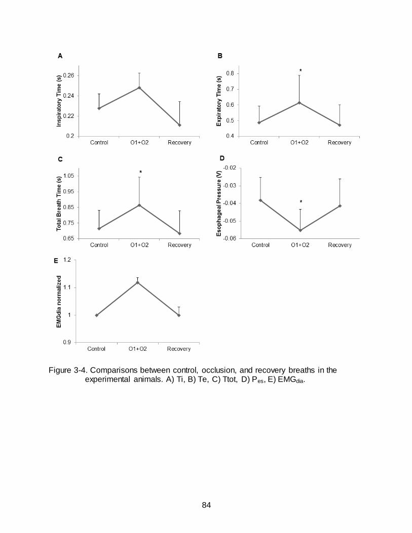
84
Figure 3-4. Comparisons between control, occlusion, and recovery breaths in the experimental animals. A) Ti, B) Te, C) Ttot, D) Pes, E) EMGdia.

85
Table 3-4. Genes of interest that were significantly regulated following ITTO.
Log fold change
p-value Gene Symbol Description
+ 1.30 0.0057 PLAU Plasminogen activator, urokinase
+ 1.07 0.0425 HTR2A Serotonin receptor 2A
+ 0.91 0.0060 TNFRSF14 Tumor necrosis factor receptor superfamily
+ 0.75 0.0098 CHRNB1 Cholinergic receptor, nicotinic, beta polypeptide 1
- 0.74 0.0316 KCNJ3 Potassium inwardly-rectifying channel
- 0.71 0.0045 COX6B2 Cytochrome c oxidase subunit
- 0.52 0.0104 DLG4 Discs, large homolog 4
- 0.45 0.0128 DRD1A Dopamine receptor D1A
- 0.42 0.0271 PRKAA2 Protein kinase alpha 2 catalytic subunit

86
Table 3-5. Highly regulated biological processes following ITTO were found with gene ontology analysis.
GO ID GO Name # of
Genes Selected
# of Genes
on Array
Fisher
p value
FDR
0006916 Anti-apoptosis 25 293 8.6 e-5 0.0497
0006950 Response to stress 93 1780 9.6 e-5 0.0277
0050790 Regulation of enzyme activity 37 575 4.8 e-4 0.0937
0000165 MAPKKK cascade 22 281 6.6 e-4 0.0964
0043066 Negative regulation of apoptosis
28 411 1.0 e-3 0.0998
0043069 Negative regulation of programmed cell death
28 413 1.1 e-3 0.0916

87
Figure 3-5. Interaction between dopamine (DRD1) and serotonin receptors (HTR2A)
under the control of MAPK1. Both DRD1 and HTR2A have actions on the same neurotransmitters and small molecules.

88
CHAPTER 4 TRACHEAL OCCLUSION CONDITIONING IN CONSCIOUS RATS MODULATES
GENE EXPRESSION PROFILE OF MEDIAL THALAMUS
Introduction
Conscious awareness of breathing requires the activation of higher brain centers.
The neural control pathway to the higher centers is thought to be a gated process. Only
information that is selectively attended to or that is above a certain threshold would be
able to pass through the gate to the cortex. The candidate brain structure involved in the
gating of various sensory afferents to the cortex is the thalamus (Kimble and Kaufman,
2004); so it is speculated that respiratory sensory information may also be gated and
relayed through the thalamus, in particular the medial thalamus. The thalamus
integrates many bidirectional connections with virtually every region of the cortex, most
notably the prefrontal cortex and the limbic system (Kimble and Kaufman, 2004;
Newman, 1995). The thalamus is an integral component in the respiratory cortical
neural pathway as tracheal obstruction activates neurons in the cerebral cortex and also
in the medial thalamus (Vovk et al., 2006). We have shown that tracheal obstruction in
anesthetized animals modulates gene expression in the medial thalamus (Chapter 3).
While it is known the repeated exposure to ITTO in conscious states changes load
compensation behavior (Pate et al., 2010), it is unknown if neuron plasticity in the
medial thalamus is induced by conscious chronic exposure to ITTO. We hypothesized
that repeated conscious exposure to ITTO would modulate the gene expression pattern
of the medial thalamus.
The thalamus is the largest structure in the diencephalon, located near the center
of the brain. Thus, it is situated in an ideal position to receive incoming sensory
information and send nerve fibers out to the cerebral cortex in multiple directions.

89
Functionally, the thalamus is believed to serve as the processing and relay station that
sensory information (one known exception is olfaction) must pass before reaching the
cortex (Kimble and Kaufman, 2004; McCormick and Bal, 1994; Sherman and Guillery,
2002). It can act as a gate controlling the flow of information to the cortex. For the well-
studied visual, auditory, and somatosensory systems different thalamic relay neurons
are responsible for relaying the specific information. Visual stimuli pass through the
lateral geniculate nucleus, auditory information through the medial geniculate body, and
somatosensory stimuli are processed by the ventrobasal complex. It was hypothesized
that respiratory afferents also carry lung and airway information to the thalamus,
specifically the medial thalamus, where it is processed and relayed to the cortex.
Midline thalamic nuclei receive projections from areas such as the periaqueductal
gray (Krout and Loewy, 2000b), the parabrachial nucleus (Krout and Loewy, 2000a), the
superior colliculus (Krout et al., 2001), and the brainstem (Krout et al., 2002). Both
sensory information as well as arousal signals converge in the thalamus and could
explain why even basic sensory information can be distorted under conditions of high
arousal, such as in post-traumatic stress disorder (Kimble and Kaufman, 2004). The
thalamus has connections to virtually every brain region, most importantly the recurrent
loop to and from the cerebral cortex, but also to the amygdala (possibly for emotional
processing) and the hippocampus (for learning and memory) (Kimble and Kaufman,
2004). A single thalamic nucleus can send afferents to multiple cortical areas (Herrero
et al., 2002). The cortex then feeds back onto different thalamic nuclei to either enhance
or suppress information. Indeed, anatomical studies have shown that about 50% of
thalamic connections are coming from the cortex. It is believed that corticothalamic
90
feedback functions in “egocentric selection”, which refers to the ability of cortical
neurons to analyze thalamic input, select certain sensory features, and then amplify the
transmission of these features by feedback to the thalamus (Suga et al., 2002). Other
top-down connections such as from the cingulated gyrus and prefrontal cortex feeding
back onto the thalamic neurons help in selecting stimuli that are relevant, salient, and
novel (Kimble and Kaufman, 2004). In other instances, the thalamus may impair rather
than facilitate the processing of environmental stimuli, such as during extreme stress or
trauma (Kimble and Kaufman, 2004).
Sensory information passing through the thalamus is relayed to cortical layer IV
from where it is distributed laterally along the cortex. Layer VI sends reciprocal
corticothalamic connections back to the thalamic relay and interneurons (McCormick
and Bal, 1994). It has been suggested that the cortical neurons analyze the thalamic
input and select important features; this information is fed back to the thalamus to
enhance thalamic transmission/gating of the selected features. The thalamus consists
of three broad classes of neurons, 1) relay neurons, 2) GABAergic interneurons, and 3)
GABAergic nRt neurons (McCormick and Bal, 1994). Relay neurons receive both
excitatory (glutamatergic) and inhibitory (GABAergic) signals and the balance/ratio
between these signals is what determines the response. Other neurotransmitters and
neuromodulators also influence thalamic neurons, most notably serotonin from the
raphe nucleus (McCormick and Bal, 1994).
Previously, we observed that, with ITTO in anesthetized rats, the serotonin
receptor HTR2A was up-regulated (log2 fold change > 1, p < 0.05) (Chapter 3).
Serotonin, as well as its receptors and transporter have been implicated in the stress

91
response, anxiety, and depression (Graeff et al., 1996; Harada et al., 2008; Heisler et
al., 2007; Weisstaub et al., 2006). Chronic exposure to tracheal occlusions in conscious,
rats is stressful and may show an even greater modulation of serotonin and/or its
targets in the thalamus.
It was hypothesized that 10 days of exposure to 10 minute trials of ITTO in
chronically instrumented, conscious rats would induce gene expression changes in the
medial thalamus; specifically, upregulation of genes that have been implicated in the
response to stress, anxiety, and/or depression, such as genes in the serotonergic
system.
Materials and Methods
Animals
Eight male Sprague-Dawley rats (299 g ± 43.05) were housed two per cage in a
temperature-controlled room (72°F) on a 12:12 light:dark cycle, and with free access to
food and water. All animal experiments were approved by the Institutional Animal Care
and Use Committee of the University of Florida.
Surgical Procedures
Placement of tracheal occluder
Rats were anesthetized using inhaled isoflurane gas (2-5% in O2). Buprenorphine
(0.01-0.05 mg/kg BW) and carpofen (5mg/kg BW) were administered preoperatively via
subcutaneous injection. The eyes were coated with petroleum ointment to prevent
drying. Incision sites were shaved and sterilized with povidine-iodine topical antiseptic
solution. The trachea was exposed ventrally in the neck via a skin incision and blunt
dissection of surrounding connective tissues. An expandable cuff was sutured around
the trachea, two cartilage rings caudal to the larynx. The actuating tube was routed

92
subcutaneously and externalized, between the scapulae. The tube was anchored to the
skin using the closing sutures. The neck incision was then closed using an interrupted
suture pattern.
Analgesia and postoperative care
Preoperative analgesia consisted of buprenorphine (0.01-0.05 mg/kg BW) and
carpofen (5mg/kg BW) administered via subcutaneous injection. Following surgical
instrumentation rats were administered warm normal saline (0.01-0.02 ml/g BW) to
ensure proper hydration. Postoperative analgesia was provided for at least three days
using buprenorphine (0.01-0.05 mg/kg BW given every 12-24 hours) and carprofen
(5mg/kg BW given every 24 hours). Rats were closely observed for any signs of distress
or pain.
Experimental Protocol
Rats were placed in a plethysmograph and the externalized occlude actuator was
connected to a saline-filled syringe (Figure 4-1). The syringe was used to inflate and
deflate the cuff bladder. Inflation of the cuff compressed the trachea completely,
occluding the airway during both inspiration and expiration. Deflation restored the
trachea back to its original condition to allow unobstructed breathing. The control group
data collection consisted of 15 min of recording with no experimental manipulation. The
experimental group received a 2.5 min background control recording period, followed by
a 10 min experimental session of ITTO, and ended with a 2.5 min post-test control
period. During the experimental session the rats underwent repeated trials of 3-10
breath occlusions followed by approximately 30 s of recovery (Figure 4-2). Occlusion
and control sessions were performed daily for 10 days. On day 10, within 10 min of
completing the ITTO trial, the rats were sacrificed via overdose of anesthetic. The

93
medial thalamus was quickly excised, frozen in liquid nitrogen, and stored at -80°C until
further use.
Microarray Analysis
Isolation of total RNA, amplification of RNA, microarray hybridization, statistical
analysis, and gene ontology and pathway analysis was performed in the same way as
in the previous study (Chapter 2). Genes were considered differentially expressed if the
p-value was ≤ 0.05 and the log2 fold change was ≥ 0.58.
Results
Modulation of Gene Expression Profile Following ITTO
Statistical analysis of the microarray data showed that a total of 661 genes were
altered (p < 0.05, log2 fold change ≥ 0.58) following the 10 day occlusion protocol, with
250 down-regulated and 411 up-regulated (Appendix Table C-1). Table 4-1 shows
some of the significantly differentially modulated genes. These genes were chosen
based on their potential role thalamic neuron functioning, as well as their implication in
stress, anxiety, and depression. The glutamic acid decarboxylase subtypes 1 and 2
(GAD1 and GAD2), corticotrophin releasing hormone binding protein (CRHBP), and the
serotonin receptor subtypes 1A and 2A (HTR1A and HTR2A) were found to be
significantly up-regulated. Down-regulated genes included short stature homeobox 2
(SHOX2), cholecystokinin (CCK), protein kinase C (PRKCG), metabotropic glutamate
receptor subtype 4 (GRM4), and a potassium inwardly-rectifying channel (KCNJ9).
Gene Ontology and Pathway Analysis
Table 4-2 shows some of the GO categories for biological processes that were
significantly over-represented among the regulated genes (for full list see Appendix

94
Table D-1). Neurotransmitter signaling and learning and memory were some of the most
important differentially modulated processes.
Pathway Studio was used to visualize changes of gene expression following
tracheal occlusions. The balance between the activity of up- and down-regulated genes
involved in these pathways determines the response. Figure 4-3 shows the significantly
regulated genes and pathways involved in learning and/or memory. The up-regulated
KCJN and GRM genes promote learning and/or memory, while the down-regulated HTR
and GAD genes inhibited these pathways. Counteractive interactions between up- and
down-regulated genes exist between the HTR and KCNJ pathways.
Modulated pathways for cellular processes are depicted in Figure 4-4. Cell
proliferation is positively regulated by the down-regulated HTR and GAD genes and
negatively regulated by the up-regulated GRM4, suggesting that this pathway could be
less active following chronic exposure to ITTO. Cell differentiation appears to be
promoted due to the positive regulation by DLH4 and the inhibition by GAD2. Several
genes are involved in neuroprotection but the exact regulation is unknown. Cell death is
inhibited by HTR1A and CRHBP but also by CCK, complicating a prediction on the
direction of regulation on this pathway.
Figure 4-5 demonstrates the modulated pathways for cell signaling. Most up-
regulated genes, except for GRM4, positively regulate calcium (Ca2+) export, membrane
polarization, and synaptic transmission. The up-regulated DLG4 inhibits HTR1A, which
was found to be down-regulated. HTR1A and HTR2A have opposing effects on long-
term synaptic potentiation, membrane polarization, and synaptic transmission.

95
Discussion
Thalamic Firing Mode and Sensory Gating
Transmission in the thalamic relay neurons occurs via one of two neuronal firing
modes, called tonic and burst (Ramcharan et al., 2000). This response depends on the
cells’ membrane potential and the activity of T- (transient) and L- (long-lasting) type
Ca2+ channels (Cheong et al., 2008; Sherman and Guillery, 2002). Tonic firing occurs at
relatively depolarized membrane potentials when the T-channels are inactivated, and
the firing of single action potentials is dependent on suprathreshold activation. Burst
firing occurs when the membrane is hyperpolarized. The T-channels are de-inactivated
and the next suprathreshold depolarization activates the channels to produce an inward
Ca2+ current. This results in a low-threshold, all-or-none voltage spike that is usually
large enough to fire a high frequency cluster of action potentials. L-type Ca2+ channels
are involved in the production of afterhyperpolarization, an important factor in
determining the firing rate of neuronal cells. Specifically, Cheong et al. (Cheong et al.,
2008) have shown that increased Ca2+ influx via these channels augments
afterhyperpolarization, which leads to both decreased tonic firing rates in thalamic relay
neurons and increased gating of pain stimuli.
It has been proposed that switching between firing modes occurs in order to
liberate the cerebral cortex from bothersome interference from the outside world
(McCormick and Bal, 1994), or when the organism is attending to a specific stimulus
(Sherman, 1996, 2001; Sherman and Guillery, 2002). During tonic firing each
depolarization from a sensory stimulus produces one action potential. This linear
relationship attains a faithful and accurate transmission of information through the
thalamus to the cortex. Burst firing, on the other hand, is non-linear because

96
spontaneous activity can occur in the absence of a stimulus. This high signal-to-noise
ratio improves the initial detectability of a stimulus. Sherman et al. (Sherman and
Guillery, 2002) thus hypothesized that thalamic relay cells fire in burst mode when an
important stimulus is detected, after which the cell switches to tonic mode for accurate
relay of that input. A finding supporting this hypothesis is that tonic firing is increased
the more alert the animal is (Ramcharan et al., 2000; Swadlow and Gusev, 2001).
Furthermore, increased bursting and decreased tonic firing of thalamic neurons has
been shown to reduce pain responses, suggesting that burst firing is associated with
gating out of afferent sensory signals (Cheong et al., 2008).
Switching between firing modes requires a shift in membrane potential that is
sufficiently sustained to inactivate or de-inactivate the T-channels. Ionotropic receptors
are too fast acting to promote this shift; the slow metabotropic receptors are most likely
responsible for the sustained voltage change. Specifically, studies have demonstrated
that depolarization (inactivate T-channels) occurs through metabotropic glutamate
receptors from the cortex and hyperpolarization (de-inactivate T-channels) through
GABA receptors from reticular and/or interneuronal inputs (Sherman, 1996, 2001;
Sherman and Guillery, 2002). Several neurotransmitters, such as serotonin, glutamate,
acetylcholine, and norepinephrine, have been shown to facilitate the transition between
thalamic firing modes (McCormick and Bal, 1994). Information on several genes that
were found to be significantly modulated following chronic ITTO and that could be
important in the thalamic firing and thus gating activity is presented below.
KCNJ9 (or GIRK channel or Cir3 channels, potassium inwardly-rectifying
channel, subfamily J, member 9): G-protein inwardly rectifying potassium (GIRK)
97
channels mediate the synaptic actions of numerous neurotransmitters in the mammalian
brain and play an important role in the regulation of neuronal excitability in most brain
regions through activation of various G-protein-coupled receptors (Saenz del Burgo et
al., 2008). Activation of GIRK channels causes membrane hyperpolarization, and thus
the channels play an important role in the inhibitory regulation of neuronal excitability
(Kobayashi et al., 2004). GIRK channels are widely expressed in brain nuclei and are
co-expressed with serotonergic, GABAergic, glutamatergic, and cholinergic neurons
throughout the brain (Saenz del Burgo et al., 2008). Thus, the interplay between the
neurotransmitters and GIRK channels in addition to hyperpolarization could result in
switching of the thalamic firing mode. GIRK2-deficient mice have been shown to exhibit
reduced anxiety and elevated motor activity (Kobayashi et al., 2004); thus an
upregulation in GIRK as found in this study could point to increased anxiety.
GRM4 (or mGluR4, metabotropic glutamate receptor 4): Glutamatergic
neurotransmission is involved in most aspects of normal brain function and can be
perturbed in many neuropathologic conditions. L-glutamate is the major excitatory
neurotransmitter in the central nervous system and activates both ionotropic and
metabotropic glutamate receptors. Grm4 belongs to group III metabotropic glutamate
receptors. Agonists of group III mGluRs have been shown to exert antidepressant-like
effects, possibly due to a decrease in excitatory glutamatergic neurotransmission (Klak
et al., 2007).
A recent study in protein lipase C-knockout mice demonstrated that switching
between tonic and burst firing in thalamic neurons occurs through the simultaneous
modulation of T- and L-type Ca2+ channels possibly through a transduction cascade that

98
includes metabotropic glutamate receptors and protein kinase C (Cheong et al., 2008).
These mice, which show decreased visceral pain responses, exhibited increased
bursting and decreased tonic firing in thalamic neurons, suggesting that burst firing acts
as an inhibitor of pain signal transmission to the cortex.
PRKCG (protein kinase C, gamma): Protein kinase C (PRKC) is a family of
serine- and threonine-specific protein kinases that can be activated by Ca2+ and the
second messenger diacylglycerol. These molecules phosphorylate a wide variety of
protein targets and are involved in diverse cellular signaling pathways. The gamma
subunit of PRKC is expressed solely in neurons in the brain and spinal cord (Saito and
Shirai, 2002). This specific kinase has been implicated in several neuronal functions,
including long term potentiation and long term depression (Saito and Shirai, 2002). It
has also been shown to associate directly with the GluR4 AMPA receptor subunit;
GluR4 phosphorylation would allow for regulation of synaptic function and plasticity
(Correia et al., 2003). As mentioned above, PRKC is involved in switching between
burst and tonic firing. Specifically, down-regulation of PRKC activity in thalamic relay
neurons has been attributed to reduced pain responses, or increased sensory gating
(Cheong et al., 2008). In the present study, we found an up-regulation of PRKC,
suggesting that chronic exposure to ITTO results in decreased gating in the thalamus.
CCK (cholecystokinin): Cholecystokinin is one of the most abundant
neuropeptides in the brain and acts as a neurotransmitter and neuromodulator of
dopamine, serotonin, endogenous opioids, GABA and excitatory amino acids (Harro
and Vasar, 1991). These characteristics support an important role in regulation of many
behavioral phenomena, including anxiety and learning and memory. Indeed, CCK
99
agonists have been shown to be anxiogenic and CCK antagonists are anxiolytic in a
variety of animal species (for reviews see (Harro et al., 1993; Rotzinger and Vaccarino,
2003). CCK has also been demonstrated to have close interaction with GABAergic
inhibitory neurotransmission, mediated probably through CCK-B receptors, which could
be the neurochemical substrate for anxious behavior (Harro and Vasar, 1991).Whole-
cell patch clamp experiments have shown that CCK depolarizes somatosensory cortex
neurons long-lastingly and thus may lead to prolonged discharge of these
corticothalamic glutamatergic neurons and slow depolarization of thalamocortical
neurons, shifting the firing mode from burst- to tonic-firing mode, thus being critical in
sensory information processing (Chung et al., 2009).
Chronic Exposure to ITTO Modulates Genes Involved in Stress, Anxiety, and Depression
Chronic airway occlusion, as occurs in diseases such as chronic obstructive
pulmonary disease, has been implicated in an increased incidence of anxiety and
depression (Di Marco et al., 2006; Omachi et al., 2009). In the present study, several
genes were found to be significantly altered following tracheal occlusions that play
important roles in the development of depressive and other psychological disorders.
GAD1 (or GAD67, glutamate decarboxylase) and GAD2 (or GAD65): GAD1
and GAD2 are two isoforms of glutamate decarboxylase. These enzymes catalyze the
reaction to synthesize GABA from glutamate and are responsible for keeping cortical
GABA levels at steady state (Soghomonian and Martin, 1998). Thus, a reduced
expression of GAD1 and GAD2 may lead to decreased GABA levels and less inhibition
of downstream targets. Decreased GAD67 expression has been implicated in
schizophrenia and bipolar disorder with psychosis (Guidotti et al., 2000).

100
CRHBP (corticotrophin releasing hormone – binding protein): CRHBP is an
important modulatory protein that negatively regulates corticotrophin releasing hormone
(CRH) activity. CRHBP binds to CRH and thus reduces the ability of CRH to activate the
CRHR1 and CRHR2 receptors (Jahn et al., 2002). CRHBP is a physiologically relevant
reservoir of endogenous CRH, as 40-60% of human brain CRH is bound by CRHBP.
CRH is released in response to stress. A reduction in CRHBP would lead to less binding
of CRH and more free CRH which can then activate its receptors and elevate the stress
response. In a CRHBP-deficient mouse model, Karolyi et al. (Karolyi et al., 1999) have
demonstrated increased anxiogenic behavior as tested on the elevated plus maze and
open field. A decrease in CRHBP has also been suggested to play a role in the
pathogenesis of major depressive disorder by inhibiting the function of CRH (Van Den
Eede et al., 2005).
HTR (serotonin receptors): The serotonin system has been shown to play a
critical role in a variety of human psychopathological conditions, particularly mood and
anxiety disorders (Hensler, 2006). Antidepressant treatment has thus focused on
modulating serotonergic neurotransmission (Jones and Blackburn, 2002). One of the
challenges of the serotonin system is the sheer complexity of it, with 14 known receptor
varieties categorized into seven receptor subtypes (Hoyer et al., 2002).
The HTR1A subtype exists on pre-synaptic neurons in raphe nuclei as well as on
post-synaptic neurons in other brain regions, such as the hippocampus and the
thalamus. Agonists have different effects depending on the location of the receptors, in
that agonists to pre-synaptic receptors result in anxiolytic behaviors while agonists to
post-synaptic receptors lead to anti-depressive behaviors (Schreiber and De Vry,

101
1993)). Kennett et al. (Kennett et al., 1987) have demonstrated that the 5-HTR1A
agonist 8-hydroxy-2-(di-n-propylamino) tetralin (8-OH-DPAT) may have rapid
antidepressant properties. Partial HTR1A may also be effective in the treatment of
generalized anxiety disorder (Jones and Blackburn, 2002) and schizophrenia (Millan,
2000).
HTR1A knockout mice show elevated anxiety levels in open-field, elevated-zero
maze, and novel-object assays (Heisler et al., 1998) and are less reactive, more
anxious, and possibly less aggressive than the wild-types (Ramboz et al., 1998).
Dysfunction of this receptor has been suggested to also play a role in the genesis of
major depressive disorder in humans (Savitz et al., 2009). PET studies in patients with
posttraumatic stress and panic disorders (Neumeister et al., 2004) and depression
(Drevets et al., 1999) have shown reduced HTR1A receptor binding potential and
reduced receptor availability (Nash et al., 2008).
In the present study we found a down-regulation of serotonin receptors after 10
days of ITTO. In the previous study (Chapter 3) we have shown that the serotonin
receptor HTR2A was rapidly up-regulated with acute occlusions. This is in general
agreement with the suggestion that anxiety is the result of a hypersensitive serotonin
system; whereas impulsivity and depression is the result of a hyposensitive serotonin
system (Schreiber and De Vry, 1993).
Chronic Exposure to ITTO Modulates SHOX2
SHOX2 (or Prx3 or OG12X or SHOT): SHOX2 is a homeobox gene expressed
mainly in the thalamus in adult rats, more specifically in those thalamic relay nuclei that
coordinate and integrate sensory information to be sent to the sensory cortex (van
Schaick et al., 1997). Despite its critical location for gating of sensory information, no

102
reports have been published on modulated SHOX2 gene expression. Homeobox genes
are the largest class of transcription factors instrumental in cell-specific gene
expression. Their expression patterns are mostly restricted during development of
embryonic brain, and some persist during adult life. Aberrations in homeobox genes can
cause several genetic disorders, in the case of SHOX2 it is thought to be responsible for
idiopathic short stature in Turner syndrome patients (Rao et al., 1997).
Chronic Exposure to ITTO Modulates Pathways Involved in Learning and Memory, Cell Processes, and Cell Signaling
Ten days of ITTO resulted in a behavioral adaption in order to cope with the stress
of the occlusion trials. These adaptations were characterized by decreased exploratory
behavior, increased submissive state, and even breath holding (Pate et al., 2010). This
finding of learned helplessness and memory of previous occlusion trials is consistent
with the altered molecular pathways of learning and/or memory. In the present study,
most of the genes that were found to be up-regulated have been shown to increase
learning and/or memory.
Modulated cellular processes included cell proliferation, differentiation,
neuroprotection, and cell death. A common trend in the analysis of these pathways was
the inhibitory regulation of cell death by up-regulated genes, while neuroprotection was
positively regulated. Cell proliferation seemed to be down-regulated because the genes
that are activating this process showed decreased expression. However, cell
differentiation appeared to be increased, both by positive regulation of up-regulated
genes as well as by inhibitory regulation of down-regulated ones.
Differentially regulated genes involved in cell signaling were Ca2+ transport,
membrane polarization, synaptic potentiation and transmission. Involvement of the
103
various genes is less clear and often in opposite direction. The balance of up- and
down-regulation of genes in these pathways seems to be important in determining the
final outcome.
Conclusions
Contrary to our hypothesis that repeated ITTO would induce increases in 5-
HTR2A gene expression, we found a decrease in the expression of this receptor
subtype along with a decrease in 5-HTR1A. This finding could be explained by the
opposite action of the serotonergic system depending on the duration of the stress
stimulus (Graeff et al., 1997; Schreiber and De Vry, 1993). An acute stress, such as one
trial of ITTO, would produce anxiety due to up-regulation of components in the
serotonergic system, while chronic exposure to a stress such as repeated trials of ITTO
would produce depression-like behavior due to down-regulation of this system.
Repeated ITTO elicited changes in the gene expression profile of the medial
thalamus involved in neuronal firing mode, suggesting a modulation of respiratory
afferent information gating. A change in gating of information to higher brain centers
could result in a different behavioral response to the ITTO respiratory stimulus. The
respiratory load compensation response has been shown to be altered in conscious rats
and following 10 days of repeated ITTO (Pate et al., 2010). This alteration may be due
to a change in the gating pattern that influenced the behavioral control of breathing.

104
Figure 4-1. Schematic of the experimental protocol for repeated ITTO. Rats were placed in a plethysmograph and the actuator tube of the tracheal cuff was connected to a saline-filled syringe.

105
Figure 4-2. Representative plethysmograph pressure traces for one occlusion trial on
day 10. Cuff pressure indicates the period of occlusion. The large deflection of the signal at the beginning of occlusion is ascribed to a movement artifact
when the rat twitches due to application of occlusion.

106
Table 4-1. Candidate genes significantly differentially regulated following chronic ITTO.
Log fold change
p-value Gene Symbol Description
- 2.14 0.0043 GAD1 Glutamic acid decarboxylase 1
- 1.61 0.0256 GAD2 Glutamic acid decarboxylase 2
- 1.38 0.0039 CRHBP Corticotropin releasing hormone binding protein
- 0.78 0.0396 HTR1A Serotonin receptor 1A
- 0.59 0.0023 HTR2A Serotonin receptor 2A
+ 1.37 0.0009 SHOX2 Short stature homeobox 2
+ 1.36 0.0402 CCK Cholecystokinin
+ 1.22 0.0040 PRKCG Protein kinase C, gamma
+ 1.09 0.0085 GRM4 Glutamate receptor, metabotropic 4
+ 1.07 0.0060 KCNJ9 Potassium inwardly-rectifying channel
Table 4-2. Significantly modulated Gene Ontology Biological Processes.
Name # of Entities Overlap p-value
Synaptic transmission 247 17 2.19 e-12
Learning and/or memory 42 5 1.11 e-5
Neurotransmitter Transport 62 5 7.55 e-5
Neurotransmitter Secretion 48 4 3.60 e-4
Regulation of neuronal synaptic plasticity 26 3 7.85 e-4
Regulation of neurotransmitter secretion 27 3 8.79 e-4
Learning 31 3 1.32 e-3

107
Figure 4-3. Pathway analysis of transcripts (p < 0.05) involved in the biological processes of learning and/or memory.

108
Figure 4-4. Pathway analysis of transcripts (p < 0.05) involved in cell processes.
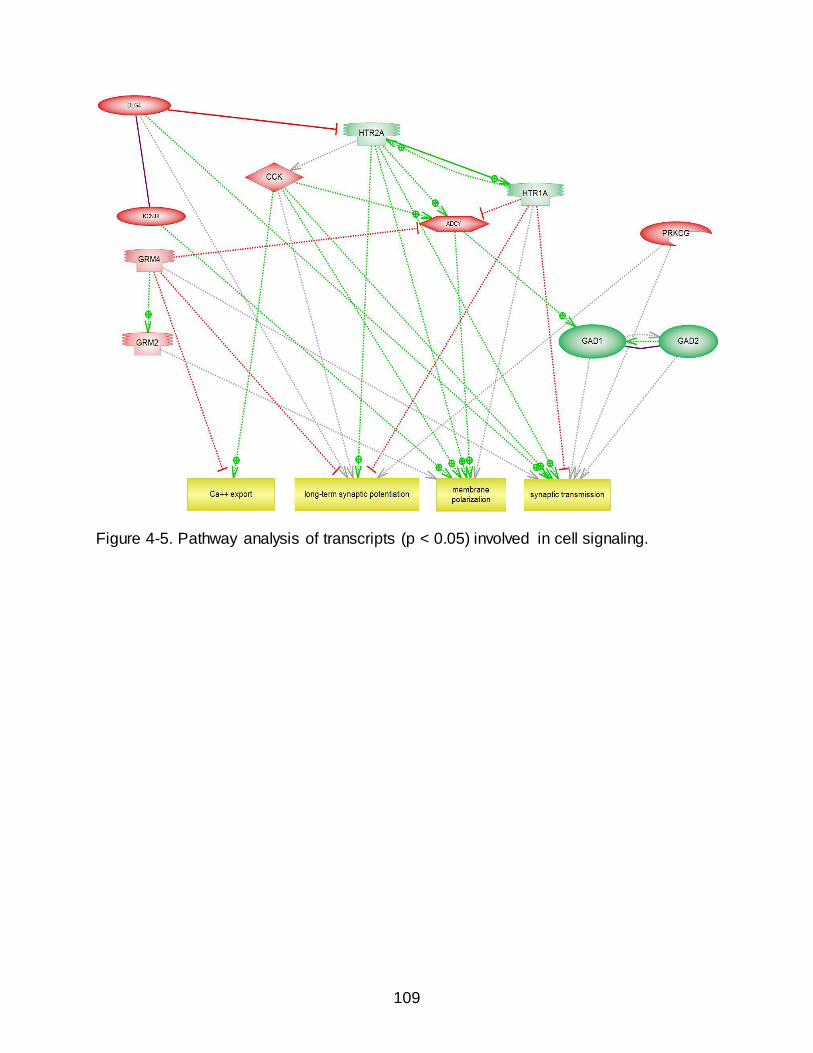
109
Figure 4-5. Pathway analysis of transcripts (p < 0.05) involved in cell signaling.

110
CHAPTER 5 SUMMARIES AND CONCLUSIONS
Summary of Study Findings
Study #1 Summary
The purpose of this study was to determine the effects of EMST on pulmonary
function and maximal expiratory pressure generating capacity, and to determine
whether this specific respiratory muscle training affects oxygen consumption during an
incremental exercise test, swimming performance, and perception of exertion and
breathlessness in highly trained collegiate swimmers. The EMST group was compared
to an AFT group that functioned as a placebo control.
The EMST intervention elicited significant increases in MEP but not the other
measured pulmonary function variables (FEV1.0, FVC, FEV1.0/FVC). Post-training, MEP
increased by 29.86% in the EMST group and 7.86% in the AFT group. These results
demonstrate that the EMST pressure-threshold training device provided a sufficiently
strong stimulus to increase expiratory pressure generating capacity in these highly
trained swimmers, while the AFT non-resistance air-flow training device did not
significantly increase expiratory muscle strength as measure by MEP.
Swimming performance improved more in the EMST than the AFT group. Average
swim time during the all-out 6 x 100 meter freestyle interval test decreased by 1.44% in
the EMST group and by 0.29% in the AFT group, however, the change was not
statistically significantly different between the groups (p = 0.204). Post-training, ratings
of breathlessness tended to worsen in the AFT group and ratings of perceived exertion
tended to improve in the EMST group. The results demonstrate that EMST has a trend
to improve swimming times in a maximal effort interval swim test that may give these

111
highly competitive athletes an improved performance during a swim competition, and
EMST has a trend to improve subjective ratings of perceived exertion and
breathlessness during maximum effort swimming. EMST is more effective than AFT in
conditioning expiratory muscles of highly trained athletes and is an easy method that
could be used as an adjunct for swimmer’s athletic conditioning. These promising
results in improving performance merit further investigation into training of the
respiratory system in athletes.
Study #2 Summary
This study evaluated the responses of a 10-minute bout of ITTO on the gene
expression profile of the medial thalamus in urethane anaesthetized rats. This study
was important to establish a role of the medial thalamus as a component in the
respiratory mechanosensory neural pathway. Analyses of breath timing responses to
ITTO corroborated the existence of the ventilatory load compensation reflex in the
anaesthetized animal. Gene expression profiles were measured using Agilent
Technology Oligo Microarrays. Tracheal occlusions modulated a total of 588 genes
(p < 0.05, log2 fold change ≥ 0.58), of which 261 were up-regulated and 327 were down-
regulated. A significant up-regulation of the serotonin HTR2A receptor and significant
down-regulation of the dopamine DRD1 receptor genes were found. Pathway analysis
was performed targeting serotonin and dopamine receptor pathways. The MAPK1 gene
was significantly down-regulated. MAPK1 is an inhibitory regulator of the serotonin
HTR2A receptor and facilitatory regulator for the dopamine DRD1 receptor. Down-
regulation of MAPK1 may be related to the up-regulation of HTR2A and down-regulation
of DRD1 suggesting an interaction in the medial thalamus serotonin-dopamine pathway
elicited by airway obstruction. Gene ontology analysis showed gene expression

112
changes related to anti-apoptosis, response to stress, and the MAPKKK cascade. The
results of this study demonstrate an immediate change in gene expression in thalamic
stress/anxiety/depression-pathways, providing evidence for the involvement of the
medial thalamus to respond to airway occlusions.
Study #3 Summary
Based on the results of study #2, the experiments in study #3 were undertaken to
evaluate the gene expression profile of the medial thalamus following chronic
conditioning to ITTO (daily 10-minute occlusion trials for 10 days) in conscious rats.
Chronic tracheal obstruction conditioning modulated 611 genes (p < 0.05), of which 411
were up-regulated and 250 were down-regulated. There was a significant down-
regulation of the genes encoding GAD1, GAD2, CRHBP, HTR1A, and HTR2A. Up-
regulated genes included SHOX2, CCK, PRKCG, GRM4, and KCNJ9. Significantly
regulated gene ontology categories were learning and/or memory, neurotransmitter
synthesis, transport, and secretion, and synaptic transmission and plasticity. Pathway
analysis showed the involvement of the significantly modulated genes in learning and/or
memory, cell processes, and cell signaling.
The results of this microarray study demonstrate that repeated ITTO in conscious
rats elicited changes in the gene expression profile of the medial thalamus involved in
neuronal firing mode, suggesting a modulation of respiratory afferent information gating.
A change in gating would influence the behavioral control breathing and thus alter the
respiratory load compensation response. It further implicated a role of chronic exposure
to airway occlusions in development of stress, anxiety, and depression.

113
Discussion
The Role of Serotonin in Response to Respiratory Stimuli
Any stressor threatening the homeostasis of an organism can initiate neural and
behavioral responses to adapt to the specific situation. The activity of neurotransmitters
is critical for these changes to occur. Based on the studies in chapters 3 and 4, one of
the neurotransmitter systems involved in respiratory stimuli is serotonin. Pharmacologic
agents targeting the serotoninergic system are used to treat a variety of disorders; some
functions of serotonin include the regulation of mood, appetite, sleep, and memory and
learning. Serotonergic fibers from the raphe nucleus travel to midline, intralaminar and
association nuclei of the thalamus and from there mostly to limbic forebrain areas and
some to sensory and motor cortices, suggesting that serotonergic fibers to the thalamus
may exert a significant influence on affective and cognitive functions involved in
emotional and cognitive behaviors (Vertes et al., 2010). Modulation of components of
the serotonergic system in the thalamus could thus be responsible to changes in
affective perception and behavior to a stimulus. In study 2 (Chapter 3), the serotonin
receptor HTR2A was found to be up-regulated following just one trial of ITTO in
anesthetized rats. In study 3 (chapter 4), there was a down-regulation of the serotonin
receptors subtype HTR2A and HTR1A after ten days of repeated ITTO in conscious
animals. These findings are in agreement with other studies that show that serotonin
can modulate anxiety and depression in opposite directions, with high serotonergic
activity being associated with anxiety and low activity with depression (Graeff et al.,
1997; Schreiber and De Vry, 1993).

114
The Effects of Respiratory Training
The respiratory system could be one of the factors limiting exercise performance in
highly trained athletes (Dempsey, 2006). Study 1 corroborates this hypothesis by
demonstrating that a specific respiratory muscle strength training slightly, albeit
statistically not significant, improved swimming performance. Pulmonary function (FVC,
FEV1.0) did not change, which is consistent with the notion that the pulmonary system
does not structurally adapt to physical training (Dempsey et al., 1990). However, EMST
did significantly increase the maximum expiratory pressure capacity, indicating that the
expiratory muscles responded to the training load by an increase in strength. This
increase could improve the response to the expiratory flow limitation imposed by the
water pressure, and in turn reduce and/or delay the perception of breathlessness.
The results from the animal studies in chapters 3 and 4 using ITTO shed some
light on the potential underlying mechanisms for the positive effects of EMST. We found
changes on the gene level in the medial thalamus, indicating that the supra-pontine
brain areas play a role in influencing the control of breathing. As described in chapter 1,
the thalamus is the structure implicated in the gating of sensory information to higher
brain centers such as cortical areas and the limbic system (Figure 1-1). Modulation of
thalamic function and thus gating could suggest altered feedback to these brain areas
and thus a different response pattern to the stimuli. More specifically, the trend of
decreased perception of breathlessness and exertion during the interval swim test in the
EMST group could have been due to increased gating of the uncomfortable respiratory
stimulus.
Breathlessness, either in combination with peripheral muscle fatigue or by itself, is
the most common reason for reduction or termination of exercise (Killian and Campbell,

115
1995). During a maximal effort exercise, the subject is aware of both the increasing
central motor command and the decline in power output (Killian and Gandevia, 1996);
the awareness of these two factors gives rise to the sensation of fatigue (Jones and
Killian, 2000). The sensation of breathlessness likely depends on several different
mechanisms that are involved in the regulation of breathing including feed-forward and
feedback mechanisms (Figure 2-11) (Chonan et al., 1990b).
Figure 5-1 shows a schematic model of the effects of respiratory muscle training
on perception based on my studies. With increasing exercise intensity, there usually is
increased feedback from receptors in the respiratory muscles, chest wall, lungs, lower
and upper airways, and chemoreceptors to the thalamus. Respiratory muscle training
(repeated ITTO) could modulate this feedback due to improved strength of these
muscles and thus could result in a change of how the thalamus handles the influx of
respiratory information, so that the perception of breathlessness is delayed or reduced.
The ITTO animal studies are suggestive of a change in function of the thalamic gating
process. The combined factors of reduced feedback and change in gating within the
thalamus could lead to decreased signaling to the limbic system and the sensory cortex,
and in turn decrease perception.
Methodological Considerations and Directions for Future Studies
The present set of experiments highlighted a potential ergogenic effect of EMST in
a group of highly trained swimmers and contributed to a better understanding of the role
of the medial thalamus in response to loaded respiratory stimuli. Limitations to these
studies and proposals for future experiments that could expand this body of work are
discussed below.

116
Fatigue due to Regular Training
During the EMST study the swimmers were on a three-week training cycle with
each week having a different focus such as endurance, sprint, strength, and recovery,
which was repeated every three weeks. Accordingly, the swimmers’ general fatigue
levels varied to some extent within the training cycle. One week of training (Monday
through Saturday) consisted of nine two-hour swim sessions at 5,000-8,000 m, plus
additional dry land and weight training workouts. Therefore, most of our laboratory
testing was performed early in the week to take advantage of the recovery Sunday. The
6 x 100 m swim tests were administered at the beginning of the Tuesday afternoon
swim practice after a specific standard warm-up. In order to minimize variability, an
interval swim test was chosen instead of a time-trial. Other studies involving highly
trained swimmers have used either time-trials (Kilding et al., 2010; Lindh et al., 2008;
Vandenbogaerde and Hopkins, 2010) or interval tests (Psycharakis, 2010; Seifert et al.,
2010; Wells et al., 2005).
Respiratory Training Stress Stimulus
As seen from study 3 (chapter 4), repeated ITTO induced gene expression
changes in the conscious rat that are associated with anxiety and depression. This is
consistent with evidence showing an increased rate of anxiety and depression in
patients with repeated acute or chronic airway occlusion, such as asthma and COPD
(Moussas et al., 2008). It also implicates ITTO as a negative stressor. However, in our
experiments the animals were healthy and had no intrinsic respiratory muscle weakness
as is characteristic of asthma, COPD, or various neuromuscular diseases (Barbarito et
al., 2001). Respiratory muscle training in humans is probably a positive stressor due to
the motivation and expectation of the individual; therefore, this training should not

117
induce anxiety/depression but rather a positive outlook toward improvement of
performance. Thus, we cannot be absolutely certain that the respiratory stimulus of
ITTO per se elicited the changes in gene expression or if feedback from higher brain
centers, especially from the limbic system, were involved. However, study 3 also
showed an up-regulation of the learning and/or memory pathways and study 2 (chapter
3) demonstrated an up-regulation of the stress response and negative regulation in
apoptosis in anesthetized animals immediately following the single trial of ITTO,
suggesting that this type of stressor was not necessarily a negative one. To my
knowledge, our animal model is the first to examine gene expression changes in a
subcortical area in response to an intrinsic respiratory stimulus that has been shown to
induce respiratory muscle hypertrophy (Smith et al., 2010).
O2max Testing
The incremental O2 test on the swim ergometer was not a true maximal test, but
submaximal because most swimmers were able to complete the testing protocol before
the characteristic plateau phase of the O2 curve was reached. To my knowledge, this
particular swim ergometer has never been used in previous studies, even though it has
some important advantages to other ergometers as discussed in chapter 2. For a future
study, adjustments to stroke frequency and/or time spent at each resistance level
should be made, depending on the fitness level of the participant group. In this case
with highly trained swimmers, a slightly higher constant stroke frequency may be more
appropriate to assess a true O2 max value.

118
Prevalence of Respiratory Disease in Swimmers
Asthma is a common occurrence in elite athletes, especially in endurance sport
such as swimming. The most recent study of 200 top Finnish swimmers revealed that
the prevalence of asthma was higher in these swimmers than in the general population
(Paivinen et al., 2010). The study found that physician-diagnosed asthma was reported
by 32 swimmers (16%), including 24 (12%) with exercise-induced asthma. Asthmatic
symptoms during swimming were described by 84 subjects (42%) with most symptoms
occurring when swimming exceeded speeds corresponding to the lactic/anaerobic
threshold. In our study with UF swimmers, exclusion criteria included the occurrence of
bronchoconstriction and taking medication for respiratory disease. Unfortunately, these
criteria complicated the recruiting process since about one third of the swimmers asked
had physician-diagnosed asthma and were taking medication. Future studies should
include asthmatic athletes to see the possibly different effect of EMST in this population.
Specificity of Medial Thalamic Nuclei
The medial thalamus consists of multiple nuclei, midline and intralaminar nuclei,
that may have different specific functions. Furthermore, the medial thalamus contains a
variety of anatomically and functionally heterogeneous neurons (Benarroch, 2008), such
as glutamatergic projection neurons, inhibitory interneurons, and relay neurons. The
thalamic microarray sample in the experiments presented here was not targeted to a
specific nucleus or group of neurons, thus gene expression may be multifactorial.
Future studies may be carried out to determine the different type of neurons comprising
the midline and intralaminar nuclei and the connectivity between other brain areas and
these specific neurons.

119
Genomics Versus Proteomics
The use of microarray technology has become a popular method to study changes
in gene expression in pathologies and following specific stimulus interventions.
However, a change in mRNA levels of a particular gene does not necessarily translate
into the same change in protein levels of that gene. Most microarray studies that use
protein validation (in the form of Western blots, for example) show modulation of mRNA
and protein in the same direction and with similar fold changes. Some studies, however,
have demonstrated distinctly different fold changes or even opposite regulation of
mRNA and the associated protein. Thus, in future studies, proteomics could be used to
assess protein changes in certain brain regions involved in the respiratory control
network.
Conclusions
The three studies presented in this dissertation examined the effects of respiratory
muscle training in humans and in a rat model. EMST in swimmers showed a significant
increase in expiratory pressure generating capacity and trends for improvements in
swimming performance and feelings of breathlessness and perceived exertion. Tracheal
occlusions in anesthetized and conscious rats showed changes in the gene expression
profile of the medial thalamus, indicating that this brain area responds to respiratory
loading and may be involved in gating and relaying respiratory information to higher
brain centers.

120
Figure 5-1. Model for possible effects of respiratory muscle training on perception.

121
APPENDIX A RATING SCALES

122

123
APPENDIX B LIST OF MODULATED GENES FOLLOWING ACUTE ITTO
Table B-1. List of modulated genes following acute ITTO, p < 0.05 and log2 fold change ≥ 0.4
log2 FC p-value TargetID Symbol Name
-2.99 0.0466 AA899244
-1.48 0.0196 ENSRNOT00000047881
-1.43 0.0418 AA900238
-1.33 0.0004 AW143927
-1.33 0.0388 XM_237191 Fzd7_predicted frizzled homolog 7 (Drosophila) (predicted)
-1.31 0.0474 CB547739
-1.29 0.0377 NM_001025147 Gpr160 G protein-coupled receptor 160
-1.28 0.0011 BQ196489
-1.28 0.0220 AI102771
-1.27 0.0214 BC099085
-1.25 0.0436 XM_001077586 LOC691293 similar to reproductive homeobox on chromosome X, 7
-1.25 0.0414 NM_013167 Ucp3 uncoupling protein 3 (mitochondrial, proton carrier)
-1.23 0.0144 TC541367
-1.20 0.0249 D84486
-1.20 0.0401 NM_147165 Gpx6 glutathione peroxidase 6
-1.15 0.0200 BI291339
-1.13 0.0019 NM_153721 Cxcl7 chemokine (C-X-C motif) ligand 7
-1.12 0.0300 XM_577774 LOC502310 similar to Cytochrome P450 2B12
(CYPIIB12) -1.12 0.0304 AI555248
-1.10 0.0063 XM_222561
-1.09 0.0265 NM_147213 LOC259245 alpha-2u globulin PGCL5
-1.08 0.0026 AA997228
-1.08 0.0345 AA956566
-1.08 0.0011 BF391695
-1.08 0.0098 AA818571
-1.06 0.0429 NM_133533 Cd79b CD79B antigen
-1.06 0.0209 A_44_P473318
-1.06 0.0411 XM_001076906 LOC687057 similar to Calponin-2 (Calponin H2, smooth muscle) (Neutral calponin)
-1.06 0.0069 NM_001025628 Ropn1 ropporin, rhophilin associated protein 1
-1.05 0.0109 BI296967
-1.05 0.0117 TC541883
-1.04 0.0444 XM_217136 Cd3g CD3 antigen, gamma polypeptide
-1.04 0.0432 A_44_P319138
-1.03 0.0215 BF559727
-1.02 0.0407 BE097028
-0.99 0.0059 XM_340956 LOC360684 similar to eyes absent 4 isoform a
-0.99 0.0102 NM_012969 Irs1 insulin receptor substrate 1
-0.98 0.0310 BI296249
-0.98 0.0472 NM_031537 LOC24906 Robo-1
-0.98 0.0395 BF396663
-0.98 0.0121 AA944970

124
-0.97 0.0278 BI296064
-0.96 0.0134 XM_001069898 LOC684331 hypothetical protein LOC684331
-0.96 0.0430 AA800882
-0.95 0.0184 BF281960
-0.95 0.0300 TC530472
-0.93 0.0389 BI282122
-0.93 0.0124 A_44_P527770
-0.91 0.0390 XM_344756 Pou4f2 POU domain, class 4, transcription factor 2
-0.91 0.0270 XM_001075247 LOC686680 similar to membrane-spanning 4-domains, subfamily A, member 5
-0.91 0.0392 XM_344473
-0.91 0.0270 BE115850
-0.91 0.0456 XM_345276
-0.91 0.0064 XM_001053946 LOC679957 similar to G protein-coupled receptor 18
-0.91 0.0489 NM_175755 Ppm1f protein phosphatase 1F (PP2C domain containing)
-0.90 0.0018 NM_012629 Prl prolactin
-0.90 0.0411 A_44_P340418
-0.89 0.0495 XM_001072511 LOC684923 similar to chromosome 9 open reading
frame 79 -0.88 0.0350 AA926044
-0.88 0.0382 BE113640
-0.88 0.0087 AA850212
-0.88 0.0430 XM_225068 Irx4_predicted Iroquois related homeobox 4 (Drosophila) (predicted)
-0.88 0.0221 AJ224441
-0.87 0.0101 A_44_P889765
-0.87 0.0247 CB547880
-0.86 0.0128 BI293639
-0.86 0.0077 NM_138898 Phlpb phospholipase B
-0.85 0.0177 AA925378
-0.85 0.0477 BF409820
-0.85 0.0182 AW530584
-0.83 0.0433 NM_147139
-0.83 0.0361 TC548367
-0.83 0.0018 TC548927
-0.83 0.0165 NM_012741 LOC25087 K-kininogen
-0.83 0.0421 AI406624
-0.83 0.0197 NM_001014058 Usp18 ubiquitin specific peptidase 18
-0.82 0.0125 XM_232952
-0.82 0.0241 AI072433
-0.82 0.0279 NM_198727 LOC288750 hypothetical protein
-0.82 0.0058 TC537158
-0.81 0.0438 AA819842
-0.81 0.0350 XM_345868 Hdac7a histone deacetylase 7A
-0.81 0.0050 AY325249
-0.80 0.0016 CB547640
-0.80 0.0272 XM_232342 Cd163_predicted
CD163 antigen (predicted)
-0.80 0.0088 TC543535
-0.79 0.0109 XM_001079735 LOC687705 similar to misshapen-like kinase 1 isoform 1

125
-0.78 0.0312 AA924420
-0.78 0.0058 AI175421
-0.77 0.0468 XM_343922 Polr2a_mapped polymerase (RNA) II (DNA directed) polypeptide A (mapped)
-0.77 0.0247 XM_222215 RGD1565800_predicted
similar to hypothetical protein FLJ20674 (predicted)
-0.77 0.0163 BE108142
-0.76 0.0109 XM_341912 Xylt1 xylosyltransferase 1
-0.76 0.0348 XM_573321 LOC498113 similar to This CDS feature is included to
show the translation of the corresponding V_region. Presently translation qualifiers on V_region features are illegal
-0.76 0.0184 NM_001024782 Lrrc8 leucine-rich repeat-containing 8
-0.76 0.0452 TC544952
-0.75 0.0399 NM_053398 Gfra3 glial cell line derived neurotrophic factor family receptor alpha 3
-0.75 0.0113 CB547650
-0.75 0.0173 XM_214313 Fcho1_predicted FCH domain only 1 (predicted)
-0.75 0.0207 NM_001003401 Enc1 ectodermal-neural cortex 1
-0.74 0.0283 BF563208
-0.74 0.0448 BF285026
-0.74 0.0316 NM_031610 Kcnj3 potassium inwardly-rectifying channel,
subfamily J, member 3 -0.73 0.0472 BQ207264
-0.73 0.0242 NM_021669 Ghrl ghrelin precursor
-0.72 0.0362 A_44_P598999
-0.72 0.0432 CF109839
-0.72 0.0351 A_44_P255625
-0.72 0.0031 A_44_P984772
-0.71 0.0192 BC089106
-0.71 0.0045 NM_001039085 Cox6b2 cytochrome c oxidase subunit VIb testes-specific isoform precursor
-0.71 0.0495 NM_031697 St3gal3 ST3 beta-galactoside alpha-2,3-sialyltransferase 3
-0.71 0.0316 AA849518
-0.71 0.0327 BF408914
-0.70 0.0242 NM_001001368 Olr943_predicte
d
olfactory receptor 943 (predicted)
-0.70 0.0035 AI012782
-0.70 0.0218 XM_345112
-0.70 0.0231 NM_138536 Ttl tubulin tyrosine ligase
-0.70 0.0170 NM_001003929 Cntfr ciliary neurotrophic factor receptor
-0.70 0.0269 NM_001030034 Rhbdf1 rhomboid family 1 (Drosophila)
-0.69 0.0361 XM_220699 Rutbc1_predicted
RUN and TBC1 domain containing 1 (predicted)
-0.69 0.0178 BF417211
-0.68 0.0389 XM_220330 RGD1309452_predicted
similar to RIKEN cDNA 9530066K23 (predicted)
-0.67 0.0389 XM_230039 Dusp19_predict
ed
dual specificity phosphatase 19 (predicted)
-0.67 0.0407 BE106909
-0.67 0.0126 NM_198727 LOC288750 hypothetical protein
-0.67 0.0368 ENSRNOT00000020530
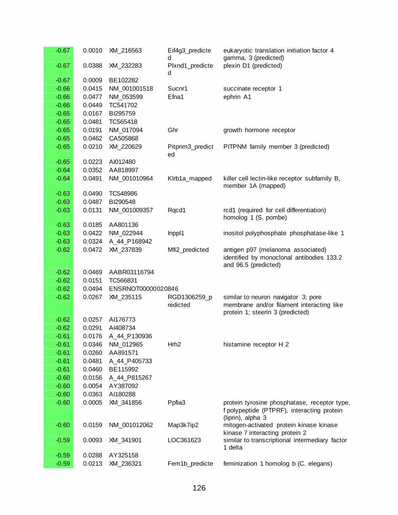
126
-0.67 0.0010 XM_216563 Eif4g3_predicted
eukaryotic translation initiation factor 4 gamma, 3 (predicted)
-0.67 0.0388 XM_232283 Plxnd1_predicted
plexin D1 (predicted)
-0.67 0.0009 BE102282
-0.66 0.0415 NM_001001518 Sucnr1 succinate receptor 1
-0.66 0.0477 NM_053599 Efna1 ephrin A1
-0.66 0.0449 TC541702
-0.65 0.0167 BI295759
-0.65 0.0481 TC565418
-0.65 0.0191 NM_017094 Ghr growth hormone receptor
-0.65 0.0462 CA505868
-0.65 0.0210 XM_220629 Pitpnm3_predict
ed
PITPNM family member 3 (predicted)
-0.65 0.0223 AI012480
-0.64 0.0352 AA818997
-0.64 0.0491 NM_001010964 Klrb1a_mapped killer cell lectin-like receptor subfamily B, member 1A (mapped)
-0.63 0.0490 TC548986
-0.63 0.0487 BI290548
-0.63 0.0131 NM_001009357 Rqcd1 rcd1 (required for cell differentiation) homolog 1 (S. pombe)
-0.63 0.0185 AA801136
-0.63 0.0422 NM_022944 Inppl1 inositol polyphosphate phosphatase-like 1
-0.63 0.0324 A_44_P168942
-0.62 0.0472 XM_237839 Mfi2_predicted antigen p97 (melanoma associated)
identified by monoclonal antibodies 133.2 and 96.5 (predicted)
-0.62 0.0469 AABR03116794
-0.62 0.0151 TC566831
-0.62 0.0494 ENSRNOT00000020846
-0.62 0.0267 XM_235115 RGD1306259_p
redicted
similar to neuron navigator 3; pore
membrane and/or filament interacting like protein 1; steerin 3 (predicted)
-0.62 0.0257 AI176773
-0.62 0.0291 AI408734
-0.61 0.0176 A_44_P130936
-0.61 0.0346 NM_012965 Hrh2 histamine receptor H 2
-0.61 0.0260 AA891571
-0.61 0.0481 A_44_P405733
-0.61 0.0460 BE115992
-0.60 0.0156 A_44_P815267
-0.60 0.0054 AY387092
-0.60 0.0363 AI180288
-0.60 0.0005 XM_341856 Ppfia3 protein tyrosine phosphatase, receptor type,
f polypeptide (PTPRF), interacting protein (liprin), alpha 3
-0.60 0.0159 NM_001012062 Map3k7ip2 mitogen-activated protein kinase kinase
kinase 7 interacting protein 2 -0.59 0.0093 XM_341901 LOC361623 similar to transcriptional intermediary factor
1 delta
-0.59 0.0288 AY325158
-0.59 0.0213 XM_236321 Fem1b_predicte feminization 1 homolog b (C. elegans)

127
d (predicted)
-0.58 0.0061 XM_243040 Clstn1 calsyntenin 1
-0.58 0.0410 AA925246
-0.58 0.0456 XM_001079501 LOC687643 similar to 60S ribosomal protein L13 (A52)
-0.58 0.0040 TC562460
-0.58 0.0368 AA800318
-0.58 0.0140 AI008687
-0.57 0.0230 NM_001030042 Rad9b RAD9 homolog B (S. cerevisiae)
-0.57 0.0438 NM_053788 Stx1a syntaxin 1A (brain)
-0.56 0.0255 NM_032613 Lasp1 LIM and SH3 protein 1
-0.55 0.0457 XM_343168 RGD1307067_predicted
LOC362840 (predicted)
-0.55 0.0359 TC532979
-0.55 0.0112 NM_001014089 Lrrc35 leucine rich repeat containing 35
-0.55 0.0292 XM_217026 Irak4_predicted interleukin-1 receptor-associated kinase 4 (predicted)
-0.54 0.0172 U42423 Kcnj3 potassium inwardly-rectifying channel,
subfamily J, member 3 -0.54 0.0143 AI029408
-0.54 0.0043 XM_234543 RGD1307503_predicted
similar to Hypothetical protein KIAA0297/KIAA0329 (predicted)
-0.54 0.0470 TC525304
-0.54 0.0042 AI233717
-0.54 0.0342 XM_214968 Mtap7_predicted microtubule-associated protein 7 (predicted)
-0.54 0.0495 XM_235049 Pctk2 PCTAIRE-motif protein kinase 2
-0.54 0.0367 AI179957
-0.54 0.0489 XM_343492 LOC363153 similar to cyclic AMP-regulated phosphoprotein, 21 isoform 2
-0.53 0.0262 XM_221216 Cd7_predicted CD7 antigen (predicted)
-0.53 0.0427 ENSRNOT00000034929
-0.53 0.0122 AI411183
-0.53 0.0046 XM_343483 Dag1 dystroglycan 1
-0.53 0.0371 NM_022853 Slc30a1 solute carrier family 30 (zinc transporter),
member 1 -0.53 0.0456 BF549324
-0.53 0.0120 XM_001055528 LOC679267 hypothetical protein LOC679267
-0.53 0.0151 NM_053535 Enpp1 ectonucleotide pyrophosphatase/phosphodiesterase 1
-0.53 0.0393 XM_574352 RGD1566222_predicted
similar to HSU79303 protein (predicted)
-0.53 0.0390 CO406134
-0.52 0.0198 XM_226561 Pgbd5_predicte
d
piggyBac transposable element derived 5
(predicted) -0.52 0.0186 A_44_P621328
-0.52 0.0138 XM_233829 RGD1560519_predicted
similar to thyroid adenoma associated (predicted)
-0.52 0.0105 NM_019621 Dlgh4 discs, large homolog 4 (Drosophila)
-0.52 0.0374 XM_221189 LOC287867 similar to myosin, heavy polypeptide 9, non-muscle
-0.52 0.0270 NM_001033691 Irf7 interferon regulatory factor 7
-0.52 0.0213 XM_001055574 LOC680076 similar to ankyrin repeat and SOCS box-
containing protein 14 -0.51 0.0385 TC551983

128
-0.51 0.0374 NM_001000604 Olr784_predicted
olfactory receptor 784 (predicted)
-0.51 0.0138 NM_001012345 Dgat2 diacylglycerol O-acyltransferase homolog 2 (mouse)
-0.51 0.0279 XR_007436 LOC302833 similar to CG2691-PA
-0.51 0.0280 XM_226431 RGD1310170_p
redicted
similar to beta-1,3-galactosyltransferase-
related protein (predicted) -0.51 0.0372 XM_219785 Gldc_predicted glycine decarboxylase (predicted)
-0.50 0.0063 NM_177931 Orc1l origin recognition complex, subunit 1-like (S.cereviaiae)
-0.50 0.0147 A_44_P370411
-0.50 0.0390 A_44_P985259
-0.50 0.0295 AI104089
-0.50 0.0484 XM_217409 Bmpr2 bone morphogenic protein receptor, type II (serine/threonine kinase)
-0.50 0.0234 AA946181
-0.50 0.0283 XM_225138 Dapk1_predicte
d
death associated protein kinase 1
(predicted) -0.49 0.0314 XM_001080050 LOC365985 similar to adenylate kinase 5 isoform 1
-0.49 0.0238 XM_220163
-0.49 0.0342 AW914780
-0.49 0.0136 AW532611
-0.49 0.0036 NM_001033706 LOC361571 similar to RIKEN cDNA 2410004H02
-0.49 0.0329 AI101330
-0.49 0.0131 XM_001053570 LOC679578 similar to Protein C18orf1
-0.49 0.0258 AI232943
-0.48 0.0081 AA817769
-0.48 0.0095 BM392264
-0.48 0.0064 XM_224841 Odz3_predicted odd Oz/ten-m homolog 3 (Drosophila) (predicted)
-0.48 0.0188 XM_219341
-0.48 0.0305 BQ211663
-0.48 0.0212 NM_031070 Nell2 nel-like 2 homolog (chicken)
-0.47 0.0145 AI011604
-0.47 0.0381 NM_023991 Prkaa2 protein kinase, AMP-activated, alpha 2 catalytic subunit
-0.47 0.0297 TC565055
-0.47 0.0136 CK598443
-0.47 0.0490 XM_222179
-0.47 0.0223 XM_001070874 Dusp7 dual specificity phosphatase 7
-0.47 0.0417 NM_053535 Enpp1 ectonucleotide
pyrophosphatase/phosphodiesterase 1 -0.47 0.0205 XM_001079870 LOC687711 similar to small nuclear ribonucleoprotein D3
-0.47 0.0255 XM_213769 Vps37b_predicted
vacuolar protein sorting 37B (yeast) (predicted)
-0.47 0.0252 XM_230784 RGD1562262_predicted
similar to centrosomal Nek2-associated protein 1 (predicted)
-0.46 0.0413 NM_023991 Prkaa2 protein kinase, AMP-activated, alpha 2
catalytic subunit -0.46 0.0451 NM_053788 Stx1a syntaxin 1A (brain)
-0.46 0.0414 XM_001054112 RGD1561985_predicted
similar to dystrobrevin alpha isoform 1 (predicted)
-0.46 0.0124 XM_226089

129
-0.46 0.0287 CO562195
-0.46 0.0341 AI171999
-0.46 0.0443 XM_001064905 RGD1560736_predicted
similar to solute carrier family 9 (sodium/hydrogen exchanger), isoform 9
(predicted) -0.46 0.0451 NM_001000178 Olr176_predicte
d olfactory receptor 176 (predicted)
-0.45 0.0284 NM_019621 Dlgh4 discs, large homolog 4 (Drosophila)
-0.45 0.0255 NM_001014217 LOC363251 similar to 1700029B21Rik protein
-0.45 0.0059 XM_228778
-0.45 0.0339 AW920552
-0.45 0.0136 NM_198749 Rab15 RAB15, member RAS onocogene family
-0.45 0.0129 NM_012546 Drd1a dopamine receptor D1A
-0.45 0.0324 ENSRNOT00000046703
-0.45 0.0173 NM_001013121 Snapc2 small nuclear RNA activating complex, polypeptide 2
-0.45 0.0133 XM_221964 RGD1311314 similar to RIKEN cDNA 6530401C20
-0.45 0.0086 XM_340870 RGD1305547_p
redicted
similar to RIKEN cDNA 2810417D08
(predicted) -0.45 0.0330 AI233855
-0.44 0.0134 BM986218
-0.44 0.0229 XM_228196 Ptk9l_predicted protein tyrosine kinase 9-like (A6-related protein) (predicted)
-0.44 0.0078 AW918520
-0.44 0.0283 NM_023991 Prkaa2 protein kinase, AMP-activated, alpha 2 catalytic subunit
-0.44 0.0350 BF410589
-0.44 0.0486 NM_080691 Cacng3 calcium channel, voltage-dependent,
gamma subunit 3 -0.44 0.0330 TC541972
-0.44 0.0435 AW914778
-0.44 0.0385 XM_575106 LOC499768 similar to TBC1 domain family, member 13
-0.44 0.0245 NM_175762 Ldlr low density lipoprotein receptor
-0.44 0.0378 NM_023991 Prkaa2 protein kinase, AMP-activated, alpha 2 catalytic subunit
-0.43 0.0334 NM_080585 Napa N-ethylmaleimide sensitive fusion protein attachment protein alpha
-0.43 0.0137 XM_001070611 Celsr2 cadherin EGF LAG seven-pass G-type
receptor 2 -0.43 0.0485 NM_019621 Dlgh4 discs, large homolog 4 (Drosophila)
-0.43 0.0433 NM_031027 Dpyd dihydropyrimidine dehydrogenase
-0.43 0.0499 NM_001000566 Olr542_predicted
olfactory receptor 542 (predicted)
-0.43 0.0153 XM_243980 RGD1561652_predicted
similar to oxysterol-binding protein-like protein 10 (predicted)
-0.43 0.0129 AY234417
-0.43 0.0080 M15402
-0.43 0.0150 NM_001014093 Parp16 poly (ADP-ribose) polymerase family,
member 16 -0.43 0.0388 XM_001069774 LOC684297 similar to PHD finger protein 8
-0.43 0.0211 NM_053535 Enpp1 ectonucleotide pyrophosphatase/phosphodiesterase 1
-0.43 0.0020 ENSRNOT00000030885

130
-0.43 0.0284 XM_225214 Diras2_predicted
DIRAS family, GTP-binding RAS-like 2 (predicted)
-0.43 0.0402 NM_001024331 Rab43 Ras-related protein RAB43
-0.42 0.0290 NM_001007656 Mapre3 microtubule-associated protein, RP/EB family, member 3
-0.42 0.0267 BI286685
-0.42 0.0130 NM_017093 Akt2 thymoma viral proto-oncogene 2
-0.42 0.0272 NM_023991 Prkaa2 protein kinase, AMP-activated, alpha 2
catalytic subunit -0.42 0.0271 A_44_P536567
-0.42 0.0453 NM_001003711 Jph4 junctophilin 4
-0.42 0.0279 XM_001070712 LOC689414 similar to alternative testis transcripts open reading frame A CG4241-PA, isoform A
-0.42 0.0369 NM_134351 Mat2a methionine adenosyltransferase II, alpha
-0.42 0.0286 XM_001058968 LOC680806 similar to Transcription initiation factor TFIID 105 kDa subunit (TAFII-105) (TAFII105)
-0.42 0.0030 CF111640
-0.42 0.0469 NM_172022 Prosapip1 ProSAPiP1 protein
-0.42 0.0286 XM_001072657 Efna3 ephrin A3
-0.42 0.0467 AA955473
-0.42 0.0450 XM_227627 RGD1309567_p
redicted
similar to hypothetical protein FLJ20300
(predicted) -0.41 0.0247 XM_342700 Herc6 potential ubiquitin ligase
-0.41 0.0115 XM_347344 LOC368190 similar to Williams-Beuren syndrome critical region 18
-0.41 0.0078 XM_234904 Shc2_predicted src homology 2 domain-containing transforming protein C2 (predicted)
-0.41 0.0391 NM_001009667 RGD1308031 similar to RIKEN cDNA 2510048L02
-0.41 0.0193 BF565121
-0.41 0.0192 XM_219703 RGD1563721_p
redicted
similar to Cezanne 2 protein (predicted)
-0.41 0.0416 XM_230722 Tcf15_predicted transcription factor 15 (predicted)
-0.41 0.0495 TC558413
-0.41 0.0116 XM_575540
-0.41 0.0224 XM_238302 RGD1562883_predicted
similar to livin inhibitor of apoptosis isoform beta (predicted)
-0.41 0.0418 NM_173101 Myo1e myosin IE
-0.41 0.0376 XM_236747 Cdcp1_predicted
CUB domain containing protein 1 (predicted)
-0.41 0.0497 NM_017222 Slc10a2 solute carrier family 10, member 2
-0.41 0.0321 TC560423
-0.41 0.0055 A_44_P354420
-0.40 0.0326 AY325217
-0.40 0.0173 AI013472
-0.40 0.0266 XM_574558 RGD1562278_p
redicted
similar to KTSR5831 (predicted)
-0.40 0.0174 NM_053535 Enpp1 ectonucleotide pyrophosphatase/phosphodiesterase 1
-0.40 0.0072 TC543636
-0.40 0.0191 NM_053984 Gjb4 gap junction membrane channel protein beta 4
-0.40 0.0158 XM_217432 Arpc2_predicted actin related protein 2/3 complex, subunit 2
(predicted)

131
-0.40 0.0055 AW143308
-0.40 0.0280 BF390070
-0.40 0.0261 NM_017025 Ldha lactate dehydrogenase A
-0.40 0.0208 NM_130829 Palm paralemmin
2.42 0.0100 XM_218843 RGD1562653_predicted
similar to hypothetical protein D030069K18 (predicted)
2.22 0.0031 XM_001055017 LOC501126 similar to hypothetical protein MGC26733
1.85 0.0158 XM_235839
1.84 0.0001 XM_234720 Dnah11 dynein, axonemal, heavy polypeptide 11
1.79 0.0334 NM_012630 Prlr prolactin receptor
1.78 0.0325 TC562412
1.78 0.0104 TC523276
1.76 0.0223 XM_214760 RGD1310945_predicted
similar to hypothetical protein FLJ23305 (predicted)
1.59 0.0071 XM_218574 RGD1308141_p
redicted
similar to BC013491 protein (predicted)
1.57 0.0083 AI599504
1.56 0.0429 NM_057184 Chrna6 cholinergic receptor, nicotinic, alpha polypeptide 6
1.48 0.0124 XM_343290 Dmc1h_predicted
disrupted meiotic cDNA 1 homolog (yeast) (predicted)
1.47 0.0321 AA817758
1.46 0.0056 AA925518
1.41 0.0095 AI178246
1.41 0.0182 NM_001012176 Tsga2 testis specific gene A2
1.36 0.0255 NM_001004263 Itgb6 integrin, beta 6
1.35 0.0118 AI111365
1.35 0.0183 XM_576582 RGD1565062_p
redicted
similar to PF20 (predicted)
1.33 0.0140 NM_012630 Prlr prolactin receptor
1.32 0.0102 XM_236597 RGD1564871_predicted
similar to Thioredoxin domain containing protein 6 (Thioredoxin-like protein 2)
(predicted) 1.30 0.0057 NM_013085 Plau plasminogen activator, urokinase
1.28 0.0333 BQ194305
1.27 0.0001 AI044662
1.26 0.0117 XM_344019 RGD1559942_predicted
similar to hypothetical protein (predicted)
1.24 0.0091 NM_053526 Cpn1 carboxypeptidase N, polypeptide 1, 50kD
1.22 0.0346 BE107038
1.22 0.0148 NP516904
1.19 0.0113 XM_001063975 LOC500700 similar to chromosome 14 open reading frame 145
1.19 0.0448 A_44_P347283
1.17 0.0003 CB606444
1.15 0.0418 NM_030865 Myoc myocilin
1.14 0.0321 XM_345077
1.11 0.0459 AA998060
1.11 0.0210 NM_001003977 Gk11 glandular kallikrein 11
1.10 0.0277 XM_344863 Cabp5_predicte
d
calcium binding protein 5 (predicted)
1.10 0.0358 XM_342904 RGD1560700_predicted
similar to palmitoyl-protein thioesterase (predicted)

132
1.07 0.0491 XM_001055031 LOC680102 similar to mab-21-like 2
1.07 0.0425 NM_017254 Htr2a 5-hydroxytryptamine (serotonin) receptor 2A
1.06 0.0465 XM_236991 Tdrd6_predicted tudor domain containing 6 (predicted)
1.05 0.0209 NM_001030043 RGD1311300 similar to T cell receptor V delta 6
1.05 0.0482 NM_017144 Tnni3 troponin I type 3 (cardiac)
1.05 0.0178 AI575619
1.04 0.0198 NM_001007721 Emp2 epithelial membrane protein 2
1.03 0.0252 XM_236325 Cln6_predicted ceroid-lipofuscinosis, neuronal 6 (predicted)
1.03 0.0262 XM_237386 RGD1305311_predicted
similar to hypothetical protein FLJ22527 (predicted)
1.03 0.0151 U81826
1.03 0.0412 BF389308
1.03 0.0312 A_44_P408197
1.02 0.0104 BF286372
1.02 0.0170 XM_343481 RGD1306603_predicted
similar to RIKEN cDNA D330022A01 gene (predicted)
1.01 0.0390 TC525017
1.01 0.0278 CV795283
1.01 0.0093 XM_217031 RGD1305928_p
redicted
hypothetical LOC300207 (predicted)
0.99 0.0139 BF416970
0.99 0.0290 NM_023962 Pdgfd platelet-derived growth factor, D polypeptide
0.99 0.0313 AA925922
0.99 0.0320 AI170067
0.98 0.0493 AI031036
0.97 0.0482 ENSRNOT00000002541
0.97 0.0024 BF282074
0.97 0.0470 NM_023969 Edg7 putative G protein-coupled receptor snGPCR32
0.96 0.0318 NM_173045 Zc3hav1 zinc finger CCCH type, antiviral 1
0.96 0.0158 XM_345792
0.96 0.0148 BU758972
0.96 0.0185 XM_220513 Cias1_predicted cold autoinflammatory syndrome 1 homolog (human) (predicted)
0.96 0.0493 AI007821
0.95 0.0258 NM_001007692 Nfatc2ip nuclear factor of activated T-cells,
cytoplasmic, calcineurin-dependent 2 interacting protein
0.94 0.0146 XM_001057993 LOC680611 similar to B-cell leukemia/lymphoma 3
0.94 0.0126 BF550234
0.93 0.0270 XM_232620 Mybl1_predicted myeloblastosis oncogene-like 1 (predicted)
0.93 0.0339 AI413060
0.93 0.0335 XM_001059031 Hoxa5 homeo box A5
0.92 0.0294 XM_220008 Pik3ap1_predict
ed
phosphoinositide-3-kinase adaptor protein 1
(predicted) 0.92 0.0375 XM_341392 Bm259 BM259 protein
0.92 0.0305 XM_001065586 RGD1562236_predicted
similar to breast cancer membrane protein 101 (predicted)
0.92 0.0178 A_44_P858612
0.92 0.0430 A_44_P368416
0.91 0.0412 XM_234755
0.91 0.0260 NM_001033688 Dsc2 desmocollin 2

133
0.91 0.0164 XM_219738 RGD1565442_predicted
similar to X-linked lymphocyte regulated gene 4 (predicted)
0.91 0.0060 NM_001015034 Tnfrsf14 tumor necrosis factor receptor superfamily, member 14 (herpesvirus entry mediator)
0.91 0.0498 XM_001073797 Tll1_predicted tolloid-like 1 (predicted)
0.90 0.0361 NM_001008823 Ka39 type I keratin KA39
0.90 0.0495 NR_002149 E030032D13Rik E030032D13Rik gene
0.90 0.0478 XM_577934 RGD1563918_p
redicted
similar to putative homeobox protein
(predicted) 0.90 0.0148 BQ209930
0.90 0.0337 NM_053408 Chst3 carbohydrate (chondroitin 6/keratan) sulfotransferase 3
0.89 0.0216 NM_001014158 LOC361487 similar to KRAB-containing zinc-finger protein KRAZ1
0.88 0.0425 AW915462
0.88 0.0274 NM_013136 Mak male germ cell-associated kinase
0.87 0.0139 AA819090
0.87 0.0388 BE115639
0.87 0.0168 XM_001063356 RGD1311381_p
redicted
similar to hypothetical protein FLJ20037
(predicted) 0.87 0.0372 AA924018
0.87 0.0058 XM_001075395 RGD1564575_predicted
RGD1564575 (predicted)
0.86 0.0093 BF403009
0.86 0.0416 AA997400
0.85 0.0123 TC525592
0.85 0.0121 CA507495 S100a8 S100 calcium binding protein A8 (calgranulin A)
0.85 0.0458 AI171652
0.85 0.0071 XM_222983 RGD1561133_p
redicted
similar to putative pheromone receptor (Go-
VN2) (predicted) 0.84 0.0149 XM_215934 Wfdc5_predicte
d WAP four-disulfide core domain 5 (predicted)
0.84 0.0366 XM_001053211 LOC678739 similar to phosphorylase kinase alpha 2
0.83 0.0151 AI228628
0.82 0.0149 AI029401
0.82 0.0461 AI070915
0.82 0.0395 CB547054
0.81 0.0387 XM_236331 RGD1310670_predicted
similar to hypothetical protein FLJ12476 (predicted)
0.80 0.0366 XM_215558 RGD1307595_p
redicted
similar to RIKEN cDNA 1700018B24
(predicted) 0.80 0.0405 XM_223116
0.80 0.0248 AI145286
0.79 0.0116 BF289433
0.79 0.0251 AA956634
0.79 0.0489 BF548111
0.78 0.0324 XM_216310 Casc1_predicted
cancer susceptibility candidate 1 (predicted)
0.78 0.0373 NM_033485 Pawr PRKC, apoptosis, WT1, regulator
0.78 0.0467 AA900966
0.78 0.0072 NM_181376 Spats1 spermatogenesis associated, serine-rich 1
0.77 0.0080 XM_225779
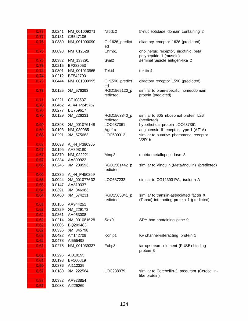
134
0.77 0.0241 NM_001009271 Nt5dc2 5'-nucleotidase domain containing 2
0.77 0.0131 CB547106
0.76 0.0380 NM_001000090 Olr1626_predicted
olfactory receptor 1626 (predicted)
0.75 0.0098 NM_012528 Chrnb1 cholinergic receptor, nicotinic, beta polypeptide 1 (muscle)
0.75 0.0382 NM_133291 Sval2 seminal vesicle antigen-like 2
0.75 0.0215 BF283053
0.74 0.0301 NM_001013965 Tekt4 tektin 4
0.74 0.0212 BF542793
0.73 0.0444 NM_001000995 Olr1590_predict
ed
olfactory receptor 1590 (predicted)
0.73 0.0125 XM_576393 RGD1565120_predicted
similar to brain-specific homeodomain protein (predicted)
0.71 0.0221 CF108537
0.70 0.0462 A_44_P245767
0.70 0.0277 BU759617
0.70 0.0129 XM_226231 RGD1563840_predicted
similar to 60S ribosomal protein L26 (predicted)
0.69 0.0393 XM_001076148 LOC687361 hypothetical protein LOC687361
0.69 0.0193 NM_030985 Agtr1a angiotensin II receptor, type 1 (AT1A)
0.68 0.0291 XM_575663 LOC500312 similar to putative pheromone receptor
V2R1b 0.67 0.0038 A_44_P380365
0.67 0.0195 AA893180
0.67 0.0379 NM_022221 Mmp8 matrix metallopeptidase 8
0.67 0.0334 AA899922
0.66 0.0246 XM_230593 RGD1561442_predicted
similar to Vinculin (Metavinculin) (predicted)
0.66 0.0335 A_44_P450259
0.65 0.0044 XM_001077632 LOC687232 similar to CG12393-PA, isoform A
0.65 0.0147 AA819337
0.64 0.0391 XM_346983
0.64 0.0460 XM_574231 RGD1565341_predicted
similar to translin-associated factor X (Tsnax) interacting protein 1 (predicted)
0.63 0.0155 AA944251
0.63 0.0329 XM_229173
0.62 0.0361 AA963008
0.62 0.0214 XM_001081628 Sox9 SRY-box containing gene 9
0.62 0.0006 BQ209483
0.62 0.0336 XM_345798
0.62 0.0422 AY142709 Kcnip1 Kv channel-interacting protein 1
0.62 0.0478 AI555498
0.61 0.0278 NM_001039337 Fubp3 far upstream element (FUSE) binding
protein 3 0.61 0.0296 AI010195
0.61 0.0193 BF560819
0.59 0.0376 AI112329
0.57 0.0180 XM_222564 LOC288979 similar to Cerebellin-2 precursor (Cerebellin-like protein)
0.57 0.0332 AA923854
0.57 0.0083 AI229269

135
0.57 0.0152 TC524716
0.56 0.0121 XM_001081628 Sox9 SRY-box containing gene 9
0.56 0.0272 AW143519
0.56 0.0116 AI059089
0.56 0.0272 XM_227622 Sdfr2_predicted stromal cell derived factor receptor 2 (predicted)
0.56 0.0049 AA924721
0.56 0.0060 AI170621
0.56 0.0364 XM_216733 RGD1565716_predicted
similar to RIKEN cDNA 4921529O18 (predicted)
0.55 0.0229 TC555070
0.55 0.0131 NM_020101 Centa2 centaurin, alpha 2
0.55 0.0486 XR_006043 LOC681331 similar to apoptosis inhibitor 5
0.55 0.0273 XM_341550 Akr1cl1_predicte
d
aldo-keto reductase family 1, member C-like
1 (predicted) 0.55 0.0432 TC566391
0.55 0.0465 AI177135
0.54 0.0010 XM_001068622 RGD1561916_predicted
similar to testes development-related NYD-SP22 isoform 1 (predicted)
0.54 0.0321 AW253256
0.54 0.0385 AW920939
0.54 0.0217 XM_001072036 LOC684554 similar to Thrombospondin-3 precursor
0.54 0.0190 XM_001053365 RGD1561975_predicted
similar to TGFB-induced factor 2 (predicted)
0.54 0.0231 TC519685
0.54 0.0405 NM_001000473 Olr1325_predict
ed
olfactory receptor 1325 (predicted)
0.54 0.0466 A_44_P791333
0.53 0.0370 XM_236675 Glb1_mapped galactosidase, beta 1 (mapped)
0.53 0.0385 NM_001034139 Lrrc8e leucine rich repeat containing 8 family, member E
0.53 0.0122 XM_225947 Trim36_predicted
tripartite motif protein 36 (predicted)
0.52 0.0183 XM_230509 RGD1566077_p
redicted
similar to RIKEN cDNA A130038L21
(predicted) 0.51 0.0287 AI233208
0.51 0.0481 TC546713
0.51 0.0446 TC519466
0.51 0.0428 CB742296
0.51 0.0467 NM_198772 MGC72974 Unknown (protein for MGC:72974)
0.51 0.0137 AI043958
0.50 0.0103 NM_053619 C5r1 complement component 5, receptor 1
0.50 0.0339 BQ783165
0.50 0.0320 XM_342281 Muc1 mucin 1, transmembrane
0.50 0.0294 XM_218644 Klk12_predicted kallikrein 12 (predicted)
0.49 0.0097 NM_001024800 Txndc1 thioredoxin domain containing 1
0.49 0.0472 XM_001063356 RGD1311381_predicted
similar to hypothetical protein FLJ20037 (predicted)
0.49 0.0444 AI406369
0.48 0.0444 AA924847
0.48 0.0280 XM_345338
0.48 0.0482 TC519225

136
0.48 0.0287 XM_221343 RGD1305614_predicted
similar to IGF-II mRNA-binding protein 2 (predicted)
0.48 0.0492 XM_216518
0.48 0.0497 XM_216331 RGD1305828_predicted
similar to hypothetical protein (predicted)
0.48 0.0166 BM391896
0.47 0.0020 NM_013091 Tnfrsf1a tumor necrosis factor receptor superfamily,
member 1a 0.47 0.0260 NM_001014051 LOC311548 similar to RIKEN cDNA 4930509O20
0.47 0.0220 NM_001001016 Olr1174_predicted
olfactory receptor 1174 (predicted)
0.47 0.0391 XM_232320 RGD1562038_predicted
similar to putative voltage-gated calcium channel alpha(2)delta-4 subunit (predicted)
0.47 0.0045 NM_001011997 Tmod3 tropomodulin 3
0.47 0.0443 AA925130
0.46 0.0271 NM_023981 Csf1 colony stimulating factor 1 (macrophage)
0.46 0.0256 CF111186
0.46 0.0106 TC542803
0.46 0.0377 NM_001013944 RGD1309051 similar to chromosome 14 open reading
frame 50 0.46 0.0371 NM_019273 Kcnmb1 potassium large conductance calcium-
activated channel, subfamily M, beta
member 1 0.46 0.0426 XM_001077512 RGD1565716_p
redicted similar to RIKEN cDNA 4921529O18 (predicted)
0.46 0.0070 XM_001058665 LOC681839 hypothetical protein LOC681839
0.45 0.0405 NM_207601 Plp2_mapped proteolipid protein 2 (mapped)
0.45 0.0286 AY064511 Ua20 putative UA20 protein
0.45 0.0487 XM_223527 Sh3tc1_predicted
SH3 domain and tetratricopeptide repeats 1 (predicted)
0.45 0.0248 XM_343728
0.45 0.0068 NM_172223 Pxmp4 peroxisomal membrane protein 4
0.45 0.0136 NM_138848 Podxl podocalyxin-like
0.45 0.0407 NM_012620 Serpine1 serine (or cysteine) peptidase inhibitor,
clade E, member 1 0.44 0.0085 TC556623
0.44 0.0373 NM_214828 Olr1378_predicted
olfactory receptor 1378 (predicted)
0.44 0.0149 XM_227540 Rsbn1_predicted
rosbin, round spermatid basic protein 1 (predicted)
0.44 0.0496 NM_001025039 LOC499602 hypothetical protein LOC499602
0.44 0.0314 XM_213813 Lrrc43 leucine rich repeat containing 43
0.44 0.0147 XM_217218 RGD1311381_p
redicted
similar to hypothetical protein FLJ20037
(predicted) 0.43 0.0178 XM_346182
0.43 0.0350 NM_012824 Apoc1 apolipoprotein C-I
0.43 0.0210 TC523657
0.43 0.0426 NM_130741 Lcn2 lipocalin 2
0.42 0.0004 XM_001081958 Fyttd1 forty-two-three domain containing 1
0.42 0.0316 AW525988
0.42 0.0188 AI169706
0.42 0.0234 NM_001009650 Taar5 trace amine associated receptor 5
0.42 0.0241 AI030156

137
0.41 0.0330 NM_053399 Nrtn neurturin
0.41 0.0057 XM_236938 RGD1310693_predicted
similar to RIKEN cDNA 1700027N10 (predicted)
0.41 0.0089 XM_001065902 Syncrip synaptotagmin binding, cytoplasmic RNA interacting protein
0.41 0.0304 AW916350
0.41 0.0113 NM_012735 Hk2 hexokinase 2
0.41 0.0347 XM_222564 LOC288979 similar to Cerebellin-2 precursor (Cerebellin-
like protein) 0.41 0.0499 XM_576074 RGD1560978_p
redicted similar to hypothetical protein (predicted)
0.40 0.0419 XM_343348
0.40 0.0449 BF407782
0.40 0.0345 NM_017064 Stat5a signal transducer and activator of transcription 5A
0.40 0.0307 NM_183326 Gabra1 gamma-aminobutyric acid A receptor, alpha
1 0.40 0.0460 NM_031003 Abat 4-aminobutyrate aminotransferase
0.40 0.0270 NM_001012235 Impact imprinted and ancient
0.40 0.0110 TC523460
0.40 0.0305 XM_214962 Tm2d3_predicted
TM2 domain containing 3 (predicted)
0.40 0.0148 XM_343652
0.40 0.0261 XM_342959 RGD1310427_predicted
similar to KIAA0090 protein (predicted)
0.40 0.0182 XM_001079851 LOC691849 hypothetical protein LOC691849

138
APPENDIX C LIST OF MODULATED GENES FOLLOWING REPEATED ITTO
Table C-1. List of modulated genes following repeated ITTO, p < 0.05 and log2 fold change ≥ 0.58
Log2 FC p-value TargetID Symbol Description
-2.830 0.019 NM_012881 Spp1 secreted phosphoprotein 1
-2.787 0.022 NM_012881 Spp1 secreted phosphoprotein 1
-2.722 0.019 NM_012881 Spp1 secreted phosphoprotein 1
-2.719 0.024 NM_012881 Spp1 secreted phosphoprotein 1
-2.698 0.020 NM_012881 Spp1 secreted phosphoprotein 1
-2.676 0.027 NM_012881 Spp1 secreted phosphoprotein 1
-2.665 0.023 NM_012881 Spp1 secreted phosphoprotein 1
-2.617 0.026 NM_012881 Spp1 secreted phosphoprotein 1
-2.601 0.020 NM_012881 Spp1 secreted phosphoprotein 1
-2.601 0.027 NM_012881 Spp1 secreted phosphoprotein 1
-2.138 0.004 NM_017007 Gad1 glutamic acid decarboxylase 1
-2.126 0.001 ENSRNOT00000012564
RGD1564797_predicted
similar to empty spiracles-like protein 2 (predicted)
-2.062 0.009 NM_031782 Slc32a1 solute carrier family 32 (GABA vesicular transporter), member 1
-1.741 0.038 NM_181370 Hs3st2 heparan sulfate (glucosamine) 3-O-sulfotransferase
2 -1.669 0.002 TC617840 - -
-1.614 0.026 NM_012563 Gad2 glutamic acid decarboxylase 2
-1.525 0.012 NM_017007 Gad1 glutamic acid decarboxylase 1
-1.463 0.002 NM_001014183
RGD1306991
similar to Protein C20orf103 precursor
-1.401 0.011 NM_013007 Pnoc prepronociceptin
-1.398 0.003 NM_017122 Hpca hippocalcin
-1.382 0.004 NM_139183 Crhbp corticotropin releasing hormone binding protein
-1.342 0.016 CB327622 - -
-1.305 0.010 NM_001037351
Tnnc2 troponin C type 2 (fast)
-1.294 0.025 NM_001008
880
Scn4b sodium channel, voltage-gated, type IV, beta
-1.232 0.037 ENSRNOT00000001048
Man1a_predicted
mannosidase 1, alpha (predicted)
-1.184 0.006 NM_001047973
LOC503325 hypothetical protein LOC503325
-1.132 0.000 DY471163 - -
-1.120 0.006 TC603004 - -
-1.109 0.025 NM_017007 Gad1 glutamic acid decarboxylase 1
-1.077 0.018 TC626556 - -
-1.074 0.011 NM_012798 Mal myelin and lymphocyte protein, T-cell differentiation
protein -1.074 0.013 NM_057196 Baiap2 brain-specific angiogenesis inhibitor 1-associated
protein 2
-1.066 0.008 NM_032071 Synj2 synaptojanin 2
-1.052 0.004 NM_001077201
Caln1_predicted
calneuron 1 (predicted)
-1.048 0.005 NM_031736 Slc27a2 solute carrier family 27 (fatty acid transporter),

139
member 2
-1.043 0.002 TC645081 - -
-1.038 0.021 TC633482 Pde6h phosphodiesterase 6H, cGMP-specific, cone, gamma
-1.031 0.017 NM_001033656
Man1a_predicted
mannosidase 1, alpha (predicted)
-1.010 0.000 NM_001037
206
Bphl biphenyl hydrolase-like (serine hydrolase, breast
epithelial mucin-associated antigen) -0.985 0.008 TC603491 - -
-0.980 0.038 NM_053976 Krt1-18 keratin complex 1, acidic, gene 18
-0.976 0.019 XM_578417 RGD1562829_predicted
similar to RAS-like, estrogen-regulated, growth-inhibitor (predicted)
-0.976 0.006 NM_031059 Msx1 homeo box, msh-like 1
-0.970 0.012 DV722143 - -
-0.964 0.026 BF287496 - -
-0.956 0.021 ENSRNOT00000031175
Crabp1 cellular retinoic acid binding protein 1
-0.952 0.003 ENSRNOT0
0000010623
RGD156135
7_predicted
similar to LIM domain only 3 (predicted)
-0.952 0.023 TC600004 - -
-0.943 0.021 NM_012752 Cd24 CD24 antigen
-0.943 0.010 XM_232202 Adamts9_predicted
a disintegrin-like and metalloprotease (reprolysin type) with thrombospondin type 1 motif, 9
(predicted) -0.939 0.036 NM_031742 Kcnh1 potassium voltage-gated channel, subfamily H
(eag-related), member 1
-0.939 0.013 XM_001070642
RGD1564871_predicted
similar to Thioredoxin domain containing protein 6 (Thioredoxin-like protein 2) (predicted)
-0.921 0.000 ENSRNOT0
0000049507
RGD156065
8_predicted
similar to serine (or cysteine) proteinase inhibitor,
clade B, member 1b (predicted) -0.908 0.023 TC631315 - -
-0.905 0.018 NM_012733 Rbp1 retinol binding protein 1, cellular
-0.905 0.011 ENSRNOT00000016433
RGD1310371
similar to RIKEN cDNA 1700026D08
-0.903 0.004 XM_217250 Ephb1 Eph receptor B1
-0.899 0.000 NM_031834 Sult1a1 sulfotransferase family 1A, phenol-preferring, member 1
-0.897 0.014 XM_213354 Dnah9 dynein, axonemal, heavy polypeptide 9
-0.894 0.018 NM_012825 Aqp4 aquaporin 4
-0.885 0.022 BF567171 LOC689106 hypothetical protein LOC689106
-0.880 0.008 BF556399 - -
-0.877 0.004 ENSRNOT0
0000023378
Capsl_predi
cted
calcyphosine-like (predicted)
-0.865 0.039 ENSRNOT00000010850
Cnr1 cannabinoid receptor 1 (brain)
-0.860 0.007 NM_012825 Aqp4 aquaporin 4
-0.856 0.007 ENSRNOT00000010623
RGD1561357_predicted
similar to LIM domain only 3 (predicted)
-0.856 0.010 NM_001013
953
RGD130535
1
hypothetical LOC300663
-0.838 0.005 NM_001024882
Ccdc19 coiled-coil domain containing 19
-0.836 0.021 NM_053878 Cplx2 complexin 2
-0.835 0.016 ENSRNOT0 Rshl3_predi radial spokehead-like 3 (predicted)

140
0000036449 cted
-0.833 0.033 NM_199093 Serping1 serine (or cysteine) peptidase inhibitor, clade G, member 1
-0.831 0.022 NM_001012235
Impact imprinted and ancient
-0.830 0.021 NM_057196 Baiap2 brain-specific angiogenesis inhibitor 1-associated
protein 2 -0.826 0.014 NM_024140 Nrgn neurogranin
-0.825 0.049 NM_001033656
Man1a_predicted
mannosidase 1, alpha (predicted)
-0.824 0.006 NM_001005765
Rap1a RAS-related protein 1a
-0.820 0.012 BC092207 LOC365476 similar to chromosome 10 open reading frame 79
-0.811 0.008 TC611803 - -
-0.806 0.014 NM_031736 Slc27a2 solute carrier family 27 (fatty acid transporter),
member 2 -0.804 0.043 NM_017210 Dio3 deiodinase, iodothyronine, type III
-0.800 0.002 ENSRNOT00000016911
Efhb_predicted
EF hand domain family, member B (predicted)
-0.795 0.003 NM_012825 Aqp4 aquaporin 4
-0.793 0.020 XM_001054111
LOC689106 hypothetical protein LOC689106
-0.791 0.030 XM_343481 RGD130660
3_predicted
similar to RIKEN cDNA D330022A01 gene
(predicted) -0.789 0.006 NM_031509 Gsta3 glutathione S-transferase A3
-0.783 0.006 ENSRNOT00000038579
RGD1560137_predicted
similar to expressed sequence AU021034 (predicted)
-0.777 0.040 J05276 Htr1a 5-hydroxytryptamine (serotonin) receptor 1A
-0.774 0.035 ENSRNOT00000013483
LOC679875 similar to MAGE-like protein 2 (Protein nS7)
-0.773 0.026 TC598708 - -
-0.772 0.023 XR_007590 RGD156300
2_predicted
similar to hypothetical protein DKFZp434O0527
(predicted) -0.769 0.007 NM_012825 Aqp4 aquaporin 4
-0.765 0.011 AW915033 Traf3_predicted
Tnf receptor-associated factor 3 (predicted)
-0.764 0.013 ENSRNOT00000012469
Amn_predicted
amnionless (predicted)
-0.755 0.011 XM_573504 RGD156265
8_predicted
similar to RIKEN cDNA 1700009P17 (predicted)
-0.754 0.013 NM_145880 Lhx1 LIM homeobox protein 1
-0.754 0.000 NM_001025664
Wsb1 WD repeat and SOCS box-containing 1
-0.749 0.020 ENSRNOT00000024222
Ccdc37_predicted
coiled-coil domain containing 37 (predicted)
-0.749 0.026 BC092207 LOC365476 similar to chromosome 10 open reading frame 79
-0.748 0.006 NM_012825 Aqp4 aquaporin 4
-0.745 0.015 NM_001013
953
RGD130535
1
hypothetical LOC300663
-0.743 0.028 TC607764 - -
-0.742 0.038 ENSRNOT00000010850
Cnr1 cannabinoid receptor 1 (brain)
-0.741 0.018 XM_341694 Dynlrb2_predicted
dynein light chain roadblock-type 2 (predicted)

141
-0.729 0.028 BC107936 - -
-0.724 0.003 NM_053688 Pde6h phosphodiesterase 6H, cGMP-specific, cone, gamma
-0.723 0.011 CX569250 - -
-0.723 0.010 ENSRNOT00000003841
RGD1561932_predicted
similar to novel protein (predicted)
-0.717 0.025 CK366954 LOC691455 similar to calmodulin-like 4
-0.716 0.049 TC584035 - -
-0.715 0.047 ENSRNOT0
0000010850
Cnr1 cannabinoid receptor 1 (brain)
-0.713 0.021 XM_341952 RGD1562437_predicted
similar to transcription elongation regulator 1-like (predicted)
-0.705 0.039 TC596255 - -
-0.701 0.049 ENSRNOT00000010850
Cnr1 cannabinoid receptor 1 (brain)
-0.697 0.029 BF556560 - -
-0.697 0.006 ENSRNOT0
0000007789
Nnmt_predic
ted
nicotinamide N-methyltransferase (predicted)
-0.696 0.001 NM_001012176
Tsga2 testis specific gene A2
-0.694 0.041 ENSRNOT00000016433
RGD1310371
similar to RIKEN cDNA 1700026D08
-0.694 0.042 ENSRNOT0
0000010850
Cnr1 cannabinoid receptor 1 (brain)
-0.690 0.042 ENSRNOT00000010850
Cnr1 cannabinoid receptor 1 (brain)
-0.690 0.017 ENSRNOT00000036374
RGD1310677_predicted
similar to RIKEN cDNA 4632412N22 gene (predicted)
-0.689 0.011 NM_012825 Aqp4 aquaporin 4
-0.688 0.015 TC634886 - -
-0.685 0.004 NM_023020 Tmeff1 transmembrane protein with EGF-like and two
follistatin-like domains 1 -0.684 0.005 TC607399 Arnt2 aryl hydrocarbon receptor nuclear translocator 2
-0.682 0.003 XM_001080361
LOC691960 similar to solute carrier family 28, member 2
-0.682 0.036 XM_342561 RGD1305725_predicted
similar to chromosome 20 open reading frame 102 (predicted)
-0.681 0.005 ENSRNOT0
0000008005
Pole4_predi
cted
polymerase (DNA-directed), epsilon 4 (p12 subunit)
(predicted) -0.680 0.011 ENSRNOT0
0000027055 RGD1307059_predicted
similar to RIKEN cDNA 1110035L05 (predicted)
-0.680 0.007 NM_001009920
Yc2 glutathione S-transferase Yc2 subunit
-0.679 0.009 NM_012825 Aqp4 aquaporin 4
-0.677 0.038 NM_017258 Btg1 B-cell translocation gene 1, anti-proliferative
-0.676 0.008 NM_138506 Adra2c adrenergic receptor, alpha 2c
-0.673 0.005 XM_220252 - -
-0.672 0.023 NM_212541 Slc44a4 solute carrier family 44, member 4
-0.672 0.007 XM_575803 RGD156191
6_predicted
similar to testes development-related NYD-SP22
isoform 1 (predicted) -0.672 0.035 ENSRNOT0
0000029587 Dpysl4 dihydropyrimidinase-like 4
-0.672 0.045 NM_001000388
Olr417_predicted
olfactory receptor 417 (predicted)

142
-0.670 0.006 NM_145786 Iiig9 IIIG9 protein
-0.669 0.023 TC617303 - -
-0.669 0.009 BC092654 Col16a1 procollagen, type XVI, alpha 1
-0.668 0.014 NM_013099 Mc4r melanocortin 4 receptor
-0.668 0.018 TC599619 - -
-0.667 0.025 ENSRNOT00000044831
Ttll2_predicted
tubulin tyrosine ligase-like family, member 2 (predicted)
-0.667 0.022 NM_012667 Tacr1 tachykinin receptor 1
-0.666 0.005 NM_001031647
Dnali1 dynein, axonemal, light intermediate polypeptide 1
-0.666 0.004 NM_053018 Cd9 CD9 antigen
-0.664 0.016 NM_001025
293
Mbp myelin basic protein
-0.663 0.006 ENSRNOT00000026491
RGD1305311_predicted
similar to hypothetical protein FLJ22527 (predicted)
-0.663 0.024 TC614187 - -
-0.661 0.010 ENSRNOT00000035948
RGD1565611_predicted
RGD1565611 (predicted)
-0.660 0.001 NM_053660 Gng10 guanine nucleotide binding protein (G protein),
gamma 10 -0.660 0.003 CV102938 - -
-0.658 0.003 TC624015 - -
-0.657 0.018 AI113235 - -
-0.656 0.049 CX571008 Pla2g3_predicted
phospholipase A2, group III (predicted)
-0.656 0.035 BI297059 Ibrdc3_predicted
IBR domain containing 3 (predicted)
-0.655 0.016 NM_031140 Vim vimentin
-0.655 0.003 ENSRNOT0
0000020568
Crtac1 cartilage acidic protein 1
-0.654 0.002 TC617271 - -
-0.653 0.047 A_44_P729134
- -
-0.652 0.030 XM_343747 LOC363424 similar to spermatogenesis associated glutamate (E)-rich protein 2
-0.652 0.029 TC603496 - -
-0.651 0.002 ENSRNOT0
0000033449
Samd7_pre
dicted
sterile alpha motif domain containing 7 (predicted)
-0.651 0.002 NM_001079701
RGD1310641
similar to hypothetical protein
-0.650 0.023 NM_133293 Gata3 GATA binding protein 3
-0.649 0.047 TC633399 - -
-0.649 0.024 ENSRNOT00000017847
Bm259 BM259 protein
-0.648 0.049 XM_001067
026
RGD156609
7_predicted
similar to Anillin (predicted)
-0.648 0.046 NM_012667 Tacr1 tachykinin receptor 1
-0.647 0.007 TC593623 - -
-0.647 0.005 ENSRNOT00000012490
RGD1561171_predicted
similar to RIKEN cDNA 6330407D12 (predicted)
-0.647 0.018 XM_229809 - -
-0.646 0.005 TC643803 - -
-0.644 0.013 NM_012825 Aqp4 aquaporin 4

143
-0.644 0.033 NM_012667 Tacr1 tachykinin receptor 1
-0.644 0.014 ENSRNOT00000047663
LOC689415 similar to Metallothionein-2 (MT-2) (Metallothionein-II) (MT-II)
-0.642 0.041 TC578855 Ptger2 prostaglandin E receptor 2, subtype EP2
-0.641 0.028 NM_021703 Akap14 A kinase (PRKA) anchor protein 14
-0.641 0.023 NM_053330 Rpl21 ribosomal protein L21
-0.640 0.016 NM_022620 Kpl2 KPL2 protein
-0.640 0.016 NM_012825 Aqp4 aquaporin 4
-0.638 0.001 ENSRNOT00000026491
RGD1305311_predicted
similar to hypothetical protein FLJ22527 (predicted)
-0.638 0.015 NM_053456 Plcl1 phospholipase C-like 1
-0.636 0.019 NM_001012
235
Impact imprinted and ancient
-0.635 0.004 TC583130 - -
-0.635 0.012 TC601388 - -
-0.634 0.028 ENSRNOT00000037023
RGD1562705_predicted
similar to Shb-like adapter protein, Shf - human (predicted)
-0.632 0.035 NM_019168 Arg2 arginase 2
-0.629 0.037 NM_053832 Foxj1 forkhead box J1
-0.626 0.026 NM_001024367
LOC501619 similar to 40S ribosomal protein S29
-0.625 0.041 ENSRNOT0
0000010850
Cnr1 cannabinoid receptor 1 (brain)
-0.625 0.007 ENSRNOT00000027261
RGD1565856_predicted
similar to Hypothetical 55.1 kDa protein F09G8.5 in chromosome III (predicted)
-0.625 0.015 NM_012825 Aqp4 aquaporin 4
-0.625 0.012 XR_008801 Elmod1_predicted
ELMO domain containing 1 (predicted)
-0.625 0.027 NM_012667 Tacr1 tachykinin receptor 1
-0.624 0.037 XM_342195 RGD130773
5_predicted
similar to hypothetical protein FLJ11795 (predicted)
-0.623 0.024 NM_138837 Pou3f3 POU domain, class 3, transcription factor 3
-0.621 0.020 NM_001012235
Impact imprinted and ancient
-0.621 0.010 NM_001031647
Dnali1 dynein, axonemal, light intermediate polypeptide 1
-0.621 0.000 XM_218313 Gpr126_pre
dicted
G protein-coupled receptor 126 (predicted)
-0.620 0.016 NM_012825 Aqp4 aquaporin 4
-0.620 0.017 AW915353 Anp32e acidic (leucine-rich) nuclear phosphoprotein 32 family, member E
-0.620 0.015 ENSRNOT00000057692
Slitrk5_predicted
SLIT and NTRK-like family, member 5 (predicted)
-0.620 0.001 TC587992 RGD131062
3
similar to RIKEN cDNA 2010005O13
-0.619 0.016 DV729125 - -
-0.619 0.011 TC622410 Bbox1 butyrobetaine (gamma), 2-oxoglutarate dioxygenase 1 (gamma-butyrobetaine hydroxylase)
-0.618 0.040 NM_213627 Zdhhc23 zinc finger, DHHC domain containing 23
-0.616 0.014 NM_012809 Cnp1 cyclic nucleotide phosphodiesterase 1
-0.615 0.020 NM_001014221
LOC363337 similar to RIKEN cDNA 1700081O22
-0.614 0.017 XM_576882 LOC501474 similar to Myosin-9B (Myosin IXb) (Unconventional
myosin-9b)

144
-0.613 0.013 NM_001013151
Gna14 guanine nucleotide binding protein, alpha 14
-0.612 0.005 A_44_P358649
- -
-0.612 0.012 XR_009412 RGD156300
0_predicted
similar to hypothetical protein MGC26856
(predicted) -0.612 0.024 ENSRNOT0
0000045285 RGD1564852_predicted
similar to hypothetical protein FLJ14503 (predicted)
-0.611 0.018 NM_053019 Avpr1a arginine vasopressin receptor 1A
-0.611 0.010 NM_001004269
Jam3 junctional adhesion molecule 3
-0.608 0.002 TC647877 - -
-0.607 0.001 NM_031339 Parg poly (ADP-ribose) glycohydrolase
-0.606 0.033 AA801133 RGD156310
9_predicted
RGD1563109 (predicted)
-0.606 0.045 ENSRNOT00000017151
Mlf1_predicted
myeloid leukemia factor 1 (predicted)
-0.603 0.009 AI236782 RGD1308324_predicted
similar to RIKEN cDNA 5730469D23 (predicted)
-0.602 0.020 NM_019291 Ca2 carbonic anhydrase 2
-0.600 0.037 TC646852 - -
-0.599 0.006 NM_001005
765
Rap1a RAS-related protein 1a
-0.598 0.023 NM_001044277
MGC94891 hypothetical protein LOC681210
-0.597 0.033 NM_001007710
Acpl2 acid phosphatase-like 2
-0.597 0.028 NM_022202 Grm8 glutamate receptor, metabotropic 8
-0.597 0.020 DV717011 - -
-0.597 0.009 NM_022629 Bbox1 butyrobetaine (gamma), 2-oxoglutarate
dioxygenase 1 (gamma-butyrobetaine hydroxylase) -0.596 0.012 XR_005527 LOC679620 similar to CG4329-PA, isoform A
-0.596 0.031 NM_001004080
Gsn gelsolin
-0.594 0.004 NM_001047912
RGD1307739
similar to CG3306-PA
-0.594 0.001 ENSRNOT0
0000001728
LOC690211 similar to Disco-interacting protein 2 homolog
-0.594 0.026 NM_001033893
RGD1308356
similar to Hypothetical protein KIAA0341
-0.593 0.003 CF109910 - -
-0.592 0.011 A_44_P490025
- -
-0.589 0.018 NM_012614 Npy neuropeptide Y
-0.587 0.009 NM_031322 Lrp4 low density lipoprotein receptor-related protein 4
-0.586 0.045 NM_012667 Tacr1 tachykinin receptor 1
-0.586 0.041 ENSRNOT0
0000044595
Lrrc4c_predi
cted
leucine rich repeat containing 4C (predicted)
-0.585 0.002 ENSRNOT00000013408
Htr2a 5-hydroxytryptamine (serotonin) receptor 2A
-0.584 0.019 BC092654 Col16a1 procollagen, type XVI, alpha 1
-0.583 0.020 ENSRNOT00000012061
RGD1563441_predicted
similar to RIKEN cDNA A030009H04 (predicted)
-0.583 0.031 ENSRNOT0 Slc24a4_pre solute carrier family 24 (sodium/potassium/calcium
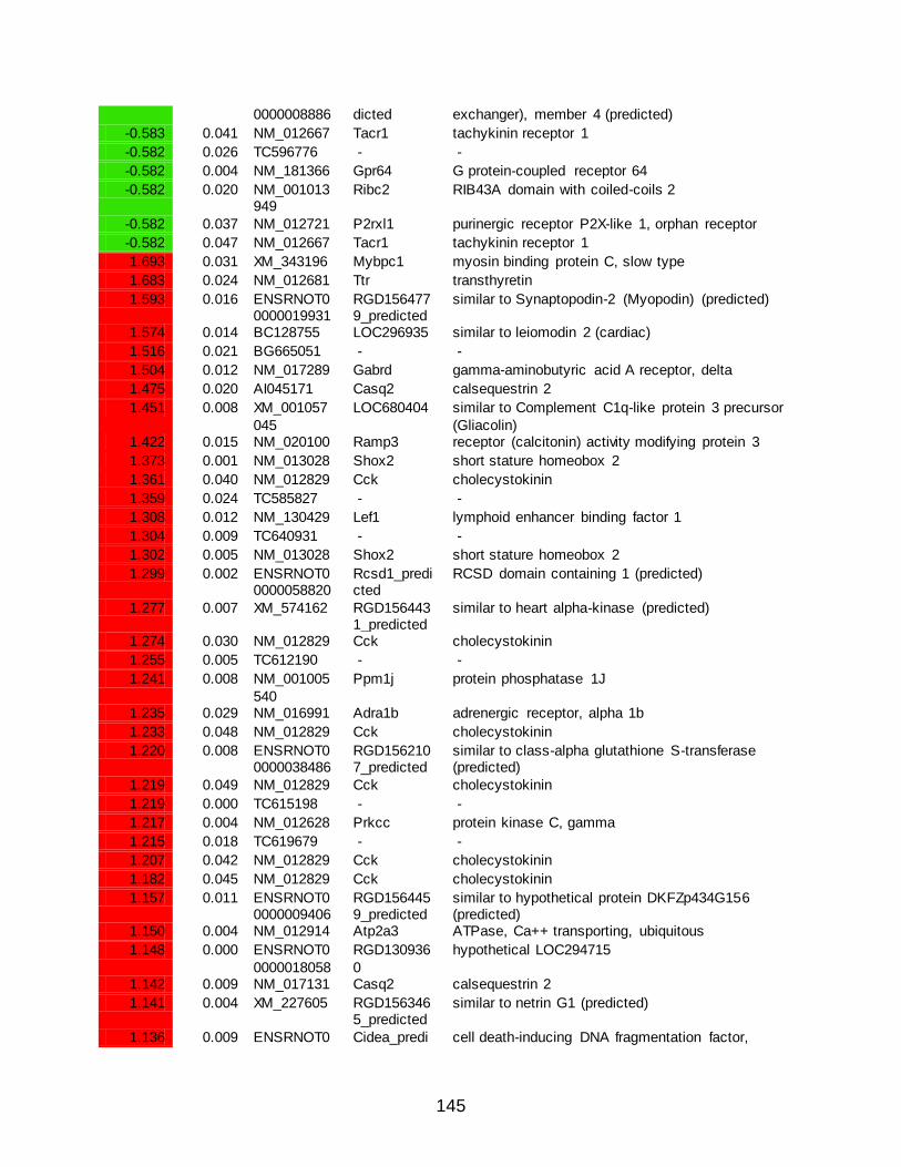
145
0000008886 dicted exchanger), member 4 (predicted)
-0.583 0.041 NM_012667 Tacr1 tachykinin receptor 1
-0.582 0.026 TC596776 - -
-0.582 0.004 NM_181366 Gpr64 G protein-coupled receptor 64
-0.582 0.020 NM_001013949
Ribc2 RIB43A domain with coiled-coils 2
-0.582 0.037 NM_012721 P2rxl1 purinergic receptor P2X-like 1, orphan receptor
-0.582 0.047 NM_012667 Tacr1 tachykinin receptor 1
1.693 0.031 XM_343196 Mybpc1 myosin binding protein C, slow type
1.683 0.024 NM_012681 Ttr transthyretin
1.593 0.016 ENSRNOT00000019931
RGD1564779_predicted
similar to Synaptopodin-2 (Myopodin) (predicted)
1.574 0.014 BC128755 LOC296935 similar to leiomodin 2 (cardiac)
1.516 0.021 BG665051 - -
1.504 0.012 NM_017289 Gabrd gamma-aminobutyric acid A receptor, delta
1.475 0.020 AI045171 Casq2 calsequestrin 2
1.451 0.008 XM_001057
045
LOC680404 similar to Complement C1q-like protein 3 precursor
(Gliacolin) 1.422 0.015 NM_020100 Ramp3 receptor (calcitonin) activity modifying protein 3
1.373 0.001 NM_013028 Shox2 short stature homeobox 2
1.361 0.040 NM_012829 Cck cholecystokinin
1.359 0.024 TC585827 - -
1.308 0.012 NM_130429 Lef1 lymphoid enhancer binding factor 1
1.304 0.009 TC640931 - -
1.302 0.005 NM_013028 Shox2 short stature homeobox 2
1.299 0.002 ENSRNOT00000058820
Rcsd1_predicted
RCSD domain containing 1 (predicted)
1.277 0.007 XM_574162 RGD1564431_predicted
similar to heart alpha-kinase (predicted)
1.274 0.030 NM_012829 Cck cholecystokinin
1.255 0.005 TC612190 - -
1.241 0.008 NM_001005
540
Ppm1j protein phosphatase 1J
1.235 0.029 NM_016991 Adra1b adrenergic receptor, alpha 1b
1.233 0.048 NM_012829 Cck cholecystokinin
1.220 0.008 ENSRNOT00000038486
RGD1562107_predicted
similar to class-alpha glutathione S-transferase (predicted)
1.219 0.049 NM_012829 Cck cholecystokinin
1.219 0.000 TC615198 - -
1.217 0.004 NM_012628 Prkcc protein kinase C, gamma
1.215 0.018 TC619679 - -
1.207 0.042 NM_012829 Cck cholecystokinin
1.182 0.045 NM_012829 Cck cholecystokinin
1.157 0.011 ENSRNOT00000009406
RGD1564459_predicted
similar to hypothetical protein DKFZp434G156 (predicted)
1.150 0.004 NM_012914 Atp2a3 ATPase, Ca++ transporting, ubiquitous
1.148 0.000 ENSRNOT0
0000018058
RGD130936
0
hypothetical LOC294715
1.142 0.009 NM_017131 Casq2 calsequestrin 2
1.141 0.004 XM_227605 RGD1563465_predicted
similar to netrin G1 (predicted)
1.136 0.009 ENSRNOT0 Cidea_predi cell death-inducing DNA fragmentation factor,

146
0000024968 cted alpha subunit-like effector A (predicted)
1.134 0.003 TC596653 - -
1.124 0.015 TC614984 - -
1.115 0.025 NM_016991 Adra1b adrenergic receptor, alpha 1b
1.113 0.039 BC103629 Ptpn3 protein tyrosine phosphatase, non-receptor type 3
1.107 0.011 NM_053699 Cited4 Cbp/p300-interacting transactivator, with Glu/Asp-rich carboxy-terminal domain, 4
1.096 0.012 TC610674 - -
1.089 0.008 NM_022666 Grm4 glutamate receptor, metabotropic 4
1.081 0.002 CB547657 - -
1.079 0.020 XM_343196 Mybpc1 myosin binding protein C, slow type
1.075 0.006 NM_019340 Rgs3 regulator of G-protein signalling 3
1.069 0.006 NM_053834 Kcnj9 potassium inwardly-rectifying channel, subfamily J, member 9
1.060 0.016 AW917391 - -
1.050 0.014 AF366899 Adra2b adrenergic receptor, alpha 2b
1.047 0.033 AW143334 - -
1.044 0.037 TC601767 - -
1.041 0.029 ENSRNOT0
0000009406
RGD156445
9_predicted
similar to hypothetical protein DKFZp434G156
(predicted) 1.040 0.007 XM_343983 - -
1.037 0.015 NM_017214 Rgs4 regulator of G-protein signaling 4
1.037 0.017 ENSRNOT00000018677
RGD1304904_predicted
similar to mitochondrial glycerol 3-phosphate acyltransferase (predicted)
1.037 0.011 NM_001013032
Npy1r neuropeptide Y receptor Y1
1.020 0.036 XM_001072
492
LOC684921 similar to Complement C1q-like protein 3 precursor
(Gliacolin) 1.017 0.003 BE101695 - -
1.010 0.012 NM_016991 Adra1b adrenergic receptor, alpha 1b
1.003 0.037 ENSRNOT00000005072
LOC498289 similar to Opsin-3 (Encephalopsin) (Panopsin)
1.002 0.022 AW916327 RGD1307396_predicted
similar to RIKEN cDNA 6330406I15 (predicted)
0.996 0.007 NM_001002
829
Rasl11a RAS-like family 11 member A
0.990 0.004 TC601560 - -
0.983 0.007 NM_053506 Hrh3 histamine receptor H3
0.981 0.016 XM_001058055
LOC681708 similar to transmembrane protein 41a
0.981 0.048 ENSRNOT00000004193
RGD1310166_predicted
similar to Chromodomain-helicase-DNA-binding protein 1 (CHD-1) (predicted)
0.973 0.004 ENSRNOT0
0000008692
Fstl4_predic
ted
follistatin-like 4 (predicted)
0.969 0.010 TC645916 - -
0.966 0.004 TC626053 - -
0.965 0.003 ENSRNOT00000021471
LOC679812 similar to Pleckstrin homology domain-containing family G member 1
0.963 0.002 NM_173138 Dlgap3 discs, large (Drosophila) homolog-associated protein 3
0.959 0.006 ENSRNOT0
0000007993
LOC679668 similar to leucine-rich repeat transmembrane
neuronal 1 0.959 0.040 BF393607 LOC689147 hypothetical protein LOC689147

147
0.959 0.011 NM_053613 Rtn4r reticulon 4 receptor
0.958 0.010 TC608608 - -
0.950 0.008 AF039218 Cit citron
0.946 0.015 TC585018 - -
0.938 0.014 NM_024354 Chrna4 cholinergic receptor, nicotinic, alpha polypeptide 4
0.937 0.008 BI395573 - -
0.934 0.017 XM_342800 Tox_predicted
thymocyte selection-associated HMG box gene (predicted)
0.934 0.010 ENSRNOT00000042872
LOC689770 similar to osteoclast inhibitory lectin
0.931 0.049 NM_013157 Ass argininosuccinate synthetase
0.929 0.038 ENSRNOT0
0000018058
RGD130936
0
hypothetical LOC294715
0.929 0.009 CF107721 - -
0.924 0.002 A_44_P723917
- -
0.924 0.041 NM_023960 Kcnmb4 potassium large conductance calcium-activated channel, subfamily M, beta member 4
0.923 0.013 NM_017214 Rgs4 regulator of G-protein signaling 4
0.922 0.029 AW919694 LOC679158 similar to SRY (sex determining region Y)-box 3
0.920 0.026 TC598660 - -
0.919 0.015 TC623297 - -
0.916 0.020 XM_219747 Trpm6 transient receptor potential cation channel,
subfamily M, member 6 0.915 0.011 NM_019230 Slc22a3 solute carrier family 22, member 3
0.914 0.028 TC594011 - -
0.913 0.020 NM_053402 Wnt4 wingless-related MMTV integration site 4
0.909 0.003 NM_057142 Lrrc7 leucine rich repeat containing 7
0.905 0.005 ENSRNOT00000014661
Tox_predicted
thymocyte selection-associated HMG box gene (predicted)
0.903 0.005 NM_001011974
Akap2 A kinase (PRKA) anchor protein 2
0.899 0.010 NM_053788 Stx1a syntaxin 1A (brain)
0.898 0.008 ENSRNOT0
0000003568
Ptpn4 protein tyrosine phosphatase, non-receptor type 4
0.898 0.001 NM_001025145
Zfp365 zinc finger protein 365
0.893 0.011 ENSRNOT00000001205
RGD1307396_predicted
similar to RIKEN cDNA 6330406I15 (predicted)
0.892 0.004 TC630382 - -
0.887 0.021 AW142807 - -
0.887 0.033 BF523192 - -
0.883 0.015 NM_053804 Kcnk4 potassium channel, subfamily K, member 4
0.881 0.015 ENSRNOT0
0000004963
Usp43_predi
cted
ubiquitin specific protease 43 (predicted)
0.881 0.011 NM_001037492
Slc41a3 solute carrier family 41, member 3
0.879 0.007 NM_031826 Fbn2 fibrillin 2
0.878 0.010 NM_021676 Shank3 SH3/ankyrin domain gene 3
0.878 0.040 XM_344544 Angpt2 angiopoietin 2
0.874 0.013 NM_053788 Stx1a syntaxin 1A (brain)
0.871 0.007 M92075 Grm2 glutamate receptor, metabotropic 2
0.870 0.020 NM_080587 Gabra4 gamma-aminobutyric acid (GABA-A) receptor,

148
subunit alpha 4
0.868 0.019 NM_001012101
Coro2a coronin, actin binding protein 2A
0.867 0.009 ENSRNOT00000046812
RGD1563465_predicted
similar to netrin G1 (predicted)
0.866 0.016 NM_138502 Mgll monoglyceride lipase
0.865 0.013 TC600845 Ptger3 prostaglandin E receptor 3 (subtype EP3)
0.860 0.014 TC637759 Adarb1 adenosine deaminase, RNA-specific, B1
0.858 0.027 NM_139217 Kcnc2 potassium voltage gated channel, Shaw-related
subfamily, member 2 0.854 0.028 NM_001007
611 RGD1359691
hypothetical LOC287534
0.853 0.001 AI502122 - -
0.853 0.017 NM_184045 Srpk3 serine/arginine-rich protein specific kinase 3
0.853 0.009 XM_235546 Upk3a_predicted
uroplakin 3A (predicted)
0.849 0.002 U90444 Adarb1 adenosine deaminase, RNA-specific, B1
0.847 0.024 TC622940 - -
0.846 0.004 ENSRNOT0
0000029415
Tnrc9_predi
cted
trinucleotide repeat containing 9 (predicted)
0.846 0.017 NM_019372 Ppm2c protein phosphatase 2C, magnesium dependent, catalytic subunit
0.844 0.014 NM_053788 Stx1a syntaxin 1A (brain)
0.842 0.043 XM_223087 Lamb3 laminin, beta 3
0.840 0.023 NM_022302 Efcbp1 EF hand calcium binding protein 1
0.840 0.027 NM_022297 Ddah1 dimethylarginine dimethylaminohydrolase 1
0.836 0.044 CR469283 - -
0.835 0.013 NM_053788 Stx1a syntaxin 1A (brain)
0.834 0.015 ENSRNOT00000051793
RGD1310117_predicted
hypothetical LOC298591 (predicted)
0.832 0.016 DV713993 - -
0.829 0.026 NM_031686 Scn7a sodium channel, voltage-gated, type VII, alpha
0.829 0.016 ENSRNOT0
0000051566
LOC679869 similar to transcription factor 7-like 2, T-cell
specific, HMG-box 0.828 0.008 DV718743 - -
0.827 0.015 NM_053788 Stx1a syntaxin 1A (brain)
0.827 0.015 NM_053788 Stx1a syntaxin 1A (brain)
0.826 0.026 BG673684 - -
0.825 0.044 TC600862 Rnf152_predicted
ring finger protein 152 (predicted)
0.819 0.002 BF522086 - -
0.819 0.016 ENSRNOT00000026262
RGD1561144_predicted
similar to N-acetylglucosamine 6-O-sulfotransferase (predicted)
0.819 0.026 NM_053788 Stx1a syntaxin 1A (brain)
0.818 0.005 NM_012704 Ptger3 prostaglandin E receptor 3 (subtype EP3)
0.818 0.036 ENSRNOT0
0000020727
Cpne7_pred
icted
copine VII (predicted)
0.817 0.025 TC608942 - -
0.816 0.022 CK603271 LOC681708 similar to transmembrane protein 41a
0.815 0.003 NM_031826 Fbn2 fibrillin 2
0.815 0.015 TC590536 - -
0.814 0.006 AW142828 LOC293589 putative GTP-binding protein
0.812 0.011 AA957183 Cit citron

149
0.812 0.004 NM_138909 Foxe1 forkhead box E1 (thyroid transcription factor 2)
0.808 0.003 L04739 - -
0.804 0.016 ENSRNOT00000050400
Gpr123_predicted
G protein-coupled receptor 123 (predicted)
0.803 0.006 TC586471 - -
0.802 0.023 NM_053788 Stx1a syntaxin 1A (brain)
0.802 0.025 XM_344130 Inhbb inhibin beta-B
0.800 0.046 ENSRNOT00000022774
Angpt2 angiopoietin 2
0.800 0.021 NM_053788 Stx1a syntaxin 1A (brain)
0.798 0.045 XM_001072
408
LOC684906 similar to zinc finger, matrin type 4
0.797 0.002 NM_080482 Dbccr1 deleted in bladder cancer chromosome region candidate 1 (human)
0.797 0.008 AW143275 - -
0.795 0.001 CB545680 - -
0.794 0.012 ENSRNOT00000021253
Chst9_predicted
carbohydrate (N-acetylgalactosamine 4-0) sulfotransferase 9 (predicted)
0.794 0.033 NM_153473 Myo7a myosin VIIA
0.793 0.026 NM_001004
268
RGD130327
1
similar to chromosome 1 open reading frame 172
0.789 0.011 NM_001007235
Itpr1 inositol 1,4,5-triphosphate receptor 1
0.788 0.002 NM_031977 Src Rous sarcoma oncogene
0.788 0.008 AW920092 - -
0.786 0.017 TC576606 Coro2a coronin, actin binding protein 2A
0.784 0.008 NM_053917 Inpp4b inositol polyphosphate-4-phosphatase, type II
0.783 0.006 NM_001003401
Enc1 ectodermal-neural cortex 1
0.783 0.013 NM_001000
779
Olr1424_pre
dicted
olfactory receptor 1424 (predicted)
0.782 0.031 TC620908 - -
0.782 0.028 TC616402 - -
0.781 0.005 ENSRNOT00000056116
LOC690366 similar to vang, van gogh-like 1
0.780 0.012 ENSRNOT00000021432
Vil1_predicted
villin 1 (predicted)
0.778 0.028 AW917894 - -
0.778 0.008 XM_341784 Cdc42ep5_p
redicted
CDC42 effector protein (Rho GTPase binding) 5
(predicted) 0.777 0.019 NM_057190 Nelf nasal embryonic LHRH factor
0.774 0.025 TC623960 - -
0.773 0.002 NM_031977 Src Rous sarcoma oncogene
0.773 0.028 DV725289 - -
0.771 0.013 ENSRNOT00000007242
RGD1308745_predicted
similar to E430002G05Rik protein (predicted)
0.771 0.021 NM_013026 Sdc1 syndecan 1
0.771 0.012 AA997148 Morf4l1 mortality factor 4 like 1
0.764 0.048 NM_001024791
Epn3 epsin 3
0.762 0.003 ENSRNOT0
0000009896
Cyp46a1_pr
edicted
cytochrome P450, family 46, subfamily a,
polypeptide 1 (predicted) 0.762 0.003 NM_001033 Slc6a17 solute carrier family 6 (neurotransmitter

150
079 transporter), member 17
0.761 0.019 BF563765 - -
0.760 0.022 A_44_P417133
- -
0.759 0.022 NM_001012056
LOC307660 carboxylesterase 615
0.758 0.015 TC599342 - -
0.758 0.047 ENSRNOT0
0000026419
Gbx2 gastrulation brain homeobox 2
0.758 0.031 NM_017288 Scn1b sodium channel, voltage-gated, type I, beta
0.757 0.011 NM_031085 Prkch protein kinase C, eta
0.757 0.006 NM_053698 Cited2 Cbp/p300-interacting transactivator, with Glu/Asp-rich carboxy-terminal domain, 2
0.757 0.000 TC620686 - -
0.754 0.006 NM_001007235
Itpr1 inositol 1,4,5-triphosphate receptor 1
0.753 0.008 ENSRNOT0
0000002498
Pkp2 plakophilin 2
0.750 0.019 NM_017230 Padi3 peptidyl arginine deiminase, type III
0.749 0.002 NM_001034855
Gpr153 G protein-coupled receptor 153
0.747 0.038 ENSRNOT00000012757
Papln_predicted
papilin, proteoglycan-like sulfated glycoprotein (predicted)
0.747 0.011 NM_001007
235
Itpr1 inositol 1,4,5-triphosphate receptor 1
0.745 0.034 NM_053981 Kcnj12 potassium inwardly-rectifying channel, subfamily J, member 12
0.744 0.044 CB546657 - -
0.743 0.009 NM_001007235
Itpr1 inositol 1,4,5-triphosphate receptor 1
0.742 0.015 NM_001007
235
Itpr1 inositol 1,4,5-triphosphate receptor 1
0.742 0.021 ENSRNOT00000021036
Cda_predicted
cytidine deaminase (predicted)
0.742 0.007 TC610464 - -
0.741 0.035 NM_138849 Bk brain and kidney protein
0.740 0.013 ENSRNOT00000044039
RGD1565245_predicted
similar to Histone H2B 291B (predicted)
0.739 0.008 NM_001034
933
Arsa arylsulfatase A
0.738 0.008 A_44_P918476
- -
0.737 0.014 XM_231560 - -
0.735 0.006 NM_001029911
Cit citron
0.734 0.002 NM_031977 Src Rous sarcoma oncogene
0.734 0.011 NM_001007
235
Itpr1 inositol 1,4,5-triphosphate receptor 1
0.731 0.006 TC599051 - -
0.731 0.027 TC596940 - -
0.730 0.004 NM_031977 Src Rous sarcoma oncogene
0.729 0.004 NM_031977 Src Rous sarcoma oncogene
0.727 0.011 NM_031730 Kcnd2 potassium voltage gated channel, Shal-related family, member 2

151
0.724 0.038 A_44_P323955
- -
0.722 0.033 NM_172224 Impa2 inositol (myo)-1(or 4)-monophosphatase 2
0.720 0.023 NM_053311 Atp2b1 ATPase, Ca++ transporting, plasma membrane 1
0.719 0.029 ENSRNOT00000039225
RGD1310037_predicted
similar to RIKEN cDNA C230093N12 (predicted)
0.719 0.001 AF468695 Plekha5 pleckstrin homology domain containing, family A
member 5 0.718 0.011 A_44_P199
343 - -
0.715 0.008 NM_001007235
Itpr1 inositol 1,4,5-triphosphate receptor 1
0.714 0.002 NM_001014
035
LOC309957 similar to myocyte enhancer factor 2C
0.714 0.009 TC580645 LOC685491 similar to retinoblastoma binding protein 4
0.712 0.012 NM_012596 Lepr leptin receptor
0.711 0.019 XM_219476 Ifitm6_predicted
interferon induced transmembrane protein 6 (predicted)
0.710 0.020 NM_001024999
RGD1307787
similar to RIKEN cDNA 9130017C17 gene
0.710 0.021 A_44_P792
124
RGD156058
7_predicted
similar to Eph receptor A4 (predicted)
0.710 0.040 ENSRNOT00000044378
Grid2ip_predicted
glutamate receptor, ionotropic, delta 2 (Grid2) interacting protein 1 (predicted)
0.709 0.050 NM_133307 Prkcd protein kinase C, delta
0.709 0.002 NM_173145 Dlgap4 discs, large homolog-associated protein 4 (Drosophila)
0.709 0.013 XM_216761 - -
0.709 0.008 NM_001007
235
Itpr1 inositol 1,4,5-triphosphate receptor 1
0.708 0.016 ENSRNOT00000019442
Shc3 src homology 2 domain-containing transforming protein C3
0.708 0.050 BF565038 - -
0.707 0.011 CB547706 - -
0.707 0.006 TC597957 - -
0.705 0.005 NM_031977 Src Rous sarcoma oncogene
0.705 0.000 CF110435 - -
0.704 0.039 XM_345977 Cpne4_predicted
copine IV (predicted)
0.704 0.001 ENSRNOT0
0000012219
Rab6b_pred
icted
RAB6B, member RAS oncogene family (predicted)
0.703 0.008 ENSRNOT00000023084
Lrrn6a leucine rich repeat neuronal 6A
0.703 0.017 TC601674 - -
0.703 0.017 NM_001007235
Itpr1 inositol 1,4,5-triphosphate receptor 1
0.702 0.023 ENSRNOT0
0000009883
Il12rb2 interleukin 12 receptor, beta 2
0.702 0.041 AJ617619 Syt1 synaptotagmin I
0.699 0.021 NM_030841 Nptxr neuronal pentraxin receptor
0.698 0.023 NM_031684 Slc29a1 solute carrier family 29 (nucleoside transporters), member 1
0.697 0.013 NM_001007235
Itpr1 inositol 1,4,5-triphosphate receptor 1

152
0.696 0.001 XM_346384 Grin2a glutamate receptor, ionotropic, N-methyl D-aspartate 2A
0.692 0.014 AA943125 - -
0.692 0.006 A_44_P668786
- -
0.691 0.008 NM_199381 NAPE-PLD N-acyl-phosphatidylethanolamine-hydrolyzing
phospholipase D 0.691 0.014 NM_001025
680 Gpr4 G protein-coupled receptor 4
0.691 0.016 NM_012517 Cacna1c calcium channel, voltage-dependent, L type, alpha 1C subunit
0.690 0.037 NM_199402 Spata20 spermatogenesis associated 20
0.689 0.017 TC605809 - -
0.689 0.020 ENSRNOT0
0000056721
Galnt9_pred
icted
UDP-N-acetyl-alpha-D-galactosamine:polypeptide
N-acetylgalactosaminyltransferase 9 (predicted) 0.687 0.020 DY471743 - -
0.685 0.003 XM_001077297
LOC691506 similar to Tetratricopeptide repeat protein 19 (TPR repeat protein 19)
0.684 0.029 TC601709 - -
0.683 0.018 NM_207598 Abca7 ATP-binding cassette, sub-family A (ABC1), member 7
0.682 0.006 XM_344728 Nkd1_predic
ted
naked cuticle 1 homolog (Drosophila) (predicted)
0.682 0.026 XR_007322 RGD1563459_predicted
RGD1563459 (predicted)
0.681 0.038 AW534376 - -
0.681 0.033 TC598660 - -
0.680 0.008 NM_031321 Slit3 slit homolog 3 (Drosophila)
0.678 0.016 AW143756 Amotl1_predicted
angiomotin-like 1 (predicted)
0.677 0.017 ENSRNOT0
0000005176
Wnt3_predic
ted
wingless-type MMTV integration site family,
member 3 (predicted) 0.675 0.032 BG663107 Akap12 A kinase (PRKA) anchor protein (gravin) 12
0.674 0.042 NM_017238 Vipr2 vasoactive intestinal peptide receptor 2
0.673 0.015 TC633364 - -
0.672 0.037 A_44_P921044
- -
0.671 0.030 ENSRNOT00000046700
Trpc3 transient receptor potential cation channel, subfamily C, member 3
0.671 0.033 BF286307 - -
0.670 0.002 TC598831 - -
0.669 0.002 NM_001011
989
Gns glucosamine (N-acetyl)-6-sulfatase
0.668 0.017 TC599772 - -
0.668 0.007 AW142827 RGD1306212_predicted
similar to hypothetical protein DKFZp566N034 (predicted)
0.667 0.006 NM_001034131
Foxp1 forkhead box P1
0.667 0.007 CB608947 - -
0.666 0.014 ENSRNOT0
0000010620
Sema7a_pr
edicted
sema domain, immunoglobulin domain (Ig), and
GPI membrane anchor, (semaphorin) 7A (predicted)
0.665 0.021 TC611170 - -
0.663 0.026 NM_053311 Atp2b1 ATPase, Ca++ transporting, plasma membrane 1

153
0.662 0.015 NM_053774 Usp2 ubiquitin specific peptidase 2
0.661 0.013 NM_199381 NAPE-PLD N-acyl-phosphatidylethanolamine-hydrolyzing phospholipase D
0.660 0.004 NM_053945 Rims2 regulating synaptic membrane exocytosis 2
0.659 0.007 BI395616 - -
0.658 0.008 NM_012971 Kcna4 potassium voltage-gated channel, shaker-related subfamily, member 4
0.657 0.022 NM_012517 Cacna1c calcium channel, voltage-dependent, L type, alpha
1C subunit 0.654 0.010 NM_133559 Pcsk4 proprotein convertase subtilisin/kexin type 4
0.653 0.039 BC089892 RGD1566180_predicted
RGD1566180 (predicted)
0.653 0.018 TC646523 - -
0.652 0.002 TC631242 - -
0.652 0.003 ENSRNOT00000001182
Auts2_predicted
autism susceptibility candidate 2 (predicted)
0.651 0.043 AI511358 - -
0.650 0.008 TC647640 - -
0.648 0.021 NM_012517 Cacna1c calcium channel, voltage-dependent, L type, alpha
1C subunit 0.648 0.004 AW144045 Slc9a3r1 solute carrier family 9 (sodium/hydrogen
exchanger), isoform 3 regulator 1
0.646 0.010 BM986517 Nudt4 nudix (nucleoside diphosphate linked moiety X)-type motif 4
0.645 0.006 ENSRNOT0
0000015795
RGD130815
3_predicted
similar to RIKEN cDNA 2700085M18 (predicted)
0.644 0.006 NM_019329 Cntn3 contactin 3
0.643 0.028 A_44_P812699
Hrsp12 heat-responsive protein 12
0.643 0.016 ENSRNOT00000013009
Slitrk3_predicted
SLIT and NTRK-like family, member 3 (predicted)
0.643 0.003 NM_031977 Src Rous sarcoma oncogene
0.641 0.005 NM_012886 Timp3 tissue inhibitor of metalloproteinase 3 (Sorsby
fundus dystrophy, pseudoinflammatory) 0.641 0.012 BC086332 Adprtl1 ADP-ribosyltransferase (NAD+; poly (ADP-ribose)
polymerase)-like 1
0.641 0.007 TC596471 - -
0.641 0.011 ENSRNOT00000040110
LOC691335 similar to septin 6
0.640 0.016 NM_019344 Rgs8 regulator of G-protein signaling 8
0.639 0.045 XM_230765 E2f1 E2F transcription factor 1
0.638 0.039 NM_053788 Stx1a syntaxin 1A (brain)
0.638 0.004 NM_031977 Src Rous sarcoma oncogene
0.634 0.001 XM_343260 Bai1_predict
ed
brain-specific angiogenesis inhibitor 1 (predicted)
0.633 0.004 NM_012727 Camk4 calcium/calmodulin-dependent protein kinase IV
0.632 0.028 TC590363 - -
0.631 0.008 NM_001014271
LOC367515 similar to RIKEN cDNA 1700081O22
0.631 0.005 ENSRNOT00000049753
Mgat5b_predicted
mannoside acetylglucosaminyltransferase 5, isoenzyme B (predicted)
0.630 0.002 CO399814 - -
0.629 0.014 ENSRNOT0
0000002498
Pkp2 plakophilin 2

154
0.629 0.028 NM_199381 NAPE-PLD N-acyl-phosphatidylethanolamine-hydrolyzing phospholipase D
0.629 0.020 NM_053931 5-Sep septin 5
0.628 0.037 TC600452 - -
0.627 0.044 XR_009350 RGD1561060_predicted
similar to hypothetical protein 4930474N05 (predicted)
0.627 0.026 TC606407 - -
0.627 0.022 NM_178021 Hspa5bp1 heat shock 70kDa protein 5 binding protein 1
0.626 0.012 NM_199381 NAPE-PLD N-acyl-phosphatidylethanolamine-hydrolyzing
phospholipase D 0.626 0.014 ENSRNOT0
0000025516 Elmo1_predicted
engulfment and cell motility 1, ced-12 homolog (C. elegans) (predicted)
0.624 0.013 AI008119 - -
0.624 0.007 XM_217648 Hspa12a_predicted
heat shock 70kDa protein 12A (predicted)
0.624 0.011 NM_199381 NAPE-PLD N-acyl-phosphatidylethanolamine-hydrolyzing
phospholipase D 0.621 0.005 ENSRNOT0
0000035826 RGD1304563_predicted
similar to RIKEN cDNA 4831426I19 (predicted)
0.621 0.018 AW142951 - -
0.620 0.019 NM_012517 Cacna1c calcium channel, voltage-dependent, L type, alpha 1C subunit
0.619 0.013 NM_053698 Cited2 Cbp/p300-interacting transactivator, with Glu/Asp-
rich carboxy-terminal domain, 2 0.618 0.033 XM_341147 Rcsd1_predi
cted RCSD domain containing 1 (predicted)
0.618 0.009 NM_031977 Src Rous sarcoma oncogene
0.616 0.025 NM_199381 NAPE-PLD N-acyl-phosphatidylethanolamine-hydrolyzing phospholipase D
0.614 0.040 ENSRNOT0
0000040706
RGD130980
8_predicted
similar to apolipoprotein L2; apolipoprotein L-II
(predicted) 0.613 0.010 ENSRNOT0
0000035926 Arhgap22_predicted
Rho GTPase activating protein 22 (predicted)
0.611 0.018 XM_342632 Pftk1_predicted
PFTAIRE protein kinase 1 (predicted)
0.610 0.001 NM_021658 Hcn4 hyperpolarization-activated, cyclic nucleotide-gated
K+ 4 0.609 0.005 AJ617619 Syt1 synaptotagmin I
0.608 0.015 AW251647 - -
0.608 0.013 NM_012517 Cacna1c calcium channel, voltage-dependent, L type, alpha 1C subunit
0.607 0.001 AI059890 RGD1560744_predicted
similar to ring finger protein 111 (predicted)
0.607 0.021 NM_012746 Pcsk2 proprotein convertase subtilisin/kexin type 2
0.607 0.013 NM_012517 Cacna1c calcium channel, voltage-dependent, L type, alpha
1C subunit 0.606 0.006 NM_019179 Tyms thymidylate synthase
0.606 0.004 TC636367 - -
0.606 0.039 NM_012517 Cacna1c calcium channel, voltage-dependent, L type, alpha 1C subunit
0.606 0.008 TC602078 - -
0.605 0.001 NM_053949 Kcnh2 potassium voltage-gated channel, subfamily H (eag-related), member 2
0.604 0.020 NM_001039 Zdhhc18 zinc finger, DHHC domain containing 18

155
339
0.604 0.036 TC625842 - -
0.604 0.003 XM_001066889
RGD1561030_predicted
similar to DEP domain containing 6 (predicted)
0.604 0.031 NM_053351 Cacng2 calcium channel, voltage-dependent, gamma subunit 2
0.603 0.012 BE107979 - -
0.603 0.037 ENSRNOT0
0000054890
Inpp5a_pred
icted
inositol polyphosphate-5-phosphatase A (predicted)
0.602 0.018 NM_012746 Pcsk2 proprotein convertase subtilisin/kexin type 2
0.601 0.015 ENSRNOT00000019408
Zfhx3_predicted
zinc finger homeobox 3 (predicted)
0.601 0.011 A_44_P989601
- -
0.601 0.013 NM_199381 NAPE-PLD N-acyl-phosphatidylethanolamine-hydrolyzing
phospholipase D 0.600 0.004 TC594701 - -
0.599 0.023 TC592480 - -
0.598 0.025 NM_012517 Cacna1c calcium channel, voltage-dependent, L type, alpha 1C subunit
0.597 0.022 XM_342320 - -
0.596 0.010 NM_053851 Cacnb2 calcium channel, voltage-dependent, beta 2 subunit
0.595 0.000 AW523545 Btbd3_predicted
BTB (POZ) domain containing 3 (predicted)
0.594 0.024 NM_199381 NAPE-PLD N-acyl-phosphatidylethanolamine-hydrolyzing
phospholipase D 0.594 0.021 TC644730 - -
0.594 0.021 XM_341223 RGD1562860_predicted
similar to RIKEN cDNA 2310045A20 (predicted)
0.593 0.012 ENSRNOT00000057506
Rora_predicted
RAR-related orphan receptor alpha (predicted)
0.593 0.005 XM_001076
955
RGD156555
6_predicted
similar to cajalin 2 isoform a (predicted)
0.591 0.033 ENSRNOT00000019956
Sema4g_predicted
sema domain, immunoglobulin domain (Ig), transmembrane domain (TM) and short
cytoplasmic domain, (semaphorin) 4G (predicted) 0.591 0.002 NM_139333 Prpf19 PRP19/PSO4 pre-mRNA processing factor 19
homolog (S. cerevisiae)
0.588 0.010 NM_053698 Cited2 Cbp/p300-interacting transactivator, with Glu/Asp-rich carboxy-terminal domain, 2
0.588 0.003 ENSRNOT0
0000022469
Loxl2_predic
ted
lysyl oxidase-like 2 (predicted)
0.587 0.027 TC612255 - -
0.587 0.013 NM_017032 Pde4d phosphodiesterase 4D, cAMP specific
0.587 0.005 TC627587 - -
0.587 0.028 CB547681 - -
0.587 0.007 TC596530 - -
0.587 0.023 NM_017345 L1cam L1 cell adhesion molecule
0.587 0.007 NM_012588 Igfbp3 insulin-like growth factor binding protein 3
0.587 0.007 NM_134373 Avpi1 arginine vasopressin-induced 1
0.587 0.030 TC619875 - -
0.586 0.006 NM_001034855
Gpr153 G protein-coupled receptor 153
0.586 0.007 ENSRNOT0 LOC685076 similar to phosphoglucomutase 2-like 1

156
0000022963
0.585 0.039 AI111924 RGD1562963_predicted
similar to chromosome 6 open reading frame 52 (predicted)
0.585 0.003 XM_341506 RGD1306058_predicted
similar to RIKEN cDNA 1110007C09 (predicted)
0.584 0.025 AI237691 LOC314964 similar to PHD finger protein 20-like 1 isoform 1
0.584 0.038 NM_199381 NAPE-PLD N-acyl-phosphatidylethanolamine-hydrolyzing
phospholipase D 0.584 0.023 NM_031977 Src Rous sarcoma oncogene
0.584 0.002 AJ617619 Syt1 synaptotagmin I
0.583 0.014 NM_031601 Cacna1g calcium channel, voltage-dependent, T type, alpha 1G subunit
0.583 0.001 XM_576480 LOC501065 similar to chemokine-like factor super family 7
0.583 0.004 ENSRNOT00000046486
Tiam1 T-cell lymphoma invasion and metastasis 1
0.583 0.036 BF563262 - -
0.582 0.037 ENSRNOT0
0000009178
Stk32c_pred
icted
serine/threonine kinase 32C (predicted)
0.582 0.016 NM_031610 Kcnj3 potassium inwardly-rectifying channel, subfamily J, member 3
0.581 0.027 BC086332 Adprtl1 ADP-ribosyltransferase (NAD+; poly (ADP-ribose) polymerase)-like 1

157
APPENDIX D LIST OF MODULATED GENE ONTOLOGY BIOLOGICAL PROCESSES FOLLOWING
REPEATED ITTO
Table D-1. List of modulated GO groups following repeated ITTO with p < 0.01 Name # of
Entities
Expanded #
of Entities
Over
lap
p-value
Synaptic transmission 247 247 17 2.189 e-12
Melanogenesis 51 687 29 6.496 e-9
Gap junction regulation 51 644 26 1.317 e-7
Neuropeptide signaling pathway 127 127 9 2.965 e-7
Ion transport 619 619 18 4.204 e-7
Elevation of cytosolic calcium ion concentration 99 99 8 5.032 e-7
Nervous system development 441 441 15 5.834 e-7
Signal transduction 3207 3207 46 1.515 e-6
Skeletal myogenesis control 70 558 21 9.190 e-6
Cation transport 106 106 7 1.017 e-5
Learning and-or memory 42 42 5 1.112 e-5
Feeding behavior 44 44 5 1.403 e-5
Cell-cell signaling 334 334 11 2.612 e-5
Gonadotrope cell activation 71 702 23 3.048 e-5
Multicellular organismal development 1100 1100 21 3.303 e-5
Dentate gyrus development 2 2 2 4.898 e-5
Negative regulation of fibroblast growth factor receptor signaling pathway
2 2 2 4.898 e-5
Glutamate decarboxylation to succinate 2 2 2 4.898 e-5
Ectoderm formation 2 2 2 4.898 e-5
Response to drug 190 190 8 6.203 e-5
Neurotransmitter transport 62 62 5 7.550 e-5
Calcium ion transport 145 145 7 7.66 e-5
Drinking behavior 13 13 3 9.235 e-5
Response to glucocorticoid stimulus 67 67 5 1.095 e-4
G-protein signaling, coupled to cyclic nucleotide second
messenger 42 42 4 2.134 e-4
Transport 1962 1962 28 2.621 e-4
Striated muscle contraction 45 45 4 2.794 e-4
Negative regulation of appetite 4 4 2 2.912 e-4
Embryonic epithelial tube formation 4 4 2 2.912 e-4
Negative regulation of blood pressure 19 19 3 3.033 e-4
Negative regulation of adenylate cyclase activity 19 19 3 3.033 e-4
Axonogenesis 84 84 5 3.180 e-4
Regulation of cell migration 47 47 4 3.309 e-4
Neurotransmitter secretion 48 48 4 3.590 e-4
Negative regulation of bone mineralization 5 5 2 4.830 e-4
Behavior 52 52 4 4.887 e-4
Inner ear morphogenesis 52 52 4 4.887 e-4
Kidney development 53 53 4 5.257 e-4
Intracellular signaling cascade 472 472 11 5.352 e-4
Opioidr -> CREB/ELK-SRF/STAT3 signaling 32 51 5 5.560 e-4
Negative regulation of neuron differentiation 24 24 3 6.174 e-4
Embryonic limb morphogenesis 56 56 4 6.490 e-4

158
Regulation of bone remodeling 6 6 2 7.212 e-4
Embryonic digestive tract morphogenesis 6 6 2 7.212 e-4
Regulation of neuronal synaptic plasticity 26 26 3 7.850 e-4
Cell death 60 60 4 8.427 e-4
Regulation of neurotransmitter secretion 27 27 3 8.785 e-4
Hemocyte development 7 7 2 0.0010
Regulation of synaptic transmission, glutamatergic 7 7 2 0.0010
Response to steroid hormone stimulus 29 29 3 0.0011
Regulation of transcription 521 521 11 0.0012
Regulation of blood pressure 67 67 4 0.0013
Learning 31 31 3 0.0013
Monoamine transport 8 8 2 0.0013
Adrenergicra -> STAT3 signaling 22 38 4 0.0016
Adrenergicrb -> CREB signaling 28 64 5 0.0016
Behavioral response to cocaine 9 9 2 0.0017
Regulation of cytoskeleton organization and biogenesis 9 9 2 0.0017
Mast cell degranulation 9 9 2 0.0017
Cholecystokininr -> ELK-SRF signaling 23 39 4 0.0017 Negative regulation of transcription from RNA polymerase II promoter
247 247 7 0.0019
Anatomical structure formation 35 35 3 0.0019
VIPR -> CREB/CEBP signaling 31 67 5 0.0019
Thyroid hormone metabolic process 10 10 2 0.0021
Thyroid hormone generation 10 10 2 0.0021
Adrenergicra -> ELK-SRF signaling 26 42 4 0.0023
Forebrain development 79 79 4 0.0023
Anterior-posterior pattern formation 79 79 4 0.0023
Prostaglandinir -> ATF1/ELK-SRF/CREB signaling 35 70 5 0.0023
T-cell receptor -> ATF/CREB signaling pathway 49 71 5 0.0025
Positive regulation of cell cycle 11 11 2 0.0026
Vasopressinr1 -> STAT signaling 16 44 4 0.0027
Regulation of G-protein coupled receptor protein signaling pathway
41 41 3 0.0030
Neurotransmitter biosynthetic process 12 12 2 0.0031
Neurite development 43 43 3 0.0034
Prostaglandinfr -> ATF1/ELK-SRF/CREB signaling 28 47 4 0.0035
Tube morphogenesis 13 13 2 0.0036
Regulation of Wnt receptor signaling pathway 13 13 2 0.0036
Cell differentiation 691 691 12 0.0037
Adenosiner -> NF-kb signaling 19 49 4 0.0040
Epithelial cell differentiation 46 46 3 0.0041
Regulation of ossification 14 14 2 0.0042
Cerebellum development 14 14 2 0.0042
Dorsal-ventral pattern formation 47 47 3 0.0044
Heart development 156 156 5 0.0050
GHR -> ELK-SRF/MYC signaling 25 53 4 0.0053
Vasopressinr2 -> CREB/ELK-SRF/AP-1/EGR signaling 56 121 6 0.0055
Response to estradiol stimulus 16 16 2 0.0055
Organ morphogenesis 228 228 6 0.0056
Response to nutrient 104 104 4 0.0063
KIT -> MITF signaling 28 56 4 0.0065

159
Hair follicle morphogenesis 18 18 2 0.0070
Midbrain-hindbrain boundary morphogenesis 1 1 1 0.0070
Hard palate development 1 1 1 0.0070
Positive regulation of mitochondrial depolarization 1 1 1 0.0070
Negative regulation of serotonin secretion 1 1 1 0.0070 Negative regulation of gamma-aminobutyric acid secretion
1 1 1 0.0070
Negative regulation of glycogen catabolic process 1 1 1 0.0070
Negative regulation of collateral sprouting of intact axon in response to injury
1 1 1 0.0070
Negative regulation of hepatocyte growth factor
biosynthetic process 1 1 1
0.0070
Positive regulation of mismatch repair 1 1 1 0.0070 Positive regulation of circadian sleep-wake cycle, REM sleep
1 1 1 0.0070
Positive regulation of insulin receptor signaling pathway 1 1 1 0.0070
Positive regulation of epinephrine secretion 1 1 1 0.0070
Positive regulation of protein oligomerization 1 1 1 0.0070
Positive regulation of collagen biosynthetic process 1 1 1 0.0070
Histamine uptake 1 1 1 0.0070
Vasoconstriction of artery involved in baroreceptor response to lowering of systemic arterial blood pressure
1 1 1 0.0070
Phagolysosome formation 1 1 1 0.0070
Keratan sulfate biosynthetic process 1 1 1 0.0070
5-phosphoribose 1-diphosphate biosynthetic process 1 1 1 0.0070
Protein-pyridoxal-5-phosphate linkage 1 1 1 0.0070
Rhombomere 2 development 1 1 1 0.0070
Synaptic vesicle amine transport 1 1 1 0.0070
Voluntary musculoskeletal movement 1 1 1 0.0070
Prolactinr -> STAT signaling 10 10 2 0.0075
Neuroprotection 19 19 2 0.0077
Lamellipodium biogenesis 19 19 2 0.0077
Lipid metabolic process 327 327 7 0.0085
Positive regulation of cell adhesion 20 20 2 0.0086
Inactivation of MAPK activity 20 20 2 0.0086
Endoderm development 20 20 2 0.0086
Fciger -> ELK-SRF signaling 34 62 4 0.0093
Nk cell activation 59 525 14 0.0094
IL8R -> CREB/EGR signaling 33 33 3 0.0095

160
LIST OF REFERENCES
1995. Standardization of Spirometry, 1994 Update. American Thoracic Society. Am J
Respir Crit Care Med 152, 1107-1136.
1999. Dyspnea. Mechanisms, assessment, and management: a consensus statement.
American Thoracic Society. Am J Respir Crit Care Med 159, 321-340.
Aaron, E.A., Seow, K.C., Johnson, B.D., Dempsey, J.A., 1992. Oxygen cost of exercise hyperpnea: implications for performance. J Appl Physiol 72, 1818-1825.
Alitto, H.J., Usrey, W.M., 2003. Corticothalamic feedback and sensory processing. Curr Opin Neurobiol 13, 440-445.
Aliverti, A., 2008. Lung and chest wall mechanics during exercise: Effects of expiratory flow limitation. Respir Physiol Neurobiol.
Amonette, W.E., Dupler, T.L., 2002. The effects of respiratory muscle training on
VO2max, the ventilatory threshold and pulmonary function. J Exerc Physiol Online 5, 29-35.
Arias-Carrion, O., Poppel, E., 2007. Dopamine, learning, and reward-seeking behavior. Acta Neurobiol Exp (Wars) 67, 481-488.
Armour, J., Donnelly, P.M., Bye, P.T., 1993. The large lungs of elite swimmers: an
increased alveolar number? Eur Respir J 6, 237-247.
Armstrong, N., Davies, B., 1981. An ergometric analysis of age group swimmers. Br J
Sports Med 15, 20-26.
Athanasopoulos, D., Louvaris, Z., Cherouveim, E., Andrianopoulos, V., Roussos, C., Zakynthinos, S.G., Vogiatzis, I., 2010. Expiratory muscle loading increases intercostal
muscle blood flow during leg exercise in healthy humans. J Appl Physiol.
Babcock, M.A., Pegelow, D.F., McClaran, S.R., Suman, O.E., Dempsey, J.A., 1995.
Contribution of diaphragmatic power output to exercise-induced diaphragm fatigue. J Appl Physiol 78, 1710-1719.
Baker, S., Davenport, P., Sapienza, C., 2005. Examination of strength training and
detraining effects in expiratory muscles. J Speech Lang Hear Res 48, 1325-1333.
Barbarito, N., Ceriana, P., Nava, S., 2001. Respiratory muscles in chronic obstructive
pulmonary disease and asthma. Minerva Anestesiol 67, 653-658.
Benarroch, E.E., 2008. The midline and intralaminar thalamic nuclei: anatomic and functional specificity and implications in neurologic disease. Neurology 71, 944-949.

161
Bishop, B., Settle, S., Hirsch, J., 1981. Single motor unit activity in the diaphragm of cat during pressure breathing. J Appl Physiol 50, 348-357.
Borg, G., 2008. A general scale to rate symptoms and feelings related to problems of ergonomic and organizational importance. G Ital Med Lav Ergon 30, A8-10.
Botteron, K.N., Raichle, M.E., Drevets, W.C., Heath, A.C., Todd, R.D., 2002. Volumetric reduction in left subgenual prefrontal cortex in early onset depression. Biol Psychiatry 51, 342-344.
Boutellier, U., Buchel, R., Kundert, A., Spengler, C., 1992. The respiratory system as an exercise limiting factor in normal trained subjects. Eur J Appl Physiol Occup Physiol 65,
347-353.
Bradley, G.W., 1972. The response of the respiratory system to elastic loading in cats. Respir Physiol 16, 142-160.
Bremner, J.D., Narayan, M., Anderson, E.R., Staib, L.H., Miller, H.L., Charney, D.S., 2000. Hippocampal volume reduction in major depression. Am J Psychiatry 157, 115-
118.
Bullitt, E., 1990. Expression of c-fos-like protein as a marker for neuronal activity following noxious stimulation in the rat. J Comp Neurol 296, 517-530.
Campbell, E.J., Gandevia, S.C., Killian, K.J., Mahutte, C.K., Rigg, J.R., 1980. Changes in the perception of inspiratory resistive loads during partial curarization. J Physiol 309,
93-100.
Carlsson, A., Waters, N., Carlsson, M.L., 1999. Neurotransmitter interactions in schizophrenia-therapeutic implications. Eur Arch Psychiatry Clin Neurosci 249 Suppl 4,
37-43.
Carvalho, N.S., Ribeiro, P.R., Ribeiro, M., Nunes Mdo, P., Cukier, A., Stelmach, R.,
2007. Comparing asthma and chronic obstructive pulmonary disease in terms of symptoms of anxiety and depression. J Bras Pneumol 33, 1-6.
Chan, P.Y., Davenport, P.W., 2008. Respiratory-related evoked potential measures of
respiratory sensory gating. J Appl Physiol 105, 1106-1113.
Charney, D.S., Woods, S.W., Goodman, W.K., Heninger, G.R., 1987. Serotonin function
in anxiety. II. Effects of the serotonin agonist MCPP in panic disorder patients and healthy subjects. Psychopharmacology (Berl) 92, 14-24.
Chen, Z., Eldridge, F.L., Wagner, P.G., 1992. Respiratory-associated thalamic activity is
related to level of respiratory drive. Respir Physiol 90, 99-113.

162
Cheong, E., Lee, S., Choi, B.J., Sun, M., Lee, C.J., Shin, H.S., 2008. Tuning thalamic firing modes via simultaneous modulation of T- and L-type Ca2+ channels controls pain
sensory gating in the thalamus. J Neurosci 28, 13331-13340.
Chiara, T., Martin, A.D., Davenport, P.W., Bolser, D.C., 2006. Expiratory muscle
strength training in persons with multiple sclerosis having mild to moderate disability: effect on maximal expiratory pressure, pulmonary function, and maximal voluntary cough. Arch Phys Med Rehabil 87, 468-473.
Chonan, T., Altose, M.D., Cherniack, N.S., 1990a. Effects of expiratory resistive loading on the sensation of dyspnea. J Appl Physiol 69, 91-95.
Chonan, T., Mulholland, M.B., Leitner, J., Altose, M.D., Cherniack, N.S., 1990b. Sensation of dyspnea during hypercapnia, exercise, and voluntary hyperventilation. J Appl Physiol 68, 2100-2106.
Chung, L., Moore, S.D., Cox, C.L., 2009. Cholecystokinin action on layer 6b neurons in somatosensory cortex. Brain Res 1282, 10-19.
Clanton, T.L., Dixon, G.F., Drake, J., Gadek, J.E., 1987. Effects of swim training on lung volumes and inspiratory muscle conditioning. J Appl Physiol 62, 39-46.
Clark, F.J., von Euler, C., 1972. On the regulation of depth and rate of breathing. J
Physiol 222, 267-295.
Cordain, L., Tucker, A., Moon, D., Stager, J.M., 1990. Lung volumes and maximal
respiratory pressures in collegiate swimmers and runners. Res Q Exerc Sport 61, 70-74.
Corfield, D.R., Fink, G.R., Ramsay, S.C., Murphy, K., Harty, H.R., Watson, J.D., Adams,
L., Frackowiak, R.S., Guz, A., 1995. Evidence for limbic system activation during CO2-stimulated breathing in man. J Physiol 488 ( Pt 1), 77-84.
Correia, S.S., Duarte, C.B., Faro, C.J., Pires, E.V., Carvalho, A.L., 2003. Protein kinase C gamma associates directly with the GluR4 alpha-amino-3-hydroxy-5-methyl-4-isoxazole propionate receptor subunit. Effect on receptor phosphorylation. J Biol Chem
278, 6307-6313.
Corry, I., Powers, N., 1982. Maximal aerobic power measurement in runners and
swimmers. Br J Sports Med 16, 154-160.
Davenport, P.W., Frazier, D.T., Zechman, F.W., Jr., 1981. The effect of the resistive loading of inspiration and expiration on pulmonary stretch receptor discharge. Respir
Physiol 43, 299-314.
Davenport, P.W., Freed, A.N., Rex, K.A., 1984. The effect of sulfur dioxide on the
response of rabbits to expiratory loads. Respir Physiol 56, 359-368.

163
Davenport, P.W., Reep, R.L., Thompson, F.J., 2010. Phrenic nerve afferent activation of neurons in the cat SI cerebral cortex. J Physiol 588, 873-886.
Davenport, P.W., Vovk, A., 2009. Cortical and subcortical central neural pathways in respiratory sensations. Respir Physiol Neurobiol 167, 72-86.
Davenport, P.W., Wozniak, J.A., 1986. Effect of expiratory loading on expiratory duration and pulmonary stretch receptor discharge. J Appl Physiol 61, 1857-1863.
Dempsey, J.A., 1986. J.B. Wolffe memorial lecture. Is the lung built for exercise? Med
Sci Sports Exerc 18, 143-155.
Dempsey, J.A., 2006. Is the healthy respiratory system (always) built for exercise? J
Physiol 576, 339-340.
Dempsey, J.A., Johnson, B.D., Saupe, K.W., 1990. Adaptations and limitations in the pulmonary system during exercise. Chest 97, 81S-87S.
Dempsey, J.A., Miller, J.D., Romer, L., Amann, M., Smith, C.A., 2008. Exercise-induced respiratory muscle work: effects on blood flow, fatigue and performance. Adv Exp Med
Biol 605, 209-212.
Dempsey, J.A., Romer, L., Rodman, J., Miller, J., Smith, C., 2006. Consequences of exercise-induced respiratory muscle work. Respir Physiol Neurobiol 151, 242-250.
Di Chiara, G., Bassareo, V., 2007. Reward system and addiction: what dopamine does and doesn't do. Curr Opin Pharmacol 7, 69-76.
Di Marco, F., Verga, M., Reggente, M., Maria Casanova, F., Santus, P., Blasi, F., Allegra, L., Centanni, S., 2006. Anxiety and depression in COPD patients: The roles of gender and disease severity. Respir Med 100, 1767-1774.
Dodd, D.S., Yarom, J., Loring, S.H., Engel, L.A., 1988. O2 cost of inspiratory and expiratory resistive breathing in humans. J Appl Physiol 65, 2518-2523.
Doherty, M., Dimitriou, L., 1997. Comparison of lung volume in Greek swimmers, land based athletes, and sedentary controls using allometric scaling. Br J Sports Med 31, 337-341.
Downey, A.E., Chenoweth, L.M., Townsend, D.K., Ranum, J.D., Ferguson, C.S., Harms, C.A., 2007. Effects of inspiratory muscle training on exercise responses in
normoxia and hypoxia. Respir Physiol Neurobiol 156, 137-146.
Drevets, W.C., Frank, E., Price, J.C., Kupfer, D.J., Holt, D., Greer, P.J., Huang, Y.Y., Gautier, C., Mathis, C., 1999. PET imaging of serotonin 1A receptor binding in
depression. Biological Psychiatry 46, 1375-1387.

164
Edwards, A.M., Walker, R.E., 2009. Inspiratory muscle training and endurance: a central metabolic control perspective. Int J Sports Physiol Perform 4, 122-128.
Fairbarn, M.S., Coutts, K.C., Pardy, R.L., McKenzie, D.C., 1991. Improved respiratory muscle endurance of highly trained cyclists and the effects on maximal exercise
performance. Int J Sports Med 12, 66-70.
Fernandes, L., Fonseca, J., Martins, S., Delgado, L., Pereira, A.C., Vaz, M., Branco, G., 2010. Association of anxiety with asthma: subjective and objective outcome measures.
Psychosomatics 51, 39-46.
Freedman, R., Adler, L.E., Gerhardt, G.A., Waldo, M., Baker, N., Rose, G.M., Drebing,
C., Nagamoto, H., Bickford-Wimer, P., Franks, R., 1987. Neurobiological studies of sensory gating in schizophrenia. Schizophr Bull 13, 669-678.
Fuller, D., Sullivan, J., Fregosi, R.F., 1996. Expiratory muscle endurance performance
after exhaustive submaximal exercise. J Appl Physiol 80, 1495-1502.
Gandevia, S.C., Killian, K.J., Campbell, E.J., 1981. The effect of respiratory muscle
fatigue on respiratory sensations. Clin Sci (Lond) 60, 463-466.
Gaudreau, J.D., Gagnon, P., 2005. Psychotogenic drugs and delirium pathogenesis: the central role of the thalamus. Med Hypotheses 64, 471-475.
Gething, A.D., Williams, M., Davies, B., 2004. Inspiratory resistive loading improves cycling capacity: a placebo controlled trial. Br J Sports Med 38, 730-736.
Goodwin, R.D., Pine, D.S., 2002. Respiratory disease and panic attacks among adults in the United States. Chest 122, 645-650.
Gosselink, R., Kovacs, L., Ketelaer, P., Carton, H., Decramer, M., 2000. Respiratory
muscle weakness and respiratory muscle training in severely disabled multiple sclerosis patients. Arch Phys Med Rehabil 81, 747-751.
Graeff, F.G., Guimaraes, F.S., De Andrade, T.G., Deakin, J.F., 1996. Role of 5-HT in stress, anxiety, and depression. Pharmacol Biochem Behav 54, 129-141.
Graeff, F.G., Viana, M.B., Mora, P.O., 1997. Dual role of 5-HT in defense and anxiety.
Neurosci Biobehav Rev 21, 791-799.
Griffiths, L.A., McConnell, A.K., 2007. The influence of inspiratory and expiratory muscle
training upon rowing performance. Eur J Appl Physiol 99, 457-466.
Guidotti, A., Auta, J., Davis, J.M., Di-Giorgi-Gerevini, V., Dwivedi, Y., Grayson, D.R., Impagnatiello, F., Pandey, G., Pesold, C., Sharma, R., Uzunov, D., Costa, E., 2000.
Decrease in reelin and glutamic acid decarboxylase67 (GAD67) expression in schizophrenia and bipolar disorder: a postmortem brain study. Arch Gen Psychiatry 57,
1061-1069.

165
Guz, A., 1997. Brain, breathing and breathlessness. Respir Physiol 109, 197-204.
Hakkinen, K., Komi, P.V., 1983. Electromyographic changes during strength training
and detraining. Med Sci Sports Exerc 15, 455-460.
Harada, K., Yamaji, T., Matsuoka, N., 2008. Activation of the serotonin 5-HT(2C)
receptor is involved in the enhanced anxiety in rats after single-prolonged stress. Pharmacol Biochem Behav 89, 11-16.
Harms, C.A., Wetter, T.J., St Croix, C.M., Pegelow, D.F., Dempsey, J.A., 2000. Effects
of respiratory muscle work on exercise performance. J Appl Physiol 89, 131-138.
Harro, J., Vasar, E., 1991. Cholecystokinin-induced anxiety: how is it reflected in studies
on exploratory behaviour? Neurosci Biobehav Rev 15, 473-477.
Harro, J., Vasar, E., Bradwejn, J., 1993. CCK in animal and human research on anxiety. Trends Pharmacol Sci 14, 244-249.
Harver, A., Mahler, D.A., Daubenspeck, J.A., 1989. Targeted inspiratory muscle training improves respiratory muscle function and reduces dyspnea in patients with chronic
obstructive pulmonary disease. Ann Intern Med 111, 117-124.
Hasler, G., Gergen, P.J., Kleinbaum, D.G., Ajdacic, V., Gamma, A., Eich, D., Rossler, W., Angst, J., 2005. Asthma and panic in young adults: a 20-year prospective
community study. Am J Respir Crit Care Med 171, 1224-1230.
Heijtz, R.D., Kolb, B., Forssberg, H., 2007. Motor inhibitory role of dopamine D1
receptors: implications for ADHD. Physiol Behav 92, 155-160.
Heisler, L.K., Chu, H.M., Brennan, T.J., Danao, J.A., Bajwa, P., Parsons, L.H., Tecott, L.H., 1998. Elevated anxiety and antidepressant-like responses in serotonin 5-HT1A
receptor mutant mice. Proc Natl Acad Sci U S A 95, 15049-15054.
Heisler, L.K., Zhou, L., Bajwa, P., Hsu, J., Tecott, L.H., 2007. Serotonin 5-HT(2C)
receptors regulate anxiety-like behavior. Genes Brain Behav 6, 491-496.
Henke, K.G., Sharratt, M., Pegelow, D., Dempsey, J.A., 1988. Regulation of end-expiratory lung volume during exercise. J Appl Physiol 64, 135-146.
Hensler, J.G., 2006. Serotonergic modulation of the limbic system. Neuroscience & Biobehavioral Reviews 30, 203-214.
Herrero, M.T., Barcia, C., Navarro, J.M., 2002. Functional anatomy of thalamus and basal ganglia. Childs Nerv Syst 18, 386-404.
Hertel, P., Nomikos, G.G., Iurlo, M., Svensson, T.H., 1996. Risperidone: regional effects
in vivo on release and metabolism of dopamine and serotonin in the rat brain. Psychopharmacology (Berl) 124, 74-86.
166
Hill, J.M., 2000. Discharge of group IV phrenic afferent fibers increases during diaphragmatic fatigue. Brain Res 856, 240-244.
Hoffman-Ruddy, B., (2001). Expiratory Pressure Threshold Loading in High-Risk Performers, Communication Sciences and Disorders. University of Florida, Gainesville,
FL.
Holm, P., Sattler, A., Fregosi, R.F., 2004. Endurance training of respiratory muscles improves cycling performance in fit young cyclists. BMC Physiol 4, 9.
Holmberg, H.C., Rosdahl, H., Svedenhag, J., 2007. Lung function, arterial saturation and oxygen uptake in elite cross country skiers: influence of exercise mode. Scand J
Med Sci Sports 17, 437-444.
Holmer, I., 1972. Oxygen uptake during swimming in man. J Appl Physiol 33, 502-509.
Holmer, I., Lundin, A., Eriksson, B.O., 1974. Maximum oxygen uptake during swimming
and running by elite swimmers. J Appl Physiol 36, 711-714.
Homma, I., Masaoka, Y., (1999). Nonchemical and behavioral effects on breathing, in:
Altose, M.D., Kawakami, Y. (Eds.), Control of Breathing in Health and Disease. Marcel Dekker, New York, pp. 89-104.
Hong, S.K., Cerretelli, P., Cruz, J.C., Rahn, H., 1969. Mechanics of respiration during
submersion in water. J Appl Physiol 27, 535-538.
Hoyer, D., Hannon, J.P., Martin, G.R., 2002. Molecular, pharmacological and functional
diversity of 5-HT receptors. Pharmacology Biochemistry and Behavior 71, 533-554.
Hughes, T.R., Mao, M., Jones, A.R., Burchard, J., Marton, M.J., Shannon, K.W., Lefkowitz, S.M., Ziman, M., Schelter, J.M., Meyer, M.R., Kobayashi, S., Davis, C., Dai,
H., He, Y.D., Stephaniants, S.B., Cavet, G., Walker, W.L., West, A., Coffey, E., Shoemaker, D.D., Stoughton, R., Blanchard, A.P., Friend, S.H., Linsley, P.S., 2001.
Expression profiling using microarrays fabricated by an ink-jet oligonucleotide synthesizer. Nat Biotechnol 19, 342-347.
Iandelli, I., Aliverti, A., Kayser, B., Dellaca, R., Cala, S.J., Duranti, R., Kelly, S., Scano,
G., Sliwinski, P., Yan, S., Macklem, P.T., Pedotti, A., 2002. Determinants of exercise performance in normal men with externally imposed expiratory flow limitation. J Appl
Physiol 92, 1943-1952.
Jahn, O., Eckart, K., Brauns, O., Tezval, H., Spiess, J., 2002. The binding protein of corticotropin-releasing factor: ligand-binding site and subunit structure. Proc Natl Acad
Sci U S A 99, 12055-12060.
Javanbakht, A., 2006. Sensory gating deficits, pattern completion, and disturbed fronto-
limbic balance, a model for description of hallucinations and delusions in schizophrenia. Med Hypotheses 67, 1173-1184.

167
Johnson, B.D., Babcock, M.A., Suman, O.E., Dempsey, J.A., 1993. Exercise-induced diaphragmatic fatigue in healthy humans. J Physiol 460, 385-405.
Jones, B.J., Blackburn, T.P., 2002. The medical benefit of 5-HT research. Pharmacology Biochemistry and Behavior 71, 555-568.
Jones, E.G., Leavitt, R.Y., 1974. Retrograde axonal transport and the demonstration of non-specific projections to the cerebral cortex and striatum from thalamic intralaminar nuclei in the rat, cat and monkey. J Comp Neurol 154, 349-377.
Jones, N.L., Killian, K.J., 2000. Exercise limitation in health and disease. N Engl J Med 343, 632-641.
Karolyi, I.J., Burrows, H.L., Ramesh, T.M., Nakajima, M., Lesh, J.S., Seong, E., Camper, S.A., Seasholtz, A.F., 1999. Altered anxiety and weight gain in corticotropin-releasing hormone-binding protein-deficient mice. Proc Natl Acad Sci U S A 96, 11595-
11600.
Kayser, B., Sliwinski, P., Yan, S., Tobiasz, M., Macklem, P.T., 1997. Respiratory effort
sensation during exercise with induced expiratory-flow limitation in healthy humans. J Appl Physiol 83, 936-947.
Kearon, M.C., Summers, E., Jones, N.L., Campbell, E.J., Killian, K.J., 1991. Breathing
during prolonged exercise in humans. J Physiol 442, 477-487.
Kellerman, B.A., Martin, A.D., Davenport, P.W., 2000. Inspiratory strengthening effect
on resistive load detection and magnitude estimation. Med Sci Sports Exerc 32, 1859-1867.
Kennett, G.A., Dourish, C.T., Curzon, G., 1987. Antidepressant-like action of 5-HT1A
agonists and conventional antidepressants in an animal model of depression. Eur J Pharmacol 134, 265-274.
Kilding, A.E., Brown, S., McConnell, A.K., 2010. Inspiratory muscle training improves 100 and 200 m swimming performance. Eur J Appl Physiol 108, 505-511.
Killian, K.J., Campbell, E.J., (1995). Dyspnea, in: Roussos, C. (Ed.), The Thorax, Part
B: Applied Physiology. Marcel Dekker, New York.
Killian, K.J., Gandevia, S.C., (1996). Sense of Effort and Dyspnea, in: Adams, L., Guz,
A. (Eds.), Respiratory Sensation. Marcel Dekker, New York, pp. 181-200.
Killian, K.J., Mahutte, C.K., Campbell, E.J., 1981. Magnitude scaling of externally added loads to breathing. Am Rev Respir Dis 123, 12-15.
Kim, J., Davenport, P., Sapienza, C., 2009. Effect of expiratory muscle strength training on elderly cough function. Arch Gerontol Geriatr 48, 361-366.

168
Kimble, M., Kaufman, M., 2004. Clinical correlates of neurological change in posttraumatic stress disorder: an overview of critical systems. Psychiatr Clin North Am
27, 49-65, viii.
Klak, K., Palucha, A., Branski, P., Sowa, M., Pilc, A., 2007. Combined administration of
PHCCC, a positive allosteric modulator of mGlu4 receptors and ACPT-I, mGlu III receptor agonist evokes antidepressant-like effects in rats. Amino Acids 32, 169-172.
Kobayashi, T., Washiyama, K., Ikeda, K., 2004. Inhibition of G protein-activated
inwardly rectifying K+ channels by various antidepressant drugs. Neuropsychopharmacology 29, 1841-1851.
Koehler, R.C., Bishop, B., 1979. Expiratory duration and abdominal muscle responses to elastic and resistive loading. J Appl Physiol 46, 730-737.
Koga, S., Shiojiri, T., Shibasaki, M., Kondo, N., Fukuba, Y., Barstow, T.J., 1999.
Kinetics of oxygen uptake during supine and upright heavy exercise. J Appl Physiol 87, 253-260.
Konstantaki, M., Trowbridge, E.A., Swaine, I.L., 1998. The relationship between blood lactate and heart rate responses to swim bench exercise and women's competitive water polo. J Sports Sci 16, 251-256.
Krishnan, B.S., Zintel, T., McParland, C., Gallagher, C.G., 2000. Evolution of inspiratory and expiratory muscle pressures during endurance exercise. J Appl Physiol 88, 234-
245.
Kroff, J., (2008). New Insights into Respiratory Muscle Function in an Athletic Population. Stellenbosch University.
Krout, K.E., Belzer, R.E., Loewy, A.D., 2002. Brainstem projections to midline and intralaminar thalamic nuclei of the rat. J Comp Neurol 448, 53-101.
Krout, K.E., Loewy, A.D., 2000a. Parabrachial nucleus projections to midline and intralaminar thalamic nuclei of the rat. J Comp Neurol 428, 475-494.
Krout, K.E., Loewy, A.D., 2000b. Periaqueductal gray matter projections to midline and
intralaminar thalamic nuclei of the rat. J Comp Neurol 424, 111-141.
Krout, K.E., Loewy, A.D., Westby, G.W., Redgrave, P., 2001. Superior colliculus
projections to midline and intralaminar thalamic nuclei of the rat. J Comp Neurol 431, 198-216.
Kunik, M.E., Roundy, K., Veazey, C., Souchek, J., Richardson, P., Wray, N.P., Stanley,
M.A., 2005. Surprisingly high prevalence of anxiety and depression in chronic breathing disorders. Chest 127, 1205-1211.

169
Kyroussis, D., Mills, G.H., Polkey, M.I., Hamnegard, C.H., Koulouris, N., Green, M., Moxham, J., 1996. Abdominal muscle fatigue after maximal ventilation in humans. J
Appl Physiol 81, 1477-1483.
Lieberman, J.A., Mailman, R.B., Duncan, G., Sikich, L., Chakos, M., Nichols, D.E.,
Kraus, J.E., 1998. Serotonergic basis of antipsychotic drug effects in schizophrenia. Biological Psychiatry 44, 1099-1117.
Lindh, A.M., Peyrebrune, M.C., Ingham, S.A., Bailey, D.M., Folland, J.P., 2008. Sodium
bicarbonate improves swimming performance. Int J Sports Med 29, 519-523.
Lisboa, C., Villafranca, C., Leiva, A., Cruz, E., Pertuze, J., Borzone, G., 1997.
Inspiratory muscle training in chronic airflow limitation: effect on exercise performance. Eur Respir J 10, 537-542.
Loke, J., Mahler, D.A., Virgulto, J.A., 1982. Respiratory muscle fatigue after marathon
running. J Appl Physiol 52, 821-824.
Lomax, M.E., McConnell, A.K., 2003. Inspiratory muscle fatigue in swimmers after a
single 200 m swim. J Sports Sci 21, 659-664.
Mador, M.J., Acevedo, F.A., 1991. Effect of respiratory muscle fatigue on subsequent exercise performance. J Appl Physiol 70, 2059-2065.
Mador, M.J., Magalang, U.J., Rodis, A., Kufel, T.J., 1993. Diaphragmatic fatigue after exercise in healthy human subjects. Am Rev Respir Dis 148, 1571-1575.
Magel, J.R., Faulkner, J.A., 1967. Maximum oxygen uptakes of college swimmers. J Appl Physiol 22, 929-933.
Maglischo, E.W., (2003). Physiological Responses to Exercise, Swimming Fastest: The
Essential Reference on Technique, Training, and Program Design. Human Kinetics.
Manning, H.L., Schwartzstein, R.M., 1995. Pathophysiology of dyspnea. N Engl J Med
333, 1547-1553.
McConnell, A.K., Griffiths, L.A., 2010. Acute Cardiorespiratory Responses to Inspiratory Pressure Threshold Loading. Med Sci Sports Exerc.
McCormick, D.A., Bal, T., 1994. Sensory gating mechanisms of the thalamus. Curr Opin Neurobiol 4, 550-556.
McKay, E.E., Braund, R.W., Chalmers, R.J., Williams, C.S., 1983. Physical work capacity and lung function in competitive swimmers. Br J Sports Med 17, 27-33.
McKay, L.C., Evans, K.C., Frackowiak, R.S., Corfield, D.R., 2003. Neural correlates of
voluntary breathing in humans. J Appl Physiol 95, 1170-1178.

170
Millan, M.J., 2000. Improving the treatment of schizophrenia: focus on serotonin (5-HT)(1A) receptors. J Pharmacol Exp Ther 295, 853-861.
Mitchell, J.H., 1990. J.B. Wolffe memorial lecture. Neural control of the circulation during exercise. Med Sci Sports Exerc 22, 141-154.
Mizuno, M., 1991. Human respiratory muscles: fibre morphology and capillary supply. Eur Respir J 4, 587-601.
Mizuno, M., Secher, N.H., 1989. Histochemical characteristics of human expiratory and
inspiratory intercostal muscles. J Appl Physiol 67, 592-598.
Moritani, T., deVries, H.A., 1979. Neural factors versus hypertrophy in the time course
of muscle strength gain. Am J Phys Med 58, 115-130.
Mota, S., Guell, R., Barreiro, E., Solanes, I., Ramirez-Sarmiento, A., Orozco-Levi, M., Casan, P., Gea, J., Sanchis, J., 2007. Clinical outcomes of expiratory muscle training in
severe COPD patients. Respir Med 101, 516-524.
Moussas, G., Tselebis, A., Karkanias, A., Stamouli, D., Ilias, I., Bratis, D., Vassila-Demi,
K., 2008. A comparative study of anxiety and depression in patients with bronchial asthma, chronic obstructive pulmonary disease and tuberculosis in a general hospital of chest diseases. Ann Gen Psychiatry 7, 7.
Nash, J.R., Sargent, P.A., Rabiner, E.A., Hood, S.D., Argyropoulos, S.V., Potokar, J.P., Grasby, P.M., Nutt, D.J., 2008. Serotonin 5-HT1A receptor binding in people with panic
disorder: positron emission tomography study. Br J Psychiatry 193, 229-234.
Neumeister, A., Bain, E., Nugent, A.C., Carson, R.E., Bonne, O., Luckenbaugh, D.A., Eckelman, W., Herscovitch, P., Charney, D.S., Drevets, W.C., 2004. Reduced serotonin
type 1A receptor binding in panic disorder. J Neurosci 24, 589-591.
Newman, J., 1995. Thalamic contributions to attention and consciousness. Conscious
Cogn 4, 172-193.
Nikitin, A., Egorov, S., Daraselia, N., Mazo, I., 2003. Pathway studio--the analysis and navigation of molecular networks. Bioinformatics 19, 2155-2157.
O'Donnell, D.E., Banzett, R.B., Carrieri-Kohlman, V., Casaburi, R., Davenport, P.W., Gandevia, S.C., Gelb, A.F., Mahler, D.A., Webb, K.A., 2007. Pathophysiology of
dyspnea in chronic obstructive pulmonary disease: a roundtable. Proc Am Thorac Soc 4, 145-168.
Omachi, T.A., Katz, P.P., Yelin, E.H., Gregorich, S.E., Iribarren, C., Blanc, P.D., Eisner,
M.D., 2009. Depression and health-related quality of life in chronic obstructive pulmonary disease. Am J Med 122, 778 e779-715.

171
Ongur, D., Drevets, W.C., Price, J.L., 1998. Glial reduction in the subgenual prefrontal cortex in mood disorders. Proc Natl Acad Sci U S A 95, 13290-13295.
Paivinen, M.K., Keskinen, K.L., Tikkanen, H.O., 2010. Swimming and asthma: factors underlying respiratory symptoms in competitive swimmers. Clin Respir J 4, 97-103.
Pate, K.M., Hotchkiss, M., Bernhardt, V., Shahan, P., Vovk, A., Davenport, P.W., 2008. Tracheal obstruction elicited c-Fos expression in the somatosensory and affective central neural pathways. Am. J. Respir. Crit. Care Med. 177.
Pate, K.M., Scheuer, D.A., Davenport, P.W., 2010. Behavioral & physiological changes associated with chronic tracheal obstructions in conscious rats. FASEB J 24.
Paxinos, G., Watson, C., 1998. The Rat Brain in Stereotaxic Coordinates. Academic Press, New York.
Phillipson, E.A., 1974. Vagal control of breathing pattern independent of lung inflation in
conscious dogs. J Appl Physiol 37, 183-189.
Pitts, T., Bolser, D., Rosenbek, J., Troche, M., Okun, M., Sapienza, C., 2008. Impact of
Expiratory Muscle Strength Training on Voluntary Cough and Swallow Function in Parkinson Disease. Chest.
Polla, B., D'Antona, G., Bottinelli, R., Reggiani, C., 2004. Respiratory muscle fibres:
specialisation and plasticity. Thorax 59, 808-817.
Potts, A.D., Charlton, J.E., Smith, H.M., 2002. Bilateral arm power imbalance in swim
bench exercise to exhaustion. J Sports Sci 20, 975-979.
Psycharakis, S.G., 2010. A Longitudinal Analysis on the Validity and Reliability of ratings of Perceived Exertion for Elite Swimmers. J Strength Cond Res.
Ramboz, S., Oosting, R., Amara, D.A., Kung, H.F., Blier, P., Mendelsohn, M., Mann, J.J., Brunner, D., Hen, R., 1998. Serotonin receptor 1A knockout: an animal model of
anxiety-related disorder. Proc Natl Acad Sci U S A 95, 14476-14481.
Ramcharan, E.J., Gnadt, J.W., Sherman, S.M., 2000. Burst and tonic firing in thalamic cells of unanesthetized, behaving monkeys. Vis Neurosci 17, 55-62.
Rao, E., Weiss, B., Fukami, M., Rump, A., Niesler, B., Mertz, A., Muroya, K., Binder, G., Kirsch, S., Winkelmann, M., Nordsiek, G., Heinrich, U., Breuning, M.H., Ranke, M.B.,
Rosenthal, A., Ogata, T., Rappold, G.A., 1997. Pseudoautosomal deletions encompassing a novel homeobox gene cause growth failure in idiopathic short stature and Turner syndrome. Nat Genet 16, 54-63.
Romer, L.M., Lovering, A.T., Haverkamp, H.C., Pegelow, D.F., Dempsey, J.A., 2006. Effect of inspiratory muscle work on peripheral fatigue of locomotor muscles in healthy
humans. J Physiol 571, 425-439.

172
Romer, L.M., McConnell, A.K., Jones, D.A., 2002. Effects of inspiratory muscle training on time-trial performance in trained cyclists. J Sports Sci 20, 547-562.
Romer, L.M., Miller, J.D., Haverkamp, H.C., Pegelow, D.F., Dempsey, J.A., 2007. Inspiratory muscles do not limit maximal incremental exercise performance in healthy
subjects. Respir Physiol Neurobiol 156, 353-361.
Rong, C., Bei, H., Yun, M., Yuzhu, W., Mingwu, Z., 2008. Lung function and cytokine levels in professional athletes. J Asthma 45, 343-348.
Rotzinger, S., Vaccarino, F.J., 2003. Cholecystokinin receptor subtypes: role in the modulation of anxiety-related and reward-related behaviours in animal models. J
Psychiatry Neurosci 28, 171-181.
Rowland, T., Bougault, V., Walther, G., Nottin, S., Vinett, A., Obert, P., 2008. Cardiac responses to swim bench exercise in age-group swimmers and non-athletic children. J
Sci Med Sport.
Saenz del Burgo, L., Cortes, R., Mengod, G., Zarate, J., Echevarria, E., Salles, J., 2008.
Distribution and neurochemical characterization of neurons expressing GIRK channels in the rat brain. J Comp Neurol 510, 581-606.
Saito, N., Shirai, Y., 2002. Protein kinase C gamma (PKC gamma): function of neuron
specific isotype. J Biochem 132, 683-687.
Sale, D.G., 1988. Neural adaptation to resistance training. Med Sci Sports Exerc 20,
S135-145.
Saleem, A.F., Sapienza, C.M., Okun, M.S., 2005. Respiratory muscle strength training: treatment and response duration in a patient with early idiopathic Parkinson's disease.
NeuroRehabilitation 20, 323-333.
Saltin, B., Calbet, J.A., 2006. Point: in health and in a normoxic environment, VO2 max
is limited primarily by cardiac output and locomotor muscle blood flow. J Appl Physiol 100, 744-745.
Sapienza, C.M., Davenport, P.W., Martin, A.D., 2002. Expiratory muscle training
increases pressure support in high school band students. J Voice 16, 495-501.
Sasaki, M., Kurosawa, H., Kohzuki, M., 2005. Effects of inspiratory and expiratory
muscle training in normal subjects. J Jpn Phys Ther Assoc 8, 29-37.
Savitz, J., Lucki, I., Drevets, W.C., 2009. 5-HT(1A) receptor function in major depressive disorder. Prog Neurobiol 88, 17-31.
Schreiber, R., De Vry, J., 1993. 5-HT1A receptor ligands in animal models of anxiety, impulsivity and depression: multiple mechanisms of action? Prog
Neuropsychopharmacol Biol Psychiatry 17, 87-104.

173
Seifert, L., Komar, J., Lepretre, P.M., Lemaitre, F., Chavallard, F., Alberty, M., Houel, N., Hausswirth, C., Chollet, D., Hellard, P., 2010. Swim Specialty Affects Energy Cost
and Motor Organization. Int J Sports Med.
Sherman, S.M., 1996. Dual response modes in lateral geniculate neurons: mechanisms
and functions. Vis Neurosci 13, 205-213.
Sherman, S.M., 2001. Thalamic relay functions. Prog Brain Res 134, 51-69.
Sherman, S.M., Guillery, R.W., 2002. The role of the thalamus in the flow of information
to the cortex. Philos Trans R Soc Lond B Biol Sci 357, 1695-1708.
Shyu, B.C., Kiritsy-Roy, J.A., Morrow, T.J., Casey, K.L., 1992. Neurophysiological,
pharmacological and behavioral evidence for medial thalamic mediation of cocaine-induced dopaminergic analgesia. Brain Res 572, 216-223.
Sieck, G.C., Prakash, Y.S., (1997). The diaphragm muscle, in: Miller, A.D., Bianchi,
A.L., Bishop, B.P. (Eds.), Neural Control of the Respiratory Muscles. CRC, Boca Raton, FL, pp. 7-20.
Smeltzer, S.C., Lavietes, M.H., Cook, S.D., 1996. Expiratory training in multiple sclerosis. Arch Phys Med Rehabil 77, 909-912.
Smith, B.K., Martin, A.D., Vandenborne, K., Davenport, P.W., 2010. Respiratory muscle
phenotype and cross-sectional area following repeated tracheal occlusion. Am J Respir Crit Care Med 181.
Soghomonian, J.J., Martin, D.L., 1998. Two isoforms of glutamate decarboxylase: why? Trends Pharmacol Sci 19, 500-505.
Sonetti, D.A., Wetter, T.J., Pegelow, D.F., Dempsey, J.A., 2001. Effects of respiratory
muscle training versus placebo on endurance exercise performance. Respir Physiol 127, 185-199.
Sperlich, B., Fricke, H., de Marees, M., Linville, J.W., Mester, J., 2009. Does respiratory muscle training increase physical performance? Mil Med 174, 977-982.
St Clair Gibson, A., Lambert, E.V., Rauch, L.H., Tucker, R., Baden, D.A., Foster, C.,
Noakes, T.D., 2006. The role of information processing between the brain and peripheral physiological systems in pacing and perception of effort. Sports Med 36, 705-
722.
Stuart, D.G., Collings, W.D., 1959. Comparison of vital capacity and maximum breathing capacity of athletes and non-athletes. J Appl Physiol 14, 507-509.
Suga, N., Xiao, Z., Ma, X., Ji, W., 2002. Plasticity and corticofugal modulation for hearing in adult animals. Neuron 36, 9-18.

174
Suzuki, S., Sato, M., Okubo, T., 1995. Expiratory muscle training and sensation of respiratory effort during exercise in normal subjects. Thorax 50, 366-370.
Suzuki, S., Suzuki, J., Okubo, T., 1991. Expiratory muscle fatigue in normal subjects. J Appl Physiol 70, 2632-2639.
Swadlow, H.A., Gusev, A.G., 2001. The impact of 'bursting' thalamic impulses at a neocortical synapse. Nat Neurosci 4, 402-408.
Swaine, I.L., Zanker, C.L., 1996. The reproducibility of cardiopulmonary responses to
exercise using a swim bench. Int J Sports Med 17, 140-144.
Taylor, B.J., How, S.C., Romer, L.M., 2006. Exercise-induced abdominal muscle fatigue
in healthy humans. J Appl Physiol 100, 1554-1562.
Taylor, B.J., Romer, L.M., 2008. Effect of expiratory muscle fatigue on exercise tolerance and locomotor muscle fatigue in healthy humans. J Appl Physiol 104, 1442-
1451.
ten Brinke, A., Ouwerkerk, M.E., Zwinderman, A.H., Spinhoven, P., Bel, E.H., 2001.
Psychopathology in patients with severe asthma is associated with increased health care utilization. Am J Respir Crit Care Med 163, 1093-1096.
Valenca, A.M., Falcao, R., Freire, R.C., Nascimento, I., Nascentes, R., Zin, W.A., Nardi,
A.E., 2006. The relationship between the severity of asthma and comorbidities with anxiety and depressive disorders. Rev Bras Psiquiatr 28, 206-208.
Van Den Eede, F., Van Broeckhoven, C., Claes, S.J., 2005. Corticotropin-releasing factor-binding protein, stress and major depression. Ageing Res Rev 4, 213-239.
van Schaick, H.S., Smidt, M.P., Rovescalli, A.C., Luijten, M., van der Kleij, A.A., Asoh,
S., Kozak, C.A., Nirenberg, M., Burbach, J.P., 1997. Homeobox gene Prx3 expression in rodent brain and extraneural tissues. Proc Natl Acad Sci U S A 94, 12993-12998.
Vandenbogaerde, T.J., Hopkins, W.G., 2010. Monitoring acute effects on athletic performance with mixed linear modeling. Med Sci Sports Exerc 42, 1339-1344.
Verges, S., Boutellier, U., Spengler, C.M., 2008a. Effect of respiratory muscle
endurance training on respiratory sensations, respiratory control and exercise performance: a 15-year experience. Respir Physiol Neurobiol 161, 16-22.
Verges, S., Kruttli, U., Stahl, B., Frigg, R., Spengler, C.M., 2008b. Respiratory control, respiratory sensations and cycling endurance after respiratory muscle endurance training. Adv Exp Med Biol 605, 239-244.
Verges, S., Sager, Y., Erni, C., Spengler, C.M., 2007. Expiratory muscle fatigue impairs exercise performance. Eur J Appl Physiol 101, 225-232.

175
Verges, S., Schulz, C., Perret, C., Spengler, C.M., 2006. Impaired abdominal muscle contractility after high-intensity exhaustive exercise assessed by magnetic stimulation.
Muscle Nerve 34, 423-430.
Vermeire, S.T., Audenaert, K.R., Dobbeleir, A.A., De Meester, R.H., De Vos, F.J.,
Peremans, K.Y., 2009. Evaluation of the brain 5-HT2A receptor binding index in dogs with anxiety disorders, measured with 123I-5I-R91150 and SPECT. J Nucl Med 50, 284-289.
Vertes, R.P., Linley, S.B., Hoover, W.B., 2010. Pattern of distribution of serotonergic fibers to the thalamus of the rat. Brain Struct Funct 215, 1-28.
Volianitis, S., McConnell, A.K., Koutedakis, Y., McNaughton, L., Backx, K., Jones, D.A., 2001. Inspiratory muscle training improves rowing performance. Med Sci Sports Exerc 33, 803-809.
Vovk, A., Pate, K.M., Davenport, P., (2006). Respiratory response to transient, reversible tracheal obstruction in rats, 2006 Neuroscience Meeting Planner. Society for
Neuroscience, Atlanta, GA.
Wagner, P.D., 1996. A theoretical analysis of factors determining VO2 MAX at sea level and altitude. Respir Physiol 106, 329-343.
Wagner, P.D., 2006. Counterpoint: in health and in normoxic environment VO2max is limited primarily by cardiac output and locomotor muscle blood flow. J Appl Physiol 100,
745-747; discussion 747-748.
Weiner, P., Magadle, R., Beckerman, M., Weiner, M., Berar-Yanay, N., 2003a. Comparison of specific expiratory, inspiratory, and combined muscle training programs
in COPD. Chest 124, 1357-1364.
Weiner, P., Magadle, R., Beckerman, M., Weiner, M., Berar-Yanay, N., 2003b. Specific
expiratory muscle training in COPD. Chest 124, 468-473.
Weisstaub, N.V., Zhou, M., Lira, A., Lambe, E., Gonzalez-Maeso, J., Hornung, J.P., Sibille, E., Underwood, M., Itohara, S., Dauer, W.T., Ansorge, M.S., Morelli, E., Mann,
J.J., Toth, M., Aghajanian, G., Sealfon, S.C., Hen, R., Gingrich, J.A., 2006. Cortical 5-HT2A receptor signaling modulates anxiety-like behaviors in mice. Science 313, 536-
540.
Wells, G.D., Plyley, M., Thomas, S., Goodman, L., Duffin, J., 2005. Effects of concurrent inspiratory and expiratory muscle training on respiratory and exercise performance in
competitive swimmers. Eur J Appl Physiol 94, 527-540.
Wylegala, J.A., Pendergast, D.R., Gosselin, L.E., Warkander, D.E., Lundgren, C.E.,
2007. Respiratory muscle training improves swimming endurance in divers. Eur J Appl Physiol 99, 393-404.

176
Yates, J.S., Davenport, P.W., Reep, R.L., 1994. Thalamocortical projections activated by phrenic nerve afferents in the cat. Neurosci Lett 180, 114-118.
Young, K.A., Holcomb, L.A., Yazdani, U., Hicks, P.B., German, D.C., 2004. Elevated neuron number in the limbic thalamus in major depression. Am J Psychiatry 161, 1270-
1277.
Zechman, F.W., Frazier, D.T., Lally, D.A., 1976. Respiratory volume-time relationships during resistive loading in the cat. J Appl Physiol 40, 177-183.
Zhang, W., Davenport, P.W., 2003. Activation of thalamic ventroposteriolateral neurons by phrenic nerve afferents in cats and rats. J Appl Physiol 94, 220-226.
177
BIOGRAPHICAL SKETCH
Vipa Bernhardt was born in Riyadh, Saudi Arabia and grew up in Bad Hersfeld,
Germany, earning her Abitur from the Modellschule Obersberg in 2002. She graduated
summa cum laude with a Bachelor of Science degree in Neurological Sciences and a
Bachelor of Arts degree in Music Performance from the University of Florida in 2006.
Throughout her undergraduate years she received an athletic scholarship for
being a member of the varsity swim team. Vipa is a five-time Southeastern Conference
(SEC) champion and a 19-time All-American and still holds two University of Florida
school records. In 2003 she received the Tracy Caulkins Outstanding Freshman Award
and in 2006 she was named UF’s most valuable swimmer. International competitions for
her home country Germany included European Championships, World University
Games, World Championships and the 2004 Summer Olympic Games. For her success
in both her studies and as a swimmer, she was recognized as the SEC Women’s
Scholar Athlete for two consecutive years.
Vipa entered UF’s Interdisciplinary Program in 2006 and, shortly after, joined the
laboratory of Dr. Paul W. Davenport, where she could combine her interests in science
and sports by studying the effects of respiratory muscle training in swimmers. She has
accepted a post-doctoral research associate position at the National Aquatics and
Sports Medicine Institute at Washington State University.