![Therapy for a Patient with Periodontal Abscess: Case Report · periodontitis or during the course of periodontal therapy [10]. In non-periodontitis-related abscesses, ... surgical](https://static.fdocuments.in/doc/165x107/5af36ac27f8b9a154c8cdeb5/therapy-for-a-patient-with-periodontal-abscess-case-report-or-during-the-course.jpg)
THE EFFECTS OF NON-SURGICAL PERIODONTAL THERAPY …
45
i THE EFFECTS OF NON-SURGICAL PERIODONTAL THERAPY (NSPT) ON PERIODONTAL PARAMETERS, LEVELS OF INFLAMMATORY MARKERS AND KIDNEY FUNCTION INDICATORS IN CHRONIC KIDNEY DISEASE PATIENTS WITH CHRONIC PERIODONTITIS AHMED CHAUDHRY UNIVERSITI SAINS MALAYSIA 2019
Transcript of THE EFFECTS OF NON-SURGICAL PERIODONTAL THERAPY …

i
THE EFFECTS OF NON-SURGICAL
PERIODONTAL THERAPY (NSPT) ON
PERIODONTAL PARAMETERS, LEVELS OF
INFLAMMATORY MARKERS AND KIDNEY
FUNCTION INDICATORS IN CHRONIC KIDNEY
DISEASE PATIENTS WITH CHRONIC
PERIODONTITIS
AHMED CHAUDHRY
UNIVERSITI SAINS MALAYSIA
2019

i
THE EFFECTS OF NON-SURGICAL
PERIODONTAL THERAPY (NSPT) ON
PERIODONTAL PARAMETERS, LEVELS OF
INFLAMMATORY MARKERS AND KIDNEY
FUNCTION INDICATORS IN CHRONIC KIDNEY
DISEASE PATIENTS WITH CHRONIC
PERIODONTITIS
by
AHMED CHAUDHRY
Thesis submitted in fulfillment of the requirement
for the degree of
Master of Science
July 2019

ii
ACKNOWLEDGEMENT
All praises to my creator ALMIGHTY ALLAH for blessing me with this opportunity
to undertake my Master in one of the most prestigious institutes of the world and also
for my sound health and mind to complete my project on time. Afterward, I would like
to pay my respects to my Holy Prophet Muhammad (P.B.U.H) whose blessings and
mercy were an integral part of this journey as they ever had been all my life.
I wish to thank my worthy supervisors and co-researchers Dr. Siti Lailatul Akmar
Zainuddin, Dr. Nur Karyatee Kassim, Prof. Azreen Syazril Adnan, Dr. Haslina Taib,
Dr. Basaruddin Ahmad, Dr. Hanim Afzan Ibrahim. Without their assistance and
dedicated involvement in every step throughout the process, this project would have
never been accomplished. I would like to thank you very much for your support and
guidance along the way. I also want to pay my regards to my fellow researchers
especially Dr. Nik Aloesnisa and Dr. Aiman Mohammad Azmi, Dr. Nur Diyanah Ab
Wahid, who helped me a lot in my data collection.
I also want to appreciate the School of Dental Sciences, its staff especially Siti Nor
Azlian Mat Hassan, Sakinatul Aisyah and all the personnel from Endocrine and TDM
labs for their tremendous support and help throughout. Also, I would pay my sincere
regards to Roche Diagnostics for their generous research grant to conduct my research
timely.

iii
I dedicate this thesis to my parents Prof. Dr. Khalid Ch and Naseem Akhter. I would
like to thank them for inspiring me to never stop dreaming big and making me believe
that everything is possible as long as you have faith. Their prayers, wishes, and
encouragement were all along the way as they had been throughout my life. I am so
proud of you for bringing me where I am right now, and I sincerely hope, that I also
make you feel proud of me. I would also like to extend my thanks to my other family
members, my beloved Chachu, my aunts, all my beloved brothers, my dearest sisters,
and my two favorite sisters-in-law. Whatever I am or ever will be, is because of my
family and I feel eternally grateful to all of them for their unconditional love, support,
sacrifices, and guidance throughout my life. These people are my pride and a
continuous source of inspiration for me to achieve something big in life. I also want to
extend my gratitude to one of the most important people of my life, Dr. Neelam Khalid
for her perpetual backing, for being my strength throughout the time, and for always
standing by me through every thick and thin.
In the end, I would like to thank all my dearest colleagues who were a constant source
of support for me in this tenure. Few worthy mentions would be Dr. Usman Rashid,
Dr. Sarmad Saif, Dr. Saba Asif, Dr. Paras Ahmed, Dr. Samiya Riaz, Dr. Rehan
Rafique, Dr. Raheem Cheema, Dr. Minahil Maqbool, Dr. Sohaib Arshad, Dr. Imran
Moheet Alam, Dr. Jawaad Ahmed, Dr. Yousaf Athar, and Abdulwali Sabo.

iv
TABLE OF CONTENTS
ACKNOWLEDGEMENT ......................................................................................... ii
TABLE OF CONTENTS ......................................................................................... iv
LIST OF FIGURES ................................................................................................ viii
LIST OF TABLES ..................................................................................................... x
LIST OF APPENDICES ......................................................................................... xii
LIST OF ABBREVATIONS .................................................................................. xiii
ABSTRAK…………….. ........................................................................................ xvii
ABSTRACT………. ................................................................................................ xix
CHAPTER 1 INTRODUCTION.......................................................................... 1
1.1 Background of the study .................................................................................. 1
1.2 Justification of the study/ Study Rationale ....................................................... 4
1.3 Objectives ......................................................................................................... 6
1.3.1 General: ........................................................................................... 6
1.3.2 Specific: ........................................................................................... 6
1.4 Research Question(s) ....................................................................................... 7
CHAPTER 2 LITERATURE REVIEW ............................................................. 8
2.1 Periodontitis ..................................................................................................... 8
2.1.1 Prevalence of periodontitis ............................................................ 16
2.1.2 Risk Factors for Periodontitis ........................................................ 17
2.1.3 Pathogenesis of Periodontitis ........................................................ 21
2.2 Chronic Kidney Disease (CKD) ..................................................................... 26
2.2.1 Definition of CKD ......................................................................... 26
2.2.2 Prevalence of CKD ........................................................................ 27
2.2.3 Diagnosis and staging of CKD ...................................................... 28
2.2.4 Staging of CKD ............................................................................. 30

v
2.2.5 Risk factors and Pathophysiology of CKD ................................... 32
2.2.6 Clinical manifestations of CKD; systemic and oral ...................... 35
2.3 Bidirectional relationship between CKD and Periodontitis ........................... 38
2.3.1 CKD as a non-traditional risk factor for Periodontitis .................. 38
2.3.2 Periodontitis as a non-traditional risk factor for CKD .................. 43
2.4 Inflammatory markers .................................................................................... 48
2.4.1 C-Reactive Protein (CRP) and high sensitivity-CRP .................... 48
2.4.2 Interleukin 6 (IL-6) ........................................................................ 52
2.4.3 Association of CRP & IL-6 with Chronic kidney disease ............. 54
2.4.4 Association of CRP & IL-6 with Chronic Periodontitis ................ 59
2.5 Kidney function indicators (Serum creatinine and eGFR) ............................. 61
2.5.1 Serum creatinine ............................................................................ 61
2.5.2 Serum Urea .................................................................................... 64
2.6 Periodontal Therapy ....................................................................................... 69
2.6.1 Non-surgical periodontal therapy (NSPT)..................................... 69
2.6.2 Periodontal therapy effects on inflammatory markers: ................. 71
CHAPTER 3 MATERIALS AND METHODS ................................................ 74
3.1 Study Design .................................................................................................. 74
3.2 Ethical Clearance ........................................................................................... 74
3.3 Subject Recruitment ....................................................................................... 75
3.3.1 Study population ............................................................................ 75
3.3.2 Case definition for Chronic periodontitis diagnosis ...................... 75
3.3.3 Subject Criteria .............................................................................. 77
3.3.4 Sample size calculation .................................................................. 78
3.3.5 Sampling Method .......................................................................... 81
3.4 Data collection ............................................................................................... 81
3.5 Measurements ................................................................................................ 82

vi
3.5.1 Periodontal pocket depth (PPD) .................................................... 82
3.5.2 Clinical attachment loss (CAL) ..................................................... 83
3.5.3 Gingival Bleeding Index (GBI) ..................................................... 84
3.5.4 Plaque score (PS) ........................................................................... 84
3.6 Inter-Examiner Reproducibility ..................................................................... 84
3.7 Blood sample collection ................................................................................. 85
3.8 Laboratory Procedures ................................................................................... 86
3.8.1 Measurement of hs-CRP level ....................................................... 86
3.8.2 Measurement of Interleukin-6 (IL-6) ............................................ 87
3.8.3 Measurement of Serum urea .......................................................... 89
3.8.4 Measurement of Serum creatinine ................................................. 90
3.8.5 Calculation of eGFR through Creatinine clearance ....................... 91
3.9 Statistical Analysis ......................................................................................... 93
CHAPTER 4 RESULTS ..................................................................................... 95
4.1 Demographic and medical characteristics analysis of the subjects ............... 95
4.2 Analysis of the number of teeth present in all subjects of this study ............. 98
4.3 Periodontal parameters analysis ................................................................... 100
4.4 Inflammatory markers analysis .................................................................... 108
4.5 Kidney function indicators analysis in group 2 ............................................ 112
CHAPTER 5 DISCUSSION ............................................................................. 114
5.1 Demographic analysis of the subjects .......................................................... 115
5.2 Analysis of the mean number of teeth .......................................................... 117
5.3 Analysis of Periodontal parameters ............................................................. 118
5.4 Analysis of Inflammatory markers (hs-CRP, IL-6) ..................................... 127
5.5 Kidney Function Indicators .......................................................................... 140
CHAPTER 6 CONCLUSIONS AND FUTURE RECOMMENDATIONS . 145
6.1 Conclusions .................................................................................................. 145

vii
6.2 Limitations and Future recommendations .................................................... 146
REFERENCES
APPENDICES

viii
LIST OF FIGURES
Page
Figure 2.1 An illustration of (A) Normal periodontium and (B) Diseased
periodontium.
9
Figure 2.2 Classification of periodontal disease 10
Figure 2.3 Measurement of PPD and CAL 12
Figure 2.4 Oral cavity of the patient showing profuse spontaneous
gingival bleeding
13
Figure 2.5 Presence of dental calculus (hardened dental plaque at the
lingual surface of lower anterior)
14
Figure 2.6 An overview of the classification of periodontitis 15
Figure 2.7 Polymicrobial synergy and dysbiosis in susceptible hosts
causes periodontitis
23
Figure 2.8 Inflammatory mechanisms leading to bone loss in
periodontitis
25
Figure 2.9 Classification of CKD using GFR and ACR categories 31
Figure 2.10 Risk factors for Chronic kidney disease 34
Figure 2.11 Outline of the proposed relationship between Chronic
periodontitis and CVD
46
Figure 2.12 Crystal structure of C-reactive protein complexed with
phosphocholine
49

ix
Figure 2.13 Causes and consequences of high inflammation in CKD
patients
58
Figure 2.14 Biological plausibility diagram that periodontal infection
may increase the serum levels of C-reactive protein and IL-6
60
Figure 3.1 Study flow chart 94
Figure 4.1 Mean number of teeth present in group 1 and 2 99

x
LIST OF TABLES
Page
Table 2.1 Studies showing effects of NSPT on inflammatory
markers
72
Table 3.1 Parameters and their values used for sample size
estimation
80
Table 4.1 Demographic and medical characteristics of the study
population
96
Table 4.2 Comparison of the mean number of teeth present in group
1 and 2
98
Table 4.3.1 Periodontal parameters analysis at baseline 101
Table 4.3.2 Post Non-surgical periodontal therapy analysis of PPD
(mm) in group 1 and 2
102
Table 4.3.3 Post Non-surgical periodontal therapy analysis of CAL
(mm) in group 1 and 2
103
Table 4.3.4 Post Non-surgical periodontal therapy analysis of GBI%
in group 1 and 2
104
Table 4.3.5 Post Non-surgical periodontal therapy analysis of PS% in
group 1 and 2
105
Table 4.3.6 Coefficient of linear regression (mean changes)
comparing group 2 and group 1 for periodontal parameters
after adjusting for variation at baseline (n=66)
107
Table 4.4.1 Inflammatory markers analysis at baseline 108

xi
Table 4.4.2 Post Non-surgical periodontal therapy analysis of hs-CRP
(mg/L) in group 1 and 2
109
Table 4.4.3 Post Non-surgical periodontal therapy analysis of IL-6
(pg/mL)in group 1 and 2
110
Table 4.4.4 Coefficient of linear regression (mean changes)
comparing group 2 and group 1 for hs-CRP, IL-6 after
adjusting for variation at baseline (n=66)
111
Table 4.5.1 Post Non-surgical periodontal therapy analysis of Serum
urea (mmol/L) in group 2
112
Table 4.5.2 Post Non-surgical periodontal therapy analysis of eGFR
(mL/min/1.73m2) in group 2
113

xii
LIST OF APPENDICES
APPENDIX A JEPeM Ethical Approval
APPENDIX B Patient consent form (English and Bahasa Malaysia version)
APPENDIX C (A) Periodontal chart for periodontal pocket depth and clinical
attacchment level
(B) Periodontal chart for gingival bleeding index and plaque
score
APPENDIX D Patient history form
APPENDIX E Temporary Practicioner Certificate (MDC)
APPENDIX F Verification of Turnitin software
APPENDIX G COBAS INTEGRA® 400 plus analyzer- Roche Diagnostics for
measurement of hs-CRP
APPENDIX H COBAS® 6000 analyzer- Roche Diagnostics for measurement
of IL-6
APPENDIX I Abbott Architect c8000 for measurement of serum urea and
serum creatinine
APPENDIX J Conferences/Oral presentations
Certificate of participation in Professional and Development
workshops
Oral Presesentaion (IADR 2019)
Poster Presentation (MIDS 2019) (Abstract)
Oral Presesentaion (PGRD 2019) (Abstract)
Bleaching Worskhop participation
xiii
LIST OF ABBREVATIONS
% Percentage
AAP American Academy of Periodontology
ALP Alkaline phosphatase
B2M Beta-2 microglobulin
B-cells B lymphocyte cells
BTP Beta-trace protein
BUN Blood urea nitrogen (BUN)
CAL Clinical attachment loss
CAPD Continuous ambulatory peritoneal dialysis
CEJ Cemento enamel junction
CI Confidence interval
CHD Corornay heart disease
CKD Chronic kidney disease
CKD-EPI Chronic Kidney Disease Epidemiology Collaboration
CLC Cardiotrophin like cytokine
CLSI Clinical and Laboratory Standard Institute
CNTF Ciliary neurotrophic factor
CP Chronic periodontitis
CRP C-reactive protein
CT-1 Cardiotropin-1
CVD Cardiovascular disease
DM Diabetes mellitus
ELISA Enzyme-linked immunosorbent assay

xiv
eNOS Endothelial nitric oxide synthase
ESRD End-stage renal disease
FDA Food and Drug Administration USA
GBI Gingival bleeding index
GCF Gingival crevicular fluid
GFR Glomerular filtration rate
GLD Glutamate dehydrogenase
Hb Hemoglobin
HD Hemodialysis
HPT Hypertension
hs-CRP High sensitivity C-reactive protein
HUSM Hospital Universiti Sains Malaysia
ICC Intraclass correlation coefficient
IHD Ischemic heart disease
IL-11 Interleukin 11
IL-17 Interleukin 17
IL-27 Interleukin 27
IL-31 Interleukin 31
IL-6 Interleukin 6
IL-6R Interleukin 6 receptor
IMT Intima- media thickness
JEPeM Jawatankuasa Etika Penyelidikan Manusia
KDIGO Kidney Disease: Improving Global Outcomes
LIF Leukemia inhibitory factor
MDRD Modification of Diet in Renal Disease
xv
mg/L Milligram/litre
mL Milliliter
mm Millimeter
mmol/L millimoles per liter
MW Molecular weight
NADH Nicotinamide adenine dinucleotide
NFκB Nuclear factor κB
NH3 Ammonia
NHANES National Health and Nutrition Examination Survey
NP Neuropoietin
NSPT Non-surgical periodontal therapy
OSM Oncostatin M
P. gingivalis Porphyromonas gingivalis
PDL Periodontal ligament
pg/mL Picogram/milliliter
PPD Periodontal pocket depth
PPSG Pusat Pengajian Sains Pergigian
PS Plaque score
RANKL Receptor Activator of Nuclear Factor kappa-B Ligand
RCT Randomized controlled trial
ROS Reactive oxygen species
SAA Serum amyloid A
SCr Serum creatinine
SD Standard deviation
SEM Standard Error of the Mean
xvi
SRP Scaling and root planing
TNF-α Tumor necrosis factor alpha
TRL Toll-like receptors
us-CRP Ultra-sensitive CRP
USM Universiti Sains Malaysia
USA United States of America
WHO World Health Organization

xvii
KESAN TERAPI PERIODONTAL TANPA PEMBEDAHAN KE ATAS
PARAMETER PERIODONTAL, PARAS PENANDA KERADANGAN DAN
PENUNJUK FUNGSI GINJAL DALAM PESAKIT PENYAKIT GINJAL
KRONIK DAN PERIODONTIK KRONIK
ABSTRAK
Penyakit ginjal kronik (PGK) berkait dengan penyakit periodontium kerana
keadaan hiperinflamasi kedua-dua penyakit tersebut. Maka penyakit periodontium
dikatakan sebagai faktor risiko bukan tradisi kepada penyakit ginjal kronik. Terapi
periodontium tanpa pembedahan adalah terapi piawai periodontitis. Akan tetapi,
sedikit diketahui akibat kesan terapi ini ke atas parameter periodontium di dalam
pesakit PGK pre-dialisis dan periodontitis pada populasi setempat. Kajian ini bertujuan
untuk menyelidik dan perbandingan kesan terapi periodontium tanpa pembedahan
keatas parameter periodontium klinikal dan paras kepekatan serum penanda inflamasi
(hs-CRP, IL-6) pada pesakit PGK beserta periodontitis kronik dan periodontitis kronik
sahaja, dan juga kesan terapi periodontium tanpa pembedahan keatas penunjuk fungsi
ginjal di dalam pesakit PGK. Seramai 66 subjek terdiri dari 33 pesakit periodontitis
kronik yang tiada penyakit sistemik (kumpulan 1) dan 33 pesakit PGK pre-dialisis
peringkat III dan IV beserta periodontitis kronik (kumpulan 2) di daftar. Parameter
periodontium klinikal termasuk kedalaman poket periodontium, kehilangan atakmen
klinikal, index perdarahan gingiva dan skor plak dinilai semasa temujanji pertama dan
selepas terapi periodontium tanpa pembedahan, enam minggu kemudian (temujanji
kedua). Serum darah juga diambil semasa temujanji untuk analisis hs-CRP, IL-6, urea
serum and kreatinin serum (untuk anggaran kadar filtrasi glomerulus). Pesakit PGK
beserta periodontitis kronik (kumpulan 2) mempunyai paras parameter periodontium
klinikal lebih tinggi bererti (p<0.05) pada temujanji pertama dibanding dengan pesakit

xviii
kronik periodontitis sahaja (kumpulan 1). Paras penanda inflamasi (hs-CRP and IL-6)
juga lebih tinggi bererti (p<0.05) pada pesakit kumpulan 2 dibanding kumpulan 1.
Penurunan bererti (p<0.05) didapati pada semua parameter periodontium klinikal dan
penanda inflamasi pada kedua-dua kumpulan selepas terapi periodontium tanpa
pembedahan. Akan tetapi, tiada kemajuan bererti dilihat pada kadar filtrasi glomerulus
(urea serum and eKFG) pada pesakit kumpulan 2 selepas terapi. Kajian ini
menunjukkan lebih keterukan periodontitis dan beban inflamasi terhadap pesakit PGK
beserta periodontitis dibanding dengan pesakit periodontits sahaja. Kedua-dua
kumpulan memberi respon yang baik selepas terapi dengan penurunan paras keterukan
periodontitis dan beban inflamasi. Sedikit kemajuan terhadap eKFG pada pesakit PGK
selepas terapi dan sepatutnya dikaji pada masa hadapan. Oleh itu, kesihatan
periodontium pesakit PGK perlu dipantau dan diperiksa untuk intervensi penyakit
periodontium.

xix
THE EFFECTS OF NON-SURGICAL PERIODONTAL THERAPY (NSPT)
ON PERIODONTAL PARAMETERS, LEVELS OF INFLAMMATORY
MARKERS AND KIDNEY FUNCTION INDICATORS IN CHRONIC
KIDNEY DISEASE PATIENTS WITH CHRONIC PERIODONTITIS
ABSTRACT
Chronic kidney disease (CKD) is associated with periodontal disease due to
the hyperinflammatory state in both conditions. Hence periodontal disease has
emerged as a non-traditional risk factor for CKD. Non-surgical periodontal therapy
(NSPT) is a standard treatment for periodontitis. However, limited is known about the
effect of NSPT on periodontal parameters in pre-dialysis CKD patients with chronic
periodontitis (CP) in our local population. This study was aimed to investigate and
compare the effects of non-surgical periodontal therapy (NSPT) on clinical periodontal
parameters and the levels of inflammatory markers (hs-CRP, IL-6) in CKD patients
with CP and CP only patients. Moreover, the aim was to determine the effects of NSPT
on kidney function indicators in CKD and CP patients. A total of 66 patients which
consisted of 33 chronic periodontitis patient with no medical illness (Group 1) and 33
pre-dialysis CKD stage III and IV patients with chronic periodontitis (Group 2) were
enrolled. Clinical periodontal parameters including periodontal pocket depth (PPD),
clinical attachment loss (CAL), gingival bleeding index (GBI) and plaque score (PS)
were evaluated during the first visit and six weeks following NSPT (second visit).
Blood samples were also obtained during both visits for the analysis of hs-CRP, IL-6,
serum urea and serum creatinine (for estimation of GFR).CKD patients with chronic
periodontitis (group 2) had shown significantly higher (p<0.05) levels of clinical
periodontal parameters at baseline as compared to the patients with chronic
periodontitis only (group 1). Inflammatory markers (hs-CRP and IL-6) levels were

xx
also found significantly higher (p<0.05) in group 2 as compared to group 1 patients.
Significant reduction (p<0.05) was recorded in all the clinical periodontal parameters
and inflammatory markers in both groups following NSPT. However, the mean
difference of 0.27 for serum urea levels and 0.21 for eGFR showed mild improvement
of kidney function in group 2 patients following NSPT. The clinical periodontal
parameters and levels of inflammatory markers improved in both the groups following
NSPT. Although kidney function indicators showed no significant difference
following NSPT, there was a slight improvement. Thus NSPT may play a role be
helpful in halting the progression of CKD Therefore, the periodontal health of CKD
patients’ needs to be monitored and screened for early dental interventions.

1
CHAPTER 1
INTRODUCTION
1.1 Background of the study
The periodontium is a specialized tissue that acts as a supporting structure for
functional teeth and occlusal interactions. It mainly consists of four tissues namely the
gingiva, periodontal ligament (PDL), root cementum and alveolar bone. These
components of periodontium play a pivotal role in the support of teeth in their alveolar
bone (Palumbo, 2011). Destruction of the periodontium is associated with many
factors including trauma, aging, infections, orthodontic tooth movement, systemic
disorders, and genetic diseases. However, the role of periodontal disease in this
destruction seems imperative. Periodontitis is a chronic inflammatory disease that
compromises the stability of periodontium (Hajishengallis, 2015a). In periodontal
disease, chronic inflammation is caused by the bacteria present on the tooth surface. A
gingival crevice is produced with the downward growth of epithelium from oral
mucosa due to the destruction of cells on both the surface of the tooth root and the
cementum covering the root surface (Arzate et al., 2015). Among many contributing
factors to the pathogenesis of periodontal disease, the growth and maturation of plaque
biofilm by bacterial colonization is considered as a primary etiological factor (Kinane
et al., 2017). The appalling consequences of this periodontal disease include
edentulism (tooth loss) and contribution to systemic inflammation. The loss of teeth
may cause severe damage to both the masticatory function and aesthetics leading to a
decrease in the quality of life. Periodontal disease can also act as a risk factor for many
acquired systemic disease (Nguyen et al., 2017).

2
Chronic Kidney Disease (CKD) is a generic term used for the progressive loss
of kidney functions. Under normal circumstances, the kidneys are responsible for
several functions in the body including adjustment of fluid volume and the acid-base
balance of plasma; emission of nitrogenous waste from the body; synthesis of
erythropoietin, 1,25-dihydroxycholecalciferol and renin; and metabolism of different
drugs (Gibson, 2007). The functions of kidneys are assessed on the basis of glomerular
filtration rate (GFR) (total amount of fluid filtered through all of the functioning
nephrons per unit of time). The current international guidelines define CKD as a
decrease in GFR as of less than 60 mL/min per 1·73 m2 or presence of kidney damage
markers or both for at least three months duration irrespective of the underlying cause.
Based on the decrease of GFR, CKD has been categorized into five stages with stage
5 being known as End-Stage Renal Disease (ESRD) (GFR <15 mL/min/l .73m2)
characterized by bilateral, continuing deterioration of the nephrons (functional units of
kidney) ("K/DOQI clinical practice guidelines," 2002). The long-term sustainability
of life becomes very difficult once a person reaches the stage 5 of CKD as the kidneys
lose the ability to carry out their essential functions leading to imbalanced homeostasis
resulting in uremia (urea in blood), retention of harmful products like creatinine (by-
product of muscle metabolism) and loss of metabolic and endocrine functions of the
body (Levin et al., 2013). In all high-income and middle-income countries, the leading
causes of CKD are diabetes and hypertension. Treatment options for the patients of
ESRD include kidney replacement therapy (dialysis or kidney transplantation), or
conservative treatment (also called palliation or non-dialytic care) (Webster et al.,
2017).
Inflammation is the tissue's biological reaction to harmful stimuli like
pathogens or irritants (Ferrero-Miliani et al., 2007). Previous research has shown that

3
inflammation and its markers are key factors that make vital contributions to the
disastrous outcomes of CKD (Akchurin and Kaskel, 2015). The evidence from the
previous research supports the fact of an increase in serum inflammatory cytokines
with a decrease in the renal functions of the kidneys (Kir et al., 2012). Other theories
state that increased levels of advanced glycation end products might serve as
contributing factors to vascular inflammation due to reduced renal clearance (Martins
et al., 2016). Many studies have also proposed the role of dental infections, such as
periodontal disease, in the increased inflammatory burden of CKD patients
(Ariyamuthu et al., 2013; Craig, 2008; Fisher et al., 2008b; Kshirsagar et al., 2007b).
Atherosclerotic vascular disease and infection are important causes of morbidity and
mortality in CKD patients and accounts for almost 38% of annual mortality with nearly
50% of all reported deaths in CKD (dialysis stage) patients (Cengiz et al., 2007).
Inflammation has been found to play a significant role in atherosclerotic pathogenesis
in ESRD patients with an increase in inflammatory markers such as C-reactive protein
(CRP) and Interleukin 6 (IL-6). These inflammatory markers stand out to be a powerful
risk predictor for the worsening of kidney functions and cardiovascular disease
development in CKD patients (Selim et al., 2006; Stenvinkel and Alvestrand, 2002).
Researchers in the past two decades have been trying to establish a link
between periodontal disease and other systemic diseases. Recent advances in both the
fields of medical and dental have suggested a strong relationship of periodontal disease
with other systemic disease such as diabetes (Preshaw et al., 2012), pneumonia
(Iwasaki et al., 2018), cardiovascular disease (Chistiakov et al., 2016; Tonetti and Van
Dyke, 2013), adverse pregnancy outcomes (Vettore et al., 2006) and CKD
(Chambrone et al., 2013; Wehmeyer et al., 2013). The relationship of periodontitis has
inflammation linked pathogenesis with these systemic diseases (Tonetti et al., 2007).

4
The concept of periodontitis is based on the circulation of mediators such as CRP, IL-
6 and tumor necrosis factor‐α (TNF‐α). This type of response is ignited either by local
bacterial infection causing significant inflammatory damage to the periodontal tissue
or by the systemic spread of bacteria or their toxins and products during the vicious
cycle of periodontal disease (Kumar et al., 2013). The proposed concept is that
periodontitis, being an inflammatory disease, markedly increases the already present
systemic inflammatory burden of these diseases thus proving itself as a risk factor for
many systemic diseases including CKD (Choudhury, 2010; Fisher and Taylor, 2009).
The increase in the levels of CRP and IL-6 in periodontal disease and their marked
decrease after non-surgical periodontal therapy (NSPT) also supports the above
concept (Paraskevas et al., 2008b; Zhang et al., 2016). Based on the above evidence,
the fundamental hypothesis of this research project was developed as below
CKD subjects compared to non-CKD subjects have a higher prevalence of
periodontal infections that worsen the systemic inflammatory status leading to the poor
renal outcome. Improving oral health status by non-surgical periodontal therapy
(NSPT) and better oral health care can improve the quality of life of these patients by
improving their inflammatory status and renal function.
1.2 Justification of the study/ Study Rationale
CKD is ranked fourteenth in the list of leading causes of death worldwide and
was reported to be the cause of 864 226 deaths (or 1·5% of deaths worldwide) in 2012
according to WHO global health estimate (Webster et al., 2017). The role of chronic
periodontitis in the etiopathogenesis of CKD and other systemic diseases seems pivotal
(Chen et al., 2011; Fisher et al., 2008b; Ricardo et al., 2015). The current study mainly
focuses on the influence of chronic periodontitis on renal dysfunctions of CKD patients

5
and the impact of NSPT in improving clinical periodontal status as well as systemic
inflammatory profile in these subjects. Also, the dearth of scientific evidence regarding
the effects of NSPT on systemic inflammatory status of CKD patients also justifies the
need for this clinical trial. There are multiple benefits of this study to the participating
subjects and the community.
This study will help us to better understand the two-way relationship between
chronic periodontitis and CKD. Moreover, the assessment of inflammatory markers
(hs-CRP, IL-6), serum urea and eGFR may serve as important biomarkers in the
diagnosis of CKD and also for the prognostic follow-up. Also, targeted therapy aimed
at improving these inflammatory markers may serve as a useful adjunct for the
treatment of CKD. It will also help us to understand the effects of non-surgical
periodontal therapy (scaling and root planning) in the patients having both the diseases
which will be very useful to improve the CKD condition in its reversible stages (Ш
and Ⅳ) and stop its progression to irreversible stage Ⅴ, which marks an irreversible
threatening effect on the morbidity and mortality of CKD subjects and requires dialysis
thus increasing the cost and putting extra burden on these patients. This study will also
serve as a link between medical and dental professionals thus increasing their
awareness and knowledge of the subject and study data will serve as a baseline for
future referrals.

6
1.3 Objectives
1.3.1 General:
To investigate the changes in periodontal parameters, inflammatory markers and
kidney function indicators of CKD with chronic periodontitis patients and chronic
periodontitis only patients following non-surgical periodontal therapy (NSPT).
1.3.2 Specific:
1. To asses and compare the mean changes in the periodontal parameters (periodontal
pocket depth (PPD), clinical attachment loss (CAL), gingival bleeding index (GBI)
and plaque score (PS)) before and after a non-surgical periodontal therapy (NSPT)
in CKD with chronic periodontitis patients and chronic periodontitis only patients.
2. To asses and compare the mean changes in the hs-CRP and IL-6 before and after a
non-surgical periodontal therapy (NSPT) in CKD with chronic periodontitis
patients and chronic periodontitis only patients.
3. To asses and compare the mean changes in the serum urea before and after a non-
surgical periodontal therapy (NSPT) therapy in CKD patients with chronic
periodontitis.
4. To asses and compare the mean changes in the eGFR before and after a non-surgical
periodontal therapy (NSPT) in CKD patients with chronic periodontitis.
5. To determine the effect size of the non-surgical periodontal therapy (NSPT) on the
change in periodontal parameters and inflammatory markers (hs-CRP, IL-6) in
CKD with chronic periodontitis patients and chronic periodontitis only patients.

7
6. To determine the effect size of the non-surgical periodontal therapy (NSPT) on the
change in kidney function indicators (serum urea, eGFR) in CKD with chronic
periodontitis patients.
1.4 Research Question(s)
1 What is the difference in the measurements of clinical periodontal parameters,
levels of inflammatory markers (hs-CRP, IL-6) and in CKD with chronic
periodontitis patients and chronic periodontitis only patients at the baseline?
2 What is the difference of change in the clinical periodontal parameters before and
after non-surgical periodontal therapy (NSPT) in CKD with chronic periodontitis
patients and chronic periodontitis only patients?
3 What is the difference of change in the levels of inflammatory markers (hs-CRP
and IL-6), before and after non-surgical periodontal therapy (NSPT) in CKD with
chronic periodontitis patients and chronic periodontitis only patients?
4 Does non-surgical periodontal therapy (NSPT) have any effect on kidney function
indicators (serum urea, eGFR) in CKD patients with chronic periodontitis?

8
CHAPTER 2
LITERATURE REVIEW
2.1 Periodontitis
The periodontium comprises of the investing and supporting tissues of the
tooth. It has been divided into two parts: the gingiva and the attachment apparatus
(Palumbo, 2011). The attachment apparatus of normal periodontium consists of
Periodontal ligaments: Connective tissues surrounding the root and attaching it to
the alveolar bone. Their role is to provide resistance against the occlusal forces
(shock absorption).
Cementum: calcified avascular mesenchymal tissue that shapes the outer covering
of the anatomic root.
Alveolar Bone: Portion of the maxilla and mandible that forms and supports the
tooth sockets (alveoli) (Palumbo, 2011).
To differentiate between the healthy and diseased state of the gingiva, one must be
aware of the normal/healthy state (figure 2.1A). With its firm and resilient consistency,
stippled surface texture, coral pink color, and the contour according to the shape of the
tooth, it is attached to the tooth at a certain level known as the cementoenamel junction.
The probing depth of normal gingival sulcus in humans is 2-3 mm (Newman et al.,
2002).

9
Figure 2.1: An illustration of (A) Normal periodontium and (B) diseased
periodontium. Adopted from (Elahi et al., 2017).

10
Periodontal disease is usually caused by pathogenic microbiota present in the
biofilm or layer of dental plaque flanking the teeth on a daily basis (Pihlstrom et al.,
2005). The two most common types of periodontal disease are plaque-induced
gingivitis and periodontitis (figure 2.2).
Figure 2.2: Classification of periodontal disease
The mildest form of periodontal disease is called gingivitis, also known as
plaque-induced gingivitis, which is restricted to gingival tissue only and induced due
to bacterial plaque accumulation at the gingival margin. It may clinically present as
swollen, red gingiva that bleeds easily on probing, without loss of periodontal structure
(figure 2.1B). It is highly prevalent and can be promptly reversed by effective oral
hygiene (Albandar and Rams, 2002).
A more aggressive form of periodontal disease is called periodontitis. It can be
explained as the inflammation that spreads deep into the tissues and causes loss of both
supporting connective tissues and alveolar bone. The formation of soft tissue pockets
or deep crevices between the gingiva and tooth root known as periodontal pockets are
associated with periodontitis (figure 2.1B). The severe form of periodontitis may lead
to loosening of teeth in their sockets, pain, discomfort and ultimate tooth loss which
Gingivitis
Periodontal disease
Periodontitis

11
affects the effective mastication and also decrease the quality of life of the patient
(Nguyen et al., 2017; Pihlstrom et al., 2005).
Clinical presentation of periodontitis
Periodontitis can be clinically diagnosed by measuring clinical parameters of
periodontitis such as periodontal pocket depth (PPD), clinical attachment loss (CAL),
gingival bleeding index (GBI) and plaque score (PS).
In order to determine the presence and extent of the periodontal lesion,
periodontal probing is one of the most useful diagnostic tools. The measurement from
the margin of the gingiva to the base of gingival sulcus is called periodontal probing
depth (PPD). Whereas, clinical attachment loss (CAL) is defined as the distance
between the base of the probable periodontal pocket and a fixed point on the crown of
the tooth such as CEJ (cementoenamel junction) (Newman et al., 2016) (figure 2.3).

12
Figure 2.3: Measurement of PPD and CAL (Adopted from (Cafiero and Matarasso,
2013)).

13
Gingival bleeding is one of the prime symptoms of inflammation in periodontal
tissues. It is measured by gently probing the inflamed gingiva and recorded as positive
if bleeding appears within few seconds of probing due to ulceration of pocket
epithelium and fragile underlying vasculature of periodontal tissues (Armitage, 2004)
(figure 2.4).
Figure 2.4: Oral cavity of the patient showing profuse spontaneous gingival bleeding
(adopted from (Khan et al., 2012)).

14
Dental plaque is a structured, hard yellow-grayish substance that adheres
persistently to intraoral hard surfaces and is composed of bacteria in a matrix salivary
glycoproteins and extracellular polysaccharides. It is considered as the primary
etiological factor in gingivitis and periodontitis development. The measurement of
plaque score (PS) has a very important diagnostic value in periodontal disease
(O'Leary, 1967) (figure 2.5).
Figure 2.5: Presence of dental calculus (hardened dental plaque at the lingual surface
of the lower anterior)
Classification of periodontitis
Classification of periodontitis is of fundamental importance for the prompt diagnosis
and treatment of the disease. The most accepted and acknowledged classification of
periodontal disease was given by Armitage back in 1999 which subclassified the
disease into chronic periodontitis, aggressive periodontitis, periodontitis as a
manifestation of systemic disease, necrotizing periodontal diseases and periodontal

15
abscesses (Armitage, 1999). However 17 years later, in a World workshop, a
consensus was made by the workgroup by reviewing, debating and agreeing on overall
conclusions of the five position papers published by different prominent personals in
the field of periodontology (Papapanou et al., 2018). The conclusion of this
classification can be seen in the following figure 2.6.
Figure 2.6: An overview of the classification of periodontitis.

16
2.1.1 Prevalence of periodontitis
The attempts made to determine the prevalence and severity of periodontal
diseases were undermined due to a lack of consensus on the declaration of universal
clinical criteria to define the presence and severity of the disease. For example, the
unanimity on types of clinical measurements to be recorded during the examination
(e.g. probing depth, clinical attachment level, bleeding upon probing, etc.) and the
inclusion criteria (whether to include all teeth or to perform a partial sampling within
an individual need to be examine) couldn’t be achieved in the previous studies related
to this topic. The American Academy of Periodontology (AAP) addressed this problem
by developing a set of definitions to be used for the standardization of prevalence and
severity of periodontal disease all over the world (Craig, 2016).
According to a report on the global economic impact of dental diseases, severe
periodontitis is declared as the sixth most common disease globally affecting 743
million people (10.8% of the total world population) aged between 15 and 99. The
prevalence and incidence of periodontitis were reported to be stagnated over the past
20 years. Hispanic Americans were reported to be more affected by periodontitis
followed by non-Hispanic blacks and Asian-Americans (Frencken et al., 2017).
A National Health and Nutrition Examination Survey conducted in 2009-2014
reported that an estimated 42% of dentate US adults 30 years or older had periodontitis,
with 7.8% having severe periodontitis. The prevalence of severe periodontitis was
found to be mostly among adults of 65 years or older, non-Hispanic blacks, Mexican
Americans and smokers (Eke et al., 2012).

17
According to the National oral health survey done by the Ministry of Health of
Malaysia in 2013, the prevalence of periodontitis and severe periodontitis was reported
48.5% and 18.2 % respectively (Khan et al., 2015). These numbers are likely to
increase persistently because of the continuous growing and aging of many
populations.
2.1.2 Risk Factors for Periodontitis
A large number of studies have been done to identify the risk factors involved
in the development and progression of periodontal disease (Genco and Borgnakke,
2013; Van Dyke and Dave, 2005). The most prominent risk factors identified by
different studies include male gender, smoking, increased age, the presence of
anaerobic bacteria (like Porphyromonas gingivalis, Tannerella forsythia, and others)
in the biofilm, diabetes mellitus, obesity, and decreased socioeconomic status (Van
Dyke and Dave, 2005). The previous literature revealed that the development and
progression of periodontal disease are attributed to lifestyle rather than genetics. Risk
factors for periodontitis are placed in the following groups for ease of understanding
(Genco and Borgnakke, 2013).
Oral microorganisms
Gender, smoking, and alcohol (lifestyle)
Diabetes and Cardiovascular disease
Obesity and metabolic syndrome
Osteoporosis, dietary calcium, and vitamin D
Stress
Genetic factors.

18
2.1.2(a) Oral microorganisms
It is a well-established fact that periodontal disease is mainly a bacterial
infection involving the dental plaque or biofilm. The identification of some of the most
prominent periodontal pathogens of subgingival microbiome has been made. These
pathogens of complex polymicrobial communities are believed to be strongly
associated with the etiology and pathogenesis of periodontitis and are known to be
resistant against antimicrobial agents and host defense (Genco and Borgnakke, 2013;
Pihlstrom et al., 2005).
The prominent anaerobic microbes associated with the pathogenesis of
periodontitis are Porphyromonas gingivalis, Tannerella forsythia, and the spirochete
Treponema denticola (together known as the famous “Red complex”). Some other
species of bacteria including Actinobacillus actinomycetemcomitans, Prevotella
intermedia, Fusobacterium and Campylobacter species have also been reported to be
of importance in the pathogenesis of this disease (Pihlstrom et al., 2005).
2.1.2(b) Gender
Being of the male sex is one of the prominent risk factor for periodontitis. This
difference of more prevalence of periodontitis in males than in females is linked to the
difference in the lifestyles of two genders rather than genetics (Genco and Borgnakke,
2013). According to the data collected by NHANES in 2009–2010 male gender had a
50% higher prevalence of periodontitis than females (33% more mild, 28% more
moderate and 180% more severe periodontitis) (Eke et al., 2012).

19
2.1.2(c) Tobacco Smoking and Alcohol
Cigarette smoking has long been linked with periodontal disease and tooth loss.
Most of the studies showed poorer oral hygiene in smokers compared with
nonsmokers. Cigarette smoke contains more than 4,000 toxic substances such as
carbon monoxide, carcinogens, oxidizing radicals, and nicotine. The relation of
smoking with periodontitis has been explained on the basis of the number of pathways
with which tobacco smoking can compromise periodontal health (Genco and
Borgnakke, 2013). Previous literature states that tobacco smoking can cause an
increase in periodontal pathogens like Porphyromonas gingivalis, Treponema
denticola, and Tannerella forsythia, leading to an increase in the risk of development
of periodontitis (Haffajee and Socransky, 2009). Also smoking has been reported to
reduce the gingival blood flow (Bergstrom and Bostrom, 2001), cause
polymorphonuclear neutrophil phagocytosis (Söder et al., 1999), increase the cytokine
production (e.g., interleukin-1) (Loos et al., 2004) and delay the periodontal healing
thus proving to be an independent risk factor for periodontitis.
Alcohol consumption (dose dependent) has also been reported as one of the
risk factors for periodontal disease (Tezal et al., 2004).
2.1.2(d) Diabetes
Diabetes and periodontitis have been reported to be in a two-way relationship
with inflammation being a central feature of both diseases (Lalla and Papapanou,
2011). Up-regulation of inflammatory processes and inflammatory markers such as
interleukin-1b and prostaglandin E2 in the periodontal tissues of diabetic patients (type
1 and 2) have been reported (Engebretson et al., 2004; Salvi et al., 1997).
20
Diabetes is strongly associated with impaired wound healing, increased
monocyte response to dental plaque antigens and compromised neutrophil chemotactic
responses all of which lead to increased local tissue destruction (Deshpande et al.,
2010). Hyperglycemia can lead to increased inflammation, apoptosis and oxidative
stress resulting in enhanced periodontal destruction (Genco and Borgnakke, 2013).
2.1.2(e) Obesity
Numerous studies in the past have presented evidence of the association of
obesity and periodontal disease. There is evidence suggesting that obesity-associated
changes in the pro-inflammatory and immune responses may lead to increased
susceptibility to periodontal disease (Chaffee and Weston, 2010).
2.1.2(f) Stress
Stress is associated with poor oral hygiene, increased secretion of
glucocorticoids that can undermine the immune functions, significantly increase
insulin resistance and may cause an increased risk of periodontitis (Peruzzo et al.,
2007).
21
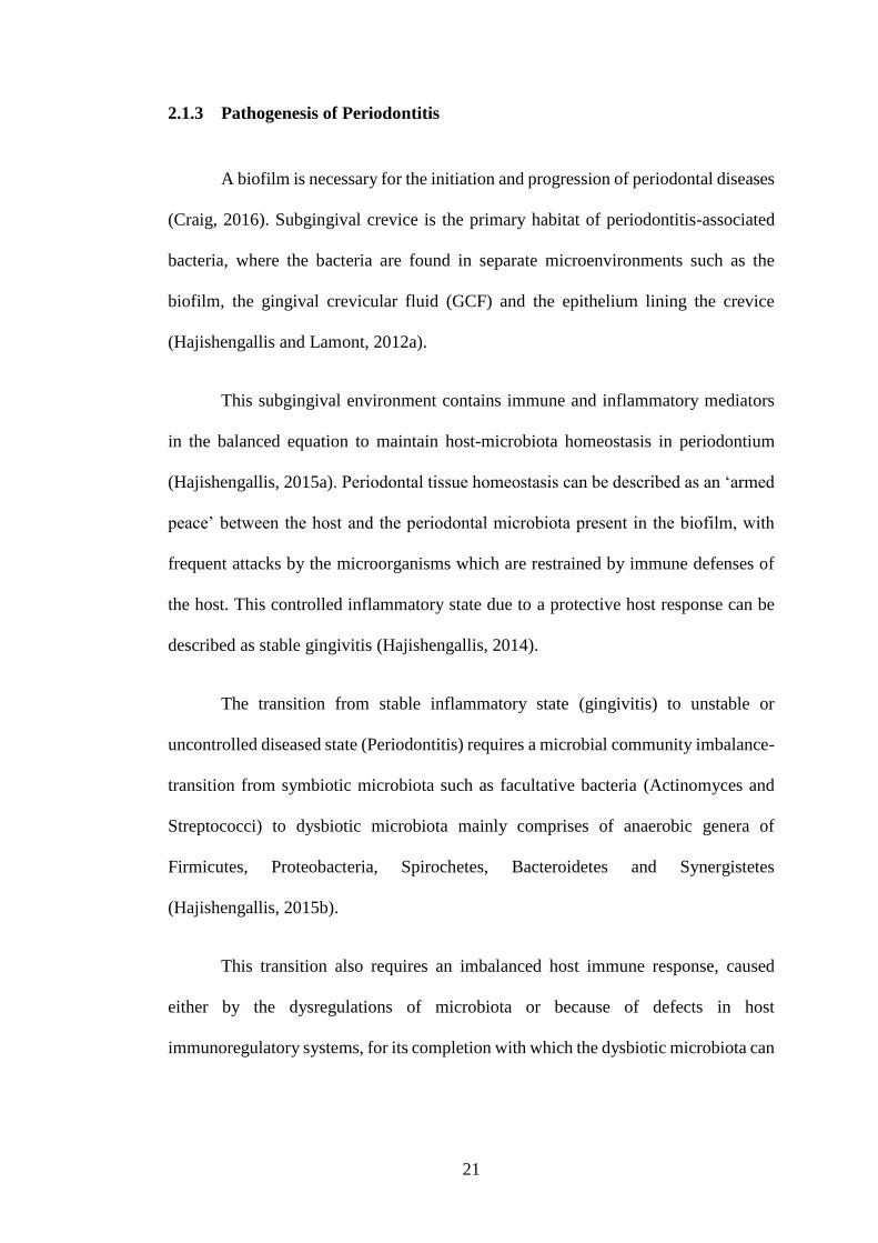
2.1.3 Pathogenesis of Periodontitis
A biofilm is necessary for the initiation and progression of periodontal diseases
(Craig, 2016). Subgingival crevice is the primary habitat of periodontitis-associated
bacteria, where the bacteria are found in separate microenvironments such as the
biofilm, the gingival crevicular fluid (GCF) and the epithelium lining the crevice
(Hajishengallis and Lamont, 2012a).
This subgingival environment contains immune and inflammatory mediators
in the balanced equation to maintain host-microbiota homeostasis in periodontium
(Hajishengallis, 2015a). Periodontal tissue homeostasis can be described as an ‘armed
peace’ between the host and the periodontal microbiota present in the biofilm, with
frequent attacks by the microorganisms which are restrained by immune defenses of
the host. This controlled inflammatory state due to a protective host response can be
described as stable gingivitis (Hajishengallis, 2014).
The transition from stable inflammatory state (gingivitis) to unstable or
uncontrolled diseased state (Periodontitis) requires a microbial community imbalance-
transition from symbiotic microbiota such as facultative bacteria (Actinomyces and
Streptococci) to dysbiotic microbiota mainly comprises of anaerobic genera of
Firmicutes, Proteobacteria, Spirochetes, Bacteroidetes and Synergistetes
(Hajishengallis, 2015b).
This transition also requires an imbalanced host immune response, caused
either by the dysregulations of microbiota or because of defects in host
immunoregulatory systems, for its completion with which the dysbiotic microbiota can

22
engage through series of complex inflammatory interactions (Hajishengallis, 2014)
(figure 2.7).
Previously, bacterial species like Porphyromonas gingivalis, Tannerella
forsythia and Treponema denticola (also known as red complex), were considered as
primary etiological agents of periodontitis (Socransky et al., 1998a).
23
Figure 2.7: Polymicrobial synergy and dysbiosis in susceptible hosts causes
periodontitis. Adopted from (Hajishengallis, 2015a)

24
However recent developments based on independent metagenomic and
mechanistic approaches suggested polymicrobial synergy and dysbiosis in the
pathogenesis of periodontitis (the ‘PSD model’). This model suggests that P. gingivalis
is pathogenic because of its ability to encourage dysbiotic microbial communities and
may act as a keystone pathogen, but it requires the help of accessory pathogens and
overactivation by commensal bacteria known as pathobionts to cause disruption in the
homeostasis and promote destructive inflammatory state in the suspected individuals
(Hajishengallis and Lamont, 2012b) (figure 2.7).
The host immune of the body causes the initial release of neutrophils at the site
of inflammation. neutrophils fail to control the microbial dysbiosis, which can thus
penetrate the connective tissue and interact with additional immune cell types, such as
dendritic cells (DCs), macrophages (Mw), and Gamma delta T (γ T) cells; a subset of
innate-like lymphocytes. These cells are then responsible for the production of
proinflammatory mediators (such as the bone-resorptive cytokines tumor necrosis
factor (TNF), interleukin (IL)-1b, and IL-17) also the regulation of development of T
helper (Th) cells, which also contribute to and exaggerate the inflammatory response
(figure 2.8).
IL-17, by interacting with immune and connective tissue cell types such as
neutrophils, fibroblasts, and osteoblasts, induces the production of CXC chemokines,
matrix metalloproteinases (MMPs) and other tissue-destructive molecules such as
reactive oxygen species (ROS), Receptor activator of nuclear factor kappa-Β ligand
(RANKL). RANKL causes the maturation of osteoclast precursors (OCPs) leading to
bone resorption in periodontitis (figure 2.8) (Hajishengallis, 2014).