THE EFFECTS OF KINESIO® TAPE ON ACROMIOHUMERAL …
87
THE EFFECTS OF KINESIO® TAPE ON ACROMIOHUMERAL DISTANCE IN PATIENTS WITH SUBACROMIAL IMPINGEMENT A Thesis Submitted to the Graduate Faculty of the North Dakota State University of Agriculture and Applied Science By Nicholas Avery Sample In Partial Fulfillment of the Requirements for the Degree of MASTER OF SCIENCE Major Program: Advanced Athletic Training February 2018 Fargo, North Dakota
Transcript of THE EFFECTS OF KINESIO® TAPE ON ACROMIOHUMERAL …

THE EFFECTS OF KINESIO® TAPE ON ACROMIOHUMERAL DISTANCE IN PATIENTS
WITH SUBACROMIAL IMPINGEMENT
A Thesis Submitted to the Graduate Faculty
of the North Dakota State University
of Agriculture and Applied Science
By
Nicholas Avery Sample
In Partial Fulfillment of the Requirements for the Degree of
MASTER OF SCIENCE
Major Program: Advanced Athletic Training
February 2018
Fargo, North Dakota

North Dakota State University
Graduate School
Title
The Effects of Kinesio® Tape on Acromiohumeral Distance in Patients with
Subacromial Impingement
By
Nicholas Avery Sample
The Supervisory Committee certifies that this disquisition complies with North
Dakota State University’s regulations and meets the accepted standards for the
degree of
MASTER OF SCIENCE
SUPERVISORY COMMITTEE:
Katie Lyman
Chair
Kara Gange
Kathleen Woken
Approved: 03/01/2018 Yeong Rhee
Date Department Chair

iii
ABSTRACT
Kinesio® Tape has the potential to optimize the treatment of subacromial impingement
syndrome. This research project investigated the effect Kinesio® Tape has on patient-reported outcome
measures and acromiohumeral distance in patients with subacromial impingement syndrome. Twenty
volunteers exhibiting subacromial impingement syndrome symptoms were divided into two groups, one
receiving Kinesio® Tape inhibition technique of the supraspinatus and deltoid muscles and the other
receiving a sham Kinesio® Tape. Patient-reported SPADI scores and acromiohumeral distance measured
by diagnostic ultrasound were recorded at 24- and 48-hour intervals. SPADI scores of both groups were
statistically significantly lower at the 48-hour interval. No statistically significant change in
acromiohumeral distance was found at any interval. Therefore, Kinesio® Tape on the supraspinatus and
deltoid muscles alleviated symptoms related to subacromial impingement syndrome as reported by
patient-outcome data but did not alter the subacromial space according to diagnostic ultrasound
scanning.

iv
ACKNOWLEDGEMENTS
There are many individuals I would like to acknowledge for their support during this research
project. First, I would like to thank my committee chair Dr. Lyman for providing assistance and edits from
the start. Her support and drive allowed me to make this project as strong as possible. I would also like
to thank Kassiann Landin for dedicating her time as one of the researchers as well as assisting with
collecting data and formatting. Additionally, I appreciate the support and constructive criticism received
from the rest of my committee, Dr. Kara Gange and Kathleen Woken. This project could not have
transpired as it did without everyone involved. Lastly, thank you to all my friends and family who have
been my support system for these two years. I am lucky to have each and every one of you in my life.

v
TABLE OF CONTENTS
ABSTRACT ......................................................................................................................................... iii
ACKNOWLEDGEMENTS ....................................................................................................................... iv
LIST OF TABLES .............................................................................................................................. viii
LIST OF FIGURES .............................................................................................................................. ix
LIST OF ABBREVIATIONS .................................................................................................................... x
LIST OF SYMBOLS ............................................................................................................................. xi
CHAPTER 1. INTRODUCTION ............................................................................................................... 1
1.1. Overview of the Problem ........................................................................................................... 1
1.2. Statement of Purpose ............................................................................................................... 2
1.3. Research Questions .................................................................................................................. 2
1.4. Dependent Variable .................................................................................................................. 2
1.5. Independent Variable ................................................................................................................ 2
1.6. Limitations ............................................................................................................................... 2
1.7. Delimitations ............................................................................................................................ 3
1.8. Assumptions ............................................................................................................................. 3
1.9. Significance of Study ................................................................................................................. 3
1.10. Definitions .............................................................................................................................. 4
CHAPTER 2. LITERATURE REVIEW ....................................................................................................... 5
2.1. Shoulder Anatomy .................................................................................................................... 5
2.1.1. Bony Anatomy ................................................................................................................... 5
2.1.2. Muscular Anatomy ............................................................................................................. 7
2.2. Subacromial Impingement Syndrome ......................................................................................... 9
2.2.1. Etiology ........................................................................................................................... 10
2.2.2. Diagnosis and Treatment .................................................................................................. 14
2.3. Kinesio® Tape ....................................................................................................................... 19
2.3.1. Pain Regulation ................................................................................................................ 20
vi
2.3.2. Muscle Activity Regulation ................................................................................................ 21
2.3.3. Kinesio Taping® for Subacromial Impingement Syndrome ................................................. 24
2.3.4. Effects on the Subacromial Space ..................................................................................... 28
2.4. Diagnostic Ultrasound ............................................................................................................. 30
2.5 Conclusion .............................................................................................................................. 33
CHAPTER 3. METHODOLOGY ............................................................................................................. 35
3.1. Participants ............................................................................................................................ 35
3.2. Setting ................................................................................................................................... 35
3.3. Equipment and Instruments .................................................................................................... 36
3.4. Procedures ............................................................................................................................. 36
3.5. Data Analysis.......................................................................................................................... 41
3.6. Conclusion ............................................................................................................................. 41
CHAPTER 4. MANUSCRIPT ................................................................................................................ 43
4.1. Abstract ................................................................................................................................. 43
4.2. Introduction ........................................................................................................................... 44
4.3. Methods ................................................................................................................................. 46
4.3.1. Subjects .......................................................................................................................... 46
4.3.2. Study Design and Protocol ................................................................................................ 48
4.3.3. Intervention ..................................................................................................................... 49
4.3.4. Statistical Analysis ............................................................................................................ 51
4.4. Results ................................................................................................................................... 51
4.5. Discussion .............................................................................................................................. 54
4.6. Conclusions ............................................................................................................................ 58
REFERENCES .................................................................................................................................... 59
APPENDIX A. INFORMED CONSENT ................................................................................................... 64
APPENDIX B. TAKE HOME INSTRUCTIONS ......................................................................................... 69

vii
APPENDIX C. HEALTH HISTORY QUESTIONNAIRE .............................................................................. 71
APPENDIX D. SHOULDER PAIN AND DISABILITY INDEX ...................................................................... 75
APPENDIX E. INSTITUTIONAL REVIEW BOARD APPROVAL .................................................................. 76

viii
LIST OF TABLES
Table Page
1: Sensitivity, Specificity and Predictive Values in Clinical Diagnostic Tests .................................... 16
2: Sensitivity, Specificity and Likelihood Ratios in Clinical Diagnostic Tests .................................... 17
3: Functional Assessment Results of Manual Therapy and Kinesio® Taping Groups....................... 26
4: Length of Kinesio® Tape at Various Tensions ......................................................................... 40
5: Special Testing Results of Participants .................................................................................... 47
6: Effect of Kinesio® Tape on Mean Self-Reported SPADI Scores Over Time ................................ 53
7: Effect of Kinesio® Tape on AHD at 0° Over Time .................................................................... 54
8: Effect of Kinesio® Tape on AHD at 60° Over Time .................................................................. 54
9: Effect of Kinesio® Tape on AHD at 90° Over Time .................................................................. 54
10: Effect of Kinesio® Tape on AHD at 120° Over Time ................................................................ 54

ix
LIST OF FIGURES
Figure Page
1. The Three Types of Acromions7 ............................................................................................. 11
2. Kinesio® Tape Lifting Mechanism73 ........................................................................................ 20
3. Completed Kinesio® Tape Application .................................................................................... 40

x
LIST OF ABBREVIATIONS
AHD .................................................................Acromiohumeral Distance
SPADI ..............................................................Shoulder Pain and Disability Index
KTAI ................................................................Kinesio Taping Association International®
US ...................................................................Diagnostic Ultrasound
SC ....................................................................Sternoclavicular
AC....................................................................Acromioclavicular
GH ...................................................................Glenohumeral
ROM.................................................................Range of Motion
MMT ................................................................Manual Muscle Test
PPV ..................................................................Positive Predictive Value
NPV .................................................................Negative Predictive Value
SIT ..................................................................Subacromial injection test
VAS ..................................................................Visual Analog Scale
DASH ...............................................................Disabilities of the Arm, Shoulder, and Hand
MRI ..................................................................Magnetic Resonance Imaging
CT ....................................................................Computed Tomography
HHQ .................................................................Health History Questionare
CKTP ................................................................Certified Kinesio Tape® Practitioner
BLV ..................................................................Baseline Value
PIV1 .................................................................Post-Intervention Value 1
PIV2 .................................................................Post-Intervention Value 2
PTRV ................................................................Post-Tape Removal Value
ANOVA .............................................................Analysis of Variance

xi
LIST OF SYMBOLS
® ....................................................................Registered Trademark
.....................................................................Trademark
° ......................................................................Degrees
± .....................................................................Plus/Minus
< .....................................................................Less Than
> .....................................................................Greater Than
= .....................................................................Equals

1
CHAPTER 1. INTRODUCTION
1.1. Overview of the Problem
Subacromial impingement syndrome is the most commonly diagnosed shoulder condition in the
general population, accounting for 44-65% of all shoulder complaints.1-6 This diagnosis is often used as a
broad term that encompasses multiple specific pathologies including supraspinatus tendinopathy,
subacromial bursitis, and biceps tendinopathy. 1,3-5,7 Even with the high prevalence of this condition, the
etiology and proper course of treatment is unclear. Once diagnosed, treatment often consists of
stretching and/or rehabilitation exercises to correct any imbalances. In addition, modalities such as ice,
electrical stimulation, soft tissue mobilization, therapeutic ultrasound, and Kinesio® Tape may be used to
alleviate symptoms related to subacromial impingement syndrome.6,8-11 Unfortunately, there is no specific
treatment that has been proven to be the best for patients with subacromial impingement syndrome.
Clinicians must use a holistic approach to evaluate this condition and knowledge of the treatment options
available is vital to positive patient outcomes.
Kinesio® Tape is a widely used modality first created in the 1970s by Dr. Kenzo Kase.12,13 With a
multitude of theorized benefits, but minimal evidence supporting or refuting them, it is understandable
that the use remains controversial. Among these theorized applications is the regulation of muscle
activity.14-17 When applied from a muscle’s insertion to its origin, Kinesio® Tape is thought to inhibit the
targeted muscle, which can be beneficial if symptoms are due to overactive musculature.13 With
subacromial impingement syndrome, the deltoid and supraspinatus muscles are often overactive and pull
the humeral head superiorly, decreasing the subacromial space.5,7,18,19 Kinesio® Tape, applied using the
inhibition method, may decrease activity of these muscles therefore increasing the subacromial space and
lessening symptoms related to subacromial impingement syndrome.
To quantify the subacromial space, diagnostic ultrasound has been shown to be a valid and
reliable imaging technique.20-24 Diagnostic ultrasound is a noninvasive, inexpensive imaging technique
that can be used to visualize musculoskeletal structures in real time.22,25 The subacromial space has been
quantified in the literature with various landmarks, but the most common and representative

2
measurement involves the inferior aspect of the acromion and the superior aspect of the humerus20,24,26
and will be the method used in this study.
1.2. Statement of Purpose
The purpose of this study was to determine if application of Kinesio® Tape for inhibiting the
supraspinatus and deltoid has an effect on acromiohumeral distance (AHD) as well as pain measures for
patients suffering from subacromial impingement.
1.3. Research Questions
1) What are the differences in AHD when Kinesio® Tape is applied on individuals who are
diagnosed with subacromial impingement?
2) What are the differences on participants’ perceived shoulder disability and pain levels with and
without Kinesio® Tape applications?
1.4. Dependent Variable
The focal dependent variable in this study was the AHD measured with diagnostic ultrasound
before and after Kinesio® Tape application. In addition, Shoulder Pain and Disability Index (SPADI)
scores were recorded to quantify outcome measures.
1.5. Independent Variable
The independent variable in this study was the application of Kinesio® Tape.
1.6. Limitations
This research study was not without limitations due to the numerous variables present. First, this
study was limited to participants between the ages of 18 and 55; therefore, the results are not
generalizable to populations outside of this age range including pediatric and geriatric patients.
Additionally, participants presented with varying severity of subacromial impingement. Parameters for
differentiating severities of this condition have not been clearly outlined in previous literature, but the
Kinesio® Tape may impact them differently. These limitations were outside of the current study’s scope
and future research should focus on developing methodologies to reduce these factors.

3
1.7. Delimitations
This study was delimited to the Fargo-Moorhead area of North Dakota and Minnesota, United
States. To be included in the study, participants must have had symptomatic shoulder pain that was
diagnosed as subacromial impingement syndrome by the researcher using the clinical prediction rule
given by Park et al.27 Participants had to be symptomatic at the start of the study, which occurred in the
spring and fall of 2017. Due to time and resource constraints, this study was conducted over the course
of 48 hours and did not include any long-term progression. Additionally, activity levels of the participants
as well as shoulder complex usage were not accounted for outside of instructing the participants to
continue their typical daily activities. The final delimitation for this study involved the application of
Kinesio® Tape. Dr. Kenzo Kase and Kinesio Taping Association International® (KTAI) recommend using
three strips to improve symptoms related to rotator cuff impingement or dysfunction: supraspinatus
inhibition, deltoid inhibition, and glenohumeral joint mechanical correction. In this study, the
glenohumeral joint mechanical correction strip was not used as this would interfere with the diagnostic
ultrasound transducer placement on the shoulder joint. These factors are outside the scope of the current
study and should be the focus of future research. The researchers of the current study considered
numerous variables and reviewed the available literature on subacromial impingement, Kinesio® Tape,
and diagnostic ultrasound in order to formulate an appropriate methodology.
1.8. Assumptions
There were a few assumptions that were made throughout this research study. Since participants
were instructed to continue with their normal daily routine, it was assumed that subjects honestly and
accurately reported any vigorous activity (e.g. weight lifting). It was also assumed that the participants
would remove the Kinesio® Tape if they felt any discomfort or irritation.
1.9. Significance of Study
Kinesio® Tape is a modality used by many members of the medical community including athletic
trainers and physical therapists. However, use of Kinesio® Tape continues to be a controversial
treatment option due to lack of consistent evidence. While the effects of Kinesio® Tape on shoulders is a
popular topic, no previous research design had investigated the effect this modality has on the objective

4
measure of AHD in individuals with symptomatic subacromial impingement syndrome. Overall, this study
aids clinicians in determining if Kinesio® Tape is a viable treatment option in patients with subacromial
impingement syndrome.
1.10 Definitions
Subacromial impingement syndrome: The symptomatic irritation of structures between the
superior aspect of the humerus and the inferior aspect of the acromion.3,4
Acromiohumeral Distance (AHD): The distance between acromion and the superior humeral
head.28 For this study, the AHD will be measured under diagnostic ultrasound as a perpendicular line
from the hyperechoic lateral acromion to the hyperechoic humeral head to represent the distance
between the cortical layer of the acromion and cortical layer of the superior humeral head.
Kinesio® Tape: A kinesthetic tape composed of a polymer elastic strand wrapped by cotton fibers
that mimics the thickness and flexibility of skin. Able to stretch approximately 55-60% of its resting
length, Kinesio® Tape is used to alter muscle activation, increase proprioception, decrease pain, increase
lymphatic drainage, and provide mechanical support.13
Kinesio® Tape inhibition method: A Kinesio Taping® Methods technique used to decrease
muscle activity. The tape is applied from the target muscle’s insertion to its origin with 15-25% of
available tension.13
Diagnostic ultrasound: a non-invasive imaging technique that uses a transducer containing a
crystal sound head. This transducer creates sound waves which interact with soft tissues to produce an
image.29

5
CHAPTER 2. LITERATURE REVIEW
Shoulder pain is the most common musculoskeletal complaint across all ages.1-3,30 Of all shoulder
pain, subacromial impingement syndrome is the most commonly diagnosed disorder, accounting for 44-
65% of all shoulder pathologies.1-6 Subacromial impingement syndrome is a term that can be used for a
variety of specific pathologies, but most commonly involves supraspinatus tendinopathy or subacromial
bursitis.4,7 For this literature review, subacromial impingement syndrome is broadly defined as the
symptomatic irritation of structures between the superior aspect of the humerus and the inferior aspect
of the acromion. Due to the common nature of this condition, various treatments are constantly being
investigated. One of these treatments is Kinesio® Tape, which has been theorized to decrease symptoms
of subacromial impingement syndrome by increasing the acromiohumeral distance (AHD), thus increasing
the space available for structures in the area.13 The purpose of this literature review is to give
background information on subacromial impingement syndrome with regards to anatomy, diagnosis,
treatment, Kinesio® Tape, and Diagnostic Ultrasound (US).
2.1. Shoulder Anatomy
An understanding of the musculoskeletal anatomy present at any specific area is vital to
comprehend the epidemiology behind an injury. The shoulder complex is an intricate structure; with
multiple motions, muscular attachments, bony articulations, nerves, and ligamentous attachments, the
anatomy of the shoulder is often not well understood. Of particular importance to this research study is
how the bony and muscular anatomy of the shoulder relates to subacromial impingement syndrome.
2.1.1 Bony Anatomy
There are four main bones that comprise the shoulder complex: sternum, clavicle, scapula, and
humerus.31,32 The sternum is the flat bone centered over the chest and allows for the attachment of the
12 rib bones as well as the clavicle.33 The clavicle, known as a collarbone to the lay person, is the s-
shaped bone that runs from the sternum to the tip of the shoulder.31 This bone serves to attach the
upper extremity to the trunk through the sternoclavicular (SC) joint.31,33,34 The distal end of the clavicle
articulates with the scapula to form the acromioclavicular (AC) joint.31,34,35 The scapula (also known as
the shoulder blade) is the irregularly shaped bone that lays primarily on the upper, lateral thorax and

6
includes the glenoid fossa in which the humeral head is positioned, forming the glenohumeral (GH)
joint.36 This bone is responsible for accessory motions that allow the glenohumeral joint to have a vast
range of motion.36 This literature review will focus on the AC and GH joints, as these are typically the
most important when discussing subacromial impingement. However, the SC joint as well as the
scapulothoracic articulation, due to its role in scapular dyskinesia, should be considered by clinicians
when evaluating a shoulder injury.
The AC joint is imperative when discussing subacromial impingement syndrome as it is directly
related to the superior border of the subacromial space. As the name implies, the subacromial space is
located beneath the acromion of the scapula. At the AC joint, the acromion and coracoid process of the
scapula articulate with the distal end of the clavicle.35 Reinforced by the acromioclavicular,
coracoacromial, and coracoclavicular ligaments, there are three degrees of freedom available at the AC
joint: internal/external rotation, upward/downward rotation, and anterior/posterior scapular tipping.30-
32,34,35 This large amount of motion available contributes significantly to subacromial impingement
syndrome as any abnormalities or pathologies involving the AC joint impact the area as a whole.
The glenohumeral (GH) joint, is the articulation between the glenoid fossa of the scapula and the
head of the humerus and is often regarded as more important than the AC joint when discussing
subacromial impingement syndrome.32,36,37 Frequently described as “a golf ball on a tee,” this joint is
responsible for the largest range of motion in the human body.37 As a ball-and-socket joint, the motions
available include flexion/extension, vertical and horizontal abduction/adduction, internal/external rotation,
and circumduction.9,32,36 The stability at the GH joint is almost entirely due to ligamentous support of the
superior, middle, and inferior GH ligaments and muscular support of the rotator cuff, deltoids, and
trapezious.30,38 The GH joint is of particular importance to subacromial impingement syndrome due to the
vast majority of shoulder motion that is accomplished at this joint. Located directly below the acromion,
the subacromial space utilizes the head of the humerus as the inferior border.5 In essence, any motion at
the shoulder that results in humeral head motion will affect the subacromial space.
With two bony borders of the acromion and the humeral head, the subacromial space is a
delicate area susceptible to many pathologies. In this space, the supraspinatus tendon passes and inserts

7
on the head of the humerus and is the second most often injured structure of the shoulder, behind only
the acromioclavicular ligament.39,40 Other tissues present in the subacromial area include the subacromial
bursa and long head of the biceps tendon.5,41 Subacromial impingement syndrome can encompass
pathology to any of these structures, including but not limited to partial thickness rotator cuff tears,
rotator cuff and biceps tendinopathy, and subacromial bursitis.4,7
2.1.2. Muscular Anatomy
There are 19 muscles that allow for movement of the scapula or the humerus in every plane of
motion.31,32,34 While all of these muscles are imperative to movement, this portion of the literature review
will focus on specific musculature that directly affects motion at the glenohumeral joint related to
subacromial impingement syndrome. The supraspinatus, subscapularis, infraspinatus, teres minor,
deltoids, trapezious, and biceps brachii are muscles of particular importance when discussing this
diagnosis.
With the rotator cuff musculature accounting for a large amount of motion available at the
shoulder as well as the stabilization of the humerus, it may be the most important group of muscles when
investigating shoulder pathologies.5,7 Originating on the posterior scapula above the spine, the
supraspinatus is arguably the most important muscle to consider when discussing subacromial
impingement as it is the most often inflamed muscle of the rotator cuff.2,3,5,7,42 The supraspinatus is a
muscle of interest when considering the subacromial space because it passes directly through this space
and inserts on the apex of the humeral head.43-45 The supraspinatus functions primarily to abduct the
humerus and stabilize the humeral head, although there is a component that is responsible for external
rotation of the humerus as well.43,44 While the supraspinatus contributes a small amount of external
rotation, this motion is primarily achieved by the teres minor and infraspinatus working in tandem.43
These muscles also aid in horizontal abduction and humeral head stabilization and the teres minor plays a
small role in glenohumeral extension.31,43 Both the teres minor and infraspinatus originate on the
posterior aspect of the scapula, with the infraspinatus directly below the scapular spine and the teres
minor immediately beneath the infraspinatus.45 The insertions of the infraspinatus and teres minor on the
posterosuperior and posterolateral humeral head respectively allow these muscles to take a large role in

8
external rotation.45,46 The final rotator cuff muscle, the subscapularis, is the primary muscle credited for
internal rotation of the humerus, as well as GH stabilization.43 Unlike the other rotator cuff muscles, the
subscapularis originates on the anterior aspect of the scapula and inserts on the anterior aspect of the
humeral head.45,46 Similar to the rest of the rotator cuff, the subscapularis plays a large role in humeral
head stabilization.31,32,34 While certainly an important and arguably the most important component to the
shoulder complex, the rotator cuff musculature cannot stand alone and other muscle groups must be
considered as well.
Outside of the rotator cuff, the deltoid has been shown to have the largest impact on
subacromial impingement.5,7,19 There are three components to the deltoids: posterior, middle, and
anterior, and while they have a common insertion on the deltoid tubercle of the proximal humerus; their
differing origins allow for unique actions in each.47 The anterior deltoid originates on the lateral clavicle
and is the prime flexor of the shoulder.32,43 The anterior deltoid also aids in abduction, horizontal
adduction, and internal rotation.43 The origin of the middle third of the deltoid on the acromion process
allows primarily for abduction while also aiding in shoulder flexion.43,47 Notably, this portion of the deltoid
also provides the lateral border for the subacromial space. Finally, the posterior deltoid, initiating on the
spine of the scapula, is primarily responsible for shoulder extension and horizontal abduction, while also
contributing to shoulder abduction and external rotation.43,47 If the rotator cuff musculature, particularly
the internal and/or external rotators, of an individual is weak due to pathology, decreased strength, or
lack of neuromuscular control, the deltoid may become overactive to assist these movements.7 An
overactive deltoid is often a factor in subacromial impingement as it leads to a narrowing of the
subacromial space as it pulls the humeral head into a resting state of elevation that is unopposed by the
diminished inferior forces of the internal and external rotator.5,7,18,19
While not having an attachment on the humerus, the trapezius is an important muscle to
consider when evaluating subacromial impingement.18,48,49 Similar to the deltoid, the trapezius has three
sections: upper, middle, and lower. The upper and lower segments primarily function to rotate the
scapula upward and downward respectively, while the middle trapezius primarily retracts the scapula.31,50
With insertions on the lateral clavicle and acromion process, the trapezius also aids with acromion

9
elevation. A weak trapezius may lead to depression in this area, preceding a smaller subacromial space
and higher likelihood of impingement.18,48
Lastly, the biceps brachii muscle often plays a multifactorial role in subacromial shoulder
impingement.51 Primarily tasked with elbow flexion, this muscle is composed of two heads, the long head
and short head, although the former is often viewed as the more intricate structure due to its origin on
the supraglenoid tuberosity and superior aspect of the glenoid labrum.52 With this origin, the biceps
brachii provides assistance with shoulder flexion.53 Similar to the aforementioned muscles, the long head
of the biceps tendon is another structure that runs directly through the subacromial space and is often
damaged due to subacromial impingement.51
The preceding is by no means a comprehensive overview of the musculature involved at the
shoulder, and certain factors must be considered on a case-by-case basis. However, the musculature
highlighted in this section tend to play the most prominent role in subacromial impingement syndrome.
The deltoid and supraspinatus particularly are often viewed as the most significant muscles with this
pathology as these muscles directly contribute to superior translation of the humeral head, thus
narrowing the subacromial space.
A thorough knowledge of the musculoskeletal anatomy present at the shoulder complex is
essential in order to fully understand any pathology at the shoulder. The anatomy related to subacromial
impingement syndrome is of particular importance as this condition has the highest prevalence of all
shoulder pathologies. In terms of bony anatomy, the scapula, clavicle, and humerus, as well as the
articulations that these bones share, have the highest influence on the subacromial space. From a
muscular anatomy viewpoint, the most important muscles are the deltoid, trapezius, and rotator cuff as
these contribute the most to movement and stability at the GH joint. With knowledge of the anatomy, the
possible etiologies of the condition can be better understood and discussed.
2.2. Subacromial Impingement Syndrome
Subacromial impingement syndrome is a complex and poorly understood pathology.1-6 In
addition, it is the most commonly diagnosed shoulder disorder in the general population, accounting for
44-65% of all shoulder complaints.1-6 Subacromial impingement syndrome is typically defined as the

10
symptomatic irritation of structures between the superior aspect of the humerus and the inferior aspect
of the acromion. This diagnosis is often used as a catch-all term for more specific pathologies which may
occur in the subacromial space including supraspinatus tendinopathy, subacromial bursitis, and long head
of the biceps tendinopathy.1,3-5,7 Any one or combination of these structures may be damaged in
subacromial impingement syndrome and differentiation is difficult without the use of advanced imaging or
arthroscopic surgery.4 Many causes of subacromial impingement syndrome have been theorized, but
there is no universal constant in all cases and specific factors contributing to subacromial impingement
syndrome must be examined on an individual basis.
2.2.1. Etiology
The theorized etiology of subacromial impingement syndrome has changed drastically in the past
decades. In 1972, Neer proposed that 100% of subacromial impingement syndrome pathologies could be
attributed to the acromion.35 In the years since, various causes of subacromial impingement syndrome
have been described and are accepted in the medical community. These causes may be broken down
into six categories: anatomical/mechanical, rotator cuff dysfunction, hypermobility, restrictive movement,
scapular instability, and posture.5,7 This literature review will examine each of these, as a thorough
understanding of the different possible causes of subacromial impingement syndrome is imperative when
discussing the pathology.
One of the oldest and most well-known etiologies of subacromial impingement syndrome, the
anatomical/mechanical group, has been examined thoroughly in the literature.7,35,39 This is an extension
of Neer’s hypothesis about subacromial impingement syndrome and encompasses any morphological
abnormalities of the acromion including acromion shape, acromion bone spurs, and os acromiale.5,7,35
Figure 1 illustrates the three different acromion shapes which have been described in the literature: flat,
curved, and hooked.7,35,39 While results are controversial, it seems that the shape, as well as the angle,
plays an important role in subacromial impingement syndrome. Hooked acromions, lower angles, and
bone spurs have been found more often in patients with subacromial impingement syndrome and rotator
cuff tears.39,54 In cases without acromion abnormalities, Neer35 hypothesized that subacromial
impingement syndrome was caused by the shape of the coracoclavicular ligament, as the length of this

11
ligament was thought to partially dictate the size of the subacromial space, and the thickness of the
ligament places an added pressure on structures in the area.5,7 The final specific pathology related to this
group is posterosuperior glenohumeral impingement, also known as internal impingement.7,55 While the
previous causes have been based on morphological differences in the acromion, this classification occurs
when the anatomical variance originates from the superior surface of the humerus, which is the inferior
border of the subacromial space.5,7,55 This pathology most often causes damage during overhead
motions, as elevation of the humerus decreases the subacromial space.7,55 Any outgrowth of bone on the
superior aspect of the humerus thus narrows this space further.7,55 While anatomical and mechanical
variances play a significant role in subacromial impingement syndrome, other factors must be examined.
Figure 1. The Three Types of Acromions7
The second category that has been theorized to be a contributor of subacromial impingement
syndrome is rotator cuff dysfunction. Rotator cuff dysfunction can contribute to subacromial impingement
syndrome through one of two pathways: degenerative tendinopathy or overuse injuries.7 Degenerative
tendinopathy occurs most often in later years of life and can by symptomatic or asymptomatic.56 It has
been reported that as high as 54% of asymptomatic patients over the age of sixty have some form of
degenerative tendinopathy.56 Overuse injuries occur often in sports, particularly in repetitive overhead
sports such as baseball and volleyball.7,40,57 Both degenerative and overuse tendinopathies lead to
weakness in the rotator cuff musculature, which may compromise stability of the glenohumeral joint.5,7,43

12
Specifically, the teres minor, infraspinatus, and subscapularis play a large role in stability during shoulder
elevation.5,7 The insertions of these muscles aid in pulling the humeral head inferiorly to counteract the
superior pull of the deltoid.5,7,45 With a weakness in these muscles or tendons, the forces from the deltoid
cannot be overcome and the humeral head shifts superiorly, narrowing the subacromial space and
contributing to subacromial impingement syndrome.5,7
Similar to a lack of stability associated with rotator cuff dysfunctions, the mobility of the GH joint
may play a large role in subacromial impingement syndrome. A hypermobile GH joint leads to an increase
in humeral head translation on the glenoid fossa in various directions during normal kinematics.5,7 This is
of particular importance in GH flexion and elevation as a superior translation of one to three mm has
been demonstrated in the first 30-60° of motion.58 An increase in this translation is likely due to the
inability of stabilizing factors including anatomical morphology, rotator cuff, and GH ligaments to
overcome the pull of the deltoid.5,7,45 The mean distance between the superior humeral head and the
acromion has been described to be between 9 and 15 mm in healthy individuals in studies using
radiographs and/or diagnostic ultrasound.22,25,59,60 Any measurement less than nine mm can increase the
risk of patients of developing symptoms of subacromial impingement syndrome.25,60 Therefore, any
increase in superior translation beyond normal could decrease the subacromial space and be a causative
factor of subacromial impingement syndrome5,7.
In direct opposition to hypermobility, restrictive processes can also play a role in subacromial
impingement syndrome.5,7,61 Restrictive motion at the GH joint most often occurs due to tightness in the
posterior capsule associated with adhesive capsulitis or surgical procedures.7 These restrictions cause
glenohumeral pathomechanics similar to hypermobile shoulders. For the shoulder to obtain full range of
motion (ROM) and stability, the humeral head must undergo arthrokinematic movements including rolling
and gliding, which may be hampered with posterior capsule tightness. One study specifically examined
the translation of the humeral head after posterior capsule tightening in cadavers and an increase in both
superior and anterior translation was found during passive shoulder flexion.61 This increase in translation
can cause subacromial impingement syndrome as the subacromial space is further compressed in these
13
cases.5,7,59 When evaluating a case of subacromial impingement syndrome, both hyper- or hypomobility
of the joint must be addressed as either one of these factors can be the causative factor5,7.
Recently receiving increased attention, the etiology of subacromial impingement syndrome in
some cases can be traced to scapular instability.5,7,9,36 During GH elevation, the scapula should undergo
upward rotation, allowing the humerus to achieve the full ROM.9 In patients with scapular instability or
scapular dyskinesis, the scapula does not properly rotate and excessive humeral head rotation or
translation occurs as compensation.5,7,9 Scapular dyskinesis may be due to multiple factors, although
muscular abnormalities seem to be the leading cause.9 Several muscles directly attach to the scapula and
provide scapular movement; these include but are not limited to: the rhomboids, trapezius, levator
scapulae, and serratus anterior.31,32 An imbalance or weakness in any of the muscles often leads to
scapular instability.5,7,9,36 Excessive restrictions at the scapulothoracic articulation can also lead to
scapular dysfunction and a restriction in free movement.7,31 The clear relation between the scapula and
the humerus is an important factor to investigate in subacromial impingement syndrome cases as the
true etiology may come from either of these components.
The final collectively accepted theory behind subacromial impingement syndrome is rooted in
posture. The term “slouched posture” refers to a combination of three pathological postures: forward
head, forward shoulder, and thoracic spine flexion.5 While any one of these may cause pathomechanics,
the combination of all three is the most concerning when discussing subacromial impingement syndrome.
This postural abnormality relates a tightness of the anterior musculature and weakness of posterior
musculature, which, in turn, places the scapula in an increased anterior tilt and decreases the
subacromial space.5,7 In addition, cervical spine flexion has been correlated to abnormal scapular
position.5,62 Cervical spine flexion of 25° has been demonstrated to decrease scapular posterior tilting as
well as increase scapular upward rotation in shoulder elevation, both of which decrease the subacromial
space.62 While conducting a postural examination from the sagittal plane, clinicians are able to objectively
visualize posture which has been shown to have a significant effect on subacromial impingement
syndrome and treatment focusing on such is growing in the clinical setting.

14
As evidenced, subacromial impingement syndrome is a complicated pathology which may include
a variety of structures and the etiology of which is difficult to find and varies by case. The more
recognized and accepted etiologies include anatomical/mechanical abnormalities, rotator cuff dysfunction,
hypermobility, restrictive movement, scapular instability, and posture.5,7 In order to find the true cause of
subacromial impingement syndrome, a detailed evaluation must be performed by a trained professional
who understands the biomechanics involved in the shoulder complex.
2.2.2. Diagnosis and Treatment
The prominence of shoulder pain surpasses any other musculoskeletal condition and the
complexity of the joint can make accurate diagnosis challenging.1-3,30 The difficulty of diagnosing
subacromial impingement syndrome is prevalent primarily due to the multifactorial etiology behind the
condition and lack of consensus on diagnosis technique. Typically, subacromial impingement syndrome is
diagnosed through a detailed clinical examination including history, manual muscle tests, and diagnostic
tests. There is little consensus on a gold standard for diagnosis, with studies using MRI, diagnostic US or
surgery as a reference standard. Therefore, the accuracy of special tests must be understood by the
clinician1,2,27,63-65
While there is an extensive list of diagnostic tests for shoulder assessment, there are fewer that
are accepted as useful for subacromial impingement syndrome. The tests that are most often utilized in
an evaluation and reviewed in the literature include Neer’s,1,2,27,63,64 Hawkins-Kennedy,1,2,27,63,64 Painful
Arc,1,27,63,64 Drop Arm,1,2,27 External Rotation Resistance,27,63,64 and Empty Can.2,63,64 Difficulty in assessing
the accuracy of some of these tests presents in the form of the technique used, as some tests are less
defined and may be interpreted differently by clinicians. These tests should all be performed with the
patient standing to increase reliability unless otherwise noted. Neer’s test is performed by the clinician
stabilizing the scapula of the seated patient with one hand and passively flexing the humerus until pain is
produced or full flexion is achieved.66 A positive Neer’s test is any production of pain. The Hawkins-
Kennedy test involves the clinician placing the patient in 90° of shoulder flexion and elbow flexion.67 The
examiner then passively internally rotates the arm and notes any pain, which indicates a positive result.
For the Painful Arc test, the patient actively abducts the arm through the full ROM and then lowers the

15
arm back to the side.1,27,63,64 A positive result is considered if there is pain produced between 60° and
120°. In the Drop Arm test, the patient is passively placed in to 90° of shoulder abduction and instructed
to lower arm back to neutral.65 A positive test is observed with the inability of the patient to lower the
shoulders in a controlled manner due to pain. To perform the External Rotation Resistance test, also
considered the Infraspinatus Manual Muscle test (MMT), the clinician resists external rotation with the
shoulder fully adducted and the elbow in 90° of flexion.65 A positive result is considered if weakness is
noted by the clinician as compared bilaterally or if pain is reproduced. For the Empty Can test, also
considered the Supraspinatus MMT, the patient is asked to flex their shoulders to 90° in the scapular
plane with full elbow extension and internal rotation.2,63,68 The examiner then applies downward pressure
on the upper surface of the arm. The test is considered positive if the clinician notices weakness
compared bilaterally. While these tests are commonly performed in similar ways, some slight variations
are noted between studies that could impact accuracy of the test and the diagnostic values found in
studies.65
There have been multiple studies which have examined the diagnostic value of typical diagnostic
tests used for subacromial impingement syndrome as well as related tests.1,2,27,63,64 One such study
conducted by Çalıs et al1 investigated the sensitivity, specificity, positive predictive value (PPV) and
negative predictive value (NPV) of seven tests: Neer’s, Hawkins-Kennedy, Horizontal Adduction, Painful
Arc, Drop Arm, Yergason’s, and Speed’s. There were 120 patients recruited for this study; five patients
had bilateral shoulder pain for a total of 125 pathological shoulders. The subjects were separated into
two groups after undergoing the subacromial injection test (SIT), wherein an injection of 10 cc of 1%
lidocaine was placed in the subacromial space. Marked reduction in pain or ROM after 30 minutes was
considered a positive test and subjects with a positive SIT were diagnosed with subacromial impingement
syndrome and placed in the experimental group. The patients also underwent an MRI to further examine
the structures involved and classify the type of impingement. After the MRI, each subject underwent each
of the listed tests performed by two physicians. Results of the study are listed in Table 1. The Hawkins-
Kennedy test recorded the highest sensitivity at 92.1%, but also recorded the lowest specificity (25.0%).
In contrast, the Drop Arm test was found to have the highest specificity (97.2%), but also the lowest

16
sensitivity (7.8%). The sensitivity of a test indicates its ability to result with a true positive and the
specificity of a test represents its ability to detect true negatives. Furthermore, a high sensitivity indicates
that the test is useful in identifying subjects without the condition, while a high specificity is useful in
identifying patients with the condition. Therefore, if a test with high sensitivity returns a negative result,
the clinician can be confident in ruling out the pathology. On the other hand, if a test with high specificity
returns with a positive result, the condition is likely present. The results of this study indicate that there is
no single test with high sensitivity and specificity and a combination of tests and thorough evaluation
must be implemented by the clinician in order to diagnose subacromial impingement syndrome.
Table 1: Sensitivity, Specificity and Predictive Values in Clinical Diagnostic Tests
Test Sensitivity (%) Specificity (%) Accuracy (%) PPV(%) NPV(%)
Hawkins-Kennedy Neer Horizontal Adduction Speed Yergason Painful Arc Drop Arm
92.1 88.7 82.0 68.5 37.0 32.5 7.8
25.0 30.5 27.7 55.5 86.1 80.5 97.2
72.8 72.0 66.4 64.8 51.2 46.4 33.6
75.2 75.9 73.7 79.2 86.8 80.5 87.5
56.2 52.3 38.4 41.6 35.6 32.5 29.9
A similar study investigated the diagnostic value of seven tests for subacromial impingement
syndrome: Neer’s, Hawkins-Kennedy, Painful Arc, Empty Can, Full Can, Resisted External Rotation, and
Resisted Abduction.63 This study recruited 34 patients that were diagnosed with subacromial
impingement syndrome. Diagnosis was made using ultrasonography and patients were classified into one
of three groups after imaging: full-thickness rotator cuff tear, partial-thickness rotator cuff tear, or
subacromial/subdeltoid bursitis. The tests were performed immediately after ultrasound imaging by a
physiotherapist who was blinded to the results. Similar to the previous study, the Hawkins-Kennedy test
was found to have the highest sensitivity (74.1%), but had a lower specificity (50.0%). In addition, the
external rotation resistance test was found to have a specificity of 100%, although the sensitivity of the
test was low (34.5%). This pattern was consistent with the majority of the tests in that a test with higher
sensitivity often had lower specificity and vice-versa. Full results from this study are presented in Table 2.
This study concluded that these diagnostic tests for subacromial impingement syndrome may not be as
17
accurate as desired by a clinician. In addition, a combination of tests as well as a strong understanding of
subacromial impingement syndrome are necessary for an accurate diagnosis.
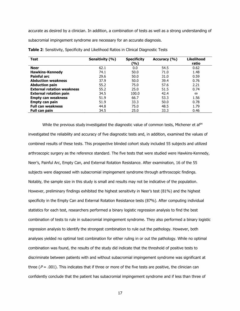
Table 2: Sensitivity, Specificity and Likelihood Ratios in Clinical Diagnostic Tests
Test Sensitivity (%) Specificity
(%) Accuracy (%) Likelihood
ratio
Neer Hawkins-Kennedy
Painful arc Abduction weakness
Abduction pain
External rotation weakness External rotation pain
Empty can weakness Empty can pain
Full can weakness Full can pain
62.1 74.1
29.6 37.9
55.2
55.2 34.5
51.9 51.9
44.8 34.5
0.0 50.0
50.0 50.0
75.0
25.0 100.0
66.7 33.3
75.0 25.0
54.5 71.0
31.0 39.4
57.6
51.5 42.4
53.3 50.0
48.5 33.3
0.62 1.48
0.59 0.76
2.21
0.74 ∞
1.56 0.78
1.79 0.46
While the previous study investigated the diagnostic value of common tests, Michener et al64
investigated the reliability and accuracy of five diagnostic tests and, in addition, examined the values of
combined results of these tests. This prospective blinded cohort study included 55 subjects and utilized
arthroscopic surgery as the reference standard. The five tests that were studied were Hawkins-Kennedy,
Neer’s, Painful Arc, Empty Can, and External Rotation Resistance. After examination, 16 of the 55
subjects were diagnosed with subacromial impingement syndrome through arthroscopic findings.
Notably, the sample size in this study is small and results may not be indicative of the population.
However, preliminary findings exhibited the highest sensitivity in Neer’s test (81%) and the highest
specificity in the Empty Can and External Rotation Resistance tests (87%). After computing individual
statistics for each test, researchers performed a binary logistic regression analysis to find the best
combination of tests to rule in subacromial impingement syndrome. They also performed a binary logistic
regression analysis to identify the strongest combination to rule out the pathology. However, both
analyses yielded no optimal test combination for either ruling in or out the pathology. While no optimal
combination was found, the results of the study did indicate that the threshold of positive tests to
discriminate between patients with and without subacromial impingement syndrome was significant at
three (P = .001). This indicates that if three or more of the five tests are positive, the clinician can
confidently conclude that the patient has subacromial impingement syndrome and if less than three of

18
the tests are positive, the likelihood of the presence of the pathology is unlikely. This concept of three
tests holding significance in differentiating patients with and without subacromial impingement syndrome
has been reinforced by other research and may aid clinicians in diagnosis.27
Systematic reviews and meta analyses on the diagnostic value of subacromial impingement tests
have also been performed.2,65 One such study conducted by Alquanaee et al2 included 16 studies in their
review primarily focusing on five diagnostic tests: Neer’s, Hawkins-Kennedy, Empty Can, Drop Arm, and
Lift-Off. While few studies have examined the Lift-Off test for subacromial impingement syndrome, this
systematic review found that this test had the highest diagnostic accuracy with a positive likelihood ratio
of 16.47 and a specificity and sensitivity of 97% and 42%, respectively. Additionally, Neer’s test was
found to have the highest pooled sensitivity at 78%, although, similar to most individual studies, the
specificity of Neer’s test was lower (58%). The Hawkins-Kennedy test followed this same pattern as well
(sensitivity: 74%, specificity: 54%). These results indicate that two diagnostic tests may be useful in
ruling out subacromial impingement syndrome in the case of a negative result, but a positive result does
not definitively indicate the pathology is present. One conclusion from this study was the diagnostic value
of the lift-off test being much higher than any other test examined. Also, as in previous studies, most
tests sacrifice sensitivity for specificity or specificity for sensitivity and there are no cases where both are
high.
In one of the most extensive systematic reviews on special tests for subacromial impingement
syndrome, Hanchard et al65 examined studies of diagnostic accuracy of tests for shoulder impingement
and pathologies related to shoulder impingement. The authors concluded that evidence upon which to
base selection of special tests for subacromial impingement syndrome is insufficient due, in large part, to
the diversity of performance of these tests.65 Nevertheless, the research indicates some trends in the
diagnostic tests including the abilities of certain tests to be highly sensitive (Neer’s, Hawkins-Kennedy)
and others to be highly specific (Drop Arm, Lift-Off, Painful Arc, External Rotation Resistance). The
accepted clinical prediction rule is given by Park et al27 and states that if the Hawkins-Kennedy, painful
arc, and external rotation resistance test are positive, there is a 95% accuracy in diagnosing subacromial

19
impingement syndrome (+LR: 10.6). While there is no consensus, it is important for clinicians to
understand where the literature is valuable and where there are shortcomings.
Once the diagnosis of subacromial impingement is made, treatments for the condition must be
considered. One common treatment approach is to manage the acromiohumeral distance (AHD). This
measurement has been theorized to be diminished in patients with subacromial impingement
syndrome.20-24,69 A study performed by Mackenzie et al28 was designed to discover if AHD was a cause or
effect of subacromial impingement. This descriptive review examined the literature available on
subacromial impingement as well as internal impingement to better understand the etiology behind these
two conditions. The researchers concluded that the hypothesis of AHD being a cause for subacromial
impingement syndrome has not been definitively established and no conclusion can be made on this
subject. Likely, as many authors have suggested, the cause of subacromial impingement syndrome is
multifactorial. However, maintenance of the subacromial space may be important for managing
symptoms of subacromial impingement syndrome regardless of whether the relationship is a cause or
effect. In addition, there are many treatment options available including therapeutic exercise, manual
therapy, laser therapy, modalities, and taping. One form of taping that has been suggested to improve
symptoms related to subacromial impingement syndrome by increasing the AHD is Kinesio® Tape.
2.3. Kinesio® Tape
Kinesio® Tape originated in the 1970s by way of a Japanese Chiropractor, Dr. Kenzo Kase, and
use has subsequently increased steadily, particularly in the last decade.12 One of the biggest contributors
to the growing popularity of Kinesio® Tape was the Olympic Games as athletes from around the world
were televised with brightly colored tape used to treat various musculoskeletal conditions.12 Kinesio®
Tape is an elastic therapeutic tape that can stretch up to 60% of its resting length in order to
treat various musculoskeletal and neuromuscular maladies including pain, proprioceptive deficits,
swelling, under/overactive muscles, and postural alignment.12,13,70-72 The theory behind Kinesio® Tape
differs depending on the intended result. For example, Kinesio® Tape is claimed to aid with swelling by
lifting the epidermis from the underlying layers of skin, thus opening the subcutaneous interstitial area
and the body’s natural lymphatic system and allowing fluid to flow out of the area (Figure 2).6,13,73

20
Additionally, Kinesio® Tape may be used to facilitate or inhibit musculature by stimulating
mechanoreceptors in the tissue which may increase or decrease muscle activity based on the direction of
tape application.13 The need for evidence-based treatment options has led to an increase of Kinesio®
Tape research in recent years as researchers try to support or refute the claims made by the inventor.
Figure 2. Kinesio® Tape Lifting Mechanism73
2.3.1. Pain Regulation
Treatment of pain is an important part of Kinesio® Tape and has been researched extensively,
albeit with varying results. Parreira et al74 conducted a systematic review to determine the clinical efficacy
of Kinesio® Tape. Included in this systematic review were randomized controlled trials that had been
published in peer-reviewed journals. Additional inclusion criteria included participants with
musculoskeletal conditions, application following the Kinesio Taping Method®, and outcome measures of
pain intensity, disability, quality of life, return to work, and global impression of recovery. A total of 12
studies were reviewed and the authors found no significance or minimal significance with many different
applications and goals. Many of the articles indicated that while a slight decrease in pain is noted in the
short-term, this reduction may be too small to be clinically significant. An earlier systematic review
similarly found inadequate evidence to support the use of Kinesio® Tape following musculoskeletal
injury.75 Six articles were included in this review, with two indicating there was no benefit in Kinesio®
Tape, one reporting no difference between Kinesio® Tape and other treatments, and three suggesting
short-term benefits. While the authors concluded there is insufficient evidence for the efficacy of
Kinesio® Tape, they also mentioned that a possible benefit could not be disregarded.

21
In contrast, a study examining the pain control aspect of Kinesio® Tape in patients with acute
whiplash indicated diminished pain in the treatment compared to a sham group.76 This study employed a
cervical extensor inhibition technique as well as a space correction technique over the midcervical region
in the experimental group, while a control group received Kinesio® Tape in the same pattern with no
tension applied to the tape. Pain levels were taken immediately after Kinesio® Tape application as well
as 24 hours post application. Reported pain levels were reduced both immediately and 24 hours after
Kinesio® Tape application in the experimental group compared to the control group. Additionally, a
review of the research into Kinesio® Tape for myofascial pain conducted by Wu et al73 reported
significant benefits found in multiple studies due to the lifting of the subcutaneous layer surrounding a
myofascial trigger point, allowing for increased blood flow, increased lymphatic drainage, and muscle
relaxation. Overall, the literature on the effectiveness of Kinesio® Tape to treat pain varies substantially,
with some articles reporting no benefits and others indicating short-term pain relief.
2.3.2. Muscle Activity Regulation
Muscle activity regulation through facilitating or inhibiting musculature is one of the most
common research questions used when applying Kinesio® Tape.14-17,77-80 Specifically, in one study, the
effects of Kinesio® Tape on muscle facilitation of the biceps brachii were examined using a handheld
dynamometer77. Sixteen participants completed the double-blind, repeated measures study in which four
different Kinesio® Tape application techniques were utilized: proximal-to-distal, distal-to-proximal,
horizontal, and no tape. The proximal-to-distal technique, theorized by Kase to facilitate a muscle,13 was
applied in a “Y” strip with anchors on the anterior shoulder. A 30% tension was applied throughout the
length of the tape until it terminated with an anchor above the radial tuberosity. The distal-to-proximal
technique, intended to inhibit a muscle, was applied as a “Y” strip; however, the first anchor was placed
above the radial tuberosity. A 30% tension was then placed throughout the tape until it terminated in two
anchors on the anterior shoulder. The Kinesio® Tape tails were placed around the muscle belly in both of
these techniques. The horizontal Kinesio® Tape application was applied with two “I” strips horizontal to
the biceps area. It should be noted that this is not a technique approved by the Kinesio Taping
Association International® (KTAI). The final group tested had no tape applied and served as the control.

22
Each subject completed a maximal isometric contraction four times, once with each Kinesio® Tape
application. Evaluation of peak force was performed using a hand-held dynamometer as the subject sat
on a stool with his/her back and arm against a wall at the elbow flexed at 90°. There was no statistical
difference (P > .05) in force when comparing the proximal-to-distal, distal-to-proximal, and no tape.
However, the two horizontal “I” strips produced statically significant higher muscle peak forces than no
tape (P = .003), proximal-to-distal group (P = .001), and distal-to-proximal group (P = .001). This study
concluded that traditional methods of Kinesio® Taping to facilitate or inhibit musculature are
unsupported; however, Kinesio® Tape application in different manners does seem to have different
effects on muscle strength. Therefore, there may be some efficacy behind the use of Kinesio® Tape for
this effect, but more research must be conducted to establish a consistent method.
Other authors have agreed that Kinesio® Tape does not alter the isokinetic or isometric muscle
strength through inhibitory or faciliatory pathways.14-17 In one such study, a total of 112 pathological
knees that were diagnosed with patellofemoral pain syndrome underwent treatment with Kinesio® Tape
to observe its effects on joint position, sense, isokinetic strength, pain, and functional limitation.17
Subjects were split into two groups: Kinesio® Tape and sham tape. The Kinesio® Tape group received
faciliatory tape to the vastus medialis oblique (VMO) as well as mechanical correction of the patella. The
former taping technique involved 30% tension from origin to insertion of the VMO while the latter was
applied with medium tension across the patella. The sham group received two pieces of tape, one seven
cm above the superior patellar pole and the other seven cm below the inferior patellar pole. Outcome
measures of pain, isokinetic strength, and proprioception were among those recorded. The Visual Analog
Scale (VAS) was used for pain and an isokinetic dynamometer was used for isokinetic strength and
proprioception. Patients were tested both before and after the Kinesio® Tape application. Isokinetic
strength was not significantly improved in the Kinesio® Tape group when compared to the sham group
(P > .05). However, other outcome measures including pain (P < .001) and joint position sense (P <
.001) yielded positive results in favor of the Kinesio® Tape. The authors of this study concluded that
while an increase in strength of the quadriceps is not supported, Kinesio® Tape may be able to show
benefits in the realm of joint position sense and pain.

23
In contrast, other authors have reached the alternative conclusion that Kinesio® Tape can
immediately increase muscle strength.78-81 In a 2016 study conducted by Kim et al78, 20 healthy subjects
were divided into two groups: Kinesio® Tape and non-elastic tape. Baseline isometric grip strength was
recorded using a handheld dynamometer. The first group received Kinesio® Tape for facilitation of the
wrist extensors with 50% tension from origin to insertion. The second group received a non-elastic tape
with the same pattern: origin to insertion of the wrist extensors, but with no tension. Immediately after
the taping, participants completed a second grip strength trial using the same procedure as the baseline
testing. The Kinesio® Tape group showed a significant increase in maximal isometric grip strength
immediately after tape application (P < .05). This measurement increased from 31.6 ± 7.1 kg prior to
tape application to 33.1 ± 8.4 kg post application. Meanwhile, the non-elastic group showed no
significant change from pretest to posttest (P > .05). The authors concluded that Kinesio® Tape can
immediately increase the isometric grip strength when applied to facilitate the wrist extensor
musculature. A separate study examined how facilitating the flexor digitorum superficialis instead of the
wrist extensors with Kinesio® tape would affect grip strength as measured with a handheld
dynamometer.79 The authors of this study incorporated three groups: experimental, sham, and control.
The experimental group received Kinesio® Tape with 25-35% tension from the muscle origin to insertion,
the sham group received Kinesio® Tape with no tension, and the control group had no tape applied.
Following Kinesio® Tape application, the experimental group was found to have an average increase in
right hand grip strength of 1.9kg/F after 30 minutes, 2.5kg/F after 24 hours, and 2.3kg/F after 48 hours
(P < .05) compared to baseline measurements. This trend was similarly seen in the left hand, but there
was no statistically significant difference between any measurements in the sham or the control group.
This study may indicate more clinically significant results than prior research designs as the grip strength
continued to be heightened in the control group 48 hours after initial Kinesio® Tape application.
There have been minimal research projects that have examined the effect that Kinesio® Tape
has on muscular strength in unhealthy individuals. One of these studies specifically examined the
isokinetic knee flexion and extension strength in football players with a knee injury.81 Ten subjects were
recruited at a physical therapy clinic to undergo this study which used an isokinetic dynamometer to

24
measure outcomes including peak torque, total work, and average power. Subjects completed
standardized baseline testing with the isokinetic dynamometer after familiarizing themselves with the
protocol. Participants were instructed to push as hard as possible against the resistance arm at three
velocities: 60°/s, 120°/s, and 180°/s for both flexion and extension. After baseline testing, Kinesio® Tape
was applied to the subjects. A facilitation application technique of both the quadriceps and hamstring
muscle groups was employed with a tape-off tension from origin to insertion. Participants then completed
a second battery of isokinetic knee flexion and extension measurements with the isokinetic dynamometer
at the same velocities as the baseline. A significant increase from baseline was measured in peak torque
and total work at 120°/s and 180°/s of knee flexion (P < .05). In addition, a significant increase in
average power of extension at 180°/s was indicated in the results (P < .05). The authors concluded that
while Kinesio® Tape is not the main therapy for increasing knee muscle function in injured athletes, it
may be an effective adjunct therapy.
Studies on this topic often examine the quadriceps or forearm muscle groups, and the
generalizability to other muscle groups may not be adequate. In addition, many available studies do not
examine an unhealthy population, and results may not be the same when working with this group. The
research that is available is conflicting in the efficacy of Kinesio® Tape for altering muscular function.
The variation in application methods as well as data collection methods or demographic differences are
likely at fault for the varying results found in the literature. Overall, while no definitive consensus
statement may be made on the use of Kinesio® Tape to facilitate or inhibit a muscle’s function,
numerous authors have concluded that this treatment can provide a positive treatment outcome.
2.3.3. Kinesio® Tape for Subacromial Impingement Syndrome
As stated previously, treatments for subacromial impingement syndrome are thoroughly analyzed
in the literature. One of the newer treatments gaining popularity is Kinesio® Tape, although many
clinicians may be hesitant to employ this modality for a multitude of reasons including lack of knowledge
of the application technique or of the literature. Multiple studies have been conducted which compare
Kinesio® Tape to other common treatments for subacromial impingement syndrome in patients with the
condition.6,10,11,82

25
Pekyavas et al10 compared the short-term effects of high-level laser, manual therapy, and
Kinesio® Tape in patients with subacromial impingement syndrome. The researchers utilized the Kinesio
Tape Method® application of inhibiting the deltoid and the supraspinatus and measured pain as well as
shoulder range of motion. Participants were split into four groups. The first group received exercise as
treatment (EX); the second group received exercise and Kinesio® Tape (EX+KT); the third group
received exercise, Kinesio® Tape, and manual therapy (EX+KT+MT); and the final group received
exercise, Kinesio® Tape, manual therapy, and high-intensity laser therapy (EX+KT+MT+HILT).
Researchers report that all three treatments were more effective in treating pain, as measured by the
Shoulder Pain and Disability Index (SPADI), related to subacromial impingement syndrome when
compared to the EX group (P < .05); however, the EX+KT+MT and EX+KT+MT+HILT groups were found
to have a statistically significant decrease in pain compared to the EX+KT group as well. Also, the EX+KT
group, unlike manual therapy and high-level laser, was not shown to provide a significant change in
range of motion (P > .05), but did indicate reduced pain as recorded by the SPADI (P = .02). This study
concluded that exercise in conjunction with manual therapy and Kinesio® Tape or a combination of
Kinesio® Tape, manual therapy, and high-intensity laser therapy was most effective in minimizing pain as
well as disability and increasing ROM in patients with subacromial impingement syndrome.
Similar to Pekyavas et al, Kaya et al8 compared Kinesio® Tape with exercise to manual therapy
with exercise for individuals suffering from subacromial impingement syndrome over the course of six
weeks. In this research, the Kinesio® Tape application followed the general guidelines for rotator cuff
impingement/tendonitis given by Kase et al13 and added tape as deemed necessary by a special
assessment. The general application included three strips: supraspinatus inhibition, deltoid inhibition, and
glenohumeral mechanical correction. Additional inhibition, facilitation, or mechanical correction strips
were included upon evaluation of musculature and posture. Research measures included pain measured
by the Visual Analog Scale (VAS), disability determined with the Disability of Arm Shoulder and Hand
Questionnaire (DASH) and supraspinatus thickness assessed with diagnostic US. Kinesio® Tape and
exercise was found to be equally as beneficial as manual therapy with exercise, and also surpassed the
latter in reducing night pain (Table 3). Kaya et al concluded that exercise in conjunction with either

26
manual therapy or Kinesio® Tape have similar outcomes on reducing pain and disability in patients with
subacromial impingement syndrome, with Kinesio® Tape additionally offering decreased pain at night.
Table 3: Functional Assessment Results of Manual Therapy and Kinesio® Taping Groups
Group 1 (exercise + MT) Group 2 (exercise + KT) Between Groups (P)
Outcome Measure Pre, Mean
Post, Mean
Within groups (P)
Pre, Mean
Post, Mean
Within groups, P
Pre Post
Pain (VAS) Pain at rest (cm) Pain during activity (cm) Pain at night (cm) Function DASH score Tendon thickness Affected side (cm) Nonaffected side (cm)
3.11 7.84 5.15
64.97
5.97 5.44
1.50 5.11 3.19
35.61
5.74 5.48
< .001* < .001* < .001*
< .001*
.08 .46
2.89 7.21 4.92
65.01
6.19 5.91
1.82 3.92 1.28
38.71
6.17 6.00
.04*
< .001* < .001*
< .001*
.84 .35
.79 .19 .80
.99
.43 .08
.58 .57 .01* .46 .17 .08
*P < .05
A similar study investigated the role of Kinesio® Tape and a home exercise program versus
physical therapy modalities including ultrasound, transcutaneous electrical nerve stimulation, exercise,
and hot pack.82 The Kinesio® Tape application in this study was a space and lymphatic correction
technique using three strips focusing on the supraspinatus, deltoid, and teres minor, respectively. The
base of the first strip began just below the greater tuberosity of the humerus. The patient was then
asked to adduct the shoulder and laterally flex the neck to the opposite side. The rest of the I strip was
then placed along the spine of the scapula with 15-25% tension. The second strip was a Y strip that
began three cm below the deltoid tuberosity. The anterior and posterior tails were placed on the anterior
and posterior aspect of the deltoid with 15-25% tension. The final strip was initiated on the lower facet of
the greater tuberosity. The patient was then instructed to abduct and internally rotate the humerus and
the rest of the I strip was placed on the axillary border of the scapula with 15-25% tension. After two
weeks, participants in both groups had significant improvements in the Disability of Arm, Shoulder, and
Hand (DASH) scale. In the modalities group, the DASH interquartile range was measured at 39.5-73.5
before treatment and decreased to 23-48 post treatment (P < .001). Similarly, the interquartile range of
DASH scores in the Kinesio® Tape group decreased from 50.3-70.3 prior to the intervention to 17.8-32
at the conclusion of the study (P < .001). The two groups were not statistically different after these two
weeks (P > .05); however, outcome measures recorded after one week indicated an increased benefit in
the Kinesio® Tape group compared to the modalities group (P < .01). The researchers concluded that

27
Kinesio® Tape and physical therapy modalities have similar long-term outcomes in patients with
subacromial impingement syndrome, but Kinesio® Tape may be more effective for short-term relief,
which may facilitate patient compliance with prescribed exercises.
Kinesio® Tape was compared to another common treatment, injection therapy, for patients with
subacromial impingement syndrome.11 Participants were divided into two groups, one of which received
Kinesio® Tape while the other received Betamethasone plus Prilocaine. Both groups also completed a
three-month physical therapy program consisting of exercises and stretching. As found in the other
studies examined, there was a significant difference in both groups when compared to baseline (P < .05),
but the groups themselves did not statistically differ (P > .05) apart from active shoulder flexion ROM in
favor of the injection group (P = .04). This study concluded that Kinesio® Tape is a comparable
treatment to injection therapy and may be beneficial when non-invasive techniques are preferred.
While many studies have indicated a noticeable benefit with Kinesio® Tape for subacromial
impingement syndrome, this finding is not always consistent.83 In a randomized, double-blind, clinical trial
that compared Kinesio® Tape to a sham, improvements in the treatment group were only significant in
immediate active shoulder abduction (P = .005). The authors of this study recruited 42 subjects that had
been referred to a physical therapy clinic due to shoulder pain. Kinesio® Tape was applied for rotator
cuff tear/impingement as recommended by Kase13 in one group and applied as a non-therapeutic sham in
the other group. The treatment group received three strips: one to inhibit the supraspinatus, one to
inhibit the deltoid, and one mechanical correction strip on the GH joint. Outcomes of disability, general
shoulder ROM, and pain were measured by the SPADI, a standard goniometer, and the VAS, respectively,
after one and six days. The only result of statistical significance on day one was that of active abduction,
which increased by a mean of 19.1° compared to baseline (P = .005). Results from day six indicated that
both groups had improved over baseline, but were not statistically different (P > .05). The conclusions
reached by the authors was that Kinesio® Tape may immediately increase pain-free shoulder abduction
in symptomatic patients, but the efficacy for use as pain or disability control is unfounded.
Kinesio® Tape has been shown to have positive outcomes when used as treatment for
subacromial impingement syndrome, but the literature varies in some aspects. The majority of these

28
studies support the use of Kinesio® Tape as treatment for subacromial impingement syndrome and show
that it is equally as, and in some cases, more beneficial than more traditional options including exercise,
manual therapy, high-intensity laser therapy, and physical therapy modalities. More research is necessary
in order to understand the mechanism by which these benefits occur.
2.3.4. Effects on the Subacromial Space
The effects of Kinesio® Tape on the subacromial space was the subject of one study conducted
by Luque-Suarez et al.84 This study used diagnostic US to measure the distance between the humerus
and the acromion at different shoulder joint angles before and after application of Kinesio® Tape. Forty-
nine healthy participants were randomized into three different groups based on Kinesio® Tape
application: anterior-to-posterior, posterior-to-anterior, and sham. Upon arrival, participants’ AHD was
measured with diagnostic US at 0° and 60° of active shoulder elevation in the scapular plane. Two
measurements were taken at each angle, one from the most anterior part of the acromial arch to the
superior humeral head and the second one-centimeter posterior to this position. The mean of these two
measurements was recorded. The participants in the first group were placed in humeral adduction and
full external rotation and had Kinesio® Tape applied with an anchor on the coracoid process and 100%
tension applied throughout the tape until the terminal anchor was placed on the superior angle of the
scapula. The second group received the same technique, except the Kinesio® Tape was initiated on the
superior scapular angle and terminated on the coracoid process. The final group received a sham taping
that appeared visually identical to groups one and two, but with no tension. All three groups immediately
underwent the same diagnostic US procedure. The results of this study found a statistically significant
average increase in AHD for both groups one (1.158 mm, P < .001) and two (0.856mm, P = .006) when
compared to the sham group (0.128mm). The difference in increase between groups one and two was
not statistically significant (P > .05). There was also no statistical difference between joint angles of 0°
and 60° of shoulder elevation (P > .05). Authors of this study concluded that Kinesio® Tape immediately
increases the AHD in healthy tissue at both 0° and 60° of shoulder elevation, but the direction of pull of
the Kinesio® Tape does not make a difference.

29
Notably, this study incorporated a Kinesio® Tape application method that is not the advised
method to treat subacromial impingement syndrome. Kase et al13 recommends Kinesio® Tape for deltoid
and supraspinatus inhibition to treat this pathology and neither an anterior-posterior nor posterior-
anterior technique satisfies this recommendation. Another concern with the application of the Kinesio®
Tape in this study is the amount of tension the researchers applied to the tape. Both techniques
incorporated a 100% tension with the tape; however, a stretch of over 35% is not indicated when
applying the tape to affect the pathological muscles associated with subacromial impingement syndrome.
A third limitation to this study was the timing between Kinesio® Tape application and AHD measurement.
The study stated that measurement was taken “immediately” after application; however, this is a
subjective statement and it is recommended that approximately 30 minutes should pass for physiological
effects due to tape application46. While this study is one of the only designs to specifically assess the
effects of Kinesio® Tape on AHD, the validity and reliability must be considered and altered in future
research.
A separate study on the effects of Kinesio® Tape on the subacromial space was conducted by
Lyman et al85 to address some of the limitations of the previous study. These authors compared the
effects of various components the Kinesio Taping® Method recommended by Kase et al:13 deltoid
inhibition, supraspinatus inhibition, and a combination of both deltoid and supraspinatus inhibition. Forty-
eight healthy participants were recruited for this study. The AHD of all participants was measured with
diagnostic ultrasound prior to and five minutes after Kinesio® Tape application. The baseline and post-
taping values were compared and the authors concluded that inhibition of the deltoid significantly
increased the subacromial space (P < .05) and inhibition of the supraspinatus increased the subacromial
space, but not at a significant level (.05 < P < .10). Combined inhibition technique of the deltoid and
supraspinatus did not yield an increase in AHD. The results of this study agree with those of Luque-
Suarez et al84 in that Kinesio® Tape does impact the AHD in healthy individuals.
While there is some available research regarding Kinesio® Tape, the lack of consistency in
application method provides a barrier for quality and comparable studies. Even with this barrier, many
studies have returned results for the efficacy of Kinesio® Tape; however, more research must be

30
conducted with consistent guidelines to build on these claims. After an exhaustive review of the
literature, there were no studies found that investigated the effects of Kinesio® Tape applied according
to published guidelines on the AHD in subjects with diagnosed subacromial impingement syndrome.
2.4. Diagnostic Ultrasound
Diagnostic ultrasound (US) is an imaging technique that is well-known for its use in imaging fetal
development, but has recently grown in practice for musculoskeletal conditions.22,23,25,37 Diagnostic US
uses a coupling medium (often gel) to transmit sound waves into the tissue, which then creates a
sonogram from which the clinician can evaluate defects in soft tissue.29 Compared to other imaging
techniques, diagnostic US is quick, inexpensive, and noninvasive.22,25 Additionally, the results are
available immediately and no radiation is required as in radiographs. While there are limitations to
ultrasonography, certain situations such as examining superficial muscles and tendons may call for
diagnostic US as the imaging technique of choice.22,25,29 This portion of the literature review will focus on
diagnostic US as is related specifically to the shoulder complex.
The aforementioned benefits of diagnostic US allow for easy diagnosis of superficial conditions,
and the reliability and validity of the imaging tool specific to the shoulder has been reported
extensively.20-24,86,87 A systematic review conducted by Mccreesh et al22 focused on studies describing the
reliability of various radiological methods (MRI, CT, diagnostic US, or radiographs) for measuring AHD.
The authors included 18 articles for their review that met the inclusion criteria. Of these 18 articles, 10
employed diagnostic US as their radiological method for measuring AHD, four used radiographs, two used
MRI, and two employed a combined method of MRI and CT. Most of these studies reported reliability as
an intraclass correlation coefficient (ICC). An ICC is a correlation between two variables and ranges from
0.00 to 1.00. An ICC under 0.50 is considered to indicate poor reliability, while values between 0.51 and
0.75 indicate moderate reliability, values between 0.76 and 0.90 indicate good reliability, and values
above 0.90 indicate excellent reliability. All 10 studies examining reliability with diagnostic US reported
high intrarater reliability (ICC > 0.75), but lower interrater reliability (ICC < 0.70). Similarly, MRI and CT
measures had high reliability for measuring the AHD (ICC > .75), but there were a lower number of
quality studies available for these imagining techniques. In contrast, radiographic methods had

31
inconsistent results in reliability with various methodologies and no specific comparison was possible.
Overall, the authors concluded that diagnostic US and MRI/CT are reliable methods with which to
measure AHD.
A separate study specifically examined both the interrater and intrarater reliability associated with
diagnostic US when used for measuring AHD and supraspinatus tendon thickness.23 The participants in
this study were broken down into two groups, healthy subjects and patients with rotator cuff
tendinopathy. Subjects in the symptomatic group were included if diagnosis of rotator cuff tendinopathy
could be made through diagnostic US. Two examiners with experience in diagnostic US calculated both
interrater and intrarater reliability. Sonographic measurements of the AHD and the supraspinatus
thickness were recorded. AHD was measured with the subject seated with their elbow bent to 90° and
their hand resting on their lap. The transducer was then placed longitudinally along the center of the
acromion and was moved forward until the most anterior part of the acromion was in view along with the
humeral head. The AHD was recorded as the shortest distance between the inferolateral acromion and
the humeral head. For the supraspinatus thickness, the subject was seated with their palm on their iliac
crest and the elbow directed posteriorly. The US transducer was placed on the acromion and moved
laterally until the supraspinatus tendon was visible. The transducer was then moved anteriorly into the
intraarticular portion of the glenohumeral joint where two measurements were taken at separate points.
The average of these two measurements were recorded as the supraspinatus thickness. Interrater and
intrarater reliability expressed as ICCs were calculated for each group, as well as for both AHD
measurement and supraspinatus tendon thickness. The interrater ICC for the AHD was reported at 0.95
with a 95% confidence interval (CI) of 0.88-0.98, while ICC for supraspinatus tendon thickness was
reported at 0.94 with a 95% CI of 0.80-0.98. Intrarater reliability was found to be similar, with ICCs
ranging from 0.92-0.98 and narrow 95% CIs. With these high ICC values reported, the researchers
concluded diagnostic US is a highly reliable method for measuring AHD and supraspinatus tendon
thickness.
Another study examining the interrater reliability of diagnostic US was conducted in real time.87
This study recruited 10 patients for a total of 20 shoulders. Two examiners with two years of experience

32
with diagnostic US recorded AHD measurements in each shoulder. Three measurements were taken on
each shoulder: one with the shoulder in a neutral position, one in 60° of passive shoulder abduction, and
the last in 60° of active shoulder abduction. ICC values were calculated for each of the positions. The ICC
values were reported as 0.88 for neutral, 0.65 for passive abduction, and 0.68 for active abduction. While
these values are lower than the previous study,23 the authors were still able to conclude that diagnostic
US has fair-to-good interrater reliability, with the highest reliability noted in a neutral position. The
reliability of diagnostic US has been studied extensively, with positive results indicating use of the
imaging modality for AHD and supraspinatus evaluation is justified.
The validity of diagnostic US in relation to AHD was investigated to compare the AHD of 200
individuals in the coronal axis associated to this measurement in radiographs.24 In addition to comparing
the measurements of the two imaging techniques, participants were also divided into separate groups
depending on the degree of rotator cuff injury. There was a significant correlation in AHD measurements
in every group between the sonographic and radiographic images, with no group reporting a correlation
coefficient (r) less than 0.77 (P > .8). A correlation coefficient (r) is a number between -1 and +1 that
illustrates the amount of relationship between two variables, with -1 being a direct inverse relationship
and +1 being a direct correlation. The authors concluded that diagnostic ultrasound is a valid technique
compared to radiographs when measuring AHD.
With the reliability and validity of diagnostic US concluded to be strong, an association between
AHD and subacromial impingement syndrome can be examined with this technique. To evaluate this
association, a systematic review examining five articles on this topic was conducted.25 Exclusion criteria
for this systematic review included studies completed on asymptomatic individuals only and patients with
conditions other than rotator cuff disease. In addition, studies had to describe the details of diagnostic US
to quantify AHD to be included. While there was no common US unit used in the studies, the frequencies
ranged from 5-12.5 MHz, which are generally accepted values for superficial structures20-25,31,69. The
authors concluded that patients with rotator cuff disease demonstrated decreased AHD compared to
asymptomatic patients. With a significant difference observed with this measurement, AHD is an
important component when examining subacromial impingement syndrome. Furthermore, the ability to

33
increase the AHD may help alleviate symptoms related to this pathology. This systematic review also
noted the method of measuring the AHD varied in the five studies with respect to anatomical landmarks.
Three of the studies measured the AHD using the acromion and humeral head as landmarks.20,24,26
Another study used the acromion and greater tuberosity as measurement landmarks,86 significantly
increasing the reported AHD as the greater tuberosity is both inferior and lateral to the humeral head.
The third variation of measurement utilized the superior aspect of the supraspinatus tendon as the
superior landmark.69 This method does not account for any space superior to the tendon and inferior to
the acromion and is not truly representative of the subacromial space. The distance between the inferior
aspect of the acromion and the superior aspect of the humeral head is the most often used and most
representative measurement of the subacromial space.20,24,26
With regards to the AHD, diagnostic US has been shown to be a reliable and valid imaging
technique when used properly by trained professionals.20-24,86,87 The AHD is an important concept when
discussing subacromial impingement syndrome. While a direct cause and effect relationship is not
supported in the literature, there is a relationship between the two as this measurement has been found
to be smaller in patients with the pathology when compared to control groups.22,24,25 The AHD should be
measured using an US device at a frequency between 5 and 12.5 MHz and should utilize the inferior
aspect of the acromion and the superior aspect of the humeral head as bony landmarks.25 Increasing the
AHD can help relieve the signs and symptoms associated with subacromial impingement syndrome and
methods that may aid in this aspect of rehabilitation should be evaluated using ultrasonography.3,5,20,84
2.5. Conclusion
In summary, future research is warranted to determine the effect Kinesio® Tape has on the AHD
in patients with subacromial impingement syndrome. Since AHD has been shown to be decreased in
patients with subacromial impingement syndrome, the ability to increase the space may lessen the
symptoms reported in the short-term. Furthermore, symptom reduction would allow for greater patient
compliance in a rehabilitation program designed to target the true etiology behind the condition. One
method that has been theorized to increase this measurement is Kinesio® Tape, specifically through
inhibition of the deltoid and supraspinatus. With regards to measuring AHD, diagnostic US is supported in

34
the literature to be a valid and reliable imaging method. Therefore, diagnostic US may be used to
measure the AHD in symptomatic patients when treated with Kinesio® Tape in order to objectively
examine any effect the treatment has on subacromial impingement syndrome.

35
CHAPTER 3. METHODOLOGY
The purpose of this study was to determine if application of Kinesio® Tape has an effect on
acromiohumeral distance (AHD) for patients suffering from subacromial impingement. This study used
diagnostic ultrasound to compare the effects of Kinesio Taping Methods® to a sham taping technique.
This chapter describes the population of the study, setting of the study, data collection instrumentation,
procedures, and the data analysis. The research was guided by the following research questions:
1) What are the differences in AHD when Kinesio® Tape is applied on individuals who are
diagnosed with subacromial impingement?
2) What are the differences on participants’ perceived shoulder disability and pain levels with
and without Kinesio® Tape applications?
3.1. Participants
A convenience sample of 20 participants between the ages of 18 and 55 were recruited through
email listserv, word-of-mouth, physical therapist referral, and doctor referral in the Fargo-Moorhead area.
The maximum age of 55 was set due to a study by Sher et al56 that found that 54% of asymptomatic
participants aged 60 or over had some level of rotator cuff degeneration. Participants had to have been
diagnosed with subacromial impingement syndrome and have been experiencing symptoms for at least
one month. Exclusion criteria for the study included history of shoulder surgery in the past year, acute
injury, or neck or elbow pain. Additionally, any contradictions for Kinesio® Tape, including allergy to
adhesive, malignancy sites, cellulitis, skin infection, open wounds, diabetes, or fragile skin,13 were cause
for exclusion. Subjects were compensated for their participation with twenty dollars following completion
of the study. Informed written and verbal consent was obtained from each participant before enrollment
and baseline demographic and clinical data were collected by the Health History Questionnaire (HHQ).
3.2. Setting
This study was conducted in the Athletic Training Laboratory in the Bentson Bunker Fieldhouse
on the campus of North Dakota State University, Room 14, 1301 Centennial Blvd. Fargo, ND 58102 in the
spring and fall of 2017. This laboratory was used because the equipment (Kinesio® Tape, Terason

36
t3200™ Diagnostic Ultrasound) for this study is located at this site. In addition, participants had easy
access to this room as they were recruited from the surrounding area.
3.3. Equipment and Instruments
In order to visualize and measure the subacromial space, the Terason t3200™ Diagnostic
Ultrasound (MedCorp, LLC., Tampa, FL) and the 15L4 Linear transducer (4.0-15.0 MHz) (MedCorp, LLC.,
Tampa, FL) were used with Aquasonic® 100 ultrasound gel (Parker Laboratories, INC., Fairfield, NJ) as
the coupling medium. The AHD was measured from the lateral aspect of the cortical layer of the
acromion to the superior aspect of the cortical layer of the humeral head for each participant.67,69,81
Kinesio® Tex Tape was used as the elastic tape that has been theorized to alleviate symptoms of
subacromial impingement syndrome.6,11,13,82,84 Kinesio® Tape can help increase or decrease muscular
activity by providing sensory information from the skin and muscle to the brain which can alter the neural
signals that stimulate a given muscle. The recommendation given by Kase et al13 involves using the
Kinesio® Tex Tape to inhibit the supraspinatus and the deltoid, as overactivity of these muscles can
decrease the AHD.5,7,18,19
The Shoulder Pain and Disability Index (SPADI) was used as a patient-reported outcomes
measure to subjectively quantify each participant’s pain and disability throughout the study. The SPADI
consists of thirteen questions and two subcategories: pain (five questions) and disability (eight
questions). Each question is ranked as a score from zero to ten, with zero indicating no pain or disability
and ten indicating extreme pain or disability. The SPADI has been shown to be a reliable and valid self-
assessment of any shoulder pathology, and compares favorably with other shoulder-specific
questionnaires88,89. Additionally, using the SPADI to measure patient-reported outcomes in patients with
subacromial impingement syndrome has been validated.90
3.4. Procedures
Participants for this research project were recruited through North Dakota State University email
listserv, word-of-mouth, physical therapist referral, and doctor referral in the surrounding area. The first
20 people that met the inclusion criteria were included in this study. Prior to data collection, this research
project was approved by the North Dakota State University Institutional Review Board. Research was

37
conducted in the spring and fall of 2017 in Room 14 of the Bentson Bunker Fieldhouse on the NDSU
campus. Upon arrival of the participants to the lab, each participant completed necessary paperwork
including the HHQ and Informed Consent. Participants were excluded from the study if they indicated any
of the following on the HHQ: neurological impairment (i.e., Parkinson’s disease; nerve entrapment;
multiple sclerosis; ALS; paresthesia); prior history of general medical conditions involving joints, muscles,
bones, or connective tissue of the upper extremity (i.e., osteoarthritis; fibromyalgia; Lyme disease); or
reported allergy to adhesive or Kinesio® Tex Tape.
Subjects were asked to report to the research laboratory twice throughout the duration of the
study. Following completion of necessary forms on the initial appointment, participants underwent a
variety of special tests in order to confirm the diagnosis of subacromial impingement syndrome. The
three tests that were performed were the Painful Arc test, Hawkins-Kennedy impingement test, and
External Rotation Muscle test as the combination of these tests gives the highest likelihood of diagnosing
subacromial impingement syndrome.6 For continuance in the study, the subjects were required to have
positive test results for at least two of these three diagnostic tests as performed by a trained clinician.
The Painful Arc test was performed first. For this test, the subject was standing upright and asked to
abduct his/her arms as high as possible before returning them back to neutral. For consistency, each
subject performed this test in anatomical position, which was cued by instructing the subject to keep
his/her thumbs up throughout the movement. A positive result was indicated if pain was present between
60 and 120° of shoulder abduction. The Hawkins-Kennedy impingement test was also conducted with the
subject standing. The clinician passively flexed the participant’s shoulder and elbow to 90°. Once in this
position, the clinician supported the subjects arm at the elbow and applied and internal rotation force to
the forearm. A positive result was indicated if pain was produced upon passive internal rotation. The final
test, the External Rotation Muscle test, was also performed with the patient standing. The participant was
instructed to place his/her shoulder in adduction at his/her side and flex his/her elbow to 90° with his/her
palm facing in. The clinician then placed one hand on the scapula for support and applied a medially
directed force to the distal forearm with the other hand while the participant resisted this movement. This
test was performed on both shoulders to determine any weakness. A positive result for the External

38
Rotation Muscle test was indicated if the test resulted in pain or weakness was noted as compared
bilaterally. If positive results were indicated for at least two of the three tests, the subject qualified for
the study. If a negative result was found with more than one of the three tests, the participant did not
qualify and was dismissed from the study without compensation.
Subjects that qualified for the study then completed the Shoulder Pain and Disability Index
(SPADI) as a baseline measurement. Next, subjects were randomly assigned into two groups: the
experimental group and the control group. This randomization was determined prior to arrival of any
participant. A random number generator picked numbers between one and twenty. The first ten numbers
selected by the random number generator were placed in the experimental group and the control group
consisted of the remaining ten participants. The experimental group received Kinesio® Tape with
tension, while the control group received the same Kinesio® Tape pattern but without tension, resulting
in a sham application.
Following randomization, a clinician trained in diagnostic ultrasound and blinded to the
randomization measured each participant’s AHD. The subject was instructed to be seated in an upright
posture with his/her arm relaxed at his/her side in neutral position and the elbow extended.29 The
anterior aspect of the acromion process was palpated. The Terason t3200 diagnostic ultrasound unit
was set to either medium or high frequency depending on the participants’ body type with a depth of
four cm. The transducer that was used was a 15L4 linear transducer with frequencies between 4.0 and
15.0 MHz (The medium to high frequency used was between 8.0 and 15.0MHz). The clinician then
applied ultrasound gel to the transducer and placed the transducer in the coronal-oblique plane over the
acromion process. The transducer was then moved inferiorly until the superior aspect of the humeral
head was visible. The transducer was then toggled as necessary to refine the image and obtain a
hyperechoic acromion and humeral head. At this point, the screen was frozen and a perpendicular line
was drawn from the cortical layer of the lateral acromion to cortical layer of the superior humeral head.
This line was measured and recorded as the AHD. Additionally, marks were made on the patient at the
proximal and distal ends of the transducer for repeatable measures. The screen was then unfrozen and
the transducer was removed from the participant’s skin. Next, a second measurement of the AHD was

39
obtained by the same clinician with the same procedure. These two measurements were averaged for the
initial AHD measurement. A clinician then placed an inclinometer on the midshaft of the humerus in order
to measure the angle of the glenohumeral joint as it was placed in 60, 90, and 120 degrees of abduction.
At each angle of abduction, the subject was instructed to and hold the position while the clinician
measured the AHD with the procedure previously outlined. The clinician then wiped the participant’s skin
dry and exited the lab to ensure blinding of the clinician to the Kinesio® Tape application.
A second clinician who is a Certified Kinesio Tape® Practitioner (CKTP) then applied the
Kinesio® Tape. The subject’s skin was cleaned with an isopropyl alcohol preparation pad and excess hair
was trimmed. While the skin dried further, the clinician cut a strip of Kinesio® Tape to match the
distance between the greater tuberosity and the most medial point of the scapular spine with the tissue
on stretch. A second strip was cut to match the distance from the deltoid tuberosity to the middle of the
clavicle with the tissue on stretch. Both strips were then cut in half length-wise until one-half block
remained intact on one side of the tape, forming two Y strips. All edges were then rounded to enhance
tape adherence. A tension measurement table (Table 4) was followed in order to ensure consistency in
treatments: a distance that matches a 15% tension of the given size was measured, and marks were
placed at the start and ends of the tape measure. The first Y strip was placed to inhibit activity of the
supraspinatus. The base was applied with no tension to the greater tuberosity of the humerus. The
muscle was then placed in a stretched position by instructing the subject to fully horizontally adduct
his/her shoulder and pronate his/her hand. The superior tail was applied directly superior to the scapular
spine with 15% tension and the inferior tail was applied with the same tension directly inferior to the
scapular spine. The final half block of each tail was applied with no tension. Next, the second strip was
applied to inhibit the anterior and posterior deltoid. The base was applied at the deltoid tuberosity with
no tension. The shoulder was then placed in extension to stretch the anterior deltoid and the tape was
applied following the muscle belly of the anterior deltoid with 15% tension. Next, the shoulder was
placed in flexion to stretch the posterior deltoid, and the tape was applied along the muscle belly of the
posterior deltoid with 15% tension. Both tails terminated with no tension over the final half block. The
two strips of tape were then rubbed with the paper backing to activate the adhesive. The control group
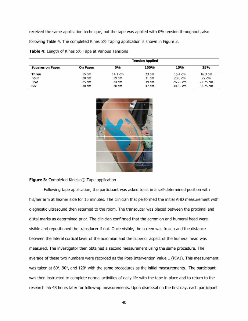
40
received the same application technique, but the tape was applied with 0% tension throughout, also
following Table 4. The completed Kinesio® Taping application is shown in Figure 3.
Table 4: Length of Kinesio® Tape at Various Tensions
Tension Applied
Squares on Paper On Paper 0% 100% 15% 25%
Three Four Five Six
15 cm 20 cm 25 cm 30 cm
14.1 cm 19 cm 24 cm 28 cm
23 cm 31 cm 39 cm 47 cm
15.4 cm 20.8 cm 26.25 cm 30.85 cm
16.3 cm 22 cm
27.75 cm 32.75 cm
Figure 3: Completed Kinesio® Tape application
Following tape application, the participant was asked to sit in a self-determined position with
his/her arm at his/her side for 15 minutes. The clinician that performed the initial AHD measurement with
diagnostic ultrasound then returned to the room. The transducer was placed between the proximal and
distal marks as determined prior. The clinician confirmed that the acromion and humeral head were
visible and repositioned the transducer if not. Once visible, the screen was frozen and the distance
between the lateral cortical layer of the acromion and the superior aspect of the humeral head was
measured. The investigator then obtained a second measurement using the same procedure. The
average of these two numbers were recorded as the Post-Intervention Value 1 (PIV1). This measurement
was taken at 60°, 90°, and 120° with the same procedures as the initial measurements. The participant
was then instructed to complete normal activities of daily life with the tape in place and to return to the
research lab 48 hours later for follow-up measurements. Upon dismissal on the first day, each participant

41
was given a SPADI form to complete 24 hours after the initial measurement which they were instructed
to return to the researcher upon follow-up.
Upon arrival for the follow-up appointment, the clinician confirmed that the Kinesio® Tape
remained in place on the subject since initial application. All participants returned with the tape in place
and completed a third SPADI while the clinician prepared the diagnostic ultrasound. The clinician then
measured the AHD in the subjects using the procedure outlined prior. Two measurements were taken at
each angle and averaged for the Post-Intervention Value 2 (PIV2). Next, the Kinesio® Tape was removed
and the skin was cleansed with an alcohol preparation pad. Following tape removal, the participant was
asked to sit in a self-determined position with his/her arm at his/her side for 15 minutes. After 15
minutes, an AHD measurement was taken twice using the procedure outlined prior at each angle of
abduction. These measurements were then averaged and recorded as the Post-Tape Removal Value
(PTRV). Upon completion, the participant received twenty dollars for his/her time and cooperation and
was dismissed.
3.5. Data Analysis
Statistical analysis for the approved research was computed using SPSS software (Version 23.0).
Two-way repeated measures ANOVA with a significance of P < .05 was conducted in order to compare
the mean differences between the type of tape application. Post hoc statistical significance was
determined by Tukey’s honestly significant difference test.
3.6. Conclusion
The purpose of this study was to determine if application of Kinesio® Tape has an effect on AHD
for patients suffering from subacromial impingement. The accepted clinical prediction rule for diagnosing
subacromial impingement syndrome given by Park et al27 was used as inclusion criteria for the
participants. Diagnostic ultrasound was used to quantify the AHD throughout the course of the study.
Kinesio® Tape was applied to the participants with either a Kinesio Taping Methods® technique or a
sham technique to compare outcomes. In addition to the objective diagnostic ultrasound measurements,
the participants completed the SPADI three times throughout the course of the study. This prospective

42
research will be used to determine the effect Kinesio® Tape has on subacromial impingement syndrome
in order to expand treatment options and increase patient outcomes.

43
CHAPTER 4. MANUSCRIPT
4.1. Abstract
[Study Design] Randomized Controlled Trial
[Background] Subacromial impingement syndrome is the most commonly diagnosed shoulder
pathology but is complex and poorly understood.1-6 Kinesio® Tape has the potential to optimize the
treatment of subacromial impingement syndrome.6,10,11,82 There is a need for objective measurements of
the subacromial space in response to Kinesio® Tape to understand the mechanism by which this
modality may alleviate symptoms.
[Objectives] To determine the effect Kinesio® Tape has on patient-reported outcome measures
and acromiohumeral distance (AHD) in patients with subacromial impingement syndrome.
[Methods] This study consisted of twenty volunteers exhibiting subacromial impingement
syndrome symptoms as determined by the combination of three orthopedic Special Tests. Participants
were divided into two groups, one receiving Kinesio® Tape inhibition technique (15% tension from
insertion to origin) of the supraspinatus and deltoid muscles and the other receiving a sham Kinesio®
Tape technique (0% tension from insertion to origin) of the same muscles. Patient-reported SPADI scores
and AHD measured by diagnostic ultrasound at various angles of shoulder abduction were recorded at
24- and 48-hour intervals.
[Results] SPADI scores of both the experimental and sham groups were statistically significant
when compared between baseline and 24 hours, baseline and 48 hours, and the 24- and 48-hour
intervals. The participants showed no statistically significant change in AHD at any interval in the study.

44
[Conclusions] Inhibition technique of Kinesio® Tape on the supraspinatus and deltoid muscles
alleviated pain and disability symptoms related to subacromial impingement syndrome as reported by a
patient outcome survey, but did not alter the subacromial space according to diagnostic ultrasound
scanning.
[Level of Evidence] Therapy, level 2b
[Key Words] Diagnostic ultrasound, subacromial space, SPADI
4.2. Introduction
Subacromial impingement syndrome is the most commonly diagnosed shoulder condition in the
general population, accounting for 44-65% of all shoulder complaints.1-6 Subacromial impingement
syndrome is often used as a catch-all term for more specific pathologies occurring in the subacromial
space including supraspinatus tendinopathy, subacromial bursitis, and long head of the biceps
tendinopathy.1,3-5,7 As a whole, patients diagnosed with subacromial impingement syndrome demonstrate
mild to severe pain, particularly with overhead motions. Even with the high prevalence of diagnosis, the
proper course of treatment is highly individualized and often unclear to clinicians. One theorized cause of
subacromial impingement is overactivity of the supraspinatus and the deltoid.5,7,18,19 Due to the
attachments of the supraspinatus and deltoid, overactivity in these muscles leads to a decrease in the
subacromial space as the humeral head is pulled superiorly. One proposed modality to aid in recovery
from subacromial impingement syndrome is Kinesio® Tape to decrease the activity of the supraspinatus
and deltoid.13
Kinesio® Tape is an elastic therapeutic tape that can stretch up to 60% of its resting length in
order to treat various musculoskeletal and neuromuscular maladies including pain, proprioceptive deficits,
swelling, under/overactive muscles, and postural alignment.12,13,70-72 Kinesio® Tape is theorized to aid

45
these conditions through various pathways, including altering muscle activation by stimulating
mechanoreceptors in muscles, lifting the epidermis to open the interstitial subcutaneous area, and
increasing kinesthetic awareness by stimulating proprioceptors.6,13,73 Research on Kinesio® Tape is fickle
due to inconsistencies in taping methods and types of tape applied. The creator of Kinesio® Tape has
published recommendations both on proper taping techniques and techniques for specific pathologies,13
but these are seldom followed in the literature.
Diagnostic Ultrasound (US) is an imaging technique that clinicians have recently used in practice
for visualizing musculoskeletal structures and conditions.22,23,25,37 Diagnostic US is beneficial over other
imaging techniques because it is inexpensive and noninvasive. Additionally, the images are available in
real time; therefore, movements throughout a range of motion can be visualized as opposed to still
pictures. Diagnostic US has been shown to be valid and reliable for measuring the subacromial space,
which can be quantified in terms of the AHD.20-24,86,87 While a cause and effect relationship cannot be
determined, subjects with subacromial impingement syndrome often have a smaller AHD than
asymptomatic individuals.22,24,25
Currently, there is one study that examined the effects of Kinesio® Tape applied as
recommended for subacromial impingement syndrome on AHD; however, it should be noted the research
was conducted on participants with no known history of subacromial impingement.85 Authors of this study
found that inhibition of the deltoid increased the AHD (P < .05). In contrast, inhibition of only the
supraspinatus did not increase the AHD (P > .05). Interestingly, a combination of applying both
techniques, as recommended by Kase et al,13 did not yield an increase in AHD (P > .05). The current
study aimed to challenge these results reported by Lyman et al85 and to examine the effects Kinesio®

46
Tape has on the AHD in symptomatic individuals, as well as obtain subjective patient outcome measures
of pain and disability.
The ability to justify certain treatment options with evidence-based recommendations is
imperative to clinicians. The literature is lacking on studies objectively evaluating the effects of Kinesio®
Tape on the subacromial space in patients with subacromial impingement syndrome. We hypothesized
that, when applying Kinesio® Tape with the Kinesio Taping® Method to inhibit the supraspinatus and
deltoid, symptoms of pain and disability related to subacromial impingement syndrome would decrease
and there would be a corresponding increase in AHD.
4.3. Methods
4.3.1 Subjects
Twenty adults (f=10, m=10) ranging in age from 18 to 51 (M=24.55, SD=8.876) volunteered for
this study through word-of-mouth and email recruitment. Inclusion criteria for this study were previous
diagnosis of shoulder impingement from a healthcare provider and pain in the affected shoulder for at
least one month. Factors excluding participants were shoulder surgery in the past year, acute injury, neck
or elbow pain, history of general medical conditions to joints, muscles, bones, or connective tissue, or
any contradictions for Kinesio® Tape, including allergy to adhesive, malignancy sites, cellulitis, skin
infection, open wounds, diabetes, or fragile skin.13 To screen for the inclusion and exclusion criteria,
participants completed a health history questionnaire. Additionally, an athletic trainer performed three
orthopedic special tests to confirm the presence of subacromial impingement syndrome: the Painful Arc
Test, Hawkins-Kennedy Impingement Test, and the External Rotation Muscle Test. These three tests
were chosen as they have the highest post-test probability of any combined tests, with 90% probability
of subacromial impingement syndrome if two positive results are found and 95% if all three tests are
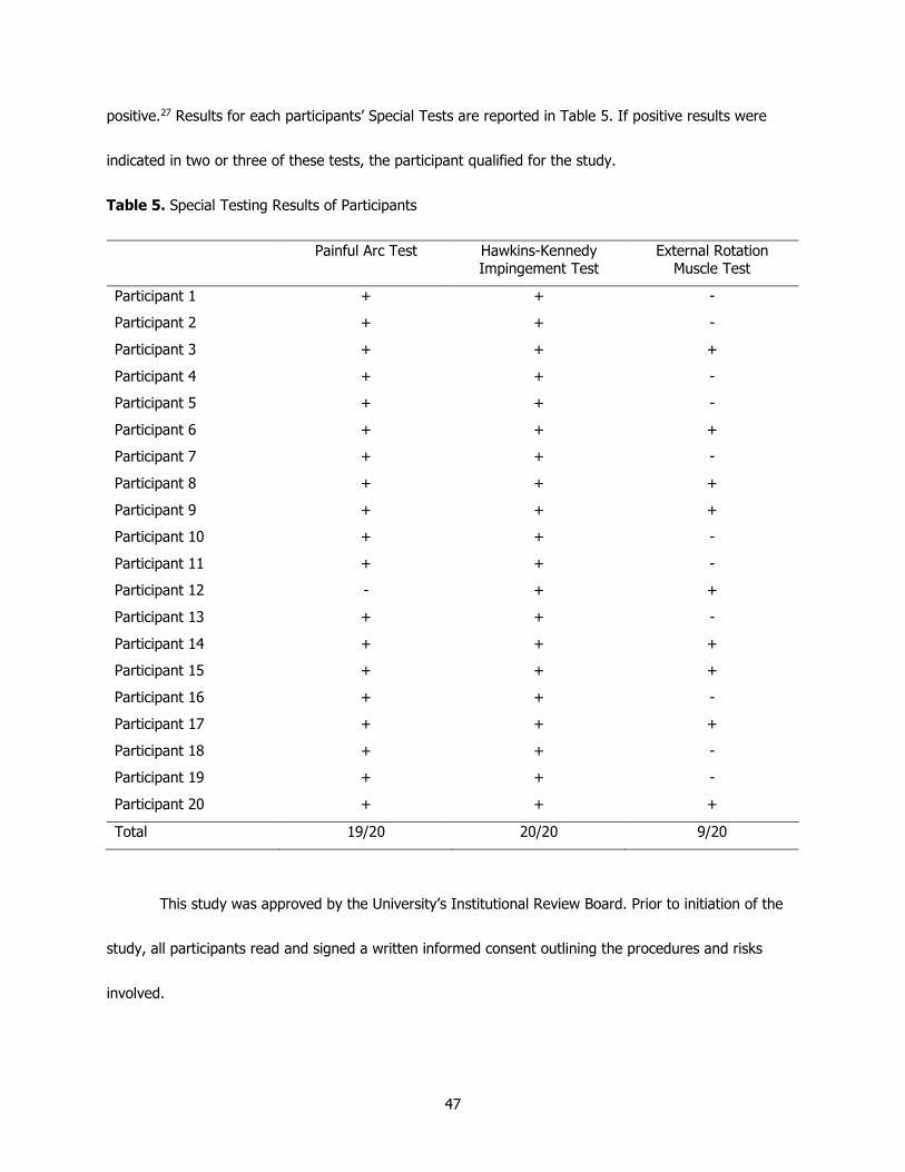
47
positive.27 Results for each participants’ Special Tests are reported in Table 5. If positive results were
indicated in two or three of these tests, the participant qualified for the study.
Table 5. Special Testing Results of Participants
Painful Arc Test Hawkins-Kennedy Impingement Test
External Rotation Muscle Test
Participant 1 + + -
Participant 2 + + -
Participant 3 + + +
Participant 4 + + -
Participant 5 + + -
Participant 6 + + +
Participant 7 + + -
Participant 8 + + +
Participant 9 + + +
Participant 10 + + -
Participant 11 + + -
Participant 12 - + +
Participant 13 + + -
Participant 14 + + +
Participant 15 + + +
Participant 16 + + -
Participant 17 + + +
Participant 18 + + -
Participant 19 + + -
Participant 20 + + +
Total 19/20 20/20 9/20
This study was approved by the University’s Institutional Review Board. Prior to initiation of the
study, all participants read and signed a written informed consent outlining the procedures and risks
involved.

48
4.3.2. Study Design and Protocol
This within-subject research study utilized a pretest/posttest design with subjects randomly
divided into two groups: experimental and control. All participants completed the Shoulder Pain and
Disability Index (SPADI) as a baseline measurement. Next, a clinician trained in diagnostic ultrasound
with four years of experience and blinded to the randomization measured each participant’s baseline
AHD. All diagnostic imaging was completed using the preset shoulder parameters on a Terason t3200™
diagnostic ultrasound machine (United Medical Instruments Inc, San Jose, California) with a 15L4 linear
transducer (4 MHz to 15 MHz) (Teratech Corporation, Burlington, Massachusetts) and Aquasonic® 100
ultrasound gel (Parker Laboratories, INC., Fairfield, NJ) as the coupling medium. The subject was
instructed to be seated in an upright posture with his/her arm relaxed at his/her side in neutral position
and the elbow extended.29 The anterior aspect of the acromion process was palpated. The transducer
was placed in the coronal-oblique plane over the acromion process and was then moved inferiorly until
the superior aspect of the humeral head was visible. The transducer was then toggled as necessary to
refine the image and obtain a hyperechoic acromion and humeral head. At this point, the screen was
frozen and a perpendicular line was drawn from the cortical layer of the lateral acromion to cortical layer
of the superior humeral head. This line was measured and recorded as the baseline AHD value (BLV).
Two measurements were obtained and averaged in order to ensure intratester reliability. The participant
then had an inclinometer placed on the midshaft of the humerus by a second clinician. The participant
was asked to abduct his/her humerus to 60° and hold this position while the ultrasound clinician
measured the AHD with the procedure previously outlined. Additionally, the participant was asked to
abduct his/her arm to 90° and 120°, with the ultrasound clinician measuring AHD at each position.

49
4.3.3. Intervention
The second clinician, a Certified Kinesio Tape® Practitioner (CKTP), then applied the Kinesio®
Tape. The experimental group received Kinesio® Tape with 15% tension from insertion to origin applied
to inhibit the supraspinatus and the deltoid while the control group received identical tape with 0%
tension in a sham technique. Two Y-strips of Kinesio® Tape were cut to match the length of the
participant’s supraspinatus and deltoid. In order to address the methodological issues of tension, the
researchers measured varying tensions with corresponding measurements. Marks of appropriate
measurement were applied to a participant’s skin in order to ensure proper tension was applied to the
Kinesio® Tape. These values are reported as a tension measurement table in Table 4.
Tape application for the supraspinatus was conducted by placing a mark on the participant’s
greater tuberosity and, using the tension measurement table, a distance was measured with the humerus
fully pronated and horizontally abducted to stretch the muscle; a mark was placed where the tape was to
terminate on the scapular spine. The base of the tape was applied with 0% tension posterior to the
participant’s greater tuberosity with the arm at rest. The humerus was then again placed in the pronated
and horizontally abducted position to stretch the supraspinatus and the two tails of the tape were placed
to encompass the breadth of the supraspinatus. The second strip was applied from insertion to origin of
the deltoid in a manner suggested by the creator of the tape to inhibit an overactive muscle. For this
application, a mark was placed on the participant’s deltoid tuberosity and a distance was measured to
ensure proper tension. A mark was placed where this strip was to terminate, approximately mid-clavicle.
The base was then applied with 0% tension on the deltoid tuberosity. For the anterior tail, the participant
was placed in slight humeral extension and for the posterior tail, the participant was placed in slight
humeral flexion in order to stretch the specific portion of the deltoid targeted.

50
Table 4: Length of Kinesio® Tape at Various Tensions
Tension Applied
Squares on Paper On Paper 0% 100% 15% 25%
Three Four Five Six
15 cm 20 cm 25 cm 30 cm
14.1 cm 19 cm 24 cm 28 cm
23 cm 31 cm 39 cm 47 cm
15.4 cm 20.8 cm 26.25 cm 30.85 cm
16.3 cm 22 cm
27.75 cm 32.75 cm
Following tape application, the participant was asked to sit in a self-determined position with
his/her arm at his/her side for 15 minutes. The clinician who performed the initial AHD measurement with
diagnostic ultrasound then returned to the room. This clinician then measured the AHD at 0°, 60°, 90°,
and 120° using the same methods as previously described to obtain the Post-Intervention Value 1 (PIV1).
The participant was then instructed to complete normal activities of daily living with the tape still intact
and to return to the research lab 48 hours later for follow-up measurements. Following all AHD
measurements, each participant was given a SPADI form to complete 24 hours after the initial
measurement which they were instructed to return to the researchers upon follow-up.
Upon arrival for the follow-up appointment, the clinician confirmed that the Kinesio® Tape
remained in place on the subject. All participants returned with the tape in place and completed a third
SPADI, which enabled researchers to evaluate patient outcomes at three separate time intervals. The
ultrasound clinician then measured the AHD in the subjects using the procedure outlined prior. Two
measurements were taken at each angle and averaged for the Post-Intervention Value 2 (PIV2). Next,
the Kinesio® Tape was removed and the skin was cleansed with an alcohol preparation pad. Following
tape removal, the participant was asked to sit in a self-determined position with his/her arm at his/her
side for 15 minutes. After 15 minutes, an AHD measurement was taken twice using the procedure

51
outlined prior at each angle of abduction. These measurements were then averaged and recorded as the
Post-Tape Removal Value (PTRV).
4.3.4. Statistical Analysis
Statistical analysis for the approved research was computed using SPSS software (Version 23.0).
Patient outcome data (SPADI) were analyzed with a repeated measures analysis of variance (ANOVA)
model. In order to analyze differences in AHD, additional ANOVAs were employed to examine the with-in
subject and between group models at the four separate intervals. All tests used a significance level of
5%. No data transformations were necessary.
4.4. Results
The repeated measures ANOVA model for the SPADI measurements was statistically significant in
terms of both time (F[1.56, 23.44] = 37.06, P < .001) and the interaction of group and time (F[1.56,
23.44] = 7.01, P = .007). The between-subjects effect due to taping technique was significant only at the
10% level (F[1, 15] = 3.11, P = .098). Therefore, pain reduction was significant over time, but the
difference between the two groups was not significant at the 5% level. In the pain subset of the SPADI,
scores were significantly lower in both groups over time (F[1.823, 27.35] = 31.00, P < .001), but were
only significant at the 10% level in the interaction of group and time (F[1.823, 27.35] = 2.96, P = .073)
and the between-subjects effect (F[1, 15] = 3.91, P = .067). Additionally, the disability subset yielded
significant results in terms of time (F[1.52, 22.80] = 25.88, P < .001) and the interaction of group and
time (F[1.52, 22.80] = 9.81, P = .002). However, in this subset, the between-subjects effect due to
technique was not significant (F[1, 15] = 2.28, P = .152). All participants, regardless of taping technique,
reported lower SPADI scores at the conclusion of the experiment (Table 5).

52
No statistically significant difference was found in AHD at any of the four angles or any of the
diagnostic ultrasound scans (Tables 6-9). Overall, US measurements of participants’ AHD in either group
did not differ from their baseline measurements immediately after taping, after 48 hours, or after tape
removal.

53
Table 6: Effect of Kinesio® Tape on Mean Self-Reported SPADI Scores Over Time
SPADI Pain Subset Score SPADI Disability Subset Score SPADI Total Score
Time Base 24H 48H Base 24H 48H Base 24H 48H
Experimental
Control
25.2±7.2 16.8±6.2 11.7±6.0 23.6±9.52 12.2±6.29 12.0±8.79 48.8±15.9 29.0±10.45 22.3±14.48
17.7±3.37 13.1±7.34 10.4±6.70 12.1±6.23 10.8±7.29 9.2±6.99 29.8±8.43 23.9±13.89 21.3±12.89

54
Table 7: Effect of Kinesio® Tape on AHD at 0° Over Time
Acromiohumeral Distance (mm)
BLV PIV1 PIV2 PTRV
Experimental 12.1±1.97 12.5±1.73 12.3±1.63 12.2±2.31
Control 13.1±1.59 12.7±1.26 12.7±0.98 12.4±0.85
Table 8: Effect of Kinesio® Tape on AHD at 60° Over Time
Acromiohumeral Distance (mm)
BLV PIV1 PIV2 PTRV
Experimental 10.6±1.72 9.58±1.80 9.53±2.08 9.78±1.73
Control 10.5±1.90 10.2±2.09 10.3±2.06 9.84±2.43
Table 9: Effect of Kinesio® Tape on AHD at 90° Over Time
Acromiohumeral Distance (mm)
BLV PIV1 PIV2 PTRV
Experimental 10.2±1.37 9.73±1.53 9.08±1.55 9.41±1.26
Control 10.3±2.53 10.3±2.63 9.97±2.92 9.32±1.59
Table 10: Effect of Kinesio® Tape on AHD at 120° Over Time
Acromiohumeral Distance (mm)
BLV PIV1 PIV2 PTRV
Experimental 9.23±2.75 8.65±2.63 8.31±2.13 9.31±3.04
Control 8.94±2.33 8.12±2.19 7.83±1.60 8.68±2.27
4.5. Discussion
The prevalence of subacromial impingement syndrome beckons new treatments, one of which is
using Kinesio® Tape in conjunction with other typical therapies. This is the first study to quantify the
55
effect Kinesio® Tape has on the AHD in people with subacromial impingement syndrome. The results of
this research will give practitioners a better understanding of how this modality may aid patients with
subacromial impingement syndrome.
To qualify for the study, participants had to demonstrate the presence of pathology, indicated by
three orthopedic special tests: Painful Arc, Hawkins-Kennedy, and External Rotation Muscle tests.
Interestingly, all participants except one demonstrated a positive Painful Arc, and all participants were
found to have positive Hawkins-Kennedy test results. The External Rotation Muscle test varied the most,
as nine participants were found to have weakness or pain on the affected side, but eleven did not
function any differently during this test as compared bilaterally. Park et al27 established the clinical
prediction rule of using these three tests by comparing eight tests used individually and in combination.
The combination of the Painful Arc, Hawkins-Kennedy, and External Rotation Muscle tests indicated the
highest post-test probability. However, when examining the results closer, the overall accuracy of the
Painful Arc (76.1%) and Hawkins-Kennedy (69.7%) tests outperformed that of the External Rotation
Muscle test (58.7%), which may explain the differences observed in the current study.
Participants in both groups reported decreased pain and disability outcomes; however, the SPADI
results of the experimental group improved significantly more than the control group. The experimental
groups’ total SPADI scores decreased 26.5 points, but the control groups’ SPADI score only decreased by
8.5 points. The creators of the SPADI determined the minimal detectable change with a 90% confidence
interval to be 13 points; therefore, the experimental group satisfies this finding, but the control groups’
decrease may have been due to chance. The AHD did not change significantly when compared across
groups. These results suggest that while Kinesio® Tape did not alter the space available for the
subacromial structures, it apparently decreased pain through a separate pathway and this decrease was

56
more substantial in the group with tension placed through the Kinesio® Tape. The results of this
experiment only allow for speculation on what this pathway may be, but other authors have suggested
pain modulation due to the gate control theory or due to increased afferent feedback as opposed to
simply increasing the AHD.91
The findings of the research compliment previous findings that Kinesio® Tape may reduce pain
associated with subacromial impingement syndrome. A previous study by Kaya et al8 who compared relief
of subacromial impingement through Kinesio® Tape and exercise to manual therapy and exercise found
equal benefits in each treatment. A similar study compared patient-reported outcomes between a group
receiving Kinesio® Tape and a group receiving physical therapy modalities.82 The Kinesio® Tape group
reported better outcomes than the physical therapy modalities group after the first week and showed
similar results after two weeks. Previous literature as well as the results of the current study make a case
that Kinesio® Tape can be applied to aid in the reduction of pain associated with the symptoms of
subacromial impingement syndrome, thereby aiding in patient compliance with exercises to correct
pathomechanics associated with the condition.
While Kinesio® Tape is often found to be a valid treatment in subacromial impingement
syndrome cases, this finding is not always consistent. Thelen et al83 conducted a study to examine the
efficacy of Kinesio® Tape for shoulder pain related to rotator cuff tendonitis or impingement. Using
similar taping techniques as the current study, the authors did not find any statistically significant
decrease in pain immediately after, 3 days after, or 6 days after application, but did find significant
increase in immediate shoulder abduction (P = .005). Interestingly, both the experimental and the sham
group reported better outcomes at the end of the study; however, it was not significant (P > .05).

57
Results reported by Thelen et al align with the current study, where both the experimental group and the
control group experienced a decrease in pain and disability.
Research examining the effect of Kinesio® Tape on the AHD is minimal, although it was the
subject of one study conducted by Luque-Suarez et al.84 In this study, healthy participants were divided
into three groups, with different taping techniques in each. The authors found an increase in AHD in the
two experimental groups as compared to the control group and concluded that Kinesio® Tape can
immediately increase the AHD in healthy tissue. Notably, neither of the two experimental groups utilized
the taping techniques recommended by Kase et al13 which were applied to the participants in the current
research project. The results of the current study contrast the results reported by Luque-Suarez et al, as
no change in AHD was significant at any time period or angle of abduction.
Similarly, a recently published manuscript examined how different Kinesio® Tape applications
affect the subacromial space as measured by diagnostic US. Lyman et al85 conducted a study
investigating three different applications: inhibition of the deltoid, inhibition of the supraspinatus, and
inhibition of both deltoid and supraspinatus. AHD was then measured with diagnostic US after a five
minute period of time. Results of this study indicated an initial increase in AHD with deltoid inhibition and
supraspinatus inhibition, but not when used in combination. The current research study aimed to address
some limitations of this study including recruiting symptomatic individuals, measuring AHD at different
angles of humeral abduction, and obtaining subjective SPADI measurements in addition to the US values.
Additionally, the current research project utilized a combined taping method of deltoid and supraspinatus
inhibition, for which Lyman et al also found no statistically significant increase in AHD.
This research study is not without limitations due to the numerous variables present. First, this
study was limited to participants between the ages of 18 and 55; therefore, the results will not be

58
generalizable to populations outside of this age range including pediatric and geriatric patients.
Additionally, participants clearly presented with varying severity of subacromial impingement as noted by
their baseline SPADI scores. Parameters for differentiating severities of this condition have not been
clearly outlined in previous literature, but Kinesio® Tape may impact them differently. Finally, the AHD
measurement at 120° of glenohumeral abduction was difficult to obtain and ultimately unreliable in this
study. Researchers of the current study recommend using alternate methods to measure AHD at this
angle of abduction if this is a desired measurement.
Future research on this topic should aim to investigate the reason why SPADI scores were
decreased in all participants regardless of taping tension. Studying muscle activation in participants with
subacromial impingement syndrome with regards to Kinesio® Tape may give insight into the mechanism
of pain reduction. Additionally, since the AHD did not statistically significantly increase with the Kinesio®
Tape application, future research should consider various Kinesio® Taping techniques to explore if any
technique may increase the AHD in symptomatic individuals.
4.6. Conclusions
The results support the use of Kinesio® Tape to decrease pain and disability in patients with
subacromial impingement syndrome. SPADI scores were lower in the group who received the inhibition
technique of the supraspinatus and deltoid compared to the control group with no tension applied, but
both groups significantly improved from their baseline. The AHD was not statistically affected with either
group or at any of the four angles measured. The pathway by which Kinesio® Tape decreases pain
remains a question; however, Kinesio® Tape may confidently be used to aid patients with pain and
disability in the treatment of subacromial impingement syndrome.

59
REFERENCES
1. Calis M, Akgun K, Birtane M, Karacan I, Calis H, Tuzun F. Diagnostic values of clinical diagnostic
tests in subacromial impingement syndrome. Ann Rheum Dis. 2000;59(1):44-47. 2. Alqunaee M, Galvin R, Fahey T. Diagnostic accuracy of clinical tests for subacromial impingement
syndrome: a systematic review and meta-analysis. Arch Phys Med Rehabil. 2012;93(2):229-236.
3. de Witte PB, Nagels J, van Arkel ER, Visser CP, Nelissen RG, de Groot JH. Study protocol subacromial impingement syndrome: the identification of pathophysiologic mechanisms (SISTIM).
BMC Musculoskelet Disord. 2011;12:282. 4. Koester MC, George MS, Kuhn JE. Shoulder impingement syndrome. Am J Med.
2005;118(5):452-455.
5. Michener LA, McClure PW, Karduna AR. Anatomical and biomechanical mechanisms of subacromial impingement syndrome. Clin Biomech (Bristol, Avon). 2003;18(5):369-379.
6. Dong W, Goost H, Lin XB, et al. Treatments for shoulder impingement syndrome: a PRISMA systematic review and network meta-analysis. Medicine (Baltimore). 2015;94(10):e510.
7. Lewis JS, Green AS, Dekel S. The Aetiology of Subacromial Impingement Syndrome. Physiotherapy. 2001;87(9):458-469.
8. Kaya DO, Baltaci G, Toprak U, Atay AO. The clinical and sonographic effects of kinesiotaping and
exercise in comparison with manual therapy and exercise for patients with subacromial impingement syndrome: a preliminary trial. J Manipulative Physiol Ther. 2014;37(6):422-432.
9. Moura KF, Monteiro RL, Lucareli PR, Fukuda TY. REHABILITATION OF SUBACROMIAL PAIN SYNDROME EMPHASIZING SCAPULAR DYSKINESIS IN AMATEUR ATHLETES: A CASE SERIES. Int J Sports Phys Ther. 2016;11(4):552-563.
10. Pekyavas NO, Baltaci G. Short-term effects of high-intensity laser therapy, manual therapy, and Kinesio taping in patients with subacromial impingement syndrome. Lasers Med Sci. 2016;31(6):1133-1141.
11. Subasi V, Cakir T, Arica Z, et al. Comparison of efficacy of kinesiological taping and subacromial
injection therapy in subacromial impingement syndrome. Clin Rheumatol. 2016;35(3):741-746.
12. Williams S, Whatman C, Hume PA, Sheerin K. Kinesio taping in treatment and prevention of sports injuries: a meta-analysis of the evidence for its effectiveness. Sports Med. 2012;42(2):153-
164. 13. Kase K, Wallis J, Kase T. Clinical Therapeutic Applications of the Kinesio Taping ® Method. 2nd
ed2003. 14. Cai C, Au IP, An W, Cheung RT. Facilitatory and inhibitory effects of Kinesio tape: Fact or fad? J
Sci Med Sport. 2016;19(2):109-112.
15. Fernandes de Jesus J, de Almeida Novello A, Bezerra Nakaoka G, Curcio Dos Reis A, Fukuda TY, Fernandes Bryk F. Kinesio taping effect on quadriceps strength and lower limb function of healthy
individuals: A blinded, controlled, randomized, clinical trial. Phys Ther Sport. 2016;18:27-31. 16. Korman P, Straburzynska-Lupa A, Rutkowski R, et al. Kinesio Taping Does Not Alter Quadriceps
Isokinetic Strength and Power in Healthy Nonathletic Men: A Prospective Crossover Study.
Biomed Res Int. 2015;2015:626257. 17. Kurt EE, Buyukturan O, Erdem HR, Tuncay F, Sezgin H. Short-term effects of kinesio tape on joint
position sense, isokinetic measurements, and clinical parameters in patellofemoral pain syndrome. J Phys Ther Sci. 2016;28(7):2034-2040.
18. Kolber MJ, Hanney WJ, Cheatham SW, Salamh PA, Masaracchio M, Liu X. Shoulder Joint and Muscle Characteristics Among Weight-training Participants with and without Impingement
Syndrome. J Strength Cond Res. 2016.
19. Reddy AS, Mohr KJ, Pink MM, Jobe FW. Electromyographic analysis of the deltoid and rotator cuff muscles in persons with subacromial impingement. J Shoulder Elbow Surg. 2000;9(6):519-523.
20. Desmeules F, Minville L, Riederer B, Cote CH, Fremont P. Acromio-humeral distance variation measured by ultrasonography and its association with the outcome of rehabilitation for shoulder
impingement syndrome. Clin J Sport Med. 2004;14(4):197-205.
60
21. Leong HT, Tsui S, Ying M, Leung VY, Fu SN. Ultrasound measurements on acromio-humeral
distance and supraspinatus tendon thickness: test-retest reliability and correlations with shoulder rotational strengths. J Sci Med Sport. 2012;15(4):284-291.
22. McCreesh KM, Crotty JM, Lewis JS. Acromiohumeral distance measurement in rotator cuff tendinopathy: is there a reliable, clinically applicable method? A systematic review. Br J Sports Med. 2015;49(5):298-305.
23. McCreesh KM, Anjum S, Crotty JM, Lewis JS. Ultrasound measures of supraspinatus tendon thickness and acromiohumeral distance in rotator cuff tendinopathy are reliable. J Clin Ultrasound. 2016;44(3):159-166.
24. Azzoni R, Cabitza P. Sonographic versus radiographic measurement of the subacromial space
width. Chir Organi Mov. 2004;89(2):143-150. 25. Seitz AL, Michener LA. Ultrasonographic measures of subacromial space in patients with rotator
cuff disease: A systematic review. J Clin Ultrasound. 2011;39(3):146-154.
26. Wang HK, Lin JJ, Pan SL, Wang TG. Sonographic evaluations in elite college baseball athletes. Scand J Med Sci Sports. 2005;15(1):29-35.
27. Park HB, Yokota A, Gill HS, El Rassi G, McFarland EG. Diagnostic accuracy of clinical tests for the different degrees of subacromial impingement syndrome. J Bone Joint Surg Am. 2005;87(7):1446-1455.
28. Mackenzie TA, Herrington L, Horlsey I, Cools A. An evidence-based review of current perceptions with regard to the subacromial space in shoulder impingement syndromes: Is it important and
what influences it? Clin Biomech (Bristol, Avon). 2015;30(7):641-648. 29. Jacobson JA. Fundamentals of Musculoskeletal Ultrasound. 2nd ed. Philadelphia, PA: Elsevier
Inc.; 2013. 30. Mazzocca AD, Arciero RA, Bicos J. Evaluation and treatment of acromioclavicular joint injuries.
Am J Sports Med. 2007;35(2):316-329.
31. Starkey C, Brown SD, Ryan J. Examination of Orthopedic and Athletic Injuries. Third ed: F.A. Davis Company; 2010.
32. Prentice WE. Principles of Athletic Training: A Competency-Based Approach. Fifteenth ed: McGraw-Hill; 2014.
33. Musgrove J. The Costo-Sternal Articulations. J Anat Physiol. 1892;27(Pt 1):1-4.
34. Jones SA. Pocket Anatomy and Physiology. Second ed: F.A. Davis Company; 2013. 35. Neer CS, 2nd. Anterior acromioplasty for the chronic impingement syndrome in the shoulder.
1972. J Bone Joint Surg Am. 2005;87(6):1399. 36. Ludewig PM, Reynolds JF. The association of scapular kinematics and glenohumeral joint
pathologies. J Orthop Sports Phys Ther. 2009;39(2):90-104.
37. Krzyzanowski W. The use of ultrasound in the assessment of the glenoid labrum of the glenohumeral joint. Part I: Ultrasound anatomy and examination technique. J Ultrason. 2012;12(49):164-177.
38. Park YH, Lee JY, Moon SH, et al. MR arthrography of the labral capsular ligamentous complex in
the shoulder: imaging variations and pitfalls. AJR Am J Roentgenol. 2000;175(3):667-672. 39. Balke M, Schmidt C, Dedy N, Banerjee M, Bouillon B, Liem D. Correlation of acromial morphology
with impingement syndrome and rotator cuff tears. Acta Orthop. 2013;84(2):178-183.
40. Dick R, Ferrara MS, Agel J, et al. Descriptive epidemiology of collegiate men's football injuries: National Collegiate Athletic Association Injury Surveillance System, 1988-1989 through 2003-
2004. J Athl Train. 2007;42(2):221-233. 41. Price GE. Rheumatology: 6. Localized rheumatism. Cmaj. 2000;163(2):176-183.
42. Gschwend N, Ivosevic-Radovanovic D, Patte D. Rotator cuff tear--relationship between clinical
and anatomopathological findings. Arch Orthop Trauma Surg. 1988;107(1):7-15. 43. Escamilla RF, Yamashiro K, Paulos L, Andrews JR. Shoulder muscle activity and function in
common shoulder rehabilitation exercises. Sports Med. 2009;39(8):663-685. 44. Hill LJ, Jelsing EJ, Terry MJ, Strommen JA. Evaluation, treatment, and outcomes of suprascapular
neuropathy: a 5-year review. Pm r. 2014;6(9):774-780.

61
45. Curtis AS, Burbank KM, Tierney JJ, Scheller AD, Curran AR. The insertional footprint of the rotator
cuff: an anatomic study. Arthroscopy. 2006;22(6):609.e601. 46. Dugas JR, Campbell DA, Warren RF, Robie BH, Millett PJ. Anatomy and dimensions of rotator cuff
insertions. J Shoulder Elbow Surg. 2002;11(5):498-503. 47. Leijnse JN, Han SH, Kwon YH. Morphology of deltoid origin and end tendons--a generic model. J
Anat. 2008;213(6):733-742.
48. Fennell J, Phadke CP, Mochizuki G, Ismail F, Boulias C. Shoulder Retractor Strengthening Exercise to Minimize Rhomboid Muscle Activity and Subacromial Impingement. Physiother Can. 2016;68(1):24-28.
49. Michener LA, Sharma S, Cools AM, Timmons MK. Relative scapular muscle activity ratios are
altered in subacromial pain syndrome. J Shoulder Elbow Surg. 2016. 50. Guazzelli Filho J, Furlani J, De Freitas V. Electromyographic study of the trapezius muscle in free
movements of the arm. Electromyogr Clin Neurophysiol. 1991;31(2):93-98.
51. Elser F, Braun S, Dewing CB, Giphart JE, Millett PJ. Anatomy, function, injuries, and treatment of the long head of the biceps brachii tendon. Arthroscopy. 2011;27(4):581-592.
52. Zappia M, Reginelli A, Russo A, et al. Long head of the biceps tendon and rotator interval. Musculoskelet Surg. 2013;97 Suppl 2:S99-108.
53. Chalmers PN, Cip J, Trombley R, et al. Glenohumeral Function of the Long Head of the Biceps
Muscle: An Electromyographic Analysis. Orthop J Sports Med. 2014;2(2):2325967114523902. 54. Bigliani LU, Ticker JB, Flatow EL, Soslowsky LJ, Mow VC. The relationship of acromial architecture
to rotator cuff disease. Clin Sports Med. 1991;10(4):823-838. 55. Jobe CM. Superior glenoid impingement. Orthop Clin North Am. 1997;28(2):137-143.
56. Sher JS, Uribe JW, Posada A, Murphy BJ, Zlatkin MB. Abnormal findings on magnetic resonance images of asymptomatic shoulders. J Bone Joint Surg Am. 1995;77(1):10-15.
57. Menge TJ, Byram IR, Boykin RE, Bushnell BD. Labrum and rotator cuff injuries in the throwing
athlete. Phys Sportsmed. 2015;43(1):65-72. 58. Ludewig PM, Cook TM. Translations of the humerus in persons with shoulder impingement
symptoms. J Orthop Sports Phys Ther. 2002;32(6):248-259. 59. Flatow EL, Soslowsky LJ, Ticker JB, et al. Excursion of the rotator cuff under the acromion.
Patterns of subacromial contact. Am J Sports Med. 1994;22(6):779-788.
60. Petersson CJ, Redlund-Johnell I. The subacromial space in normal shoulder radiographs. Acta Orthop Scand. 1984;55(1):57-58.
61. Harryman DT, 2nd, Sidles JA, Clark JM, McQuade KJ, Gibb TD, Matsen FA, 3rd. Translation of the humeral head on the glenoid with passive glenohumeral motion. J Bone Joint Surg Am. 1990;72(9):1334-1343.
62. Ludewig PM, Cook TM. The effect of head position on scapular orientation and muscle activity during shoulder elevation. J Occup Rehabil. 1996;6(3):147-158.
63. Kelly SM, Brittle N, Allen GM. The value of physical tests for subacromial impingement syndrome: a study of diagnostic accuracy. Clin Rehabil. 2010;24(2):149-158.
64. Michener LA, Walsworth MK, Doukas WC, Murphy KP. Reliability and diagnostic accuracy of 5 physical examination tests and combination of tests for subacromial impingement. Arch Phys Med Rehabil. 2009;90(11):1898-1903.
65. Hanchard NCA, Lenza M, Handoll HHG, Takwoingi Y. Physical tests for shoulder impingements and local lesions of bursa, tendon or labrum that may accompany impingement. Cochrane Database of Systematic Reviews. 2013(4).
66. Neer CS, 2nd, Welsh RP. The shoulder in sports. Orthop Clin North Am. 1977;8(3):583-591.
67. Hawkins RJ, Kennedy JC. Impingement syndrome in athletes. Am J Sports Med. 1980;8(3):151-
158. 68. Jobe FW, Jobe CM. Painful athletic injuries of the shoulder. Clin Orthop Relat Res.
1983(173):117-124.

62
69. Girometti R, De Candia A, Sbuelz M, Toso F, Zuiani C, Bazzocchi M. Supraspinatus tendon US
morphology in basketball players: correlation with main pathologic models of secondary impingement syndrome in young overhead athletes. Preliminary report. Radiol Med. 2006;111(1):42-52.
70. Alvarez-Alvarez S, Jose FG, Rodriguez-Fernandez AL, Gueita-Rodriguez J, Waller BJ. Effects of
Kinesio(R) Tape in low back muscle fatigue: randomized, controlled, doubled-blinded clinical trial
on healthy subjects. J Back Musculoskelet Rehabil. 2014;27(2):203-212. 71. Lee YJ, Kim JY, Kim SY, Kim KH. The effects of trunk kinesio taping on balance ability and gait
function in stroke patients. J Phys Ther Sci. 2016;28(8):2385-2388. 72. Rojhani-Shirazi Z, Amirian S, Meftahi N. Effects of Ankle Kinesio Taping on Postural Control in
Stroke Patients. J Stroke Cerebrovasc Dis. 2015;24(11):2565-2571. 73. Wu WT, Hong CZ, Chou LW. The Kinesio Taping Method for Myofascial Pain Control. Evid Based
Complement Alternat Med. 2015;2015:950519.
74. Parreira Pdo C, Costa Lda C, Hespanhol LC, Jr., Lopes AD, Costa LO. Current evidence does not support the use of Kinesio Taping in clinical practice: a systematic review. J Physiother. 2014;60(1):31-39.
75. Mostafavifar M, Wertz J, Borchers J. A systematic review of the effectiveness of kinesio taping for
musculoskeletal injury. Phys Sportsmed. 2012;40(4):33-40.
76. Gonzalez-Iglesias J, Fernandez-de-Las-Penas C, Cleland JA, Huijbregts P, Del Rosario Gutierrez-Vega M. Short-term effects of cervical kinesio taping on pain and cervical range of motion in
patients with acute whiplash injury: a randomized clinical trial. J Orthop Sports Phys Ther. 2009;39(7):515-521.
77. Vered E, Oved L, Zilberg D, Kalichman L. Influence of kinesio tape application direction on peak force of biceps brachii muscle: A repeated measurement study. J Bodyw Mov Ther. 2016;20(1):203-207.
78. Kim JY, Kim SY. Effects of kinesio tape compared with non-elastic tape on hand grip strength. J Phys Ther Sci. 2016;28(5):1565-1568.
79. Lemos TV, Pereira KC, Protassio CC, Lucas LB, Matheus JP. The effect of Kinesio Taping on handgrip strength. J Phys Ther Sci. 2015;27(3):567-570.
80. Ozturk G, Kulcu DG, Mesci N, Silte AD, Aydog E. Efficacy of kinesio tape application on pain and
muscle strength in patients with myofascial pain syndrome: a placebo-controlled trial. J Phys Ther Sci. 2016;28(4):1074-1079.
81. Hong S, Shim J, Kim S, Namkoong S, Roh H. Effect of kinesio taping on the isokinetic muscle function in football athletes with a knee injury. J Phys Ther Sci. 2016;28(1):218-222.
82. Kaya E, Zinnuroglu M, Tugcu I. Kinesio taping compared to physical therapy modalities for the
treatment of shoulder impingement syndrome. Clin Rheumatol. 2011;30(2):201-207. 83. Thelen MD, Dauber JA, Stoneman PD. The clinical efficacy of kinesio tape for shoulder pain: a
randomized, double-blinded, clinical trial. J Orthop Sports Phys Ther. 2008;38(7):389-395. 84. Luque-Suarez A, Navarro-Ledesma S, Petocz P, Hancock MJ, Hush J. Short term effects of
kinesiotaping on acromiohumeral distance in asymptomatic subjects: a randomised controlled trial. Man Ther. 2013;18(6):573-577.
85. Lyman KJ, Gange KN, Hanson TA, Mellinger CD. Effects of 3 Different Elastic Therapeutic Taping
Methods on the Subacromial Joint Space. Journal of Manipulative and Physiological Therapeutics. 2017;40(7):494-500.
86. Cholewinski JJ, Kusz DJ, Wojciechowski P, Cielinski LS, Zoladz MP. Ultrasound measurement of rotator cuff thickness and acromio-humeral distance in the diagnosis of subacromial impingement
syndrome of the shoulder. Knee Surg Sports Traumatol Arthrosc. 2008;16(4):408-414.
87. Mackenzie TA, Bdaiwi AH, Herrington L, Cools A. Inter-rater Reliability of Real-Time Ultrasound to Measure Acromiohumeral Distance. Pm r. 2016;8(7):629-634.
88. Roach KE, Budiman-Mak E, Songsiridej N, Lertratanakul Y. Development of a shoulder pain and disability index. Arthritis Care Res. 1991;4(4):143-149.
89. Paul A, Lewis M, Shadforth MF, Croft PR, Van Der Windt DA, Hay EM. A comparison of four shoulder-specific questionnaires in primary care. Ann Rheum Dis. 2004;63(10):1293-1299.

63
90. Cloke DJ, Lynn SE, Watson H, Steen IN, Purdy S, Williams JR. A comparison of functional,
patient-based scores in subacromial impingement. J Shoulder Elbow Surg. 2005;14(4):380-384. 91. Shakeri H, Keshavarz R, Arab AM, Ebrahimi I. Clinical effectiveness of kinesiological taping on
pain and pain-free shoulder range of motion in patients with shoulder impingement syndrome: a randomized, double blinded, placebo-controlled trial. Int J Sports Phys Ther. 2013;8(6):800-810.

64
APPENDIX A. INFORMED CONSENT
NDSU North Dakota State University
Health, Nutrition and Exercises Science Department #2620, PO Box 6050 Fargo, ND 58108-6050 701-231-5590
Title of Research Study: The Effects of Kinesio® Tape on Acromiohumeral Distance in Patients with Subacromial Impingement Syndrome
This study is being conducted by:
Dr. Katie Lyman, HNES Assistant Professor, [email protected], office number: (701)231-8208
Nicholas Sample, HNES Advanced Athletic Training Masters Student, [email protected], cell number: (314) 971-5548
Why am I being asked to take part in this research study?
We are looking for 20 participants between the ages of 18 and 55 in the Fargo-Moorhead region. You will not be allowed to participate if you have had: 1) shoulder surgery in the past year; 2) symptoms related to impingement occurring for less than one month; 3) have a neurological impairment (Parkinson’s, nerve entrapment, multiple sclerosis, ALS, etc.); 4) prior history of general medical conditions involving joints, muscles, bones or connective tissue such as fibromyalgia, osteoporosis, etc., 5) reported allergies to Kinesio Tex Tape® or any other adhesive material, or 6) any contraindications for the usage of Kinesio® Tape.
What is the reason for doing the study?
Kinesio® Tape is a modality used by many members of the medical community including athletic trainers and physical therapists. However, use of Kinesio® Tape continues to be a controversial treatment option due to lack of consistent evidence. While the effects of Kinesio® Tape on shoulders is a popular topic, no previous research design has investigated the effect that the recommended application has on the objective measure of acromiohumeral distance in individuals with symptomatic impingement. Overall, this study will aid clinicians in determining if Kinesio® Tape is a viable treatment option in patients with subacromial impingement syndrome.

65
What will I be asked to do? What information will be collected about me?
You will come to the Bentson Bunker Fieldhouse, room 14 wearing a shirt that can be removed or has easy access to the shoulder. You will be asked to read the consent form, ask any questions you may have, sign the consent form, and complete the Health History Questionnaire. Next, a certified athletic trainer will test your shoulder by attempting to reproduce your pain. If the athletic trainer confirms the diagnosis of subacromial impingement, you will be asked to complete the SPADI and your shoulder will be cleaned and excess body hair will be trimmed with scissors. Then, an ultrasound probe with ultrasound gel will be placed on your lateral shoulder. You will be asked to hold your shoulder in 4 different positions, as tolerated, for the measurements. After your shoulder has been measured, it will be cleansed again if necessary and then taped with Kinesio® Tape. A second series of ultrasound measurements will then be taken. If possible, we would like you to wear the tape for 48 hours while you complete your normal daily activities including working out, showering, etc. You will be given a second SPADI form to fill out at the 24 hour mark. If at any time you feel discomfort, you should remove the tape and contact Katie Lyman. If you are able to wear the tape for the entire 48-hour period, we want you to return to the lab to undergo follow up ultrasound measurements and to remove the Kinesio® Tape. The researchers have been trained and are qualified to perform all of the techniques (diagnostic ultrasound use and Kinesio Taping® Methods).
Where is the study going to take place, and how long will it take?
The study will be completed on the North Dakota State University campus in the Bentson Bunker Fieldhouse Research Laboratory in room 14. Filling out all of the paperwork (consent form, HHQ) and all of testing will be completed in two visits spaced 48 hours apart. Each session will take no more than 45 minutes each.
What are the risks and discomforts?
You may feel slight physical discomfort after the Kinesio Tape has been applied. The session will be stopped at any time and tape removed upon request. Most people do not have any adverse reaction to the Kinesio® Tape and do not notice it as they are completing activities of daily life. During the ultrasound measurement, you may be asked to hold your shoulder in an uncomfortable position. If pain with this position is significant, this measurement will not be taken. It is not possible to identify all potential risks in research procedures, but the researchers have taken reasonable safeguards to minimize any known risks to the participant.
What are the benefits to me?
This study could yield useful information, including a treatment technique that may alleviate your symptoms related to subacromial impingement syndrome. However, you may not get any benefit from being in this research study.

66
What are the benefits to other people?
This study could discover useful information for athletic trainers, physical therapists, and any other health care professionals who use the Kinesio Taping® Methods for subacromial impingement syndrome.
Do I have to take part in the study?
Your participation in this research is your choice. If you decide to participate in the study, you may change your mind and stop participating at any time without penalty or loss of benefits to which you are already entitled.
What will it cost me to participate?
If serious injury or distress occurs during or as a result of this research, you may be taken to local care facilities where your own personal insurance will be needed to cover medical costs. We deem the risk of this to be low based on the precautions taken and the populations being studied.
What are the alternatives to being in this research study?
Instead of being in this research study, you can choose not to participate.
Who will see the information that I give?
We will keep private all research records that identify you. Your information will be combined with information from other people taking part in the study. When we write about the study, we will write about the combined information that we have gathered. We may publish the results of the study; however, we will keep your name and other identifying information private.
We will make every effort to prevent anyone who is not on the research team from knowing that you gave us information, or what that information is. For example, your name will be kept separate from your research records. Your name and research records will be stored in different places under lock and key. If you withdraw before the research is over, your information will be removed at your request, and we will not collect additional information about you.
If you withdraw before the research is over, your information will be (retained in the research record) OR (removed at your request), and we will not collect additional information about you.

67
Can my taking part in the study end early?
If you fail to show up to all sessions, you may be removed from the study. You may elect to end your participation in the study at any time.
Will I receive any compensation for taking part in this study?
You will be compensated $20 for the successful completion of the two sessions.
What happens if I am injured because of this research?
If you receive an injury in the course of taking part in the research, you should contact Dr. Katie Lyman at the following phone number (218) 443-6446, or Nicholas Sample (314) 971-5548. If needed, he/she may refer you to local care facilities. Payment for this treatment must be provided by you and your third party payer (such as health insurance or Medicare). This does not mean that you are releasing or waiving any legal right you might have against the researcher or NDSU as a result of your participation in this research.
What if I have questions?
Before you decide whether to accept this invitation to take part in the research study, please ask any questions that might come to mind now. Later, if you have any questions about the study, you can contact either researcher: Dr. Katie Lyman at (218) 443-6446 or [email protected] and/or Nicholas Sample at (314) 971-5548 or [email protected]
What are my rights as a research participant?
You have rights as a participant in research. If you have questions about your rights, or complaints about this research, you may talk to the researcher or contact the NDSU Human Research Protection Program by:
• Telephone: 701.231.8995 or toll-free 1-855-800-6717
• Email: [email protected]
• Mail: NDSU HRPP Office, NDSU Dept. 4000, PO Box 6050, Fargo, ND 58108-6050.
The role of the Human Research Protection Program is to see that your rights are protected in this research; more information about your rights can be found at: www.ndsu.edu/irb .

68
Documentation of Informed Consent:
You are freely making a decision whether to be in this research study. Signing this form means that
1. you have read and understood this consent form 2. you have had your questions answered, and 3. you have decided to be in the study.
You will be given a copy of this consent form to keep.
Your signature Date
Your printed name
Signature of researcher explaining study Date
Printed name of researcher explaining study

69
APPENDIX B. TAKE HOME INSTRUCTIONS
Kinesio® Tape
What is Kinesio® Tape?
Kinesio® Tape is a specific type of tape that is applied to an area of the body to:
• Increase circulation to the tissues under the taped area • Decrease swelling by raising the tissue and relieving the pressures beneath the
skin surface.
Depending on the direction it is applied, Kinesio Tape® will also:
• Help strengthen a weakened muscle by providing information from the skin and
muscles to the brain to increase muscle activity; or
• Help decrease pain and muscle spasm by providing information from the skin
and muscles to the brain to decrease muscle activity
The tape is waterproof and has holes to allow air circulation. It is hypoallergenic and
does not contain latex, reducing the chances of allergic reaction.
How is Kinesio® Tape used?
The tape is applied to the area by a trained rehabilitation therapist. It works best if it is
left on for three days.
Some individuals are bothered by the tape. It might feel itchy or uncomfortable at first.
Try to keep the tape on for at least 24 hours before removing. Each time the tape is
applied, try to increase the wearing time until it stays on for three days. Once the tape
is removed, it will not stick to the skin again.
Removing the tape
• The tape comes off easiest when wet.
• You can also apply olive oil or baby oil on the tape and let it soak in.
• Remove tape in the direction the hair grows.
• As you pull the tape with one hand, use the fingers of your other hand to press
against the skin.
• Rub the skin as you remove the tape to help reduce sensitivity.

70
How do I care for myself?
Watch for skin problems around the taped area. Redness under and around the tape
may be normal, as the tape increases circulation. It should go away within 24 hours.
Remove the tape right away and call your Certified Kinesio Tape® Practioner if:
• Redness lasts more than 24 hours.
• Blisters appear on the skin.
• Itching occurs under the tape.
You can shower or bathe. The cotton fabric over the adhesive will absorb water, but will
dry in about 20 minutes.
• Blot the tape’s wet areas dry with a towel. Do not rub the tape, as this will
cause the edges to loosen.
• Do not use a hairdryer to dry the tape. The heat will harden the acrylic glue
making it very hard and uncomfortable to remove.
If the tape gets loose edges, carefully trim the loose edges with scissors. Do not get too
close to the skin.
Questions?
If you have any problems or any questions, please email or call Nick Sample at
[email protected] or (314) 971-5548.

71
APPENDIX C. HEALTH HISTORY QUESTIONNAIRE (HHQ)

72

73

74

75
APPENDIX D. SHOULDER PAIN AND DISABILITY INDEX (SPADI)

76
APPENDIX E. INSTITUTIONAL REVIEW BOARD APPROVAL
March 6, 2017 Dr. Katie Lyman Health, Nutrition & Exercise Sciences IRB Approval of Protocol #HE17152, “The Effects of Kinesio® Tape on Acromihumeral Distance in Patients with Subacromial Impingement Syndrome” Co-investigator(s) and research team: Nicholas Sample, Kassian Landin Approval period: 3/6/2017 to 3/5/2018 Continuing Review Report Due: 2/1/2018 Research site(s): NDSU Funding Agency: n/a Review Type: Expedited category # 4 IRB approval is based on the revised protocol submission (received 2/17/2017). Additional approval from the IRB is required: o Prior to implementation of any changes to the protocol (Protocol Amendment Request Form). o For continuation of the project beyond the approval period (Continuing Review/Completion Report Form). A reminder is typically sent approximately 4 weeks prior to the expiration date; timely submission of the report the responsibility of the PI. To avoid a lapse in approval, suspension of recruitment, and/or data collection, a report must be received, and the protocol reviewed and approved prior to the expiration date. Other institutional approvals: • Research projects may be subject to further review and approval/disapproval. A report is required for: o Any research-related injuries, adverse events, or other unanticipated problems involving risks to participants or others within 72 hours of known occurrence (Report of Unanticipated Problem or Serious Adverse Event Form). o Any significant new findings that may affect risks to participants. o Closure of the project (Continuing Review/Completion Report Form). Research records are subject to random or directed audits at any time to verify compliance with human subjects protection regulations and NDSU policies. Thank you for cooperating with NDSU IRB procedures, and best wishes for a successful study. Sincerely, Kristy Shirley, CIP, Research Compliance Administrator For more information regarding IRB Office submissions and guidelines, please consult www.ndsu.edu/irb. This Institution has an approved FederalWide Assurance with the Department of Health and Human Services: FWA00002439.
INSTITUTIONAL REVIEW BOARD
NDSU Dept 4000 | PO Box 6050 | Fargo ND 58108-6050 | 701.231.8995 | Fax 701.231.8098 | ndsu.edu/irb
Shipping address: Research 1, 1735 NDSU Research Park Drive, Fargo ND 58102
NDSU is an EO/AA university.