The Effect osf Chorioallantoic Grafts on the...
18
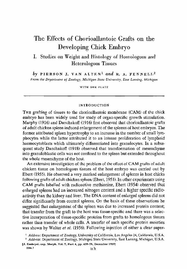
The Effects of Chorioallantoic Grafts on the Developing Chick Embryo I. Studies on Weight and Histology of Homologous and Heterologous Tissues by PIERSON J. VAN ALTEN 1 and R. A. FENNELL 2 From the Department of Zoology, Michigan State University, East Lansing, Michigan WITH ONE PLATE INTRODUCTION THE grafting of tissues to the chorioallantoic membrane (CAM) of the chick embryo has been widely used for study of organ-specific growth stimulation. Murphy (1916) and Danchakoff (1916) first observed that chorioallantoic grafts of adult chicken spleen induced enlargement of the spleens of host embryos. The former attributed spleen hypertrophy to an increase in the number of small lym- phocytes while the latter attributed it to an intense proliferation of lymphoid haemocytoblasts which ultimately differentiated into granulocytes. In a subse- quent study Danchakoff (1918) observed that transformation of mesenchyme into granuloblastic cells was not confined to the spleen but extended throughout the whole mesenchyme of the host. An extensive investigation of the problem of the effect of CAM grafts of adult chicken tissue on homologous tissues of the host embryo was carried out by Ebert (1955). He observed a very marked enlargement of spleens in host chicks following grafts of adult chicken spleen (Ebert, 1951). In other experiments using CAM grafts labelled with radioactive methionine, Ebert (1954) observed that enlarged spleens had an increased nitrogen content and a higher specific radio- activity than the kidney and liver. The DNA content of enlarged spleens did not differ significantly from control spleens. On the basis of these observations he suggested that enlargement of the spleen was due to increased protein content; that transfer from the graft to the host was tissue-specific and there was a selec- tive incorporation of tissue-specific proteins from grafts to homologous tissues rather than transfer of whole cells. A transfer of such specific protein moieties was shown by Walter et al. (1956). Following injection of either a clear super- 1 Address: Department of Zoology, University of California, Los Angeles 24, California, U.S.A. 2 Address: Department of Zoology, Michigan State University, East Lansing, Michigan, U.S.A. [J. Embryol. exp. Morph. Vol. 7, Part 4, pp. 459-75, December 1959] 5584.7 H h
Transcript of The Effect osf Chorioallantoic Grafts on the...
The Effects of Chorioallantoic Grafts on theDeveloping Chick Embryo
I. Studies on Weight and Histology of Homologous andHeterologous Tissues
by PIERSON J. VAN ALTEN1 and R. A. FENNELL2
From the Department of Zoology, Michigan State University, East Lansing, Michigan
WITH ONE PLATE
INTRODUCTION
T H E grafting of tissues to the chorioallantoic membrane (CAM) of the chickembryo has been widely used for study of organ-specific growth stimulation.Murphy (1916) and Danchakoff (1916) first observed that chorioallantoic graftsof adult chicken spleen induced enlargement of the spleens of host embryos. Theformer attributed spleen hypertrophy to an increase in the number of small lym-phocytes while the latter attributed it to an intense proliferation of lymphoidhaemocytoblasts which ultimately differentiated into granulocytes. In a subse-quent study Danchakoff (1918) observed that transformation of mesenchymeinto granuloblastic cells was not confined to the spleen but extended throughoutthe whole mesenchyme of the host.
An extensive investigation of the problem of the effect of CAM grafts of adultchicken tissue on homologous tissues of the host embryo was carried out byEbert (1955). He observed a very marked enlargement of spleens in host chicksfollowing grafts of adult chicken spleen (Ebert, 1951). In other experiments usingCAM grafts labelled with radioactive methionine, Ebert (1954) observed thatenlarged spleens had an increased nitrogen content and a higher specific radio-activity than the kidney and liver. The DNA content of enlarged spleens did notdiffer significantly from control spleens. On the basis of these observations hesuggested that enlargement of the spleen was due to increased protein content;that transfer from the graft to the host was tissue-specific and there was a selec-tive incorporation of tissue-specific proteins from grafts to homologous tissuesrather than transfer of whole cells. A transfer of such specific protein moietieswas shown by Walter et al. (1956). Following injection of either a clear super-
1 Address: Department of Zoology, University of California, Los Angeles 24, California, U.S.A.2 Address: Department of Zoology, Michigan State University, East Lansing, Michigan, U.S.A.
[J. Embryol. exp. Morph. Vol. 7, Part 4, pp. 459-75, December 1959]5584.7 H h
460 P. J. VAN ALTEN AND R. A. FENNELL
natant of S35-labelled homogenized liver or heart into 9-day-old embryos, ahigher specific activity was observed in the respective host tissues of liver andheart. Similar studies have also been conducted by Ebert (1957; 1958 a, b). Thisinvestigator was able to demonstrate the localization of tissue-specific fractionsin homologous organs following intravenous injections of cytoplasmic materials.Selective localization was obtained following the injection of microsomal andsupernatant fractions, but not with nuclear and mitochondrial fractions. On theother hand, Van Haeften (1958) observed significant enlargement of the hostspleen following grafting of cell-free homogenates of spleen to the CAM.
An alternative hypothesis, that of a 'graft-versw.s-host reaction', has beenadvanced to explain splenic enlargement following introduction of adult spleenor blood-cells into the chick embryo. Simonsen (1957) observed that 18-day chickembryos, injected intravenously with adult chicken spleen-cells or blood-cells,manifested symptoms of severe haemolytic anaemia, and that marked hyper-trophy occurred in the spleen, liver, and thymus about 2 weeks after hatching.He further observed that reticulo-endothelial cells replaced erythropoietic andmyelopoietic cells in the bone-marrow. The enlargement of the spleen, thehaemolytic anaemia, and the histological changes in the bone-marrow he attri-buted to an immune reaction of the injected cells against the host and coloniza-tion of the host's tissues by injected cells. Recently, Cock & Simonsen (1958)have shown that when blood from one inbred line of adult chickens was injectedinto newly hatched chicks of a cross between two highly inbred lines, grossenlargement of the spleen and liver occurred. On the other hand, when Fi bloodwas injected into Fi chicks only a relatively slight enlargement occurred, andthis they attributed to the antigenic diversity within one of the inbred lines.Terasaki (1959) has shown that when adult chicken lymphocytes were injectedinto chick embryos there was a marked splenic enlargement in the host. On theother hand, adult monocytes and thymocytes did not cause significant enlarge-ment of the spleen. Billingham & Brent (1957) also attribute the production of'runt disease' in mice and the mortality observed following injections of A-strainmice with C57 spleen-cells to the immunological reactions produced by theinoculated adult spleen-cells against the tissue antigens present in their younghosts.
Evidence of a cellular immune reaction on the part of adult spleen-cells in anembryonic environment has been demonstrated in the chick by Ebert (1957;1958 a, b) and in the larval salamander by De Lanney (1958). The formerinvestigator observed that following intracoelomic grafts of adult spleen on the4th day of incubation, a profound effect was produced on the vascular system,leading to death in 30 per cent, of the host embryos. The latter investigatorobserved that when adult salamander spleen was grafted into a pocket in thedorsal fin or in the coelom of the larval salamander, Taricha towsa, growth ofthe host spleen was suppressed.
From this review of the literature it would seem that in order to ascertain if
E F F E C T S OF C H O R I O A L L A N T O I C GRAFTS. I 461
organ-specific growth stimulation exists some organ should be studied which isnot known to be engaged in haematopoiesis. The histogenesis of the duodenumand histochemistry of the mucopolysaccharides in the connective tissues of thisorgan have been described by Van Alten & Fennell (1957). The objectives of thepresent investigation were to study: (1) the effects of chorioallantoic grafts ofembryonic duodenum and various adult organs (spleen, liver, heart, skin, brain,and duodenum) upon the weight of homologous and heterologous organs in thedeveloping chick embryo; (2) the eifects of soluble extracts of adult and em-bryonic chicken organs on the host embryo when injected into the yolk sac; and(3) the histology of the various embryonic organs following CAM transplants.
MATERIALS AND METHODS
Chickens of both sexes, of different breeds and of ages ranging from 3 monthsto about one year after hatching were used as donors of tissues for grafting.Embryonic duodena were obtained from embryos incubated 15,16,17,18, and20 days.
The procedure of the chorioallantoic transplantations is given in detail byVan Alten (1958). The approach has been to compare weight differences of thetotal host embryo and various host organs (spleen, liver, heart, intestine, andduodenum) following: (1) chorioallantoic transplants of fresh adult organs;(2) grafts of adult chicken duodenum treated with 95 per cent, ethanol at—20° C. for 24 hours, lyophilization, or heat (80° C. for 20 minutes); (3) shamoperations in which the complete chorioallantoic transplantation procedurewas carried out, but only a drop of sterile Ringer's solution, or a small piece of2 per cent, agar, was put on the CAM; (4) transplants of adult rat duodenum; and(5) grafts of 15-, 16-, 17-, 18-, and 20-day embryonic duodena.
The chicken was killed by decapitation and various tissues (duodenum,spleen, liver, heart, skin, and brain) were quickly removed and cut into small(2-3 mm.3) pieces in chick Ringer's solution. In order to sterilize the duodenumit was placed in 200 ml. of chick Ringer's solution containing 1,000 mg. ofchloromycetin for 10 minutes. In order to determine if the tissue was sterilizedan occasional piece of tissue was streaked on a blood-agar plate. In no case wascontamination found. One of the prepared pieces of tissue was then placed at thebifurcation point of blood-vessels on the CAM. The above procedure was alsoused for adult rat duodenum.
In order to ascertain the effect of soluble cell-free material of the duodenum,liver, and heart of the adult chicken on the chick embryo, the following pro-cedure was carried out. Tissues, either adult or 20-day embryonic chick, werehomogenized in a Waring blendor with a 1 to 5 ratio of 015 M saline (bufferedto pH 7-4 with 0 005 M phosphate buffer) for 20 minutes at 4° C , after which thehomogenate was centrifuged at about 500 g. for 30 minutes at 4° C. The super-natant after Seitz filtration was used for injection into the yolk sac.
462 P. J. VAN ALTEN AND R. A. FENNELL
The statistics used in comparing the weight of the chick, spleen, liver, heart,intestine, and duodenum following various treatments to the CAM, were ananalysis of variance (Dixon & Massey, 1951) and the multiple range test forheteroscedastic means (Duncan, 1957).
Following various CAM grafts, host tissues of spleen, liver, heart, duodenum,and CAM were fixed in Bouin's fixative for 24 hours and embedded in paraffinby routine methods of tissue preparation. Tissue sections were cut at 5 p.. Alltissues were routinely stained with haematoxylin and eosin, Gomori's trichromestain (Gomori, 1950), the triple stain of Himes & Moriber (1956), and theAzure II eosin procedure (Lillie, 1954). For elastic tissue Weigert's resorcinfuchsin was used and Van Giesen's picro-acid fuchsin was used for collagenoustissue.
RESULTS
The effect of chorioallantoic transplants of adult and embryonic chicken tissueson the weight of homologous and heterologous tissues of the host embryo
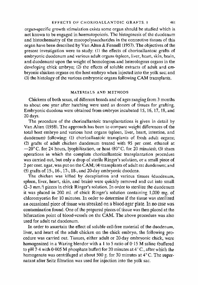
It is evident from Table 1 that, following adult duodenal grafts, there was asignificant reduction in weight of the whole embryo and a marked enlargementof the spleen, liver, and heart, and that there was no significant weight differencein the intestine or duodenum. It can also be seen that after liver grafts the liverand heart showed a significant enlargement over those of controls (sham opera-tion). However, the liver and heart were significantly larger after grafts ofduodenum than after liver grafts. When alcohol-inactivated adult chicken duo-denum was transplanted to the CAM it was observed that there was a decreasein weight of both the whole embryo and the duodenum.
TABLE 1
Fresh weights of whole embryos and of homologous and heterologous organsfollowing chorioallantoic transplants
Treatment
A. Grafts of adultchicken duodenum .
B. Grafts of adultchicken liver .
C. Sham operationD. Grafts of inactivated
adult chicken duo-denum .
Statistical F test .Statistical multiple range
test1.
No.
33
2925
21
Wholechick
(g.)
16-25
18-1318-86
17-609-41*
(BQ (BD)
Mean weight of host and host organs
Spleen
(mg-)
36-41
16-971200
131922-19*
(BCD)
Liver
(g.)
0-6609
0-54210-4716
0-451430-77*
(CD)
Heart
(g.)
0-2115
0178601592
0144828-75*
(CD)
Intestine
(g.)
0-3963
0-41480-3924
0-37570-75
(ABCD)
Duodenum
(mg.)
6708
670861-96
55-375-72*
(ABC) (CD)
1 Any two means appearing together within the same parentheses are not significantly differentat the 5 per cent, level.
EFFECTS OF CHORIOALLANTOIC GRAFTS. I 463
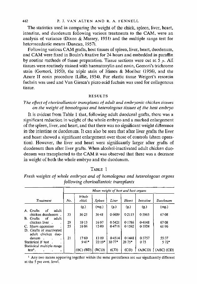
In Table 2 it is evident that the spleen and heart were significantly larger whenbrain was grafted to the CAM than when control procedures were used. Theduodenum was larger, but other significant growth changes were not observedafter grafts of rat duodenum.
TABLE 2
Fresh weights of whole embryos and of homologous and heterologous organsfollowing chorioallantoic transplants
Treatment
A. Grafts of adultchicken duo-denum
B. Grafts of adultchicken brain .
C. Grafts of adultrat duodenum .
D. Sham operationStatistical F test .Statistical multiple
range test1 .
No.
19
28
2126
Wholechick
(g.)
20-63
23-26
22-5822-53
705*
(BCD)
Meat
Spleen
(mg.)
54-66
17-49
9-899-83
75-91*
(CD)
i weight of host and host organs
Liver
(g.)
0-7158
0-5681
0-52670-5142
24-29*
(BC) (CD)
Heart
(g.)
0-2121
0-2026
01590015465-67*
(AB)(CD)
Intestine
(g-)
0-5778
0-6143
0-57000-56921-43
(ABCD)
Duodenum
(mg.)
58-73
58-60
62-9355-773-12*
(ABC) (ABD)
1 Any two means appearing together within the same parentheses are not significantly differentat the 5 per cent, level.
TABLE 3
Fresh weights of whole embryos and of homologous and heterologous organsfollowing chorioallantoic transplants
Treatment
A. Grafts of adultchicken duo-denum
B. Grafts of adultchicken lyophil-ized duodenum.
C. Sham operationStatistical F test .Statistical multiple
range test1 .
No.
20
3828
Wholechick
(g.)
21-36
231623-354-86*
(BC)
Mean
Spleen
(mg.)
43-31
13-891310
103-21*
(BC)
weight of host and host organs
Liver
(g.)
0-7280
0-52530-5393
36-88*
(BC)
Heart
(g.)
0-2405
0186101636
14-29*
Intestine
(g.)
0-6285
0-57370-59253-33
(ABC)
Duodenum
(mg.)
64-72
5419640610-52*
(AC)
1 Any two means appearing together within the same parentheses are not significantly differentat the 5 per cent, level.
It is evident on the basis of the multiple range test (Table 3) that, followingadult duodenal grafts, the weight of the whole embryo was significantly less and
464 P. J. VAN ALTEN AND R. A. FENNELL
the weights of the spleen, liver, and heart were significantly more than afterlyophilized duodenal grafts, as well as (in agreement with Table 1) more thanin controls. On the other hand, when lyophilized duodenum was placed on theCAM the weight of the host heart was significantly higher, while the duodenumwas significantly lower than in controls.
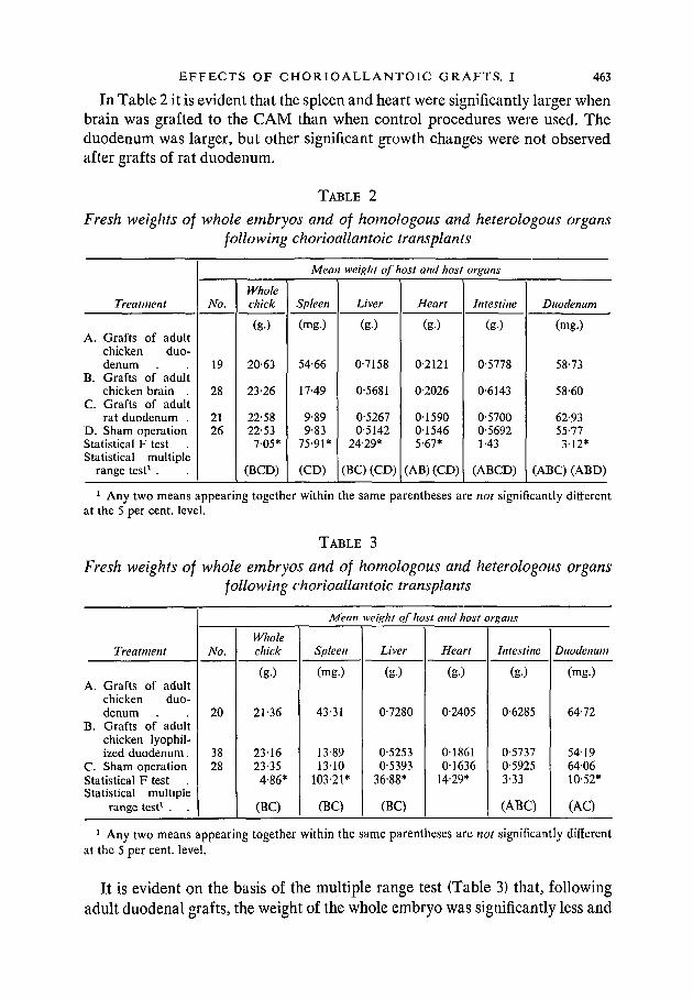
Table 4 again confirms the effects of a graft of adult chicken duodenum.Following grafts of either heated or alcohol-extracted chicken duodenum therewere no significant weight differences from controls in either the whole embryoor the individual organs.
TABLE 4
Fresh weights of whole embryos and of homologous and heterologous organsfollowing chorioallantoic transplants
Treatment
A. Grafts of adult chickenduodenum.
B. Grafts of heated adultchicken duodenum(80° C. for 20 minutes)
C. Grafts of alcohol ex-tract of adult chickenduodenum.
D. Sham operation .Statistical F testStatistical multiple range
t e s t 1 . . . .
No.
34
30
3627
Mean
Wholechick
(g.)
23-45
24-85
24-4124-81409*
(BCD)
weight of host and host organs
Spleen
(mg.)
46-63
13-22
12-38120536-46*
(BCD)
Liver
(g.)
0-7229
0-5713
0-54940-5378
25-77*
(BCD)
Heart
(g.)
0-2185
01917
01853018008-75*
(BCD)
Intestine
(g.)
0-6412
0-6607
0-61420-6622213
(ABCD)
Duodenum
(mg.)
66-51
73-43
66-6471-773-95*
(AC) (BD)
1 Any two means appearing together within the same parentheses are not significantly differentat the 5 per cent, level.
TABLE 5
Fresh weights of whole embryos and of homologous and heterologous organsfollowing chorioallantoic transplants
Treatment
A. Grafts of adult chickenheart.
B. Grafts of adult chickenskin . . . .
C. Sham operation .Statistical F testStatistical multiple range
test1 . . . .
No.
19
2535
Wholechick
(g.)
24-89
24-9725-32
0-37
(ABC)
Mean weight of host and host organ.
Spleen
(mg.)
1606
28-861407
7-54*
(AC)
Liver
(g.)
0-5684
0-65400-56406-25*
(AC)
Heart
(g.)
01763
01868018711-85
(ABC)
Intestine
(g-)
0-6221
0-66880-67372-14
(ABC)
Duodenum
(mg.)
71-11
82-3272113-38*
(AC)
1 Any two means appearing together within the same parentheses are not significantly differentat the 5 per cent, level.
EFFECTS OF CHORIOALLANTOIC GRAFTS. I 465
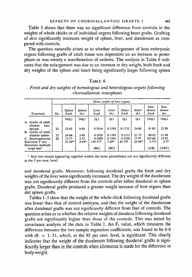
Table 5 shows that there was no significant difference from controls in theweights of whole chicks or of individual organs following heart grafts. Graftingof skin significantly increases weight of spleen, liver, and duodenum as com-pared with controls.
The question naturally arises as to whether enlargement of host embryonicorgans following grafts of adult tissue was dependent on an increase in proto-plasm or was merely a manifestation of oedema. The analysis in Table 6 indi-cates that the enlargement was due to an increase in dry weight, both fresh anddry weights of the spleen and heart being significantly larger following spleen
TABLE 6
Fresh and dry weights of homologous and heterologous organs followingchorioallantoic transplants
Treatment
A. Grafts of adultchicken duo-denum .
B. Grafts of adultchicken spleen
C. Sham operationStatistical F test .Statistical multiple
range test1
Mean weight of host organs
No.
25
2528
Spleenfresh
(mg.)
32-65
240413-3721-69*
Spleendry
(mg.)
9-66
5 062-334-44*
Liverfresh
(g.)
0-7014
0-59300-5909
139-57*
(BC)
Liverdry
(g.)
01598
01383014245-29*
(BC)
Heartfresh
(g.)
01733
0151501250
45-79*
Heartdry
(g.)
24-84
21-7517-9725-64*
Duo-denumfresh
(mg.)
91-83
90-62103-66
3-71*
(AB)
Duo-denum
dry
(mg.)
12-58
11-5612-49
2-77
(ABC)
1 Any two means appearing together within the same parentheses are not significantly differentat the 5 per cent, level.
and duodenal grafts. Moreover, following duodenal grafts the fresh and dryweights of the liver were significantly increased. The dry weight of the duodenumwas not significantly different from the controls after either duodenal or spleengrafts. Duodenal grafts produced a greater weight increase of host organs thandid spleen grafts.
Tables 1-3 show that the weight of the whole chick following duodenal graftswas lower than that of control embryos, and that the weight of the duodenumafter duodenal grafts was not significantly different from that of controls. Thequestion arises as to whether the relative weights of duodena following duodenalgrafts are significantly higher than those of the controls. This was tested bycovariance analysis of the data in Table 1. An Fi value, which measures thedifference between the two sample regression coefficients, was found to be 6*6with df. = 1, 51, which, at the 95 per cent, level, is significant. This clearlyindicates that the weight of the duodenum following duodenal grafts is signi-ficantly larger than in the controls when allowance is made for the difference inbody-weight.
466 P. J. VAN ALTEN AND R. A. FENNELL
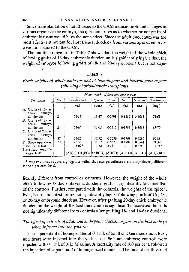
Since transplantation of adult tissue to the CAM induces profound changes invarious organs of the embryo, the question arises as to whether or not grafts ofembryonic tissue would have the same effect. Since the adult duodenum was themost effective stimulant for host tissues, duodena from various ages of embryoswere transplanted to the CAM.
The multiple range test in Table 7 shows that the weight of the whole chickfollowing grafts of 16-day embryonic duodenum is significantly higher than theweight of embryos following grafts of 18- and 20-day duodena but is not signi-
TABLE 7
Fresh weights of whole embryos and of homologous and heterologous organsfollowing chorioallantoic transplants
Treatment
A. Grafts of 16-daychick embryoduodenum
B. Grafts of 18-daychick embryoduodenum
C. Grafts of 20-daychick embryoduodenum
D. Sham operationStatistical F test .Statistical multiple
range test1 .
Mean weight of host and host organs
No.
24
24
3722
Whole chick
(g.)
26-13
24-66
24-8625-56
5-07*
(AD) (CD) (BC)
Spleen
(mg.)
13-47
1305
12-7211-621-62
(ABCD)
Liver
(g.)
0-5908
0-5717
0-56300-55772-33
(ABCD)
Heart
(g-)
01817
01796
0176801709
0
(ABCD)
Intestine
(g.)
0-6613
0-6658
0-62860-65410-875
(ABCD)
Duodenum
(mg.)
74-65
67-70
60-8871-398-75*
(AD) (BD)
1 Any two means appearing together within the same parentheses are not significantly differentat the 5 per cent, level.
ficantly different from control experiments. However, the weight of the wholechick following 18-day embryonic duodenal grafts is significantly less than thatof the controls. Further, compared with the controls, the weights of the spleen,liver, heart, and intestine are not significantly higher following grafts of 16-, 18-,or 20-day embryonic duodena. However, after grafting 20-day chick embryonicduodenum the weight of the host duodenum is significantly decreased, but it isnot significantly different from controls after grafting 18- and 16-day duodena.
The effect of extracts of adult and embryonic chicken organs on the host embryowhen injected into the yolk sac
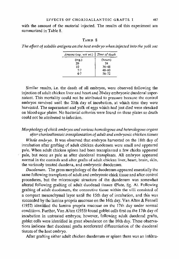
The supernatant of homogenates of 0 1 ml. of adult chicken duodenum, liver,and heart were injected into the yolk sac of 96-hour embryos; controls wereinjected with 0-1 ml. of 0-15 M saline. A mortality rate of 100 per cent, followedthe injection of supernatant of homogenized duodena. The time of death varied
EFFECTS OF CHORIOALLANTOIC GRAFTS. I 467
with the amount of the material injected. The results of this experiment aresummarized in Table 8.
TABLE 8
The effect of soluble antigens on the host embryo when injected into the yolk sac
Amount (wig. wet wt.)
(mg.)20107-76-7
Time of death
(hours)24
36-4848-6056-72
Similar results, i.e. the death of all embryos, were observed following theinjection of adult chicken liver and heart and 20-day embryonic duodenal super-natant. This mortality could not be attributed to pressure because the controlembryos survived until the 20th day of incubation, at which time they wereharvested. The supernatant and yolk of eggs which had just died were streakedon blood-agar plates. No bacterial colonies were found on these plates so deathcould not be attributed to infection.
Morphology of chick embryos and various homologous and heterologous organsafter chorioallantoic transplantation of adult and embryonic chicken tissues
Whole embryo. It was observed that embryos harvested on the 18th day ofincubation after grafting of adult chicken duodenum were small and appearedpale. When adult chicken spleen had been transplanted a few chicks appearedpale, but none as pale as after duodenal transplants. All embryos appearednormal in the controls and after grafts of adult chicken liver, heart, brain, skin,the variously treated duodena, and embryonic duodenum.
Duodenum. The gross morphology of the duodenum appeared essentially thesame following transplants of adult and embryonic chick tissue and after controlprocedures, but the microscopic structure of the duodenum was somewhataltered following grafting of adult duodenal tissues (Plate, fig. A). Followinggrafting of adult duodenum, the connective tissue within the villi consisted ofa compact mesenchymal layer until the 15th day of incubation, and this wassucceeded by the lamina propria mucosae on the 16th day. Van Alten & Fennell(1957) identified the lamina propria mucosae on the 17th day under normalconditions. Further, Van Alten (1955) found goblet cells first on the 17th day ofincubation in untreated embryos; however, following adult duodenal grafts,goblet cells were identified in great abundance on the 16th day. These observa-tions indicate that duodenal grafts accelerated differentiation of the duodenaltissues of the host embryo.
After grafting either adult chicken duodenum or spleen there was an infiltra-
468 P. J. VAN ALTEN AND R. A. FENNELL
tion of lymphocytes into the duodenal tissue, but this was not observed followingliver, heart, brain, skin, or embryonic duodenal grafts (Plate, fig. B).
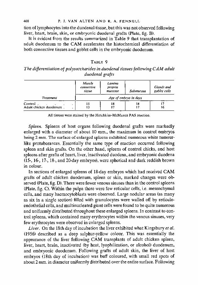
It is evident from the results summarized in Table 9 that transplantation ofadult duodenum to the CAM accelerates the histochemical differentiation ofboth connective tissues and goblet cells in the embryonic duodenum.
TABLE 9
The differentiation of polysaccharides in duodenal tissues following CAM adultduodenal grafts
Treatment
C o n t r o l . . . . .A d u l t c h i c k e n d u o d e n u m .
Muscleconnective
tissue
Laminapropriamucosae Submucosa
Glands andgoblet cells
Age of embryo in days
1513
1817
1817
1716
All tissues were stained by the Hotchkiss-McManus PAS reaction.
Spleen. Spleens of host organs following duodenal grafts were markedlyenlarged with a diameter of about 10 mm., the maximum in control embryosbeing 2 mm. The surface of enlarged spleens exhibited numerous white tumour-like protuberances. Essentially the same type of reaction occurred followingspleen and skin grafts. On the other hand, spleens of control chicks, and hostspleens after grafts of heart, liver, inactivated duodena, and embryonic duodena(15-, 16-, 17-, 18-, and 20-day embryos), were spherical and dark reddish brownin colour.
In sections of enlarged spleens of 18-day embryos which had received CAMgrafts of adult chicken duodenum, spleen or skin, marked changes were ob-served (Plate, fig. D). There were fewer venous sinuses than in the control spleens(Plate, fig. C). Within the pulpa there were few reticular cells, i.e. mesenchymalcells, and many haemocytoblasts were observed. Large nodular areas (as manyas six in a single section) filled with granulocytes were walled off by reticulo-endothelial cells, and multinucleated giant cells were found to be quite numerousand uniformly distributed throughout these enlarged spleens. In contrast to con-trol spleens, which contained many erythrocytes within the venous sinuses, veryfew erythrocytes were observed in enlarged spleens.
Liver. On the 18th day of incubation the liver exhibited what Kingsbury et al.(1956) described as a deep sulphur-yellow colour. This was essentially theappearance of the liver following CAM transplants of adult chicken spleen,liver, heart, brain, inactivated (by heat, lyophilization, or alcohol) duodenum,and embryonic duodenum. Following grafts of adult skin, the liver of hostembryos (18th day of incubation) was buff coloured, with small red spots ofabout 2 mm. in diameter uniformly distributed over the entire surface. Following
EFFECTS OF CHORIOALLANTOIC GRAFTS. I 469
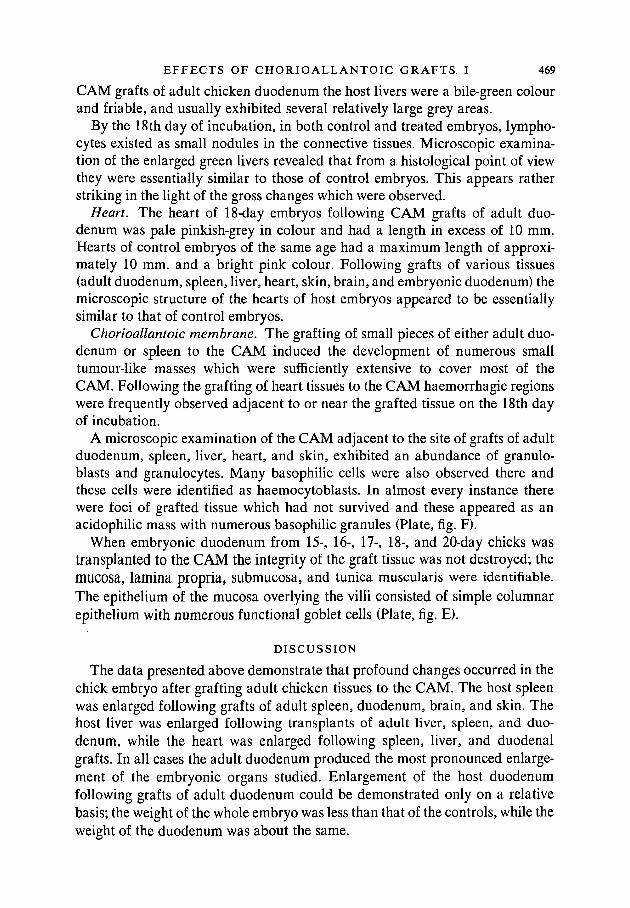
CAM grafts of adult chicken duodenum the host livers were a bile-green colourand friable, and usually exhibited several relatively large grey areas.
By the 18th day of incubation, in both control and treated embryos, lympho-cytes existed as small nodules in the connective tissues. Microscopic examina-tion of the enlarged green livers revealed that from a histological point of viewthey were essentially similar to those of control embryos. This appears ratherstriking in the light of the gross changes which were observed.
Heart. The heart of 18-day embryos following CAM grafts of adult duo-denum was pale pinkish-grey in colour and had a length in excess of 10 mm.Hearts of control embryos of the same age had a maximum length of approxi-mately 10 mm. and a bright pink colour. Following grafts of various tissues(adult duodenum, spleen, liver, heart, skin, brain, and embryonic duodenum) themicroscopic structure of the hearts of host embryos appeared to be essentiallysimilar to that of control embryos.
Chorioallantoic membrane. The grafting of small pieces of either adult duo-denum or spleen to the CAM induced the development of numerous smalltumour-like masses which were sufficiently extensive to cover most of theCAM. Following the grafting of heart tissues to the CAM haemorrhagic regionswere frequently observed adjacent to or near the grafted tissue on the 18th dayof incubation.
A microscopic examination of the CAM adjacent to the site of grafts of adultduodenum, spleen, liver, heart, and skin, exhibited an abundance of granulo-blasts and granulocytes. Many basophilic cells were also observed there andthese cells were identified as haemocytoblasts. In almost every instance therewere foci of grafted tissue which had not survived and these appeared as anacidophilic mass with numerous basophilic granules (Plate, fig. F).
When embryonic duodenum from 15-, 16-, 17-, 18-, and 20-day chicks wastransplanted to the CAM the integrity of the graft tissue was not destroyed; themucosa, lamina propria, submucosa, and tunica muscularis were identifiable.The epithelium of the mucosa overlying the villi consisted of simple columnarepithelium with numerous functional goblet cells (Plate, fig. E).
DISCUSSION
The data presented above demonstrate that profound changes occurred in thechick embryo after grafting adult chicken tissues to the CAM. The host spleenwas enlarged following grafts of adult spleen, duodenum, brain, and skin. Thehost liver was enlarged following transplants of adult liver, spleen, and duo-denum, while the heart was enlarged following spleen, liver, and duodenalgrafts. In all cases the adult duodenum produced the most pronounced enlarge-ment of the embryonic organs studied. Enlargement of the host duodenumfollowing grafts of adult duodenum could be demonstrated only on a relativebasis; the weight of the whole embryo was less than that of the controls, while theweight of the duodenum was about the same.
470 P. J. VAN ALTEN AND R. A. FENNELL
Histological studies of the enlarged spleen showed that the enlargement wasprimarily caused by granulopoiesis regardless of which adult organ (spleen,duodenum, skin, or brain) was used to stimulate it. Although the gross appear-ance of the host liver (it was bile-green in colour) and of the heart (it was enlargedand pale) was altered following CAM grafts of adult chicken duodenum, themicroscopic anatomy was similar to that of the controls.
The hypothesis of organ-specific growth stimulation as proposed by Weiss(1947) and Ebert (1955) has been based largely on the observations of Murphy(1916), Danchakoff (1916), Willier (1924), Weiss (1947), and Ebert (1951). Theseworkers found that when a small fragment of an adult chicken organ was trans-planted to the vascular bed of an embryo (either CAM or vascular area of theblastoderm) it greatly stimulated growth of the homologous embryonic organs.Weiss & Andres (1952) observed an increase in the mitotic rate of the embryonickidney after injections of kidney brei into the CAM blood-vessels, and also in themitotic rate of the kidney and liver following injections of mesonephric brei(Andres, 1955).
Observations made during the course of this study do not entirely support thehypothesis of organ-specific growth stimulation. Van Alten (1959) also observedthat the antigens prepared from spleens stimulated by adult duodenal graftsexhibited a different antigenic pattern than control spleens, and that antigensprepared from duodena stimulated by adult spleen grafts exhibited a differentantigenic pattern from control duodena. Danchakoff (1918) observed thatenlargement after CAM transplants of adult spleen was not confined to thespleen but extended throughout the whole mesenchyme of the host. Andres's(1955) studies were not organ-specific after injection of mesonephric brei. Heobserved that mitotic indices of the liver increased 23 per cent, while themesonephros increased 46 per cent. However, Levy (1956) was unable to demon-strate the retrogression of the mesonephros after CAM grafts of 18-day em-bryonic mesonephros or metanephros which would be expected if organ-specificgrowth stimulation had occurred. Also, Wilson & Leduc (1947) have demon-strated that a number of agents (pulped liver of mice and guinea-pigs, boiledand autolysed liver, pulped kidney, and boiled egg-yolk) produced an increase inthe mitotic rates in mouse livers on the 5th day after injection. Further, Saetren(1956) has observed that after injections of macerated homologous tissue into theperitoneal cavity of rats following partial nephrectomy or removal of a portionof the liver, there was a marked inhibition of mitoses in regenerating portions ofkidney and liver. Steuart (see Ebert, 19586) has confirmed this observation onthe kidney and, further, has shown that liver homogenate suppresses the mitoticactivity in the remaining kidney stump but that kidney homogenate is morespecific. However, it should be kept in mind that the systems studied by Andresand those of Wilson & Leduc, Saetren, and Steuart are quite different.
The tracer studies of Ebert (1954) showed that when labelled tissues wereplaced on the CAM, homologous organs had a higher specific activity than
EFFECTS OF CHORIOALLANTOIC GRAFTS. I 471
heterologous organs. However, Horn & House (1955) observed that when theyinjected tagged homogenates of liver, kidney, spleen, and thymus into youngmice the uptake value of the spleen was consistently higher than other organs.They suggested that the spleen was the most effective organ of the reticulo-endothelial system for removing foreign protein from the circulation.
An alternative hypothesis of graft-vm^s-host reaction was proposed bySimonsen (1957) to account for the enlargement of embryonic spleens followinggrafts of adult spleen. Billingham et al. (1956) observed a 95 per cent, mortalityof chick embryos following injection of adult blood. Death occurred toward theend of incubation and was attributed to an infective agent. However, Simonsen(1957) and Cock & Simonsen (1958) believe that adult spleen or blood-cells cancolonize host lymphoid organs, in which they multiply and react immunologi-cally against the host. Recently, Terasaki (1959) has shown that when adultchicken lymphocytes were injected into chick embryos there was a markedsplenic enlargement in the host. On the other hand, adult monocytes and thymo-cytes did not cause significant enlargement of the spleen. The generalized effects,i.e. enlargement of spleen, liver, and heart, observed in these studies followinggrafting of adult chicken duodenum may be accounted for on the basis that thisorgan contains much lymphocytic tissue and thus produces a graft-vmw.s-hostreaction. However, this hypothesis fails to give a satisfactory explanation as towhy brain grafts, which contain very few lymphocytes, produced splenic en-largement while adult liver transplants, which contain many lymphocytes, didnot elicit splenic enlargement. Further, the present study also shows that adultduodenal grafts, which contain fewer lymphocytes than spleen, elicited a greaterenlargement of the host spleen than did splenic grafts. However, these results arein contrast to Ebert's (1959), who obtained no splenic enlargement in the embryoafter grafting of adult brain tissue and observed that adult liver grafts produceda marked stimulation (about 40 per cent, as much as adult spleen) of the em-bryonic spleen. The graft-vmw,s-host hypothesis also fails to explain why themicroscopic morphology of the adult grafted tissue was destroyed and replacedby a myeloid metaplastic centre, a type of reaction to the CAM which has alsobeen observed by Van Haeften (1958) with cell-free material. Simonsen (1957),on the basis of direct injections of adult cells, postulates that enlargement ofhost spleens was due to colonization of the host organ by injected cells. Ebert(1954), on the basis of DNA content of the enlarged spleens, ruled out the trans-fer of whole cells. Recently, however, Ebert (1958 a, b) has re-evaluated thisquestion in the light of experiments involving serial transplantation of stimulatedspleens; he observed no dilution of growth-promoting activity by serial passage.On the basis of this observation he proffers as the simplest explanation thecolonization of the host spleen by whole cells from the donor, these cells beingcapable of reproduction. Nevertheless, he was not able to identify large popula-tions of adult cells in the host spleen by histological analysis and thus is unableto rule out the possibility of subcellular material as the causative agent.
472 P. J. VAN ALTEN AND R. A. FENNELL
Recently, Van Haeften (1958) grafted a cell-free homogenate of adult spleen tothe CAM and observed hypertrophy of the host homologous organ. However,Ebert (1958a) did not observe stimulation of the homologous organ with cell-freematerial but only 'predominant localization'. By placing spleen grafts in mem-brane filters, which prevent the passage of cells, Ebert (19586) observed amodest stimulation of the host spleen.
It was shown in the preceding paragraphs that both the organ-specific growthstimulation and the graft-vern^-host hypothesis fail to adequately explain theresults observed following grafts of adult duodenal tissue. It may be postulatedthat the duodenum contains substances which are non-specific and cause variousorgans to enlarge. Levi-Montalcini (1952) has observed enlargement of thespleen and liver of the chick after transplantation of mouse sarcoma 37 or 180 tothe allantoic vesicle. The enlarged liver was described as 'deeply suffused withbile'. Van Alten (1959) observed an increase in the number of antigens in boththe duodenum and spleen after grafts of adult duodenum. This would furtherindicate that some sort of non-specific effect was being produced.
Heart enlargement was probably due to a compensatory reaction related tothe anaemic condition of the chicks. This anaemic condition probably was dueto the fact that the spleen was almost totally given over to the production ofgranulocytes rather than erythrocytes. The grafts were made on the 9th day ofincubation, at which time Fennell (1947) observed that the definitive erythro-cytes had replaced the primitive erythrocytes to become the most numerous typein peripheral blood. Thus, spleen and liver enlargement may be due in part toremoval of primitive generation of blood-cells and foreign substances from theblood vascular system. On the other hand, heart enlargement may be due, inpart, to compensation.
It was also observed in the course of this study that when either normal duo-denum, liver, or heart were injected into the yolk sac on the 4th day of incuba-tion, they caused all the embryos to die within 72 hours. This observation is inkeeping with that of Fennell (1947) who, using a much more dilute inoculum ofminced normal liver, observed that 67 per cent, of the embryos died within 5 daysafter inoculation. He further observed that injections of normal liver-mince pro-duced blood changes which meet the requirements for haemocytoblastosis.
SUMMARY
1. It was observed that following CAM grafts of adult chicken duodenumthere was a marked decrease in the absolute weight of the host, a markedincrease in the weight of the spleen, liver, and heart, and a relative weight in-crease in the duodenum. Further, following grafts of adult skin and brain thespleen and liver were significantly heavier. Following liver grafts the liver andheart showed a significant increase in weight. Adult chicken spleen grafts causeda marked increase in the weight of the spleen and heart. Further, it was observed
EFFECTS OF CHORIOALLANTOIC GRAFTS. I 473
that, regardless of what tissue was used for grafting, 9 days later the morpho-logical integrity of the graft was destroyed and the area was replaced by a mye-loid metaplastic centre. On the other hand, embryonic duodena retained theirintegrity and continued to differentiate.
2. Grafting of adult duodenum caused acceleration of tissue differentiation ofthe host duodenum. The polysaccharides in the connective tissue and gobletcells of the duodenum differentiated at least 24 hours earlier than in controlchicks. Following grafting of duodenum, spleen, and skin, the host spleenexhibited a marked increase in granuloblasts and granulocytes. The heart andliver following grafts were essentially like those in control embryos.
3. Treatment of the adult duodenum prior to grafting with either 95 per cent,alcohol for 24 hours at — 20° C , lyophilization, or heating at 80° C. for 20minutes, resulted in inactivation, the host not being affected.
4. When duodena of 15-, 16-, 17-, 18-, and 20-day embryos were grafted to theCAM, no changes in the weight of host spleen, liver, or heart were found. Fol-lowing 20-day duodenal grafts the weight of the duodenum was significantlydecreased.
5. All embryos receiving adult duodenum, liver, and heart extracts into theyolk sac at 4 days of incubation, died within 72 hours after inoculation.
6. The results are discussed in the light of organ-specific growth stimulationand grait-versus-host reaction, both of which fail to adequately explain all theresults observed.
ACKNOWLEDGEMENTS
The authors are greatly indebted to Drs. A. S. Fox and J. R. Shaver for theirhelp and many valuable suggestions, and to Dr. P. J. Clark for his help with thestatistical analysis. We are also grateful to Dr. J. D. Ebert, Department ofEmbryology, Carnegie Institution of Washington, for a critical discussion of themanuscript. We also wish to thank Mr. P. G. Coleman, photographer, Agricul-tural Experiment Station, Michigan State University, for the photomicrographs.
The material reported in this paper is part of a thesis submitted by P. J. VanAlten to the Department of Zoology, Michigan State University, in partialfulfilment of the requirements for the Ph.D. degree, and was carried out duringthe tenure of a pre-doctoral fellowship (CF 6731), Cancer Division, PublicHealth Service.
R E F E R E N C E S
ANDRES, G. (1955). Growth reactions of mesonephros and liver to intravascular injections ofembryonic liver and kidney suspensions in the chick embryo. J. exp. Zool. 130, 221-49.
BILLINGHAM, R. E., & BRENT, L. (1957). A simple method for inducing tolerance of skin homo-grafts in mice. Transpl. Bull. 4, 67-71.
& MEDAWAR, P. B. (1956). Quantitative studies on tissue transplantation immunity.III. Actively acquired tolerance. Phil. Trans. B, 239, 357-414.
COCK, A. G., & SIMONSEN, M. (1958). Immunological attack on newborn chickens by injectedadult cells. Immunology, 1, 103-10.
474 P. J. VAN ALTEN AND R. A. F E N N E L L
DANCHAKOFF, V. (1916). Equivalence of different hematopoietic anlages (by method of stimulationof their stem cells). Amer. J. Anat. 20, 255-327.(1918). Equivalence of different hematopoietic anlages (by method of stimulation of their
stem cells). II. Graphs of adult spleen on the allantois and response of the allantoic tissues.Amer. J. Anat. 24, 127-89.
DE LANNEY, L. E. (1958). Influence of adult amphibian spleen on the development of embryosand larvae: an immune response? In The Chemical Basis of Development, ed. W. D. McElroy& B. Glass, pp. 562-8. Baltimore: Johns Hopkins Press.
DIXON, W. J., & MASSEY, Jr., F. J. (1951). Introduction to Statistical Analysis. New York:McGraw-Hill Book Company.
DUNCAN, D. B. (1957). Multiple range tests for correlated and heteroscedastic means. Biometrics,13, 164-76.
EBERT, J. D. (1951). Ontogenetic change in the antigenic specificity of the chick spleen. Physiol.Zoo/. 24,20-41.(1954). The effects of chorioallantoic transplants of adult chicken tissues on homologous
tissues of the host chick embryo. Proc. nat. Acad. Sci. Wash. 40, 337-47.(1955). Some aspects of protein biosynthesis in development. In Aspects of Synthesis andOrder in Growth, ed. D. Rudnick, pp. 69-112. Princeton, N.J.: Princeton UniversityPress.(1957). Annual Report of the Director of the Department of Embryology. Yearb. Carneg.
Instn. 56, 297-356.(1958a). Immunochemical analysis of development. In The Chemical Basis of Development,
ed. W. D. McElroy & B. Glass, pp. 526-45. Baltimore: Johns Hopkins Press.(19586). Annual Report of the Director of the Department of Embryology. Yearb. Carneg.
Instn. 57, 309-72.(1959). Personal communication.
FENNELL, R. A. (1947). The relation between age, number, and types of cells in the peripheralcirculation of chicken embryos under normal and experimental conditions. / . agric. Res. 74,217-39.
GOMORI, G. (1950). A rapid one-step trichrome stain. Amer. J. din. Path. 20, 661-4.HIMES, M., & MORIBER, L. (1956). A triple stain for desoxyribonucleic acid, polysaccharides, and
proteins. Stain Tech. 31, 67-70.HORN, E. C , & HOUSE, M. E. (1955). A test of specific uptake from organ homogenates by homo-
logous organs in the young mouse. / . Elisha Mitchell sci. Soc. 71, 171.KINGSBURY, J. W., ALEXANDERSON, M., & KORNSTEIN, E. A. (1956). The development of the liver
in the chick. Anat. Rec. 124, 165-88.LEVI-MONTALCINI, R. (1952). Effects of mouse tumor transplantation on the nervous system.
Ann. N.Y. Acad. Sci. 55, 330-43.LEVY, C. K. (1956). The role of extrinsic factors in the retrogression of the mesonephros of the
embryo chick. University of North Carolina Ph.D. thesis.LILLIE, R. D. (1954). Histopathological Technic and Practical Histochemistry. New York:
Blakiston Co.MURPHY, J. B. (1916). The effect of adult chicken organ grafts on the chick embryo. / . exp. Med.
24,1-6.SAETREN, H. (1956). A principle of auto-regulation of growth. Production of organ specific
mitose-inhibitors in kidney and liver. Exp. Cell Res. 11, 229-34.SIMONSEN, M. (1957). The impact on the developing embryo and newborn animal of adult homo-
logous cells. Acta path, microbiol. scand. 40, 480-500.TERASAKI, P. I. (1959). Identification of the type of blood-cell responsible for the graft-vmus-
host homograft reaction in chicks. / . Embryol. exp. Morph. 7, -VAN ALTEN, P. J. (1955). Development and histogenesis of the avian digestive tract with special
reference to the histochemistry of the connective tissue. Michigan State University, unpub-lished MS. thesis.(1958). The effect of chorioallantoic grafts on the developing chick embryo with special
reference to the development of antigens in the duodenum. Michigan State University,unpublished Ph.D. thesis.
/. Embryol. exp. Morph. Vol. 7, Part 4
P. J. VAN ALTEN and R. A. FENNELL
E F F E C T S OF C H O R I O A L L A N T O I C GRAFTS. I 475
VAN ALTEN, P. J. (1959). The effects of chorioallantoic grafts on the developing chick embryo.II. Studies of adult antigens in the duodenum and spleen. / . Embryol. exp. Morph. 7, 476-86.& FENNELL, R. A. (1957). Histogenesis and histochemistry of the digestive tract of chick
embryos. Anat. Rec. 127, 677-96.VAN HAEFTEN, F. F. (1958). Differentiation induced by chorioallantoic grafting of tissue homo-
genate. Ada physiol. pharm. Neerl. 7, 1-34.WALTER, H., ALLMANN, D. W., & MAHLER, H. R. (1956). Influence of adult tissue homogenates on
formation of similar embryonic proteins. Science, 124, 1251-2.WEISS, P. (1947). The problem of specificity in growth and development. Yale J. Biol. Med. 19,
235-78.& ANDRES, G. (1952). Experiments on the fate of embryonic cells (chick) disseminated by
the vascular route. J. exp. Zool. 121, 449-88.WILLIER, B. H. (1924). The endocrine glands and the development of the chick. I. The effects of
thyroid grafts. Amer. J. Anat. 33, 67-103.WILSON, J. W., & LEDUC, E. H. (1947). Mitotic rate in mouse liver following intraperitoneal
injection of liver, kidney and egg yolk. Anat. Rec. 97, 471-94.
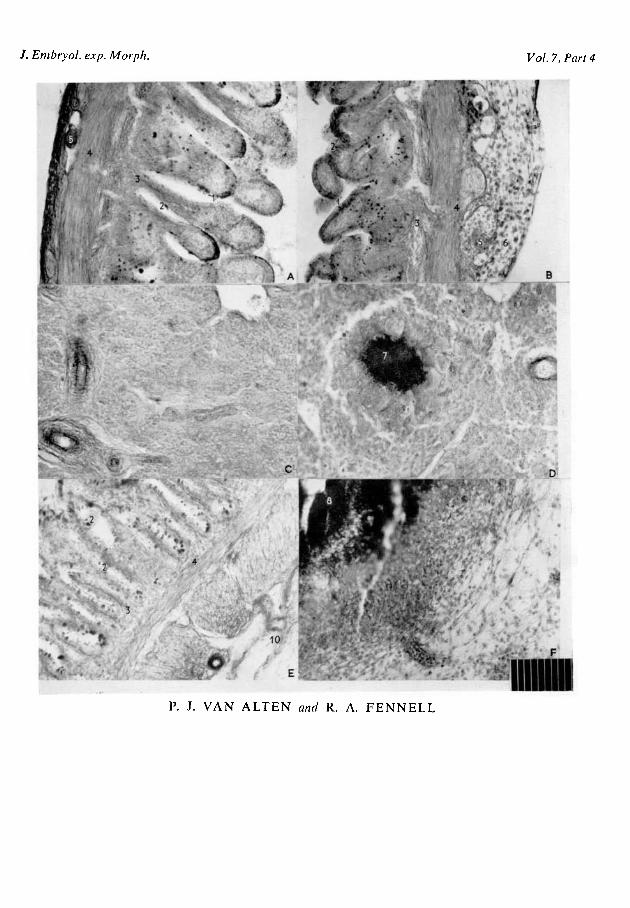
E X P L A N A T I O N OF PLATE
FIG. A. Photomicrograph of a cross-section through the duodenum of a control 18-day em-bryo. 1. epithelium; 2. goblet cell; 3. lamina propria; 4. tunica muscularis; 5. blood-vessel.
FIG. B. Photomicrograph of a cross-section through the duodenum of an 18-day embryofollowing adult chicken duodenal grafts. 1. epithelium; 2. goblet cell; 3. lamina propria; 4. tunicamuscularis; 5. blood-vessel; 6. lymphocytes.
FIG. C. Photomicrograph of a cross-section through a spleen of a control 18-day embryo show-ing the reticular structure.
FIG. D. Photomicrograph of a cross-section through a spleen of an 18-day embryo followingadult duodenal grafts. 7. granulocytic nodule surrounded by multinucleated giant cells.
FIG. E. Photomicrograph of a cross-section of the CAM through a 15-day embryonic duodenalgraft on the 18th day of incubation. 2. goblet cell; 3. lamina propria; 4. tunica muscularis;10. chorioallantoic membrane.
FIG. F. Photomicrograph of a cross-section of the CAM through an adult duodenal graft onthe 18th day of incubation. 8. foci of degenerating adult duodenal tissue surrounded by invadinglymphocytes and granulocytes.
All sections were stained by the Himes Moriber triple stain.Micrometer scale insert: 1 division = 001 mm.
(Manuscript received 23: ii: 59)
5584.7 I I