THE EFFECT OF WHOLE BODY VIBRATION ON BONE DENSITY …
40
THE EFFECT OF WHOLE BODY VIBRATION ON BONE DENSITY AND OTHER PARAMETERS IN THE EXERCISING HORSE by Kayleigh E. Maher A Thesis Submitted in Partial Fulfillment of the Requirements for the Degree of Masters of Science in Horse Science Middle Tennessee State University May 2016 Thesis Committee: Dr. Holly Spooner, Chair Dr. Rhonda Hoffman Dr. John Haffner
Transcript of THE EFFECT OF WHOLE BODY VIBRATION ON BONE DENSITY …
THE EFFECT OF WHOLE BODY VIBRATION ON BONE DENSITY AND OTHER PARAMETERS IN THE EXERCISING HORSE
by
Kayleigh E. Maher
A Thesis Submitted in Partial Fulfillment of the Requirements for the Degree of Masters of Science in Horse Science
Middle Tennessee State University May 2016
Thesis Committee:
Dr. Holly Spooner, Chair
Dr. Rhonda Hoffman
Dr. John Haffner
ii
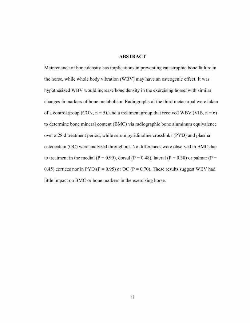
ABSTRACT Maintenance of bone density has implications in preventing catastrophic bone failure in
the horse, while whole body vibration (WBV) may have an osteogenic effect. It was
hypothesized WBV would increase bone density in the exercising horse, with similar
changes in markers of bone metabolism. Radiographs of the third metacarpal were taken
of a control group (CON, n = 5), and a treatment group that received WBV (VIB, n = 6)
to determine bone mineral content (BMC) via radiographic bone aluminum equivalence
over a 28 d treatment period, while serum pyridinoline crosslinks (PYD) and plasma
osteocalcin (OC) were analyzed throughout. No differences were observed in BMC due
to treatment in the medial (P = 0.99), dorsal (P = 0.48), lateral (P = 0.38) or palmar (P =
0.45) cortices nor in PYD (P = 0.95) or OC (P = 0.70). These results suggest WBV had
little impact on BMC or bone markers in the exercising horse.

iii
TABLE OF CONTENTS
LIST OF TABLES..............................................................................................................iv LIST OF FIGURES.............................................................................................................v CHAPTER 1. LITERATURE REVIEW.............................................................................1
Bone Growth and Remodeling................................................................................2
Bone Density in Equines.........................................................................................5
Radiographic Measurements of Bone.....................................................................7
Biochemical Measurements of Bone......................................................................7
Whole Body Vibration............................................................................................9 CHAPTER 2. THE EFFECT OF WHOLE BODY VIBRATION ON BONE DENSITYAND OTHER PARAMETERS IN THE EXERCISING HORSE..................15
Introduction..........................................................................................................15
Materials and Methods.........................................................................................16
Results..................................................................................................................20
Discussion............................................................................................................25
Conclusion...........................................................................................................29
Literature Cited....................................................................................................30
Appendices..........................................................................................................34
Appendix A: IACUC Approval...............................................................35

iv
LIST OF TABLES Table 1. Gaskin and Forearm values (cm ± SEM) of horses subjected to whole body vibration and light exercise (VIB) or light exercise (CON). Values not sharing a common superscript tend to differ (P = 0.067), while values with no superscripts are not different (P > 0.20) ..........................................................................................................................21
v
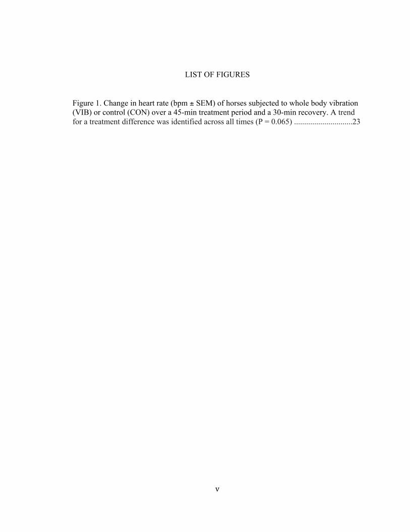
LIST OF FIGURES
Figure 1. Change in heart rate (bpm ± SEM) of horses subjected to whole body vibration (VIB) or control (CON) over a 45-min treatment period and a 30-min recovery. A trend for a treatment difference was identified across all times (P = 0.065) .............................23

1CHAPTER 1. LITERATURE REVIEW
The maintenance of bone density has been a topic of importance in research
involving human health and physiology. Interventions to maintain bone density have
been extensively researched in human and some animal models such as mice and rats
(Oxlund et al. 2003; Stuermer et al. 2014; Wei et al. 2014). More recently, research in
whole body vibration and its effect on bone density has become an important topic of
research as an aid in maintaining or increasing bone density for many different reasons.
Prevention of fractures in frail people, prevention and treatment for osteoporosis as well
as skeletal injuries (Gusi et al, 2006; Verschueren et al, 2004) are a few areas that have
been researched using whole body vibration. Whole body vibration has been used to
improve bone density of postmenopausal women (Slatkovska et al. 2010; Verschueren et
al. 2004;). Whole body vibration has also been shown to have an effect on endocrine
functions as well as muscle development and balance which in turn can affect bone
density in a positive way (Stuermer et al, 2014; Torvinen et al. 2002; Wei et al, 2014).
Injuries, lack of or inability to exercise, hormonal changes, and age are only a few
factors that can have an effect on bone density. These same factors apply to animal
models, especially the horse. Like humans and other animal models, horses require
loading and adequate levels of strain in order to maintain bone density. Skeletal strength
is one of the most difficult factors to assess in a horse especially in regards to training and
conditioning. It is difficult to predict a possible issue in the skeleton until it presents
itself. Too often a first assessment of skeletal strength takes place once an injury has
occurred; this is when skeletal weakness becomes evident (Nielsen et al, 1997). Horses
2on stall rest or minimal exercise for any reason can lose bone density rapidly, due to a
decrease in loading (Bell et al., 2001; Carrier et al., 1997; Hoekstra et al., 1999; Porr et
al., 1998). Thus finding ways to maintain skeletal strength prove important. The use of
whole body vibration on horses is a fairly new concept but has readily gained industry
acceptance for a variety of conditions, and may prove a useful intervention to maintain or
increase skeletal strength.
This review will discuss bone growth, remodeling, density and the effects of
whole body vibration on bone density and related variables including muscle strength,
effects on locomotion and endocrine function.
Bone Growth and Remodeling
Bone is a tissue that is subjected to constant turnover. The constant change in
bone is in response to mechanical loading as well as paracrine and endocrine factors
(Sims and Gooi, 2008). Bone turnover can be described as a dynamic process of
metabolic activities that work in balance with each other. Osteoclastic cells reabsorb the
old bone and osteoblasts complete the apposition of newly formed bone (Lepage et al.,
2001, Pagani et al., 2005). During bone growth and adaptations to specific changes in
loading, a bone’s shape and architecture change via cellular activity to remove and form
bone. Remodeling is a set of actions that are co-coordinated between osteoclasts,
osteoblasts and osteocytes (Sims and Gooi., 2008). Modeling includes alterations in size
and shape of bone and takes place most often in young growing animals, during long
bone growth or mechanical loading (Nielsen et al., 1997; Price et al., 1995).

3 Remodeling is how the dynamic nature of the skeleton is achieved. There are
three phases of bone remodeling; initiation, transition and termination (Matsuo and Irie,
2008). The direction of communication between the initiation phase and the transition
phase are opposite one another, from osteoblast to osteoclast precursors in the initiation
phase, and from osteoclasts to osteoblasts in the transition phase. The initiation phase
includes bone resorption and activation of osteoclasts. During the transition phase bone
resorption is inhibited, osteoclasts undergo apoptosis and osteoblast cells are recruited.
The part of the bone reabsorbed in the initiation phase is prepared for bone formation.
Following the transition phase is the termination phase. This phase includes new bone
formation, mineralization, and finally entry into quiescence (Matsuo and Irie, 2008).
Because osteoblastic bone formation is a much slower process than bone resorption, the
termination phase is much longer then the initiation phase.
In its simplest interpretation, damage stimulates bone to constantly change and
grow. Within the matrix of the bone, microdamage occurs in response to repetitive
loading cycles beyond a threshold level. According to Wolff’s law, bone and its internal
structures change in order to accommodate new stresses that are placed upon it (Prisby et
al, 2008). How a bone remodels in response to its mechanical environment ensures that
bone mass and architecture are able to withstand the often changing loads that are
required throughout the life of the animal (Price et al., 1995). The continuation of
microdamage however, can ultimately lead to a total fracture of the specific structure
(Nielsen et al., 1997; Carrier et al., 1997). Understanding the effects of microdamage and
bone remodeling are important to exercising horses.

4Understanding methods for training a horse to provide the appropriate amount of
loading and strain on the horse’s bones can prevent potential fractures and other
musculoskeletal injuries later on in life as well as to prevent wastage in horses entering
training (Carrier et al., 1997; Nielsen et al., 1997; Porr et al., 1998). Nielsen et al (1997)
characterized bone demineralization and remineralization in the third metacarpal, using
radiographic measures of bone density in young Quarter Horses entering into training.
The goal was to determine the relationship between changes in bone and injuries that
occurred during the training process. The most optically dense portion of each medial
cortex within the third metacarpal was examined in horses that completed the study
without injury compared to horses that experienced an injury. Overall, the medial
radiographic bone aluminum equivalent (RBAE) was higher in the non-injured group
than in the group that had experienced some form of bone related injury during the study
(Nielsen et al., 1997). The results of this study indicate the start of training initiated
transient changes in remodeling and increased the amount of remodeling space or area on
the bone where remodeling is taking place, which could cause a temporary increase in
resorption of mineral. In theory the increase in resorption could decrease the ability to
resist stress, which leaves the horse more susceptible at a critical time in training.
Although skeletal strength is hard to assess it would appear that skeletal mass is
the best measurable determinant (Kimmel, 1993). A decrease in RBAE values reflected a
decrease in stiffness and lowered resistance of the third metacarpal (Nielsen et al., 1997).
The decrease in resistance makes the bone less able to handle strain. A young horse
starting training may experience loads that exceed the rate of healing and modification,
5this can be difficult for the young skeleton to handle at times. Microdamage can only be
repaired via remodeling. As the workload increases the bone needs to be able to remodel
itself in order to handle the new amount of strain that is being placed on it.
Bone Density in Equines
The inability of bone to adapt to its mechanical environment can lead to injury.
Because the majority of bone modeling occurs in animals when they are younger, bone
growth and deposition in horses that are still growing can be improved by their
environment. In horses skeletal unloading has been associated with bone loss (Bell et al.,
2001; Nielsen et al., 1997; Porr et al., 1998). Bone loss is due to the continuation of
resorption and a lack of bone formation. Lameness in horses is one of the major causes of
loss of training time (Oliver et al., 1997; Rossdale et al., 1985), which can lead to stall
rest and inevitably bone loss. Bone mineral content of the third metacarpal decreased by
0.45% weekly throughout a 12-week deconditioning period (Porr et al., 1998). This loss
of BMC can lead to an increased risk of skeletal injury, especially if a horse is put back
into hard work right away.
In the racing industry, catastrophic injuries are the leading cause of track deaths
(Johnson et al., 1994). Musculoskeletal injuries are the major cause of Thoroughbred
fatalities in California (Carrier et al., 1997). A large number of the injuries observed in
California racehorses are skeletal fractures. Carrier et al. (1997) examined the incidence
of humeral fractures in relation to lay-ups in California Thoroughbred racehorses. The
return to training and racing after a lay-up of 60 days or more predisposed horses to an

6increased risk for humeral but not a pelvic fracture after returning back to work (Carrier
et al., (1997).
Loading and strain are important factors on bone mineral content in the equine
model. The effects of trotting and supplemental weight, i.e. increased loading, on the
development of the metacarpal bone was explored using radiographs and RBAE over a
108 d period (Nielsen et al., 2002). The results showed an initial decrease in RBAE from
the baseline to the pre-conditioning measurements. Because the horses were exercised
counter clockwise, the greatest increase was observed in the left foreleg, which was the
inside leg during exercise. The RBAE increased in both weight groups but remained low
in the control group. The most notable difference was the decrease in RBAE in all
treatment groups in the beginning of the study when horses were confined in stalls before
the conditioning began. This decrease in RBAE again shows the importance of loading
and weight bearing on bones.
Because bone is more adaptable in young growing horses, ensuring they get an
early start in proper bone development is important to help prevent skeletal related
injuries later in life (Hoekstra et al., 1999). Daily access to pasture has proven to increase
bone mineral content in weanlings, compared to complete stalling (Bell et al., 2001).
Both full time and 12 h per day pasture groups had greater total RBAE than the stalled
group (Bell et al., 2001). Understanding how lay-ups and stalling for extended periods of
time can affect the mineral content of a horse’s bones is important in preventing possible
injuries once the horse is put back into work.

7Radiographic Measurements of Bone
Radiographic photodensitometry has been a standardized method to measure and track
changes in bone mineral content in the distal limbs of horses. Radiographs are used to
determine radiographic aluminum equivalent (RBAE) or the optical density as compared
to the known density of aluminum (mm Al) for each cortex, using an aluminum
stepwedge attached to the radiographic cassette. Radiographic aluminum equivalent also
takes into account changes in bone density as well as overall changes in bone size
(Meakim et al., 1981). In horse research, particular attention is paid to the third
metacarpus, because the development of the RBAE method compared third metacarpal
RBAE (mm Al) of equine cadaver legs to 2 cm section of ashed bone (BMC grams) from
the same third metacarpal (Meakim el al., 1981). Optical density is plotted against mm Al
and the equation of the subsequent line is determined. Previously radiographic
photodensitometry methods used traditional film radiographs but in this digital age, a
switch from traditional to digital radiographs has required a validation of digital
technology compared to traditional radiographs (O’Connor-Robison and Nielsen, 2013).
Because digital radiographs have an increased clarity, researchers and veterinarians are
able to detect treatment differences as well as monitor changes that would be associated
with nutrition and training.
Biochemical Measurements of Bone
Biochemical markers to assess bone turnover have developed over the past 20
years (Delmas et al., 2000). Today these markers are more sensitive to detecting
abnormalities in bone turnover. There are bone markers to detect bone formation as well

8as bone resorption. Alkaline phosphatase (ALP) plays an important role in osteoid
formation and mineralization (Delmas et al., 2000). Total ALP serum originates from
various tissues including liver, bone, spleen and kidney. ALP is important in determining
liver function and bone disorders, with specific assays for bone-specific alkaline
phosphatase having been developed. Osteocalcin (OC) is another important biochemical
marker involved in bone formation. OC is synthesized by osteoblasts and contains
gamma-carboxyglutamic acid which are responsible for calcium binding properties of the
protein (Delmas et al., 2000). Serum OC is a specific marker used to assess osteoblast
function. Serum or plasma can be used to quantify osteocalcin. Biochemical markers for
bone resorption are degradation products of bone collagen. Both pyridinoline (PYD) and
deoxypyridinoline (DPD) are important bone resorption markers. Both PYD and DPD are
released when degradation of mature collagens takes place. The measurements of these
two components show a high specificity for skeletal issues (Delmas et al., 2000).
Evaluation of bone formation as a measure of osteocalcin has proven useful in
horses as well as humans (Brown et al., 1984; Lepage et al., 1990). Assessing
biochemical markers in relation to bone mineral content in horses has become an easier
process due to more sensitive assays. Osteocalcin can be a useful biochemical indicator
of bone turnover. When osteocalcin is newly synthesized a fraction of it goes into
circulation, which allows it to be measured in horses by an enzyme immunoassay. Age of
horses can effect concentrations of osteocalcin. An inverse correlation has been seen
between the age of the animal and the concentration of serum osteocalcin. There is a
decrease in the first 30 months of life showing a slowdown in the rate of bone formation

9in adult horses compared to foals. Lepage et al. (1990) found a significant inverse
correlation between age and serum osteocalcin concentrations in horses, with serum
osteocalcin concentrations being lowest in the oldest group. The decline in serum
osteocalcin suggests a decrease in bone turnover as an animal ages, which leads to the
necessity of loading on bones from a young age and the need for bone density
maintenance.
Serum pyridinoline (PYD) assays provide a quantitative measure of pyridinoline
crosslink excretion and can be measured either in horse urine or in the serum (Seibel et
al., 1992). Immunoassays have made improvements in sensitivity, which result in the
ability to measure free PYD levels in serum. The ability to measure free PYD allows for
a novel method for researching bone and cartilage degradation. Bone and cartilage
contain structural collagens such as type I and II and are cross linked with their alpha
chains as well as between adjacent molecules. This helps to provide strength and rigidity
to the collagen fibril that results. When bone or cartilage collagen is broken down or
degraded, PYD is released into circulation and excreted in the urine.
Whole Body Vibration
It is known that bone is a very dynamic structure and is constantly changing via
modeling and remodeling. When strain is over a certain threshold positive remodeling
takes place. Strain is determined by how much something deforms due to the application
of a force (Skerry, 2008). Many studies have shown the importance of strengthening the
skeleton through exercise during the years of skeletal growth during adolescence and
early adult hood. Early strengthening may be a promising way of reducing the likelihood

10of skeletal fractures as aging occurs (Bell et al., 2001; Hoekstra et al., 1999; Nielsen et
al., 1997; Xie et al., 2006). Exercise has been proven to increase bone formation,
decrease bone resorption, raise peak bone mass and enhance bone strength not only in
humans but animal models as well, specifically in equines. Although skeletal loading is
important for skeletal strength it should be approached with caution. Specific aspects of
mechanical loading can be harmful and can potentially cause skeletal issues. There is a
great deal of interest in low-intensity, high frequency vibration and how it can influence
strain and therefore bone formation. Many studies in humans as well as other mammals
show evidence that bone remodeling does not necessarily require high impact strain to
increase bone formation but that low-intensity high frequency strain can also stimulate
bone growth (Gusi et al., 2006; Oxlund et al., 2002; Xie et al., 2006).
Whole body vibration (WBV) is currently used for many different reasons not
only on humans but equines as well. One of the main uses for WBV is on
postmenopausal women to help prevent osteoporosis and maintain bone density
(Verschueren et al., 2004). The use of WBV has been proven effective in increasing bone
density in mammals such as rats (Oxlund et al., 2002; Stuermer et al., 2014; Wei et al.,
2014), but little research has documented its effects in equines.
The effect of low-level mechanical vibrations on bone resorption and formation in
the growing skeleton of eight-week old female mice was researched by Xie et al. (2006).
Compared to age-matched controls, mice that received 15 min per day of WBV treatment
had lower osetoclastic activity in both the epiphyseal and the metaphyseal regions of the
tibia by 30%. Osteoclastic activity in the metaphysis of the control mice remained
11unchanged throughout the duration of the study but was decreased in WBV mice. The
results of this study show that extremely low levels of mechanical signals have the ability
to reduce the levels of trabecular bone resorption. The bone that was formed was the
same quality as previously existing bone, which is important in any study examining
bone growth and formation.
As people age, osteoporosis and other bone diseases become an increasing
concern. In elderly people the incidence of fractures increases dramatically. Patients that
experience a hip fracture have an increased rate of mortality by roughly 12-20%,
compared to those of similar age and gender who do not suffer from a fracture (Autier et
al., 2000). Verschueren et al. (2004) conducted a randomized controlled 6- month study
examining the musculoskeletal effects of high-frequency WBV training in
postmenopausal women, with a focus on the bone density of the hip. The results of this
study showed that total hipbone mineral density (BMD) increased over time in the WBV
group, but there were no changes in BMD in the resistance training group or the age-
matched control group (Verschueren et al., 2004).
Along with their gain in BMD, women in the vibration group also showed
improved recovery of balance after performing ballistic abduction of the arms and
experienced an increase in muscle strength and a decline in fat mass (Verschueren et al.,
2004). The changes that were seen in muscle mass were similar to changes observed in
the resistance-training group. The gain in BMD during the 6-month study was shown to
be statistically unrelated to the increase in isometric or dynamic strength. This suggests
that the osteogenic effect that was observed was not mediated by reflexive muscle
12contractions. This assumption is supported by the fact that the gain the resistance-training
group saw in lower limb extension strength was not paralleled by an associated increase
in bone density. The results of this study show a promising effect of WBV on not only
bone density but its effect on muscle as well. This can be helpful for those who may not
be able to complete load-bearing exercises, such as the elderly as well as in the equine
industry with regards to skeletal injuries and rehabilitation.
WBV can assist in preventing problems such as osteoporosis without causing
further strain or damage to the person or animal receiving it. Rubin et al. (2002)
investigated the effects of mechanical intervention on the quantity and quality of
trabecular bone in the femur. This specific study used the hind limbs of sheep and
stimulated them with vertical oscillation on the cortex of the tibia for 20 min per day, 5
days per wk at 30 Hz. Bone quantity increased due to not only increasing the number of
trabeculae but also an increase in the thickness of trabeculae that already existed (Rubin
et al., 2002). The new trabeculae enhanced the stiffness and strength of the trabecular
bone, which increases the quality of that bone. Low-level mechanical stimuli presented at
a frequency that is similar to contractility of muscle, are strongly anabolic. The changes
observed by Rubin et al. (2002) are promising indications of the efficacy of WBV with
respect to treatment and prevention of diseases such as osteoporosis.
WBV may also have a hormonal effect, which in turn can also affect BMD.
Previous studies have shown that gut-derived serotonin acts as an important regulatory
factor in inhibiting an increase in the number of osteoblasts and well as an increase in
bone formation (Inose et al., 2011; Modder et al., 2010). Higher levels of circulating
13serotonin in humans increased bone turnover and reduced bone formation in the human
model (Modder et al., 2010). Wei et al. (2014) used ovariectomized rats to investigate the
effect of WBV on serum serotonin levels. The lack of estrogen in the ovariectomized rats
resulted in an increase in circulating serum serotonin. The treatment with WBV reduced
the level of circulating serum serotonin. This suggests WBV can be utilized as a non-
invasive and pharmacological alternative to preventing bone loss and can be helpful for
postmenopausal women who are experiencing a decrease in estrogen, which can lead to a
change in serotonin levels.
The use of WBV in athletics has become very popular and is now a part of many
training regimes for athletes (Torvinen et al., 2002). The same is true in the equine
industry with the to-date untested notion that WBV has a positive impact on bone and
muscle characteristics as well as stride length, blood flow and endocrine influences. The
effects of WBV on not only bone but muscular performance and balance have been
reported in several studies (Kawanabe et al., 2007; Prisby et al., 2008; Rehn et al., 2007;
Torvinen et al., 2002; Verschueren et al., 2004). The effect of a short exposure to
vibration on muscular performance and body balance of 16 healthy subjects showed that
a single 4-min bout of vibration loading had a transient increase in isometric extension
strength of the lower extremities, jump height and body balance (Torvinen et al., 2002).
These results were observed 2 min after the vibration treatment but did decrease greatly
by the 60 min mark. It is evident that a short bout of vibration is beneficial for physical
performance, yet no similar studies to-date have been performed in the athletic horse
model.
14To date only one study has addressed WBV in the equine, when the effects of
WBV were compared to a stalled group receiving light exercise (Hulak, 2015). There
were no differences seen in BMC between WBV treated and lightly exercised horses,
indicating that stall-rested horses receiving WBV treatment maintained bone density
similar to horses in light exercise (Hulak, 2015).
In all, WBV is an extensive area of research that has been studied in various
animal models as well as humans. It is known to have benefit in increasing bone mineral
density specifically in osteoporosis. On the other hand, very few studies have addressed
whether vibration treatments can stop or reduce bone loss. It is also unclear the level of
vibration magnitude required for bone density to begin to increase. More research needs
to be done in equines to determine the effects WBV has on other parameters such as
anabolic effects and endocrine functions. WBV is a non-invasive, non-pharmacological
alternative to many treatments regarding bone loss and maintenance. Going forward
WBV could be an effective treatment and preventative method in bone injuries in horses.
For horses that require stalling for rest or even those in work, WBV may be used to
maintain bone density already acquired. Also examining the effects on muscle in regards
to soreness and biomechanics (such as stride length) in horses would be very beneficial.

15CHAPTER 2. THE EFFECT OF WHOLE BODY VIBRATION ON BONE
DENSITY AND OTHER PARAMETERS IN THE EXERCISING HORSE
Introduction
In the horse lameness is the major contributor to loss of training time. Morbidity
and mortality are caused largely by skeletal injuries (Price et al., 1995). Bone injuries are
hard to predict and are often only found after they already happen. The use of WBV has
been recently introduced to the equine industry to try and combat many different issues.
It has been used in other species to impact bone density and thus its use in the horse
should be investigated.
WBV has been studied in many species including humans, mice, and sheep.
Rubin et al. (2002) found low level mechanical stimuli improved the quantity and quality
of the trabecular bone in hind legs of female sheep. WBV has also been used extensively
in postmenopausal women to maintain bone density. Hulak, 2015 studied the effects of
WBV on stalled horses compared to stalled horses receiving light exercise. This study
found that while WBV did not increase BMC in the treatment group, it did maintain
BMC in the stalled horse to the same level as those subjected to stalling with light
exercises.
The use of WBV in the equine industry is relatively new. It is being used
anecdotally to help prevent injury as well as a method of recovery from lameness and
skeletal injuries. WBV is also being used to promote blood circulation, performance,
hoof development and many other areas in the equine industry. It is thought that WBV
should have the same effects seen in other species but no research has been published
16supporting this claim to date. This project was designed to investigate the effects of WBV
on the exercising horse’s stride length, heart rate and gaskin and forearm circumference
(as a measure of muscle hypertrophy). This study also examined the effects of WBV on
bone mineral content and osteocalcin and serum PYD, markers of bone formation and
resorption, respectively, compared an exercising control group that did not experience
WBV. The hypothesis of this study is that BMC measured via RBAE’s will increase in
the treatment group receiving WBV, with corresponding changes in PYD and OC. It is
also hypothesized that stride length and gaskin/forearm circumference will increase due
to treatment. Heart rate will reflect acceptance of the treatment by the horse compared to
the control group not receiving WBV.
Materials and Methods
This study was approved by the Institutional Animal Care and Use Committee of
Middle Tennessee State University (Protocol #14-011, Appendix A).
Horses
Eleven mixed breed horses of various ages (17±4yr) were obtained from the
teaching and research herd of Middle Tennessee State University. The six mares and 5
geldings were under similar management prior to study initiation. All horses were out on
pasture for a backgrounding period from day -28 to 0. During the experimental period,
the horses were stalled in 9.3 m2 stalls. Prior to the start of the study horses were
randomly assigned into two groups either a control group (CON, n = 5) or an
experimental group (VIB, n = 6). During the stalling period all horses were fed prairie
grass hay and commercial pelleted concentrate (Purina Strategy) twice daily to maintain
17body condition. Ad libitum access to water was provided throughout the duration of the
study. All horses were weighed on days 0 and 28.
General Experimental Design
All horses were exercised daily in a free-stall, motorized, circular equine exercise
machine (EquiGym, Lexington, KY), 6 d/wk for 1h daily. The exercise protocol
consisted of 10 min at a walk, 10 min at a trot, 3 min at a canter and 7 min at a walk in
each direction. At no time throughout the study did the speed of the exerciser exceed
8m/s. In doing so this insured all horses were kept at a level considered to be low impact
exercise. The treatment group was placed on a whole body vibration plate (EquiVibe,
Malcom, NE) 5 days a week for 45 min. The vibration plate was set to 50 hertz. The
treatment group remained tied while on the vibration plate and were provided access to
prairie grass hay for the duration of the treatment.
Radiographs
Radiographs were taken on days -28, 0, and 28 of the study. Digital radiographs
of both the medial-lateral and dorsal-palmer views of the left third metacarpal were taken.
After completion of (d -28) radiographs all horses were turned out on pasture for a 28 d
wash out period with free choice exercise. For all radiographs, an aluminum stepwedge
penetrometer was attached to the radiographic cassettes. The x-ray was set to 70 kVp for
the lateral views and 72 kPv for the dorsal-palmer views, with a distance of roughly 66
cm and an exposure of 1.8 mAs. The radiographs were used to determine the bone
mineral content (BMC) of the third metacarpal using radiographic bone aluminum
equivalency (RBAE) for all bone cortices and total BMC. Total RBAE was examined
18roughly 1 cm distal to the nutrient foramen. The radiographs were analyzed using Bio
Rad Quantity One software. Using the values provided from the software a regression
model was formed using the known thickness of the aluminum stepwedge, in accordance
with the method reported by O’Connor-Robison and Nielsen (2013).
Blood Samples
Blood samples were collected on days 0, 1, 2, 7, 8, 9, 14, 15, 16, 21, 22, 23, 28,
via jugular venipuncture into 10 ml glass serum and lithium heparin tubes. Lithium
heparin tubes were immediately inverted. All samples were centrifuged and plasma and
serum were aliquoted for later analyses. All samples were stored in a -20 freezer.
Osteocalcin and serum pyridinoline crosslinks (PYD) were analyzed via colorimetric
assays previously validated in the horse (MicroVue Osteocalcin EIA Kit and MicroVue
Serum PYD EIA Kit) for days 0, 1, 2, 14, 28. All assays were completed to the
specifications of the kits (Quidel Corporation, San Diego, CA).
Heart Rate
On day 23 of the study, after daily exercise, heart rate monitors were placed on 8
of the horses, 4 control (CON) and 4 treatment (VIB). At 30 min prior to the treatment,
equine heart rate monitors (Polar Equine RS800CX, Polar Electro Inc., Lake Success,
NY) were placed on both a control horse and an experimental horse. The heart rate
monitors were attached with vet wrap on the left side of the horse with one lead slightly
below the withers and the other behind the left shoulder; the leads were placed on the
horse using water and gel as a conductant. Horses remained loose in stalls for a 30 min

19period to allow heart rate to return to baseline. The VIB horse was then placed on the
vibration plate and the CON horse paired to it was tied in an empty stall directly next to
the vibration plate stall to standardize other environmental conditions with the exception
of vibration. Both horses were provided access to prairie grass hay. Heart rate monitors
continued to record throughout the duration of the 45-min treatment. After the
completion of the vibration treatment both horses were lead back to their stalls and the
heart rate monitors were left on for 30 min post vibration.
Muscle Measurement
On days 0, 7, 14, 21, 28 forearm and gaskin circumference measurements (cm)
were taken using a standard soft measuring tape, with the location of the measurement
standardized by clipping hair to assure consistency among dates.
Stride Length Tracking
Stride length was determined on days 0 and 28 of the study. All horses, in no
particular order, were hand trotted in a straight line over a 15 m track within a sand/clay
arena. A video camera was placed approximately 18 m from the center mark to allow
video recording of the entire track. After the end of the study all videos were analyzed
using OnTrack EQUINE Software (OnTrack EQUINE, Lake Elmo, MN) movement
analysis software to measure stride length of each horse. Using the values provided from
the OnTrack software the first four strides for each horse were then averaged for each
date.
20Statistical Analysis
Statistical analysis was completed using SAS 9.2 (SAS Stat Inc., Cary, NC). An
ANOVA mixed model procedure with repeated measures was used with a Tukey-Kramer
adjustment when necessary. The data were also analyzed with a covariate to examine any
changes from baseline. The statistical model included day, treatment, and day by
treatment interaction effects.
Results
Gaskin/Forearm
No differences were observed in forearm measurements attributable to day,
treatment, or day by treatment interaction (P = 0.20, P = 0.28, P = 0.54, respectively;
Table 1). There was a day by treatment interaction observed on the gaskin measurements
that trended toward significance (P = 0.067; Table 1). There was no day (P = 0.12) or
treatment (P = 0.99) effect on gaskin measurements.
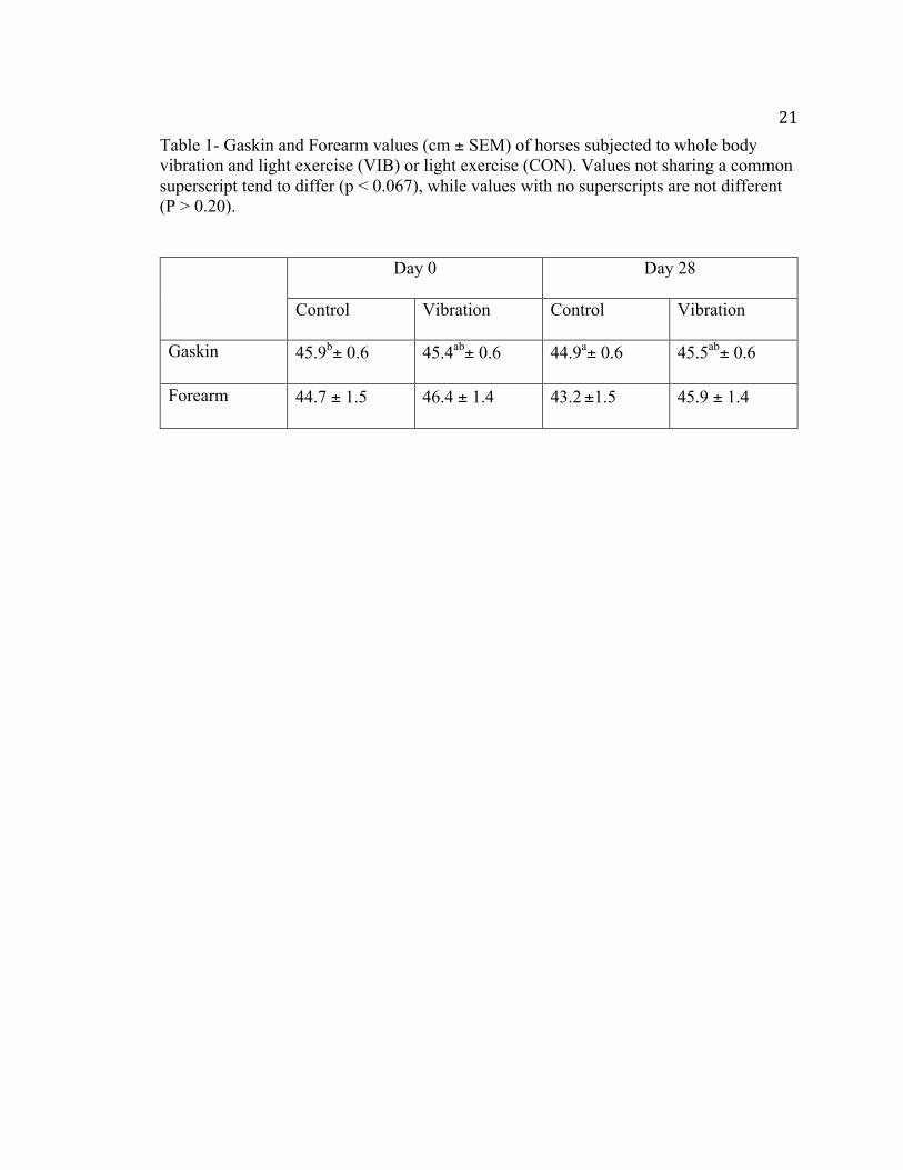
21Table 1- Gaskin and Forearm values (cm ± SEM) of horses subjected to whole body vibration and light exercise (VIB) or light exercise (CON). Values not sharing a common superscript tend to differ (p < 0.067), while values with no superscripts are not different (P > 0.20).
Day 0 Day 28
Control Vibration Control Vibration
Gaskin 45.9b± 0.6 45.4ab± 0.6 44.9a± 0.6 45.5ab± 0.6
Forearm 44.7 ± 1.5 46.4 ± 1.4 43.2 ±1.5 45.9 ± 1.4
22
Bodyweight
Body weight showed no influence of day, treatment, or date by treatment
interaction (P = 0.28, P = 0.92, P = 0.68; respectively). Mean body weight was 525 ± 24
kg on day 0 and 515 ± 24 kg on day 28.
Heart Rate
A trend for a treatment effect was observed between the CON and the VIB group
(P =0.065; Figure 1). Across all time points, CON increased on average 3.04 ± 2.8 bpm,
while VIB decreased -4.7 ± 2.8 bpm. There was no time effect or treatment by time
interaction (P = 0.94, P = 0.70, respectively).
Stride Length
Stride length at the trot exhibited a difference by day (P = 0.0002). On day 0,
stride length averaged 2.91 ± 0.08 m, where on day 28 average stride length decreased to
2.44 ± 0.08 m. There was no treatment or date by treatment interaction observed (P =
0.88, P = 0.39).
Serum PYD and OC
A day effect was observed for serum PYD (P < 0.0001). On day 0 the mean for
serum PYD levels was 1.41 ± 0.16 ng/ml, compared to day 28 where the mean levels of
serum PYD increased to 2.48 ± 0.16 ng/ml. No differences were attributable to treatment
and no day by treatment interaction was found (P = 0.95, P = 0.15). Plasma OC showed
no difference by day, treatment, or day by treatment (P = 0.75, P = 0.70, P = 0.83).

23
Figure 1- Change in heart rate (bpm ± SEM) of horses subjected to whole body vibration (VIB) or control (CON) over a 45-min treatment period and a 30-min recovery. A trend for a treatment difference was identified across all times (P = 0.065).
-15
-10
-5
0
5
10
0 15 30 45 60 75
ChangeinHeartRate(bpm
)
Time(min)
CON
VIB
24
RBAE
Data for RBAE values were analyzed for all cortices for two periods to compare
pasture turnout and stalling of all horses as well as for a treatment effect once the
treatments were initiated. Period 1 represented the pasture turnout for all horses and
period 2 represented when horses were stalled for the 28 d treatment period. No period
effect was seen in BMC in the medial cortex (P = 0.60). A period effect was observed in
the lateral cortex (P = 0.011), where during period one the mean change RBAE value was
1.87 ± 0.65 mmAl, whereas period 2 showed a decline of -1.24 ± 0.65 mmAl. No period
effect was observed in the dorsal or palmar cortices (P = 0.39 and P = 0.12, respectively).
A period effect was also identified in total BMC (P = 0.059), where during period 1 BMC
was higher (321.08 ± 833.8 mm2-Al) compared to period 2 (-2529.1 ± 833.8 mm2-Al).
During the treatment period days 0-28 there was no difference in BMC in the
medial cortex attributable to day, treatment, or interaction (P = 0.34, P = 0.99, P = 0.14,
respectively). Similarly, the effect of day, treatment and day by treatment was not
different in the dorsal cortex (P = 0.15, P = 0.48, P = 0.73, respectively). A trend toward
significance was seen in lateral BMC for a day effect (P = 0.095); day 0 mean for BMC
was 30.97 ± 0.58 mmAl when compared to day 28 which decreased to 29.72 ± 0.58
mmAl. Further, the palmar cortex had a day effect (P = 0.001), where day 0 had an
average BMC of 25.29 ± 0.79 mmAl and day 28 decreased to 23.06 ± 0.79 mmAl. No
difference was attributable to treatment or date by treatment interaction (P = 0.45, P =

250.92). Finally, total BMC exhibited a day effect (P = 0.002); total BMC on day 0
averaged 14357 ± 720 mm2-Al which decreased on day 28 to 11828 ± 720 mm2-Al.
Discussion
Until now there have been no published reports on the effects of WBV in the
exercising horse. The results of the current study show the effects of WBV on forearm
and gaskin circumference, heart rate, stride length, OC and PYD concentrations and
BMC. Application of WBV was deemed safe and no adverse effects were detected
throughout the present study.
WBV did not have an effect on forearm circumference as a measure of muscle
hypertrophy. There was a day by treatment interaction observed on the gaskin
measurement but no other interaction was seen. This could be contributed to human error
and the accuracy of each measurement taken. This would contribute to high variability,
which is reflected in the high SEM. There is no evidence to date that WBV has an effect
on muscle circumference, the current study did not find anything contrary to past
research. Previous research has shown an increase in muscle strength due to vibration-
induced muscle activity. Verschueren et al. (2004) found an increase in dynamic strength
in the group that performed static and dynamic exercises while on a vibration platform.
While vibration may have an effect on muscle strength and performance it did not
directly affect circumference. It may have proved interesting to look at muscle
composition as an alternative to circumference as such may be a better indicator of
dynamic strength in the horse, where tests to directly measure strength prove difficult to
administer and assess.

26 In this current study body weight was measured but was not expected to change
during the treatment period. Body weights were recorded at the beginning and end of the
study to make sure that a body weight necessary for maintenance was maintained. Since
no difference was observed throughout the study and the horse’s diets remained constant
no effect would be expected.
No previous research has been conducted to date on the effects of WBV on heart
rate or other cardiovascular effects in the horse. The current study found a trend for a
treatment effect between the control and vibration groups. Average heart rate decreased
in the vibration group compared to the control group. This is contrary to research
conducted in humans. Robbins et al. 2014 examined the effects of WBV on the central
and peripheral cardiovascular system. Human participants stood on a vibration plate set at
40 Hz. Vibration exposure totaled 5 min in 1 min increments and a 5 min recovery
between each testing period. No changes in heart rate, blood pressure or peripheral skin
temperature were observed. Robbins et al. (2014) did note a change in blood flow
velocity, this was suggested to be due to a high level of sensitivity of the peripheral
vascular system to vibration exposure. Another study conducted on humans found the
opposite effect, that WBV increased heart rate although the increase was modest (Liao et
al., 2015). In the horse, it might be suggested, horses in the treatment group could have
experienced a reduction in stress while receiving treatment. Experiencing the vibration
treatment may have a relaxation effect on the horses lowering cortisol levels and
therefore reducing the heart rate. Anecdotal behavior observations would support this
suggestion as horses appeared relaxed throughout treatment.
27 The current study hypothesized WBV would increase the stride length in the
horse. Stride length at the trot did exhibit a difference by day, where the average stride
length decreased from day 0 to day 28. This is likely attributable to the fact that a
different person hand trotted the horses on the two testing days. Unfortunately, using the
same person for both days was unavoidable. Although only one person hand trotted all
horses each day, the size and stride of the person may have had an effect on the
movement of the horses. The current study found no treatment or date by treatment
interaction. Further research should be done to evaluate the effect of WBV on movement
in the horse.
The use of bone markers has not been researched in regards to the effect of WBV.
Both osteocalcin (OC) and serum PYD levels were examined in this study. A day effect
was observed for serum PYD. The mean level of serum PYD increased from day 0 to day
28. When bone or cartilage collagen is degraded, PYD is released into circulation. This
can be explained by a stalling effect. The horses had been turned out on pasture until the
initiation of the study and then stalled for the 28 d treatment portion. Skeletal unloading
has been associated with loss of bone mass and decreased bone formation. Since the
horses in this study were experiencing light exercise this was not as detrimental as if they
had been on strict stall rest. Bell et al. (2001) researched the effects of daily access to
pasture on prevention of bone mineral content loss in Arabian weanlings. It was
determined that pasture turnout or 12 h/d of turnout is beneficial to maintaining and
increasing BMC. Hoekstra et al. (1999) also found an increase in BMC in pasture-reared
horses compared to stalled horses. Medial and lateral BMC were consistently higher in
28horses kept on pasture for the 56 d study. Similarly, in the current study total BMC
decreased in horses from a period of turnout to initiation of the current study when
stalling began.
Plasma osteocalcin showed no difference by day, treatment, or day by treatment
interaction. Osteocalcin makes up a portion of the non-collagenous protein in bone.
Plasma OC is used as a biochemical indicator of bone turnover. Increases in OC indicate
an increase in bone turnover and vice versa. Although horses were receiving light
exercise in the current study, having the horses stalled did not allow for enough loading
to occur to cause microcracks. Also seen in previous studies, mature horses are seen to
have lower levels of OC when compared to younger horses (Lepage et al., 1990). This
could explain the lack of difference in OC levels seen between initiation of the current
study and the end.
BMC values were evaluated for two periods to compare pasture turn out and
stalling as well as for a treatment effect once the study began. Period 1 was the time
frame where horses were out on pasture receiving free choice exercise. Period 2 was the
start of the treatment portion of the study and when stalling began. A period effect was
observed in the lateral cortex and total BMC. In both cases period 2 had a decrease in
RBAE values, which could once again be attributed to a stalling effect. During the
treatment period (days 0-28), a trend toward significance was seen in the lateral cortex
for a day effect. A similar day effect was also observed in the palmar cortex as well as
total BMC. In all instances mean BMC had decreased from day 0 to day 28. This could
potentially be explained by the fact that horses during the treatment period were
29experiencing only light exercise yet were subjected to stalling. Although horses were
receiving light exercise 6 d/wk it was not at a level of loading likely equivalent to horses
out on pasture.
Conclusion
In summary the results of this study found that bone density was not increased
due to the addition of WBV on the exercising horse. Heart rates were lower in the
treatment group, possibly due to a relaxation effect. No differences were seen in stride
length, gaskin and forearm circumference, or markers for bone metabolism. WBV is
becoming more widespread throughout the equine industry. In all research to date, across
the various species, there are many discrepancies about the application of WBV. There is
a great deal of variation in duration of treatment, length of each individual treatment and
strength of vibration. Further research needs to be done in the horse. Studies in other
species had longer durations, and since no significant change was seen in our 28 d period
a future direction would be to increase the duration of the study. Another possible study
should explore the strength of vibration used in horses, which could possibly lead to
different results. Since the horse is the largest animal WBV has been tested on then
perhaps a higher frequency should be used to see significant results. Since there was a
trend toward significance for decreased heart rate in the treatment group, perhaps a future
study should be done examining cortisol levels in relation to heart rate of horses receiving
whole body vibration to determine if it is a stress relieving activity.

30Literature Cited
Autier, P., P. Haentjens, J. Bentin, J. M. Baillon, A. R. Grivegnee, M. C. Closon, and S. Boonen. 2000. Costs induced by hip fractures: A prospective controlled study in Belgium. Osteoporos. Int. 11:373–380. doi:10.1007/s001980070102
Bell, R. A., B. D. Nielsen, K. Waite, D. Rosenstein, and M. Orth. 2001. Daily access to pasture turnout prevents loss of mineral in the third metacarpus of Arabian weanlings. J. Anim. Sci. 79:1142–1150. doi:/2001.7951142x
Brown, J. P., L. Malaval, M. C. Chapuy, P. D. Delmas, M. E. Edouard, and P. J. Meunier. 1984. Serum bone Gla-protein: a specific marker for bone formation in postmenopausal osteoporosis. Lancet 1:1091–1093. doi:10.1016/S0140-6736(84)92506-6
Carrier, T. K., L. Estberg, S. M. Stover, I. Gardner, and B. J. Johnson. 1997. Lay-up is associated with complete humeral but not pelvic fracture in California Thoroughbred racehorses. Proc. AAEP 43:271–272.
Delmas, P. D., R. Eastell, P. Garnero, M. J. Seibel, and J. Stepan. 2000. The use of biochemical markers of bone turnover in osteoporosis. Osteoporos. Int. 6:S2–S17. doi:10.1007/s001980070002
Gusi, N., A. Raimundo, and A. Leal. 2006. Low-frequency vibratory exercise reduces the risk of bone fracture more than walking: a randomized controlled trial. BMC Musculoskelet. Disord. 7:92. doi:10.1186/1471-2474-7-92
Hoekstra, K. E., B. D. Nielsen, M. W. Orth, D. S. Rosenstein, H. C. Schott, and J. E. Shelle. 1999. Comparison of bone mineral content and biochemical markers of bone metabolism in stall- vs. pasture-reared horses. Equine Vet. J. Suppl. 30:601–604. doi:10.1111/j.2042-3306.1999.tb05292.x
Hulak, E. S. 2015. Influence of whole body vibration on bone density in the stalled horse. MS Thesis. Middle Tennessee State Univ., Murfreesboro.
Inose, H., B. Zhou, V. K. Yadav, E. X. Guo, G. Karsenty, and P. Ducy. 2011. Efficacy of serotonin inhibition in mouse models of bone loss. J. Bone Miner. Res. 9:2002–2011. doi:10.1002/jbmr.439
Johnson, B. J., S. M Stover, B. M. Daft, H. Kinde, D. H. Read, B. C. Barr, M. Anderson, J. Moore, L. Woods, J. Stoltz, and P. Blanchard. 1994. Causes of death in racehorses over a 2 year period. Equine Vet. J. 4:327–330. doi:10.1111/j.2042-3306.1994.tb04395.x
31Kawanabe, K., A. Kawashima, I. Sashimoto, T. Takeda, Y. Sato, and J. Iwamoto. 2007.
Effect of whole-body vibration exercise and muscle strengthening, balance, and walking exercises on walking ability in the elderly. Keio. J. Med. 1:28–33. doi:10.2302/kjm.56.28
Kimmel, D. B. 1993. A paradigm for skeletal strength homeostasis. J. Bone Miner. Res. 8:S515–S522. doi:10.1002/jbmr.5650081317
Lepage, O. M., M. Marcoux, and A. Tremblay. 1990. Serum osteocalcin or bone Gla-protein, a biochemical marker for bone metabolism in horses: differences in serum levels with age. Can. J. Vet. Res. 54:223–226.
Lepage, O. M., B. Carstanjen, D., and D. Uebelhart. 2001. Non-invasive assessment of equine bone: An update. Vet. J. 1:10–23. doi:10.1053/tvjl.2000.0541
Liao, L. R., G. Y. F. Ng, A. Y. M. Jones, and M. Y. C. Pang. 2015. Cardiovascular stress induced by whole-body vibration exercise in individuals with chronic stroke. J. Amer. Phys. Ther. Assoc. 7:966–977. doi:10.2522/ptj.20140295
Matsuo, K., and N. Irie. 2008. Osteoclast-osteoblast communication. Arch. Biochem. Biophys. 473:201–209. doi:10.1016/j.abb.2008.03.027
Meakim, D.W., E. A. Ott, R. L. Asquith, and J. P. Feaster. 1981. Estimation of mineral content of the equine third metacarpal by radiographic photometry. J. Anim. Sci. 53:1019–1026. doi:10.2134/jas1981.5341019x
Modder, U., S. J. Achenbach, S. Amin, B. L. Riggs, L. J. Melton, and S. Khosla. 2010. Relation of serum serotonin levels to bone density and structural parameters in women. J. Bone. Miner. Res. 25:415–422. doi:10.1359/jbmr.090721
Nielsen, B. D., G. D. Potter, E. L. Morris, T. W. Odom, D. M. Senor, and J. A. Reynolds. 1997. Changes in the third metacarpal bone and frequency of bone injuries in young quarter horses during race training – observations and theoretical considerations. Equine Vet. J. 17:541–548. doi:10.1016/S0737-0806(97)80227-4
Nielsen, B. D., C. I. O’Connor, D. S. Rosenstein, H. C. Schott, and H. M. Clayton. 2002. Influence of trotting and supplemental weight on metacarpal bone development. Equine Vet. J. Suppl. 34:236–240. doi:10.1111/j.2042-3306.2002.tb05425.x
O’Connor-Robison, C. I., and B. D. Nielsen. 2013. Comparison of two software packages for determining radiographic bone aluminum equivalent values. Comp. Exer. Phys. 9:219–222. doi:10.3920/CEP13024
Oliver, A., J. P. Nurton, and A. J. Guthrie. 2007. An epizoological study of wastage in Thoroughbred racehorses in Gauteng, South Africa. J. S. Afr. Vet. Assoc. 68:125–129. doi:10.4102/jsava.v68i4.893

32Oxlund, B. S., G. Ortoft, T. T. Andreassen, and H. Oxlund. 2002. Low-intensity, high-
frequency vibration appears to prevent the decrease in strength of the femur and tibia associated with ovarieectomy of adult rats. Bone 32:69–77. doi:10.1016/S8756-3282(02)00916-X
Pagani, F., C. M. Francucci, and L. Moro. 2005. Markers of bone turnover: biochemical and clinical perspectives. J. Endocrinol. Invest. 28:8–13.
Porr, C. A., D. S. Kronfeld, L. A. Lawrence, R. S. Pleasant, and P. A. Harris. 1998. Deconditioning reduces mineral content of the third metacarpal bone in horses. J. Anim. Sci. 76:1875– 1879. doi:/1998.7671875x
Price, J. S., B. Jackson, R. Eastell, A. M. Wilson, R. G. G. Russell, L. E. Lanyon, and A. E. Goodship. 1995. The response of the skeleton to physical training: a biochemical study in horses. Bone 17:221–227. doi:10.1016/8756-3282(95)00221-X
Prisby, R. D., M. H. Lafage-Proust, L. Malaval, A. Belli, and L. Vico. 2008. Effects of whole body vibration on the skeleton and other organ systems in man and animal models: what we know and what we need to know. Ageing Res. Rev. 7:319–329. doi:10.1016/j.arr.2008.07.004
Rehn, B., J. Lidstrom, J. Skoglund, and B. Lindstrom. 2007. Effects on leg muscular performance from whole-body vibration exercise: a systemic review. Scand. J. Med. Sci. Sports 17:2–11. doi:10.1111/j.1600-0838.2006.00578.x
Robbins, D., P. Yoganathan, and M. Goss-Sampson. 2014. The influence of whole body vibration on the central and peripheral cardiovascular system. Clin. Physiol. Funct. Imaging 34:364–369. doi:10.1111/cpf.12103
Rossdale, P. D., R. Hopes, N. J. Digby, and K. Offord. 1985. Epidemiological study of wastage among racehorses 1982 and 1983. Vet. Rec. 116:66–69. doi: 10.1136/vr.116.3.66
Rubin, C., A. S. Turner, R. Muller, E. Mitra, and K. Mcleod. 2002. Quantity and quality of trabecular bone in the femur are enhanced by a strongly anabolic, noninvasive mechanical intervention. J. Bone Miner. Res. 17:349–357. doi:10.1359/jbmr.2002.17.2.349
Seibel, M. J., S. P. Robins, and J. P. Bilezikian. 1992. Urinary pyridinium crosslinks of collagen. Specific markers of bone resorption in metabolic bone disease. Trends Endocrinol. Metab. 3:263–270. doi:10:1016/1043-2760(92)90129-O
33Sims, N. A., and J. H. Gooi. 2008. Bone remodeling: multiple cellular interactions
required for coupling of bone formation and resorption. Semin. Cell. Dev. Biol. 19:444–451. doi: 10.1016/j.semcdb.2008.07.016
Skerry, T. M. 2008. The response of bone to mechanical loading and disuse: Fundamental principles and influences on osteoblast/osteocyte homeostasis. Arch. Biochem. Biophys. 473:117–123. doi:10.1016/j.abb.2008.02.028
Slatkovska, L., S. M. H. Alibhai, J. Beyene, and A. M. Cheung. 2010. Effect of whole-
body vibration on BMD: a systematic review and meta-analysis. Osteoporos Int. 21:1969–1980. doi:10.1007/s00198-010-1228-z
Stuermer, E. K., M. Komrakova, S. Sehmisch, M. Tezval, C. Dullin, N. Schaefer, J. Hallecker, and K. M. Stuermer. 2014. Whole body vibration during fracture healing intensifies the effects of estradiol and raloxifene in estrogen-deficient rats. Bone 64:187–194. doi: 10.1016/j.bone.2014.04.008
Torvinen, S., P. Kannus, H. Sievanen, T. A. H. Jarvinen, M. Pasanen, S. Kontulainen, T. L. N. Jarvinen, M. Jarvinen, P. Oja, and I. Vuori. 2002. Effect of a vibration exposure on muscular performance and body balance. Randomized cross-over study. Clin. Physiol. Funct. Imaging. 22:145–152. doi:10.1046/j.1365-2281.2002.00410.x
Verschueren, S. M. P., M. Roelants, C. Delecluse, S. Swinnen, D. Vanderschueren, and S. Boonen. 2004. Effect of 6-month whole body vibration training on hip density, muscle strength, and postural control in postmenopausal women; a randomized controlled pilot study. J. Bone Miner. Res. 19:352–359. doi:10.1359/JBMR.0301245
Wei, Q. S., L. Huang, X. Chen, H. Wang, W. Sun, S. Huo, Z. Li, and M. Deng. 2014. Effect of whole body vibration therapy on circulating serotonin levels in an ovariectomized rat model of osteoporosis. Iran. J. Basic Med. Sci. 17:62–68.
Xie, L., J. M. Jacobsen, E. S. Choi, B. Busa, L. R. Donahue, L. M. Miller, C. T. Rubin, and S. Judex. 2006. Low-level mechanical vibrations can influence bone resorption and bone formation in the growing skeleton. Bone 39:1059–1066. doi:10.1016/j.bone.2006.05.012

34
Appendices
35Appendix A: IACUC Approval
5/28/2014 Investigator(s) Name: Holly Spooner,Ph.D., John Haffner, DVM Investigator(s) Email: [email protected]; [email protected] Protocol Title: “Does vibration therapy alleviate bone density loss caused by stalling and/or influence kinematics of movement?” Protocol Number: 14-011 Dear Investigator, The MTSU Institutional Animal Use and Care Committee has reviewed your research proposal identified above and has approved your research in accordance with PHS policy. Approval is granted for three (3) years. Your study expires 5/28/2017. Please note you will need to file a Progress Report annually regarding the status of your study and submit an end-of-project report. According to MTSU Policy, an investigator is defined as anyone who has contact with animals for research purposes. Anyone meeting this definition needs to be listed on the protocol and needs to complete the IACUC training through citiprogram. If you add investigators to an approved project, please forward an updated list of investigators to the Office of Compliance before they begin to work on the project. Any change to the protocol must be submitted to the IACUC before implementing this change. Any unanticipated harms to subjects or adverse events must be reported to the Office of Compliance at (615) 494-8918. Also, all research materials must be retained by the PI or faculty advisor (if the PI is a student) for at least three (3) years after study completion. Should you have any questions or need additional information, please do not hesitate to contact me. Sincerely, Kellie Hilker Compliance Officer 615-494-8918 [email protected]