The effect of human saliva compared to Aloe vera on wound ...
9
J Med Sci, Volume 50, No. 4, 2018 Oktober : 376-384 376 *corresponding author: [email protected] The effect of human saliva compared to Aloe vera on wound healing of 2 nd degree burn injury in animal models Budi Cahyono Putro*, Ishandono Dachlan Plastic and Reconstructive Surgery, Department of Surgery, Faculty of Medicine, Public Health and Nursing, Universitas Gadjah Mada/Dr. Sardjito General Hospital, Yogyakarta DOI: http://dx.doi.org/10.19106/JMedScie/005004201801 ABSTRACT Burn injury is one of the common causes of injury that has relatively high morbidity and mortality. Several studies using herbal and traditional medicine from different countries have been documented in burn injury management. Human saliva that contains antibacterial, antifungal, antiviral and analgesic components as well as growth factors can induce re- epithelialization process in 2 nd degree burn injury. Whereas, Aloe vera that influence a physiological moist condition was proven can induce re-epithelialization process lead to faster wound healing. This study aimed to compare topical application of human saliva and A. vera on wound healing process of 2 nd degree burn injury. This was an experimental study using post-test only control group design using 27 white rats (Rattus novergicus) of Sprague Dawley strain divided into 3 groups with 9 rats in each group. Group I were applied 1 mL of human saliva, Group II were applied A. vera and Group III as control were applied NaCl. Change in body weight and macroscopic clinical assessment were observed every day for 14 days, whereas histological examination was observed on day 14. The data were presented as mean ± standard error of the mean (SEM) and analyzed using one-way analysis of variance (Anova). The result showed that the wound healing process at each treatment showed different level. The human saliva application tended to show faster wound healing process of 2 nd degree burn injury compare with A. vera or NaCl (p<0.05). In conclusion, human saliva topical application is superior compared with A. vera or NaCl on wound healing of 2 nd degree burn injury in white rat models. ABSTRAK Luka bakar merupakan salah satu penyebab umum dari trauma dan memiliki morbiditas dan mortalitas yang relative tinggi. Beberapa studi menggunakan herbal dan obat tradisional dari berbagai negara dalam penanganan luka bakar telah didokumentasi. Air liur manusia yang mengandung komponen antibakteri, antijamur, antivirus, dan analgesik serta faktor pertumbuhan dapat memacu proses re-epitelialisasi pada luka bakar derajat 2. Sedangkan Aloe vera yang dapat mempengaruhi kondisi kelembaban fisiologi terbukti dapat memacu proses re-epitelialissi sehingga mempercepat penyembuhan luka. Penelitian ini bertujuan untuk membandingkan pemberian topical air liur manusia dan A. vera pada proses penyembuhan lukabakar derajad 2. Penelitian ini merupakan penelitian eksperimental menggunakan rancangan post-test only control group menggunakan 27 tikus putih (Rattus novergicus) strain Sprague Dawley yang dibagi menjadi 3 kelompok dengan 9 ekor tikus setia kelompok. Kelompok I diberi 1 mL air liur manusia, Kelompok II diberi A. vera dan Kelompok III sebagai kontrol diberi NaCl. Perubahan berat badan dan pemeriksaan klinik maskroskopik dilakukan setiap hari selama 14 hari. Pemeriksaan histologi dilaukan pada hari ke 14. Data ditampilkan sebagai rerata ± standard error of the mean (SEM) dan dianalisis dengan anaisis varians satu jalan. Hasil penelitian menunjukkan proses penyembuhan luka pada masing-masing perlakukan menunjukkan hasil yang berbeda. Pemberian air ludah manusia cenderung menunjukkan proses penyembuhan lukabkar derajad 2 yang lebih cepat dibandingkkan pemberian A. vera atau
Transcript of The effect of human saliva compared to Aloe vera on wound ...

J Med Sci, Volume 50, No. 4, 2018 Oktober : 376-384
376*corresponding author: [email protected]
The effect of human saliva compared to Aloe vera on wound healing of 2nd degree burn injury in animal models
Budi Cahyono Putro*, Ishandono DachlanPlastic and Reconstructive Surgery, Department of Surgery, Faculty of Medicine, Public Health and Nursing, Universitas Gadjah Mada/Dr. Sardjito General Hospital, Yogyakarta
DOI: http://dx.doi.org/10.19106/JMedScie/005004201801
ABSTRACT
Burn injury is one of the common causes of injury that has relatively high morbidity and mortality. Several studies using herbal and traditional medicine from different countries have been documented in burn injury management. Human saliva that contains antibacterial, antifungal, antiviral and analgesic components as well as growth factors can induce re-epithelialization process in 2nd degree burn injury. Whereas, Aloe vera that influence a physiological moist condition was proven can induce re-epithelialization process lead to faster wound healing. This study aimed to compare topical application of human saliva and A. vera on wound healing process of 2nd degree burn injury. This was an experimental study using post-test only control group design using 27 white rats (Rattus novergicus) of Sprague Dawley strain divided into 3 groups with 9 rats in each group. Group I were applied 1 mL of human saliva, Group II were applied A. vera and Group III as control were applied NaCl. Change in body weight and macroscopic clinical assessment were observed every day for 14 days, whereas histological examination was observed on day 14. The data were presented as mean ± standard error of the mean (SEM) and analyzed using one-way analysis of variance (Anova). The result showed that the wound healing process at each treatment showed different level. The human saliva application tended to show faster wound healing process of 2nd degree burn injury compare with A. vera or NaCl (p<0.05). In conclusion, human saliva topical application is superior compared with A. vera or NaCl on wound healing of 2nd degree burn injury in white rat models.
ABSTRAK
Luka bakar merupakan salah satu penyebab umum dari trauma dan memiliki morbiditas dan mortalitas yang relative tinggi. Beberapa studi menggunakan herbal dan obat tradisional dari berbagai negara dalam penanganan luka bakar telah didokumentasi. Air liur manusia yang mengandung komponen antibakteri, antijamur, antivirus, dan analgesik serta faktor pertumbuhan dapat memacu proses re-epitelialisasi pada luka bakar derajat 2. Sedangkan Aloe vera yang dapat mempengaruhi kondisi kelembaban fisiologi terbukti dapat memacu proses re-epitelialissi sehingga mempercepat penyembuhan luka. Penelitian ini bertujuan untuk membandingkan pemberian topical air liur manusia dan A. vera pada proses penyembuhan lukabakar derajad 2. Penelitian ini merupakan penelitian eksperimental menggunakan rancangan post-test only control group menggunakan 27 tikus putih (Rattus novergicus) strain Sprague Dawley yang dibagi menjadi 3 kelompok dengan 9 ekor tikus setia kelompok. Kelompok I diberi 1 mL air liur manusia, Kelompok II diberi A. vera dan Kelompok III sebagai kontrol diberi NaCl. Perubahan berat badan dan pemeriksaan klinik maskroskopik dilakukan setiap hari selama 14 hari. Pemeriksaan histologi dilaukan pada hari ke 14. Data ditampilkan sebagai rerata ± standard error of the mean (SEM) dan dianalisis dengan anaisis varians satu jalan. Hasil penelitian menunjukkan proses penyembuhan luka pada masing-masing perlakukan menunjukkan hasil yang berbeda. Pemberian air ludah manusia cenderung menunjukkan proses penyembuhan lukabkar derajad 2 yang lebih cepat dibandingkkan pemberian A. vera atau
377
J Med Sci, Volume 50, No. 4, 2018 Oktober : 376-384
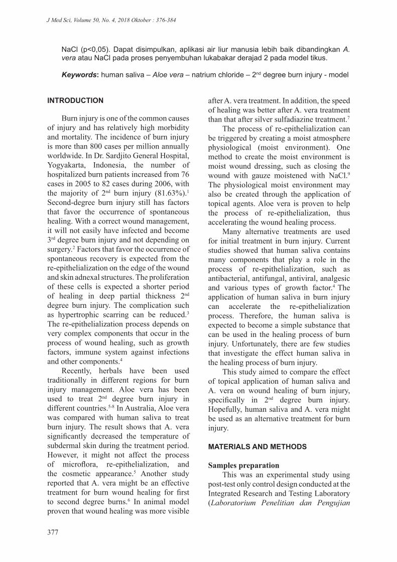
NaCl (p<0,05). Dapat disimpulkan, aplikasi air liur manusia lebih baik dibandingkan A. vera atau NaCl pada proses penyembuhan lukabakar derajad 2 pada model tikus.
Keywords: human saliva – Aloe vera – natrium chloride – 2nd degree burn injury - model
INTRODUCTION
Burn injury is one of the common causes of injury and has relatively high morbidity and mortality. The incidence of burn injury is more than 800 cases per million annually worldwide. In Dr. Sardjito General Hospital, Yogyakarta, Indonesia, the number of hospitalized burn patients increased from 76 cases in 2005 to 82 cases during 2006, with the majority of 2nd burn injury (81.63%).1
Second-degree burn injury still has factors that favor the occurrence of spontaneous healing. With a correct wound management, it will not easily have infected and become 3rd degree burn injury and not depending on surgery.2 Factors that favor the occurrence of spontaneous recovery is expected from the re-epithelialization on the edge of the wound and skin adnexal structures. The proliferation of these cells is expected a shorter period of healing in deep partial thickness 2nd degree burn injury. The complication such as hypertrophic scarring can be reduced.3
The re-epithelialization process depends on very complex components that occur in the process of wound healing, such as growth factors, immune system against infections and other components.4
Recently, herbals have been used traditionally in different regions for burn injury management. Aloe vera has been used to treat 2nd degree burn injury in different countries.5-8 In Australia, Aloe vera was compared with human saliva to treat burn injury. The result shows that A. vera significantly decreased the temperature of subdermal skin during the treatment period. However, it might not affect the process of microflora, re-epithelialization, and the cosmetic appearance.5 Another study reported that A. vera might be an effective treatment for burn wound healing for first to second degree burns.6 In animal model proven that wound healing was more visible
after A. vera treatment. In addition, the speed of healing was better after A. vera treatment than that after silver sulfadiazine treatment.7
The process of re-epithelialization can be triggered by creating a moist atmosphere physiological (moist environment). One method to create the moist environment is moist wound dressing, such as closing the wound with gauze moistened with NaCl.9
The physiological moist environment may also be created through the application of topical agents. Aloe vera is proven to help the process of re-epithelialization, thus accelerating the wound healing process.
Many alternative treatments are used for initial treatment in burn injury. Current studies showed that human saliva contains many components that play a role in the process of re-epithelialization, such as antibacterial, antifungal, antiviral, analgesic and various types of growth factor.4 The application of human saliva in burn injury can accelerate the re-epithelialization process. Therefore, the human saliva is expected to become a simple substance that can be used in the healing process of burn injury. Unfortunately, there are few studies that investigate the effect human saliva in the healing process of burn injury.
This study aimed to compare the effect of topical application of human saliva and A. vera on wound healing of burn injury, specifically in 2nd degree burn injury. Hopefully, human saliva and A. vera might be used as an alternative treatment for burn injury.
MATERIALS AND METHODS
Samples preparationThis was an experimental study using
post-test only control design conducted at the Integrated Research and Testing Laboratory (Laboratorium Penelitian dan Pengujian

378
Putro BC, et al., The effect of human saliva compared...
Terpadu/LPPT), Universitas Gadjah Mada, Yogyakarta. The human saliva samples were drawn from 6 male volunteers as much as 5 mL/volunteer/day with such conditions i.e. willing to become volunteer, aged 30-35 years old, healthy or had no systemic diseases, nor salivary gland dysfunction, did not smoke, never suffered head and neck radiotherapy, were not taking certain medications such as antihypertensives, antidepressants, antihistamines, immunosuppressant, diuretics and anticholinergics in the last 6 months, had normal blood sugar level and tested negative for HBsAg. Preparation of human saliva samples in this study was based on the WHO/IARC (International Agency for Research on Cancer) guideline for common minimum technical standards and protocols for biological resource.
Aloe vera preparation was obtained from natural A. vera plants without any further processing and taken aseptically to minimize any contamination.
Second-degree burn injury induction Second-degree burn injury was made by
placing 2 x 2 cm 75OC metal-induced heat for 5 sec. Wound healing period of 2nd burn injury was assessed clinically, macroscopically and histopathologically based on the re-epithelialization grading and the number of inflammatory cells (polymorphonuclears/neutrophils). This study was approved by the Medical and Health Research Ethics Committee of the Faculty of Medicine, Public Health and Nursing, Universitas Gadjah Mada/Dr. Sardjito General Hospital, Yogyakarta, with Ref: KE/FK/1054/EC.
Anima model procedure The animal models used in this study
were 27 white rats (Rattus novergicus) of Sprague Dawley strain. They were all female, aged 3-4 months with a body weight of 150-200 g and had met the criteria of validity for animal models. Animal models were divided into 3 groups with 9 rats in each group. Group I, we applied 1 mL of
human saliva, 3 times a day using sterile cotton buds. Group II, we applied A. vera, 3 times a day (every 4-6 hours/day: at 7.00 am, 11.00 am, and 3.00 pm). Group III, we applied NaCl as moist dressing (control), 3 times a day.
Chane in body weight and macroscopic clinical assessment were observed every day for 14 days, whereas histological examination was observed on day 14 where the healing period for second-degree burn injury was already complete. Theoretically, there are 4 grades of re-epithelialization grading for 2nd degree burn injury.7 Grade 1, the wound is still open and lined by a granulation tissue covered with fibrin, which appears red to black. At the bottom of the wound, the thickness of the residual dermis is very small, subcutaneous muscles are very close. Residual hematoma is still visible. Wound contraction is mainly superficial. Grade 2, the wound is still open and covered with a granulation tissue and fibrin, but the epidermis at the borders becomes thicker and begins to proliferate and extend. The healing process seems based on dermal cell proliferation, matrix accumulation, with a base layer of organized collagens, and the accumulation of immature matrix as wound filler. Grade 3, re-epithelialization is complete. The wound is closed with a thick new epidermis. The scar tissue at the center of wound is a very dense and unorganized matrix. The mature matrix is converging to the remodeling are. Grade 4, wounded tissue is barely visible. Deep wound contraction, matrix accumulation and remodeling lead to a strong reduction of the scar area. Hairs delimit the residual wounded area. The new epidermis is still very thick, but its surface seems to decrease: the deep remodeling and final healing of the dermis may influence the morphology of the epidermis above.
Inflammatory response was observed based on the accumulation of neutrophils (PMN) which was measured on day 14 using a microscope at 1000x magnifications, and measured per high-power fields.

379
J Med Sci, Volume 50, No. 4, 2018 Oktober : 376-384
Statistical analysis
The data were presented as mean ± standard error of the mean (SEM) and analyzed using one-way analysis of variance (Anova) continued by post hoc analysis thereafter to confirm where the differences occurred between groups, only if the result showed an overall significant difference in group means. A p value < 0.05 was considered to be significant.
RESULTS
Body weight assessment
FIGURE 1 shows the body weight of rats in each group during 14 days observation. There was a significantly difference of the body weight of the rats during the observation period (p<0.005). Moreover, post hoc analysis showed there was a significantly difference of the body weight of rats between group I and II compared to group III during the observation period (p >0.05).
FIGURE 1. Body weight of rats in each group during 14 days observation.
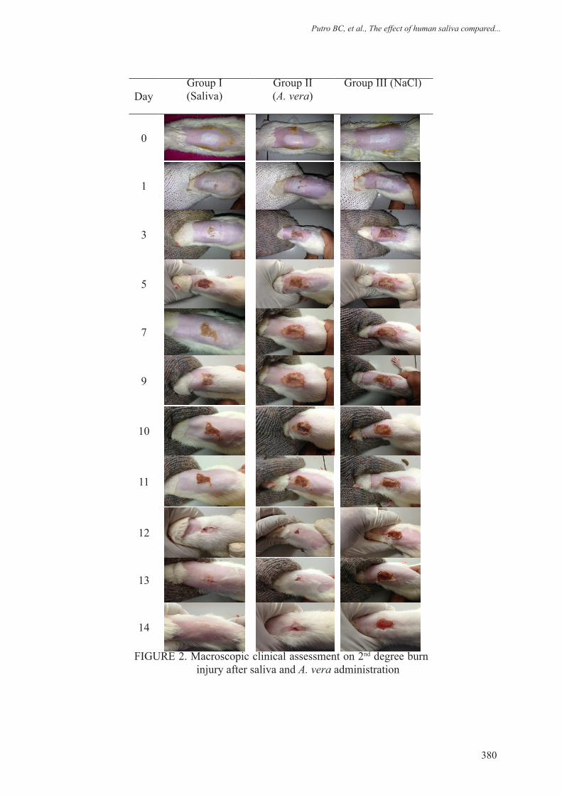
Macroscopic clinical assessment
FIGURE 2 shows macroscopic appearance of 2nd burn injury on each group
during 14 days of observation. The wound healing process at each treatment showed different level. Human saliva tended to show faster healing process.
380
Putro BC, et al., The effect of human saliva compared...
DayGroup I(Saliva)
Group II(A. vera)
Group III (NaCl)
0
1
3
5
7
9
10
11
12
13
14
FIGURE 2. Macroscopic clinical assessment on 2nd degree burn injury after saliva and A. vera administration

381
J Med Sci, Volume 50, No. 4, 2018 Oktober : 376-384
Microscopic assessment
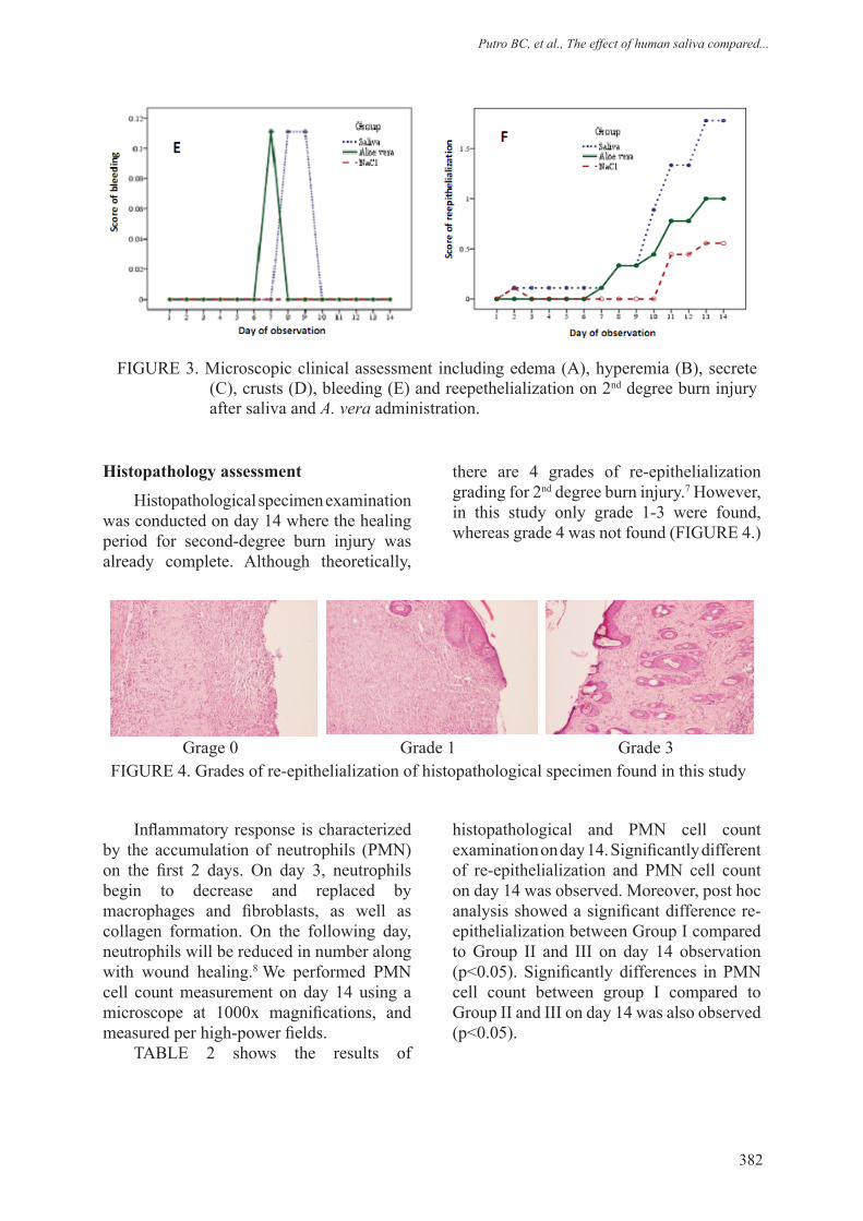
FIGURE 3 shows microscopic clinical assessment including edema (A), hyperemia (B), secrete (C), crusts (D), bleeding (E) and re-epethelialization on 2nd degree burn injury after saliva and A. vera administration. Significantly different of edema during the observation period was observed (FIGURE 3A; p<0.05). Moreover, post hoc analysis showed a significant difference between Group I compared to Group II and III during 14 days’ observation period (p<0.05). Significantly difference of hyperemia during the observation period was also observed (FIGURE 3B; p=0.000). However, post hoc analysis showed no significant difference between 3 groups during 14 days of observation period (p>0.05). Significant difference of crusts during the observation
period was also observed (FIGURE 3C; p<0.05). Furthermore, post hoc analysis showed a significant difference between Group I compared to Group III (p<0.05). Significantly difference of secrete during the observation period was also observed (FIGURE 3D; p<0.05). However, post hoc analysis showed there was no difference between 3 groups during 14 days of observation period (p>0.05). No significantly difference of bleeding during the 14 day of observation was observed (FIGURE 3E; p=0.562). This study also found significantly difference of re-epithelialization during the 14 day of observation (FIGURE 3F; p<0.005). Although, post hoc analysis showed there was no difference between 3 groups during the observation period (p>0.05).
382
Putro BC, et al., The effect of human saliva compared...
FIGURE 3. Microscopic clinical assessment including edema (A), hyperemia (B), secrete (C), crusts (D), bleeding (E) and reepethelialization on 2nd degree burn injury after saliva and A. vera administration.
histopathological and PMN cell count examination on day 14. Significantly different of re-epithelialization and PMN cell count on day 14 was observed. Moreover, post hoc analysis showed a significant difference re-epithelialization between Group I compared to Group II and III on day 14 observation (p<0.05). Significantly differences in PMN cell count between group I compared to Group II and III on day 14 was also observed (p<0.05).
Histopathology assessment
Histopathological specimen examination was conducted on day 14 where the healing period for second-degree burn injury was already complete. Although theoretically,
there are 4 grades of re-epithelialization grading for 2nd degree burn injury.7 However, in this study only grade 1-3 were found, whereas grade 4 was not found (FIGURE 4.)
Grage 0 Grade 1 Grade 3FIGURE 4. Grades of re-epithelialization of histopathological specimen found in this study
Inflammatory response is characterized by the accumulation of neutrophils (PMN) on the first 2 days. On day 3, neutrophils begin to decrease and replaced by macrophages and fibroblasts, as well as collagen formation. On the following day, neutrophils will be reduced in number along with wound healing.8 We performed PMN cell count measurement on day 14 using a microscope at 1000x magnifications, and measured per high-power fields.
TABLE 2 shows the results of

383
J Med Sci, Volume 50, No. 4, 2018 Oktober : 376-384
TABLE 2. Results of histopathological examination (re-epithelialization grading) and PMN cell count (mean ± SEM) on day 14 of each group
Group Rats (n)
Re-epithelialization
grading
PMN cell counts
(x 105/mL)Saliva 9 2.33 ± 0.11 1.33 ± 0.00A. vera 9 1.00 ± 0.00 9.89 ± 0.56
NaCl 9 0.67 ± 0.11 11.11 ± 0.62
DISCUSSION
Macroscopic assessment In our study, on day 0, right after the
induction of 2nd degree burn injury, we found reddish pale woundsin samples which were similar in each group samples. During day 1 to 14 of observation, we found a significant difference in body weight (FIGURE 1). It happened because Group III (NaCl) had a lower body weight than Group I (human saliva) and Group II (A. vera).
During day 1 to 5, we found a significant difference in edema between Group I and II. It is explained that human saliva has a better anti-inflammation effect than A. vera. On day 3, we started to find a difference in edema between group I and group II. Group I showed a decrease in edema compared to Group II. On day 4, crust was started to develop in Group I and III, in which was thin in Group I and thick in Group III. Crust is an accumulation of inflammation product or infiltration of PMN cells and necrotic tissues on the wound surface.9 On day 5 and day 6 of observation, we found only a difference in body weight between Group I and II and Group III. On day 7, we found a difference in hyperemia between Group I and Group III, in which in Group I and Group II started to diminish, while in Group III it was still visible. On day 8 and day 9, there was a difference in body weight between Group I and II and Group III. On day 10 of observation, we started to find a difference in re-epithelialization between Group I and
III, in which in Group I, re-epithelialization developed faster than in Group III. A study reported that human saliva contains growth factor, cytokine and protease inhibitor which can shorten inflammation phase and induce re-ephitelialization phase earlier.10-12
The different in crust between Group II and III showed that the crust in Group II started to diminish. During day 11 to day 14 of observation, we only found a difference in body weight between Group I and II and Group III.
According to the macroscopic clinical findings in this study, we conclude that the application of human saliva is able to fasten superficial 2nd degree burn injury healing period, referring to re-epithelialization and crust development compared to NaCl. Second degree burn injury can be a superficial/deep partial thickness (2A/2B), which the damage happens in the epidermis and some part of the dermis so that there is a possibility of spontaneous wound healing. The supporting factors for spontaneous wound healing are from the proliferation of epithelial layer (re-epithelialization) in the wound bed and skin appendages. This cell proliferation is expected to shorten the 2nd degree burn injury healing period, for not more than 4 weeks.4 Our study made a model for 2nd degree superficial partial thickness burn injury (2A) and successfully recovered within 2 weeks.
Histopathology examinationWe found a difference between Group
I and Group II and III. In Group I (human saliva), there was a complete healing, according to re-epithelialization and PMN cells count, compared to Group II (A. vera) and Group III (NaCl). A study reveals on day 14, superficial-partial thickness of 2nd degree burn injury will recover completely, and the wound re-epithelialization can be clinically assessed by its characteristic of faded pink colour.4 Wound healing mechanism is an integration of cellular changes and molecular processes, including physiological and biochemical phenomenon, thus, a clinical evaluation will be inadequate to provide

384
Putro BC, et al., The effect of human saliva compared...
information about wound healing changes. Therefore, a histopathology examination is required. Human saliva also contains hystatin which is a mitogen that enhances reepithelialization.10 Leptin in human saliva can enhance proliferation of oral kerationocytes.11,13,14
CONCLUSION
Topical application of human saliva is superior compared with A. vera and NaCl on wound healing of 2nd degree burn injury in white rats models.
ACKNOWLEDGEMNTS
We would like to thanks technicians from the Integrated Research and Testing Laboratory, Universitas Gadjah Mada, Yogyakarta for their valuables assistances during the study.
REFERENCES
1. Hettiaratchy S & Dziewulski P. ABC of burns. BMJ 2004; 329:504-6.http://dx.doi.org/10.1136/bmj.329.7457.101
2. Klingensmith ME, Chen LE, Glasgow SC, Goers TA, Melby SJ. Burns. In: The Washington manual of surgery. 5th ed. Washington: Lippincott Williams & Wilkins. 2008.
3. Pusponegoro AD. Luka. In: Sjamsuhidajat R, Jong W. 2004. Buku Ajar Ilmu Bedah, edisi 2. Jakarta: EGC. 2004.
4. Klein MB. Thermal, chemical, and electrical injuries. In: Thorne CH. editor. Grabb & Smith’s Plastic Surgery, 6th ed. Philadelphia: Lippincott Williams & Wilkins. 2007.
5. Cuttle L & Kimble RM. First aid treatment of burn injuries. Wound Pract Res 2010; 18 1:4-13.
6. Maenthaaisong R, Chaiyakunapruk N, Niruntraporn S, Kongkaew C. The efficacy of Aloe vera used for burn wound healing: a systematic review.
Burn 2007; 33: 713-8.https://doi.org/10.1016/j.burns.2006.10.384
7. Akhoondinasab MR, Akhoodinasab M, Saberi M. Comparison of healing effect of Aloe vera extract and silver sulfadiazine in burn injuries in experimental rat model. World J Plast Surg 2014; 3(1): 29-34.
8. Shahzad MN & Ahmed N. Effectiveness of Aloe vera gel compared with 1% silver sulphadiazine cream as burn wound dressing in second degree burns. JPMA 2013; 63(2): 225-30.
9. Gallagher JJ, Wolf SE, Herndon DN. Burns. In: Sabiston textbook of surgery. 18th ed. Philadelphia: Saunders Elsevier, 2007.
10. Lemo N, Marignac G, Reyes-Gomez E, Lilin T, Crosaz O, David M, et al. Cutaneous reepithelization and wound contraction after skin biopsies in rabbits: a mathematical model for healing and remodeling index. Veterinarski Arhiv 2010; 80(5):637-52.
11. Simpson DM & Ross R. The neutrophilic leucocyte in wound repair a study with antineutrophil serum. J Clin Invest 1972;51(8):2009-23. http://dx.doi.org/10.1172/JCI107007
12. Pereira DST, Lima MHM, Pontes NT, Anjos AM, Santos MT. Development of animal model for studying deep second-degree thermal burns. J Biomed Biotechnol 2012; 2012:460841.http://dx.doi.org//10.1155/2012/460841
13. Oudhoff MJ, Bolscher JGM, Nazmi K, Kalay H, van’t Hof W, Amerongen AV, et al. Histatins are the major wound-closure stimulating factors in human saliva as identified in a cell culture assay. FASEB J 2008; 22(11):3805-12. http://dx.doi.org/10.1096/fj.08-112003
14. Gröschi M, Topf HG, Kratzsch J, Dotsch J, Rascher W, Rauh M. Salivary leptin indusces increased expression of growh factors in oral keratinocytes. J Mol Endocrinol 2005; 34(2):353-66. http://dx.doi.org/10.1677/jme.1.01658