The Effect of Fixed Orthodontic Appliances and Fluoride ... · The effect of fixed orthodontic...
18
UvA-DARE is a service provided by the library of the University of Amsterdam (http://dare.uva.nl) UvA-DARE (Digital Academic Repository) The effect of fixed orthodontic appliances and fluoride mouthwash on the oral microbiome of adolescents: a randomized controlled clinical trial Koopman, J.E.; van der Kaaij, N.C.W.; Buijs, M.J.; Elyassi, Y.; van der Veen, M.H.; Crielaard, W.; ten Cate, J.M.; Zaura, E. Published in: PLoS ONE DOI: 10.1371/journal.pone.0137318 Link to publication Citation for published version (APA): Koopman, J. E., van der Kaaij, N. C. W., Buijs, M. J., Elyassi, Y., van der Veen, M. H., Crielaard, W., ... Zaura, E. (2015). The effect of fixed orthodontic appliances and fluoride mouthwash on the oral microbiome of adolescents: a randomized controlled clinical trial. PLoS ONE, 10(9), [e0137318]. https://doi.org/10.1371/journal.pone.0137318 General rights It is not permitted to download or to forward/distribute the text or part of it without the consent of the author(s) and/or copyright holder(s), other than for strictly personal, individual use, unless the work is under an open content license (like Creative Commons). Disclaimer/Complaints regulations If you believe that digital publication of certain material infringes any of your rights or (privacy) interests, please let the Library know, stating your reasons. In case of a legitimate complaint, the Library will make the material inaccessible and/or remove it from the website. Please Ask the Library: https://uba.uva.nl/en/contact, or a letter to: Library of the University of Amsterdam, Secretariat, Singel 425, 1012 WP Amsterdam, The Netherlands. You will be contacted as soon as possible. Download date: 01 Jun 2020
Transcript of The Effect of Fixed Orthodontic Appliances and Fluoride ... · The effect of fixed orthodontic...

UvA-DARE is a service provided by the library of the University of Amsterdam (http://dare.uva.nl)
UvA-DARE (Digital Academic Repository)
The effect of fixed orthodontic appliances and fluoride mouthwash on the oral microbiome ofadolescents: a randomized controlled clinical trial
Koopman, J.E.; van der Kaaij, N.C.W.; Buijs, M.J.; Elyassi, Y.; van der Veen, M.H.; Crielaard,W.; ten Cate, J.M.; Zaura, E.Published in:PLoS ONE
DOI:10.1371/journal.pone.0137318
Link to publication
Citation for published version (APA):Koopman, J. E., van der Kaaij, N. C. W., Buijs, M. J., Elyassi, Y., van der Veen, M. H., Crielaard, W., ... Zaura,E. (2015). The effect of fixed orthodontic appliances and fluoride mouthwash on the oral microbiome ofadolescents: a randomized controlled clinical trial. PLoS ONE, 10(9), [e0137318].https://doi.org/10.1371/journal.pone.0137318
General rightsIt is not permitted to download or to forward/distribute the text or part of it without the consent of the author(s) and/or copyright holder(s),other than for strictly personal, individual use, unless the work is under an open content license (like Creative Commons).
Disclaimer/Complaints regulationsIf you believe that digital publication of certain material infringes any of your rights or (privacy) interests, please let the Library know, statingyour reasons. In case of a legitimate complaint, the Library will make the material inaccessible and/or remove it from the website. Please Askthe Library: https://uba.uva.nl/en/contact, or a letter to: Library of the University of Amsterdam, Secretariat, Singel 425, 1012 WP Amsterdam,The Netherlands. You will be contacted as soon as possible.
Download date: 01 Jun 2020

RESEARCH ARTICLE
The Effect of Fixed Orthodontic Appliancesand Fluoride Mouthwash on the OralMicrobiome of Adolescents – A RandomizedControlled Clinical TrialJessica E. Koopman1☯, Nicoline C. W. van der Kaaij2☯, Mark J. Buijs1, Yassaman Elyassi1,Monique H. van der Veen1, Wim Crielaard1, Jacob M. ten Cate1, Egija Zaura1*
1 Department of Preventive Dentistry, Academic Centre for Dentistry Amsterdam, Amsterdam, TheNetherlands, 2 Department of Orthodontics, Academic Centre for Dentistry Amsterdam, Amsterdam, TheNetherlands
☯ These authors contributed equally to this work.* [email protected]
AbstractWhile the aesthetic effect of orthodontic treatment is clear, the knowledge on how it influences
the oral microbiota and the consequential effects on oral health are limited. In this randomized
controlled clinical trial we investigated the changes introduced in the oral ecosystem, during
and after orthodontic treatment with fixed appliances in combination with or without a fluoride
mouthwash, of 10–16.8 year old individuals (N = 91). We followed several clinical parameters
in time, in combination with microbiome changes using next-generation sequencing of the bac-
terial 16S rRNA gene. During the course of our study, the oral microbial community displayed
remarkable resilience towards the disturbances it was presented with. The effects of the fluo-
ridemouthwash on themicrobial composition were trivial. More pronouncedmicrobial changes
were related to gingival health status, orthodontic treatment and time. Periodontal pathogens
(e.g. Selenomonas and Porphyromonas) were highest in abundance during the orthodontic
treatment, while the health associated Streptococcus, Rothia andHaemophilus gained abun-
dance towards the end and after the orthodontic treatment. Only minor compositional changes
remained in the oral microbiome after the end of treatment. We conclude that, provided proper
oral hygiene is maintained, changes in the oral microbiome composition resulting from ortho-
dontic treatment are minimal and do not negatively affect oral health.
IntroductionThe aesthetic effects of orthodontic treatment are often readily visible; in contrast to the effectorthodontic treatment might have on the non-visible part of the oral cavity—the microbiome.
The possible changes in the oral microbiome during orthodontic treatment are likely to berelated to, the more easy observable, clinical parameters. For instance, the impaired gingival
PLOSONE | DOI:10.1371/journal.pone.0137318 September 2, 2015 1 / 17
a11111
OPEN ACCESS
Citation: Koopman JE, van der Kaaij NCW, Buijs MJ,Elyassi Y, van der Veen MH, Crielaard W, et al.(2015) The Effect of Fixed Orthodontic Appliancesand Fluoride Mouthwash on the Oral Microbiome ofAdolescents – A Randomized Controlled ClinicalTrial. PLoS ONE 10(9): e0137318. doi:10.1371/journal.pone.0137318
Editor: Chaminda Jayampath Seneviratne, NationalUniversity of Singapore, SINGAPORE
Received: March 5, 2015
Accepted: August 15, 2015
Published: September 2, 2015
Copyright: © 2015 Koopman et al. This is an openaccess article distributed under the terms of theCreative Commons Attribution License, which permitsunrestricted use, distribution, and reproduction in anymedium, provided the original author and source arecredited.
Data Availability Statement: The sequencing readsare available at NCBI’s Sequence Read Archiveunder accession number SRP055565.
Funding: The study was financially supported byElmex research / Colgate-Palmolive Europe. Theauthors declare that otherwise no competing interestsexist. The involvement of Elmex research / Colgate-Palmolive Europe in this study by financial supporthad no role in study design, data collection andanalysis, decision to publish, or preparation of themanuscript and does not alter the authors' adherence

health status [1, 2] and increased plaque formation [3, 4] that are associated with the placementof fixed orthodontic appliances. Besides, the latter could lead to the formation of white spotlesions, creating an undesirable aesthetic effect and possibly resulting in a cavity in need of res-tauration [5, 6].
So far, studies aimed to investigate the changes in bacterial taxa during orthodontic treat-ment, used culturing or targeted molecular approaches, allowing for a limited number ofopportunistic pathogenic species to be observed [7–10]. This implies that the response of theentire microbiome to orthodontic treatment is unclear, as are the possible long-term changesin bacterial composition.
A full understanding of the effects of fixed orthodontic appliances on the oral microbiomeand the consequences on clinical parameters, should allow for the preservation of a healthyoral cavity during and after orthodontic treatment, justifying orthodontic treatment.
Our aim was to investigate the changes introduced in the oral ecosystem during and afterorthodontic treatment in combination with a fluoride mouthwash. To our knowledge, this isthe first study to investigate the dynamics of the oral microbiome of adolescents during ortho-dontic treatment, and the use of a fluoride mouthwash using an open-ended molecularapproach.
Materials and Methods
Sampling and treatmentA randomized placebo-controlled parallel clinical trial was performed as described by van derKaaij et al. [11]. The study was approved by the Medical Ethical Committee of the VUMedicalCentre of the VU University of Amsterdam (VU-METc 2009/026 and Dutch trial register:NTR1817 [12]). The randomization allocation list was made in Microsoft Office Excel 2003(Microsoft, Redmond, WA, USA) using the random number generation function in the analy-sis toolpack for one variable with a discrete distribution, allocating 50% of the 120 subjects tothe test and 50% to the control group. The study was powered on the basis of the primary out-come; the data presented here were secondary outcomes.
All subjects participating in this study were scheduled to receive full fixed orthodontic appli-ances. Subjects could only be scheduled to receive full fixed orthodontic appliances if theymaintained a proper oral hygiene and had no severe gingivitis. The guidelines at the Orthodon-tic Department at ACTA state that orthodontic appliances will not be placed when the bleedingby probing score is above 2 (1: 0–5% of the sites are bleeding, 2: 6–10% of the sites are bleeding,3: 11–20% of the sites are bleeding, 4: 21–35% of the sites are bleeding, 5:> 35% of the sites arebleeding), except if immediate orthodontic treatment is indicated, for example, in case of trau-matic occlusion.
The inclusion criteria for the study were: 10–18 yrs of age, good general health, no use ofmedication and no demineralizations in need of restauration present at a buccal surface, inaddition to providing their written informed consent. A total of 120 subjects set to receive fixedorthodontic appliances in both jaws were to participate in the study. Roth Ovation Brackets(Dentsply, GAC International, Bohemia, NY, USA) were used and all were bonded followingthe same procedure and methods, using Transbond XT primer and adhesive (3M unitek, Mon-rovia, USA).
In this triple-blind study, the subjects received a randomly assigned mouthwash containing100 ppm amine-fluoride (AmF) and 150 ppm sodium-fluoride (SnF2) (Elmex caries protection,Colgate-Palmolive Europe, Therwil, Switzerland) or a placebo, also provided by Colgate-Palmolive Europe. The mouthwash was used from the time of bonding until debonding. Thesubjects were instructed not to use fluoride containing products, other than toothpaste, during
Orthodontic Treatment and the Oral Microbiome
PLOSONE | DOI:10.1371/journal.pone.0137318 September 2, 2015 2 / 17
to all PLOS ONE policies on sharing data andmaterials.
Competing Interests: The study was financiallysupported by Elmex research / Colgate-PalmoliveEurope. The authors declare that otherwise nocompeting interests exist. The involvement of Elmexresearch / Colgate-Palmolive Europe in this study byfinancial support had no role in study design, datacollection and analysis, decision to publish, orpreparation of the manuscript and does not alter theauthors' adherence to all PLOS ONE policies onsharing data and materials.
the course of the study. Their dentist was informed about the study and was asked not to applyextra fluoride during the study period. Furthermore, the subjects received oral hygiene instruc-tions after placement of the fixed appliances and were advised to use interproximal brushes toclean the areas of the tooth adjacent to the bracket underneath the orthodontic wire.
The subjects were instructed not to clean their teeth 24 h before supragingival plaque sam-ples for microbiome analysis were taken. These samples were obtained at six time-points dur-ing this study: T0 (approximately one week before placement of the fixed orthodonticappliances), T1 (six weeks after placement), T2 (twelve weeks after placement), TD (debond-ing, average of 25 months after placement), TD1 (six weeks after debonding) and TD2 (twelveweeks after debonding). Supragingival plaque was collected from the buccal surface of theupper left premolars using a sterile plastic spatula. In presence of the brackets (visits T1, T2and TD), which were placed on the middle of the tooth, the plaque was collected between thegingiva and the bracket. Gingival swelling often occurs within one or two months after place-ment of orthodontic appliances [1, 13, 14]. Hence, in cases where the gingival margin reachedthe bracket, the plaque was collected mesially and/or distally from the bracket. The plaque sam-ples were spun down for 30 s at 16.100 x g and stored at -80°C.
The number of white spot lesions of the subjects was recorded at visits T0, TD, TD1 andTD2, and is described in more detail by van der Kaaij et al. [11]. Additionally, a bleeding byprobing score was recorded at each visit for each patient. The percentage-based bleeding scorewas determined by probing each (bonded or to be bonded) tooth mesiobuccally and distobuc-cally with a periodontal probe [11]. For statistical analysis, the bleeding score was dichotomizedinto a healthy (score 1) and a gingivitis (score 2–5) group.
DNA isolation and sequencingDNA was isolated from the supragingival plaque samples as described by Zaura et al. [15]. TheV5-V7 regions of the 16S rDNA were used to prepare barcoded amplicon libraries for eachsample [16]. The equimolar pooled samples were sequenced at the Academic Medical Center(Amsterdam, the Netherlands) and Macrogen Inc. (Seoul, Republic of Korea) using the 454FLX Titanium chemistry (Roche, Basel, Switzerland). The reads are available at NCBI’sSequence Read Archive under SRP055565.
Sequencing data analysisQuantitative Insights Into Microbial Ecology (QIIME) v1.5.0 was used to analyze the sequencedata [17]. The downstream analyses and clustering into OTUs was done according to Koop-man et al. [18], with the exception that 1 ambiguous base (N = 1) was allowed. The OTUs weremanually aligned against NCBI’s nucleotide (nr/nt) collection using Megablast [19, 20] toobtain species level identification (S1 Table).
Statistical analysisThe Shannon diversity index and Bray-Curtis similarity index were calculated using PAST v3.0[21]. This program was also used to construct non-metric multidimensional scaling (nmMDS)plots based on the Bray-Curtis coefficient to visualize similarity between the samples.Stress< 0.2 (Kruskal’s stress formula 1) was used as a threshold [22].
The statistical significance of individual OTUs in relation to clinical parameters was deter-mined using QIIME’s paired t-test and correlation. The OTUs that were significant after FDRcorrection for multiple comparisons were analyzed further using IBM SPSS Statistics v21 (IBMCorp, Armonk, NY, USA). The Mann-Whitney test was used to determine if there was a statis-tically significant difference between the mouthwash groups, or gingival health status per visit
Orthodontic Treatment and the Oral Microbiome
PLOSONE | DOI:10.1371/journal.pone.0137318 September 2, 2015 3 / 17

for the phyla, genera and OTUs. The Wilcoxon Signed Ranks test was used to examine if therewas a statistically significant difference between the visits at phylum, genus and OTU level andfor the Shannon diversity index.
Results
Study populationA total number of 120 subjects participated in the study. Contribution of 22 subjects to thisstudy was discontinued because they declined further participation, moved or failed to showup. For 7 of the subjects, no supragingival plaque samples could be obtained because theybrushed their teeth prior to sampling or the quality of the reads after sequencing was poor.From the 91 remaining subjects, one or more supragingival plaque samples were obtained. Thenumber of microbiological samples obtained per visit was: T0; n = 76, T1; n = 73, T2; n = 68,TD; n = 44, TD1; n = 43 and TD2; n = 45. The number of subjects per mouthwash group pervisit and the gender ratio per visit are described in S2 Table. At the time of bonding, the averageage of the subjects was 13.3 years old (SD 1.4, range 10–16.8). There was no significant differ-ence in gingival bleeding between the group receiving the fluoride mouthwash and the groupreceiving the placebo at the baseline visit [11].
Sequencing outputOf the processed sequencing reads, 78% passed quality control and 75% (2607737 reads)remained after the removal of chimeric reads. For 31 of the samples the number of reads wastoo low (8–769 reads per sample, average 227 reads); these were excluded from further analy-ses. The remaining 349 samples had an average of 7164 reads per sample (SD 5131, range 835–28432). The reads clustered into 461 OTUs. The subsampling threshold was set at 800 readsand the remaining subset, containing an average of 49 OTUs per sample (SD 14, range 11–94),was used for further analysis.
The reads were classified into 15 phyla and, when averaged over all time-points, dominatedby Firmicutes (27%), Actinobacteria (22%), Proteobacteria (22%), Bacteroidetes (16%), Fuso-bacteria (11%) and Candidate division TM7 (1%). At a lower taxonomic level, the reads wereclassified into 149 genera, dominated by Streptococcus (12%), Neisseria (11%), Corynebacte-rium (9%), Veillonella (7%), Leptotrichia (7%) and Actinomyces (6%).
Mouthwash effectNon-metric multidimensional scaling plots were made by mouthwash group per visit. Theseplots did not show any separation of the microbial profiles based on mouthwash (Fig 1). Therewere no statistically significant differences in Shannon diversity index at any of the visits. Toassess the stability of the microbiome composition in time, the Bray-Curtis similarity indexbetween visit T0 and the subsequent visits was calculated per individual and tested for eachmouthwash group. The difference in similarity did not reach statistical significance at any ofthe time-points.
There was no significant difference in relative abundance of any bacterial phylum betweenthe two mouthwash groups at any visit.
At genus level, within the placebo group, Fusobacterium decreased significantly in abun-dance from visit T0 to T1 (P = 0.049) and from T1 to T2 (P = 0.002). Between visits T2 andTD, the level of abundance became significantly higher again (P = 0.038) (S1 Fig). In the fluo-ride mouthwash group, there was no significant difference in abundance of Fusobacteriumbetween any of the visits (S1 Fig).
Orthodontic Treatment and the Oral Microbiome
PLOSONE | DOI:10.1371/journal.pone.0137318 September 2, 2015 4 / 17

At the OTU level, the abundance of OTU381 (Kingella) was higher (P = 0.028) in the pla-cebo group compared to the fluoride group at visit T1 (S2 Fig).
Gingival healthThe gingival health status of the subjects was determined by probing. To assess the relationbetween gingival health and the supragingival plaque microbiome, we dichotomized the groupinto subjects with healthy gingiva and with gingivitis. The highest prevalence of gingivitis wasrecorded at visit TD (Fig 2). Non-metric multidimensional scaling plots based on the OTUprofiles of each subject per time-point showed that gingivitis-microbiome profiles were lessscattered, especially at visits T0, T1 and T2, in space compared to the healthy-gingiva micro-biome profiles (Fig 3).
At the phylum level, the proportion of Bacteroidetes was higher in the individuals with gin-givitis compared to those with healthy gingiva at visits T0 (P = 0.012) and T1 (P = 0.035) (S3AFig). The abundance of Candidate division TM7 was significantly elevated in individuals withgingivitis at visits T0 (P = 0.001), T1 (P = 0.029), T2 (P = 0.032) and TD2 (P = 0.037) (S3B Fig).The proportion of the phylum Fusobacteria was higher in the subjects with gingivitis at visitsT1 (P = 0.031) and TD2 (P = 0.024) (S3C Fig).
At genus level, the relative abundance of the genus Selenomonas was significantly higher inthe gingivitis group compared to the healthy group at visits T0 (P = 0.022), T1 (P = 0.041) andTD2 (P = 0.012) (S4A Fig). The same applied to Porphyromonas at visits T0, T1 and T2
Fig 1. Non-metric multidimensional scaling plots based on the three-dimensional Bray-Curtis similarity index by rinse. The plots are constructed pervisit. Subjects receiving rinse A are symbolized by ●, the subject receiving rinse B are indicated with �. Mouthwashes were administered between visits T0and TD. The stress for each individual plot is (A) 0.1543, (B) 0.1465, (C) 0.1256, (D) 0.1402, (E) 0.1495 and (F) 0.1531.
doi:10.1371/journal.pone.0137318.g001
Orthodontic Treatment and the Oral Microbiome
PLOSONE | DOI:10.1371/journal.pone.0137318 September 2, 2015 5 / 17

(P = 0.036, P = 0.010 and P = 0.033, respectively) (S4B Fig) and Johnsonella at visits T0(P = 0.0040), T1 (P = 0.013) and TD2 (P = 0.042) (S4C Fig). In contrast, the genus Derxia wassignificantly higher in the healthy group at visits T0 and T1 (P = 0.046 and P = 0.028, respec-tively) (S4D Fig). The same was observed for the genera Haemophilus at visit T0 (P = 0.021)and visit TD2 (P = 0.024) (S4E Fig) and Rothia at visit T0 (P = 0.004) (S4F Fig).
In agreement with the genus Rothia, OTU65 (Rothia) was significantly more abundant inthe healthy subjects compared to those with gingivitis at visit T0 (P = 0.011) (S5A Fig). The dif-ference in abundance in OTU351 (Streptococcus) between the two groups was significant atvisit T1 (P = 0.023) where the OTU was higher in number in the healthy group (S5B Fig). Onthe other hand, OTU424 (Johnsonella) was more abundant in the gingivitis group compared tothe healthy group at visits T0 (P = 0.032), T1 (P = 0.039) and TD (P = 0.044) (S5C Fig). TheOTUs 55, 171 and 355, all three classified as Candidate division TM7, were higher in the gingi-vitis group at visit T0 (P = 0.005, 0.006 and 0.005, respectively). OTU355 was also higher at T1(P = 0.011), while OTU55 was higher at visit T2 (P = 0.011) in the gingivitis group (S5D–S5FFig). The OTU302 (Selenomonas) was significantly higher in the gingivitis group compared tothe healthy group at T0 (P = 0.038), T1 (P = 0.045) and TD2 (P = 0.010) (S5G Fig) as wasOTU398 (Fusobacterium) at TD2 (P = 0.012) (S5H Fig).
TimeNext, we assessed the changes in microbiome of the study population in time. A non-metricmultidimensional scaling plot on OTU level was constructed of the individuals (N = 19) whosesamples were available from all six time-points. However, no discernable effects of time on the
Fig 2. Count of subjects with healthy gingiva and gingivitis per visit.
doi:10.1371/journal.pone.0137318.g002
Orthodontic Treatment and the Oral Microbiome
PLOSONE | DOI:10.1371/journal.pone.0137318 September 2, 2015 6 / 17
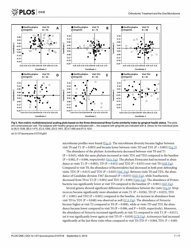
microbiome profiles were found (Fig 4). The microbiome diversity became higher betweenvisit T0 and T1 (P = 0.003) and became lower between visits TD and TD1 (P = 0.003) (Fig 5).
The abundance of the phylum Actinobacteria decreased between visit T0 and T1(P = 0.043), while the same phylum increased at visits TD1 and TD2 compared to the baseline(P = 0.002, P = 0.006, respectively) (S6A Fig). The phylum Firmicutes had increased in abun-dance at visits T1 (P = 0.005), TD (P = 0.021) and TD2 (P = 0.035) over visit T0 (S6B Fig).Compared to visit T0, the abundance of Bacteroidetes had decreased in both post-debondingvisits: TD1 (P = 0.015) and TD2 (P = 0.025) (S6C Fig). Between visits T0 and TD1, the abun-dance of Candidate division TM7 decreased (P = 0.031) (S6D Fig), while Fusobacteriadecreased from T0 to T2 (P = 0.001) and TD1 (P = 0.001) (S6E Fig). The abundance of Proteo-bacteria was significantly lower at visit TD compared to the baseline (P = 0.001) (S6F Fig).
Several genera showed significant differences in abundance between the visits (Fig 6). Strep-tococcus became significantly more abundant at visits T1 (P = 0.036), TD (P = 0.025), TD1(P< 0.001) and TD2 (P = 0.001) compared to the baseline. An increase in abundance fromvisit TD to TD1 (P = 0.048) was observed as well (S7A Fig). The abundance of Neisseriabecame higher at visit T2 compared to T0 (P = 0.008), while at visits TD and TD1 the abun-dance became lower compared to visit T0 (P = 0.006, and P = 0.029, respectively). Moreover,the abundance of Neisseria increased significantly at visit T2 compared to visit T1 (P = 0.011),yet it was significantly lower again at visit TD (P = 0.018) (S7B Fig). Actinomyces had increasedsignificantly at the last three visits when compared to visit T0 (TD: P = 0.004, TD1: P< 0.001
Fig 3. Non-metric multidimensional scaling plots based on the three-dimensional Bray-Curtis similarity index by gingival health status. The plotsare constructed per visit. The subjects with healthy gingiva are indicated with �, the subjects with gingivitis are indicated with ●. Stress for the individual plotsis (A) 0.1539, (B) 0.1474, (C) 0.1265, (D) 0.1401, (E) 0.1495 and (F) 0.1531.
doi:10.1371/journal.pone.0137318.g003
Orthodontic Treatment and the Oral Microbiome
PLOSONE | DOI:10.1371/journal.pone.0137318 September 2, 2015 7 / 17

and TD2: P< 0.001) (S7C Fig). Both Veillonella (S7D Fig) and Porphyromonas (S7E Fig) wereonly at visit TD significantly more abundant when compared to visit T0 (P = 0.0033 andP = 0.0011, respectively). Additionally, the abundance of Porphyromonas decreased signifi-cantly between T2 and TD (P = 0.017). For Leptotrichia, the abundance became significantlylower at TD1 (P< 0.001) and TD2 (P = 0.037) compared to the baseline (S7F Fig). The abun-dance of Campylobacter had decreased at the last three visits compared to visit T0 (TD:P = 0.033, TD1: P< 0.001 and TD2: P< 0.001) (S7G Fig). At both visits T1 and TD, Prevotellahad increased in abundance compared to visit T0 (P = 0.004 and P = 0.001, respectively), whileat TD1 the abundance had become significantly smaller again (P = 0.010) (S7H Fig). For thegenus Haemophilus, the only significant increase in abundance was between visits TD and TD1(P = 0.033) (S7I Fig). The abundance of the genus Fusobacterium was significantly lower at T2and TD1 compared to the baseline (P> 0.001 and P = 0.043, respectively) (S7K Fig). Theabundance of Rothia was higher in the last three visits compared to the baseline (TD:P = 0.009, TD1: P< 0.001, TD2: P> 0.001) (S7L Fig).
At the OTU level, the abundance of OTU28 (Actinomyces) was higher at TD1 (P< 0.001)and TD2 (P = 0.001) compared to visit T0 (S8A Fig). When compared to visit T0, the
Fig 4. Non-metric multidimensional scaling plot based on the three-dimensional Bray-Curtis similarity index by time. The plot consists of samples ofall subjects (N = 19) who were present at all six time points. The stress for this plot is 0.1836.
doi:10.1371/journal.pone.0137318.g004
Orthodontic Treatment and the Oral Microbiome
PLOSONE | DOI:10.1371/journal.pone.0137318 September 2, 2015 8 / 17

abundance of OTU65 (Rothia) was higher in the last three visits (TD: P 0.009, TD1: P< 0.001,and TD2: P< 0.001) (S8B Fig). In addition, both OTU28 and OTU65 were elevated signifi-cantly between visits TD and TD1 (P = 0.049 and P = 0.002, respectively). The abundance ofOTU351 (Streptococcus) became higher between visits TD and TD1 (P = 0.033) and was signif-icantly higher compared to visit T0 at visit TD1 (P< 0.001) and visit TD2 (P = 0.002) (S8CFig). In comparison to the baseline, the abundance of OTU398 (Fusobacterium) was lower atvisit T2 (P< 0.001) and at visit TD1 (P = 0.043) (S8D Fig). The abundance of OTU143 (Lepto-trichia) decreased significantly between visits TD and TD1 (P = 0.003). Moreover, at visit TD1,the abundance of OTU143 was significantly smaller compared to visit T0 (P = 0.007) (S8E Fig).The abundance of OTU151 (Campylobacter) was lower at visit TD compared to visit T2(P = 0.032) and at TD1 the abundance was lower compared to visit TD (P = 0.001). At both vis-its TD1 and TD2, the abundance of OTU151 was significantly lower compared to visit T0(P< 0.001 and P< 0.001, respectively) (S8F Fig). When compared to visit T0, the abundanceof OTU302 (Selenomonas) had increased at visits T1 (P = 0.002), T2 (P< 0.001) and TD(0.029), while the abundance had decreased at visit TD1 (P = 0.003) (S8G Fig).
Fig 5. Shannon diversity index for the entire study population per visit. T0; N = 76, T1; N = 73, T2; N = 68, TD; N = 44, TD1; N = 43, TD2; N = 45.Statistical significance (P < 0.05) was determined using the Wilcoxon Signed Ranks test.
doi:10.1371/journal.pone.0137318.g005
Orthodontic Treatment and the Oral Microbiome
PLOSONE | DOI:10.1371/journal.pone.0137318 September 2, 2015 9 / 17

DiscussionThe results of our study indicate that the fluoride mouthwash had little effect on the adolescentoral microbiome composition during fixed orthodontic appliance treatment. More pronouncedwere the microbial changes observed in relation to gingival health status and orthodontic treat-ment. Yet, the resilience of these adolescent oral communities was noteworthy in regard to theinterference caused by the orthodontic treatment, fluoride mouthwash and the physiologicalchanges of puberty itself. There was no observable shift in the composition of the total commu-nity in time (Fig 4). A remaining change in abundance was observed for a few genera (Fig 6)and, interestingly, most genera that did increase in abundance in time were associated with ahealthy oral cavity.
In this study, an amine fluoride (AmF) combined with stannous fluoride (SnF2) mouthwashwas used to reduce the amount of demineralization, since fluoride is a well-established anti-caries agent [23] and caries is an infectious bacterial disease. Compliance is regarded as a draw-back in studies aiming to observe the effect of a mouthwash. Nonetheless, van der Kaaij et al.[11] observed that the use of an AmF/SnF2 mouthwash inhibited formation of white spotlesions during this study. Likewise, Øgaard et al. [24] observed that there was no difference inwhite spot lesions before and after orthodontic treatment of patients using an AmF/SnF2mouthwash. Madléna et al. [25] observed a decrease in plaque index, gingival index and bleed-ing on probing within one month in orthodontic patients using AmF/SnF2 toothpaste, regard-less if the toothpaste was combined with an AmF/SnF2 mouthrinse. Van Loveren et al. [26] didobserve dental plaque shifting towards less acidogenic plaque, yet there was no significant dif-ference in bacterial composition after the use of AmF/SnF2 products compared to fluoride-freeperiods. This is similar to our findings, as we did not observe a clear effect of the fluoride
Fig 6. Average proportions of the genera that differed significantly in abundance between one or more of the visits.
doi:10.1371/journal.pone.0137318.g006
Orthodontic Treatment and the Oral Microbiome
PLOSONE | DOI:10.1371/journal.pone.0137318 September 2, 2015 10 / 17

mouthwash on the microbial composition. Although it is suggested that fluoride has antibacte-rial properties, its main effect appears to be on the demineralization and remineralization pro-cesses in the oral cavity [27–30]
We did observe that the abundance of several bacterial taxa was associated with the gingivalhealth status of the subjects. Gingivitis during orthodontic treatment is presumably related to pla-que accumulation caused by the newly created retention sites and consequently impaired oralhygiene [5]. Yet, it is not only the orthodontic treatment that is related to the onset of gingivitis inthese subjects, for ‘puberty itself’ is also associated with increased gingivitis [31–33]. Generally,orthodontic treatment takes place during adolescence, as was the case in our study. During thisperiod, the human body experiences many (e.g. behavioral and hormonal) changes [34].
The exact reason why gingivitis becomes prevalent in this age-group is unclear but hor-monal changes are likely to play a part. Our study did not include a control group of adoles-cents that did not receive orthodontic treatment. Therefore it is difficult to discern whichmicrobial changes are related to the orthodontic treatment, and which ones to the onset ofpuberty. Thus far, most studies regarding the (changes in the) oral microbiome during adoles-cence or orthodontic treatment have focused on a limited number of bacteria, due to the natureof their techniques.
The use of an open ended molecular approach allowed us to detect Candidate division TM7(and OTUs 55, 171, 355) (S3B and S5D–S5F Figs). Next-generation sequencing has demon-strated that these bacteria, of which only recently a member was grown as a pure laboratoryculture [35], are widespread in the human oral cavity [36]. Crielaard et al. [37] reported thatCandidate division TM7 increased with advancing age, in a study regarding children aged 3–18years. Duran-Pinedo et al. [38] presumed a role for Candidate division TM7 in periodontitis.We found Candidate division TM7 to be associated with gingivitis, in accordance with Huanget al. [39].
Interestingly, we observed the presence of the genus Derxia (S4D Fig), although low inabundance in our study population, to be related to a healthy state of the gingiva. Members ofthis genus are known to fix nitrogen in different environmental habitats [40, 41]. Recently Der-xia has been observed as a member of the human (and canine) oral cavity [42–44], yet its rolein this particular environment remains to be elucidated.
Well-known inhabitants of the oral cavity are members of the genus Prevotella; oftenassociated with an unhealthy state of the periodontium [45]. Moreover, an increase of Prevo-tella intermedia has been associated with orthodontic treatment [9, 14, 46]. In addition, vanGastel et al. [46] observed a decrease of P. intermedia after the removal of the orthodonticappliances. This coincides with our finding of the abundance of the genus Prevotella (S7HFig). Hence, there appears to be an association between orthodontic treatment and the prev-alence of Prevotella, although Choi et al. [7] did not find a significant decrease of Prevotellaafter orthodontic treatment was ended. This discrepancy might be due to difference in sam-pling sites or detection techniques.
In this study, we found that the genus Actinomyces increased with time (S7C Fig), whileOTU28 (Actinomyces naeslundii) increased mainly after debonding (S8A Fig). According toDelaney et al. [47] the levels of Actinomyces naeslundii are higher in prepubertal subjects com-pared to postpubertal subjects. Gusberti et al. [48] observed that the levels of the species Actino-myces odontolyticus elevate during puberty. Tanner et al. [49] found Actinomyces sp. to beassociated with gingivitis, whereas Tsuruda et al. [50] observed a relation between Actinomycesspecies and healthy pubertal children. These diverse findings indicate that the role of Actino-myces in the oral microbiome cannot be determined on genus level, yet it does not explain thecontradictory findings of the study by Delaney et al. [47] and our own results. Although sam-pling site and used technique might again be of influence.
Orthodontic Treatment and the Oral Microbiome
PLOSONE | DOI:10.1371/journal.pone.0137318 September 2, 2015 11 / 17

The genus Veillonella had previously been shown to increase during adolescence [37, 51]. Inthis study population however, the abundance of Veillonella remained stable throughout time(S7D Fig). In addition, the abundance of Veillonella was not significantly different between thetwo mouthwash groups or between the healthy and gingivitis groups.
Both the genus Campylobacter (S7G Fig) and OTU151 (Campylobacter gracilis) (S8F Fig)decreased with time. A similar pattern of decrease has been observed for Campylobacter rectus[7–9]. This decrease could be explained primarily by the reduction of retention sites due to thealignment of the teeth and secondly by the removal of the orthodontic fixed appliances, causingan additional loss of retention sites.
A similar decrease in time was observed for the genera Porphyromonas (S7E Fig) and Sele-nomonas (S7J Fig). Additionally, we found that Porphyromonas (S4B Fig), Selenomonas (S4AFig) and OTU302 (Selenomonas) (S5G Fig) were associated with gingivitis. Members of boththese genera are among the main periodontal pathogens [39, 52]. Therefore their decrease intime might be considered desirable. Why they decrease in time, if it is e.g. the reduction inretention sites through alignment of the teeth or hormonal changes in the host, remainsunclear.
Neisseria became lower in abundance during the advancement of the visits (S7B Fig), inagreement with Moore et al. [51], who found this genus to be more associated with prepubertalchildren than older children. Thus far, most studies investigating the oral microbiome duringorthodontic treatment or puberty did not target members of the genus Neisseria. Nonetheless,Tanner et al. [49] found Neisseria elongata to be associated with reduced gingivitis in ortho-dontic patients. They made the same observation for Fusobacterium periodonticum.
Tsuruda et al. [50] found Fusobacterium sp. to be more abundant in pubertal children withgingivitis compared to healthy children. Fusobacterium nucleatum is regarded as a bridgingorganism in the formation of dental biofilms [53]. This might explain our observation that thegenus Fusobacterium decreases during the orthodontic treatment, yet increases again in time(S7K Fig). The additional retention sites created by the brackets leave Fusobacterium superflu-ous in the formation of biofilms. On the other hand, Wojcicki et al. [54] found that Fusobacter-ium sp. was lower in their circumpubertal group compared to a younger and older test group,suggesting that the presence of Fusobacterium sp. is influenced by the physiological maturity ofthe host.
In contrast to Fusobacterium, the abundance of the genus Streptococcus (S7A Fig) andOTU351 (Streptococcus) (S8C Fig) showed an increase in time without decreasing first.Increase in Streptococcus abundance in puberty has been observed before [51], although wecannot identify this member of the genus Streptococcus on species level, we speculate that it isassociated with a healthy state of the gingiva.
Haemophilus (S4E Fig), Rothia (S4F Fig) and OTU65 (Rothia) (S5A Fig) were associatedwith a healthy state of the gingiva as well. Their increase after debonding appeared to coincidewith the decrease in gingivitis after debonding (Fig 2). Members of these two genera were usu-ally not included as target micro-organisms in studies of the oral microbiome during pubertyor orthodontic treatment. Although the role of Haemophilus in health and disease of the oralcavity remains somewhat ambiguous, Rothia is generally associated with health [15, 55].
In conclusion, the effects of the fluoride mouthwash on the adolescent microbiome wereindiscernible and promoted neither health nor disease associated bacterial growth. Yet, van derKaaij et al. [11] did observe fewer demineralizations in subjects using the fluoride mouthwashcompared to those using the placebo. Thus, the use of a fluoride mouthwash during orthodon-tic treatment might be beneficial for the health status of the oral cavity.
Nevertheless, we did observe changes in the abundance of various bacteria. In general, thebacteria that were associated with periodontal pathogenesis decreased in abundance in time,
Orthodontic Treatment and the Oral Microbiome
PLOSONE | DOI:10.1371/journal.pone.0137318 September 2, 2015 12 / 17

while the abundance of the health related bacteria increased, suggesting that orthodontic treat-ment during puberty does not have a lasting negative effect on the gingival health status. Still,the lack of an age-related control group not receiving orthodontic treatment precludes us frommaking a clear distinction between microbial changes instigated by puberty and the effects onthe oral ecology caused by orthodontic treatment with fixed appliances. A future study includ-ing such a control group would be necessary to determine which microbial changes are trulycaused by the presence of orthodontic appliances, allowing for the maintenance of a healthyoral microbiome during orthodontic treatment.
Supporting InformationS1 Fig. Difference in abundance of the genus Fusobacterium between mouthwash groupsper visit. The read count is displayed on the y-axis. Mouthwashes were administered betweenvisits T0 and TD. Statistical significance (P< 0.05) was determined using the Mann-Whitneytest between the two groups per visit, or the Wilcoxon Signed Ranks test within the same groupbetween different visits. The boxes represent the median and interquartile range (IQR), thewhiskers represent the minimum and maximum values. Outliers more than 1.5x IQR aredepicted by �, and more than 3x IQR by$.(TIF)
S2 Fig. Difference in abundance of OTU381 (Kingella) between mouthwash groups pervisit. The read count is displayed on the y-axis. Mouthwashes were administered between visitsT0 and TD. Statistical significance (P< 0.05) was determined using the Mann-Whitney testbetween the two groups per visit, or the Wilcoxon Signed Ranks test within the same groupbetween different visits. The boxes represent the median and IQR, the whiskers represent theminimum and maximum values. Outliers more than 1.5x IQR are depicted by �, and morethan 3x IQR by $.(TIF)
S3 Fig. Difference in abundance of the phyla Bacteroidetes (A), TM7 (B) and Fusobacter-ium (C) based on gingival health status per visit. The read count is displayed on the y-axis.Statistical significance (P< 0.05) was determined using the Mann-Whitney test. The boxesrepresent the median and IQR, the whiskers represent the minimum and maximum values.Outliers more than 1.5x IQR are depicted by �, and more than 3x IQR by$.(TIF)
S4 Fig. Difference in abundance of the genera Selenomonas (A), Porphyromonas (B), John-sonella (C), Derxia (D),Haemophilus (E) and Rothia (F) based on gingival health status pervisit. The read count is displayed on the y-axis. Statistical significance (P< 0.05) was deter-mined using the Mann-Whitney test. The boxes represent the median and IQR, the whiskersrepresent the minimum and maximum values. Outliers more than 1.5x IQR are depicted by �,and more than 3x IQR by$.(TIF)
S5 Fig. Difference in OTU abundance based on gingival health status per visit. The readcount is displayed on the y-axis. Statistical significance (P< 0.05) was determined using theMann-Whitney test. The boxes represent the median and IQR, the whiskers represent the min-imum and maximum values. Outliers more than 1.5x IQR are depicted by �, and more than 3xIQR by$.(TIF)
Orthodontic Treatment and the Oral Microbiome
PLOSONE | DOI:10.1371/journal.pone.0137318 September 2, 2015 13 / 17

S6 Fig. Difference in phylum abundance between visits for Actinobacteria (A), Firmicutes(B), Bacteroidetes (C), TM7 (D), Fusobacteria (E) and Proteobacteria (F). The read count isdisplayed on the y-axis. Statistical significance (P< 0.05) was determined using the WilcoxonSigned Ranks test. The boxes represent the median and IQR, the whiskers represent the mini-mum and maximum values. Outliers more than 1.5x IQR are depicted by �, and more than 3xIQR by$.(TIF)
S7 Fig. Difference in genus abundance between visits. The read count is displayed on the y-axis. Statistical significance (P< 0.05) was determined using the Wilcoxon Signed Ranks test.The boxes represent the median and IQR, the whiskers represent the minimum and maximumvalues. Outliers more than 1.5x IQR are depicted by �, and more than 3x IQR by$.(TIF)
S8 Fig. Difference in OTU abundance between visits. The read count is displayed on the y-axis. Statistical significance (P< 0.05) was determined using the Wilcoxon Signed Ranks test.The boxes represent the median and IQR, the whiskers represent the minimum and maximumvalues. Outliers more than 1.5x IQR are depicted by �, and more than 3x IQR by$.(TIF)
S1 Table. BLAST results of the OTUs.(PDF)
S2 Table. Number of subjects per group per visit.(PDF)
AcknowledgmentsWe like to thank Marja Jakobs of the Academic Medical Center (Amsterdam) for her assistancewith pyrosequencing. We thank Rianne Faaij and Merlijn Marsman for data collection.
Author ContributionsConceived and designed the experiments: NCWvdK MHvdV JMtC EZ. Performed the experi-ments: JEK NCWvdKMJB. Analyzed the data: JEK NCWvdK YE. Contributed reagents/materials/analysis tools: WC. Wrote the paper: JEK. Revised the manuscript: JEK NCWvdKMHvdV JMtCWC EZ.
References1. van Gastel J, Quirynen M, TeughelsW, CouckeW, Carels C. Longitudinal changes in microbiology and
clinical periodontal variables after placement of fixed orthodontic appliances. Journal of periodontology.2008; 79(11):2078–86. doi: 10.1902/jop.2008.080153 PMID: 18980516.
2. Gong Y, Lu J, Ding X. Clinical, microbiologic, and immunologic factors of orthodontic treatment-inducedgingival enlargement. American journal of orthodontics and dentofacial orthopedics: official publicationof the American Association of Orthodontists, its constituent societies, and the American Board ofOrthodontics. 2011; 140(1):58–64. doi: 10.1016/j.ajodo.2010.02.033 PMID: 21724088.
3. Sukontapatipark W, el-Agroudi MA, Selliseth NJ, Thunold K, Selvig KA. Bacterial colonization associ-ated with fixed orthodontic appliances. A scanning electron microscopy study. European journal oforthodontics. 2001; 23(5):475–84. PMID: 11668867.
4. Demling A, Heuer W, Elter C, Heidenblut T, Bach FW, Schwestka-Polly R, et al. Analysis of supra- andsubgingival long-term biofilm formation on orthodontic bands. European journal of orthodontics. 2009;31(2):202–6. doi: 10.1093/ejo/cjn090 PMID: 19304761.
5. Ren Y, JongsmaMA, Mei L, van der Mei HC, Busscher HJ. Orthodontic treatment with fixed appliancesand biofilm formation—a potential public health threat? Clinical oral investigations. 2014; 18(7):1711–8.doi: 10.1007/s00784-014-1240-3 PMID: 24728529.
Orthodontic Treatment and the Oral Microbiome
PLOSONE | DOI:10.1371/journal.pone.0137318 September 2, 2015 14 / 17

6. Mattousch TJ, van der Veen MH, Zentner A. Caries lesions after orthodontic treatment followed byquantitative light-induced fluorescence: a 2-year follow-up. European journal of orthodontics. 2007; 29-(3):294–8. doi: 10.1093/ejo/cjm008 PMID: 17483494.
7. Choi DS, Cha BK, Jost-Brinkmann PG, Lee SY, Chang BS, Jang I, et al. Microbiologic changes in sub-gingival plaque after removal of fixed orthodontic appliances. The Angle orthodontist. 2009; 79(6):1149–55. doi: 10.2319/111808-593R.1 PMID: 19852608.
8. Thornberg MJ, Riolo CS, Bayirli B, Riolo ML, Van Tubergen EA, Kulbersh R. Periodontal pathogen lev-els in adolescents before, during, and after fixed orthodontic appliance therapy. American journal oforthodontics and dentofacial orthopedics: official publication of the American Association of Orthodon-tists, its constituent societies, and the American Board of Orthodontics. 2009; 135(1):95–8. doi: 10.1016/j.ajodo.2007.02.057 PMID: 19121507.
9. Kim SH, Choi DS, Jang I, Cha BK, Jost-Brinkmann PG, Song JS. Microbiologic changes in subgingivalplaque before and during the early period of orthodontic treatment. The Angle orthodontist. 2012; 82-(2):254–60. doi: 10.2319/030311-156.1 PMID: 21827233.
10. Torlakovic L, Paster BJ, Ogaard B, Olsen I. Changes in the supragingival microbiota surrounding brack-ets of upper central incisors during orthodontic treatment. Acta odontologica Scandinavica. 2013; 71-(6):1547–54. doi: 10.3109/00016357.2013.776107 PMID: 24180590.
11. van der Kaaij NC, van der Veen MH, van der Kaaij MA, Ten Cate JM. A prospective, randomized pla-cebo-controlled clinical trial on the effects of a fluoride rinse on white spot lesion development andbleeding in orthodontic patients. European journal of oral sciences. 2015; 123(3):186–93. doi: 10.1111/eos.12186 PMID: 25913893.
12. www.trialregister.nl.
13. Zachrisson BU, Zachrisson S. Gingival condition associated with partial orthodontic treatment. Actaodontologica Scandinavica. 1972; 30(1):127–36. PMID: 4504433.
14. Ristic M, Vlahovic Svabic M, Sasic M, Zelic O. Clinical and microbiological effects of fixed orthodonticappliances on periodontal tissues in adolescents. Orthodontics & craniofacial research. 2007; 10-(4):187–95. doi: 10.1111/j.1601-6343.2007.00396.x PMID: 17973685.
15. Zaura E, Keijser BJ, Huse SM, Crielaard W. Defining the healthy "core microbiome" of oral microbialcommunities. BMCmicrobiology. 2009; 9:259. doi: 10.1186/1471-2180-9-259 PMID: 20003481;PubMed Central PMCID: PMC2805672.
16. Kraneveld EA, Buijs MJ, Bonder MJ, Visser M, Keijser BJ, CrielaardW, et al. The relation between oralCandida load and bacterial microbiome profiles in Dutch older adults. PloS one. 2012; 7(8):e42770.doi: 10.1371/journal.pone.0042770 PMID: 22900048; PubMed Central PMCID: PMC3416775.
17. Caporaso JG, Kuczynski J, Stombaugh J, Bittinger K, Bushman FD, Costello EK, et al. QIIME allowsanalysis of high-throughput community sequencing data. Nature methods. 2010; 7(5):335–6. doi: 10.1038/nmeth.f.303 PMID: 20383131; PubMed Central PMCID: PMC3156573.
18. Koopman JE, Roling WF, Buijs MJ, Sissons CH, ten Cate JM, Keijser BJ, et al. Stability and resilienceof oral microcosms toward acidification and Candida outgrowth by arginine supplementation. MicrobEcol. 2015; 69(2):422–33. doi: 10.1007/s00248-014-0535-x PMID: 25433583.
19. Zhang Z, Schwartz S, Wagner L, Miller W. A greedy algorithm for aligning DNA sequences. Journal ofcomputational biology: a journal of computational molecular cell biology. 2000; 7(1–2):203–14. doi: 10.1089/10665270050081478 PMID: 10890397.
20. Morgulis A, Coulouris G, Raytselis Y, Madden TL, Agarwala R, Schaffer AA. Database indexing for pro-duction MegaBLAST searches. Bioinformatics. 2008; 24(16):1757–64. doi: 10.1093/bioinformatics/btn322 PMID: 18567917; PubMed Central PMCID: PMC2696921.
21. HammerØ, Harper DAT, Ryan PD. PAST: Paleontological statistics software package for educationand data analysis. Palaeontologia Electronica. 2001; 4(1):9.
22. Clarke KR. Non-parametric multivariate analyses of changes in community structure. Australian Jour-nal of Ecology. 1993; 18(1):117–43. doi: 10.1111/j.1442-9993.1993.tb00438.x
23. van Loveren C, Gerardu VA, Sissons CH, van BekkumM, ten Cate JM. Effect of various rinsing proto-cols after use of amine fluoride/stannous fluoride toothpaste on the bacterial composition of dental pla-que. Caries research. 2009; 43(6):462–7. doi: 10.1159/000264683 PMID: 20016176.
24. Øgaard B, Alm AA, Larsson E, Adolfsson U. A prospective, randomized clinical study on the effects ofan amine fluoride/stannous fluoride toothpaste/mouthrinse on plaque, gingivitis and initial caries lesiondevelopment in orthodontic patients. European journal of orthodontics. 2006; 28(1):8–12. doi: 10.1093/ejo/cji075 PMID: 16230329.
25. Madléna M, Bánóczy J, Götz G, Márton S, Kaán M Jr., Nagy G. Effects of amine and stannous fluorideson plaque accumulation and gingival health in orthodontic patients treated with fixed appliances: a pilotstudy. Oral health and dental management. 2012; 11(2):57–61. PMID: 22692271.
Orthodontic Treatment and the Oral Microbiome
PLOSONE | DOI:10.1371/journal.pone.0137318 September 2, 2015 15 / 17

26. Gerardu VA, Buijs MJ, ten Cate JM, van Loveren C. The effect of a single application of 40% chlorhexi-dine varnish on the numbers of salivary mutans streptococci and acidogenicity of dental plaque. Cariesresearch. 2003; 37(5):369–73. PMID: 12925829.
27. Rosin-Grget K, Peros K, Sutej I, Basic K. The cariostatic mechanisms of fluoride. Acta medica acade-mica. 2013; 42(2):179–88. doi: 10.5644/ama2006-124.85 PMID: 24308397.
28. ten Cate JM. The need for antibacterial approaches to improve caries control. Advances in dentalresearch. 2009; 21(1):8–12. doi: 10.1177/0895937409335591 PMID: 19661048.
29. van Loveren C. Antimicrobial activity of fluoride and its in vivo importance: identification of researchquestions. Caries research. 2001; 35 Suppl 1:65–70. PMID: 11359062.
30. ten Cate JM. Contemporary perspective on the use of fluoride products in caries prevention. Britishdental journal. 2013; 214(4):161–7. doi: 10.1038/sj.bdj.2013.162 PMID: 23429124.
31. Morishita M, Aoyama H, Tokumoto K, Iwamoto Y. The concentration of salivary steroid hormones andthe prevalence of gingivitis at puberty. Advances in dental research. 1988; 2(2):397–400. PMID:3271037.
32. Nakagawa S, Fujii H, Machida Y, Okuda K. A longitudinal study from prepuberty to puberty of gingivitis.Correlation between the occurrence of Prevotella intermedia and sex hormones. Journal of clinical peri-odontology. 1994; 21(10):658–65. PMID: 7852609.
33. Mombelli A, Lang NP, Burgin WB, Gusberti FA. Microbial changes associated with the development ofpuberty gingivitis. Journal of periodontal research. 1990; 25(6):331–8. PMID: 2148945.
34. Vetter-O'Hagen CS, Spear LP. Hormonal and physical markers of puberty and their relationship to ado-lescent-typical novelty-directed behavior. Developmental psychobiology. 2012; 54(5):523–35. doi: 10.1002/dev.20610 PMID: 21953609; PubMed Central PMCID: PMC3288810.
35. Soro V, Dutton LC, Sprague SV, Nobbs AH, Ireland AJ, Sandy JR, et al. Axenic culture of a candidatedivision TM7 bacterium from the human oral cavity and biofilm interactions with other oral bacteria.Applied and environmental microbiology. 2014; 80(20):6480–9. doi: 10.1128/AEM.01827-14 PMID:25107981; PubMed Central PMCID: PMC4178647.
36. He X, McLean JS, Edlund A, Yooseph S, Hall AP, Liu SY, et al. Cultivation of a human-associated TM7phylotype reveals a reduced genome and epibiotic parasitic lifestyle. Proceedings of the National Acad-emy of Sciences of the United States of America. 2015; 112(1):244–9. doi: 10.1073/pnas.1419038112PMID: 25535390.
37. CrielaardW, Zaura E, Schuller AA, Huse SM, Montijn RC, Keijser BJ. Exploring the oral microbiota ofchildren at various developmental stages of their dentition in the relation to their oral health. BMCmedi-cal genomics. 2011; 4:22. doi: 10.1186/1755-8794-4-22 PMID: 21371338; PubMed Central PMCID:PMC3058002.
38. Duran-Pinedo AE, Chen T, Teles R, Starr JR, Wang X, Krishnan K, et al. Community-wide transcrip-tome of the oral microbiome in subjects with and without periodontitis. The ISME journal. 2014; 8(8):1659–72. doi: 10.1038/ismej.2014.23 PMID: 24599074.
39. Huang S, Yang F, Zeng X, Chen J, Li R, Wen T, et al. Preliminary characterization of the oral microbiotaof Chinese adults with and without gingivitis. BMC oral health. 2011; 11:33. doi: 10.1186/1472-6831-11-33 PMID: 22152152; PubMed Central PMCID: PMC3254127.
40. John RC, Itah AY, Essien JP, Ikpe DI. Fate of nitrogen-fixing bacteria in crude oil contaminated wetlandultisol. Bulletin of environmental contamination and toxicology. 2011; 87(3):343–53. doi: 10.1007/s00128-011-0320-1 PMID: 21755289; PubMed Central PMCID: PMC3155754.
41. ChenWM, HuangWC, Sheu SY. Derxia lacustris sp. nov., a nitrogen-fixing bacterium isolated from afreshwater lake. International journal of systematic and evolutionary microbiology. 2013; 63(Pt 3):965–70. PMID: 22659506.
42. Hu YJ, Shao ZY, Wang Q, Jiang YT, Ma R, Tang ZS, et al. Exploring the dynamic core microbiome ofplaque microbiota during head-and-neck radiotherapy using pyrosequencing. PloS one. 2013; 8(2):e56343. doi: 10.1371/journal.pone.0056343 PMID: 23437114; PubMed Central PMCID: PMC3578878.
43. Sturgeon A, Stull JW, Costa MC, Weese JS. Metagenomic analysis of the canine oral cavity asrevealed by high-throughput pyrosequencing of the 16S rRNA gene. Veterinary microbiology. 2013;162(2–4):891–8. doi: 10.1016/j.vetmic.2012.11.018 PMID: 23228621.
44. JiangW, Ling Z, Lin X, Chen Y, Zhang J, Yu J, et al. Pyrosequencing analysis of oral microbiota shiftingin various caries states in childhood. Microb Ecol. 2014; 67(4):962–9. doi: 10.1007/s00248-014-0372-yPMID: 24504329.
45. Nadkarni MA, Browne GV, Chhour KL, Byun R, Nguyen KA, Chapple CC, et al. Pattern of distribution ofPrevotella species/phylotypes associated with healthy gingiva and periodontal disease. European jour-nal of clinical microbiology & infectious diseases: official publication of the European Society of ClinicalMicrobiology. 2012; 31(11):2989–99. doi: 10.1007/s10096-012-1651-5 PMID: 22684253.
Orthodontic Treatment and the Oral Microbiome
PLOSONE | DOI:10.1371/journal.pone.0137318 September 2, 2015 16 / 17

46. van Gastel J, Quirynen M, TeughelsW, CouckeW, Carels C. Longitudinal changes in microbiology andclinical periodontal parameters after removal of fixed orthodontic appliances. European journal of ortho-dontics. 2011; 33(1):15–21. doi: 10.1093/ejo/cjq032 PMID: 20671070.
47. Delaney JE, Ratzan SK, Kornman KS. Subgingival microbiota associated with puberty: studies of pre-,circum-, and postpubertal human females. Pediatric dentistry. 1986; 8(4):268–75. PMID: 3472173.
48. Gusberti FA, Mombelli A, Lang NP, Minder CE. Changes in subgingival microbiota during puberty. A 4-year longitudinal study. Journal of clinical periodontology. 1990; 17(10):685–92. PMID: 2262580.
49. Tanner AC, Sonis AL, Lif Holgerson P, Starr JR, Nunez Y, Kressirer CA, et al. White-spot lesions andgingivitis microbiotas in orthodontic patients. J Dent Res. 2012; 91(9):853–8. doi: 10.1177/0022034512455031 PMID: 22837552; PubMed Central PMCID: PMC3420397.
50. Tsuruda K, Miyake Y, Suginaka H, Okamoto H, Iwamoto Y. Microbiological features of gingivitis inpubertal children. Journal of clinical periodontology. 1995; 22(4):316–20. PMID: 7622639.
51. MooreWE, Burmeister JA, Brooks CN, Ranney RR, Hinkelmann KH, Schieken RM, et al. Investigationof the influences of puberty, genetics, and environment on the composition of subgingival periodontalfloras. Infection and immunity. 1993; 61(7):2891–8. PMID: 8514392; PubMed Central PMCID:PMC280936.
52. Mysak J, Podzimek S, Sommerova P, Lyuya-Mi Y, Bartova J, Janatova T, et al. Porphyromonas gingi-valis: major periodontopathic pathogen overview. Journal of immunology research. 2014;2014:476068. doi: 10.1155/2014/476068 PMID: 24741603; PubMed Central PMCID: PMC3984870.
53. Wright CJ, Burns LH, Jack AA, Back CR, Dutton LC, Nobbs AH, et al. Microbial interactions in buildingof communities. Molecular oral microbiology. 2013; 28(2):83–101. doi: 10.1111/omi.12012 PMID:23253299; PubMed Central PMCID: PMC3600090.
54. Wojcicki CJ, Harper DS, Robinson PJ. Differences in periodontal disease-associated microorganismsof subgingival plaque in prepubertal, pubertal and postpubertal children. Journal of periodontology.1987; 58(4):219–23. doi: 10.1902/jop.1987.58.4.219 PMID: 3473218.
55. Fujinaka H, Takeshita T, Sato H, Yamamoto T, Nakamura J, Hase T, et al. Relationship of periodontalclinical parameters with bacterial composition in human dental plaque. Archives of microbiology. 2013;195(6):371–83. doi: 10.1007/s00203-013-0883-9 PMID: 23539062.
Orthodontic Treatment and the Oral Microbiome
PLOSONE | DOI:10.1371/journal.pone.0137318 September 2, 2015 17 / 17