The effect of ambient temperature on recovery of surgical ...
50
James Madison University JMU Scholarly Commons Masters eses e Graduate School Summer 2017 e effect of ambient temperature on recovery of surgical stress in Sprague-Dawley rats Romie D. Powell James Madison University Follow this and additional works at: hps://commons.lib.jmu.edu/master201019 Part of the Animal Experimentation and Research Commons , and the Other Physiology Commons is esis is brought to you for free and open access by the e Graduate School at JMU Scholarly Commons. It has been accepted for inclusion in Masters eses by an authorized administrator of JMU Scholarly Commons. For more information, please contact [email protected]. Recommended Citation Powell, Romie D., "e effect of ambient temperature on recovery of surgical stress in Sprague-Dawley rats" (2017). Masters eses. 492. hps://commons.lib.jmu.edu/master201019/492
Transcript of The effect of ambient temperature on recovery of surgical ...
James Madison UniversityJMU Scholarly Commons
Masters Theses The Graduate School
Summer 2017
The effect of ambient temperature on recovery ofsurgical stress in Sprague-Dawley ratsRomie D. PowellJames Madison University
Follow this and additional works at: https://commons.lib.jmu.edu/master201019Part of the Animal Experimentation and Research Commons, and the Other Physiology
Commons
This Thesis is brought to you for free and open access by the The Graduate School at JMU Scholarly Commons. It has been accepted for inclusion inMasters Theses by an authorized administrator of JMU Scholarly Commons. For more information, please contact [email protected].
Recommended CitationPowell, Romie D., "The effect of ambient temperature on recovery of surgical stress in Sprague-Dawley rats" (2017). Masters Theses.492.https://commons.lib.jmu.edu/master201019/492
The Effect of Ambient Temperature on Recovery of Surgical Stress in Sprague-Dawley Rats
Romie Dylan Powell
A thesis submitted to the Graduate Faculty of
JAMES MADISON UNIVERSITY
In
Partial Fulfillment of the Requirements
for the degree of
Master of Science
Department of Biology
August 2017
FACULTY COMMITTEE:
Committee Chair: Dr. Justin Brown
Committee Members/ Readers:
Dr. Mark Gabriele
Dr. T.J. Hyn

ii
Acknowledgements
I would like to thank my advisor Dr. Justin Brown for his support as well as my
committee members Drs. Mark Gabriele and T.J. Hynd for their guidance and advice.
Also, thanks to Dr. Nusrat Jahan for her statistical approach of analyzing circadian
rhythms. I would also like to thank my lab mates, Benito Blanchfield-Felice, Kelly
Burke, Elli Flora, Samantha Hetrick, Kelcy Jackson, and Alex Schmidt. I greatly
appreciate the graduate students for helping me when they could and allowing me to help
with their research as well. A special thanks for the hard work of the Facilities Manager,
Mike Love, for monitoring the vivarium and assisting with our rodents. I would like to
thank James Madison University for giving me the opportunity to earn my Master of
Science. Finally, I am most grateful for my family, for guiding and inspiring me to be my
best.

iii
Table of Contents
Acknowledgements……………………………………………………………….………ii Preface I………………………………………………………………………….……….iv Preface II……………………………………………………………………….....……….v Abstract………………………………………………………………………….……….vii I.Introduction…………………………………………………………………...................1 Statement of the Problem……………………………………………………...…..1 Background……………………………………………………………..……..…..2 Thermoregulation……………………………………………………………...…..2 Thermal Neutral Zone…………………………………………………………......5 Thermal Stress………………………………………………………………….....5 Ambient Temperature in Surgical Recovery……………………………………...7 How to Address the Problem……………………………………………………...8 Implications………………………………………………………………………..9 II. Methods…………………………………………………………………………….....10 III. Results………………………………………………………………………………..17 Core Temperature Recovery……………………………………………………..18 Circadian Rhythm……………………………………………………………......18 Body Weight……………………………………………………………………..23 Food and Water Consumption……………………………………………….......27 Survival…………………………………………………………………………..31 IV. Discussion Core Temperature Recovery…………………………………………....…..........32 Circadian Rhythm……………………………………………………………......33 Body Weight, Food, and Water Measurements……………..……………….......34 Survival…………………………………………………………………………..37 V. Conclusion…………………………………………………………………….……...38 VI. References……………………………………………………………………..……..40

iv
Preface I
List of Abbreviations
● Sprague-Dawley (SD)● Ambient temperature (Tamb)● Core temperature (Tc)● Non-shivering thermogenesis (NST)● Selected ambient temperature (STa)● Thermal neutral zone (TNZ)● Lower critical temperature (LCT)● Upper critical temperature (UCT)

v
PrefaceII
TablesTable 1. Experimental Design. 27°C is the control group because it is the rat’s preferred Tamb according to previous findings. UCT: Upper Critical Temperature. LCT: Lower Critical Temperature in relation to the thermoneutral zone of the rats. (pg. 13) Table 2. The Survival Percentage of the Tamb Groups. (pg. 31)
FiguresFigure 1. Cage Top Warmer. It rests atop the cage and below the HEPA filter top and circulates temperature-controlled air to hold the rat’s Tamb steady at the desired level. (pg. 11) Figure 2. Radiotelemetry Probe. Measures the Tc and MA from the abdominal cavity of the rodent. (pg. 12) Figure 3. The Typical Tc Recovery Response. MI388 is an example of a rat housed at Tamb 27°C that returned to 36.5°C within a day and displayed circadian rhythm a day after the surgery. This rat returned to normal circadian rhythm after the second photoperiod. (pg. 17) Figure 4. Return of Tc Following Surgical Procedure. Tamb 33°C returned to a normal Tc faster than the other Tamb groups versus the Tamb 21°C which took longer (Kruskal-Wallis, p=0.6545). (pg. 18) Figure 5. Averages of all the Tamb Circadian Rhythms in the Recovery Period. The data presented here is of a 2 hour interval average from hour 40 (~1.75 days) to hour 168 (7 days) after surgery. A. 21°C. B. 24°C. C. 27°C. D. 30°C. E. 33°C F. Data from all Tamb groups. (pg. 19) Figure 6. The 10-4 Period Comparison of the Establishment of Circadian Rhythm. The warmer Tamb groups, and the control group (27°C), stabilize circadian rhythm faster than the colder Tamb groups (ANOVA, p=0.1367). (pg. 20) Figure 7. A. MI388 2 Hour Average. This illustrates the circadian rhythm and reduces the variation. B. Spectral Density of MI388. The highest spectrum dictates early periodicity of circadian rhythm establishment. (pg. 22) Figure 8. Spectral Density Comparison of Establishing Circadian Rhythm. The 21°C group has a delay in the return of circadian rhythm while the 27°C group established circadian Tc rhythms the soonest (Kruskal-Wallis, p=0.3227). (pg. 23)

vi
Figure 9. The Percent of Original Body Weight Lost in Each Tamb Group During the Recovery Week. Data presented as mean. N=5 in each group. (pg. 24) Figure 10. The Average Weight Change Per Day Following Surgery. 21°C lost the most weight on Day 1 but regained that weight back around Day 4. *Significant difference between 21°C and 33°C on Day 1 (Tukey post hoc, p=0.006). (pg. 24) Figure 11. The Total Amount of Weight Lost Following the Surgery. The 21°C group lost the most weight during the week but it was not significant in comparison to the other groups (Kruskal-Wallis, p=0.292). (pg. 25) Figure 12. The amount of weight lost between Day 0 and Day 1. There was a significant difference between 21°C and 33°C. *Indicates significant difference between groups (Tukey post hoc, p=0.006). (pg. 25) Figure 13. The Amount of Weight Change of Days 1, 3, and 7 Between the Tamb Groups. There were no differences between the groups, only between days on individual groups. The differences were between Day 1 and 7 of Tamb 21°C and Day 1 and 7 of Tamb 27°C. *Indicates significant difference between days (Nemenyi post hoc, 21°C p=0.023; 27°C p=0.02). (pg. 26) Figure 14. A. The Averages of the Food Intake Following Surgery Over 7 Days. B. The total amount of food consumed over the week (Kruskal-Wallis, p=0.098). (pg. 28) Figure 15. The amount of food consumed between Day 1, 3, and 7. The only difference observed was between Day 1 and Day 7 of the 21°C group. *Indicates significant difference between groups (Tukey post hoc, p=0.02). (pg. 29) Figure 16. A. The averages of water consumption following surgery over 7 days. B. The total amount of water consumed over the week (Kruskal-Wallis, p=0.168). (pg. 30) Figure 17. The Amount of Water Consumed Between Day 1, 3, and 7. Differences were not observed between the Tamb groups. The 27°C group had differences between Day 1 and Day 3, and Day 1 and Day 7. The 30°C groups only had differences between Day 1 and Day 7, while 33°C had differences between Day 1 and Day 3, and Day 1 and Day 7. *Indicates significant difference between days (Tukey post hoc, 27°C: p=0.041, 0.011; 30°C, p=0.019; 33°C, p=0.039, 0.004). (pg. 31)

vii
Abstract
Laboratory animals are housed at ambient temperatures ranging from 20°C –
26°C per recommended guidelines. Rats in particular typically prefer ambient
temperatures (Tamb) of ~27°C (Brown et al, 2011). When rats undergo surgical
instrumentation for experimental use, they often recover at normal room temperature
(~21°C). While this is comfortable to those maintaining them, it may lead to a cold
thermal stress for the rats. It is hypothesized that housing rats at ambient temperatures
away from their preferred Tamb could lead to a thermal stress, which adversely affects
surgical recovery. To address this, rats (220g-350g) were surgically instrumented with a
radiotelemetry probe (DSI), which allows non-invasive measurement of core temperature
(Tc). A cannula (21ga) was also inserted into the brainstem to allow microinjection of
drugs as part of a separate project. For at least 1 week post-surgery, the rats were housed
at either 21°C, 24°C, 27°C (control), 30°C, or 33°C Tamb while Tc, food and water
intake, and body weight changes were measured. Rats housed at 21°C and 24°C struggled
to recover a normal Tc initially, which delayed the onset of normal circadian cycling in
the 21°C group. Warmer Tamb (30°C & 33°C) did not alter recovery of circadian Tc
rhythms. Rats held at 21°C steadily increased food intake, while those at 33°C consumed
less as the recovery period continued, suggesting that colder rats needed more caloric
intake to thermoregulate, while those at warmer Tamb did not. Rats in the 30°C and 33°C
groups consumed more water after post-surgical Day 3 compared to the other groups.
This was due to more evaporative water loss at the higher Tamb versus the rats held at
lower Tamb. The return of body weight to pre-surgical levels in rats housed at 33°C was
delayed due to heat stress. The control group (27°C) returned to pre-surgical weight

viii
earlier than the other groups. These data suggest that rats maintained at 27°C recovered
from surgical stress more readily. These rats returned to pre-surgical body weight more
quickly and demonstrated a normal thermoregulatory circadian rhythm earlier than the
cold (21°C) rats. The rats housed at 33°C were exposed to a heat stress, which affected
weight gain and surgical recovery initially but did not affect circadian cycling during
recovery. Rats housed at 21°C were cold stressed, which affected weight gain and
thermoregulatory recovery. It is suggested that rats be maintained at their preferred Tamb
of 27 °C during the week following surgery to minimize thermal stress and thereby
facilitate recovery. This reduction in thermal stress would facilitate the return to a normal
physiologic state and consequently enables more reliable data collection from these
animals.

Introduction Statement of the Problem
Many researchers use lab animals to explore physiologic responses to various
stimuli. Using animals requires housing them within specific environmental parameters
to ensure the health of the animals. An important aspect of these housing conditions is
maintenance of appropriate ambient temperature (Tamb). This is especially important in
animals that are exposed to other stressors such as surgical instrumentation in preparation
for experimentation. It is proposed that current Tamb guidelines for rodents, such as
Sprague Dawley (SD) rats, may allow for thermal stress, which affects the animal’s
overall physiology and ability to recover from surgery. The current guidelines of the NIH
Guide for the Care and Use of Laboratory Animals suggest a Tamb range between 20°C
to 26°C (68°F to 79°F), which is comfortable for researchers and staff, but below the
preferred Tamb of ~27°C for SD rats (Brown et al., 2011). Although housing design,
bedding for nesting material, and grouping animals in cages together are used as
resources to minimize thermal stress, animals are still at great risk for thermal stress
despite adherence to housing guidelines (Institute for Laboratory Animal Research,
2011). Surgical recovery requires special care because of the stress from surgery. The
current guidelines suggest warming animals post-surgery, but the degree and duration are
not established. Without a proper establishment of Tamb guidelines following surgery,
the physiology of the animal may be abnormal, which may result in a stressed rat being
used in experimentation. This could yield invalid data and confound interpretation. It is
essential to refine such housing guidelines not only improve the care and use of
laboratory animals, but also to validate data gathered from survival surgery experiments.
The goal of this study is to determine the optimal ambient temperature range for housing
2
rats during recovery from surgical stress.
Background
The regulations set forth by the NIH Guide for the Care and Use of Laboratory
Animals aim to ensure that lab animals are housed in an environment that minimizes
stress. Thermal stress can affect the overall health of the rats as well as recovery from
experimentation and surgical instrumentation. Current guidelines suggest housing rats
between 20°C to 26°C (68°F to 79°F) (Institute for Laboratory Animal Research, 2011).
This suggested range is based on thermoregulatory behaviors noted during normal
housing. Unfortunately, for many species such as rodents, these recommendations may
not be appropriate as rectal probes were used to measure core temperature (Tc) in
defining a basis for the guidelines. These probes likely stressed the animal and therefore
potentially confounded data collected. Furthermore, current research suggests that the
preferred ambient temperature of these rats is 27°C (Gonder & Laber 2007). This is
higher than the current housing guidelines, suggesting these guidelines may lead to cold
stressed rats (Brown et al., 2011).
Thermoregulation
Thermoregulation is the body’s attempt to regulate core temperature (Tc) within
normal limits regardless of environment temperature. SD rats maintain a Tc between
36.5°C -38.5°C which fluctuates around a set point of ~37.5°C (Gordon, 1993). Body
temperature regulation relies on the nervous system’s ability to sense and integrate
thermal information from the external environment and deep within the body core. The
hypothalamus, an area of the brain responsible for much of the homeostatic regulation of
the body, contains the preoptic area (POA). This area is thought to integrate information

3
about local brain temperature and other body temperatures and to control the level of
output of thermoregulatory responses (Bicego et al., 2007). The POA compares the
body’s current Tc based on information sent from afferent neurons, to the
thermoregulatory set point, and coordinates appropriate effector responses if corrections
are needed. These various efferent mechanisms, or thermo-effectors, are used to either
cool down or heat up the body, depending on what information was perceived.
Thermo-effectors modulate the Tc through altering heat production and heat loss
mechanisms, which allow Tc to be maintained at the set point. One important thermo-
effector in rodents is brown adipose tissue. It is responsible for non-shivering
thermogenesis (NST), a mechanism used by rats to generate heat by making ATP
production inefficient (Cannon & Nedergaard, 2011). When the Tc decreases below the
set point, a large amount of lipids and glucose are combusted in brown adipose tissue to
generate heat. This allows more heat to be produced than normal as a byproduct of
metabolism. The rats will use the brown adipose tissue to generate energy for heat
without shivering. Blood leaving brown adipose tissue is nearly depleted of oxygen after
stimulating the tissue in a cold environment (Cannon & Nedergaard, 2011). After about
nine months postnatal, humans lose their brown adipose tissue as their surface area to
volume ratio decreases. This is important in rats because their surface area to mass ratio
remains high into adulthood and they do not shiver. They instead depend on brown
adipose tissue for NST.
Rats can rely on peripheral vasomotor tone to maintain their Tc as well. The tail
of rodents in particular is crucial for thermoregulation using vasoconstriction or
vasodilation to regulate heat loss. The reasons that the tail is ideal for heat release are 1)
4
its lack of fur, 2) its vascularity, and 3) its high surface area to volume ratio. Warmer
environments cause increases of blood flow through the tail from vasodilation, which
radiates heat. In contrast, a colder environment will cause tail vasculature to
vasoconstrict, decreasing blood flow and minimizing heat loss (Gordon, 1993).
When the rat’s Tc rises above or below the set point, behavioral mechanisms of
thermoregulation are activated. Rats will seek cooler environments and adopt a sprawled
posture to lose heat. In addition, some behaviors are integrated such as grooming while
also secreting saliva, which is then spread on the skin by the rat. This facilitates an
evaporative heat loss on the skin. This is analogous to humans sweating in a warm
environment. Rats do not sweat so this behavioral response in rodents facilitates heat
loss. In a colder environment, rats will burrow into their bedding or stay grouped together
to limit heat loss (Brown et al., 2011; Gordon, 1993). These behavioral thermo-effector
mechanisms allow Tc to be maintained within normal limits of a set point despite
significant changes in Tamb.
The circadian cycle also affects thermoregulation. Rats are nocturnal so their
motor activity increases at night. This generates heat, which increases Tc. Rats will
typically choose a slightly cooler environment at night to offset this heat production
(Brown et al., 2011). When at rest during the daytime, Tc decreases with the lack of
motion-generated heat and the selected ambient temperature (STa) increases in response.
STa is the preferred temperature the rat will choose depending on its thermal needs. If
given the option to choose Tamb, rats will behaviorally choose a STa that is closer to
their preferred Tamb (Brown et al., 2011; Gordon, 1993). Generally about an hour before
nightfall, rats will increase their motor activity (MA) and Tc in anticipation of the dark

5
photoperiod. The inverse is true in anticipation of the light photoperiod in the morning. In
this study, the photoperiod change occurs at 07:00 and 19:00. Therefore the time periods
between 10:00-16:00 and 22:00-04:00 show a relatively stable Tc and MA. Careful
examination of circadian thermoregulatory responses after surgery will be a key aspect of
this project.
Thermal Neutral Zone
The thermal neutral zone (TNZ) is the range of ambient temperatures that allow a
minimal need for energy expenditure while maintaining Tc (Gonder & Laber, 2007). The
lower critical temperature (LCT) and upper critical temperature (UCT) are the minimum
and maximum ranges of the TNZ before the rats use thermo-effector pathways to
generate or lose heat in order to maintain Tc at the set point. In Sprague-Dawley rats, the
preferred ambient temperature is 27°C, which is between the LCT of 26°C and the UCT
of 30°C (Brown et al., 2011). The current housing guidelines (20°C-26°C) allow housing
at Tamb below the LCT and preferred Tamb, therefore outside of the TNZ (26°C-30°C).
Thermal Stress
Housing rats at ambient temperatures outside of their TNZ, and therefore away
from their preferred Tamb, likely elicits thermal stress responses. Such changes could be
considered additional experimental variables, and should be taken into consideration
regarding data collection and interpretation. In an experiment conducted by Zylan and
Carlisle (1992), Tamb was varied to observe its effect on brown adipose tissue in rats and
the amount of oxygen consumed during non-shivering thermogenesis. During baseline
experimentation, oxygen consumption increased as Tamb decreased, indicating that 30
minutes of cold exposure was sufficient enough to induce a significant metabolic

6
response. As stated above, such thermal stress might confound data collection and
interpretation based on significant increase in energy expenditure and subsequent change
the normal physiological state of the rodent. Rats prefer an ambient temperature of 27 °C
(Brown et al., 2011). This varies slightly with circadian rhythm in which the rats prefer
slightly cooler temperatures at night and slightly warmer ones during the day. During
active periods, rats will choose Tamb below LCT; however, they prefer temperatures
above their LCT for maintenance and resting, which is essential for everyday function
(Institute for Laboratory Animal Research, 2011). Rats typically do not have the
opportunity to choose their own STa in a normal vivarium and must rely on
thermoregulatory processes to manage thermoregulation.
If an animal is housed at a Tamb below the LCT, then cold stress can chronically
activate both thermo-effector pathways that limit heat loss and also chronically increase
metabolism. An experiment conducted by David et al. (2013) found that chronic cold
stress requires greater energy expenditure to manage core body temperature, likely by
activation of brown adipose tissue and subsequent NST. This can cause the failure of
rodent models to emulate human physiology. In the same experiment, with the vivarium
temperature being lower than the TNZ, but within housing guidelines, energy expenditure
of mice was increased significantly when compared with intermediate and heated
temperatures. There was an associated shift in metabolism toward glucose utilization for
energy production (David et al., 2013). The use of metabolism to maintain core
temperature can be problematic during recovery from surgical stress because energy is
being split between recovering and thermoregulation.
Warm stress occurs when the Tamb is above the UCT, activating thermo-effectors

7
such as sweating in humans or grooming (saliva spreading on skin) in rodents to facilitate
body heat loss to the environment. According to Leon et al. (2005), mice that show these
thermoregulatory responses to heat stress are key biomarkers that may provide insight
into heat stroke pathophysiology. Raising Tamb outside the TNZ is risky in that
hyperthermia can lead to heat stroke and the eventual denaturing of vital proteins in the
body. While a cold stress does not affect cellular function in the same ways, a warm
stress does require activation of thermo-effectors to cool the core temperature to within
normal limits. During surgical recovery, however, briefly increasing the Tamb is useful
in helping the animal return to their normal Tc. To what degree and duration the Tamb
should be raised is not yet known. Warming may be appropriate in the post-surgical
period to facilitate recovery, but excessive and prolonged warming may prove equally
stressful to the animal.
Ambient Temperature in Surgical Recovery
Ambient temperature plays a significant role during surgical recovery. When rats
are given an anesthetic, they experience hypothermia, where the Tc will drop
significantly unless external warming is significant. It is important to note, hypothermia
during anesthesia from a lack of thermoregulatory control from the hypothalamus is the
most common perioperative thermal disturbance in rodents (Diaz & Becker, 2010).
Anesthetics impair the hypothalamus, thus thermoregulation as well, suggesting external
warming to prevent hypothermia is needed. However, hypothermia can be beneficial
during surgery. For example, in cardiopulmonary bypass, following traumatic brain
injury, and many other clinical circumstances, hypothermia is used to lower the demand
for oxygen, which allows aerobic metabolism to continue through greater periods of

8
compromised oxygen supply. This thereby reduces the production of anaerobic
byproducts such as superoxide radicals and lactate (Diaz & Becker, 2010). While
hypothermia can be beneficial in surgery, once anesthesia wears off, the animal must
spend metabolic energy to recover and maintain Tc. However, housing at Tamb below
the TNZ for extended periods will force animals exposed to surgical stress to use
metabolic resources to thermoregulate instead of using them for surgical recovery.
How to Address the Problem:
Current guidelines allow for housing of rats in an environment that may lead to a
cold stress, which can result in stressed animals being used in experimentation and
therefore potential collection of misleading data. The specifics of Tamb requirements
during surgical recovery are unclear as the Guide briefly mentions raising the Tamb, but
does not specify the degree or duration. If subjects are exposed to a significant surgical
stress while being housed below their LCT, the combined surgical stress could adversely
affect recovery. If housed above their UCT during recovery, the increased heat may help
return to normal Tc faster, but also has the potential to eventually cause a heat stress. It is
therefore essential to better define the housing guidelines to determine the appropriate
Tamb during surgical recovery.
It is therefore hypothesized that housing rats at ambient temperatures outside of
their thermal neutral zone leads to a thermal stress, which adversely affects surgical
recovery. This would be evidenced by alterations in food and water intake, delayed
return of body weight to pre-surgical levels, and alterations in re-establishing normal
levels, and circadian cycling of, core temperature. Rats housed at their preferred Tamb
of ~27C are anticipated to be less stressed and recover more quickly than other
9
experimental groups.
Implications
Rats are used extensively for experimentation to explore physiologic variables in
health and disease. Not only are animal models important for understanding physiology
and pathologies, but also for testing hypotheses that cannot be done in human studies
(Horvath, et al., 2015). Many times surgical instrumentation with devices that monitor
physiologic variables are required. Recovery from surgical instrumentation to a
minimally stressed state before experimentation begins is essential to the collection of
quality data from these animals. The scope of the present study is to better define
appropriate housing regulations that inform better living standards of rats, shorter
recovery periods after surgical stress, and higher quality data collection from surgically
instrumented animals. If data suggest current housing guidelines allow for thermal stress
in animals, previously collected data from animals still recovering from surgery may
need to be re-evaluated to ensure data is valid. Refinement of housing guidelines in
regards to these important issues is essential to help facilitate animal recovery from
stress, improve data quality, and thereby aid in the Reduction, Refinement, and
Replacement (“Three R’s”) of laboratory animals in scientific research (Institute for
Laboratory Animal Research, 2011).
10
Methods
The following experimentation used a previously approved surgical procedure
involving implantation of a radiotelemetry probe into the abdominal cavity, which was
used to non-invasively measure Tc (IACUC Protocol #A16-04). Also during surgery, a
microinjection cannula was inserted into the brainstem to microinject drugs that alter
neurotransmission as a part of a separate project.
Animals Used
Male and female SD rats weighing between 225 to 350g and between 7 to 10
weeks old were used in this study for numerous reasons. First, SD rats are one of the
most commonly used strains in physiological and thermoregulatory research. Secondly,
previous work that examined the preferred Tamb of rats used this strain (Brown et al.,
2011). Using SD rats in this study would allow direct comparison of findings between
studies. If the ideal Tamb for surgical recovery was the same as the unstressed rat’s
preferred temperature, it would suggest a narrow range of Tamb in which to house SD
rats. If it differed, this would suggest the effect of surgical stress alters thermoregulation
and therefore, rats experiencing this stress require special housing after surgery, which
addresses their thermoregulatory needs. Finally, the normal thermoregulatory responses
to various stimuli are well established in this readily available strain, which makes
comparison of abnormal thermoregulatory behaviors and responses easier to identify in
stressed rats.
Animal Housing
During the normal pre-surgical period, rats were housed in polycarbonate cages
(50 x 26.8 x 36.4 cm) fitted with Hepa-filter cage tops and corncob bedding (Harlan Inc).

11
Figure1:CageTopWarmer.ItrestsatopthecageandbelowtheHEPAfiltertopandcirculatestemperaturecontrolledairtoholdtherat’sTambsteadyatthedesiredlevel.
Rats would remain at Tamb of 25 ± 1°C, 40 ± 5%
relative humidity, with a
12:12 hr L:D cycle (lights
on at 0700h). Laboratory
rodent chow (Harlan
Teklad) and water were
provided ad libitum.
Environmental enrichment was provided. To maintain
Tamb after surgery, the rat’s cage was equipped with a device that circulated
temperature-controlled air into the cage under the Hepa-filter cage top with flow rate
2L/min (Figure 1). The Tamb can be controlled at values ranging from ~17 °C - 35 °C. A
thermometer was inserted into the cage to confirm Tamb was tightly regulated.
Otherwise, the rat’s environment was the same as before surgery in regards to food and
water access, circadian light rhythms, etc. All animal housing and experimentation was
done in accordance with the NIH Guide for the Care and Use of Laboratory Animals.
Surgical Procedure
Using aseptic techniques as outlined in the NIH Guide for the Care and Use of
Laboratory Animals, rats were anesthetized (75 mg/kg ketamine and 15 mg/kg xylazine
intraperitoneally). Body weight was measured before surgery to enable accurate
calculation of anesthetic dose and to provide a pre-surgical body weight for comparison.
Abdominal fur was shaved and the skin was scrubbed with a 10% Proviodine-iodine
solution. A ~2 cm midline incision at the lina alba exposed the abdominal cavity and a
sterilized radiotelemetry thermoprobe (Data Sciences, #TA-10F40) was surgically

12
Figure2:RadiotelemetryProbe.Non-invasivelymeasuresTcfromtherat.
implanted to measure Tc (Figure 2). Abdominal muscles were then closed with
individual sutures (3.0 silk) and the skin closed with surgical staples. Using stereotaxic
approaches, rats were cannulated (23 gauge; 28mm) in their brainstem allowing for
microinjection of drugs to modify neurotransmission as part of a separate study. This
study would begin after the completion of the recovery period from surgery. After
surgery is complete, rats were injected with saline (1ml/150g body weight:
intraperitoneal) to replace fluid lost during surgery. Ibuprofen analgesic (0.2mg/ml in
water supply) was administered during the surgical
recovery period. Non-steroidal anti-
inflammatory drugs (NSAIDS) have
been shown to be more efficacious
for the alleviation of post-surgical
reductions in body weight and food
and water intake following surgery than buprenorphine (Leon et al., 2005). Adding the
analgesic to the water supply would ensure delivery of the drug. In the past, analgesia
was added to rat treats for oral administration. This is no longer done because it was
difficult to confirm full ingestion of the treat and therefore unnecessary handling of the
animal was required to confirm treat consumption, which causes animal stress. Often, the
treat was refused by the rats, regardless of the flavor variation in the treat or the NSAID
used.
Experimental Groups:
Immediately following surgical instrumentation, body weight was measured to
enable calculation of body weight lost during surgery. Subjects were then randomly

13
placed in one of five experimental groups where Tamb is maintained at either 21°C,
24°C, 27°C, 30°C, or 33°C following surgery (Table 1). The 27°C group acted as the
control because this is the preferred Tamb that unstressed rats select when allowed to
choose the Tamb (Brown et al., 2011).
Table 1. Experimental Design. 27°C is the control group as it is the rat’s preferred Tamb according to previous findings. UCT: Upper Critical Temperature. LCT: Lower Critical Temperature in relation to the thermoneutral zone of the rats. Tamb During Surgical Recovery N = Experimental Question
21°C Typical Room Temp / Cold Stress 5
Does housing rats at normal room temperature cause a cold stress, which hinders surgical recovery?
24°C Mild Cold Stress 5 Does housing rats at their LCT alter recovery from surgery?
27°C Preferred Ambient Temp (Control) 5
Does housing rats at their preferred Tamb affect recovery? Other groups will be compared to this control.
30°C At UCT 5 Does housing rats at the UCT alter recovery from surgery?
33°C Above UCT / Mild warm stress 5
Does a mild warm stress facilitate or hinder surgical recovery?
Data Collection
The weight of the rat as well as food and water intake was collected daily
throughout experimentation. Measurements would occur daily at ~8AM to minimize
variation in when experimental parameters were sampled and to minimize the handling
stress on the animals. Core temperatures and motor activity were gathered every 5
minutes using the radiotelemetry probe during the recovery period. This was an
automated process because the radiotelemetry probes allow non-invasive measurement of
physiologic variables such as Tc, thereby eliminating the effect of handling stress on

14
these variables. The recovery period continued until all three of the following conditions
were met: 1) at least one week had passed since surgical instrumentation 2) body weight
had returned to pre-surgical levels, 3) a clear circadian rhythm of Tc was reestablished.
Rats that did not meet these criteria were not used in analysis. These data were organized
into smaller bins (~30 minutes) and displayed on a 24-hr axis so circadian rhythms could
be evaluated. The time to return to a normal core temperature following surgery (defined
as 36.5°C) was evaluated as well as the time to return to normal circadian rhythm.
Data Analysis
The data was analyzed to determine the effect of Tamb on both the time to
recover to normal Tc and body weight as well as the time to recover a stable circadian
rhythm for Tc. Daily food and water intake were also measured to note any significant
differences from previous days and between the Tamb temperature groups. Given that the
sample sizes of the Tamb groups were small (n=5), a Shapiro Wilkes test was performed
to determine the distribution of the data. Groups that had a normal distribution (p > 0.05)
would be tested with a One Way ANOVA with the post hoc Tukey’s procedure. Data that
was skewed (p < 0.05) was tested using the Kruskal Wallis test, followed by the Nemenyi
post hoc test.
Immediately following surgery the initial Tc recordings began. After surgery, the
rats exhibited lower core temperature due to loss of body heat, which results from the
effect of anesthesia (Diaz & Becker, 2010). It was expected that the Tc would return to
normal in the hours after surgery and then begin fluctuating within the normal range of
36.5°C and 38.5°C in the days after surgery under circadian cycle influence. To
determine the effect of Tamb on recovery to a normal Tc, the lowest value of the

15
physiological Tc range (36.5°C) was used as the marker point. This was used because
once the animal’s Tc rises above 36.5°C it will not typically decrease below this unless
an outside physiological stress causes the decrease (Gordon, 1993). Each 5-minute
interval was measured as a count or a single data point (1 count = 5 minutes). These
values were added together to then analyze for differences between the Tamb groups.
Circadian rhythm refers to the normal physiologic fluctuations in various factors,
such as Tc, that occur during the daylight to nighttime transitions. Since surgeries were
done at varying times throughout the day all Tc data was aligned at 19:00 on the day of
surgery, which is the start of the first dark phase of the photoperiod. This enabled
circadian rhythm analysis between animals because after that initial photoperiod change
they were all on the same light:dark cycling; the same as what they experienced pre-
surgery in the vivarium. Two different methods were used to determine the recovery of
circadian rhythm. Initially, the mean values from 10:00 AM – 4:00 PM versus 10:00 PM
– 4:00 AM of each rat were compared to determine if a significant difference between
day and night existed, and therefore, indicate establishment of normal circadian rhythms.
This was used as the established time period between active and sleep periods of the
rodent. Time periods outside of the 10-4 window were ignored when determining
circadian rhythms because the rat is typically in transition from light to dark or dark to
light photoperiods and therefore their Tc is unstable. Similar to the Tc recovery, every 5
minutes up to first day or night difference at which circadian rhythm was established was
counted as one data point. This was compared among the groups to determine any
differences.
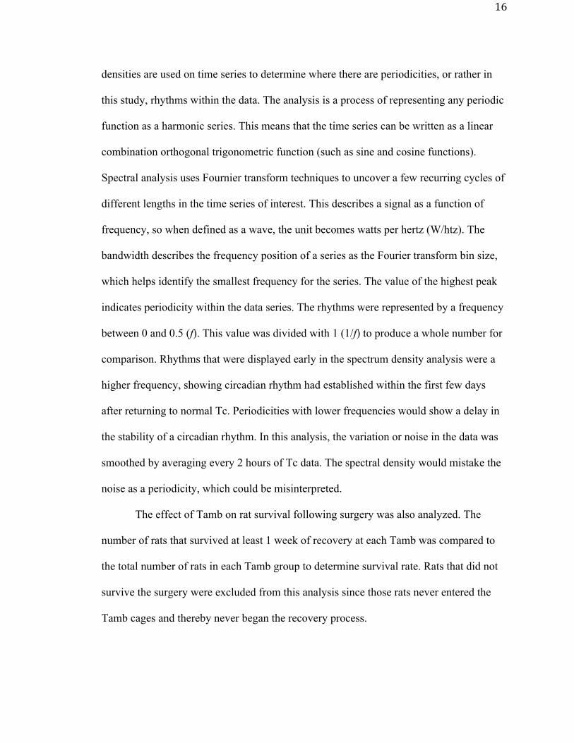
The second method to analyze circadian rhythm was a spectral density. Spectral
16
densities are used on time series to determine where there are periodicities, or rather in
this study, rhythms within the data. The analysis is a process of representing any periodic
function as a harmonic series. This means that the time series can be written as a linear
combination orthogonal trigonometric function (such as sine and cosine functions).
Spectral analysis uses Fournier transform techniques to uncover a few recurring cycles of
different lengths in the time series of interest. This describes a signal as a function of
frequency, so when defined as a wave, the unit becomes watts per hertz (W/htz). The
bandwidth describes the frequency position of a series as the Fourier transform bin size,
which helps identify the smallest frequency for the series. The value of the highest peak
indicates periodicity within the data series. The rhythms were represented by a frequency
between 0 and 0.5 (f). This value was divided with 1 (1/f) to produce a whole number for
comparison. Rhythms that were displayed early in the spectrum density analysis were a
higher frequency, showing circadian rhythm had established within the first few days
after returning to normal Tc. Periodicities with lower frequencies would show a delay in
the stability of a circadian rhythm. In this analysis, the variation or noise in the data was
smoothed by averaging every 2 hours of Tc data. The spectral density would mistake the
noise as a periodicity, which could be misinterpreted.
The effect of Tamb on rat survival following surgery was also analyzed. The
number of rats that survived at least 1 week of recovery at each Tamb was compared to
the total number of rats in each Tamb group to determine survival rate. Rats that did not
survive the surgery were excluded from this analysis since those rats never entered the
Tamb cages and thereby never began the recovery process.

17
Results
MI388 is an example of a rat from the 27°C group that returned to a normal Tc
(36.5°C -38.5°C) and displayed circadian rhythm at about 24 hours or a day after the
surgery (Figure 3). Note that after returning to 36.5°C, the Tc does not go below that
degree throughout the week. Each black box represents a dark photoperiod (nighttime),
while the unmarked areas represent the light photoperiod (daytime). The Tc rises during
the night period and decreases during the day, illustrating the circadian rhythm.
Figure 3. The Typical Tc Recovery Response. MI388 is an example of a rat housed at Tamb 27°C that returned to 36.5°C within a day and displayed circadian rhythm a day after the surgery. This rat returned to normal circadian rhythm after the second photoperiod.
Cor
e Te
mpe
ratu
re (º
C)
Elapsed Time (hr)0 24 48 72 96 120 144 168
32
33
34
35
36
37
38
39

18
Core Temperature Recovery
Figure 4 illustrates a general trend where the rats housed at Tamb 33°C return to a
normal Tc faster than the other groups whereas the 21°C group takes longer. However,
there are no significant differences between the Tamb groups. Small circles found in all
of the boxplots represent outliers in the data.
Figure 4. Return of Tc Following Surgical Procedure. Tamb 33°C returned to a normal Tc faster than the other Tamb groups versus the Tamb 21°C which took longer. (Kruskal-Wallis, p=0.6545)
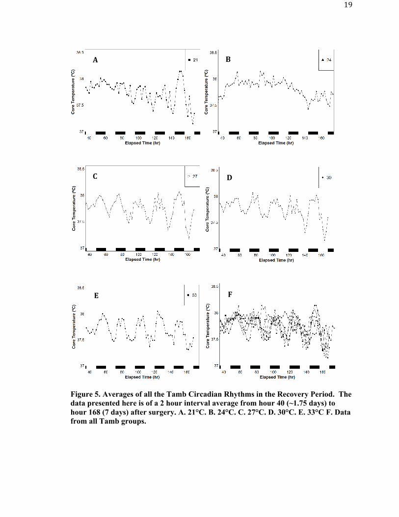
Circadian rhythm
Figure 5 is an average of all the Tc from all the Tamb groups. The data was put
into 2 hour bins to display the rhythms. The 21°C group appears to have a slight delay in
returning to circadian rhythm whereas all groups exhibit a circadian rhythm similar to
each other after 120 hours (5 days).
Retu
rn to
36.5º
C (h
r)
Ambient Temperature (ºC)21 24 27 30 33
4
8
12
16
19
Figure 5. Averages of all the Tamb Circadian Rhythms in the Recovery Period. The data presented here is of a 2 hour interval average from hour 40 (~1.75 days) to hour 168 (7 days) after surgery. A. 21°C. B. 24°C. C. 27°C. D. 30°C. E. 33°C F. Data from all Tamb groups.
A B
C D
E F
20
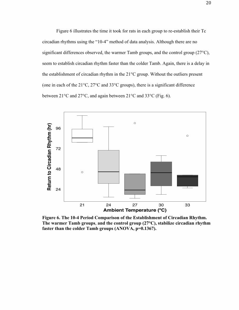
Figure 6 illustrates the time it took for rats in each group to re-establish their Tc
circadian rhythms using the “10-4” method of data analysis. Although there are no
significant differences observed, the warmer Tamb groups, and the control group (27°C),
seem to establish circadian rhythm faster than the colder Tamb. Again, there is a delay in
the establishment of circadian rhythm in the 21°C group. Without the outliers present
(one in each of the 21°C, 27°C and 33°C groups), there is a significant difference
between 21°C and 27°C, and again between 21°C and 33°C (Fig. 6).
Figure 6. The 10-4 Period Comparison of the Establishment of Circadian Rhythm. The warmer Tamb groups, and the control group (27°C), stabilize circadian rhythm faster than the colder Tamb groups (ANOVA, p=0.1367).
Retu
rn to
Circ
adian
Rhy
thm
(hr)
Ambient Temperature (ºC)21 24 27 30 33
24
48
72
96

21
The second method to determine circadian Tc rhythm recovery used spectral
density to express the data as a frequency. Figure 7A shows MI388 (Figure 3) with
smoothing applied, clearly illustrating the circadian rhythm. Figure 7B show the spectral
density with the earliest periodicity before 0.1. The larger the percentage, the sooner the
circadian rhythm was established. Figure 8 displays the intergroup comparisons of all the
rats. The results there are similar to Figure 6 which used the “10-4” method to analyze
circadian rhythm recovery. The 21°C group had the smallest percentage, meaning the
periodicity was not detected until later in the week.

22
Figure 7. A. MI388 2 Hour Average. This illustrates the circadian rhythm and reduces the variation. B. Spectral Density of MI388. The highest spectrum dictates early periodicity of circadian rhythm establishment.
Core
Tem
pera
ture
(ºC)
Time (hr)20 40 60 80 100 120 140 160
37
37.2
37.4
37.6
37.8
38
38.2
bandwidth = 0.0139
Spec
trum
(W/h
tz)
Frequency0.0 0.1 0.2 0.3 0.4 0.5
0.005
0.020
0.100
0.500
A
B

23
Figure 8. Spectral Density Comparison of Establishing Circadian Rhythm. The 21°C group has a delay in the return of circadian rhythm while the 27°C group established circadian Tc rhythms the soonest. (Kruskal-Wallis, p=0.3227) Body Weight The percent changes in rat’s body weight are illustrated in Figure 9. There was no
significant difference between the groups despite a general trend that the 21°C group
seemed to lose more weight and recover slower than the other groups while the 27°C
group seemed to recover the quickest. If the change in weight from one individual day to
the next is compared across the groups, then the rats housed at 21°C did lose significantly
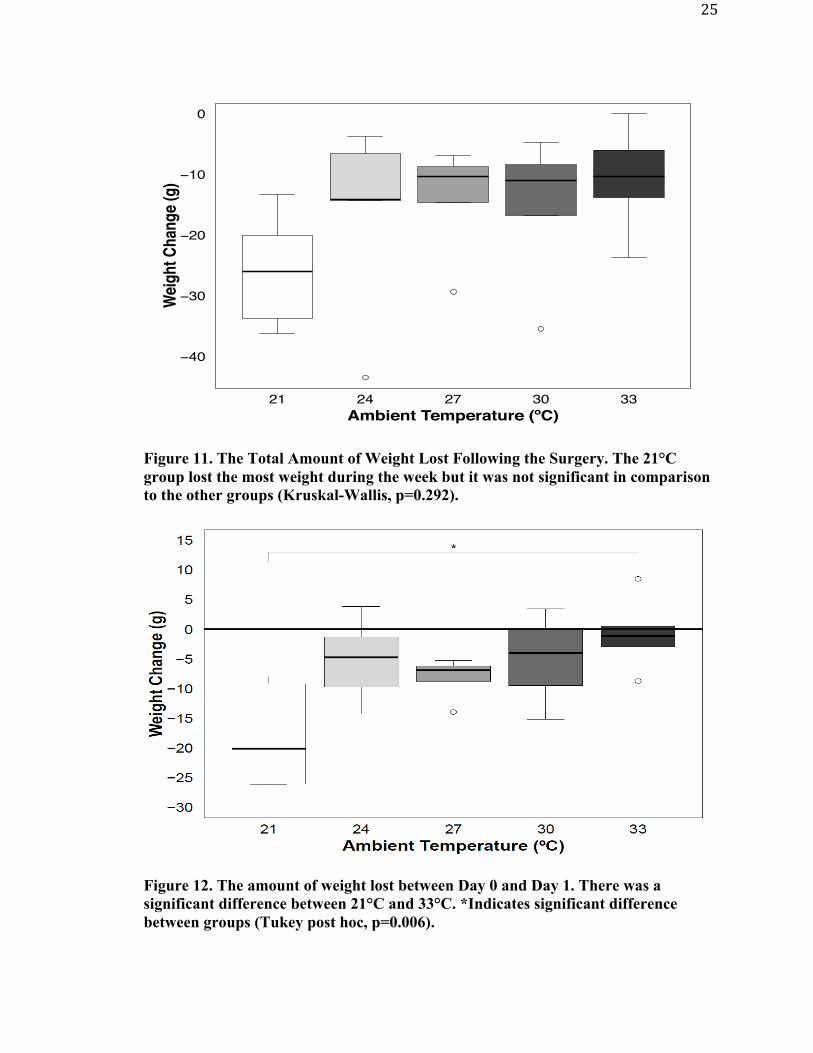
more weight on day one when compared to rats housed at 33°C (Figure 10 and 12). When
total weight lost over the entire week is analyzed (Figure 11) the 21°C group seemed to
lose the most weight but it was not significantly different than the other groups.
Freq
uenc
y (%
)
Ambient Temperature (ºC)21 24 27 30 33
4
6
8
10
12
14
16

24
Figure 9. The Percent of Original Body Weight Lost in Each Tamb Group During the Recovery Week. Data presented as mean. N=5 in each group.
Figure 10. The Average Weight Change Per Day Following Surgery. 21°C lost the most weight on Day 1 but regained that weight back around Day 4. *Significant difference between 21°C and 33°C on Day 1 (Tukey post hoc, p=0.006).
Perc
ent W
eigh
t (%
)
Time (Day)Pre−Surgery 0 1 2 3 4 5 6 7
91
94
97
100
103
106
21
24
27
30
33
25
Figure 11. The Total Amount of Weight Lost Following the Surgery. The 21°C group lost the most weight during the week but it was not significant in comparison to the other groups (Kruskal-Wallis, p=0.292).
Figure 12. The amount of weight lost between Day 0 and Day 1. There was a significant difference between 21°C and 33°C. *Indicates significant difference between groups (Tukey post hoc, p=0.006).
Wei
ght C
hang
e (g
)
Ambient Temperature (ºC)21 24 27 30 33
−40
−30
−20
−10
0

26
Finally, the body weight recovery between Days 1, 3 and 7 was analyzed within
and between groups. Day 1 followed the surgery. Day 3 was the period in which either
the weights had begun to stabilize or show minimal change. Day 7 was the final
measurement when most rats had returned to their pre-surgical weight. There were no
differences of the daily weight change between the Tamb groups, but there were
differences within the select Tamb groups on Day 1 and 7. Specifically, in the 21°C and
27°C groups the weight change on Day 1 was significantly different than the weight
change on Day 7 (Figure 13).
Figure 13. The Amount of Weight Change of Days 1, 3, and 7 Between the Tamb Groups. There were no differences between the groups, only between days on individual groups. The differences were between Day 1 and 7 of Tamb 21°C and Day 1 and 7 of Tamb 27°C. *Indicates significant difference between days (Nemenyi post hoc, 21°C p=0.023; 27°C p=0.02).

27
Food and Water Consumption
Food and water were measured daily following the surgery. Figure 14A represents
the averages of food consumed for each Tamb group. Between Day 1 and Day 2, the
24°C group ate the most food which then stabilized later in the week. In contrast, the
warmer groups ate less food, especially later in the week. In the total amount of food
consumed during the week (Figure 14B), there were no significant differences but it
seems the colder groups ate more food versus the warmer Tambs. Within the 21°C group
(Figure 15), there was a significant difference between Day 1 and Day 7 food
consumption, but there were no other differences observed in the other Tambs. There
were no differences between the Tamb groups.

28
Figure 14. A. The Averages of the Food Intake Following Surgery Over 7 Days. B. The total amount of food consumed over the week (Kruskal-Wallis, p=0.098).
21
24
27
30
33
Food
Inta
ke (g
)
Time (Day)Pre−Surgery 0 1 2 3 4 5 6 7
0
5
10
15
20Fo
od C
onsu
med
(g)
Ambient Temperature (ºC)21 24 27 30 33
80
100
120
140
160 B
A

29
Figure 15. The amount of food consumed between Day 1, 3, and 7. The only difference observed was between Day 1 and Day 7 of the 21°C group. *Indicates significant difference between groups (Tukey post hoc, p=0.02).
Water consumption shares a similar trend between the groups. The amount of
water consumed increases throughout the week (Figure 16A). The warmer groups (30°C
and 33°C) as well as the 27°C group, all seem to have an increased water consumption in
comparison to the colder Tambs despite the lack of significance in the data (Figure 16B).
The 27°C group has significant increases in water intake between Day 1 and both Day 3
and Day 7. The rats in the 30°C and 33°C groups had significant increases in water
consumption between Days 1 and 7 (Figure 17).

30
Figure 16. A. The averages of water consumption following surgery over 7 days. B. The total amount of water consumed over the week (Kruskal-Wallis, p=0.168).
21
24
27
30
33
Wat
er In
take
(g)
Time (Day)Pre−Surgery 0 1 2 3 4 5 6 7
0
10
20
30
40
50
60
Wat
er C
onsu
med
(g)
Ambient Temperature (ºC)21 24 27 30 33
200
250
300
350
400
A
B

31
Figure 17. The Amount of Water Consumed Between Day 1, 3, and 7. Differences were not observed between the Tamb groups. The 27°C group had differences between Day 1 and Day 3, and Day 1 and Day 7. The 30°C groups only had differences between Day 1 and Day 7, while 33°C had differences between Day 1 and Day 3, and Day 1 and Day 7. *Indicates significant difference between days (Tukey post hoc, 27°C: p=0.041, 0.011; 30°C, p=0.019; 33°C, p=0.039, 0.004). Survival A survival percentage was calculated for each experimental group. The 21°C and
30°C groups have the lowest survival rate with 71.43% whereas the 24°C, 27°C, and
33°C had higher survival rates of 83.33%.
Table 2. The survival percentage of the Tamb groups.
Tamb (C°) Survived 24 hours Total
surgeries % 21 5 7 71.43% 24 5 6 83.33% 27 5 6 83.33% 30 5 7 71.43% 33 5 6 83.33%
32
Discussion
Core Temperature Recovery
An important aspect of recovery from a surgical procedure is the return of Tc to
normal. With anesthesia, the blood vessels vasodilate and the hypothalamus is unable to
thermoregulate, causing heat to leave the deeper tissues and travel to the skin (Diaz &
Becker, 2010). When returning to normal Tc, the hypothalamus of rats again begins to
control thermoregulation and stimulates brown adipose tissue to generate heat using non-
shivering thermogenesis. In this study, we stated that Tc had returned to normal when the
rats Tc had reached 36.5°C because this temperature is at the lower extremity of a normal
Tc range for this strain of rat. It is important to note that while recording Tc during
recovery the Tc was above 36.5°C throughout the week, supporting the use of 36.5°C as
the demarcation point for a return to normal Tc. Interestingly, there were no differences
observed between the groups in their Tc return to 36.5°C (Figure 4). Noticeably, the 21°C
group has a much wider variation or spread compared to the other Tamb groups. The
addition of more animals may reduce this variation and perhaps show significance
between the 21°C group and others. It was expected that the rats in the 21°C group would
have difficulty returning to their normal Tc because within a colder environment, the
effects of anesthesia causing vasodilation are more drastic. Therefore, more blood is
moved to the periphery and consequently environmental heat loss is increased. Without
the aid of external heating, such as heating air-systems, this thermoregulatory impairment
can complicate recovery of a normal Tc (Diaz & Becker, 2010). The use of external heat
warmers during recovery is very helpful. Although not significant, this is shown by the
33°C group in Figure 4, having recovered the Tc faster than the rest of the group. With

33
additional animals included in the study, there could be a significant difference seen
between 21°C and 33°C groups.
Circadian Rhythm
Establishing a circadian rhythm is essential to showing normal physiologic
behavior. Not only is this rhythm seen in Tc, it is also present in physiological processes
such as motor activity, corticosterone levels, and liver metabolism. The advantage of
having circadian rhythms is being able to anticipate environmental changes and manage
our physiological processes to them (Narasimamurthy & Virshup, 2017). Experiments
that do not account for disrupted rhythms could inadvertently add another variable that
affects the data. The 10-4 periods show differences between the daytime and night time
Tc levels and thereby determine the establishment of circadian rhythm. Using this
method to analyze the data, the establishment of a circadian rhythm after surgery was
mildly delayed in the 21°C group. Although this data was not significantly different, the
delay in the 21°C group recovery could be concerning for housing SD rats at room
temperatures, which are comfortable to those caring for the rats but lead to alterations in
thermoregulatory function following surgery. This suggests the recovery process can take
longer than anticipated in these conditions. If the Tc remains unstable, then data gathered
from these animals may be misleading. The spectral density method yielded similar
results to the 10-4 periods, including a delay in the establishment of circadian rhythm in
the 21°C group. It is important to note that both the 10-4 periods and the spectral density
methods show there are negative effects when housing animals within 21°C.
In contrast to the 21°C, the warmer groups and those housed at 27°C, establish a
circadian rhythm within the first few days of recovery. This is essential to this study

34
because the 27°C Tamb is the preferred Tamb of SD rats (Brown, 2011). Without the one
outlier that is present in the 21°C data, the establishment of the circadian rhythm in this
group would be significantly delayed when compared to the 27°C and 33°C groups.
Therefore, it is essential that the current sample size (n=5/group) be increased. This
should elucidate these differences and support raising the Tamb within the first days of
recovery around 33°C and then decreasing the Tamb to 27°C. It was interesting to see
that there was a mild delay in the establishment of circadian rhythm in the 30°C and 33°C
groups, although not as delayed as the 21°C Tamb. Since these Tambs are at and above
the upper range of the preferred Tamb, these warmer temperatures are probably causing a
heat stress later in the recovery week. This likely increases their evaporative water loss
and results in an increase in their water intake seen in Figures 16 and 17 (Gordon, 1993).
Alternatively, the delay in establishment of a normal circadian rhythm could have been
caused by a period of acclimation to the warmer temperatures. However, further research
is needed to determine if acclimation does have an influence on circadian rhythms in the
warmer Tamb groups.
Weight, Food, and Water Measurements
Weight changes following surgery are often measured to ensure the rat is healthy.
This is considered to be more beneficial information because normal growth rates
following surgery can be hindered by numerous factors such as external stressors,
postoperative pain, and pain management drugs (Brennan et al., 2009). These stressors
influence the return of weight to normal pre-surgical levels. Only a few rats did not
regain their original body weight by Day 7. Thefewratsthatfailedtoregaintheirpre-
surgicalbodyweightdidnotaffecttheoveralldatatrends. The cold stress from

35
housing at 21°C caused a greater decrease in body weight compared to other groups. This
was evidenced by the difference between Day 1 weight loss between 21°C and 33°C
(Figure 10 & 12). This was likely due to an increase in metabolic rate and subsequent
loss in body weight following the surgical procedure. Metabolic rate is known to increase
when animals are maintained at Tamb below their TNZ. In an experiment conducted by
Wang et al. (2010), SD rats housed in a cold stress environment showed decreasing body
weight throughout the experiment, but in contrast, there was an increase in brown adipose
tissue, the tissue essential for non-shivering thermogenesis. Their findings suggest the
cold stress increased metabolic rate, leading to a dependence on non-shivering
thermogenesis to produce heat. Increasing food consumption would be required to meet
the now higher metabolic demands on the animal to both recover from surgery and
maintain an elevated metabolic rate. Rats housed at 21°C seemed to have increased food
consumption earlier in the recovery week, which supports this conclusion (Figure 14A).
However, once the animal adapted to the colder environment the food consumption
difference between the 21°C group and the others were negligible. They did lose the most
weight on Day 1 but they regained that weight quickly, which reduced the significance of
the total weight lost for the week. Furthermore, rats housed at 33°C quickly recovered
their Tc (Figure 4) and therefore did not require excessive food consumption to support
metabolic heat production early in the week (Figure 14). There is also more variation in
the weight loss of Day 1 in the 21°C group, whereas the 33°C group is more stabilized,
showing similar weight loss. Later in the week those rats seemed to not need to consume
as much food as the other groups. However, more animals are needed in the study to
confirm the conclusions.

36
Measuring food and water intake can be helpful in determining physiological
changes in lab animals. For example, in a study conducted by Sharp et al. (2003)
analyzing the effects of different analgesics following a major abdominal surgery, the
rat’s water consumption returned to normal within 5 days; however, food consumption
was dependent on the analgesic that was used during recovery. The recovery of normal
pre-surgical food intake varied between 5 and 12 days. Another study investigated the
effects of chronic stress and found evidence of a relationship between food intake and
body weight changes. With the increased duration and intensity of the stressor, food
intake and body weight gain would decrease from chronic restraint and immobilization
stressors (Marti et al., 1993).
Water intake is another measurement commonly used to identify health issues in
laboratory animals. Rats lose a large amount of fluid during a surgery from the
anesthesia’s effect on bladder control and evaporative water loss from exposing body
cavities during surgery. This is why saline is given following the surgery to replace the
fluid lost (Institute for Laboratory Animal Research, 2011). Furthermore, animals housed
in warmer Tamb environments were expected to consume more water to offset
evaporative water lost to the warmer environment. In fact, the warmer Tamb groups and
the control group (27°C) demonstrated a mild increase in water consumption compared to
cooler Tamb groups (Figure 16). This was especially true as the week progressed. This is
supported by the significant increase in water consumption between both Days 1 and 3
and 1 and 7 in the 27ºC group. Similar trends of increasing water consumption as the
recovery week progressed can be seen in the 30ºC and the 33ºC groups as well. These
trends are not as apparent in the cooler Tamb groups. This supports the idea that when

37
housed at warmer Tamb, the rats consume more water to replace the extra fluid lost via
evaporation from the skin and the respiratory tract to the environment.
Survival
Table 2 illustrates the survival rate of the rats that were included in the study. The
table shows that 21°C and 30°C groups had the lowest survival rate (71.43%) and 24°C,
27°C, and 33°C had the highest (83.33%). With further experiments, we expect the
percentages to change within some of these groups. From the previous data that was
gathered prior to the decontamination of the vivarium, 21°C showed a much lower
percentage, less than 50%. With further experiments, we also believe that the current
21°C group will decrease. Most likely, the cold stress environment increases metabolic
activity required to produce the required body heat to thermoregulate. Caloric intake
would have to increase to support this now increased metabolism. This is supported by
the great loss of body weight, and an increased food intake in those rats. It is interesting
to see that the 33°C survival rate is high, given that this Tamb is considered a mild heat
stress. If the recovery period was extended from 1 week to 2 weeks, there could be
destabilization of circadian rhythm, indicating a stressed rat. This may decrease
survivability. Previous attempts of housing rats in 36°C following surgery was
problematic and discontinued because of the low survival rate from the increased heat
rate. With an increased recovery period duration, we could see a decrease in the 33°C
survival rate. Further experiments are needed to determine the effects of Tamb on
survival rate.

38
Conclusion The goal of this experiment was to investigate the ideal Tamb in which SD rats
could recover following a major surgical procedure. The return to normal Tc and the
reestablishment of circadian rhythm were the main criteria for showing normal
physiologic characteristics as well as recovery of body weight, and normalization of food
and water consumption. The 33°C group returned to normal Tc the fastest while 27°C
established circadian rhythm within the first day after surgery. Food and water
consumption were also likely affected by Tamb after surgery. Rats housed in a cold
Tamb required more food intake to meet increase metabolic demands to maintain body
heat early in the week. After acclimation however, these trends dissipated. The
alterations in food consumption likely influenced the recovery of body weight to pre-
surgical levels in that the 21ºC group seemed to lose the most weight immediately after
surgery but recovered that weight quickly. Also, rats housed at 27ºC and above seemed to
require more fluid intake throughout the recovery week. This was likely done to offset
evaporative water loss at higher Tamb.
These data suggest the use of mild external heating will facilitate surgical
recovery in SD rats if that elevation in Tamb is limited to only a few days. Once the Tc
has returned to a normal level, body weight has recovered, and feeding and drinking
behaviors have been re-established, then the Tamb should be reduced to ~27°C to help
establish and maintain a circadian rhythm. Housing animals at a Tamb that is comfortable
to research staff (21°C) can be harmful. These data suggest housing at that Tamb has a
negative effect on surgical recovery. There is a delayed return to normal Tc and
establishment of circadian rhythm when SD rats are housed at 21°C. The delayed

39
establishment of circadian rhythm, along with the weight loss, could confound data
interpretation from these animals.
An important aspect to note in this study is the small sample size (n=5/group).
With further experiments, an increased sample size should help elucidate significant
differences between groups. There are many outliers affecting the data of the 21°C group
in particular. When removed, the data show significant differences between the Tamb
groups. There are trends seen within the figures that illustrate what was hypothesized.
Further research is needed to fully determine the appropriate housing during surgical
recovery.

40
References
Bicego, K. C., Barros, R.C.H., & Branco, L.G.S. (2007). Physiology of temperature regulation: Comparative aspects. Comparative Biochemistry and Physiology, Vol 147, 616-639. Brennan, M.P., Sinusas, A.J., Horvath, T.L., Collins, J.G., and Harding, M.J. (2009).
Correlation between body weight changes and postoperative pain in rats treated with meloxicam or buprenorphine. Lab animal. 38(3):87-93.
Brown, J.W., Le, N.M. (2011). The effect of thermopreference on circadian thermoregulation in Sprague-Dawley and Fisher 344 rats. Journal of Thermal Biology 37(4):309-15 Cannon, B., & Nedergaard, J. (2011). Nonshivering thermogenesis and its adequate measurement in metabolic studies. Journal of Experimental Biology, Vol 214, 242-253. David, J. M., Chatziioannou, A. F., Taschereau, R., Wang, H., & Stout, D. B. (2013). The hidden cost of housing practices: using noninvasive imaging to quantify the metabolic demands of chronic cold stress of laboratory mice. Comparative Medicine, 63(5), 386-391. Diaz, M., & Becker, D. E. (2010). Thermoregulation: physiological and clinical considerations during sedation and general anesthesia. American Dental Society of Anesthesiology, Vol 57, 25-33. Gordon, C. J. (1993). Temperature regulation in laboratory animals. New York: Cambridge University Press. Gonder, J. C., & Laber, K. (2007). A renewed look at laboratory rodent housing and management. ILAR Journal, Vol 48(1), 29-36. Horvath, G., Kekesi, G., Petrovszki, Z., & Benedek, G. (2015). Abnormal motor activity and thermoregulation in a schizophrenia rat model for translational science. PLoS ONE, 10(12), e0143751. Institute for Laboratory Animal Research (2011). Guide for the care and use of laboratory animals, 8th ed. Washington (DC): National Academies Press. Leon, L. R., DuBose, D. A., & Mason, C. W. (2005) Heat stress induces a biphasic thermoregulatory response in mice. American Journal of Physiology. 288(1) R197-R204.
41
Marti, O., Marti, J., and Armario, A. (1993). Effects of chronic stress on food intake in rats: influence of stressor intensity and duration of daily exposure. Physiology and Behavior. 55(4), 747-753. Narasimamurthy, R., and Virshup, D. M. (2017). Molecular mechanism regulating temperature compensation of the circadian clock. Frontiers in Neurology. 8(161), 1-5. Sharp, J., Zammit, T., Azar, T., and Lawson, D. (2003). Recovery of male rats from major abdominal surgery after treatment with various analgesics. Contemporary Topics . 42(6), 22-27. Wang, X., Che, H., Zhang, W., Wang, J., Ke, T., Cao, R., … Luo, W. (2015). Effects of mild chronic intermittent cold exposure on rat organs. International Journal of Biological Sciences, 11(10), 1171–1180. Zylan, K. D., & Carlisle, H. J. (1992). Effect of ambient temperature on the paradoxical metabolic reponses to norepinephrine. Pharmacology Biochemistry and Behavior, Vol 43, 577-582.