The effect of a knee ankle foot orthosis on lower limb ...usir.salford.ac.uk/35587/1/my thesis_alst...
189
The effect of a knee ankle foot orthosis on lower limb kinematics and kinetics in an individual with varus knee alignment Huda Al-fatafta School of Health Sciences University of Salford, Salford, UK Submitted in Partial Fulfilment of the Requirements of the Degree of Master of Science, March 2015
Transcript of The effect of a knee ankle foot orthosis on lower limb ...usir.salford.ac.uk/35587/1/my thesis_alst...
The effect of a knee ankle foot orthosis on lower limb
kinematics and kinetics in an individual with varus knee
alignment
Huda Al-fatafta
School of Health Sciences
University of Salford, Salford, UK
Submitted in Partial Fulfilment of the Requirements of the
Degree of Master of Science, March 2015

i
Contents
Contents ........................................................................................................................................... i
Table of Figures ............................................................................................................................. vi
List of Tables .................................................................................................................................. x
Acknowledgments............................................................................................................................ i
Abstract ........................................................................................................................................... ii
Chapter one: Literature review ................................................................................................... 1
1.1. Definition of Osteoarthritis .................................................................................................. 1
1.2. Epidemiology of Osteoarthritis ............................................................................................ 1
1.3. OA classification .................................................................................................................. 2
1.4. Sites of OA ........................................................................................................................... 2
1.5. Risk factors for OA .................................................................................................................. 3
1.5.1 Systemic factors ................................................................................................................. 4
1.5.1.1 Age............................................................................................................................... 4
1.5.1.2 Gender differences ....................................................................................................... 4
1.5.1.3 Ethnicity ....................................................................................................................... 5
1.5.1.4 Diet and nutrition ......................................................................................................... 5
1.5.1.5 Genetic factors ............................................................................................................. 6
1.5.2 Local biomechanical factors............................................................................................... 6
1.5.2.1 Muscle weakness ......................................................................................................... 6
1.5.2.2 Body mass index (BMI) and obesity ........................................................................... 6
1.5.2.3 Joint angle malalignment ............................................................................................. 7
1.5.2.4 Joint laxity and Trauma................................................................................................ 9
1.6. Knee OA ................................................................................................................................ 10
1.6.1 Prevalence of knee OA ..................................................................................................... 11
1.6.2 Clinical and Radiological criteria of knee OA ................................................................. 12
ii
1.6.2.1 The ACR criteria ....................................................................................................... 12
1.6.2.2 The Kellgren and Lawrence radiological grade ........................................................ 13
1.7. The biomechanics of walking and stair climbing .................................................................. 14
1.7.1 Walking ............................................................................................................................ 14
1.7.2 Stair climbing ................................................................................................................... 16
1.8. Gait in individuals with medial compartment knee OA ........................................................ 18
1.8.1 Temporal-spatial parameters ............................................................................................ 19
1.8.2 Kinematics ........................................................................................................................ 19
1.8.3 Kinetics............................................................................................................................. 21
1.8.3.1 Sagittal plane ............................................................................................................. 21
1.8.3.2 Frontal plane .............................................................................................................. 22
1.9. Gait adaptation strategies to reduce EKAM .......................................................................... 25
1.9.1 Lateral trunk bending ....................................................................................................... 26
1.9.2 Reducing speed ................................................................................................................ 26
1.9.3 Toe-out/toe-in walking ..................................................................................................... 27
1.10. Osteoarthritis treatments ...................................................................................................... 29
1.10.1 Surgical intervention ...................................................................................................... 29
1.10.2 Pharmacology treatments ............................................................................................... 31
1.10.3 Conservative non-pharmacology treatments .................................................................. 32
1.10.3.1 Education ................................................................................................................. 32
1.10.3.2 Physical therapy and Exercise ................................................................................. 32
1.10.3.3 Footwear and Orthoses ............................................................................................ 33
Chapter two: Methods ................................................................................................................ 46
2.1. Measurement system .............................................................................................................. 47
2.1.1 System calibration ............................................................................................................ 48
2.1.2 Reflective markers............................................................................................................ 49
iii
2.1.3 Subject procedures and calibration .................................................................................. 50
2.2. Data processing (Reduction) .................................................................................................. 51
2.2.1 Digitising and Labeling .................................................................................................... 51
2.2.2 Modeling .......................................................................................................................... 52
2.2.3 Data processing ................................................................................................................ 54
2.2.4 Joint angle and joint moment calculations ....................................................................... 56
2.3. Data analysis .......................................................................................................................... 57
Chapter three: Reliability of knee kinematics and kinetics using an interlaced stairway....53
3.1. Introduction ............................................................................................................................ 58
3. 2. Literature review ................................................................................................................... 59
3.3. Method ................................................................................................................................... 63
3.3.1 Subjects ............................................................................................................................ 63
3.3.2 Test procedures ................................................................................................................ 64
3.3.3 Outcome measures ........................................................................................................... 64
3.3.4 Statistics ........................................................................................................................... 66
3.4. Results .................................................................................................................................... 66
3.4.1 Primary outcomes ............................................................................................................. 67
3.4.1.1 Knee joint kinematics during ascending and descending .......................................... 67
3.4.1.2 Knee joint kinetics during ascending and descending ............................................... 70
3.4.1.3 Vertical ground reaction force result (VGRF) ........................................................... 72
3.4.1.4 Speed and stance time................................................................................................ 74
3.4.2 Secondary outcomes ............................................................................................................ 75
3.4.2.1 Ankle joint kinematics during stairs ascending and descending ............................... 75
3.5 Discussion ............................................................................................................................... 76
3.5.1 Discussion ........................................................................................................................ 76
3.5.2 Limitation ......................................................................................................................... 77
3.5.3 Future work ...................................................................................................................... 78
iv
3.5.4 Conclusion ........................................................................................................................ 78
Chapter four: Effect of a KAFO on kinematics and kinetics of the lower limb....................74
4.1. Test procedures ...................................................................................................................... 79
4.1.1 Participant......................................................................................................................... 79
4.1.2 Clinical examination ........................................................................................................ 79
4.1.3 Casting .............................................................................................................................. 80
4.1.4 Modifications ................................................................................................................... 81
4.1.4.1 Ossur knee valgus braces ........................................................................................... 85
4.2. Data collection ....................................................................................................................... 87
4.2.1 Walking ............................................................................................................................ 87
4.2.2 Ascending and Descending .............................................................................................. 88
4.3. Outcome measures ................................................................................................................. 90
4.3.1 Statistics ........................................................................................................................... 91
4.4. Results .................................................................................................................................... 91
4.4.1 Temporal and Spatial results ............................................................................................ 91
4.4.2 GRF results ....................................................................................................................... 92
4.4.3 Kinematics of the knee joint............................................................................................. 94
4.4.3.1 Walking ..................................................................................................................... 94
4.4.3.2 Stair ascent and descent ............................................................................................. 95
4.4. Knee kinetics in the frontal plane ........................................................................................ 100
4.4.1 Walking .......................................................................................................................... 100
4.4.2 Stair ascent and descent.................................................................................................. 100
4.5. Knee adduction angular impulse (KAAI) ............................................................................ 103
4.6. Knee kinetics in sagittal plane ............................................................................................. 103
4.7. Hip kinematics in sagittal and frontal planes ....................................................................... 106
4.7.1 Walking .......................................................................................................................... 106
4.7.2 Stair ascent and descent.................................................................................................. 106

v
4.8. Hip kinetics .......................................................................................................................... 111
4.8.1 Frontal plane ................................................................................................................... 111
4.8.2 Sagittal plane .................................................................................................................. 113
4.9. Ankle kinematics (Secondary outcomes)............................................................................. 115
4.9.1 Walking .......................................................................................................................... 115
4.9.2 Stair ascent and descent.................................................................................................. 115
Chapter five: Discussion ........................................................................................................... 120
5.1. Discussion ............................................................................................................................ 120
5.1.1 Background of discussion .............................................................................................. 120
5.1.2 Summary of results......................................................................................................... 120
5.1.3 Hypotheses ..................................................................................................................... 121
5.2.2.1 External knee adduction moment (EKAM)…………………………..……………116
5.2.2.2 Lower limb kinematics…………………………………………………………….121
5.3. Limitations ........................................................................................................................... 131
5.2. Future work .......................................................................................................................... 132
5.4. Conclusion ........................................................................................................................... 133
Chapter 6: Appendices ................................................................................................................ 134
Appendix 6 (A): Research Governance & Ethics Committee approval letter. ........................... 134
Appendix 6 (B): Demographic and examination sheet. .............................................................. 135
Appendix 6 (C): The study poster. .............................................................................................. 136
Appendix 6 (D): Consent Form. ................................................................................................. 137
Appendix 6 (E): Abbreviations....................................................................................................138
References ................................................................................................................................... 138
vi
Table of Figures
Chapter one:
Figure 1.1: Effects of age and gender on specific joints incidence of osteoarthritis, Knee joint is
the most affected joint than hip joint, especially within females than males group. ...................... 3
Figure 1.2: Mechanical axis alignment to evaluate varus and valgus deformity ............................ 9
Figure 1.3: The Grades of knee OA ............................................................................................. 14
Figure 1.4: Typical gait cycle for healthy knee individuals during walking ............................... 16
Figure 1.5: Ascending and descending gait cycles . ..................................................................... 18
Figure 1.6: KAAI which is the highlighted area under the graph . ............................................... 23
Figure 1.7: EKAM has 2 peaks and it is higher for OA group more than control group . ........... 24
Figure 1.8: Hip adduction and abduction moment for knee OA and control individuals ............ 25
Figure 1.9: The effects of toe-out gait during walking (a), stair ascent (b), and descent (c) . ...... 28
Figure 1.10: (a) full length insole, (b) a heel wedge ..................................................................... 35
Figure 1.11: The three-point pressure system applied by knee unloader braces ......................... 38
Figure 1.12: EKAM after using knee valgus brace during walking (a) and stairs stepping
(b).LLL: lower limb length .......................................................................................................... 40
Figure 1.13: Lateral bending toward the stance phase side is associated with using knee valgus
brace . ............................................................................................................................................ 40
Figure 1.14: Difference in length between a knee brace and a KAFO. ........................................ 44
Chapter two:
Figure 2.1: Stairway design with two platforms to collect the kinetics data (a): screws for fixation
(b). ................................................................................................................................................. 47
Figure 2.2: System calibration process. Red is positive X axis, green arrow: positive Y axis, blue
arrow: positive Z axis (a). L-shaped metallic structure (b), wand (c), image space (d). .............. 48
Figure 2.3: Marker reflect light back to the cameras (a), passive reflective marker (b), and cluster
marker (c). ..................................................................................................................................... 49
Figure 2.4: Location of the markers and clusters. ......................................................................... 51
vii
Figure 2.5: Modeling process. Labelling (a), modelling (b), joints' centres of motion (yellow
balls) (c). ....................................................................................................................................... 51
Figure 2.6: AMTI stairway force structures. ……………………………………………………52
Figure 2.7: Ascending gait cycle of left leg from first step to the third step. ............................... 53
Figure 2.8: Descending gait cycle for the right leg from second step until the floor. .................. 56
Chapter three:
Figure 3.1: Representation of data for sagittal knee angle (a), frontal knee angle (b), sagittal knee
moment (c), frontal knee moment (Nm/kg) and the shaded area represents the KAAI
(Nm/kg*s)(d)…………………………………………………………………………………….62
Figure 3.2: A typical vertical GRF plot…………………………………………………………63
Figure 3.3: Knee angle in two session during stair ascent (sagittal plane (a), frontal plane
(b))………………………………………………………………………………………………..64
Figure 3.4: Knee angle in two sessions during stair descent (sagittal plane (a), frontal plane (b)).
....................................................................................................................................................... 68
Figure 3.5: Averaged knee moment moments for the two sessions during stair ascent (sagittal
plane (a), frontal plane (b)).. ......................................................................................................... 70
Figure 3.6: Averaged knee moments for the two sessions during stair descent (sagittal plane (a),
frontal plane (b))……………………………………………………………………………..….68
Figure 3.7: Averaged GRF data for the two sessions during ascending (a) and descending (b)...70
Chapter four:
Figure 4.1: Casting the left side to take a negative model for participant's size. .......................... 81
Figure 4.2: The cast modification process. A four point pressure system was used to apply a
valgus correction force with long lever arms to make wearing the orthoses more comfortable (a),
modification of the medial arch (b), and removal of excessive plaster around the Achilles tendon
(c)…………………………………………………………………………………..……………79
Figure 4.3: KAFO design includes single side bar (a), and Ottobock free offset knee joint (b, c).
....................................................................................................................................................... 80

viii
Figure 4.4: KAFO design. Plastics are vacuumed over the lower parts (a). Final model of KAFO
with ankle joint (b). ....................................................................................................................... 82
Figure 4.5: Mechanism of Unloader One knee valgus brace is to transfer the load to unaffected
side. ............................................................................................................................................... 86
Figure 4.6: Control condition (a), off-the-shelf Ossur Unloader One knee valgus brace (b),
custom made Ossur Unloader One knee valgus brace (c), and KAFO (d). .................................. 87
Figure 4.7: Ascending gait cycle for left leg starts with right leg of floor. (a), left side touches the
first step (b). Right leg on the second step (c) and left side touches the third step (end of gait
cycle) (d).. ..................................................................................................................................... 88
Figure 4.8: Descending gait cycle for left side. Left leg starts descending (a), right leg on first
step (b), and gait cycle ends when the left side touches the floor (c)…………………………...86
Figure 4.9: Data transferred for sagittal hip moment (a), frontal hip moment (b), single limb
support (c) . ................................................................................................................................... 91
Figure 4.10: Sagittal hip moment (a), frontal hip moment (b), and single limb support (c)........88
Figure 4.11: Knee angles in sagittal plane during walking (a), stair ascent (b), and stair descent
(c) for the four conditions. ............................................................................................................ 97
Figure 4.12: Knee angles in frontal plane during walking (a), stair ascent (b), and stair descent
(c) for the four conditions. ............................................................................................................ 97
Figure 4.13: EKAM during walking (a), stair ascent (b), and stair descent (c) for the four
conditions. ................................................................................................................................... 101
Figure 4.14: Knee KAAI for the four conditions (error bars denote 1SD) during walking (a), stair
ascent (b), and stair descent (c). .................................................................................................. 100
Figure 4.15: Knee moment in sagittal plane during walking (a), stair ascent (b), and stair descent
(c) for the four conditions. .......................................................................................................... 101
Figure 4.16: Hip sagittal plane angles during walking (a), stair ascent (b), and stair descent (c)
for the four conditions. ................................................................................................................ 108
Figure 4.17: Hip frontal plane angle during walking (a), stair ascent (b), and stair descent (c) for
the four conditions. ..................................................................................................................... 108
Figure 4.18: Hip moment in frontal plane during walking (a), stair ascent (b), and stair descent
(c) for the four conditions. .......................................................................................................... 111

ix
Figure 4.19: Hip moment in sagittal plane during walking (a), stair ascent (b), and stair descent
(c) for the four conditions. .......................................................................................................... 113
Figure 4.20: Ankle angles in sagittal plane during walking (a), stair ascent (b), and stair descent
(c) for the four conditions. .......................................................................................................... 117
Figure 4.21: Ankle angles in frontal plane during walking (a), stair ascent (b), and stair descent
(c) for the four conditions. .......................................................................................................... 117
x
List of Tables
Chapter three:
Table 3.1: Coefficient of variation for angles and moments. ....................................................... 61
Table 3.2: Inter subject correlation coefficient (r) during stair ascent (a), and descent (b) … ..... 62
Table 3.3: Reliability knee joint kinematics variables. ................................................................. 69
Table 3.4: RMSD between two sessions....................................................................................... 70
Table 3.5: Reliability of knee joint kinetics variables. ................................................................. 72
Table 3.6: VGRF reliabilkity during ascending and descending. ................................................. 70
Table 3.7: Speed and stance time reliability during ascending and descending. .......................... 71
Table 3.8: Ankle joint reliability during ascending and descending…………………………….72
Chapter four:
Table 4.1: KAFO and knee valgus braces features........................................................................83
Table 4.2: Stance time between conditions....................................................................................89
Table 4.3: VGRF during the four conditions. ............................................................................... 90
Table 4.4: Knee angles in sagittal plane for the four conditions. Wa: walking, As: ascending, De:
descending..................................................................................................................................... 98
Table 4.5: Knee angles in frontal plane for the four conditions. Wa: walking, As: ascending, De:
descending..................................................................................................................................... 99
Table 4.6: EKAM during the four conditions. Wa: walking, As: ascending, De: descending. .... 99
Table 4.7: Knee moment during the four conditions. Wa: walking, As: ascending, De:
descending................................................................................................................................... 102
Table 4.8: Hip angles in sagittal plane during the four conditions. Wa: walking, As: ascending,
De: descending ............................................................................................................................ 109
Table 4.9: Hip angles in frontal planes for the four conditions. Wa: walking, As: ascending, De:
descending................................................................................................................................... 110
Table 4.10: Hip moment in Frontal plane during the four conditions. Wa: walking, As:
ascending, De: descending. ......................................................................................................... 112
Table 4.11: Hip Moment in Sagittal Plane during the four conditions. As: ascending, Wa:
walking, De: descending. ............................................................................................................ 114
xi
Table 4.12: Ankle angle in sagittal plane for the four conditions. Wa: walking, As: ascending,
De: descending. ........................................................................................................................... 118
Table 4.13: Ankle angles frontal plane for the four conditions. Wa: walking, As: ascending, De:
descending................................................................................................................................... 119

i
Acknowledgments
I would sincerely like to express my gratitude to a number of people without whom this thesis
would not have been written. Firstly and foremost, I wish to express my sincere thanks to my
supervisor Prof. Richard Jones. It was my pleasure to have supervisor like you with your friendly
features and your brilliant experience in research and biomechanics. Thank you very much for
your advices, your time, and your support. I always appreciate that.
Also, I would like to thank and my co-supervisors Dr. Anmin Liu and Dr. Stephen Hutchins.
Thank you very much for your help, and your kindly support. Also, I would thank all team in
University of Salford for their help and University of Jordan for the financial support.
I would like to give special thanks to my colleagues for their help, advices and participation in
my study; especially, Dr. Yousef al-Zahrani and Mr. Faisal Salem. I never have felt that I am
away from my family between you.
I also wish to acknowledge the assistance given to me by Ottobock and Ossur companies who
assisted me with knee joints and knee valgus braces to collect my data.
My gratitude finally goes to all of my lovely family members, my grandfather and best friends
for their encouragement, love, enthusiasm, and motivation during my hard times in this work. I
wish you always feel proud of me.
ii
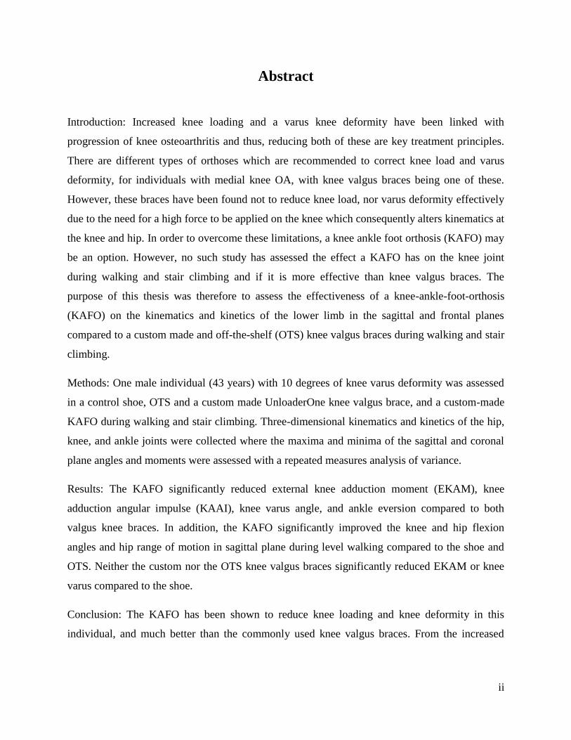
Abstract
Introduction: Increased knee loading and a varus knee deformity have been linked with
progression of knee osteoarthritis and thus, reducing both of these are key treatment principles.
There are different types of orthoses which are recommended to correct knee load and varus
deformity, for individuals with medial knee OA, with knee valgus braces being one of these.
However, these braces have been found not to reduce knee load, nor varus deformity effectively
due to the need for a high force to be applied on the knee which consequently alters kinematics at
the knee and hip. In order to overcome these limitations, a knee ankle foot orthosis (KAFO) may
be an option. However, no such study has assessed the effect a KAFO has on the knee joint
during walking and stair climbing and if it is more effective than knee valgus braces. The
purpose of this thesis was therefore to assess the effectiveness of a knee-ankle-foot-orthosis
(KAFO) on the kinematics and kinetics of the lower limb in the sagittal and frontal planes
compared to a custom made and off-the-shelf (OTS) knee valgus braces during walking and stair
climbing.
Methods: One male individual (43 years) with 10 degrees of knee varus deformity was assessed
in a control shoe, OTS and a custom made UnloaderOne knee valgus brace, and a custom-made
KAFO during walking and stair climbing. Three-dimensional kinematics and kinetics of the hip,
knee, and ankle joints were collected where the maxima and minima of the sagittal and coronal
plane angles and moments were assessed with a repeated measures analysis of variance.
Results: The KAFO significantly reduced external knee adduction moment (EKAM), knee
adduction angular impulse (KAAI), knee varus angle, and ankle eversion compared to both
valgus knee braces. In addition, the KAFO significantly improved the knee and hip flexion
angles and hip range of motion in sagittal plane during level walking compared to the shoe and
OTS. Neither the custom nor the OTS knee valgus braces significantly reduced EKAM or knee
varus compared to the shoe.
Conclusion: The KAFO has been shown to reduce knee loading and knee deformity in this
individual, and much better than the commonly used knee valgus braces. From the increased

iii
lever arm and improved fit, this could be a potential avenue for treatment of these impairments in
individuals when other conservative options are not successful.
1
Chapter one: Literature review
1.1. Definition of Osteoarthritis
There are several types of arthritis which may be grouped into four main areas: inflammatory
arthritis (such as rheumatoid arthritis and ankylosing spondylitis), soft tissue arthritis (such as
lateral epicondylitis), connective tissue arthritis (such as deratomyositis) and degenerative or
mechanical arthritis such as osteoarthritis (OA) (Arthritis Care, 2004). OA is defined as a
chronic, progressive musculoskeletal disorder characterised by articular joint cartilage
degeneration with articular bone enlargement due to new bone formation. However, it is defined
by the American Rheumatism Association as a heterogeneous group of conditions that leads to
joint symptoms and signs that are associated with the defective integrity of cartilage in addition
to the related changes in the underlying bone and at the joint margin (Altman et al., 1986).
1.2. Epidemiology of Osteoarthritis
It has been estimated that 41% of individuals aged 56 and over have OA in the UK (Dawson et
al., 2004) and around 8.4 million in the UK complain from symptoms associated with OA
(Arthritis Care, 2004). For instance, it is on record that in England and Wales alone up to 1.75
million individuals have systemic OA (Reginster, 2002) which means the condition is a serious
problem for the older population not only because OA causes a health burden, such as reducing
the quality of life and functional activity, but also an economic burden such as increased medical
costs, hospital cost, research costs, and absenteeism from work (Reginster, 2002). Evidence has
shown that between 10% and 15% of individuals aged 60 years and above have some degree of
OA, and are primarily suffering from some form of disability, in particular during walking and
climbing of stairs (Haq and Darce, 2003).
In the USA, 21 million individuals suffer from OA and this figure is continuously increasing
(D’Ambrosia, 2005), particularly in individuals aged 56 years and over due to age and obesity.
2
Also, 32.6% of the population sampled was found to complain of knee pain and 19.2%
complained from hip pain due to OA (Dawson et al., 2004).
1.3. OA classification
OA is divided into two main categories: primary and secondary. Primary (idiopathic) OA is
mainly hereditary and can be localised whereby it affects one joint, or can be generalised which
impacts on more than one joint. Secondary OA is more related to trauma, previous injury such as
hip dislocation, ligament tears, bone disorders (necrosis, osteochondritis), metabolic disease (e.g.
hemochromatosis), endocrine disease such as hyperthyroidism, and calcium deposition disease
(Altman et al., 1986). Primary OA is mainly associated with Heberden's nodes in the distal
interphalangeal joints (DIP) with other affected joints such as knee or hip joint (Petersson and
Jacobsson, 2004), and is more common than secondary OA. Moreover, it is estimated that 97%
of total knee replacements (TKR) are due to primary OA which is more prominent with the older
population, while TKRs due to secondary OA are commonly associated in the younger
population and more prominent in men than women ≥ 50 years old (Julin et al., 2010).
1.4. Sites of OA
OA is more commonly seen in weight bearing joints such as the hip, knee, and spine than in the
wrist, elbow, and shoulder joints. OA also affects most hand joints particularly the joint which
participates in the pincer grip such as the distal interphalangeal joint (DIP), proximal
interphalangeal joint (PIP), and at the base of thumb; which leads to a reduction in hand function,
deformity, and reduced joint motion (Zhang et al., 2002). OA also affects the first
metatarsophalangeal (MTP) joint which is the most affected joint in feet, and 10% of individuals
aged between 20-40 years and 44% of those aged over 80 years suffer the most from radiological
OA in the first MTP joint. In addition, in the vertebral column (spine), OA is strongly associated
with age, particularly in the cervical spine. OA has a higher recorded prevalence in the cervical
spine (84.8% men, 84.3% women) followed by the lumbar spine (71% men, 67.3% women)
(Van Saase et al., 1989). The incidence of OA in the hip joint is between 4% and 9% (Felson and

3
Zhang, 1998, Lawrence et al., 1998); whilst, the incidence of OA in the knee joint is the greatest
at between 3.4% and 30% (Kellgren and Lawrence, 1958, Felson et al., 2000, Spector and Hart,
1992) (Figure 1.1); with women more affected than men (11%, 7%, respectively) (Felson et al.,
1987).
Figure 1.4: The effects of age and gender on specific joint incidence of osteoarthritis. The knee
joint is more affected than the hip joint, especially within females (Felson et al., 2000).
1.5. Risk factors for OA
The risk factors for OA can be systemic (general) which can happen in any joint or can be local
(biomechanical) which primarily affect specific joints (Felson et al., 2000). Systemic factors
include age, gender, ethnicity, dietary, and genetic factors; whereas, biomechanical factors
include basically muscles weakness, obesity, joint alignment deformity, and ligament laxity. A
brief overview of these is given below.

4
1.5.1 Systemic factors
1.5.1.1 Age
OA is strongly related to age particularly in middle aged and older people (Beaupre et al., 2000)
due to changes in the characteristics of cartilage and also the loss of its flexibility. Thus, the
ability to provide shock absorption, and the ability to maintain the equilibrium between the
production and breakdown of cartilage cells becomes more difficult with age. Nonetheless, the
thickness of cartilage will decrease until the hydrostatic pressure inside the cartilage is decreased
and replaced with a new bone formation (Carter et al., 2004, Eckstein et al., 2002, Felson et al.,
1987). According to the Framingham study, it was also emphasised that OA is associated with
both age and gender; since, radiological OA was found in 27% of individuals aged below 70
years, but was found in 44% for individuals aged 80 years and over.
1.5.1.2 Gender differences
Gender also plays an influence whereby it is estimated that women have a higher prevalence
(11%) than men (7%) to develop symptomatic OA; In addition, the incidence of radiological OA
change is greater in women (34%) than men (31%) (Felson et al., 1987). Until 50 years of age,
men and women have the same prevalence, but after 50 years of age, women have a higher
prevalence than men due to a drop in oestrogen level caused by the menopause which reduces
the cartilage metabolism, which then increases cartilage breakdown (Beaupre et al., 2000, Nevitt
et al., 1996).

5
1.5.1.3 Ethnicity
Ethnicity has a role in the increased risk of developing some types of OA in specific countries
more than others. For example, the African American population has a higher likelihood of
developing knee OA than the white population (Anderson et al., 1988, Felson et al., 2000).
Furthermore, Felson et al. (2002) also states that different racial populations have a different
prevalence to developing knee OA in different compartments; for example, the Chinese
populations whose ages fall within 60-88 are at risk of having lateral knee OA compartment
more than Americans. Evidence of this is that 28.5% of Chinese women complain from lateral
knee OA versus 11% of Framingham women's knees, also men in China have a higher
prevalence of lateral knee OA than Framingham men’s knees by 32.3%, 8.8% respectively. This
difference can be explained by a high knee valgus alignment which is common within the
Chinese population compared to the population in the USA cohort (Felson et al., 2002). Race not
only affects OA prevalence, but also the severity of pain and the resultant limitation in activity
levels. In the USA, the black population complains of joint pain 1.9 times more, and activity
level impairment with work limitation 1.7 times more than the white population when associated
with OA (Bolen et al., 2010).
1.5.1.4 Diet and nutrition
Some vitamins have a direct impact on cartilage health and bone metabolism such as vitamin C
and vitamin D. For instance, vitamin C reduces the risk of knee OA progression three fold, and
also reduces pain severity compared to those who do not take it (McAlindon et al., 1996b).
Whereas, vitamin D, which controls chondrocytes (cartilage cell) and bone metabolism, may
reduce knee OA progression and reduce joint space narrowing by up to three fold (McAlindon et
al., 1996a), and also reduce hip OA development amongst white women (Lane et al., 1999).
6
1.5.1.5 Genetic factors
More than 50% of OA disease is related to genetic factors principally for the joints in the hand,
but approximately 39% of knee OA is related to genetic factors associated with Heberden's
nodes; specifically in women (Spector et al., 1996).
1.5.2 Local biomechanical factors
1.5.2.1 Muscle weakness
The strength of the quadriceps muscles help to dissipate shock absorption at initial contact (IC)
when the body weight is transferred suddenly to the support limb; thus, loss of muscle strength
will increase the load which is carried by the cartilage (Baker et al., 2005). Therefore, quadriceps
muscle weakness could be a risk factor and a symptom. Quadriceps weakness becomes a risk
factor when it loses its ability to control the knee joint, as it can cause knee pain and symptoms
associated with knee OA, and is a secondary reason for knee pain amongst women (Baker et al.,
2005). In addition, quadriceps weakness is strongly related to an increase in the disability and
activity level impairment of individuals (O’Reilly et al., 1998). Generally, individuals with knee
OA suffer from muscle weakness in the knee extensors (quadriceps) (Baker et al., 2005) and hip
abductors (Costa et al., 2010).
1.5.2.2 Body mass index (BMI) and obesity
Body mass index (kg/m2) is calculated by dividing mass (kg) by square of the height (M) of the
individual. Individuals who have a high body mass index have a higher incidence of developing
OA due to increase load on weight bearing joints such as the knee (Zhang et al., 2000).
When the body weight is 10% more than the average weight, this is considered as obesity
(Spector et al., 1996) which is equal to a BMI of ≥ 30 (Flegal et al., 2005). Obesity is an
important factor for the development and progression of OA (Cooper et al., 2000), and the most

7
important risk factor for primary and secondary knee OA (Spector et al., 1994). Amongst
women, it estimated that about 44 % have OA due to being overweight, while just 27 % of males
who are known to have OA are also obese (Goldin et al., 1976). Moreover, obesity increases the
risk of OA two to three times (Murphy et al., 2008) and OA progression. For instance, obese
women have a 22% greater risk to progress to knee OA and 36% to develop contra-lateral knee
OA after two years (Spector et al., 1994).
Nevertheless, it has being established that an estimated two unit decrease in one’s BMI is able to
reduce the risk of OA by 50% (Beaupre et al., 2000). Furthermore, 5.7% of body weight
reduction can reduce knee pain and physical disability by 30% and 24% respectively as
calculated using the Western Ontario and McMaster Universities Osteoarthritis Index
(WOMAC) scale after 18 months (Messier et al., 2004). A reduction of 20% in BMI by losing
weight is able to reduce knee pain 50 ± 26 mm on a visual analogue scale (VAS) and also
improve activity levels by reducing the degree of inflammation and increasing collagen
production by 32% (Richette et al., 2011).
1.5.2.3 Joint angle malalignment
Joint malalignment is a major problem in OA because it increases the load on the non-weight
bearing area and develops OA with time (Andriacchi et al. 2004). In medial compartment knee
OA, the knee varus rotates the knee centre of motion laterally and increases the load on the
medial side of the knee which further increases the load on the medial side leading to a bow-
legged deformity (Mills and Hull, 1991). This deformity eventually increases the knee load on
the medial side to 100% (Johnson et al., 1980), increasing joint instability, and functional
impairment (particularly if the varus degree angle is ≥ 50) such as the ability to sit to stand from a
chair (Chang et al., 2004). Additionally, varus alignment increases the pain during this activity
four-fold with a greater loss in the medial cartilage space (Sharma et al., 2001).
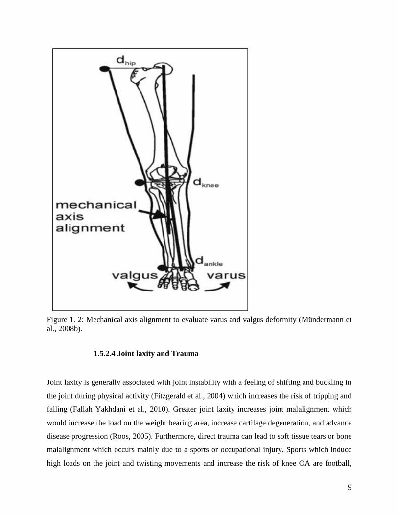
The osseous mechanical axis is used to evaluate the degree of knee varus and valgus. A full
length frontal plane radiograph during double limb support is needed to examine the medial
angle between two lines: the hip line (the line between the hip centre to the knee centre) and the

8
ankle line (the line between the knee centre to the centre of talus). This angle gives a general idea
about lower limb alignment (Figure 1.2) (Mündermann et al., 2008b). The normal angle is
between 179-181 degrees; an angle less than 179 degrees is considered as a varus deformity,
while a valgus deformity is said to be present when the angle is more than 181 degrees (Hayashi
et al., 2012). The normal varus angle is limited to between 1.5± 2 degrees (Moreland et al.,
1987); and therefore, individuals are considered to have a knee varus deformity if the varus angle
is 7.2 ±4.8 degrees or more (Cooke et al., 2003).
9
Figure 1. 2: Mechanical axis alignment to evaluate varus and valgus deformity (Mündermann et
al., 2008b).
1.5.2.4 Joint laxity and Trauma
Joint laxity is generally associated with joint instability with a feeling of shifting and buckling in
the joint during physical activity (Fitzgerald et al., 2004) which increases the risk of tripping and
falling (Fallah Yakhdani et al., 2010). Greater joint laxity increases joint malalignment which
would increase the load on the weight bearing area, increase cartilage degeneration, and advance
disease progression (Roos, 2005). Furthermore, direct trauma can lead to soft tissue tears or bone
malalignment which occurs mainly due to a sports or occupational injury. Sports which induce
high loads on the joint and twisting movements and increase the risk of knee OA are football,
10
rugby, volleyball and netball (Felson et al., 2000). Some types of occupation also have a role in
the increase of OA in specific joints; for instance, mine workers are more affected by knee OA
and lumbar degenerative than office workers (Kellgren and Lawrence, 1952). Thus, the activities
which require a high lifting of loads, kneeling, and stair climbing many times during the day
increase the risk of ligament tears (Roos et al., 1998) and subsequent OA.
For medial knee OA, injury to the anterior cruciate ligament (ACL), medial collateral ligament
injury (MCL) (Noyes et al., 1992), and meniscectomy increase the risk of knee OA developing
(Davies-Tuck et al., 2008) by increasing the knee load (Noyes et al., 1992). Further research has
demonstrated that a torn ACL has a risk factor of 60-90% in developing knee OA after 15 years
(Dayal et al., 2005) because tearing of the ACL reduces the degree of flexion to complete the
screw home mechanism (end of femur rotates laterally and externally to make knee flexion) and
increases the anterior/posterior tibial displacement which leads to increase in the knee load and
varus deformity (Andriacchi et al., 2006).
1.6. Knee OA
The knee joint is considered as a modified diathrodial hinge joint because it allows the ends of
the bones which form the joint to rotate. It is the largest joint in the human body and the most
injured joint due to the frequent stresses and strains it experiences. The stability of the knee joint
is derived mainly from ligaments and muscles rather than its osseous structure; and therefore,
any disturbance to soft tissues such as ligaments or muscles around the knee joint can impact on
knee stability (Lippert, 2006). The knee joint is one of the most common weight bearing joints
affected by OA (Ahlbäck, 1968, Oliveria et al., 1995) and one of the main causes of disability
amongst older people (18.4%), which is double the prevalence of disability caused by stroke and
pulmonary disease (Guccione et al., 1994).
The knee joint contains three compartments: the medial and lateral tibiofemoral compartment
and the patellofemoral compartment. Knee OA can impact on all the compartments of the knee,
but the medial side is the most affected part compared to other parts (Altman et al., 1986).

11
Evidence has shown that the medial compartment is ten times more likely to be affected by OA
than the lateral side (Buckwalter et al., 2004), because normally during walking the medial
compartment bears two or three times of one’s body weight (D'Lima et al., 2012), whilst on the
lateral side during walking is only 1.7 times body weight (Winby et al., 2009). This gives a
greater chance of developing OA in the medial side more than the lateral side due to the
excessive load on the medial aspect. When compared the patellofemoral joint to the medial
compartment with regards to OA incidence, a study by McAlindon, et al. (1992) demonstrated
that patellofemoral OA is present in 11% and 24% among men and women respectively;
however, medial knee OA is present in 21% and 12% among men and women.
During the developmental stage of medial knee OA, various changes in knee structures have
been noted such as radiological changes (bone enlargement and joint space narrowing), and
clinical variations (muscle weakness, inflammation, pain, and stiffness). Additional
biomechanical changes (kinematics and kinetics) may be expected during walking and stair
climbing such as a reduction in knee range of motion (ROM) mainly during flexion, and a varus
knee alignment due to the high external knee adduction moment (EKAM) being applied. These
changes will be explained in greater depth later in the thesis.
1.6.1 Prevalence of knee OA
In the UK, 10% of individuals aged 55 years and over have knee OA (Duncan et al., 2006). In
2004, it was estimated that around 0.5 million of individuals in the UK had moderate to severe
knee OA (Arthritis Care, 2004), but this increased to 4.7 million in 2010 and it is expected to
reach 5.4 million in 2020 (Arthritis Research UK, 2013). Knee OA is more common than in the
hip and women are more affected than men (14.9%, 10.1% respectively) (Guillemin et al., 2011).
According to the Framingham study, the prevalence of knee OA in adults aged ≥45 is 19.2%
(Felson et al., 1987). In the USA, women are more affected by knee OA as compared to men
(42.1% vs. 31.2% respectively) (Dillon et al., 2006).
12
1.6.2 Clinical and Radiological criteria of knee OA
OA affects all joint structures: articular bone, cartilage, subchondral bone, the synovium,
ligaments, and muscles (Lippert, 2006). Therefore, OA has both a clinical and radiological
criteria. The relationship between clinical features and radiological features are weak in the early
stage of OA. On average, 50% of individuals over 50 years of age have radiological OA without
symptoms, where also clinical features such as pain can be found without any defined
radiological criteria in early stage of OA progression, but at the later stages, the overlap between
clinical features and radiological are better matched (Peat et al., 2006). Nonetheless, OA can be
divided into symptomatic (pathological) or asymptomatic depending on whether individuals
suffer from clinical symptoms or radiological symptoms (Dieppe and Lohmander, 2005) or both.
Therefore, there are primary two classifications which are used in defining knee OA, namely the
American College of Rheumatology criteria (ACR) for clinical criterion and the Kellgren and
Lawrence grading for radiological severity (Kellgren and Lawrence, 1957).
1.6.2.1 The ACR criteria
The ACR criteria for diagnostic purposes and classification for OA of the knee, hip and hand
have been recommended for the diagnosis of OA in clinical and epidemiological studies (Brooks
and Hochberg, 2001). The sensitivity of this scale is 89% and its specificity is 88% (Altman,
1991a). Clinical features of knee OA are characterised by bony enlargement due to new bone
growth near to ligament insertions (osteophytes) or a reduced joint margin, the existence of joint
pain, morning stiffness for less than 30 minutes, loss of function, crepitus during motion (the
sound of cracking), joint instability, joint alignment deformity due to cartilage breakdown and
reduced range of motion and joint laxity (Altman et al., 1991a, Altman et al., 1990, Altman,
1991b). Local inflammation, swelling, joint effusion, muscle atrophy and pain during passive
movement and gait deformities are also clinical features of knee OA and become clear during a
physical examination (Hunter et al., 2008).

13
1.6.2.2 The Kellgren and Lawrence radiological grade
The use of a diagnostic X ray (radiograph) is the standard way to evaluate radiological knee OA
(Emrani et al., 2008) using an anterior/ posterior view with equal weight bearing on both feet and
with the toes pointing straight ahead (Jacobson, 1996). This is despite the fact that a 30 degree
flexion position is recommended because individuals reach this position during walking and the
flexed knee position increases the load on the cartilage. Thus, the real cartilage degeneration
level can be reflected in this situation more than in a full knee extension view (Davies et al.,
1999).
Radiological OA criteria are cartilage degeneration which could break down due to excessive
load (Griffin and Guilak, 2005); inflammation due to cartilage debris in the synovial fluid;
effusion; new bone growth such as osteophytes near to joint margins; and subchondral cysts
(Jacobson, 1996) due to the load increase on the non-weight bearing area of the joint because of
cartilage breakdown (Kellgren et al., 1963). Radiological OA assessment which depends on
Kellgren and Lawrence (K-L) scale is the accepted grading scale for OA by the World Health
Organisation (WHO scientific group, 1992). This scale depends on the amount of space
narrowing due to cartilage loss and osteophytes development (Kellgren and Lawrence, 1957). It
is therefore valid to examine and estimate radiological osteoarthritis severity, but has moderate
sensitivity to the real degeneration level of the articular cartilage (Kijowski et al., 2006). This
scale has five grades from zero to four. The K-L grade zero is none, K-L grade one is doubtful to
have osteoarthritis, K-L2 indicates mild OA, K-L3 indicates moderate and K-L4 is severe. For
diagnostic purposes, K-L grades which are equal to and greater than two are considered as
confirmation of the existence of OA (Kellgren and Lawrence, 1957) (Figure 1.3).

14
Figure 1. 3: The Grades of knee OA (Ryu et al., 2012).
As the knee joint structure changes due to OA, the load line position (via the ground reaction
force) is modified. These changes have a maximal influence on knee biomechanics and also
affect joints as a secondary adaptation or compensation during activities such as walking on level
ground and stair climbing. Therefore, an understanding of the biomechanical features present by
individuals with healthy lower limb joints during walking and stair climbing, especially for the
knee joint is important before discussing how knee OA causes biomechanical changes.
1.7. The biomechanics of walking and stair climbing
1.7.1 Walking
Human daily activities can be divided into three loading categories: moderate loading such as
walking; high loading such as ascending and descending stairs; and thirdly, high flexion
activities such as sit to stand or stand to sit, and squatting. However, walking is most common
daily activity undertaken by humans (Mündermann et al., 2008c).
15
Walking is considered to be a repetitive lower limb motion following an approximated pattern
which starts from one initial contact to the following initial contact for the same side (0% -
100%). A complete gait cycle is divided into two phases: stance phase (approximately 60% of
the gait cycle) when the foot is in contact with the floor, swing phase (approximately 40% of the
gait cycle) when the foot is swinging through the air (Perry, 1992). Furthermore, each phase has
sub phases such as first double limb support (1DS), single limb support, which includes early
stance (IS), mid stance (MS), and late stance (LS); and second limb support (2DS) in stance
phase. Early swing, mid swing, and late swing occur during swing phase (Baker, 2013) (Figure
1.4) (Meng, 2012).
During the first double limb support (0-10% of the gait cycle), the lower limb rapidly holds the
body mass by hip flexion, knee flexion and a neutral ankle position to allow shock absorption
during that phase. During the single limb support (10-50% of the gait cycle), the body mass is
progressed forward by hip extension and knee extension with the ankle in a dorsiflexed position.
In the second double limb support (50-60% of the gait cycle), the body weight starts to be
transferred to the opposite side by hip flexion, knee flexion, and ankle plantarflexion. After that,
in the early swing phase (60-75%) the body weight is already transferred to opposite side as the
hip and knee reach maximum flexion, while the ankle is still in plantarflexion. In the mid swing
(75%- 85% of the gait cycle), the tibia starts to be vertical when knee is extended and ankle joint
is moved to a neutral position, while the hip stays flexed. Finally, in the late stance (85%-100%
of the gait cycle) hip, knee and ankle joints start to return to the first limb support phase again
(Baker, 2013, Perry, 1992).

16
Figure 1.4: Typical gait cycle for healthy knee individuals during walking (Meng, 2012).
1.7.2 Stair climbing
Ascending and descending stairs are important daily activities and one of the main challenges for
the elderly population; primarily due to muscle weakness and a high risk of falling (Williamson
and Fried, 1996). This could be due to a reduction in the torque and power in lower limb because
of muscle weakness, biomechanical changes, and neuromuscular adaptation. Older individuals
therefore try to adapt their gait by walking slowly with shorter steps and high cadence compared
to young individuals (DeVita and Hortobagyi, 2000). Furthermore, some individuals try to
ascend and descend stairs by a stepping climb pattern (step over step) instead of the normal
climb pattern (step by step) due to pain even though this gait pattern requires high energy
consumption (Shiomi, 1994).
In a similar way to walking, ascending and ascending has two phases: stance and swing (Novak
et al., 2010). During an ascending cycle (AC), stance phase (65% ±4% of the gait cycle) has
three sub-phases (Figure 1.5):

17
Weight acceptance (0-17% of the gait cycle), during this phase the ankle reaches
maximum dorsiflexion while the hip and knee in flexion position to controlling pelvic
drop and shifting the body into an optimal position to be pulled up.
A pull-up or vertical thrust phase (vertical centre of mass displacement) which occupies
17-37% of the stair gait cycle. In this phase, hip and knee extensor muscles along with
the plantar flexors are working to pull the body mass against the gravity to full support on
the next step.
The forward continuance phase (37-65%), when ascent of a step has been completed and
progression continues by the firing of hip and knee extensors muscles.
The swing phase is subdivided into two sub-phases:
Foot clearance (65%- 82%), when the leg is raised to clear of the intermediate step by hip
and knee flexion and ankle dorsiflexion.
The foot placement phase (82-100% of the gait cycle), when the swing leg is positioned
for foot placement on the next step (McFadyen and Winter, 1988, Zachazewski et al.,
1993).
The stance phase of a descending cycle (DC), which occupies 68% ± 2% of gait cycle, is divided
into three sub-phases: weight acceptance (0-14%) when ankle starts the gait cycle with
plantarflexion to descend; forward continuance (14-34%) when the ankle moves in dorsiflexion
position to start of single limb support, forward body movement and controlled lowering (34-
68% of gait cycle) when the body’s mass is lowered onto the support limb by eccentric
contraction of the knee extensor muscles. The swing phase has two sub-phases: leg pull through
(68-84% of the gait cycle) the swing limb is pulled forward by ankle plantarflexion; the foot
placement phase (84-100% of the gait cycle) (McFadyen and Winter, 1988, Zachazewski et al.,
1993).
Ascending movements demand more energy consumption for healthy individuals than
descending because the hip and knee angles and joint moments are higher during AC than DC.

18
However, ankle dorsiflexion and plantarflexion is higher during DC than AC (Protopapadaki et
al., 2007). Mean maximum knee flexion has been shown to be 98.6 degrees during AC, 90.3
degrees during DC and 64.6 degrees during walking (Jevsevar et al., 1993). Evidence shows that
the external knee flexion moment during AC is 2.7 times more than when walking, whilst hip
flexion moments are1.5 times more than walking (Andriacch et al., 1980). Moreover, the knee
joint keeps in abduction in the stance phase of AC and DC which means that ground reaction
force (GRF) always passes medially to the knee joint during stance phase (Kowalk et al., 1996).
Yu et al. (1997b) also found that the knee varus during stair climbing is higher than walking,
while knee valgus is close to that demonstrated during walking activities.
Figure 1.5: Ascending and descending gait cycles (Novak et al., 2010).
1.8. Gait in individuals with medial compartment knee OA
Individuals with medial compartment knee OA demonstrate many adaptive gait patterns
compared to the control (healthy) population. These gait alterations are mainly related to pain
levels and the severity of OA present, and are also due to muscle weakness which consequently
leads to reduced knee and hip ranges of motion (Hurwitz et al., 2000). As the human body
systems are working relative to each other, the joints cannot work separately and as such any
deformity in one joint could impact on others directly or indirectly. Even though, individuals
19
with medial compartment knee OA complain mainly about knee problems, other joints such as
the hip could be severely affected by the application of increasing load on them which could
develop hip OA in the future (Valente et al., 2013).
1.8.1 Temporal-spatial parameters
As the speed of walking mainly affects the vertical ground reaction force magnitude (Andriacchi
et al., 1977), individuals with knee OA adapt their speed depending on the severity of the disease
(Zeni Jr et al., 2009). For example, individuals with a K-L3 classification will walk slower than
those with K-L2 (Thorp et al., 2006). Besides that, individuals with knee OA mainly have a high
cadence, shorter stride length with higher stance phase percentage and double limb support
compared to a healthy non-OA older population (Al-Zahrani and Bakheit, 2002) in order to
reduce pain by a rapid dynamic load transfer from affected side to the unaffected side (Baliunas
et al., 2002, Takacs et al., 2012). During stair climbing, individuals with medial compartment
knee OA have similar characteristics as of walking, but some differences are found between
ascending and descending the velocity is slower in AC compared to DC because the movement
is against the gravity, while the step width is more in DC compared with AC to provide balance
during DC. In addition, the time taken during double limb support is longer in AC to add
stability by means of muscle concentric contraction, while the swing phase is longer in DC to
control the body mass and control downward acceleration (Hicks-Little et al., 2012).
1.8.2 Kinematics
Medial compartment knee OA impacts on the hip, knee, and ankle joints in the sagittal and
frontal planes, but sagittal plane motion is more affected (Al-Zahrani and Bakheit, 2002).
Individuals with medial compartment knee OA were shown to have lower knee flexion values
than a control group during walking both quickly or slowly (Landry et al., 2007), and walk with
six degrees less knee ROM than normal groups (54±7 vs. 60±4) (Kaufman et al., 2001). At IC,
individuals with knee OA start with a more extended knee joint (1.8-5.3 degrees) (Mündermann
et al., 2008a). This flexion limitation depends on knee OA severity (Astephen et al., 2008a).
20
During ascending, individuals with knee OA show less knee flexion than their healthy
counterparts (Hicks-Little et al., 2011). Evidence has shown that individuals with knee OA climb
stairs with less knee range of motion and knee flexion angle (62 and 80 degrees, respectively)
than a control group (74 and 130 degrees respectively) (Walker et al., 2001). In stance phase, the
results show that the peak of knee flexion angle during ascending is earlier for healthy
individuals and individuals with knee OA (19±7 and 32±23% of the gait cycle time,
respectively) than descending (51±6 and 54±4% of the gait cycle time respectively).
Furthermore, individuals with knee OA have a delay in knee flexion compared to healthy
individuals during stair climbing (Kaufman et al., 2001).
Furthermore, Mündermann et al. (2005) found no difference between hip ROM between
individuals with medial knee OA and healthy individuals during walking. Also, Hicks-Little et
al. (2011) found that there was no significant difference in hip range of motion between
individuals with knee OA and a control group during ascending, but the individuals with knee
OA show a delay in reaching peak hip flexion.
Trunk and pelvic ranges of motion are also affected by OA. Alterations such as increased
anterior pelvic tilt (Linley et al., 2010) and lateral trunk bending towards the swing side have
been reported. Amongst unilateral medial compartment knee OA subjects, trunk bending has
been shown to be 3.7±0.9 degrees when the involved limb is loaded and 3.3± 1.4 degrees for the
non-involved limb more than the control group (Tanaka et al., 2008), which consequently leads
to asymmetry in their gait. With regards to the ankle joint, Ko et al. (2010) found that individuals
with symptomatic knee OA have a higher ankle range of motion in the sagittal plane than healthy
individuals, which can be explained as a compensatory gait due to decrease knee range of motion
during walking.
In the frontal plane, individuals with knee OA walk with a greater degree of knee varus during
stance phase due to a narrow medial joint space, while the lateral part of knee is open and also
walk with a relatively high valgus knee angle during swing phase which could be due to internal
rotation of the limb during swing phase (Gök et al., 2002). During descending, they walk with a
greater knee varus angle than a control group; however, there is no significant difference in the
21
ankle abduction angle between both groups. A delay in achieving maximum ankle ROM within
individuals with medial knee OA is noticed compared to a control group. In addition, individuals
with knee OA start ascending with high hip abduction angles and less hip adduction angles
(Hicks-Little et al., 2011).
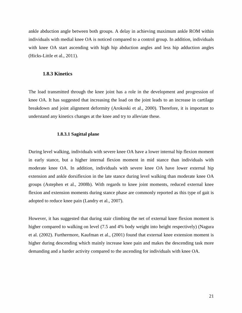
1.8.3 Kinetics
The load transmitted through the knee joint has a role in the development and progression of
knee OA. It has suggested that increasing the load on the joint leads to an increase in cartilage
breakdown and joint alignment deformity (Arokoski et al., 2000). Therefore, it is important to
understand any kinetics changes at the knee and try to alleviate these.
1.8.3.1 Sagittal plane
During level walking, individuals with severe knee OA have a lower internal hip flexion moment
in early stance, but a higher internal flexion moment in mid stance than individuals with
moderate knee OA. In addition, individuals with severe knee OA have lower external hip
extension and ankle dorsiflexion in the late stance during level walking than moderate knee OA
groups (Astephen et al., 2008b). With regards to knee joint moments, reduced external knee
flexion and extension moments during stance phase are commonly reported as this type of gait is
adopted to reduce knee pain (Landry et al., 2007).
However, it has suggested that during stair climbing the net of external knee flexion moment is
higher compared to walking on level (7.5 and 4% body weight into height respectively) (Nagura
et al. (2002). Furthermore, Kaufman et al., (2001) found that external knee extension moment is
higher during descending which mainly increase knee pain and makes the descending task more
demanding and a harder activity compared to the ascending for individuals with knee OA.

22
1.8.3.2 Frontal plane
The most important variable in the frontal plane in medial compartment knee OA is the external
knee adduction moment (EKAM) or knee varus moment which is the result of the ground
reaction force (GRF) and its distance from the knee joint centre (Hurwitz et al., 2002) passing
medially during dynamic loading. It is expressed in units of percentage of the body weight and
height (%Bw-Ht.) or Newton metres per kilogram (Nm/kg) (Andriacchi, 1994).
When knee varus is increased, the knee will rotate externally more (Mills and Hull, 1991) which
consequently leads to an increase in the lever arm distance between the GRF and the knee centre
by shifting the knee joint centre more laterally and centre of pressure more medially (Haim et al.,
2008). The EKAM has two peaks and a trough: The first peak is a sharp one after initial contact
during early stance phase, while the second one is in the late stance phase (Figure 6). The first
peak is affected by the amount of knee varus, joint space narrowing, OA severity, and
progression level, while the second one is more correlated to the amount of toe out and pain
levels. So, the first peak is more important to distinguish between different severity levels of
knee osteoarthritis groups (Sasaki and Neptune, 2010); where, individuals with severe knee OA
have a higher first external knee adduction peak than less severe and the less severe group has
lower second peak than a control group.
The knee adduction angular impulse (KAAI) is the area under the knee adduction moment curve
(Figure 1.6) (Thorp et al., 2006). Therefore, it measures the loading throughout the duration of
EKAM, and it is considered more sensitive than EKAM to distinguish between mild and
moderate knee osteoarthritis. Also, it is suggested that high KAAI is associated with greater bone
marrow lesions and cartilage degeneration (Bennell et al., 2011b). Both peaks of KAAI are
affected by walking speed, while slow speed is correlated with high KAAI. Also, the two peaks
of EKAM are affected by speed (Kean et al., 2012); however, the trough part of EKAM curve is
less affected by speed (van der Esch et al., 2011). Therefore, one should control speed by using a
metronome, for example, to eliminate speed effects on EKAM value when any conventional
treatments such as orthoses are evaluated in a gait laboratory.

23
Figure 1.6: KAAI which is the highlighted area under the graph (Thorp et al., 2006).
Individuals with medial knee OA show a higher peak EKAM and knee adduction angular
impulse (KAAI) than healthy individuals during walking (Landry et al., 2007, Linley et al.,
2010) (Figure 1.7). Moreover, the peak EKAM is higher during ascending and descending than
walking, but during descending the EKAM peak is the highest (Guo et al., 2007). Also,
symptomatic and asymptomatic individuals with knee OA do not have similar EKAM peaks; for
instance, Kean et al. (2012) stated that symptomatic individuals with knee OA have a higher
impulsive EKAM than asymptomatic group. The first peak is high for a symptomatic group only;
while the second one is high for symptomatic and asymptomatic, but normally the first one is
higher than second one.

24
Figure 1.7: EKAM has 2 peaks and it is higher for OA group more than control group (Linley et
al., 2010).
Previous studies have shown that when the EKAM increases, the joint space becomes narrower
(Miyazaki et al., 2002), stress or load on medial knee cartilage increases (Nicholas et al., 2010),
and also bone density under medial plateau increases (Thomas et al., 2000). The anterior cruciate
ligament (ACL) will also be affected by an increasing EKAM and an increased risk of ACL
tears, ligament laxity and joint instability also can result (Noyes et al., 1992). Progression of
knee OA has been shown to increase in individuals with a high EKAM level, as with a 1%
increment in EKAM, the risk of OA progression is six-fold (Andriacchi, 1994). Moreover, a 1%
EKAM increment leads to 0.63 mm loss in the joint space (Sharma et al., 2004). Therefore, the
reduction of EKAM is the primary aim of all interventions by reducing the distance between the
centre of pressure (CP) and the centre of knee (Haim et al., 2008). It is suggested that 1 mm
displacement of COP laterally can lessen the EKAM by 2% and the load on the knee joint by 1%
(Shelburne, et al., 2008). In summary, the EKAM evaluates the load on medial compartment
(Thorp et al., 2006), disease progression (Miyazaki et al., 2002), and severity of knee
osteoarthritis (Mündermann et al., 2004); and also predicts the severity of pain (Hurwitz et al.,
2000), and it is the primary outcome variable of choice in knee intervention studies.
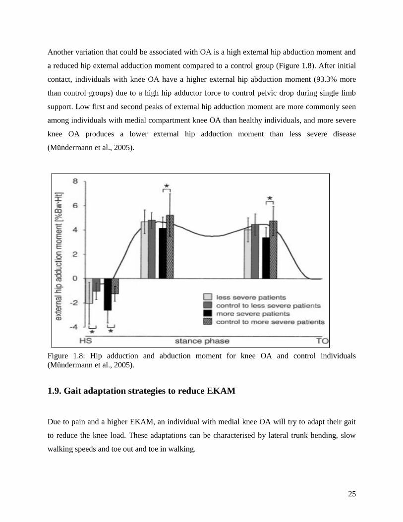
25
Another variation that could be associated with OA is a high external hip abduction moment and
a reduced hip external adduction moment compared to a control group (Figure 1.8). After initial
contact, individuals with knee OA have a higher external hip abduction moment (93.3% more
than control groups) due to a high hip adductor force to control pelvic drop during single limb
support. Low first and second peaks of external hip adduction moment are more commonly seen
among individuals with medial compartment knee OA than healthy individuals, and more severe
knee OA produces a lower external hip adduction moment than less severe disease
(Mündermann et al., 2005).
Figure 1.8: Hip adduction and abduction moment for knee OA and control individuals
(Mündermann et al., 2005).
1.9. Gait adaptation strategies to reduce EKAM
Due to pain and a higher EKAM, an individual with medial knee OA will try to adapt their gait
to reduce the knee load. These adaptations can be characterised by lateral trunk bending, slow
walking speeds and toe out and toe in walking.

26
1.9.1 Lateral trunk bending
It has been demonstrated that pelvic drop increases EKAMs significantly along with OA
progression (Zeni and Higginson 2009). It increases the EKAM by shifting the knee load more
medially toward the swinging limb (Valente et al., 2013). Also, it has suggested that a pelvic
drop increases the load on the knee and hip joints by 18.3% and 19.4% respectively (Valente et
al., 2013). To compensate for this, a high hip external abduction moment (lateral bending) will
be necessary as a gait strategy to support the pelvis and to reduce the EKAM. Mündermann et al.
(2008a) evaluated the effects of lateral bending within a healthy group of older people, and
demonstrated that lateral bending reduced hip and knee adduction moments by 57% and 56%
respectively, but increased hip and knee abduction moments during walking which suggested
that this gait pattern could be helpful for individuals with medial compartment knee OA.
However, lateral bending (a waddling gait) may increase pain in the lumbar region during
walking (Toriyama et al., 2011, Valente et al., 2013), and increase low back pain (Gombatto et
al., 2007, Mcneill et al., 1980). Furthermore, it increases disability and functional impairment for
individuals over time (Simmonds et al., 1996) which suggests that it may increase the risk of
developing hip OA in the future by increasing the load on the hip (Mündermann et al., 2005,
Toriyama et al., 2011).
1.9.2 Reducing speed
Zeni et al. (2009) suggested that speed of walking could be correlated to OA severity; individuals
with more severe knee OA walk slower than less severe individuals. Slow walking speed could
be a strategy to reduce pain, reduce knee flexion during walking, and then reduce the need for
the quadriceps muscles to control this movement; thus, reducing the knee loads (Zeni et al.,
2009, Zeni et al., 2011). Individuals with less severe knee OA demonstrated a greater EKAM
reduction with a slower gait speed compared to those with severe knee OA. The difference noted
was thought to be due to the existence of a high EKAM and varus angle alignment within
individuals with severe knee OA which a slower speed cannot reduce extensively (Mündermann
et al., 2004).
27
However, it is worthy to mention that walking slowly can reduce knee load but does not reduce
the exposure time of load because walking slowly increases KAAI (Robbins and Maly, 2009).
However, walking with a slower speed and shortened stride length is thought to reduce the
EKAM (Mündermann et al., 2004).
1.9.3 Toe-out/toe-in walking
A toe-out gait (for example with the foot externally rotated more than seven degrees) can reduce
the second peak of EKAM raised to 40 % during walking (Guo et al., 2007, Hurwitz et al., 2002)
(figure 1.9a). This may be achieved by shifting the COP laterally and reducing the applied
moment distance from the COP to the knee centre and thus reducing the EKAM (Jenkyna et al.,
2008). However, no change in first peak value was seen in either of the studies.
During ascending, toe out walking increases the first peak, while second peak decreases by 11%
(Figure 1.9b). Nevertheless, no significant differences have been shown between these
parameters during descending (figure 1.9c) (Guo et al., 2007).

28
Figure 1.9: The effects of toe-out gait during walking (a), stair ascent (b), and descent (c) Thick
black line: increased foot progression angle (toe out), Thin grey line: self-selected foot
progression angle (Guo et al., 2007).
However, evidence suggests that a toe-out gait reduces pain (Sasaki et al., 2010), but it also
increases knee flexion during walking especially at 10% of gait cycle, and the tension on the
quadriceps muscles and the load on patellofemoral joint will increase to control flexion (Jenkyn
et al., 2008). As a result, Walter et al. (2010) stated that when an individual increases the external
knee flexion moment it may increase load and compression on the knee joint; therefore toe-out
walking may not be suitable for individuals with knee OA: even though it can reduce the second
peak of EKAM, but it could increase pain during walking as a knee load is increased.
In contrast, toe-in walking (average five degrees) shows a reduction in the first peak of EKAM
by 13% and does not increase the external knee flexion moment for individuals with OA during
walking and does not affect trunk lateral bending or the tibia angle (Shull et al. 2013). Toe out
walking is accomplished by internal rotation of the forefoot and heel lateral rotation which is
29
able to shift the knee center of pressure laterally and the knee center of joint more medially
(Shull et al. 2013). Due to the fact that reducing the first peak is considered more important than
reducing the second one, it is recommended as a non-surgical treatment rather than toe-out
walking.
1.10. Osteoarthritis treatments
Many treatments have been developed for individuals with medial compartment knee OA.
Generally, all the treatments aim to reduce pain, symptoms, reduce progression of joint damage,
improve activity, and the functional level for knee OA. Therefore, different treatment options are
available to the individual depending on disease severity, pain intensity, their degree of activity
level impairment, and other knee OA symptoms such as effusion or inflammation. The
rehabilitation strategy for medial knee OA consists of surgical interventions, pharmacological
treatments, and conservative non- pharmacology treatments (Jordan et al., 2003).
1.10.1 Surgical intervention
Surgical treatment can be by arthroscopy, osteotomy, and arthroplasty surgery. Invasive
treatment (arthroscopy) is used when the medical treatment cannot manage the knee joint
inflammation. During this procedure, a small needle passes inside the knee joint to remove
cartilage and inflammatory debris. This intervention is widely used nowadays because it has a
lower risk factor than other surgical interventions (National Collaborating Centre for Chronic
Conditions, 2008), and improves the Western Ontario and McMaster Universities Osteoarthritis
Index (WOMAC), which scores within a range between 0 to 2400 points (a high score indicates
more severe symptoms). For instance, two years after surgery WOMAC is improved by 874±624
points compared to control group 897±583 points (Kirkley et al. 2008).
Surgery involving a high tibial osteotomy is another intervention used to correct the knee joint
malalignmet deformity and for pain relief. During this surgery a wedge of bone is removed from
the non-healthy side of the tibia to redistribute the body weight over both tibial condyles
30
(Benjamin and Trickey, 1969). This intervention is suitable for individuals in middle-age with
medial knee OA who have a good knee range of motion without ligament instability.
Furthermore, the aims of this surgery are to reduce pain by reducing the load on the medial side
and delay OA progression, and also to delay the time before total knee replacement is necessary
(Wright et al., 2005). In a study by Billings et al. (2000), 43 individuals with medial knee OA
had good to excellent clinical outcomes after this surgery after an average of 8.5 years post-
surgery, while 21 of these individuals had a total knee replacement after this surgery at an
average of 65 months. This result could be explained by Prodromos et al. (1985) whose study
which found that an individual with a high EKAM pre-surgery had a lower clinical outcome than
one with a low EKAM pre-surgery.
Arthroplasty (total joint replacement or partial compartment replacement) is the last and the only
solution when other interventions cannot manage the knee OA progression, where pain is
intolerable, presents at rest and night which limits the activity level of medial OA individuals
(Dreinhöfer et al., 2006). Moreover, when an individual’s activity level is impaired such that the
individual with medial knee OA cannot climb stairs, walk for six metres to one km, and
complains of pain during activity and rest, surgery will be indicated (Swift, 2012). In the UK,
around 120,000 knee joint replacement operations are conducted annually (National
Collaborating Centre for Chronic Conditions, 2008). Before deciding to perform joint
replacement surgery, it is extremely important to know the factors which are related to the
outcome after surgery such as the patient’s age as well as their fitness levels to be able to tolerate
post-surgery risk; thus, it is not recommended for very old individuals (Chang et al., 2010). Also,
obese individuals have a poor outcome after surgery compared to those with a normal weight due
to the high risk of artificial joint dislocation (Swift, 2012), and with a high risk of post-surgery
infection and skin ulcers (Järvenpää et al., 2010). As there are major risks associated with
surgery, surgery is the last resort and other options should be considered first.

31
1.10.2 Pharmacology treatments
Pharmacology treatments normally include analgesic, non-steroidal anti-inflammatory drugs
(NSAIDs), or corticosteroid injections. Analgesics are routinely used as a first choice in knee OA
treatment such as Acetaminophen which is able to reduce pain for individuals with mild knee
OA over six months, but it is less effective for more severe pain. Diclofenac and Misoprostol are
more effective for severe pain, but have more adverse events than Acetaminophen (Pincus et al.,
2001).
NSAIDs are the most commonly prescribed drugs for long term use (Brater, 1988). It has been
shown that 8.6% of the Dutch community uses it regularly and 17% of them use NSAIDs for the
long term (Leufkens et al., 1990). However, there are side effects experienced from using these
drugs. According to British Community on the Safety of Medicines, 25% of reported side effect
drugs were due to NSAIDs such as Piroxicam, Naproxen, and Ibuprofen (Update, 1986). The
side effects of these drugs are high toxicity; for instance, Indomethacin is three times more toxic
than Ibuprofen with high risk of peptic ulcer development (Griffin et al., 1991), gastrointestinal
bleeding and balance impairment after a long term use (Richy et al., 2004). However, whilst
these treatments reduce pain in the individual, there are increase in bone degeneration and OA
progression. The annual bone loss for patients who are using NSAIDs was calculated to be 3.4%
by Reid et al., (1986). Individuals who ingest NSAIDs enables them to walk with no gait
adaptations due to pain and therefore an excessive load on affected joint occurs without pain
being felt which consequently leads to increase cartilage and bone degeneration (Richmond et
al., 2009). The one aspect of these treatments is that they are only dealing with the clinical pain
symptoms and whilst of utmost importance, OA is a mechanical condition, so conservative
strategies targeting pain without the complication of toxicity are attractive treatments with
additional mechanical benefits. However, using NSAIDs reduces pain, but they increase the knee
load and knee OA progression (Schnitzer et al., 1993)

32
1.10.3 Conservative non-pharmacology treatments
Non-pharmacology treatments are mainly recommended for individuals with less severe knee
OA to improve their quality of life, reduce pain during daily activities, and delay surgery time.
These types of treatments generally include changing their lifestyle, reducing activities which
increase knee loading, performing targeted exercises, and using suitable shoes and orthoses.
1.10.3.1 Education
It is very important to inform individuals with knee OA about their health, how to manage it, and
how to adapt their lifestyle according to their health level. Such information includes the
advantages of losing weight particularly if their body mass index > 25 as it is a totally essential
recommendation and the first step in a rehabilitation program. Other recommendations are to
avoid excessive load activity and replace it with walking and low-impact aerobic fitness
exercises to avoid joint stiffness or muscles contraction (Richmond et al., 2009).
1.10.3.2 Physical therapy and Exercise
Exercise therapy has been shown to have a significant effect by reducing pain and improving the
activity levels for individuals with knee OA after three months follow up assessment (Angst et
al., 2001). The evidence shows that exercises may reduce OA progression (Slemenda et al.,
1997, Manninen et al., 2001), and may delay the requirement for total knee replacement for
individuals with severe knee OA for both men and women (Manninen et al., 2001). Different
exercises can be used for knee OA, but these rehabilitation programs have to be without
tensional load on the knee joint, contain low impact activities such as walking, swimming, and
avoid excessive load exercise such as weight lifting and running because these increase the load
on the knee joint and OA progression (Richmond et al., 2009).

33
Muscle strengthening exercises aim to improve the kinematics and kinetics of the knee joint
(Bashaw and Tingstad, 2005), and have been shown to decrease OA symptoms such as pain and
improve quality of life for knee OA by 42% (Bennell et al., 2005). Exercise can be active or
passive such as mobilization, soft tissue massage, stretching, manipulation, passive movements
to the joints and soft tissues to prevent joint contortion or muscles atrophy (National
Collaborating Centre for Chronic Conditions, 2008). Muscle strengthening has been shown to
provide pain improvement after three weeks of treatment by 73% (Hinman et al., 2003).
However, this improvement increases the walking speed in the individuals and increases the
overall load on the knee joint; and thus, increases the EKAM (Hunt et al., 2010). Thorstensson et
al. (2007) also supports this result as a quadriceps strengthening does not reduce EKAM
significantly after eight weeks for individuals with mild and moderate knee OA, and similarly for
individuals with severe knee OA after 12 weeks of therapy (King et al., 2008). Likewise, hip
muscle strengthening can improve activity levels and reduce pain during daily activity after 12
weeks, but does not significantly alter EKAM values (Bennell et al., 2010). Therefore, whilst
exercise addresses the clinical and functional impairments associated with medial knee OA, the
mechanical element is still not improved and needs addressing.
1.10.3.3 Footwear and Orthoses
Many different footwear and orthoses (assistive external device) have been designed with the
aim to reduce the mechanical load on the knee joint and thus improve clinical symptoms. These
assistive devices can be footwear, laterally-wedged insoles, unloader knee valgus braces, and
knee-ankle-foot orthoses. Each one has a specific function with the aim to reduce EKAM
(Miyazaki et al., 2002) and each will be discussed hereafter.
i. Footwear
The type of shoe which the individual with knee OA uses has to be evaluated carefully because
footwear has been shown to have different effects on the EKAM. High heeled shoes should be
avoided, as it is suggested that (1.5 inch) heel high is able to increase the second peak of EKAM
by 9-14%, and thereby increase knee load particularly with women. However, no significant

34
changes have been seen in the first peak of the EKAM (Kerrigan et al., 2005). Shoes with a six
cm heel height with a narrow heel could increase both peak values of EKAM significantly by
18.7% and 19.2% respectively and increase medial knee compression force by 23% compared to
barefoot walking (Kerrigan et al., 1998). One shoe which has been designed to reduce this
parameter is the mobility shoe (Shakoor et al., 2008) which has been shown to reduce both
EKAM and KAAI values by 8% and 6% respectively compared to an individual's own shoe, and
also increase walking speed and decrease cadence compared to a barefoot condition. This shoe is
lightweight, flexible, has a single thin insole, and a heel height of 1-1.5 cm with grooves inserted
at the main flexion point of the foot (Shakoor et al., 2008).
A second shoe has been developed called the variable stiffness shoe which has shown to produce
improvement in knee pain and function according to the WOMAC scale after one year of use.
This shoe design has 1.3-1.5 more lateral sole stiffness than on the medial side and has been
shown to be more suitable for individuals with mild knee OA than those with more severe knee
OA (Erhart‐Hledik et al., 2012). It also has been shown to reduce the EKAM after six months of
use by up to 4.7% compared to normal high street footwear (Erhart et al., 2010), and to reduce
the average of EKAM between 2.4% and 6.2% compared to control shoe after short term use
(Erhart et al., 2008).
The Gel Melbourne OA shoe is also used for medial knee OA group as a footwear intervention.
This modified shoe incorporates a lateral wedge (four to six degrees) with a variable stiffness
sole (the lateral midsole is stiffer than the medial midsole and extends to more than two thirds of
shoe length. This shoe also produces a significant reduction in EKAM values (mean 7.9 ± 8.9%,
4.7±5.4%, 3.5 ±8.2% for knee OA, healthy overweight group, and healthy weight (control)
group, respectively); and KAAI (mean 8.7±11.2%, 7.3±5.6%, 7.8±6.6% for knee OA,
overweight, and control group, respectively) (Bennell et al. 2013).
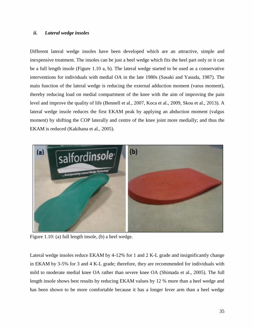
35
ii. Lateral wedge insoles
Different lateral wedge insoles have been developed which are an attractive, simple and
inexpensive treatment. The insoles can be just a heel wedge which fits the heel part only or it can
be a full length insole (Figure 1.10 a, b). The lateral wedge started to be used as a conservative
interventions for individuals with medial OA in the late 1980s (Sasaki and Yasuda, 1987). The
main function of the lateral wedge is reducing the external adduction moment (varus moment),
thereby reducing load on medial compartment of the knee with the aim of improving the pain
level and improve the quality of life (Bennell et al., 2007, Koca et al., 2009, Skou et al., 2013). A
lateral wedge insole reduces the first EKAM peak by applying an abduction moment (valgus
moment) by shifting the COP laterally and centre of the knee joint more medially; and thus the
EKAM is reduced (Kakihana et al., 2005).
Figure 1.10: (a) full length insole, (b) a heel wedge.
Lateral wedge insoles reduce EKAM by 4-12% for 1 and 2 K-L grade and insignificantly change
in EKAM by 3-5% for 3 and 4 K-L grade; therefore, they are recommended for individuals with
mild to moderate medial knee OA rather than severe knee OA (Shimada et al., 2005). The full
length insole shows best results by reducing EKAM values by 12 % more than a heel wedge and
has been shown to be more comfortable because it has a longer lever arm than a heel wedge
36
(Hinman et al., 2008) and it is able to shift COP laterally by up to five mm (Shelburne et al.,
2008). Also a full length lateral wedge characteristic is different depending on type of materials
the insole made of, such as cork, silicon, or soft foam. In addition, different wedge inclinations
have different effects. For instance, the amount of lateral wedge inclination normally ranges
between five degrees (3mm) and ten degrees (6mm). Increased inclination leads to more
significant effects on EKAM values, but is less comfortable to be used for a long time. Most
insole users have found that a five degrees lateral wedge is more comfortable than a ten degrees
one (Kerrigan et al., 2002). It is also recommended that subjects use a laterally-wedged insole for
between five to ten hours daily to get the best benefit (Toda et al., 2005).
Many studies investigating the effect of laterally-wedged insoles have been conducted during
walking activities, but only one study has been conducted on stairs. In this study the lateral
wedge insole demonstrated a significant reduction for first peak of EKAM during ascending
(0.37±0.02 Nm/Kg) and descending (0.33±0.05 Nm/Kg) and also reduced the KAAI
(Alshawabka et al., 2014).
Using a lateral wedge insole not only reduces the EKAM, but also enhances knee OA clinical
symptoms such as pain and activity level (Jones et al. 2013, Arazpour et al., 2013, Bennell et al.,
2007, Bennell et al., 2011a). For instance, a study by Baker at al. (2007) demonstrated that pain
scores can be reduced by 13.8 points on a visual analog scale (VAS) scale which is considered
clinically insignificant. However, Parkes et al. (2013) found that using a lateral wedge can only
reduce pain by 2.12 points out of 20 using the WOMAC scale when compared to a neutral
insole.
One of drawbacks of using lateral wedge insoles is even though they are a cheaper intervention
and more acceptable to use, it has been shown that the decrease in EKAM values was
insignificant for severe medial knee OA (Shimada et al., 2006). Also, using lateral wedge insoles
increases the external subtalar eversion (subtalar valgus degree) during walking (Abdallah and
Radwan, 2011, Kakihana et al., 2004, Pinto et al., 2008), and during stair climbing also
(Alshawabka et al., 2014). Also, there is a non-responder group (20%) whose EKAM increased

37
with lateral wedge insole due to high eversion angles which are increased by using lateral wedge
insole (Jones et al. 2014).
iii. Knee valgus brace (Unloader)
A knee valgus brace (or unloader brace) is designed to apply an external abduction moment
through a three point pressure system and transfer the knee load from the affected side to the
unaffected side (Richards et al., 2005, Stamenović et al., 2008 ) (Figure 1.11). The main
difference between the knee valgus brace and other conservative treatments such as lateral
wedge insoles is that a brace applies a valgus force directly on the knee joint to reduce the varus
moment (Draper et al., 2000). One study, has shown that using an insole with five degrees of
inclination reduced both the EKAM and KAAI (12%, 16.1%, respectively) more than knee
valgus brace with six degrees of valgus (7%, 8.6%, respectively) during walking for individuals
with medial knee OA, and the knee valgus brace was less acceptable to individuals with medial
knee OA than an insole (Jones et al., 2013) with the duration of wear being much less. In
contrast, a knee valgus brace (with between two to nine degrees of valgus correction has been
shown to reduce the first (2.98±1.05, 2.82±0.97 respectively) and second peak (2.78±1.01,
2.61±0.94) of EKAM better than an insole with a three to nine mm inclination for individuals
with medial knee OA(Moyer et al., (2013).
The knee valgus brace has been proven to have some biomechanical benefit for individuals with
medial OA such as reducing EKAM, pain and improving activity levels (Draper et al., 2000,
Ramsey et al., 2007, Richards et al., 2005, Self et al., 2000). EKAM values have been shown to
improve between 5.5% up to 33% (Deie et al., 2013, Fantini Pagani et al., 2010b, Lindenfeld et
al., 1997, Self et al., 2000).

38
Figure 1.11: The three-point pressure system applied by knee unloader braces (Stamenović et al.,
2008).
Knee valgus braces have also been used to evaluate the knee load for healthy participants where
a reduction in EKAM raised to 18% was found compared to a no brace condition (Fantini Pagani
et al., 2010b). Knee braces with a valgus correction angle of 4 and 8 degrees using an off-the-
shelf (OTS) type have also been shown to significantly reduce EKAM by 18%, 21%
respectively, and KAAI values by 14% and 18% respectively during walking (Fantini Pagani et
al. 2012). Also, walking with the knee valgus brace in four degrees of correction reduced KAAI
and EKAM values by 29% and 25% during walking, while these parameters were only reduced
by just 15% and 12.5% respectively when using a flexible knee brace for individuals with knee
OA (Fantini Pagani et al., 2010a).
Arazpour et al. (2013) in their study of a bespoke knee valgus brace showed that wearing a knee
valgus brace reduced the knee adduction angle and increased the knee ROM in sagittal plane
significantly, but that improvement in knee ROM was still three degrees less than that produced
by an insole (46 degrees vs. 49 degrees respectively). In contrast, total knee ROM can be
reduced significantly due to reducing knee flexion during swing phase compared to baseline
(55±9.3 and 60.1±8.8 degrees, respectively), and prevented full extension at late swing (Fantini
39
Pagani et al. 2012) which might be due to the weight of brace. Knee valgus braces have been
shown to reduce knee flexion angles by up to 10.5 degrees and knee adduction angles up to six
degrees during walking (Taghi Karimi et al., 2012). Also, no significant changes were noted in
knee ROM after using a knee valgus brace for three months during walking and stair climbing
(Al-Zahrani, 2014).
Besides the positive biomechanical benefits, using a knee valgus brace has also shown clinical
benefits such as reduce knee load and pain with improved activity levels for individuals with
medial knee OA (Draper et al., 2000, Richards et al., 2005, Shelburne et al., 2009). It has also
been suggested that a knee valgus brace is more effective in improving activity levels (walking
distance according to Lequesne scale) and pain than an insole; in particular for moderate to
severe individuals (Sattari et al., 2011). For example, using a knee valgus brace for individuals
with medial knee OA (K-L3) reduced pain by 48% on a VAS scale and improved activity levels
by 79% based on the Cincinnati knee rating system compared to healthy individuals (Lindenfeld
et al., 1997).
A knee valgus brace can be off-the-shelf (OTS) or custom made. The custom made brace shows
better overall results for pain reduction, activity improvement with better fitting, more comfort,
and proprioception level. The custom-made knee valgus braces also show a significant difference
in knee adduction angle reduction (1.3 degree), and EKAM is also reduced more with using
custom knee valgus brace compared to OTS during walking (the average peak EKAM was
5.9%± 2.0%, 6.6% ± 2.2% Nm/kg, respectively compared to the baseline (6.9% ± 1.9% Nm/kg)
and during one-stair stepping (average was 5.2% ± 2.5%, 6.3% ± 2.2% Nm/kg, respectively
compared to baseline (6.9% ± 2.3% Nm/kg ) (Figure 1.12) (Draganich et al., 2006). However,
there is a 50% increase in cost for a custom knee valgus brace in comparison to an off-the-shelf
device.

40
Figure 1.12: EKAM after using knee valgus brace during walking (a) and stairs stepping
(b).LLL: lower limb length (Draganich et al., 2006).
During stair climbing, knee valgus braces show some advantages in the treatment of medial knee
OA. For example, the custom GII knee valgus brace has been shown to reduce the varus
deformity over a period of two months use from 185 degrees to 183 degrees during walking and
also to decrease pain during ascending and descending according to Matsuno et al., (1997).
However, using UnloaderOne knee valgus brace for three month insignificantly reduced knee
adduction angle by 9.8%, 0.7%, and 9.5% during walking, stair ascent, and stair descent
respectively compared to the shoe at baseline according to Al-Zahrani study (2014).
With regards to hip moments, a knee valgus brace has been shown to produce a reduction in the
second peak of external hip abduction moment by 7.14%. This is because the knee valgus brace
is associated with lateral pelvic tilting to reduce the knee load by shifting the load closer to the
knee centre, and producing a reduction in hip adduction angle by 2.5 degrees. However, no
changes in hip flexion and extension moment were seen during walking with UnloaderOne Ossur
knee valgus brace (Taghi Karimi et al., 2012, Toriyama et al., 2011) (Figure 1.13).
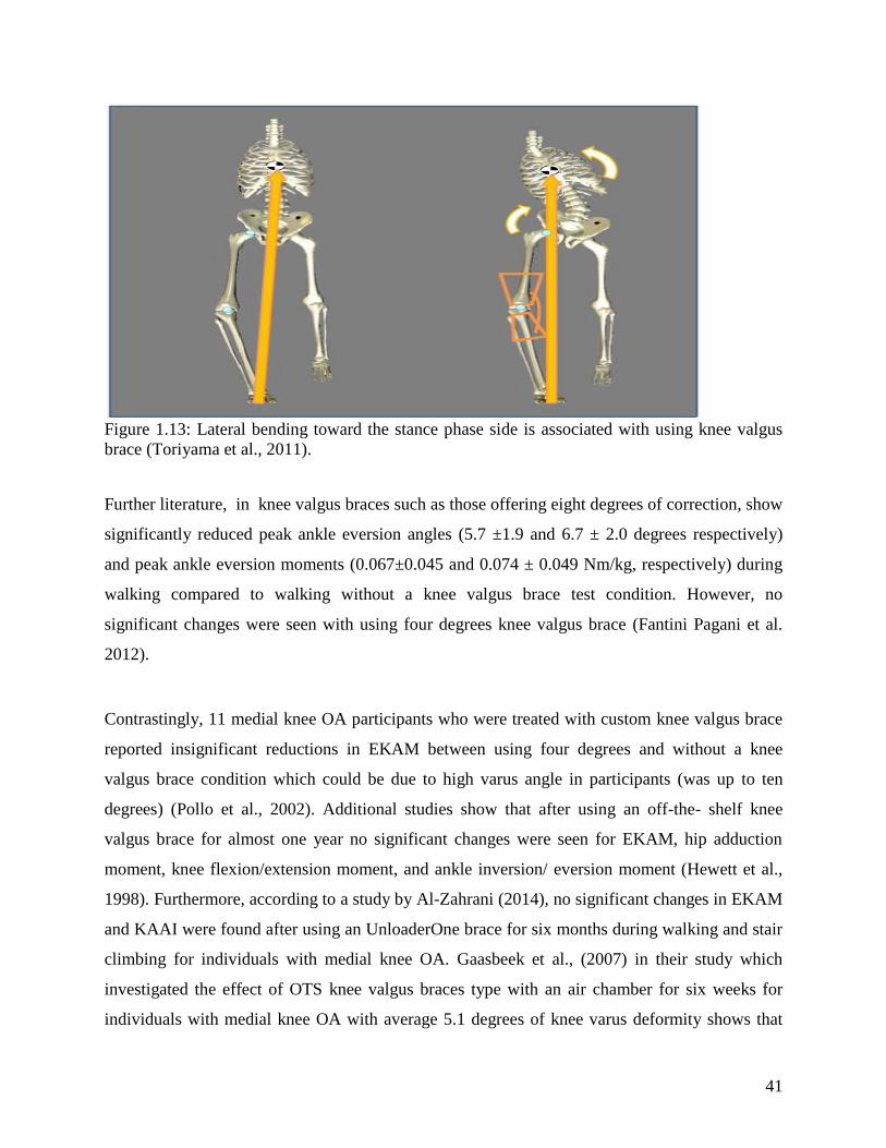
41
Figure 1.13: Lateral bending toward the stance phase side is associated with using knee valgus
brace (Toriyama et al., 2011).
Further literature, in knee valgus braces such as those offering eight degrees of correction, show
significantly reduced peak ankle eversion angles (5.7 ±1.9 and 6.7 ± 2.0 degrees respectively)
and peak ankle eversion moments (0.067±0.045 and 0.074 ± 0.049 Nm/kg, respectively) during
walking compared to walking without a knee valgus brace test condition. However, no
significant changes were seen with using four degrees knee valgus brace (Fantini Pagani et al.
2012).
Contrastingly, 11 medial knee OA participants who were treated with custom knee valgus brace
reported insignificant reductions in EKAM between using four degrees and without a knee
valgus brace condition which could be due to high varus angle in participants (was up to ten
degrees) (Pollo et al., 2002). Additional studies show that after using an off-the- shelf knee
valgus brace for almost one year no significant changes were seen for EKAM, hip adduction
moment, knee flexion/extension moment, and ankle inversion/ eversion moment (Hewett et al.,
1998). Furthermore, according to a study by Al-Zahrani (2014), no significant changes in EKAM
and KAAI were found after using an UnloaderOne brace for six months during walking and stair
climbing for individuals with medial knee OA. Gaasbeek et al., (2007) in their study which
investigated the effect of OTS knee valgus braces type with an air chamber for six weeks for
individuals with medial knee OA with average 5.1 degrees of knee varus deformity shows that

42
the peak value of EKAM did not reduced significantly and knee range of motion in sagittal plane
was reduced significantly due to reduce knee extension at mid-stance and mid swing phase.
These results could be due to the short length of knee valgus braces which reduces the ability to
apply enough force to correct the knee varus deformity, thereby the knee load.
Furthermore, using a knee valgus brace has some limitations such as the ability to rotate during
walking and providing a limitation in walking ability (Redford et al., 2005). It is difficult to fit
individuals with high body mass which leads them to abandon its usage due to pressure on distal
aspect of the tibia (Divine et al., 2005); also a knee valgus brace may restrict knee flexion and
reduce knee ROM (Richards et al., 2005, Jones et al., 2013). Additionally, some types of OTS
knee valgus braces have a bulky design, and are uncomfortable to use for long period because of
the tight straps around the knee joint (Stamenović et al., 2008). As a result, 20% of individuals
with medial knee OA stop using knee valgus brace after six months due to knee swelling or
possible development of a thrombosis (Giori, 2004) or after one year and half due to knee
instability (Liu et al., 1998). A total of 25% of individuals with knee OA reported continued
regular use of a knee valgus brace for two years; however, no radiological or clinical changes are
reported with ongoing usage, while the rest complain of skin irritation, discomfort, poor fit
(Squyer et al., 2013); thereby only 1% of 5,187 individuals with knee OA within five years were
recommended in a study to use knee valgus bracing (Dhawan et al., 2014).
iv. Knee Ankle Foot Orthoses (KAFOs)
One other option is to use a Knee Ankle Foot Orthoses (KAFOs) which are another type of lower
limb above knee orthoses to control the hip, knee and ankle joints. A KAFO design mainly
consists of a thigh shell, a shank shell, and an ankle/foot section with lateral or bilateral side bars
(uprights). They also can incorporate single or bilateral orthotic knee joints with or without an
orthotic ankle joint. Many types of KAFOs have been developed, but generally KAFO can be
conventional (metal with leather) or cosmetic (plastic or carbon fibre). A conventional KAFO
design has metal or carbon fibre side members (uprights) with proximal thigh structures made
from leather with a padded inner surface mounted on a metal superstructure. Cosmetic types can
be manufactured using thermoplastic, laminated plastic, or carbon fibre for the superstructures

43
which are less heavy than conventional KAFO, more cosmetically acceptable, and easier to don
and doff (Bowker, 1993).
KAFOs are routinely prescribed for neuromuscular diseases such as poliomyelitis, Gullian Barre
syndrome, and quadriplegia, osteomyelitis of the femur, genu valgum, genu varum, genu
recurvatum, femoral fractures, or Blount’s disease (Taktak and Bowker, 1995). A KAFO
manages a knee varus deformity by maintaining the tibia in a neutral position and controls the
ankle joint. The orthotic knee joint restricts adduction and abduction of the knee; usually by the
addition of corrective forces via the superstructure. One orthotic knee joint (the offset joint) can
correct a knee varus/ valgus deformity up to ±75 mm (Johnson et al., 2004), and produce a 50-
90% reduction in tibia varum (Whiteside, 2011). Furthermore, KAFOs with offset knee joints are
able to increase knee flexion and prevent hyper extension because the centre of movement for
the offset orthotic knee joint is located posterior to the upright which means the wearer can
achieve knee flexion with a lower flexion moment (Johnson et al., 2004). Therefore, a KAFO
can be expected to support the anatomical knee joint in all planes. Furthermore, the absence of
straps around the knee joint may be more comfortable for long term use without restricting knee
flexion unlike the knee valgus brace.
In addition, walking with a KAFO alters the knee moment in the frontal plane by applying three
point pressure to correct knee alignment deformity (Brehm et al., 2007) (Figure 1.14), which
potentially decreases load on the knee and may delay osteoarthritis progression (Hurwitz et al.,
1997). In one study, a light weight KAFO has been shown to increase speed (3%), increase knee
flexion (2.2 degrees), improve COP progression in mid-stance (1.3 mm) compared to an old
heavy weight KAFO by improving the foot and ankle rockers to give a more effective push off
for 20 polio patients (Brehm et al., 2007).
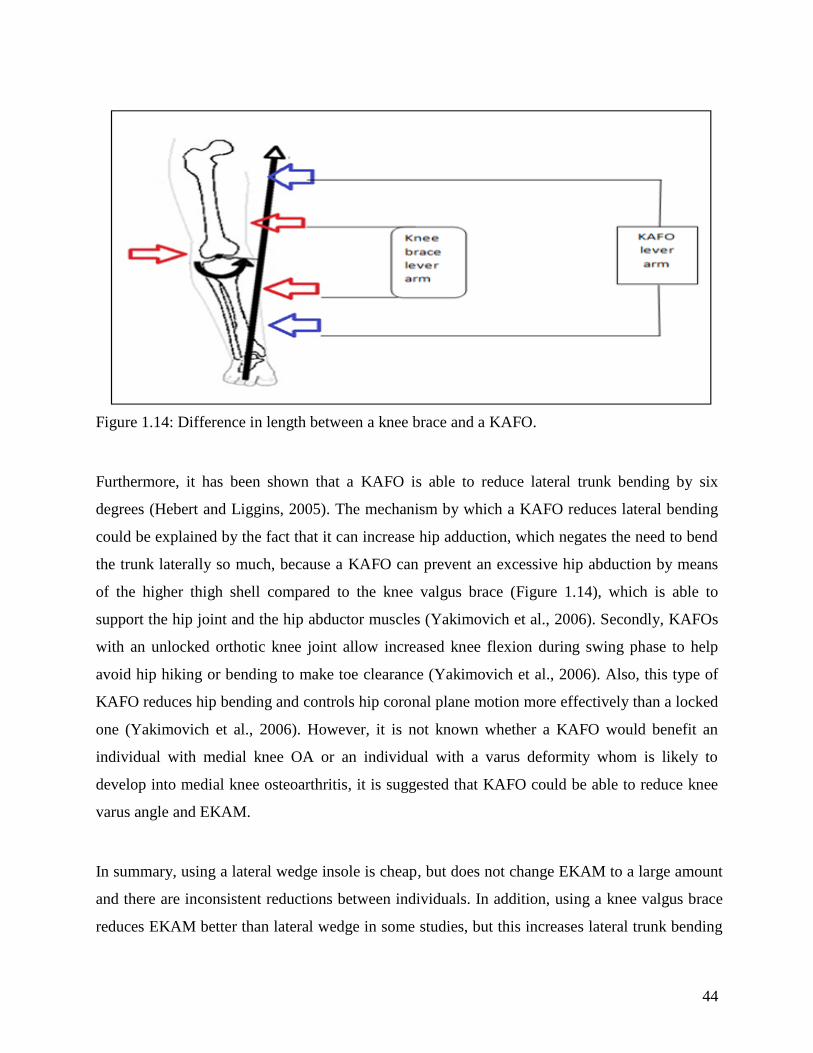
44
Figure 1.14: Difference in length between a knee brace and a KAFO.
Furthermore, it has been shown that a KAFO is able to reduce lateral trunk bending by six
degrees (Hebert and Liggins, 2005). The mechanism by which a KAFO reduces lateral bending
could be explained by the fact that it can increase hip adduction, which negates the need to bend
the trunk laterally so much, because a KAFO can prevent an excessive hip abduction by means
of the higher thigh shell compared to the knee valgus brace (Figure 1.14), which is able to
support the hip joint and the hip abductor muscles (Yakimovich et al., 2006). Secondly, KAFOs
with an unlocked orthotic knee joint allow increased knee flexion during swing phase to help
avoid hip hiking or bending to make toe clearance (Yakimovich et al., 2006). Also, this type of
KAFO reduces hip bending and controls hip coronal plane motion more effectively than a locked
one (Yakimovich et al., 2006). However, it is not known whether a KAFO would benefit an
individual with medial knee OA or an individual with a varus deformity whom is likely to
develop into medial knee osteoarthritis, it is suggested that KAFO could be able to reduce knee
varus angle and EKAM.
In summary, using a lateral wedge insole is cheap, but does not change EKAM to a large amount
and there are inconsistent reductions between individuals. In addition, using a knee valgus brace
reduces EKAM better than lateral wedge in some studies, but this increases lateral trunk bending

45
and reduces knee flexion during swing phase. Therefore, this study aims to evaluate the
biomechanical benefits of using a custom made KAFO in comparison to a standard shoe, well-
established custom-made and off-the-shelf Ossur UnloaderOne knee valgus braces on knee load
in a healthy individual with a knee varus angulation. Moreover, the hip and knee range of motion
will be examined to find which intervention is more useful to improve and support the hip and
knee joints. Ankle joint data will also be examined as a secondary outcome to evaluate the effects
of a KAFO on ankle joint alignment and range of motion. The results of the study will help us to
further understand the biomechanical effects of a KAFO in a varus knee deformity and will help
to determine if a KAFO is suitable to be used for individuals with medial knee OA as a
conservative intervention in future. However, before embarking with the comparative KAFO
study, it was necessary to examine the reliability of the interlaced stairway which was used during
the KAFO study for the stair climbing condition. Therefore, Chapter two will discuss the
methodology, while Chapter three will review several studies exploring the reliability of different
stairway designs.
Therefore, the following aims and hypotheses are presented:
To determine if a KAFO is able to reduce knee load significantly.
Hypothesis: A KAFO reduces EKAM values significantly more than custom or off-the-shelf knee
valgus braces during walking and during stair climbing in subjects with knee varus angulation.
To determine if a KAFO is able to increase hip and knee ranges of motion significantly.
Hypothesis: A KAFO increases hip and knee ROMs significantly more than custom or off-the-
shelf knee valgus braces when walking and during stair climbing in subjects with knee varus
angulation.

46
Chapter two: Methods
Chapter one detailed how a KAFO could be effective in correcting primary knee alignment
problems associated with individuals with medial compartment knee OA. A KAFO could be a
viable alternative form of orthotic intervention in this group; especially in those individuals who
are not responsive to other forms of orthotic treatment. The long corrective lever arms associated
with KAFOs compared to unloader knee valgus braces makes KAFOs potentially more suitable
to those individuals who are unable to tolerate high interface forces and for individuals whose
knees are more resistant to coronal plane correction, plus those with more advanced disease or
with excessive femuro-tibial varus angulation.
It was therefore logical to test a specific design of KAFO on the subjects suffering from a knee
varus deformity whilst undertaking specific activities in a gait laboratory setting to determine its
efficacy in improving specific primary outcome measures. Alterations to the EKAM, gait
kinetics and kinematics and other pertinent parameters were chosen as parameters to be tested to
provide a comparison to other types of knee orthoses to enable an analysis of the efficacy of a
KAFO as a form of orthotic intervention, and the feasibility of offering this type of orthosis as a
viable alternative for individuals with medial compartment knee OA. However, due to short time
of the project and the specific criteria which the subjects should have, only one subject was
accepted in the main study.
The general methodological details utilised in this thesis such as the gait laboratory equipment,
data reduction techniques, the processing of the data collection, and the data analysis procedures
which were used are detailed in the following sections. The participant demographics and also
the interventions and procedures utilised are presented in chapters three and four.
It was thought prudent to perform an initial feasibility study to confirm that the method used
during biomechanical testing was robust and repeatable and also to give an indication of the
effect size which may be expected in a future larger study.
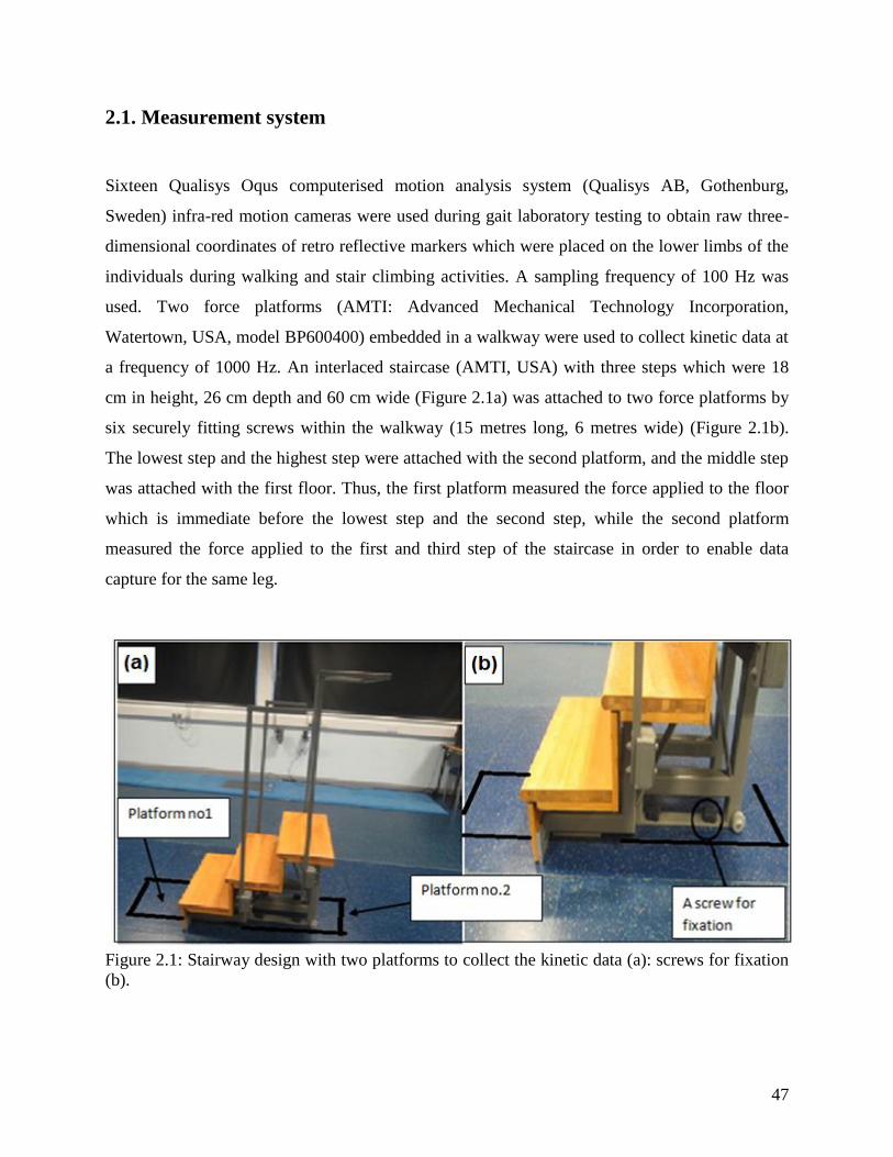
47
2.1. Measurement system
Sixteen Qualisys Oqus computerised motion analysis system (Qualisys AB, Gothenburg,
Sweden) infra-red motion cameras were used during gait laboratory testing to obtain raw three-
dimensional coordinates of retro reflective markers which were placed on the lower limbs of the
individuals during walking and stair climbing activities. A sampling frequency of 100 Hz was
used. Two force platforms (AMTI: Advanced Mechanical Technology Incorporation,
Watertown, USA, model BP600400) embedded in a walkway were used to collect kinetic data at
a frequency of 1000 Hz. An interlaced staircase (AMTI, USA) with three steps which were 18
cm in height, 26 cm depth and 60 cm wide (Figure 2.1a) was attached to two force platforms by
six securely fitting screws within the walkway (15 metres long, 6 metres wide) (Figure 2.1b).
The lowest step and the highest step were attached with the second platform, and the middle step
was attached with the first floor. Thus, the first platform measured the force applied to the floor
which is immediate before the lowest step and the second step, while the second platform
measured the force applied to the first and third step of the staircase in order to enable data
capture for the same leg.
Figure 2.1: Stairway design with two platforms to collect the kinetic data (a): screws for fixation
(b).
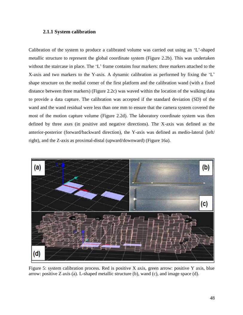
48
2.1.1 System calibration
Calibration of the system to produce a calibrated volume was carried out using an ‘L’-shaped
metallic structure to represent the global coordinate system (Figure 2.2b). This was undertaken
without the staircase in place. The ‘L’ frame contains four markers: three markers attached to the
X-axis and two markers to the Y-axis. A dynamic calibration as performed by fixing the ‘L’
shape structure on the medial corner of the first platform and the calibration wand (with a fixed
distance between three markers) (Figure 2.2c) was waved within the location of the walking data
to provide a data capture. The calibration was accepted if the standard deviation (SD) of the
wand and the wand residual were less than one mm to ensure that the camera system covered the
most of the motion capture volume (Figure 2.2d). The laboratory coordinate system was then
defined by three axes (in positive and negative directions). The X-axis was defined as the
anterior-posterior (forward/backward direction), the Y-axis was defined as medio-lateral (left/
right), and the Z-axis as proximal-distal (upward/downward) (Figure 16a).
Figure 5: system calibration process. Red is positive X axis, green arrow: positive Y axis, blue
arrow: positive Z axis (a). L-shaped metallic structure (b), wand (c), and image space (d).
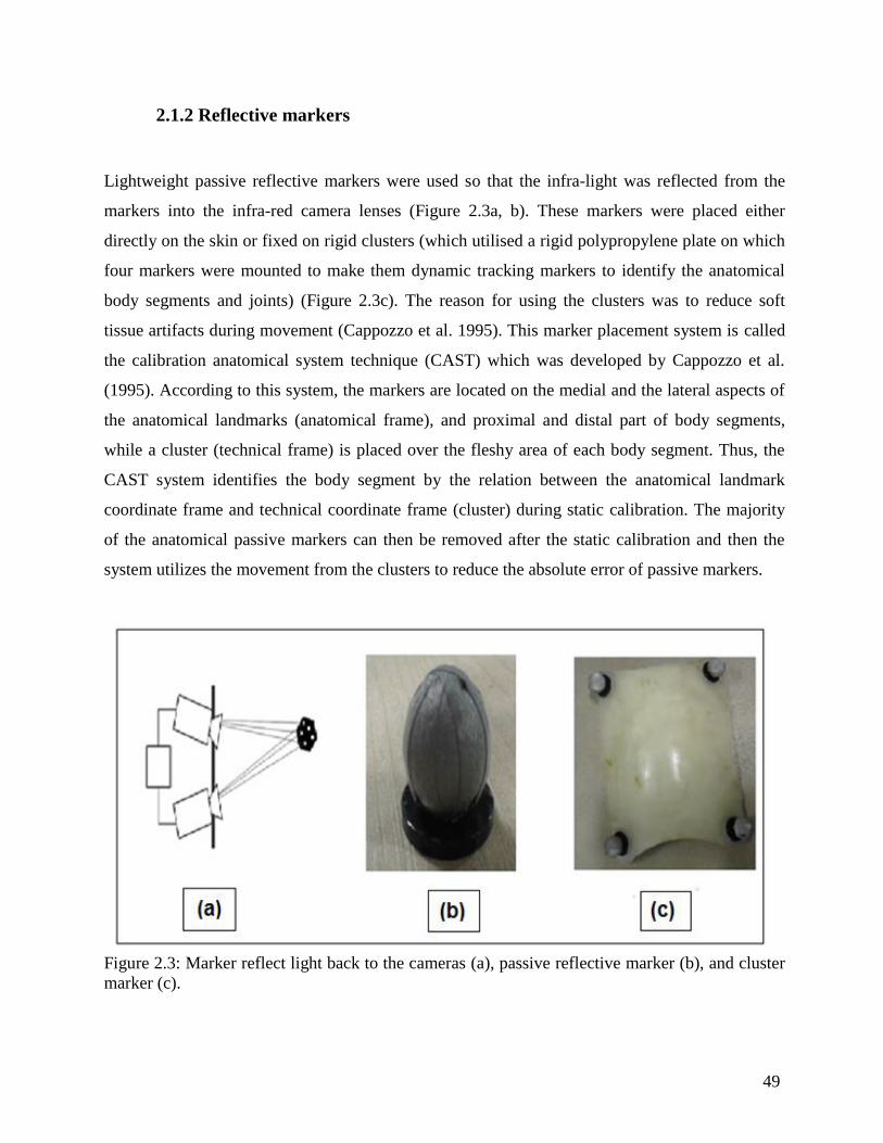
49
2.1.2 Reflective markers
Lightweight passive reflective markers were used so that the infra-light was reflected from the
markers into the infra-red camera lenses (Figure 2.3a, b). These markers were placed either
directly on the skin or fixed on rigid clusters (which utilised a rigid polypropylene plate on which
four markers were mounted to make them dynamic tracking markers to identify the anatomical
body segments and joints) (Figure 2.3c). The reason for using the clusters was to reduce soft
tissue artifacts during movement (Cappozzo et al. 1995). This marker placement system is called
the calibration anatomical system technique (CAST) which was developed by Cappozzo et al.
(1995). According to this system, the markers are located on the medial and the lateral aspects of
the anatomical landmarks (anatomical frame), and proximal and distal part of body segments,
while a cluster (technical frame) is placed over the fleshy area of each body segment. Thus, the
CAST system identifies the body segment by the relation between the anatomical landmark
coordinate frame and technical coordinate frame (cluster) during static calibration. The majority
of the anatomical passive markers can then be removed after the static calibration and then the
system utilizes the movement from the clusters to reduce the absolute error of passive markers.
Figure 2.3: Marker reflect light back to the cameras (a), passive reflective marker (b), and cluster
marker (c).
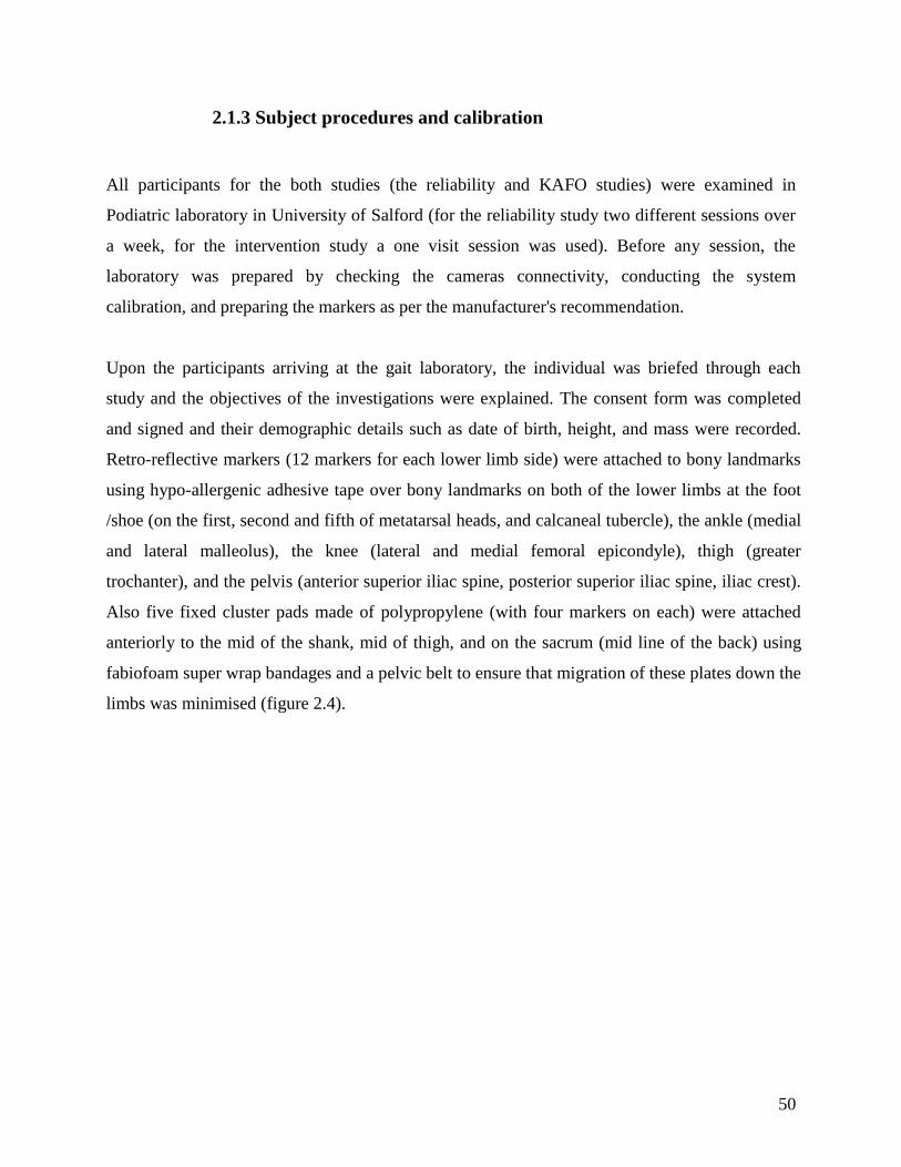
50
2.1.3 Subject procedures and calibration
All participants for the both studies (the reliability and KAFO studies) were examined in
Podiatric laboratory in University of Salford (for the reliability study two different sessions over
a week, for the intervention study a one visit session was used). Before any session, the
laboratory was prepared by checking the cameras connectivity, conducting the system
calibration, and preparing the markers as per the manufacturer's recommendation.
Upon the participants arriving at the gait laboratory, the individual was briefed through each
study and the objectives of the investigations were explained. The consent form was completed
and signed and their demographic details such as date of birth, height, and mass were recorded.
Retro-reflective markers (12 markers for each lower limb side) were attached to bony landmarks
using hypo-allergenic adhesive tape over bony landmarks on both of the lower limbs at the foot
/shoe (on the first, second and fifth of metatarsal heads, and calcaneal tubercle), the ankle (medial
and lateral malleolus), the knee (lateral and medial femoral epicondyle), thigh (greater
trochanter), and the pelvis (anterior superior iliac spine, posterior superior iliac spine, iliac crest).
Also five fixed cluster pads made of polypropylene (with four markers on each) were attached
anteriorly to the mid of the shank, mid of thigh, and on the sacrum (mid line of the back) using
fabiofoam super wrap bandages and a pelvic belt to ensure that migration of these plates down the
limbs was minimised (figure 2.4).
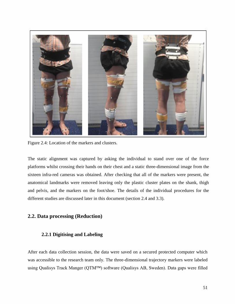
51
Figure 2.4: Location of the markers and clusters.
The static alignment was captured by asking the individual to stand over one of the force
platforms whilst crossing their hands on their chest and a static three-dimensional image from the
sixteen infra-red cameras was obtained. After checking that all of the markers were present, the
anatomical landmarks were removed leaving only the plastic cluster plates on the shank, thigh
and pelvis, and the markers on the foot/shoe. The details of the individual procedures for the
different studies are discussed later in this document (section 2.4 and 3.3).
2.2. Data processing (Reduction)
2.2.1 Digitising and Labeling
After each data collection session, the data were saved on a secured protected computer which
was accessible to the research team only. The three-dimensional trajectory markers were labeled
using Qualisys Track Manger (QTM™) software (Qualisys AB, Sweden). Data gaps were filled

52
to a maximum of ten frames using a polynomial interpolation cubic spline which is able to
smooth the small random digitising errors of the markers during the movement (Woltring e al.,
1985).
2.2.2 Modeling
All data were then exported to C3D files to be imported into Visual 3D™ software (C-Motion,
USA, version five). A six-degree of freedom model was built to give a full picture of the joints'
coordination and orientation in space after labeling the markers (Figure 2.5 a, b). This model
examines the linear movement and the angular movement in the three planes (three rotations and
three translations) by establishing a rigid body frame based on segments linking the hip, knee,
and ankle joints (Figure 2.5c). The hip joint centre was determined by anterior superior iliac
spine markers (ASIS) which uses a regression equation and it is determined by 14% of average
distance between the left and right ASIS, and also a position 30% distally and 19% posterior to
this point (Bell et al., 1990). The knee centre was determined by the medial and lateral knee
markers, and the ankle joint centre was identified by the medial and lateral malleoli. The
participant's height and mass were entered and the model then calculated the segments centre of
mass, and segment radius based on the anthropometrical indices published by Dempster (1955).

53
Figure 2.5: Modeling process. Labelling (a), modelling (b), joint centres of motion (yellow) (c).
To collect kinetic data during stair climbing, two force structures in Visual 3DTM were added.
The location and dimensions between stair corners were computed in relation to the global
coordinates and the two force platforms using AMTI force structures (figure 2.6).
54
Figure 2.6: AMTI stairway force structures.
2.2.3 Data processing
A Butterworth fourth order bi-directional low-pass filter with cut-off frequencies of 6Hz and
25Hz for kinematics and kinetics respectively was used. This type of filter accepts the low
frequency signals and prevents the high frequency data which are mainly due to noisy results
from random movement of markers (random error) and soft tissue artifacts.
During stair ascending and descending, gait cycle events needed to be identified in order to
normalise data to allow comparisons to be made between the orthotic test conditions. The gait
cycle starts when any part of the foot touches the stair structure until the same foot (ipsilateral)
touches the next step. For the ascending condition, the gait cycle starts when the foot touches the
first step and ends when the ipsilateral foot contacts the third step, while the single limb support
period starts when the non-selected left foot leaves the floor until same foot touches the second
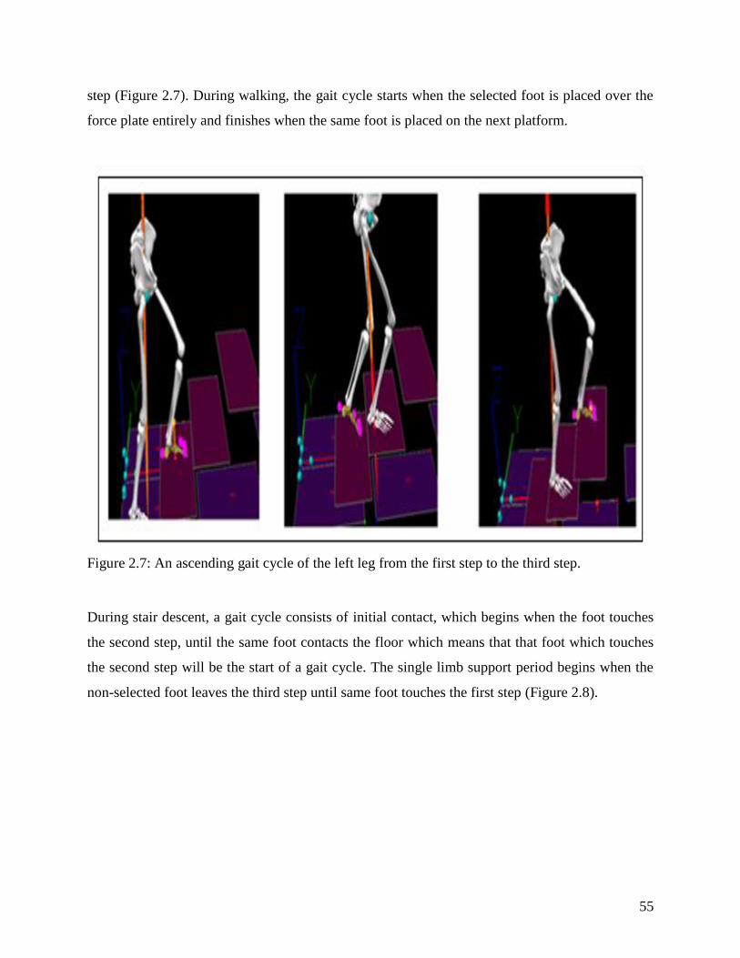
55
step (Figure 2.7). During walking, the gait cycle starts when the selected foot is placed over the
force plate entirely and finishes when the same foot is placed on the next platform.
Figure 2.7: An ascending gait cycle of the left leg from the first step to the third step.
During stair descent, a gait cycle consists of initial contact, which begins when the foot touches
the second step, until the same foot contacts the floor which means that that foot which touches
the second step will be the start of a gait cycle. The single limb support period begins when the
non-selected foot leaves the third step until same foot touches the first step (Figure 2.8).

56
Figure 2.8: Descending gait cycle for the right leg from the second step to the floor.
2.2.4 Joint angle and joint moment calculations
The angle between segments was calculated according to the relative position between the
segments using the Euler rotation sequence equivalent XYZ (flexion/extension,
abduction/adduction, internal and external rotation). For example, the proximal segment for knee
joint is the thigh, while the shank is the distal segment; therefore, knee range of motion during
the gait cycle (initial contact to ipsilateral initial contact) in the sagittal and frontal plane depends
on the orientation between these two segments.
Knee moments were computed using an inverse dynamic approach. This approach examines the
external ground reaction vectors (GRF) of the body segments and moments on the anatomical
joints which are due to internal muscle activity. The result of this approach relies on equilibrium
mathematical formulae which start by calculating the moment and force for every joint from toe
to hip (Silva and Ambrósio, 2002). During the inverse dynamic approach, the location and

57
magnitude of the mass for each segment were used to calculate the inertial moment for each
segment based on the subject's anthropometric parameters (Dempster, 1955).
2.3. Data analysis
All the data were exported as a text file to Microsoft Excel to facilitate the analysis and
presentation. Lower limb joint angles and moments were exported as well as the temporal and
spatial data which were collected during the trials.
However, before any study could be initiated with the three interventions (i.e. the bespoke and
OTS unloader braces plus the KAFO), a reliability study was needed to ensure that reliable data
could be collected and prove to be repeatable during stair climbing and descending. This will be
discussed in the next chapter.
58
Chapter three: Reliability of knee
kinematics and kinetics using an interlaced
stairway
3.1. Introduction
Stair climbing is one of the most important daily activities and the main challenge for the older
person who has muscle weakness. Previous studies have shown that 10.7% of falls in the older
population are due to stair climbing, particularly during outdoor activities (Bergland et al., 2003).
Additionally, women have a higher risk of falling compared to men amongst the 65 years old
population which leads to 230,000 of them having a hip fracture in the USA annually (Sattin,
1992). Falls due to stair usage can have an impact on the older individual by making them feel
scared of falling down again, forcing them to use handrails, modifying their gait, or reducing
their speed. Previous studies have shown that after falling down women become less confident
and use handrails more often than the men (Hamel and Cavanaugh, 2004).
In chronic diseases such as knee OA, where knee stiffness and range of motion limitations occur,
these individuals have difficulty in rising from a chair, and standing and walking for long
periods, and also when climbing and descending stairs (Kaufman et al., 2001, Whatling et al.,
2007). This limitation during stair climbing can be explained by Yu et al. (1997b) who stated that
the knee varus angle and the internal knee abduction moment are higher during stair climbing
than walking on a flat surface due to the large amount of knee flexion which is needed to ascend
or descend stairs. However, since the varus deformity is the main problem associated with
medial knee osteoarthritis, many individuals with knee OA try to avoid stair climbing, or adapt
their gait by walking slowly and decreasing the knee flexion angle (Kaufman et al., 2001), or
using assistive devices such as canes and crutches.
One additional explanation could be due to the high internal knee extension moment which is
correlated with ascending and descending (Andriacchi et al., 1980). This moment increases the

59
knee load by 12-25% (Morrison, 1969); and as a result the severity of pain forces individuals to
avoid using stairs in order to reduce their overall activity level.
The gait cycle during walking is different from the gait cycle during stair climbing. The main
difference between them is that the degree of maximum knee flexion is higher during stair
climbing than normal walking on a level surface, but differs depending on the height of the steps.
Furthermore, the ascending and descending gait cycles also consist of different tasks. For
instance, the stance phase of the ascending gait cycle contains three main phases: weight
acceptance, pull up, and forward continuation (McFadyen and Winter, 1988, Zachazewski et al.,
1993). The knee joint is the dominant joint during ascending to support the hip and ankle joint,
while the ankle joint generates more energy to facilitate moving up stairs with primarily
concentric muscle contractions. The stance phase of descending contains three main phases:
weight acceptance, forward continuance, and controlled lowering. The hip joint is the dominant
joint which supports the lower limb moving downstairs. Additionally, reduced muscle power is
needed (Svensson and Holmberg, 2007) with primarily eccentric contractions to control the body
weight against the rapid acceleration due to gravity and add more stability and safety to avoid
sudden falling down (McFadyen and Winter, 1988).
3. 2. Literature review
Lower limb biomechanics for healthy and individuals with pathology during stair climbing have
been evaluated by some studies, but unfortunately there are still some areas of little knowledge
about stairs and the reliability of different stair designs. Stairs are usually one of the daily
encountered activities; therefore, stairways have been designed to be used in gait laboratories
recently to evaluate individuals' behavior during stair ascent and descent, to discriminate
between different pathology groups and control groups, and to evaluate the benefit of using
interventions for some patient groups. From a kinematic and kinetic standpoint, reliability of the
data captured with stairs is of utmost importance, especially if one is determining the effect of an
intervention period. It could be that the variability of stair climbing is greater than steady state

60
walking as the task is more difficult, but there is a very limited evidence base for reliability
studies.
Reliability analysis is mainly measured with intraclass correlation coefficients (ICC),
Coefficients of Variation (CVs), and Correlation Coefficients (r). The error of reliability results
are examined by the standard error of measurement (SEM) and Root Mean Square Deviation
(RMSD). ICC represents the reliability among the participants by dividing the true variance by
the total variance (Bruton et al., 2000, Stratford and Goldsmith, 1997). However, CVs are
calculated by dividing standard deviation by the mean of the data and multiplied by 100% to give
score as a percentage (Bruton, et al., 2000). ICC is recommended to be used for reliability more
than CVs because the percentage expression depends on the size of observation and result could
be different clearly between small and large sample size (Chinn, 1991). Correlation Coefficient
(r) represents how two sets of data are varying together, but it does not calculate the systemic
measurement error.
Furthermore, the standard error of measurement (SEM) represents the measurement error for a
single participant without the influence by other measurements for other participants, but
influenced by error variation only, and smaller SEM’s indicate a high reliability; whereas, the
calculation of Root Mean Square Deviation (RMSD) gives the overall error in the measurement
of the joint angles (Bruton et al., 2000, Stratford and Goldsmith, 1997).
The reliability of different stairway designs to measure the kinematics and kinetics of the lower
limb has also been examined previously. Protopapadaki et al. (2007) reported the kinematic and
kinetic patterns in the lower limb (hip, knee, and ankle) during climbing four steps (with a height
of 18 cm, tread length 28.5 cm, and with a force platform embedded on step two only) for a
young healthy population (18-39 years), The results were variable (coefficient of variation)
particularly for the hip extension moment which varied from 4.65% (SD 2.99) to 21.34% (SD
19.64) for stair ascent and from 8.65% (SD 7.42) to 40.73% (SD 24.27) for stair descent (Table
3.1). This was explained by the individuals changing their trunk movement which was not
included in the data. Joint angles (hip, knee, and ankle) were less widely varied between 2.35%
(SD 1.83) to 5.77% (SD 4.18) during stair ascent and descent indicating good reliability. The

61
exception was during plantarflexion during stair ascent, which was 17.53% (SD 13.62), which
was likely to be due to gait changes during testing such as making contact with the stairs with a
flatfoot or with the forefoot only.
Table 3.1: Coefficient of variation for angles and moments during stair ascent and descent
(Protopapadaki et al., 2007).
Kowalk et al. (1996) reported the knee moment in the sagittal and frontal plane when climbing
three steps of with a 20 cm height and 25cm width with force plates embedded in two of the
stairs for ten healthy adults (22-40 years old). The results demonstrated good repeatability with
CVs ranging from 0.09- 0.45(lower CVs show less percentage error).
McFadyen et al. (1988) examined stair repeatability to evaluate the kinematics and kinetics in the
sagittal plane only for lower limb joints for three young healthy (mean 24.6 years old) men
during ascending and descending five steps (height 22cm and 28cm tread width) with two
platforms added to the second and fourth to examine the kinetic data from second to fourth step.
The results showed that inter-person variability (r) for the three joint angles was more 0.95,
while the moments varied between 0.82 and 0.98. For instance, knee moments were 0.96 and
0.88 during stair ascending and descending respectively. The hip moments showed the greatest
variability, which demonstrates that hip musculature strength was the dominant factor in to
controlling single limb support (Table 3.2). The number of participants was, however, limited.

62
Table 3.2: Inter subject correlation coefficient (r) during stair ascent (a), and descent (b)
(McFadyen et al., 1988).
Ground reaction force (GRF) reliability has been examined by some studies. Stacoff et al.,
(2005) examined GRF data for participants with three different age groups (between 33 and 60
years old) and with three different steps inclinations (mean 13.3cm height, and 29.6cm width.
This study found that the variability was higher during descending than ascending. The
coefficient of variation (CV) was between 5-10% during ascending, and 15-20% during
descending which are considered normal (i.e. more variability than that could be considered as a
gait asymmetry). Leitner et al (2011) examined the GRF reliability during stair climbing for 42
older individuals (aged 80.1 years ±6.4) using two platforms under the third and fourth step of a
staircase with six steps (17cm height, and 29 cm width). The reliability results were considered
to be moderate to good (with ICCs of between 0.739 and 0.918 and CVs between 8.79% and
12.52%) for both stair ascent and descent.
In stair climbing studies, the measurement of the kinetics depends on the design of the stairway.
All of the previous studies have primarily used two main designs of staircase: those with
embedded force platforms in the steps ( the original design) or using different rigid blocks over
the platforms to form stairs (a later design). The original design is expensive and needs setting up
at a determined place and would not allow for a quick change over for walking gait to be
collected. The later design uses different rigid blocks lengths over platforms to form stairs with
the stairs dependent on the number of platforms in gait laboratory and that they are situated on
top of them.
63
Another unique option is an interlaced stairway (Della Croce and Bonato. 2007) which was
developed to utilise the force platforms which are situated in a walkway with a secure fixation
into the surface of the forceplate. This allows the examination of the ground reaction force and
centre of pressure (COP) progression from the two platforms embedded in walkway and not
embedded in the steps. By using that unique structure, the force is examined on each step
separately, and the estimated COP coordination shows a good agreement with the horizontal
coordinates of the geometric centre of the calibration object utilized to assess accuracy and
precision of the CP estimates (max difference <6 mm) (Della Croce and Bonato. 2007).
However, the repeatability of using such a staircase design on the kinematics and kinetics of
ascent and descent has not been demonstrated as there are only three steps to the staircase design
and this may cause some variability in the movements. This is important for future studies when
assessing interventions given that individuals may come at different time points so ensuring
reliability is of utmost importance. In addition, the previous studies have not measured SEM
which is important to find out the measurement error for every participants and supports the
reliability results. Therefore, the purpose of this study was to determine the inter-session
reliability of an interlaced stairway whilst ascending and descending stairs and to measure SEM
and RMSD for inspection of the overall error in angular measurements. Thus, this study
evaluated the primary outcome of the further planned trial (knee sagittal and frontal angles and
moments) using 16 cameras in five males and five females during climbing three steps. This
information will be important for future studies which depend on interlaced stairway to evaluate
an individual's performance and interventions' effects during climbing the interlaced stairway.
3.3. Method
3.3.1 Subjects
Ten healthy participants took part in this feasibility study (five males, five females), aged 32.1
±6.8 years; height 167.9± 7 m; and mass 69.35 ±10.4 kg. The participants were recruited by
volunteering via a poster intended to recruit university students and staff (Appendix 6c). The

64
inclusion criteria included being generally fit, with no previous knee injury or knee pain, the
ability to ascend and descend stairs without using a handrail, and the ability to walk without
using external assistive devices such as crutches, and be aged between 20-60 years. The study
was approved by the University Research and Governance ethical committee and informed
consent was obtained from each individual (Appendix 6D).
3.3.2 Test procedures
After capturing the static alignment, the participants were then asked to ascend and descend the
stairs for one minute to ensure that they felt comfortable and were able to use stairs properly.
After they had familiarised themselves with the task, they were asked to ascend the three steps
with their normal self-selected walking speed, without using the handrail. When they reached the
third step, they were asked to stop for few seconds and then turn around and descend the two
steps until they reached the floor. This test was repeated five to eight times, but no more to avoid
fatigue. In the next session, every participant repeated the test again under the same conditions.
One hour was required for each session to collect the data.
3.3.3 Outcome measures
The key variables assessed included the averaged maxima of kinetic and kinematic parameters at
initial contact (0% of gait cycle), loading response (10%), mid stance (30%), late stance (50%)
and mid swing (73% of gait cycle) (Figure 3.1a), and range of motion of the knee in the sagittal
plane. The following primary outcome measures were investigated:
The average maximum frontal plane knee moment in early and late stance.
The average minimum for mid stance (Figure 3.1d).
The average of knee adduction angular impulsive (KAAI), which is the area under knee
adduction moment curve (Kean et al., 2012).
The average maximum and minimum sagittal knee moment (Figure 3.1c).
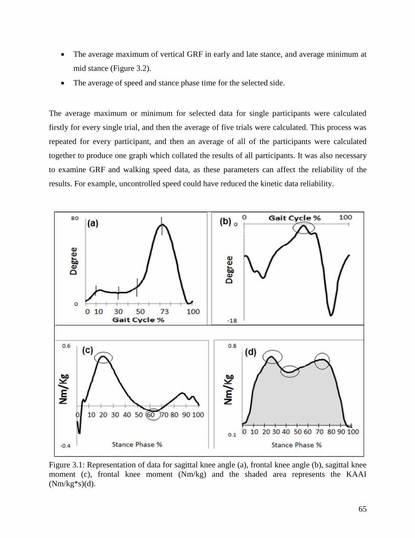
65
The average maximum of vertical GRF in early and late stance, and average minimum at
mid stance (Figure 3.2).
The average of speed and stance phase time for the selected side.
The average maximum or minimum for selected data for single participants were calculated
firstly for every single trial, and then the average of five trials were calculated. This process was
repeated for every participant, and then an average of all of the participants were calculated
together to produce one graph which collated the results of all participants. It was also necessary
to examine GRF and walking speed data, as these parameters can affect the reliability of the
results. For example, uncontrolled speed could have reduced the kinetic data reliability.
Figure 3.1: Representation of data for sagittal knee angle (a), frontal knee angle (b), sagittal knee
moment (c), frontal knee moment (Nm/kg) and the shaded area represents the KAAI
(Nm/kg*s)(d).
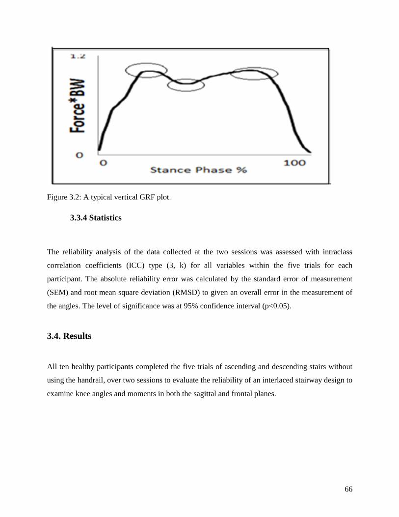
66
Figure 3.2: A typical vertical GRF plot.
3.3.4 Statistics
The reliability analysis of the data collected at the two sessions was assessed with intraclass
correlation coefficients (ICC) type (3, k) for all variables within the five trials for each
participant. The absolute reliability error was calculated by the standard error of measurement
(SEM) and root mean square deviation (RMSD) to given an overall error in the measurement of
the angles. The level of significance was at 95% confidence interval (p<0.05).
3.4. Results
All ten healthy participants completed the five trials of ascending and descending stairs without
using the handrail, over two sessions to evaluate the reliability of an interlaced stairway design to
examine knee angles and moments in both the sagittal and frontal planes.

67
3.4.1 Primary outcomes
3.4.1.1 Knee joint kinematics during ascending and descending
The average maximum for knee flexion/extension angle repeatability at initial contact (0% of
gait cycle), loading response (0-10%), mid stance (11-30%), late stance (31-50%), mid swing
(73% of gait cycle), and range of motion (difference between mid-swing and initial contact) were
excellent for ascending and descending (ranging from ICC 0.85- 0.93 and 0.96- 0.98
respectively). The SEM raised to 2.2 degree, and the RMSD raised to 3.6 degrees (Table 3.3 and
3.4, Figure 3.3 and 3.4). The average maximum of knee angle ICC in the frontal plane was 0.96
in ascending and 0.98 in descending.
Figure 3.3: Knee angle in two session during stair ascent (sagittal plane (a), frontal plane (b)).

68
Figure 3.4: Knee angle in two sessions during stair descent (sagittal plane (a), frontal plane (b)).

69
Table 3.3: Reliability knee joint kinematics variables.
Variable Ascending
Mean (±SD)
Ascending
ICC
95% CI
SEM Descending
Mean (±SD)
Descending
ICC
95% CI
SEM Lower
Bound
Upper
Bound
Lower
Bound
Upper
Bound
Sagittal knee angle
at initial contact 72.9±4.7 0.88 0.54 0.97 1.42 10.9±3.8 0.92 0.69 0.98 1.18
Sagittal knee angle
at loading response 69.2±3.8 0.90 0.61 0.97 1.16 18.6±5.8 0.90 0.60 0.97 1.78
Sagittal knee angle
in midstance 35.2±6.4 0.85 0.41 0.96 1.92 25.2±7.1 0.9 0.86 0.99 2.23
Sagittal knee angle
in terminal stance 18.4±4.9 0.86 0.46 0.96 1.46 59.6±6.9 0.95 0.81 0.98 2.13
Sagittal knee angle
in mid swing 106.6±7.4 0.93 0.72 0.98 2.27 100.2±6.8 0.98 0.93 0.99 2.15
Sagittal knee range
of motion 33.6±4.9 0.92 0.69 0.98 1.52 89.2±6.2 0.96 0.85 0.99 1.95
Maximum frontal
knee angle 3.9.1±2.1 0.96 0.85 0.99 1.51 6.3±1.5 0.98 0.94 0.99 1.98
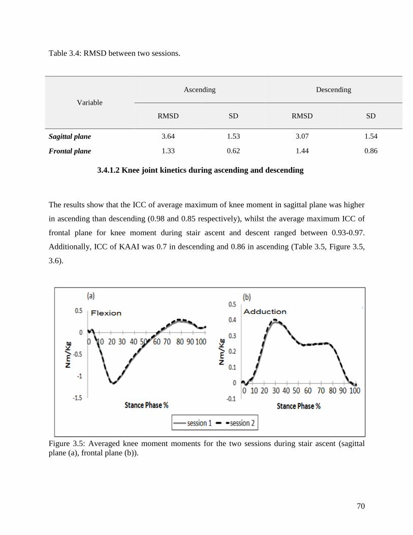
70
Table 3.4: RMSD between two sessions.
3.4.1.2 Knee joint kinetics during ascending and descending
The results show that the ICC of average maximum of knee moment in sagittal plane was higher
in ascending than descending (0.98 and 0.85 respectively), whilst the average maximum ICC of
frontal plane for knee moment during stair ascent and descent ranged between 0.93-0.97.
Additionally, ICC of KAAI was 0.7 in descending and 0.86 in ascending (Table 3.5, Figure 3.5,
3.6).
Figure 3.5: Averaged knee moment moments for the two sessions during stair ascent (sagittal
plane (a), frontal plane (b)).
Variable
Ascending Descending
RMSD SD RMSD SD
Sagittal plane 3.64 1.53 3.07 1.54
Frontal plane 1.33 0.62 1.44 0.86
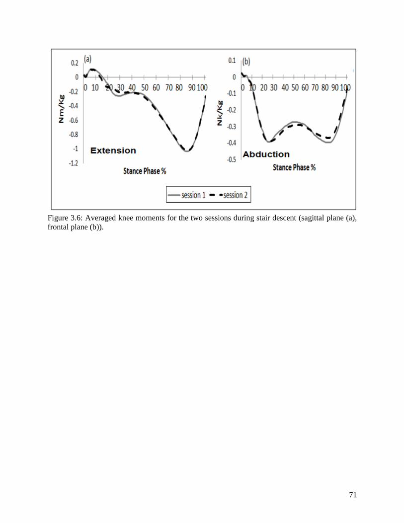
71
Figure 3.6: Averaged knee moments for the two sessions during stair descent (sagittal plane (a),
frontal plane (b)).
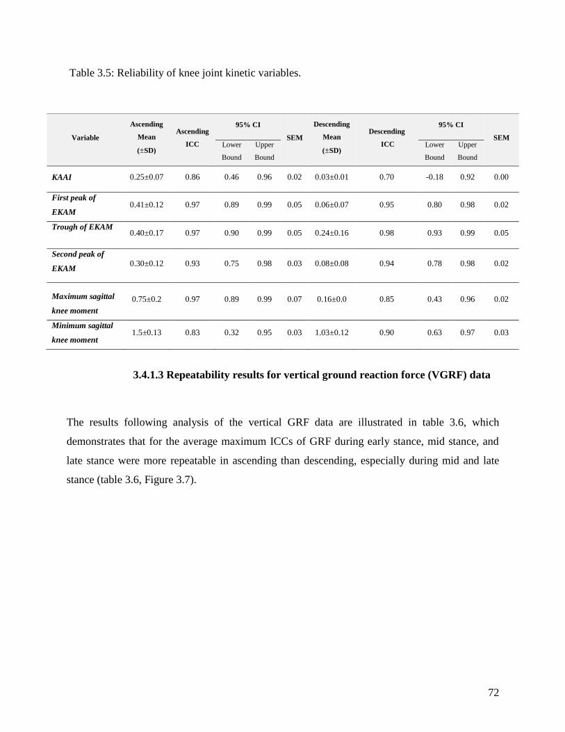
72
Table 3.5: Reliability of knee joint kinetic variables.
3.4.1.3 Repeatability results for vertical ground reaction force (VGRF) data
The results following analysis of the vertical GRF data are illustrated in table 3.6, which
demonstrates that for the average maximum ICCs of GRF during early stance, mid stance, and
late stance were more repeatable in ascending than descending, especially during mid and late
stance (table 3.6, Figure 3.7).
Variable
Ascending
Mean
(±SD)
Ascending
ICC
95% CI
SEM
Descending
Mean
(±SD)
Descending
ICC
95% CI
SEM Lower
Bound
Upper
Bound
Lower
Bound
Upper
Bound
KAAI 0.25±0.07 0.86 0.46 0.96 0.02 0.03±0.01 0.70 -0.18 0.92 0.00
First peak of
EKAM 0.41±0.12 0.97 0.89 0.99 0.05 0.06±0.07 0.95 0.80 0.98 0.02
Trough of EKAM 0.40±0.17 0.97 0.90 0.99 0.05 0.24±0.16 0.98 0.93 0.99 0.05
Second peak of
EKAM 0.30±0.12 0.93 0.75 0.98 0.03 0.08±0.08 0.94 0.78 0.98 0.02
Maximum sagittal
knee moment
0.75±0.2 0.97 0.89 0.99 0.07 0.16±0.0 0.85 0.43 0.96 0.02
Minimum sagittal
knee moment 1.5±0.13 0.83 0.32 0.95 0.03 1.03±0.12 0.90 0.63 0.97 0.03
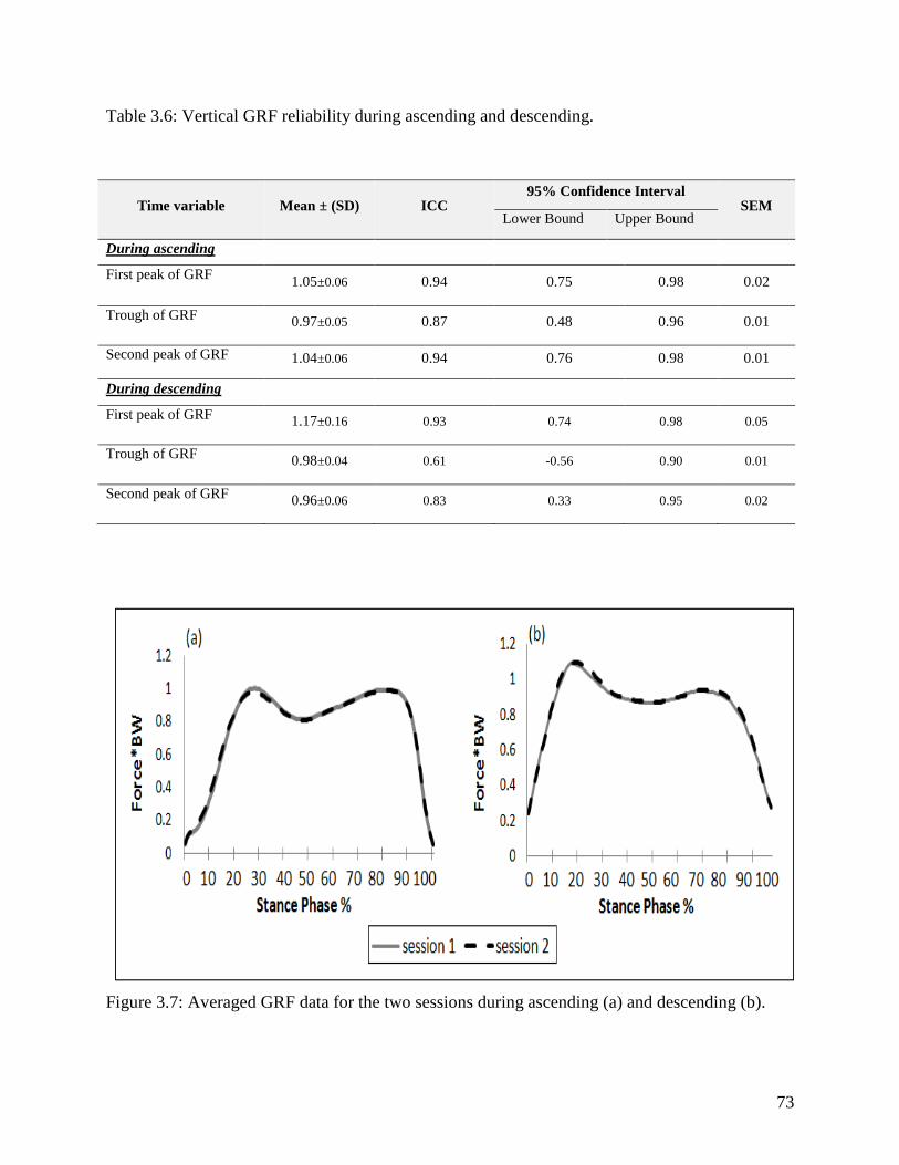
73
Table 3.6: Vertical GRF reliability during ascending and descending.
Figure 3.7: Averaged GRF data for the two sessions during ascending (a) and descending (b).
Time variable Mean ± (SD) ICC 95% Confidence Interval
SEM Lower Bound Upper Bound
During ascending
First peak of GRF 1.05±0.06 0.94 0.75 0.98 0.02
Trough of GRF 0.97±0.05 0.87 0.48 0.96 0.01
Second peak of GRF 1.04±0.06 0.94 0.76 0.98 0.01
During descending
First peak of GRF 1.17±0.16 0.93 0.74 0.98 0.05
Trough of GRF 0.98±0.04 0.61 -0.56 0.90 0.01
Second peak of GRF 0.96±0.06 0.83 0.33 0.95 0.02
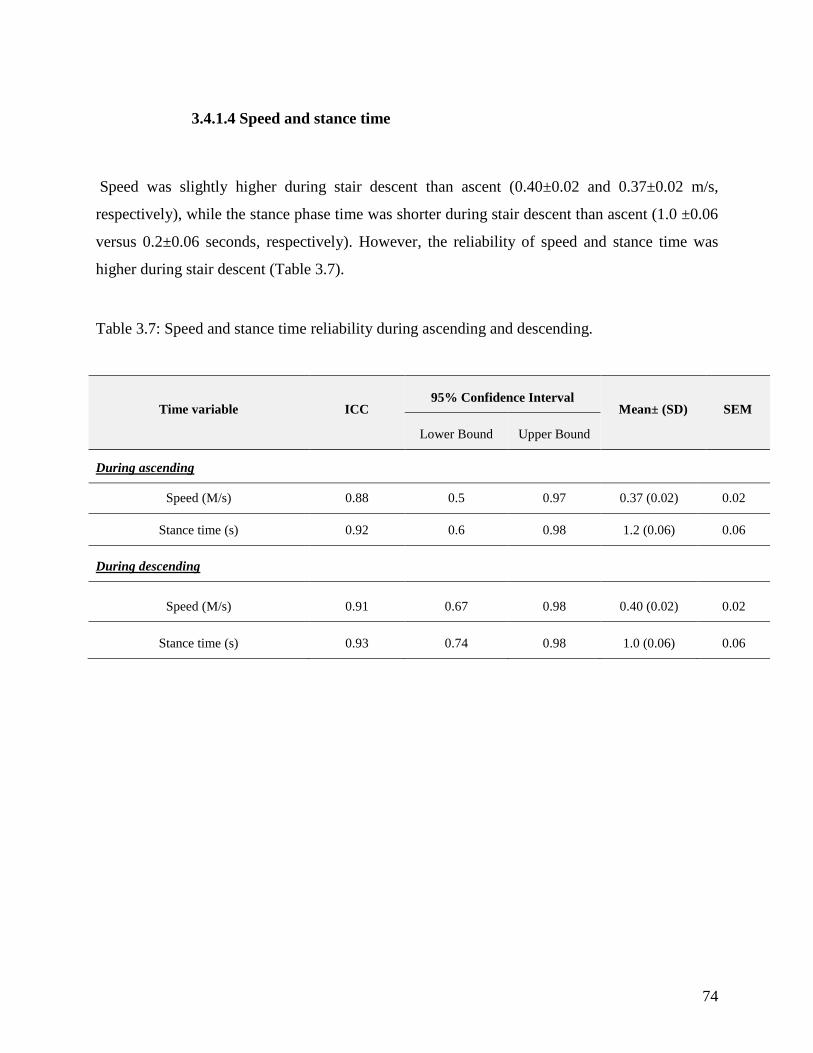
74
3.4.1.4 Speed and stance time
Speed was slightly higher during stair descent than ascent (0.40±0.02 and 0.37±0.02 m/s,
respectively), while the stance phase time was shorter during stair descent than ascent (1.0 ±0.06
versus 0.2±0.06 seconds, respectively). However, the reliability of speed and stance time was
higher during stair descent (Table 3.7).
Table 3.7: Speed and stance time reliability during ascending and descending.
Time variable ICC 95% Confidence Interval
Mean± (SD) SEM
Lower Bound Upper Bound
During ascending
Speed (M/s) 0.88 0.5 0.97 0.37 (0.02) 0.02
Stance time (s) 0.92 0.6 0.98 1.2 (0.06) 0.06
During descending
Speed (M/s) 0.91 0.67 0.98 0.40 (0.02) 0.02
Stance time (s) 0.93 0.74 0.98 1.0 (0.06) 0.06
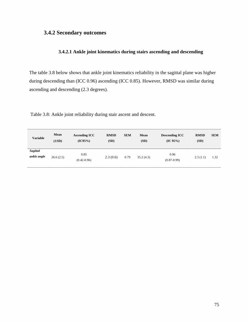
75
3.4.2 Secondary outcomes
3.4.2.1 Ankle joint kinematics during stairs ascending and descending
The table 3.8 below shows that ankle joint kinematics reliability in the sagittal plane was higher
during descending than (ICC 0.96) ascending (ICC 0.85). However, RMSD was similar during
ascending and descending (2.3 degrees).
Table 3.8: Ankle joint reliability during stair ascent and descent.
Variable Mean
(±SD)
Ascending ICC
(IC95%)
RMSD
(SD)
SEM
Mean
(SD)
Descending ICC
(IC 95%)
RMSD
(SD)
SEM
Sagittal
ankle angle 26.6 (2.5) 0.85
(0.42-0.96) 2.3 (0.6) 0.79 35.2 (4.3)
0.96
(0.87-0.99) 2.3 (1.1) 1.32

76
3.5 Discussion
3.5.1 Discussion
Stair climbing is one of the most commonly performed daily activities and can be one of most
challenging tasks for some individuals such as those with neuromuscular impairments, painful
lower limb symptoms, or the older population. Different stair designs have been used in gait
laboratories in order to distinguish gait patterns between healthy and pathological groups and
also to evaluate the efficacy of different interventions for pathology groups. In all previous
reliability studies on stairs, the two-stairway designs have primarily been used. Initial designs
embedded force platforms in the steps, but more recently different rigid blocks have been added
over the force platforms to simulate steps. However, the interlaced design which was utilised in
this study has not been evaluated before. Therefore, this study looked at the reliability of an
interlaced stairway design to examine knee kinematics and kinetics during stair ascent and
descent in ten healthy adult individuals. To the author’s knowledge no previous studies have
evaluated the reliability of this interlaced stairway design, where force platforms are embedded
in a walkway and not in the steps, to evaluate knee kinetics and kinematics in both the sagittal
and frontal planes.
The results attained in this study, as in previous studies (Leitner et al., 2011, McFadyen et al.
1988, Protopapadaki et al., 2007, Stacoff et al., 2005), show that reliability during stair ascent
and descent are not same, and the reliability of the variables in the sagittal plane are better than
in the frontal plane. In the current study, the reliability for the interlaced stairway for knee
kinematics and kinetics were high with ICCs of knee moments and angles between 0-83 and 0.97
and 0.85-0.96, respectively during stair ascent, and between 0.70- 0.98 and 0.90-0.98 during stair
descent, respectively. The results of this study show that ICCs of knee moments in the sagittal
and frontal planes were higher during stair ascent, whereas ICCs for angles were higher during
stair descent for ten healthy participants. However, all the results were placed between moderate
and excellent reliability according to Fleiss' classifications (Fleiss, 1986). To evaluate the
measurement error in this study SEM and RMSD were used. The SEM was raised to 2.2 and the
77
RMSD was raised to 3.4 and 1.4 degrees in sagittal and frontal planes (which are less than 4
degrees). This implies that this test is reliable, and would be perceived to be sensitive enough to
detect any small changes and effects of any interventions for pathological groups.
This distinction between stair ascent and descent could be explained by the uncontrolled speed
during the test, as speed has a crucial role in changing the kinetics and gait pattern within the
same session (Kean et al., 2012). Additionally, the reliability of knee kinematics was less during
stair ascent than stairs descant. This result could be related to the inconsistency of the ankle joint
position during stair ascent and our result shows that ankle reliability was less during stair ascent
(ICCs 0.88) than descent (ICCs 0.96). For example, in the same session the participants touched
the stairs with the flatfoot (ankle neutral position) or the forefoot (ankle plantarflexion), these
changes in ankle position could alter the knee flexion angle position, and thereby the knee
reliability will be less during stair ascent. In the study by Protopapadaki et al. (2007) and Yu et
al. (1997a) studies they also presented similar results since the ankle joint reliability during stair
ascent was less than during stair descent due to changes in foot placement on the steps
3.5.2 Limitation
The current study evaluated the reliability of a three step stairway, and one aspect was the
number of steps are not enough to evaluate the full reliability of the contra-lateral side at the
same time as the ipsilateral side. This is because the two platforms in the interlaced stairway
design measure kinetics between first and third step during stair ascent and between second step
and floor during stair descent . Therefore, extra steps could be added in the future (with no plates
attached or a portable plate in situ) and reliability of this stairway design will be examined again.
Moreover, the speed of the participants was not controlled during the test which could have had
an effect on the result although this does make it more generalisable as this is their natural speed.
However, controlling for speed will contain whether this was in fact a limitation.

78
3.5.3 Future work
This study has helped to evaluate the reliability of the interlaced stair way which may potentially
be used to evaluate gait for different pathological groups during stair ascent and descent and the
effect of interventions such as knee valgus braces and insoles. Further studies with more steps
should be used to show the reliability of interlaced stairways to examine both lower limbs in the
same ascent or descent. The reliability in pathology populations should also be assessed as this
may differ when the individual has pain or neuromuscular deficits.
3.5.4 Conclusion
The current results show that the interlaced stairway is a reliable method to examine the
kinematics and kinetics for healthy individuals in gait laboratory during different time intervals
with moderate to excellent reliability. A small measurement error was found during this test
showing that the interlaced stairway is a sensitive design to evaluate clinical changes after using
any interventions for pathological groups. It has further been shown that the reliability of knee
kinematics and kinetics are not same during stair ascent and descent; as kinematics is more
reliable during stair ascent while kinetics is more reliable during stair descent. .

79
Chapter four: The effect of a KAFO on
kinematics and kinetics of the lower limb.
This study investigated the effect of a KAFO with an ankle joint in comparison to both custom
and off-the-shelf (OTS) knee valgus braces on hip and knee angles and moments during walking
and climbing the interlace stairway design. Although not one of the main hypotheses, the ankle
joint angles in the sagittal and frontal planes were also examined to examine any changes. The
following details the procedures undertaken in this study, the interventions and outcome
measures and the results of the study.
4.1. Test procedures
4.1.1 Participant
One male participant, aged 45 years with a height of 168cm and mass of 86 kg, took part in the
study. The participant gave written informed consent via a form in accordance with policies of
the University Research and Governance ethical committee (Appendix 6a). The inclusion criteria
were that they were aged between 20 and 60 years old had a body mass index of ≤ 30, and a knee
varus angle of more than seven degrees. Exclusion criteria were the existence of severe knee
pain, signs of clinical or radiological knee OA, a history of ligament tears, a history of trauma
accident, a history of high tibial osteotomy or other realignment surgery, or a history of total
knee replacement on the affected side; finally, have any foot and ankle problems that would
contra-indicate the use of the KAFO.
4.1.2 Clinical examination
The maximum knee flexion and extension, active knee range of motion, and maximum passive
knee valgus and varus alignment for both lower limbs were examined manually using a
goniometer. The centre of the goniometer was fixed on the mid portion of the patella, while the
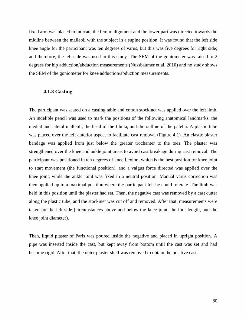
80
fixed arm was placed to indicate the femur alignment and the lower part was directed towards the
midline between the malleoli with the subject in a supine position. It was found that the left side
knee angle for the participant was ten degrees of varus, but this was five degrees for right side;
and therefore, the left side was used in this study. The SEM of the goniometer was raised to 2
degrees for hip adduction/abduction measurements (Nussbaumer et al, 2010) and no study shows
the SEM of the goniometer for knee adduction/abduction measurements.
4.1.3 Casting
The participant was seated on a casting table and cotton stockinet was applied over the left limb.
An indelible pencil was used to mark the positions of the following anatomical landmarks: the
medial and lateral malleoli, the head of the fibula, and the outline of the patella. A plastic tube
was placed over the left anterior aspect to facilitate cast removal (Figure 4.1). An elastic plaster
bandage was applied from just below the greater trochanter to the toes. The plaster was
strengthened over the knee and ankle joint areas to avoid cast breakage during cast removal. The
participant was positioned in ten degrees of knee flexion, which is the best position for knee joint
to start movement (the functional position), and a valgus force directed was applied over the
knee joint, while the ankle joint was fixed in a neutral position. Manual varus correction was
then applied up to a maximal position where the participant felt he could tolerate. The limb was
held in this position until the plaster had set. Then, the negative cast was removed by a cast cutter
along the plastic tube, and the stockinet was cut off and removed. After that, measurements were
taken for the left side (circumstances above and below the knee joint, the foot length, and the
knee joint diameter).
Then, liquid plaster of Paris was poured inside the negative and placed in upright position. A
pipe was inserted inside the cast, but kept away from bottom until the cast was set and had
become rigid. After that, the outer plaster shell was removed to obtain the positive cast.
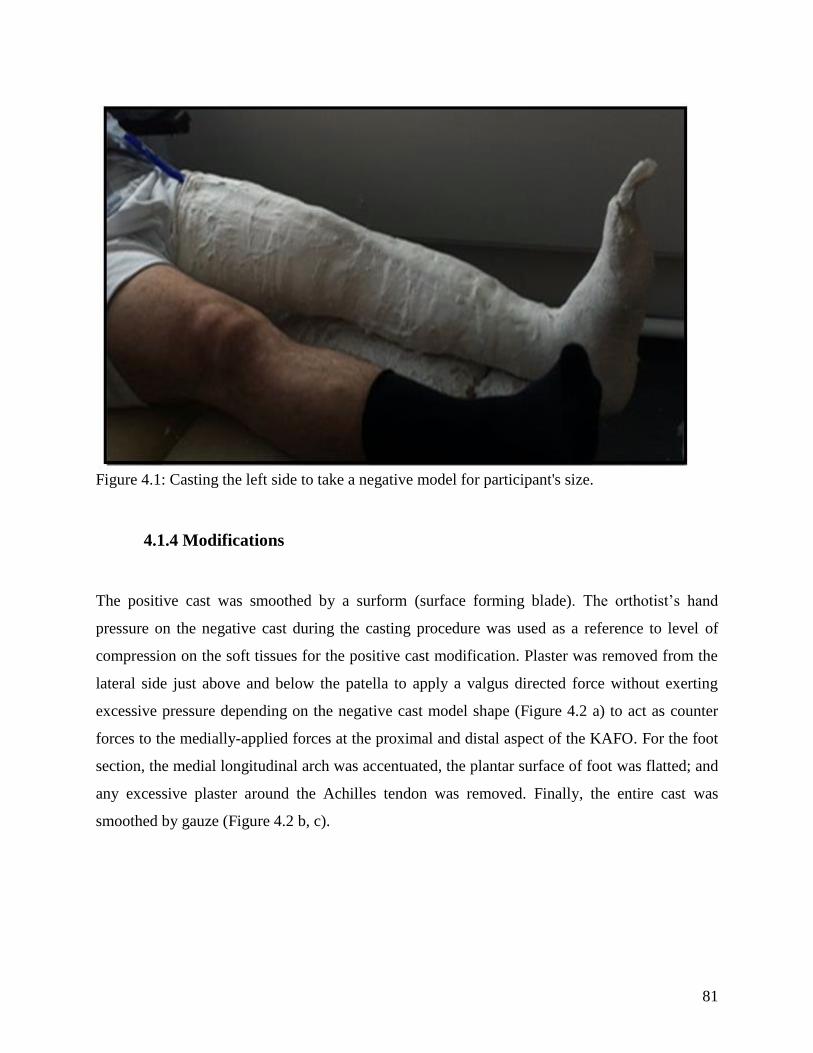
81
Figure 4.1: Casting the left side to take a negative model for participant's size.
4.1.4 Modifications
The positive cast was smoothed by a surform (surface forming blade). The orthotist’s hand
pressure on the negative cast during the casting procedure was used as a reference to level of
compression on the soft tissues for the positive cast modification. Plaster was removed from the
lateral side just above and below the patella to apply a valgus directed force without exerting
excessive pressure depending on the negative cast model shape (Figure 4.2 a) to act as counter
forces to the medially-applied forces at the proximal and distal aspect of the KAFO. For the foot
section, the medial longitudinal arch was accentuated, the plantar surface of foot was flatted; and
any excessive plaster around the Achilles tendon was removed. Finally, the entire cast was
smoothed by gauze (Figure 4.2 b, c).

82
Figure 4.2: The cast modification process. A four point pressure system was used to apply a
valgus correction force with long lever arms to make wearing the orthoses more comfortable (a),
modification of the medial arch (b), and removal of excessive plaster around the Achilles tendon
(c).
A six mm polypropylene plastic sheet was vacuum moulded over the cast. After the plastic had
dried, the medial upright was shaped over the plastic shell (Figure 4.3a). A stainless steel free
offset joint (Ottobock model 17lk1=L/R1-5, Ottobock, Germany) which tolerates up to 100kg
user weight with an eight mm thickness, 23.6mm width. Five degrees knee flexion is the neutral
position for this joint. This joint is not a completely posterior offset joint, and also it can be

83
locked or free depending on the case by just removing the washers and ring. The free joint was
used for this participant because he had good quadriceps muscle power (score of 4 on the Oxford
scale since the participant can flex his knee against gravity when apply sight resistance) and was
able to control the knee flexion and extension (Figure 4.3b). An aluminum side bar was used to
provide structural strength for the design and extended from five cm below the medial brim to
two-thirds of leg length so as to not impinge on the ankle. The joint was placed in a position as
recommended by the manufacture instructions at a position 60/40 behind the sagittal plane knee
centre; based on the external contours of the knee (Nietert 1975).
Figure 4.3: KAFO design including a single medial upright (a), and an Ottobock free offset knee
joint (b, c).
A further piece of plastic sheet was then vacuumed over the foot section to form a simple hinge
free ankle joint (Figure 4.4a). It is worthy to mention that a standard KAFO without an ankle
joint was initially tested, but it was deemed to be uncomfortable during stair use and limited the
ankle joint movement excessively. Given that the individual only had a varus deformity at the
knee and no ankle pathology, it was therefore necessary to add a simple ankle joint to the KAFO
to allow ankle joint motion and indeed is recommended rather than a standard KAFO without an
ankle joint particular if consumers habitually use stairs. The plastic shell covered two thirds of
the thigh and shin to distribute the loads applied to the lower limb over a large area. The
84
posterior part of the two shells was trimmed away from the knee joint to allow free knee flexion
during walking or stair climbing. The medial side of the thigh shell was trimmed lower than the
lateral trim line for more comfort and to prevent pinching during sitting. The shank section was
trimmed at a position five cm below the head of fibula to ensure the common peroneal nerve was
not impinged with the KAFO in situ.
After cutting the plastic shell and polishing, the side bar was fixed on the shells by screws.
Subsequently, two straps (two cm) were sewn and fixed on the medial thigh shell and one velcro
strap (five cm) for the leg shell with the loops fixed on the lateral side to apply more valgus force
and secure the KAFO to the lower limb (Figure 4.4b). Finally, the KAFO was checked to ensure
that the user felt comfortable and was able to flex the knee adequately.

85
Figure 4.4: KAFO design. Plastic was vacuumed over the lower parts (a). Final model of the
KAFO with an ankle joint (b).
4.1.4.1 Ossur knee valgus braces
The Ossur (Ossur, Iceland) UnloaderOne off-the-shelf (OTS) and custom knee valgus braces
were ordered depending on the participant's leg size. This type of unloader knee valgus brace is
used for unicompartmental knee OA. This design is easy to apply on the affected side,

86
lightweight, cosmetically acceptable, comfortable to use because of silicon pads inside the
design, and has a three points of leverage system. Additionally, it contains a polycentric free
knee joint with a zero degree extension stop, dynamic force systems applied by two straps
around the knee, and valgus directed system applied by the thigh and calf sections. The applied
force is based on the amount of user's pain relief and their tolerance level (Figure 4.5). The off-
the-shelf brace has five standard sizes depending on the circumference of the calf at a position
six inches (15cm) below the middle of the patella: x small, small, medium, large, and x large.
The custom-made design is a bespoke fit and depends on the circumstance of the thigh and calf,
and a suitable length of device which is usually shorter than OTS one and potentially generally
fits better (Table 4.1). The main function of the UnloaderOne knee valgus brace is to transfer the
load from medial compartment to the lateral side by applying a valgus moment to reduce the
EKAM and thus relieve pain. The custom device was measured and ordered by a representative
from Ossur.
Figure 4.5: The mechanism of the Unloader One knee valgus brace is to transfer the load to the
un-affected side.
Table 4.1: KAFO and knee valgus braces features.
Orthoses types Mass (Kg) Proximal part length from
knee joint centre
Distal part length from
knee joint centre
KAFO 1.20 30 cm 40 cm
Custom made knee valgus brace 0.45 17 cm 17 cm
OTS knee valgus brace 0.45 20 cm 20 cm

87
4.2. Data collection
After being fitted for the knee valgus brace and KAFO, the participant was asked to walk until he
was familiarised to all of the interventions in a gait laboratory. When the subject felt comfortable
with them, two static alignments were captured with two different shoe sizes. It was necessary to
provide a shoe size which allowed room for the KAFO and one with a normal control shoe for
the unloader braces. This is because the shoe size was changed during the test to fit the foot and
ankle section of the KAFO, but both of shoes were identical (Ecco Zen shoes). The participant
was asked to perform two activities: walking and stair climbing (ascending and descending).
4.2.1 Walking
The individual was asked to walk and climb stairs at a controlled speed using a metronome,
which is a device which produces fixed beats per minute, in each one of the conditions: walking
with control shoe, off-the-shelf knee valgus brace, custom knee valgus brace, and KAFO with
ankle joint design (Figure 4.6). The frequency of the metronome was set at 120 and 100 steps per
minute during walking and stair climbing respectively for the individual.
Figure 4.6: Control condition (a), off-the-shelf Ossur Unloader One knee valgus brace (b), custom
made Ossur Unloader One knee valgus brace (c), and KAFO (d).

88
4.2.2 Ascending and Descending
The participant was asked to ascend and descend (Figure 4.7 and 4.8 respectively) three stairs
five times without using the handrail or any other assistive devices using the control shoe, off-
the-shelf knee valgus brace (OTS), custom knee valgus brace, and KAFO (with a larger shoe
size). The shoes were identical except for shoe size. This allowed the determination of the
individual's EKAM and joints angles whilst using the different conditions. The total duration of
the test was two hours.
Figure 4.7: An ascending gait cycle for left leg starts with right leg of floor. (a), left side touches
the first step (b). Right leg on the second step (c) and the foot loads up the third step (end of the
gait cycle) (d).

89
Figure 4.8: Descending gait cycle for left side. Left leg starts descending (a), right leg on first
step (b), and gait cycle ends when the left side touches the floor (c).

90
4.3. Outcome measures
For the KAFO study, the same previous variables recorded in the reliability study such as
maxima of knee flexion angles at initial contact and mid swing (Figure 3.1a), range of the knee
motion in sagittal plane, EKAM, KAAI (Figure 3.1d), the maximum and minimum of sagittal
knee moment during gait cycle and minimum of sagittal knee moment at late stance during
walking were examined (Figure 3.1c). Moreover, vertical GRF in early and late stance were
calculated for every single trial instead of calculating the average for five trials together due to
the small sample size. Additionally, the maximum and minimum of the knee angle in the frontal
plane were evaluated during the stance phase only to examine the effects of the KAFO and knee
valgus braces on shank movement in stance phase instead of the whole gait cycle. Additional
variables which were also assessed as follows: maximum and minimum of the sagittal and
frontal angles for hip and ankle, and the three joints (hip, knee, and ankle) ROMs in the sagittal
plane (as illustrated in figure 4.9 a, b, c, d). Furthermore, the maximum and minimum of sagittal
and frontal hip moment (Figure 4.10a, b), and the maximum of single limb support moment for
every trial were evaluated (Figure 4.10c).
Figure 4.9: Data for sagittal hip angle (a), frontal hip angle (b), sagittal ankle angle (c), and
frontal ankle angle (d).
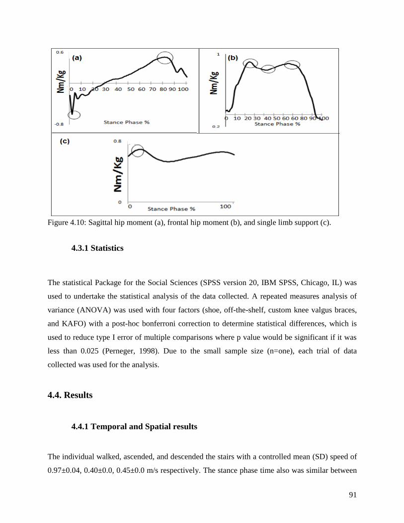
91
Figure 4.10: Sagittal hip moment (a), frontal hip moment (b), and single limb support (c).
4.3.1 Statistics
The statistical Package for the Social Sciences (SPSS version 20, IBM SPSS, Chicago, IL) was
used to undertake the statistical analysis of the data collected. A repeated measures analysis of
variance (ANOVA) was used with four factors (shoe, off-the-shelf, custom knee valgus braces,
and KAFO) with a post-hoc bonferroni correction to determine statistical differences, which is
used to reduce type I error of multiple comparisons where p value would be significant if it was
less than 0.025 (Perneger, 1998). Due to the small sample size (n=one), each trial of data
collected was used for the analysis.
4.4. Results
4.4.1 Temporal and Spatial results
The individual walked, ascended, and descended the stairs with a controlled mean (SD) speed of
0.97±0.04, 0.40±0.0, 0.45±0.0 m/s respectively. The stance phase time also was similar between
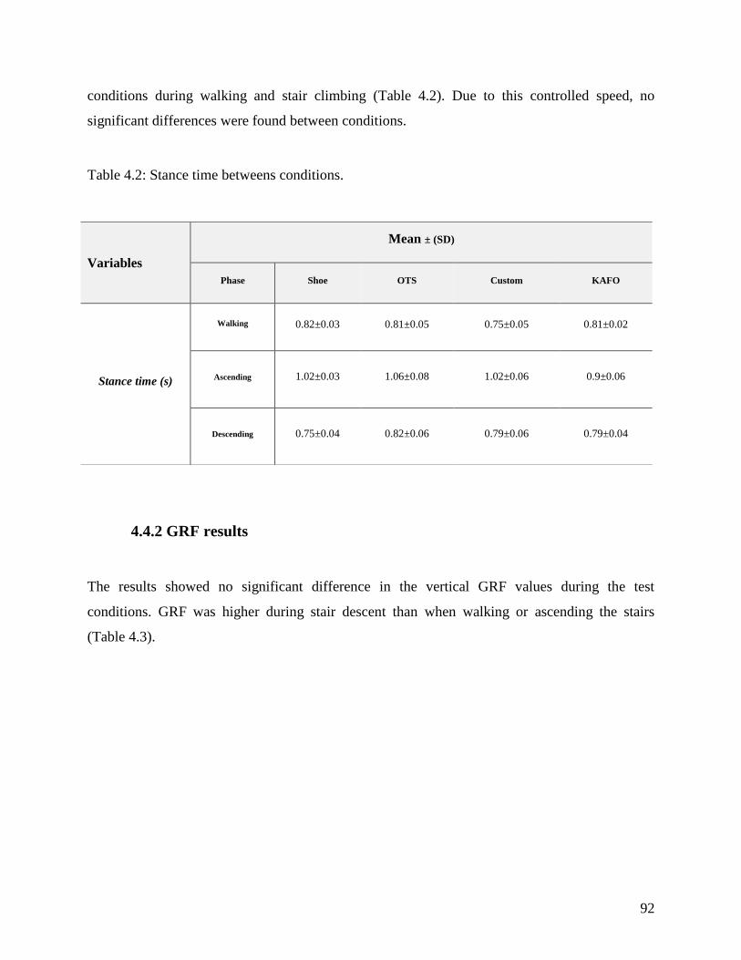
92
conditions during walking and stair climbing (Table 4.2). Due to this controlled speed, no
significant differences were found between conditions.
Table 4.2: Stance time betweens conditions.
4.4.2 GRF results
The results showed no significant difference in the vertical GRF values during the test
conditions. GRF was higher during stair descent than when walking or ascending the stairs
(Table 4.3).
Variables
Mean ± (SD)
Phase Shoe OTS Custom KAFO
Stance time (s)
Walking 0.82±0.03 0.81±0.05 0.75±0.05 0.81±0.02
Ascending
1.02±0.03
1.06±0.08 1.02±0.06 0.9±0.06
Descending
0.75±0.04
0.82±0.06 0.79±0.06 0.79±0.04

93
Table 4.3: VGRF during the four conditions.
Variables
Mean ±SD P value (IC 95%)
Shoe OTS Custom KAFO Shoe vs. OTS Shoe vs. Custom Shoe vs. KAFO KAFO
vs.Custom KAFO vs. OTS Custom vs. OTS
1. Walking
First peak
of GRF 1.03±0.01 1.06±0.02 1.09±0.01 1.10±0.0 1.0 (-0.15- 0.09) 0.29 (-0.18-0.04) 0.12 (-0.17-0.02) 1.0 (-0.10-0.08) 0.54 (-0.15-0.05) 0.99 (-0.14-0.07)
Trough of
GRF 1.01±0.01 1.01±0.0 0.99±0.01 0.99±0.0 1.0 (-0.06- 0.05) 1.0 (-0.07-0.09) 0.85(-0.02-0.05) 1.0 (-0.05-0.06) 0.15 (-0.00-0.05) 1.0 (-0.04-0.08)
Second
peak of
GRF
1.02±0.01 1.02±0.0 1.00±0.01 1.00±0.0 1.0 (-0.09- 0.10) 1.0 (-0.06-0.10) 1.00 (-0.06-0.10) 1.0 (-0.07-0.08) 0.18 (-0.01-0.05) 1.0 (-0.05-0.09)
2. Ascending
First peak of
GRF 1.04±0.1 1.03±0.0 1.01±0.1 1.08±0.1 1.0 (-0.08- 0.11) 1.0 (-0.09-0.15) 1.00 (-0.15-0.07) 0.09 (-0.15-0.01) 1.0 (-0.14-0.03) 0.29 (-0.03-0.07)
Trough of
GRF 1.03±0.01 1.02±0.0 1.04±0.0 1.06±0.01 1.0 (-0.06- 0.08) 1.0 (-0.10-0.08) 0.03 (-0.04--0.00) 1.0 (-0.013-0.08) 0.83 (-0.13-0.05) 1.0 (-0.07-0.03)
Second peak
of GRF 1.0±0.02 1.04±0.1 1.09±0.2 1.07±0.2 1.0 (-0.10- 0.18) 1.0 (-0.13-0.02) 1.0 (-0.14-0.11) 1.0 (-0.14-0.15) 1.0 (-0.10-0.14) 0.17 (-0.18 0.12)
3. Descending
First peak of
GRF 1.29±0.3 1.28±0.03 1.39±0.04 1.19±0.03 1.0 (-0.12- 0.14) 0.76 (-0.37-0.16) 0.71 (-0.14-0.34) 0.16 (-0.08-0.49) 1.0 (-0.17-0.35) 0.21 (-0.28-0.06)
Trough of
GRF 0.98±0.01 1.05±0.02 1.01±0.01 1.01±0.02 0.49 (-0.2-0.07) 0.77 (-0.10-0.04) 1.0 (-0.18-0.12) 1.0 (-0.11-0.11) 1.0 (-0.08-0.15) 1.0 (-0.09-0.17)
Second peak
of GRF 1.00±0.01 0.98±0.02 1.00±0.02 0.95±0.01 1.0 (-0.04-0.09) 1.0 (-0.09-0.10) 0.16 (-0.02-0.12) 0.80 (-0.07-0.17) 0.83 (-0.05-0.11) 1.0 (-0.15-0.12)

94
4.4.3 Kinematics of the knee joint
4.4.3.1 Walking
In the sagittal plane, the KAFO increased the knee flexion angle at initial contact (IC)
significantly compared to the shoe and OTS knee valgus brace (mean difference 8.6, 4.1 degrees,
p=0.00, p=0.03 respectively) (Figure 4.11, Table 4.4). Also, using the OTS and custom knee
valgus braces increased the knee flexion at IC in comparison to the shoe only (mean difference
4.5, 5.3 degrees, p=0.03, p=0.02 respectively) (Figure 4.11, Table 4.4).
For knee flexion in mid swing, no significant change was found between the KAFO and the knee
valgus braces compared to the shoe during walking. For minimum value of knee flexion during
the gait cycle, using the custom knee valgus brace and the OTS knee valgus braces significantly
increased the minimum value of knee flexion in comparison to the shoe (mean difference 2, 2.2
degrees, p=0.02, p=0.08 respectively), but KAFO did not increase that variable significantly
compared to the shoe.
Therefore, all conditions reduced knee ROM in sagittal plane. The KAFO reduced it
significantly during walking in comparison to the shoe (mean difference 10.5 degrees, p=0.03).
Furthermore, using OTS and custom knee valgus braces reduced it by 8.2 and 7.9 degrees,
p=0.44, p=0.05 respectively compared to the shoe (Figure 4.11, Table 4.4). However, no
significant difference was shown between KAFO and both knee valgus braces related to knee
ROM.
In the frontal plane, the maximum knee adduction angle was reduced in all conditions, but the
KAFO was superior to all in magnitude as well as statistically (Figure 4.12, Table 4.5) since the
mean difference between the KAFO and the shoe, OTS, and custom knee valgus braces was 8.2,
2.7, and 3 degrees, (p=0.00, p=0.04, p=0.00) respectively. Also, using the custom knee valgus
brace and OTS knee valgus brace significantly reduced the maximum value of the knee
95
adduction angle during walking compared to the shoe (mean difference 5.2, 5.5 degrees, p=0.0,
p=0.0 respectively) but there was no difference between the two knee valgus braces.
4.4.3.2 Stair ascent and descent
Using the KAFO insignificantly increased knee flexion at IC during stair ascent and descent
compared to the shoe, but this variable was significantly less compared to that produced by the
OTS brace during stair descent (mean difference 4.3 degrees, p=0.01) (Figure 4.11, Table 4.4).
In contrast, when using the OTS knee valgus brace this increased the degree of knee flexion at IC
during stair ascent and descent compared to the shoe, but was only significant during stair
descent (mean difference 5.2 degrees, p= 0.01). In addition, when using the custom knee valgus
brace an increase in the knee flexion at IC during stair ascent and descent was seen, but this was
not significantly reduced. When we compare both knee valgus braces together, the results show
that the OTS increased the knee flexion angle at IC during stair descent more than custom knee
valgus brace (mean difference 2 degrees, p=0.00).
At mid swing, the KAFO and OTS knee valgus brace reduced the maximum knee flexion during
stair ascent and descent, but was only significantly different during the stair ascent condition
compared to the shoe (mean difference 5.5, 3.9 degrees, p=0.05, p=0.01 respectively) (Figure
4.11, Table 4.4). However, the custom knee valgus brace reduced the maximum knee flexion
during mid-swing during both conditions (by 7 and 3 degrees respectively) compared to the shoe
but not significantly.
Furthermore, the minimum value of knee flexion during the gait cycle was considerably reduced
with the KAFO during stair ascent compared to the shoe, custom, and OTS knee valgus braces
(mean difference 3.5, 2.4, 3.8 degrees, p=0.00, p= 0.05, p=0.01 respectively), and during stair
descent with mean differences of 4.8, 5, and 3.9 degrees (p= 0.00, p=0.00, and p=0.00
respectively). Likewise, the custom knee valgus brace significantly reduced the minimum value
of knee flexion compared to the shoe during stair descent (p=0.02), but the OTS knee valgus
brace did not significantly affect the minima value of knee flexion during stair climbing. For the

96
knee ROM in the sagittal plane, no significant change was seen in any condition although
reductions were seen.
In the frontal plane, the maximum knee adduction angle during stance phase was significantly
less after using KAFO compared to all other conditions during stair ascent and descent. Up to 12
and 6 degrees mean difference (p=0.0, p=0.0 respectively) were found between the KAFO and
the shoe during stair ascent and descent respectively, raised to 15.2 and 6.8 degrees mean
difference (p=0.0, p=0.0 respectively) compared to custom knee valgus brace, and raised to 12.2
and 4.5 degrees mean difference (p=0.0, p=0.01 respectively) were found compared to OTS knee
valgus brace during stair ascent and descent respectively (Figure 4.12, Table 4.5). Moreover,
OTS knee valgus brace showed a significant reduction in knee adduction angle during stair
descent compared to the shoe (mean difference 1.5 degree, p=0.03). However, the custom knee
valgus brace increased the knee adduction raised to 3.2 degrees during stair ascent although this
was not significant.
When we compare both knee valgus braces together, the custom knee valgus brace significantly
increased the maximum value of knee adduction angle during stair ascent compared to OTS knee
valgus brace (mean difference 3 degrees, p=0.00) (Figure 4.12, Table 4.5), but no significant
changes were seen during stair descent between them.

97
Figure 4.11: Knee angles in the sagittal plane during walking (a), stair ascent (b), and stair
descent (c) for the four conditions.
Figure 4.12: Knee angles in frontal plane during walking (a), stair ascent (b), and stair descent
(c) for the four conditions.
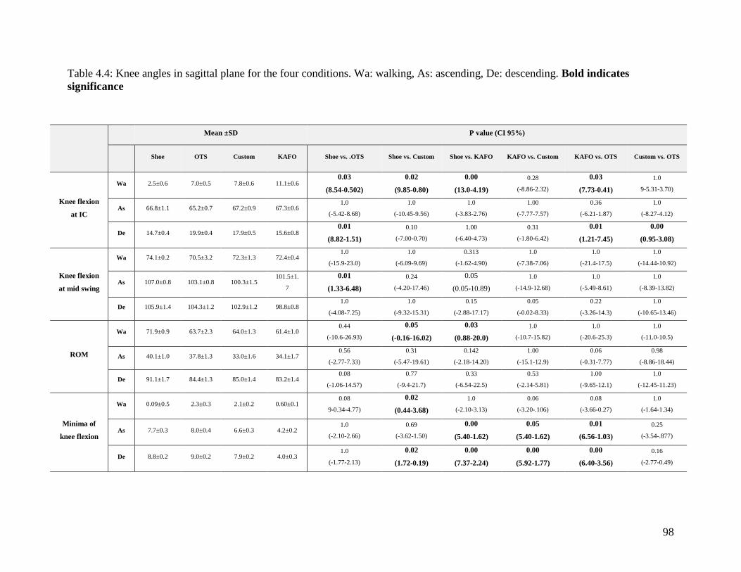
98
Table 4.4: Knee angles in sagittal plane for the four conditions. Wa: walking, As: ascending, De: descending. Bold indicates
significance
Mean ±SD P value (CI 95%)
Shoe OTS Custom KAFO Shoe vs. .OTS Shoe vs. Custom Shoe vs. KAFO KAFO vs. Custom KAFO vs. OTS Custom vs. OTS
Knee flexion
at IC
Wa 2.5±0.6 7.0±0.5 7.8±0.6 11.1±0.6 0.03
(8.54-0.502)
0.02
(9.85-0.80)
0.00
(13.0-4.19)
0.28
(-8.86-2.32)
0.03
(7.73-0.41)
1.0
9-5.31-3.70)
As 66.8±1.1 65.2±0.7 67.2±0.9 67.3±0.6 1.0
(-5.42-8.68)
1.0
(-10.45-9.56)
1.0
(-3.83-2.76)
1.00
(-7.77-7.57)
0.36
(-6.21-1.87)
1.0
(-8.27-4.12)
De 14.7±0.4 19.9±0.4 17.9±0.5 15.6±0.8 0.01
(8.82-1.51)
0.10
(-7.00-0.70)
1.00
(-6.40-4.73)
0.31
(-1.80-6.42)
0.01
(1.21-7.45)
0.00
(0.95-3.08)
Knee flexion
at mid swing
Wa 74.1±0.2 70.5±3.2 72.3±1.3 72.4±0.4 1.0
(-15.9-23.0)
1.0
(-6.09-9.69)
0.313
(-1.62-4.90)
1.0
(-7.38-7.06)
1.0
(-21.4-17.5)
1.0
(-14.44-10.92)
As 107.0±0.8 103.1±0.8 100.3±1.5 101.5±1.
7
0.01
(1.33-6.48)
0.24
(-4.20-17.46)
0.05
(0.05-10.89)
1.0
(-14.9-12.68)
1.0
(-5.49-8.61)
1.0
(-8.39-13.82)
De 105.9±1.4 104.3±1.2 102.9±1.2 98.8±0.8 1.0
(-4.08-7.25)
1.0
(-9.32-15.31)
0.15
(-2.88-17.17)
0.05
(-0.02-8.33)
0.22
(-3.26-14.3)
1.0
(-10.65-13.46)
ROM
Wa 71.9±0.9 63.7±2.3 64.0±1.3 61.4±1.0 0.44
(-10.6-26.93)
0.05
(-0.16-16.02)
0.03
(0.88-20.0)
1.0
(-10.7-15.82)
1.0
(-20.6-25.3)
1.0
(-11.0-10.5)
As 40.1±1.0 37.8±1.3 33.0±1.6 34.1±1.7 0.56
(-2.77-7.33)
0.31
(-5.47-19.61)
0.142
(-2.18-14.20)
1.00
(-15.1-12.9)
0.06
(-0.31-7.77)
0.98
(-8.86-18.44)
De 91.1±1.7 84.4±1.3 85.0±1.4 83.2±1.4 0.08
(-1.06-14.57)
0.77
(-9.4-21.7)
0.33
(-6.54-22.5)
0.53
(-2.14-5.81)
1.00
(-9.65-12.1)
1.0
(-12.45-11.23)
Minima of
knee flexion
Wa 0.09±0.5 2.3±0.3 2.1±0.2 0.60±0.1 0.08
9-0.34-4.77)
0.02
(0.44-3.68)
1.0
(-2.10-3.13)
0.06
(-3.20-.106)
0.08
(-3.66-0.27)
1.0
(-1.64-1.34)
As 7.7±0.3 8.0±0.4 6.6±0.3 4.2±0.2 1.0
(-2.10-2.66)
0.69
(-3.62-1.50)
0.00
(5.40-1.62)
0.05
(5.40-1.62)
0.01
(6.56-1.03)
0.25
(-3.54-.877)
De 8.8±0.2 9.0±0.2 7.9±0.2 4.0±0.3 1.0
(-1.77-2.13)
0.02
(1.72-0.19)
0.00
(7.37-2.24)
0.00
(5.92-1.77)
0.00
(6.40-3.56)
0.16
(-2.77-0.49)

99
Table 4.5: Knee angles in frontal plane for the four conditions. Wa: walking, As: ascending, De: descending. Bold indicates
significance.
Variables
Mean ±SD P value (CI 95%)
Phase Shoe OTS Custom KAFO Shoe vs. OTS Shoe vs. Custom Shoe vs. KAFO KAFO vs. Custom KAFO vs. OTS Custom vs. OTS
Minimum
knee
adduction
angle
Wa 0.09±0.5 2.3±0.3 2.1±0.2 0.60±0.1 0.08
(-0.34- 4.77)
0.02
(0.44- 3.68)
1.0
(-2.10- 3.13)
0.063
(-3.20-0.10)
0.083
(-3.66-0.27)
1.0
(-1.64-1.34)
As 7.7±0.3 8.0±0.4 6.6±0.3 4.2±0.2 1.0
(-2.10- 2.66)
0.69
(-3.62-1.50)
0.0
(5.40-1.62)
0.05
(4.92-0.00)
0.01
(6.56- 1.03)
0.25
(-3.54-0.87)
De 8.8±0.2 9.0±0.2 7.9±0.2 4.0±0.3 1.0
(-1.77- 2.13)
0.02
(1.72-0.02)
0.0
(7.37-2.24)
0.00
(5.92- 1.77)
0.00
(6.40- 3.56)
0.16
(-2.77-0.49)
Maximum
knee
adduction
angle
Wa 17.4±0.4 11.9±0.3 12.2±0.1 9.20±0.2 0.0
(-6.34- 4.58)
0.00
(7.57-2.85)
0.00
(-10.7-5.69)
0.00
(4.12-1.89)
0.04
(5.39-0.12)
1.0
(-1.95- 2.45)
As 30.3±0.6 30.5±0.3 33.5±0.3 18.3±0.1 1.0
(-4.44-4.87)
0.08
(-0.48-6.92)
0.00
(-15.0-8.92)
0.00
(17.1- 13.2)
0.00
-14.4- 9.97)
0.00
(1.74- 4.25)
De 21.4±0.5 19.9±0.4 22.2±0.4 15.4±0.3 0.03
(2.78-0.18)
1.0
(-2.78-4.47)
0.00
(9.53-2.40)
0.00
(8.36- 5.25)
0.01
(7.33- 1.62)
0.10
(-0.58-5.23)

100
4.4. Knee kinetics in the frontal plane
4.4.1 Walking
The external knee adduction moment was significantly reduced with the KAFO. In early stance,
the first peak of the external knee adduction moment (EKAM) was significantly reduced with the
KAFO by 11.4% (p=0.04) compared to the shoe condition and by 15% and 12.6% (p=0.07,
p=0.07 respectively) compared to the OTS and custom knee valgus braces (Figure 4.13, Table
4.6). The second peak of EKAM was also reduced by 12% compared to the shoe but not
significantly (p=0.09). The KAFO reduced the second peak of EKAM significantly more
(12.3%) than the custom knee valgus brace during walking (p=0.00) and insignificantly
compared to the OTS (9.5%, p=0.07). Neither the custom knee valgus brace, nor the OTS knee
valgus brace significantly reduced the first or second peak of the EKAM during walking
compared to the shoe.
4.4.2 Stair ascent and descent
Using the KAFO and both knee valgus braces reduced EKAM during stair climbing, but the
KAFO was superior. In early stance, the KAFO reduced the first and second peaks by 7% and
10.8% respectively compared to the shoe during ascending, but was not significantly different
(Figure 4.13, Table 4.6). During stair descent, no significant changes were shown when using the
KAFO. However, a greater reduction in the EKAM trough was seen after using KAFO in
comparison to the shoe and OTS knee valgus brace during stair ascent (16.2% and 20%, p=0.04,
p= 0.02 respectively).
Using both the custom knee valgus brace and OTS knee valgus brace reduced the first peak by
8.2% and 7%, but not significantly and the OTS brace increased the second peak of EKAM up to
4.3% compared to the shoe during stair ascent. During stair descent, both the OTS and custom
knee valgus braces increased the first peak of EKAM by 11.2% and 19.7% respectively, but this
was not statistically significant (p=0.17, p=0.38 respectively).

101
When looking at the single support aspect of the stance phase (Table 4.7), the KAFO reduced the
EKAM during walking and ascending (12.6% and 4.5% respectively) compared to the shoe and
was only significant during stair ascent (p=0.02). However, no changes were seen during stair
descent. In contrast, the custom and OTS knee valgus braces did not significantly change this
variable during walking and stair ascent, but both OTS and custom knee valgus braces increased
the EKAM during stair descent compared to the shoe, although not significantly (10%, 19%,
p=0.15, p=0.13 respectively).
Figure 4.13: EKAM during walking (a), stair ascent (b), and stair descent (c) for the four
conditions.
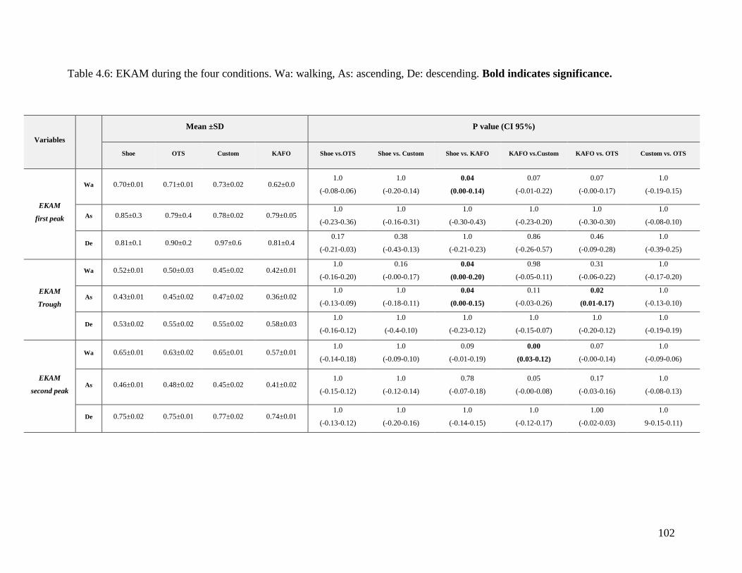
102
Table 4.6: EKAM during the four conditions. Wa: walking, As: ascending, De: descending. Bold indicates significance.
Variables
Mean ±SD P value (CI 95%)
Shoe OTS Custom KAFO Shoe vs.OTS Shoe vs. Custom Shoe vs. KAFO KAFO vs.Custom KAFO vs. OTS Custom vs. OTS
EKAM
first peak
Wa 0.70±0.01 0.71±0.01 0.73±0.02 0.62±0.0 1.0
(-0.08-0.06)
1.0
(-0.20-0.14)
0.04
(0.00-0.14)
0.07
(-0.01-0.22)
0.07
(-0.00-0.17)
1.0
(-0.19-0.15)
As 0.85±0.3 0.79±0.4 0.78±0.02 0.79±0.05 1.0
(-0.23-0.36)
1.0
(-0.16-0.31)
1.0
(-0.30-0.43)
1.0
(-0.23-0.20)
1.0
(-0.30-0.30)
1.0
(-0.08-0.10)
De 0.81±0.1 0.90±0.2 0.97±0.6 0.81±0.4 0.17
(-0.21-0.03)
0.38
(-0.43-0.13)
1.0
(-0.21-0.23)
0.86
(-0.26-0.57)
0.46
(-0.09-0.28)
1.0
(-0.39-0.25)
EKAM
Trough
Wa 0.52±0.01 0.50±0.03 0.45±0.02 0.42±0.01 1.0
(-0.16-0.20)
0.16
(-0.00-0.17)
0.04
(0.00-0.20)
0.98
(-0.05-0.11)
0.31
(-0.06-0.22)
1.0
(-0.17-0.20)
As 0.43±0.01 0.45±0.02 0.47±0.02 0.36±0.02 1.0
(-0.13-0.09)
1.0
(-0.18-0.11)
0.04
(0.00-0.15)
0.11
(-0.03-0.26)
0.02
(0.01-0.17)
1.0
(-0.13-0.10)
De 0.53±0.02 0.55±0.02 0.55±0.02 0.58±0.03 1.0
(-0.16-0.12)
1.0
(-0.4-0.10)
1.0
(-0.23-0.12)
1.0
(-0.15-0.07)
1.0
(-0.20-0.12)
1.0
(-0.19-0.19)
EKAM
second peak
Wa 0.65±0.01 0.63±0.02 0.65±0.01 0.57±0.01 1.0
(-0.14-0.18)
1.0
(-0.09-0.10)
0.09
(-0.01-0.19)
0.00
(0.03-0.12)
0.07
(-0.00-0.14)
1.0
(-0.09-0.06)
As 0.46±0.01 0.48±0.02 0.45±0.02 0.41±0.02 1.0
(-0.15-0.12)
1.0
(-0.12-0.14)
0.78
(-0.07-0.18)
0.05
(-0.00-0.08)
0.17
(-0.03-0.16)
1.0
(-0.08-0.13)
De 0.75±0.02 0.75±0.01 0.77±0.02 0.74±0.01 1.0
(-0.13-0.12)
1.0
(-0.20-0.16)
1.0
(-0.14-0.15)
1.0
(-0.12-0.17)
1.00
(-0.02-0.03)
1.0
9-0.15-0.11)

103
4.5. Knee adduction angular impulse (KAAI)
The KAAI was reduced after using KAFO compared to the shoe during walking and stair
climbing (Table 4.7), but was only statistically significant during ascending compared to the
shoe and OTS knee valgus brace (26.2%, 26.2%. p =0.00, p= 0.03 respectively). In contrast,
when using custom and OTS valgus braces there was no change in KAAI during walking and
stair ascent (p=1.0); however, using the custom knee valgus brace increased KAAI during
descending by 10.4%, p=0.06 compared to KAFO (Figure 4.14, Table 4.7).
Figure 4.14: Knee KAAI for the four conditions (error bars denote 1SD) during walking (a), stair
ascent (b), and stair descent (c).
4.6. Knee kinetics in sagittal plane
Using the KAFO and both knee valgus braces increased the knee flexion moment, but not
statistically significantly during walking and stair climbing. During late stance, the KAFO
applied more of a knee flexion moment (Figure 4.15, Table 4.6); whereas, the shoe, custom knee
valgus brace, and OTS knee valgus brace applied a greater extension moment. However, this
difference between KAFO and the shoe was not statistically significant (p=0.08).

104
For knee extension moment, no significant changes were seen during walking after using all
conditions (Figure 4.15, Table 4.6). During stair ascent, also no significant changes were shown
using KAFO and custom knee valgus brace compared to the shoe, but OTS knee valgus brace
insignificantly reduced knee extension moment more than shoe, custom knee valgus brace and
KAFO (33%,27%,46%,p=0.56, p=0.09, p=0.09 respectively). During stair descent, the knee
extension moment was reduced more by the KAFO compared to the OTS, custom knee valgus
braces, plus the shoe but not significantly (82%, 78%, 82%, p=0.46, p=0.03, p=0.37
respectively).
Figure 4.15: Knee moment in sagittal plane during walking (a), stair ascent (b), and stair descent
(c) for the four conditions.

105
Table 4.7: Knee moment during the four conditions. Wa: walking, As: ascending, De: descending. *: extension moment. Bold
indicates significance.
Variables
Mean ±SD
P value
Shoe OTS Custom KAFO Shoe vs. OTS Shoe vs. Custom Shoe vs. KAFO KAFO vs.
Custom KAFO vs. OTS Custom vs. OTS
KAAI
Wa 0.38±0.02 0.37±0.02 0.35±0.01 0.31±0.0 1.00
(-0.14-0.16)
1.00
(-0.07-0.13)
0.22
(-0.04-0.18)
0.41
(-0.04-0.12)
0.07
(-0.0-0.13)
1.00
(-0.11-0.15
As 0.42±0.0 0.42±0.0 0.41±0.0 0.31±0.01 1.00
(-0.10-0.10)
1.00
(-0.07-0.09)
0.00
(0.04-0.17)
0.05
(-0.00-0.19)
0.03
(0.013-0.20)
1.00
(-0.06-0.09)
De 0.43±0.01 0.44±0.02 0.48±0.01 0.41±0.01 1.00
(-0.18-0.15)
0.12
(-0.12-0.01)
1.0
(-0.03-0.06)
0.06
(-0.00-0.141)
1.0
(-0.12-0.18)
1.00
(-0.15-0.07)
Single Limb Support
Wa 0.71±0.01 0.71±0.02 0.73±0.03 0.62±0.0 1.000
(-0.09-0.09)
1.00
(-0.185-0.13)
0.02
(0.01-0.14)
0.07
(-0.01-0.22)
0.074
(-0.01-0.17)
1.00
(-0.19-0.15)
As 0.87±0.02 0.81±0.02 0.87±0.02 0.83±0.03 1.00
(-0.17-0.28)
1.00
(-0.152-0.142)
1.00
(-0.20-0.27)
1.00
(-0.17-0.25)
1.00
(-0.16-0.12)
0.71
(-0.20-0.08)
De 0.81±0.02 0.90±0.02 1.00±0.05 0.81±0.03 0.15
(-0.21-0.03)
0.13
(-0.442-0.066)
1.00
(-0.23-0.23)
0.32
(-0.14-0.51)
0.46
(-0.09-0.27)
1.00
(-0.42-0.23)
Knee flexion moment
Wa 0.48±0.4 0.55±0.03 0.58±0.02 0.70±0.04 1.00
(-0.28-0.15)
0.27
(-0.274-0.071)
0.33
(-0.61-0.17)
0.60
(-0.38-0.15)
0.42
(-0.44-0.14)
1.00
(-0.20-0.13)
As 0.80±0.5 0.82±0.02 0.78±0.03 0.82±0.03 1.00
(-0.25-0.21)
1.00
(-0.166-0.192)
1.00
(-0.35-0.31)
1.00
(-0.24-0.18)
1.00
(-0.19-0.20)
1.00
(-0.19-0.262)
De 1.00±0.03 0.98±0.02 0.98±0.02 1.03±0.03 1.00
(-0.14-0.17)
1.00
(-0.137-0.176)
1.00
(-0.32-0.25)
1.00
(-0.23-0.11)
1.00
(-0.2-0.15)
1.00
(-0.17-0.18)
Knee flexion moment at
late stance Wa 0.09±.03* 0.09±0.02* 0.03±0.2* 0.12±0.03
1.00
(-0.33-0.4)
0.72
(-0.14-0.07)
0.08
(-0.4-0.03)
0.21
(0.10-0.4)
0.18
(-1.12-0.54)
1.00
(-0.34-0.21)
Knee extension moment
Wa 0.28±0.02 0.27±0.02 0.28±0.0 0.24±0.01 1.00
(-0.08-0.06)
1.00
(-0.126-0.113)
1.00
(-0.17-0.08)
1.00
(-0.11-0.04)
1.00
(-0.16-0.10)
1.00
(-0.08-0.09)1
As 1.2±0.5 0.8±0.9 1.1±0.05 1.5±0.1 0.56
(-1.46-0.55)
1.00
(-0.867-0.674)
1.00
(-0.75-1.30)
0.28
(-0.26-1.00)
0.09
(-0.15-1.61
0.09
(-0.07-0.79)
De 0.51±0.2 0.62±0.2 0.64±0.01 0.11±0.05 1.00
(-1.2-1.42)
1.00
(-1.152-1.417)
0.37
(-1.17-0.35)
0.30
(-1.48-0.40)
0.46
(-1.5-0.54
1.00
(-0.51-0.55)

106
4.7. Hip kinematics in sagittal and frontal planes
4.7.1 Walking
There was a significant increase in the maximum hip flexion angle when using the KAFO in
comparison to the shoe, off-the-shelf (OTS) and custom UnloaderOne knee valgus brace during
walking (mean difference 6.6, 5.3, 6.1 degrees, p=0.0, p=0.0, p=0.01 respectively) (Figure 4.16,
Table 4.8). Furthermore, the maximum hip extension angle also increased considerably when
using KAFO during walking compared to the shoe, custom, and OTS knee valgus braces (mean
difference 0.3, 1.3, 0.9 degrees, p= 0.01, p=0.00, p=0.00 respectively). Therefore, the hip range
of motion (ROM) increased significantly during walking when using the KAFO in comparison to
the shoe, OTS, and custom knee valgus braces (mean difference 11.7, 9.8, 10 degrees, p=0.00,
p=0.002, p=0.00 respectively). However, no significant changes were seen when using the
custom and OTS knee valgus braces in hip flexion, hip extension and hip ROM compared to the
shoe.
In the frontal plane, the maximum hip abduction angle was significantly less when using the
KAFO during walking (Figure 4.17, Table 4.9). The mean difference between KAFO compared
to the shoe, OTS and custom knee valgus braces was 5.3, 5.5, 5.3 degrees respectively (p=0.0 for
all differences). Furthermore, the KAFO increased the hip adduction angle compared to the shoe
(mean difference 3.8 degrees, p=0.02). In contrast, no significant changes in maximum hip
adduction and abduction angles were seen between both knee valgus braces and the shoe during
walking.
4.7.2 Stair ascent and descent
The maximum hip flexion angle increased with the KAFO during stair ascent by an average
increase of 4 degrees compared to the shoe but this was not statistically significant. Additionally,
the OTS knee valgus brace increased hip flexion significantly during stair ascent compared to the
shoe (mean difference 3.3 degrees, p=0.02). No changes were seen with using custom knee
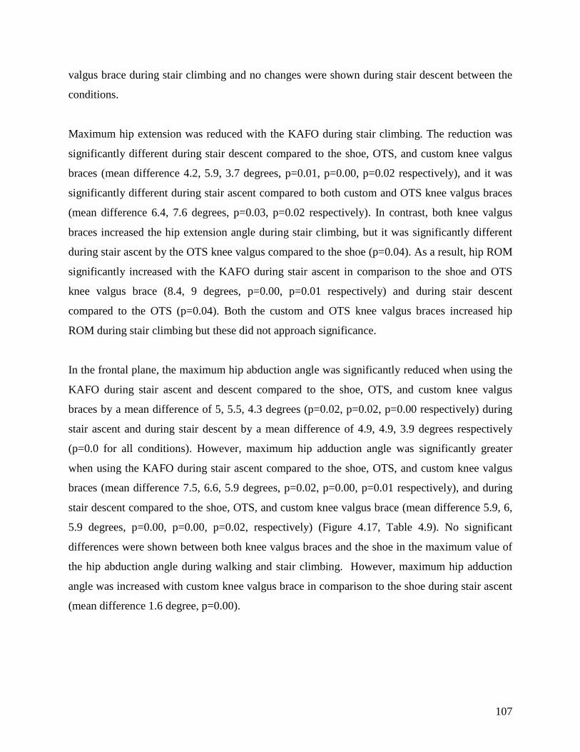
107
valgus brace during stair climbing and no changes were shown during stair descent between the
conditions.
Maximum hip extension was reduced with the KAFO during stair climbing. The reduction was
significantly different during stair descent compared to the shoe, OTS, and custom knee valgus
braces (mean difference 4.2, 5.9, 3.7 degrees, p=0.01, p=0.00, p=0.02 respectively), and it was
significantly different during stair ascent compared to both custom and OTS knee valgus braces
(mean difference 6.4, 7.6 degrees, p=0.03, p=0.02 respectively). In contrast, both knee valgus
braces increased the hip extension angle during stair climbing, but it was significantly different
during stair ascent by the OTS knee valgus compared to the shoe (p=0.04). As a result, hip ROM
significantly increased with the KAFO during stair ascent in comparison to the shoe and OTS
knee valgus brace (8.4, 9 degrees, p=0.00, p=0.01 respectively) and during stair descent
compared to the OTS (p=0.04). Both the custom and OTS knee valgus braces increased hip
ROM during stair climbing but these did not approach significance.
In the frontal plane, the maximum hip abduction angle was significantly reduced when using the
KAFO during stair ascent and descent compared to the shoe, OTS, and custom knee valgus
braces by a mean difference of 5, 5.5, 4.3 degrees (p=0.02, p=0.02, p=0.00 respectively) during
stair ascent and during stair descent by a mean difference of 4.9, 4.9, 3.9 degrees respectively
(p=0.0 for all conditions). However, maximum hip adduction angle was significantly greater
when using the KAFO during stair ascent compared to the shoe, OTS, and custom knee valgus
braces (mean difference 7.5, 6.6, 5.9 degrees, p=0.02, p=0.00, p=0.01 respectively), and during
stair descent compared to the shoe, OTS, and custom knee valgus brace (mean difference 5.9, 6,
5.9 degrees, p=0.00, p=0.00, p=0.02, respectively) (Figure 4.17, Table 4.9). No significant
differences were shown between both knee valgus braces and the shoe in the maximum value of
the hip abduction angle during walking and stair climbing. However, maximum hip adduction
angle was increased with custom knee valgus brace in comparison to the shoe during stair ascent
(mean difference 1.6 degree, p=0.00).
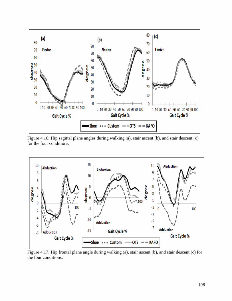
108
Figure 4.16: Hip sagittal plane angles during walking (a), stair ascent (b), and stair descent (c)
for the four conditions.
Figure 4.17: Hip frontal plane angle during walking (a), stair ascent (b), and stair descent (c) for
the four conditions.

109
Table 4.8: Hip angles in sagittal plane during the four conditions. Wa: walking, As: ascending, De: descending. Bold indicates
significance.
Variables
Mean ±SD
P value (CI 95%)
Shoe OTS Custom KAFO Shoe vs.OTS Shoe vs. Custom Shoe vs. KAFO KAFO vs.Custom KAFO vs. OTS Custom vs. OTS
Hip flexion
angle
Wa 42.0±0.4 43.3±0.5 42.5±0.7 48.6±0.4 0.85
(-4.70-2.16)
1.0
(-5.86-4.85)
0.00
(8.39-4.90)
0.01
(10.34-1.94)
0.00
(8.07-2.61)
1.0
(-2.74-4.34)
As 74.7±0.6 78.0±0.5 77.3±0.6 78.5±0.8 0.02
(5.97-0.63)
0.60
(-8.41-3.28)
0.143
(-8.94-1.38)
1.0
(-7.27-4.83)
1.0
(-5.39-4.44)
1.0
(-3.79-5.28)
De 52.0±0.7 52.5±0.4 51.0±0.8 52.0±0.5 1.0
(-5.53-4.83)
1.0
(-4.89-7.21)
1.0
(-4.72-5.04)
0.75
(-3.51-1.51)
1.0
(-2.44-3.45)
0.74
(-2.25-5.26)
Hip extension
angle
Wa 2.3±0.4 1.7±0.5 1.3±0.2 2.6±0.3 1.0
(-2.16-3.32)
0.74
(-1.70-3.98)
0.01
(1.57-8.48)
0.00
(2.93-4.85)
0.00
(1.77-7.12)
1.0
(-1.722.83)
As 15.9±0.5 19.8±0.9 17.6±0.5 11.2±0.9 0.04
(7.75-0.06)
0.58
(-5.71-2.20)
0.17
(-2.18-11.54)
0.03
(0.66-12.19)
0.02
(1.28-15.88)
0.65
(-2.93-7.25)
De 20.3±0.4 22.0±0.7 19.8±0.3 16.1±0.5 0.23
(-4.28-1.00)
1.0
(-2.67-3.66)
0.01
(1.43-7.05)
0.02
(0.80-6.69)
0.00
(2.69-9.07)
0.44
(-2.17-6.43)
ROM
Wa 39.6±0.5 41.5±0.8 41.2±0.8 51.3±0.6 1.0
(-7.95-4.18)
1.0
(-8.02-4.74)
0.00
(-15.9--7.43)
0.00
(14.5-5.54)
0.00
(14.1-5.46)
1.0
(-2.98-3.46)
As 58.8±0.5 58.2±0.7 59.6±1.0 67.2±0.6 1.0
(-3.75-4.96)
1.0
(-7.54-5.92)
0.00
(11.3-5.57)
0.05
(-15.38-0.08)
0.01
(14.9-3.13)
0.74
(-4.94-2.11)
De 31.8±0.7 30.5±1.0 31.1±1.0 35.9±0.6 1.0
(-6.13-8.71)
1.0
(-7.16-8.49)
0.13
(-9.52-1.34)
0.07
(-10.09-0.59)
0.04
(10.6-0.07)
1.0
(-8.16-6.91)
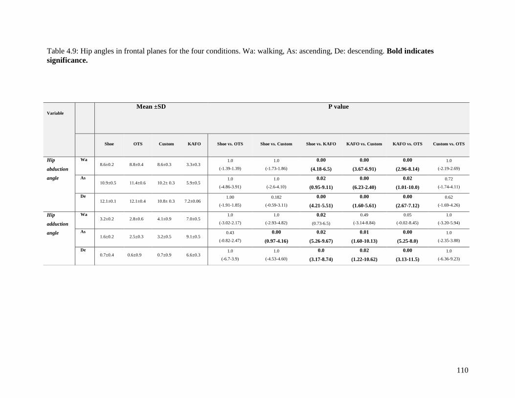
110
Table 4.9: Hip angles in frontal planes for the four conditions. Wa: walking, As: ascending, De: descending. Bold indicates
significance.
Variable
Mean ±SD P value
Shoe OTS Custom KAFO Shoe vs. OTS Shoe vs. Custom Shoe vs. KAFO KAFO vs. Custom KAFO vs. OTS Custom vs. OTS
Hip
abduction
angle
Wa 8.6±0.2 8.8±0.4 8.6±0.3 3.3±0.3
1.0
(-1.39-1.39)
1.0
(-1.73-1.86)
0.00
(4.18-6.5)
0.00
(3.67-6.91)
0.00
(2.96-8.14)
1.0
(-2.19-2.69)
As 10.9±0.5 11.4±0.6 10.2± 0.3 5.9±0.5
1.0
(-4.86-3.91)
1.0
(-2.6-4.10)
0.02
(0.95-9.11)
0.00
(6.23-2.40)
0.02
(1.01-10.0)
0.72
(-1.74-4.11)
De 12.1±0.1 12.1±0.4 10.8± 0.3 7.2±0.06
1.00
(-1.91-1.85)
0.182
(-0.59-3.11)
0.00
(4.21-5.51)
0.00
(1.60-5.61)
0.00
(2.67-7.12)
0.62
(-1.69-4.26)
Hip
adduction
angle
Wa 3.2±0.2 2.8±0.6 4.1±0.9 7.0±0.5
1.0
(-3.02-2.17)
1.0
(-2.93-4.82)
0.02
(0.73-6.5)
0.49
(-3.14-8.84)
0.05
(-0.02-8.45)
1.0
(-3.20-5.94)
As 1.6±0.2 2.5±0.3 3.2±0.5 9.1±0.5
0.43
(-0.82-2.47)
0.00
(0.97-4.16)
0.02
(5.26-9.67)
0.01
(1.60-10.13)
0.00
(5.25-8.0)
1.0
(-2.35-3.88)
De 0.7±0.4 0.6±0.9 0.7±0.9 6.6±0.3
1.0
(-6.7-3.9)
1.0
(-4.53-4.60)
0.0
(3.17-8.74)
0.02
(1.22-10.62)
0.00
(3.13-11.5)
1.0
(-6.36-9.23)
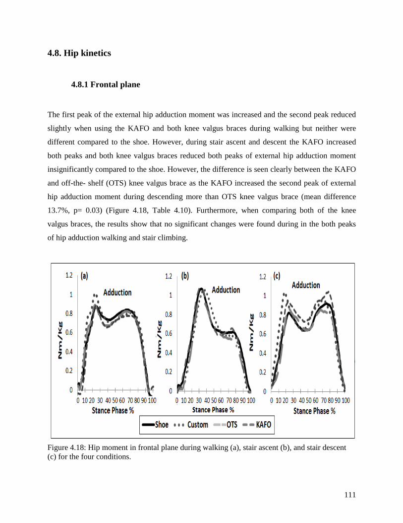
111
4.8. Hip kinetics
4.8.1 Frontal plane
The first peak of the external hip adduction moment was increased and the second peak reduced
slightly when using the KAFO and both knee valgus braces during walking but neither were
different compared to the shoe. However, during stair ascent and descent the KAFO increased
both peaks and both knee valgus braces reduced both peaks of external hip adduction moment
insignificantly compared to the shoe. However, the difference is seen clearly between the KAFO
and off-the- shelf (OTS) knee valgus brace as the KAFO increased the second peak of external
hip adduction moment during descending more than OTS knee valgus brace (mean difference
13.7%, p= 0.03) (Figure 4.18, Table 4.10). Furthermore, when comparing both of the knee
valgus braces, the results show that no significant changes were found during in the both peaks
of hip adduction walking and stair climbing.
Figure 4.18: Hip moment in frontal plane during walking (a), stair ascent (b), and stair descent
(c) for the four conditions.
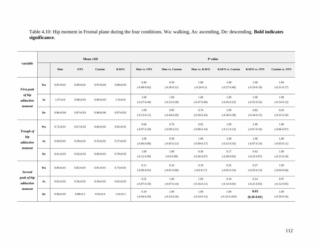
112
Table 4.10: Hip moment in Frontal plane during the four conditions. Wa: walking, As: ascending, De: descending. Bold indicates
significance.
variable
Mean ±SD
P value
Shoe OTS Custom KAFO Shoe vs. OTS Shoe vs. Custom Shoe vs. KAFO KAFO vs. Custom KAFO vs. OTS Custom vs. OTS
First peak
of hip
adduction
moment
Wa 0.87±0.01 0.90±0.01 0.97±0.04 0.90±0.03 0.40
(-0.08-0.02)
0.50
(-0.30-0.11)
1.00
(-0.24-0.1)
1.00
(-0.27-0.40)
1.00
(-0.19-0.19)
1.00
(-0.31-0.17)
As 1.07±0.0 0.98±0.05 0.99±0.03 1.10±0.0 1.00
(-0.27-0.44)
1.00
(-0.23-0.39)
1.00
(-0.47-0.40)
1.00
(-0.45-0.23)
1.00
(-0.55-0.32)
1.00
(-0.14-0.13)
De 0.86±0.04 0.87±0.03 0.98±0.06 0.97±0.03 1.00
(-0.15-0.11)
0.85
(-0.44-0.20)
0.74
(-0.39-0.16)
1.00
(-0.36-0.38)
0.82
(-0.34-0.15)
0.43
(-0.31-0.10)
Trough of
hip
adduction
moment
Wa 0.72±0.01 0.67±0.02 0.66±0.02 0.65±0.01 0.66
(-0.07-0.18)
0.76
(-0.09-0.21)
0.05
(-0.00-0.14)
1.00
(-0.11-0.13)
1.00
(-0.07-0.10)
1.00
(-0.06-0.07)
As 0.60±0.01 0.58±0.01 0.55±0.02 0.57±0.02 1.00
(-0.06-0.08)
0.59
(-0.05-0.13)
1.00
(-0.09-0.17)
1.00
(-0.15-0.16)
1.00
(-0.07-0.14)
1.00
(-0.05-0.11)
De 0.61±0.03 0.62±0.02 0.60±0.03 0.70±0.02 1.00
(-0.12-0.09)
1.00
(-0.0-0.09)
0.36
(-0.26-0.07)
0.17
(-0.28-0.05)
0.43
(-0.23-0.07)
1.00
(-0.13-0.19)
Second
peak of hip
adduction
moment
Wa 0.84±0.01 0.82±0.01 0.81±0.01 0.73±0.01 0.11
(-0.00-0.05)
0.16
(-0.01-0.06)
0.18
(-0.0-0.17)
0.55
(-0.05-0.14)
0.27
(-0.03-0.13)
1.00
(-0.04-0.04)
As 0.62±0.02 0.56±0.01 0.59±0.02 0.65±0.02 0.51
(-0.07-0.19)
1.00
(-0.07-0.14)
1.00
(-0.16-0.11)
0.19
(-0.14-0.03)
0.14
(-0.21-0.03)
0.97
(-0.12-0.05)
De 0.96±0.02 0.88±0.2 0.95±0.4 1.02±0.3 0.19
(-0.04-0.20)
1.00
(-0.23-0.26)
1.00
(-0.24-0.13)
1.00
(-0.32-0.1810
0.03
(0.26-0.01)
1.00
(-0.29-0.16)
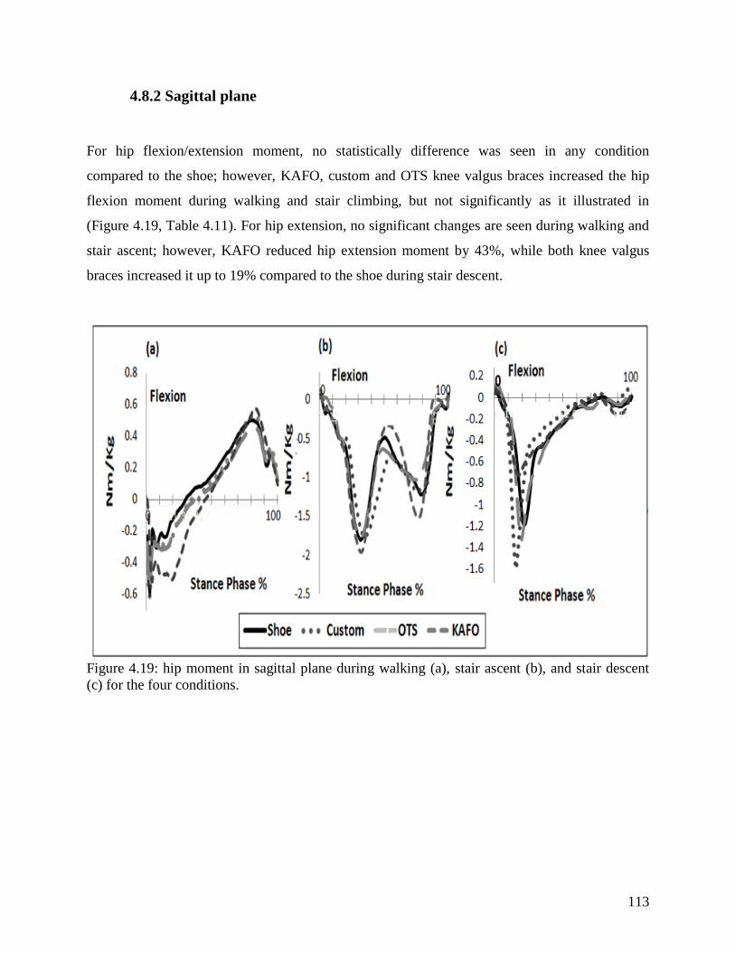
113
4.8.2 Sagittal plane
For hip flexion/extension moment, no statistically difference was seen in any condition
compared to the shoe; however, KAFO, custom and OTS knee valgus braces increased the hip
flexion moment during walking and stair climbing, but not significantly as it illustrated in
(Figure 4.19, Table 4.11). For hip extension, no significant changes are seen during walking and
stair ascent; however, KAFO reduced hip extension moment by 43%, while both knee valgus
braces increased it up to 19% compared to the shoe during stair descent.
Figure 4.19: hip moment in sagittal plane during walking (a), stair ascent (b), and stair descent
(c) for the four conditions.

114
Table 4.11: Hip moment in sagittal plane during the four conditions. As: ascending, Wa: walking, De: descending.
variable
Mean ±SD P value
Shoe OTS Custom KAFO Shoe vs.
OTS Shoe vs. Custom
Shoe vs.
KAFO
KAFO vs.
Custom KAFO vs. OTS Custom vs. OTS
Hip flexion
moment
Wa 0.49±0.03 0.45±0.03 0.50±0.02 0.57±0.2 1.0
(-0.19-0.02)
1.0
(-0.20-0.18)
0.71
(-0.28-0.21)
0.32
(-0.09-0.34)
1.00
(-0.14-0.29)
1.0
(-0.25-0.14)
As 0.09±0.02 0.16±0.03 0.16±0.0 0.14±0.03 0.62
(-0.22-0.00)
0.10
(-0.14-0.01)
0.90
(-0.32-0.15)
1.0
(-0.17-0.13)
1.0
(-0.25-0.21)
1.0
(-0.14-0.01)
De 0.06±0.01 0.11±0.01 0.10±0.03 0.13±0.02 0.67
(-0.16-0.06)
1.0
(-0.15-0.07)
0.86
(-0.24-0.11)
1.0
(-0.22-0.16)
1.0
(-0.11-0.07)
1.0
(-0.11-0.13)
Hip
extension
moment
Wa 0.60±0.04 0.61±0.03 0.67±0.02 0.61±0.02 1.0
(-0.06-0.08)
1.0
(-0.02-0.36)
1.0
(-0.15-0.17)
1.0
(-0.22-0.1)
1.0
(-0.12-0.12)
1.0
(-0.01-0.31)
As 1.8±0.2 1.7±0.1 1.7±0.01 2.1±0.01 1.0
(-1.08-0.93)
1.0
(-0.08-0.74)
0.67
(-0.47-1.15)
0.46
-0.43-1.24)
0.95
(-0.74-1.57)
1.0
(-0.03-0.38)
De 1.32±0.1 1.60±0.2 1.63±0.02 0.75±0.1 1.0
(-1.07-1.61)
1.0
(-1.30-1.90)
0.14
(-1.36-0.21)
0.29
(-2.40-0.65)
0.27
(-2.2-0.58)
1.0
(-0.47-0.53)
115
4.9. Ankle kinematics (Secondary outcomes)
4.9.1 Walking
In the sagittal plane the KAFO showed a significantly higher dorsiflexion angle during walking
than the shoe, OTS, and custom knee valgus brace (mean difference 5.6, 5.1, and 6 degrees.
p=0.00, p=0.02, p=0.00 respectively) (Figure 4.20, Table 4.12). However, no effects were seen
using either of the knee valgus braces on ankle dorsiflexion angle during walking. For ankle
plantarflexion angle, the KAFO and OTS knee valgus brace reduced plantarflexion by 3.1 and
2.3 degrees respectively compared to the shoe (but not significantly), while no change is shown
when using the custom knee valgus brace.
As a result, the ankle ROM was increased significantly with the KAFO compared to the OTS
brace (mean difference 4.2 degrees, p=0.04) and insignificantly compared to the shoe and custom
knee valgus brace (mean difference 2.2, 2.6 degrees. p=0.61, p=0.60 respectively). No
significant differences were demonstrated between the knee valgus braces and the shoe during
walking.
In the frontal plane, there was significantly less eversion ankle angle when using KAFO during
walking compared to the shoe, OTS, and custom knee valgus brace (mean difference 5.7, 5.3, 6.7
degrees respectively (p=0.0 for all conditions) (Figure 4.21, Table 4.13). No significant changes
were shown in inversion angle between the KAFO and shoe and between both knee valgus
braces (Figure 4.21, Table 4.13).
4.9.2 Stair ascent and descent
In the sagittal plane, ankle dorsiflexion angles were similar among conditions during stair ascent
and descent (Figure 4.20, Table 4.12). However, the maximum plantarflexion angle was
significantly reduced with the KAFO during stair ascent compared to the shoe, OTS, and custom
knee valgus braces (mean difference 8.9, 4.9, 5.9 degrees. p=0.04, p=0.01, p=0.02 respectively)

116
and during stair descent in comparison to the shoe (mean difference 5.9 degrees. p=0.02).
Therefore, ankle ROM in sagittal plane was reduced significantly with the KAFO during stair
ascent in comparison to the shoe (mean difference 7.8 degrees, p=0.05), and during stair descent
compared to the shoe and custom knee valgus brace (mean difference 7.8, 3.7 degrees. p=0.01,
p=0.02 respectively). Furthermore, using the custom and OTS knee valgus braces reduced the
plantarflexion angle and ROM insignificantly during stair ascent and descent respectively
compared to the shoe.
In the frontal plane, there was significantly less maximum eversion ankle angle during mid
stance when using KAFO during stair ascent compared to the shoe, OTS, and custom knee
valgus braces (mean difference 5, 5.5, 4.3 degrees. p=0.02, p=0.02, p=0.00 respectively); and
during stair descent compared to the custom knee valgus brace (mean difference 3.3 degrees,
p=0.01) (Figure 4.21, Table 4.13). Furthermore, no significant changes were shown after using
both knee valgus braces.
For ankle inversion, no significant changes were shown between KAFO compared to shoe during
stair ascent and descent respectively, but the KAFO reduced ankle inversion significantly
compared to the OTS and custom knee valgus braces during stair ascent (mean difference 3.4,
2.9 degrees, p=0.04, 0.01 respectively) (Figure 4.21, Table 4.13). Moreover, stair ascent with the
OTS knee valgus brace showed a significantly higher ankle inversion compared to shoe (mean
difference 2.9 degrees. p=0.00). Nevertheless, there were no significant differences demonstrated
during stair descent and no significant changes between both knee valgus braces.

117
Figure 4.20: Ankle angles in sagittal plane during walking (a), stair ascent (b), and stair descent
(c) for the four conditions.
Figure 4.21: Ankle angles in frontal plane during walking (a), stair ascent (b), and stair descent
(c) for the four conditions.
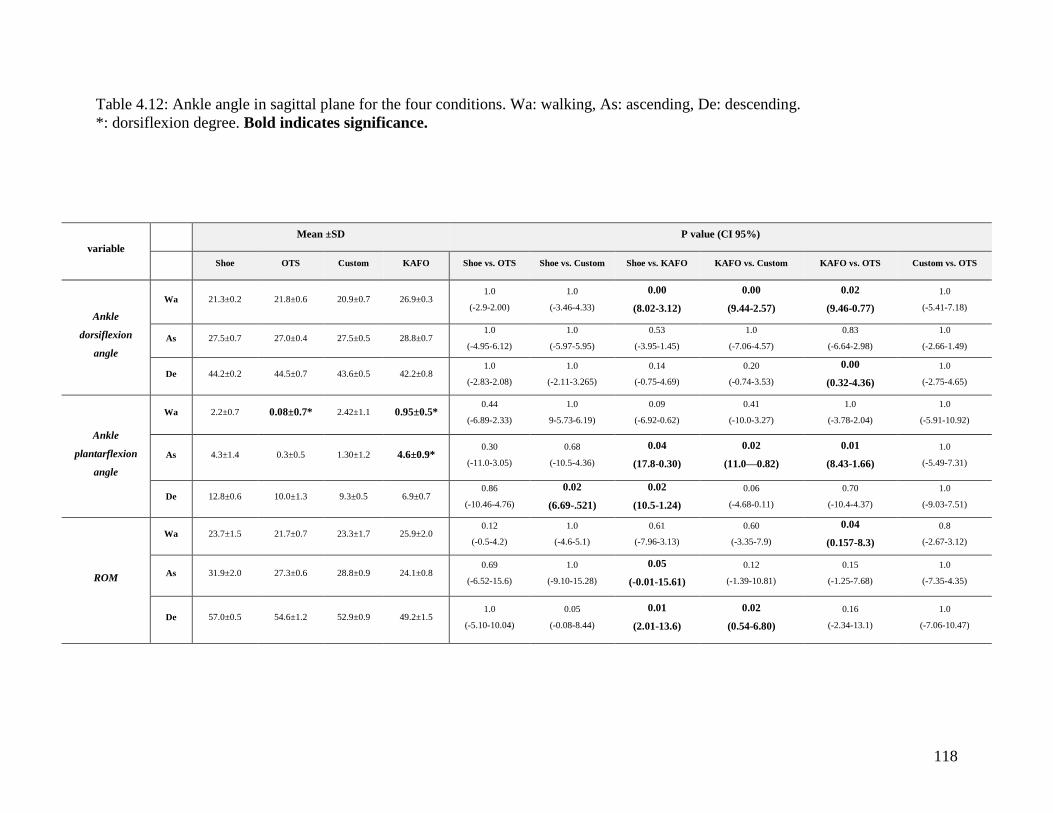
118
Table 4.12: Ankle angle in sagittal plane for the four conditions. Wa: walking, As: ascending, De: descending.
*: dorsiflexion degree. Bold indicates significance.
variable
Mean ±SD P value (CI 95%)
Shoe OTS Custom KAFO Shoe vs. OTS Shoe vs. Custom Shoe vs. KAFO KAFO vs. Custom KAFO vs. OTS Custom vs. OTS
Ankle
dorsiflexion
angle
Wa 21.3±0.2 21.8±0.6 20.9±0.7 26.9±0.3 1.0
(-2.9-2.00)
1.0
(-3.46-4.33)
0.00
(8.02-3.12)
0.00
(9.44-2.57)
0.02
(9.46-0.77)
1.0
(-5.41-7.18)
As 27.5±0.7 27.0±0.4 27.5±0.5 28.8±0.7 1.0
(-4.95-6.12)
1.0
(-5.97-5.95)
0.53
(-3.95-1.45)
1.0
(-7.06-4.57)
0.83
(-6.64-2.98)
1.0
(-2.66-1.49)
De 44.2±0.2 44.5±0.7 43.6±0.5 42.2±0.8 1.0
(-2.83-2.08)
1.0
(-2.11-3.265)
0.14
(-0.75-4.69)
0.20
(-0.74-3.53)
0.00
(0.32-4.36)
1.0
(-2.75-4.65)
Ankle
plantarflexion
angle
Wa 2.2±0.7 0.08±0.7* 2.42±1.1 0.95±0.5* 0.44
(-6.89-2.33)
1.0
9-5.73-6.19)
0.09
(-6.92-0.62)
0.41
(-10.0-3.27)
1.0
(-3.78-2.04)
1.0
(-5.91-10.92)
As 4.3±1.4 0.3±0.5 1.30±1.2 4.6±0.9* 0.30
(-11.0-3.05)
0.68
(-10.5-4.36)
0.04
(17.8-0.30)
0.02
(11.0—0.82)
0.01
(8.43-1.66)
1.0
(-5.49-7.31)
De 12.8±0.6 10.0±1.3 9.3±0.5 6.9±0.7 0.86
(-10.46-4.76)
0.02
(6.69-.521)
0.02
(10.5-1.24)
0.06
(-4.68-0.11)
0.70
(-10.4-4.37)
1.0
(-9.03-7.51)
ROM
Wa 23.7±1.5 21.7±0.7 23.3±1.7 25.9±2.0 0.12
(-0.5-4.2)
1.0
(-4.6-5.1)
0.61
(-7.96-3.13)
0.60
(-3.35-7.9)
0.04
(0.157-8.3)
0.8
(-2.67-3.12)
As 31.9±2.0 27.3±0.6 28.8±0.9 24.1±0.8 0.69
(-6.52-15.6)
1.0
(-9.10-15.28)
0.05
(-0.01-15.61)
0.12
(-1.39-10.81)
0.15
(-1.25-7.68)
1.0
(-7.35-4.35)
De 57.0±0.5 54.6±1.2 52.9±0.9 49.2±1.5 1.0
(-5.10-10.04)
0.05
(-0.08-8.44)
0.01
(2.01-13.6)
0.02
(0.54-6.80)
0.16
(-2.34-13.1)
1.0
(-7.06-10.47)
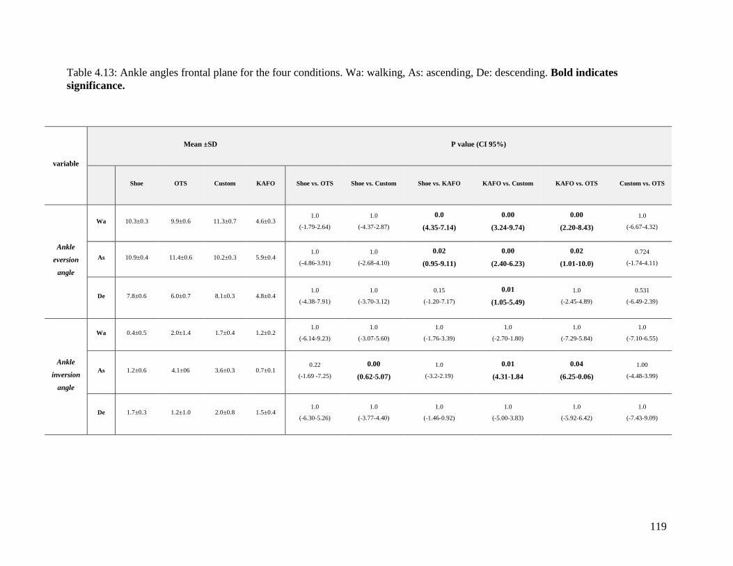
119
Table 4.13: Ankle angles frontal plane for the four conditions. Wa: walking, As: ascending, De: descending. Bold indicates
significance.
variable
Mean ±SD P value (CI 95%)
Shoe OTS Custom KAFO Shoe vs. OTS Shoe vs. Custom Shoe vs. KAFO KAFO vs. Custom KAFO vs. OTS Custom vs. OTS
Ankle
eversion
angle
Wa 10.3±0.3 9.9±0.6 11.3±0.7 4.6±0.3 1.0
(-1.79-2.64)
1.0
(-4.37-2.87)
0.0
(4.35-7.14)
0.00
(3.24-9.74)
0.00
(2.20-8.43)
1.0
(-6.67-4.32)
As 10.9±0.4 11.4±0.6 10.2±0.3 5.9±0.4 1.0
(-4.86-3.91)
1.0
(-2.68-4.10)
0.02
(0.95-9.11)
0.00
(2.40-6.23)
0.02
(1.01-10.0)
0.724
(-1.74-4.11)
De 7.8±0.6 6.0±0.7 8.1±0.3 4.8±0.4 1.0
(-4.38-7.91)
1.0
(-3.70-3.12)
0.15
(-1.20-7.17)
0.01
(1.05-5.49)
1.0
(-2.45-4.89)
0.531
(-6.49-2.39)
Ankle
inversion
angle
Wa 0.4±0.5 2.0±1.4 1.7±0.4 1.2±0.2 1.0
(-6.14-9.23)
1.0
(-3.07-5.60)
1.0
(-1.76-3.39)
1.0
(-2.70-1.80)
1.0
(-7.29-5.84)
1.0
(-7.10-6.55)
As 1.2±0.6 4.1±06 3.6±0.3 0.7±0.1 0.22
(-1.69 -7.25)
0.00
(0.62-5.07)
1.0
(-3.2-2.19)
0.01
(4.31-1.84
0.04
(6.25-0.06)
1.00
(-4.48-3.99)
De 1.7±0.3 1.2±1.0 2.0±0.8 1.5±0.4 1.0
(-6.30-5.26)
1.0
(-3.77-4.40)
1.0
(-1.46-0.92)
1.0
(-5.00-3.83)
1.0
(-5.92-6.42)
1.0
(-7.43-9.09)

120
Chapter five: Discussion
This chapter will discuss the results for the intervention study and compare our results with
previously published literature, followed by future directions for further study with a final
limitations and conclusions of the thesis.
5.1. Discussion
5.1.1Background of discussion
Custom and off-the-shelf (OTS) knee valgus braces are one of the most commonly used
conventional treatments for individuals with medial knee OA and previous results have shown
that EKAM and KAAI can be reduced by knee valgus braces (Draper et al., 2000, Deie et al.,
2013, Ramsey et al., 2007, Richards et al., 2005, Fantini Pagani et al., 2010b, Lindenfeld et al.,
1997, Self et al., 2000, Fantini Pagani et al. 2012, Fantini Pagani et al., 2010a). This study was
designed to provide evidence of the possibility of extending the conventional treatments options
available for individuals with medial knee OA and severe knee varus deformity, by evaluation of
the effect of a custom knee-ankle-foot orthosis (KAFO) on EKAM values and lower limb
kinematics. A cosmetic KAFO was compared to a custom-made and OTS knee valgus brace
which are commonly used in orthotic practice. To the author’s knowledge this is the first study
which has examined the effects of a KAFO on EKAM values for an individual with a varus
deformity during walking and stair climbing.
5.1.2 Summary of results
The KAFO was more effective than the custom or OTS knee valgus brace in reducing EKAM,
KAAI, knee varus angle, and ankle eversion during walking and stair climbing. In addition, the
KAFO improved knee and hip flexion angles and hip range of motion in the sagittal plane during

121
walking on a level surface and during stair climbing. The KAFO also reduced knee range of
motion in the sagittal plane compared to both knee valgus braces during walking and stair
climbing, but not significantly.
The KAFO also significantly reduced ankle ROM and ankle plantarflexion compared to the shoe
during stair ascent and descent. The OTS knee valgus brace reduced the knee varus angle more
than the custom one. No significant differences were found between both knee valgus braces
with regards to EKAM and KAAI. Both knee valgus braces insignificantly reduced knee range
of motion, ankle plantarflexion and ankle ROM compared to the shoe.
5.1.3 Hypotheses
5.2.2.1 External knee adduction moment (EKAM).
Hypothesis one: A KAFO reduces EKAM more than custom and off-the-shelf knee valgus
braces during walking and stair climbing.
Reducing the EKAM is one of the main aims for all interventions for medial knee OA to control
the progression of osteoarthritis (Miyazaki et al., 2002). The results have shown that the KAFO
significantly reduced the first peak of EKAM by 11.4% compared to the shoe during walking.
However, neither of the knee valgus braces showed significant changes in EKAM first or second
peaks during walking compared to the shoe. There was an insignificant difference in the second
peak when using the KAFO compared to the shoe. A 12% reduction was found compared to the
shoe during walking on a level surface.
No significant changes were seen when using either brace compared to the shoe. The custom
knee valgus brace reduced the first EKAM peak by 4.2%. This is similar to that seen in previous
studies where it has also been shown that both custom and OTS knee valgus braces do not
reduced the EKAM significantly during walking compared to a baseline condition (Gaasbeek et
al., 2007, Hewett et al., 1998, Pollo et al., 2002). This similarity in result is potentially related to

122
the high varus angle in participants in both the previous and current studies, also this gives the
indication that knee valgus brace are less effective to reduce EKAM for individuals with a severe
knee varus deformity.
In previous studies, it was found that the UnloaderOne knee valgus brace reduced EKAM values
by 5.5 % for individuals with medial knee OA during walking which was maintained over a
period of 12 months (Deie et al., 2013). Also, a study by Toriyama et al., (2011), which used the
same brace, found that using an OTS knee valgus braces reduced the first peak of EKAM
significantly by 11%, but no changes in second peak compared to a no-brace condition during
walking for individuals with medial knee OA were noted. Also, according to Moyer et al. (2013),
an adjustable UnloaderOne OTS (an adjustable knee valgus angle between two to nine degrees)
for individuals with medial knee OA with knee varus (6.6 ±3.3 degrees), reduced the first and
second peaks of EKAM by 8% and 12.7% respectively during walking compared to a no brace
condition. In contrast, our results show that both the UnloaderOne OTS and custom knee valgus
braced did not reduce EKAM significantly. This dissimilarity in results could be due to different
samples characteristics as we examined a participant without knee OA but still with a severe
knee varus deformity of 10 degrees, and therefore a different valgus position (more valgus
correction will reduce knee load more). In this current study, five degrees of valgus angulation
was set, but previous studies have used adjustable knee valgus angulation. Furthermore, different
periods of wearing a knee valgus brace (up to 12 months) have been used instead of a single test
session in this current study.
When comparing the custom and OTS knee valgus braces together, the OTS knee valgus brace
reduced EKAM and knee varus angle more effectively than the custom made knee valgus brace
but not significantly. In contrast, in the study by Draganich et al., (2006), which utilised different
types of OTS and custom knee valgus braces in the same study, found that the custom knee
valgus brace reduced the first peak of EKAM during walking by 14.5% after immediate use,
while the OTS knee valgus brace only reduced it by 3% compared to the baseline. This
difference is mainly related to high anatomical knee varus angle in our sample (ten degrees)
compared to the previous studies, different knee valgus braces, and the short length of the
123
custom knee valgus brace in this current study compared to the OTS brace could also make OTS
apply more correction force than the custom knee valgus brace.
Interlaced stairway climbing was the other activity which the participant undertook with the
shoe, OTS, custom knee valgus brace, and the KAFO as test conditions. Interlaced stairway
reliability was examined before the intervention study and proved that an interlaced stairway
could be highly reliable when used to examine knee kinetics and knee kinematics with an SEM
raised to 2.3 degrees. Furthermore, to our knowledge only one study has used stairs to examine
interventions such as an insole (Alshawabka et al., 2014), plus no previous study has examined
the effects of a KAFO on EKAM for varus individuals, and this study is only the second study
that has investigated the effect of knee valgus braces during stair climbing [the previous study
was undertaken by Al-Zahrani, (2014)].
The current study shows that the KAFO reduced first and second peaks by 7% and 10%
respectively compared to the shoe during stair ascent and no significant changes were seen
during stair descent. In addition, using custom and OTS knee valgus braces showed a non-
significant reduction in the EKAM of 8.2% and 7% respectively during stair ascent. However,
during stair descent the OTS and custom knee valgus braces increased the first peak of EKAM
by 11.2% and 19.7% respectively. This is in agreement with the Al-Zahrani (2014) study which
stated that a knee valgus brace did not reduce both peaks of EKAM significantly compared to the
baseline after three months of treatment for individuals with knee OA.
In contrast to our results, a custom knee valgus brace has been shown to reduce the average peak
of EKAM significantly during one stair stepping compared to baseline and an OTS knee valgus
brace. The OTS knee valgus brace did not reduce it significantly compared to baseline
(Draganich et al., 2006). However, this dissimilarity could be due to the different type, the design
of knee valgus braces, and the degree of valgus correction used, since the previous study used
bilateral upright knee valgus braces with bilateral polycentric knee joints and the valgus load
applied was adjustable. In this current study four degrees of knee valgus angulation (which is the
standard design for Ossur knee valgus braces) was applied with the knee valgus brace with a
single upright and single hinge joint was used. Moreover, one step was used in the previous

124
study to evaluate knee load, but three steps were used in this current study to evaluate the knee
load during one complete gait cycle. Also, it has been suggested that a knee valgus brace may
not reduce EKAM significantly during stair climbing due to the high knee varus moment during
stair climbing compared to walking on level (Al-Zahrani, 2014).
The measure of total loading during stance phase, the KAAI, was reduced significantly when
wearing the KAFO when compared to the shoe condition by 26.2% during stair ascent only;
however, the KAFO insignificantly reduced KAAI by 18.4% and 4.6% during walking and
descent respectively compared to the shoe. This means that using a KAFO reduced both EKAM
and the duration of the applied force on the medial knee compartment. Using the custom knee
valgus brace and its OTS variant did not significantly reduce the value of the KAAI during
walking (it was reduced by 7.8% by the custom one) or stair ascent, but the custom knee valgus
brace did however increase KAAI during stair descent by 10.4% compared to the shoe. This
result is similar to the Al-Zahrani (2014) study as knee valgus braces insignificantly reduced
KAAI values after three months compared to the shoe during walking and interlaced stairs way
climbing for individuals with medial knee OA.
In contrast to our result, Jones et al., (2013) found that an OTS knee valgus brace reduced KAAI
by 16.1% compared to baseline during walking activities. Furthermore, KAAI has also been
shown to be reduced by 29% and 15% according to Fantini Pagani et al. (2010a) after using a
four degree knee valgus brace and a flexible knee brace respectively. Similarly, it reduced when
using four-degrees valgus braces by 14% during walking on a level surface (Fantini Pagani et al.
2012) and 18% with using 4 degrees for healthy group compared to walking without brace
(Fantini Pagani et al., 2010b) and by 0.5% after using UnloaderOne OTS compared to walking
without a brace (Moyer et al., 2013). This variation could be due to dissimilar knee braces types,
and different sample characteristics. In the study by Fantini Pagani et al. (2012) a long knee
valgus brace was used and participants had mild knee varus alignment (mean 2.1±1.2), but in the
study by Fantini Pagani et al. (2010b) sixteen males with 8 degrees (±4 SD) of varus
participated. In this current study the participant had ten degrees of varus and both the Ossur
knee valgus braces used are shorter than the one which was used in the previous studies.

125
When comparing the KAFO and the knee valgus braces, the KAFO reduced the first peak of
EKAM by 15% and 12.6% and reduced second peak by 9.5% and 12.3 % (significantly) in
comparison to the OTS and custom knee valgus braces during walking. During stair ascent,
KAFO reduced second peak of EKAM by 9% and 14.5% significantly compared to custom and
OTS knee valgus brace respectively. During stair descent, KAFO reduced first peak of EKAM
by 16.4% and 10% in comparison to custom and OTS knee valgus braces respectively. Also,
KAAI is reduced by KAFO significantly compared to OTS and custom knee valgus brace during
walking and stair climbing. This difference between KAFO and knee valgus braces could be due
to KAFO design which can apply four point pressure over thigh, knee, and shank to correct the
tibia alignment, keep tibia in vertical position, and maintain the end of tibia and foot part in the
neutral position. This design could be able to shift the GRF more laterally and the knee joint
centre more medially, thereby reducing EKAM. Although, knee valgus brace applied three
points pressure likewise, the taller length of KAFO compared to knee valgus braces (Table 4.1)
helps to apply more force correction by KAFO than knee valgus braces and apply more control
on tibia position and foot alignment.
Even though reducing the EKAM is main aim for orthoses, the sagittal plane knee moment,
especially in areas of knee flexion such as stairs has to be examined because a high knee flexion
moment is correlated with increased knee pain and increased compression on the medial
compartment for knee OA (Walter et al. 2010). However, it has been suggested that a high knee
flexion moment only increases knee load during high flexion activities (Trepczynski et al.,
2014). Our results show that the knee flexion moment was increased after using the KAFO and
both knee braces during walking and stair climbing but this was not statistically significant. Also,
using the OTS knee valgus brace for one year does not change the knee flexion moment
significantly (Hewett et al., 1998).
Also, any change in GRF due to alteration in walking speed could change the EKAM
(Mündermann et al., 2004). To eliminate the effects of speed variation on the vertical GRF, the
speed was controlled during this current test by a metronome. Therefore, no significant changes
in vertical GRF were found during walking and stair climbing between the test conditions (Table
4.2).

126
In summary, the KAFO reduced EKAM and KAAI more effectively than both knee valgus
braces during walking and stair climbing without changing knee flexion moments. We
hypothesised that this could be due to KAFO design which applies four points of pressure to
correct knee alignment and its length cover more area to apply more correction force over a more
extensive area on the thigh and leg. No previous study has examined effects of a KAFO on
EKAM and KAAI for an individual with knee varus. Also, both knee valgus braces did not
reduce EKAM and KAAI significantly compared to some previous studies which could be due to
different knee valgus brace, degree of valgus position (2-9 degrees), short period of treatment in
current study, high varus deformity in our sample, small sample size, and the short length of
custom knee brace compared to the OTS.
Therefore, this hypothesis was accepted as the KAFO significantly reduced EKAM and
KAAI compared to the shoe and both knee valgus braces during walking and stair
climbing.
5.2.2.2 Lower limb kinematics
Hypothesis two: A KAFO would improve knee and hip ROMs more than either of the
valgus knee braces during walking and stair climbing.
Knee joint malalignment due to medial knee OA can affect the knee and hip joint in both sagittal
and frontal planes. It was suggested that a KAFO would improve knee and hip ROM more than
both valgus knee braces. Improvement in knee flexion and knee ROM are also important for
individuals with medial knee OA because it has been shown that individuals with medial knee
OA have reduced knee flexion than controls and 6 degrees less knee flexion at initial contact
(IC); (Landry et al., 2007, Mündermann et al., 2008a). Another purpose of using orthoses such as
KAFOs and knee valgus braces is to increase the knee flexion angle at initial contact and mid
swing, and increase overall sagittal plane knee ROM. This study has demonstrated that the
maximum knee flexion angle at initial contact is significantly increased by wearing a KAFO by
8.6 degrees during walking, but no significant changes in knee flexion at mid swing were noted.

127
However, it reduced knee ROM significantly (up to 10 degrees) compared to the shoe during
walking. Also, using OTS and custom knee valgus braces significantly increased knee flexion at
IC during walking by 3.5 and 5.3 degrees respectively and insignificantly reduced knee ROM by
8.2 and 7.9 degrees respectively when compared to the shoe during walking. Also, during stair
climbing, the KAFO and knee valgus braces also increased knee flexion at IC, but reduced knee
flexion at mid-swing and knee ROM, but not statistically differently between them.
As a result, the KAFO was superior in increasing knee flexion at IC and this result is mainly
related to the use of an offset knee joint which is used in KAFO design. This type of joint started
the movement from a position of 6 degrees more flexion at IC. The reduced knee flexion at mid-
swing may be associated with the weight of KAFO and the knee valgus braces could restrict
knee flexion at mid-swing because of the tight straps around knee in the knee valgus braces.
However, previous studies have found no significant differences in knee ROM during walking
and one stair stepping (Draganich et al., 2006) nor during interlaced stairway climbing (Al-
Zahrani, 2014). In contrast, previous studies have found that using a knee valgus brace reduces
knee flexion at mid swing, and total knee ROM during walking (Gasbeek et al., 2007, Jones et
al., 2013). This result could be due to light weight of knee valgus brace with single upright and
hinge joint instead of a double.
A high knee varus angle constitutes a high risk for medial knee OA subjects (Sharma et al.,
2001), and it is suggested that reducing knee varus deformity reduces EKAM and thereby knee
load (Fantini Pagani et al., 2010b, Landry et al., 2007). Also, a high knee varus angle is capable
of increasing knee load by 100% (Johnson et al., 1980); therefore, it is important to reduce the
knee varus angle using knee OA orthoses. This present study found that the knee adduction angle
was significantly decreased when using the KAFO by 8.2 degrees during walking on a level
surface compared to the shoe. Also, the custom and OTS knee valgus braces reduced it
significantly by 5.2 degrees and 5.8 degrees respectively during walking compared to the shoe,
but with no significant difference between them during walking. In previous studies, using a
custom knee valgus brace significantly reduced knee varus during walking which is similarly to
the studies by Draganich et al., (2006) and Matsuno et al., (1997). Also, our study found that
128
using the OTS knee valgus brace was more effective than a custom knee brace in reducing the
knee varus angle which is contrary to the studies by Draganich et al., (2006) and Toriyama et al.,
(2011). This variation could be due to different sample characteristics; for example previous
studies have evaluated individuals with moderate and severe knee OA, while in the existing
study the participant did not suffer from knee OA but had a knee varus deformity.
During stair climbing, the KAFO reduced the knee varus angle by 12 and 6 degrees during stair
ascent and descent respectively compared to the shoe. Both knee valgus braces insignificantly
reduced it during stair climbing up to 3 degrees which is similar to Al-Zahrani (2014) study.
When we compared the KAFO with the knee valgus braces, the results showed that the KAFO
reduced knee varus angle by up to 15 and 6.8 degrees compared to the custom knee valgus brace
and up to 12.4 and 3.5 degrees compared to OTS knee valgus brace during stair ascent and
descent respectively. The efficiency of a KAFO in reducing a knee varus angle is mainly related
to the offset joint. This type of artificial knee joint is able to correct knee deformity in the frontal
plane (knee valgus/varus) by up to 75mm (Johnson et al., 2004), also the length of the KAFO is
able to apply more force over a more extensive tissue area than knee valgus braces, so would
theoretically correct more deformity, and it is therefore assumed that the custom fit of the KAFO
on the limb has an important role in correcting the knee varus deformity and reducing EKAM
efficiently. When comparing both knee valgus braces together, the OTS was slightly superior to
the custom made knee valgus brace and reduced knee varus more. However, custom made knee
valgus braces fit better, but the one used in this study was shorter than the OTS and has a similar
mass (Table 4.1). These differences could make the OTS more able to apply increased forces
over the proximal and distal segments around the knee joint and control the knee rotation since
the OTS used for the participant in this study was also a good fit over the affected knee.
With regards to hip flexion and extension angles, Briem and Snyder‐Mackler (2009)
demonstrated that hip flexion and hip ROM is reduced and hip extension is increased for
individuals with knee OA. As a result, it would be advantageous for an orthosis to produce
improved hip ROM for individuals with medial knee OA. In this current study, the KAFO
increased hip range of motion during walking and stair ascent, while neither of the knee braces
altered hip range of motion. This may have been due to the offset joint included in the design of
129
the KAFO which shifts the GRF more posterior to the knee and more anterior to hip joint to
generate knee and hip flexion. In addition, during taking the negative cast for the participant, the
lower extremity was fixed with the knee and hip in a slightly flexion position as it is functional
position for both joints. Custom knee and OTS knee valgus braces have not been shown
previously to alter hip ROM significantly (Taghi Karimi et al., (2012) and Toriyama et al.,
(2011). However, hip ROM was reduced by the knee valgus brace during walking, but no
changes were seen during stair ascent and descent (Al-Zahrani, 2014), which is suggested to be
related to the tight strap around the thigh segment which restricted hip extension at mid swing.
For hip adduction/abduction angle and moment, it has been suggested that knee OA is associated
with a high hip abduction angle and low hip adduction moment due to the lateral bending
compensatory gait during early stance. This is used as a strategy to reduce EKAM (Briem and
Snyder‐Mackler, 2009), but unfortunately this compensation could increase back pain and the
incidence of hip OA in the future (Mündermann et al. 2008a, Toriyama et al., 2011). Therefore,
it is necessary to control the hip abduction angle and increase the hip adduction moment, and
thereby prevent hip lateral bending.
Using the KAFO reduced hip abduction, increased hip adduction angle, and also increased hip
adduction moment during walking and stair climbing more than both knee valgus braces. This
means that the KAFO did not produce an increase in pelvic tilt or lateral bending to reduce the
EKAM, but it reduced EKAM by controlling the tibia and foot alignments. However, no
significant changes in hip adduction/abduction angles were seen after using the knee valgus
braces, but both of them reduced the second peak of the hip adduction moment. In contrast to our
results, hip adduction angle is reduced by 2.5 degrees with a custom knee valgus brace according
to Toriyama et al., (2011) due to a reduction in the hip adduction moment and an increase in hip
bending to the swing phase side as a compensation to reduce EKAM. This supports the
biomechanical theory that the knee valgus brace can reduce EKAM by shifting the centre of
mass more laterally and closer to the centre of the knee. This difference could be due to the fact
that subjects with bilateral knee OA participated in the Toriyama (2011) study.
130
For the ankle joint, the KAFO reduced ankle ROM significantly, but no changes in ankle ROM
were demonstrated when using either knee valgus braces during walking or stair climbing.
However, according to the study by Al-Zahrani (2014), no significant changes in ankle ROM
were found during walking and stair descent, but it was significantly reduced during stair ascent
which could be due to the tight straps that limited shank movement and thereby ankle joint
motion.
For ankle inversion/eversion, the KAFO was superior in reducing ankle eversion than the knee
valgus braces; however, both knee valgus braces increased ankle eversion compared to the shoe.
Similarly, ankle eversion angles and moments were not significantly reduced during walking
with a 4 degree knee valgus brace according to the studies by Fantini Pagani et al. (2012) and
Hewett et al., (1998). However, the orthosis used was an 8 degree knee valgus brace (Fantini
Pagani et al. 2012); this dissimilarly in result is clearly related to the different varus angle in the
brace knee valgus angle which was used in this study (4 degrees) and the one which was used in
the previous study (8 degrees).
In conclusion, this hypothesis is partially accepted since the KAFO significantly increased
knee flexion at IC, and increased hip flexion and hip ROM more than both knee valgus
braces during walking and stair climbing; however, using the KAFO insignificantly
reduced knee ROM more than both knee valgus braces during walking and stair climbing.
Since the knee joint is one of the most affected joints by osteoarthritis, a variety of orthoses are
recommended as conservative treatments to correct the knee deformity, reduce pain, improve the
activity level, and thereby hope to delay the inevitable knee replacement. The primary
anatomical deviation in medial knee OA is a varus deformity which results in a higher EKAM.
Therefore, conservative orthotic interventions such as footwear, lateral wedges and knee valgus
braces are used to examine their effect on these parameters. However, these previous
interventions have some limitations such as changing ankle alignment, not being very effective
to reduce EKAM, they reduce knee flexion in mid swing, increase hip lateral bending, have tight
straps in knee valgus brace which causes swelling, and they may feel uncomfortable for extended
use. Therefore, the feasibility of a KAFO was examined in order to find a new conservative

131
treatment for severe knee varus and medial knee OA individuals. Thus, this study demonstrates
that the KAFO reduced EKAM, KAAI, knee varus angle, improved hip ROM, and reduced hip
lateral bending more effectively than knee valgus braces.
5.3. Limitations
The current study has some limitations such as small sample size and the subject used having a
knee varus deformity without complaining from pain or knee OA or any previous injury. As a
result, one of the future plans is to increase the sample size to include medial compartment knee
OA participants in a future investigation. Another limitation of this study is that it evaluated the
immediate effects of KAFO and knee braces on the primary outcome measures. A longitudinal
study could be helpful to examine the long term effects of using KAFO on the knee load and OA
progression. Furthermore, because of that reason it is difficult to know if these orthoses would
reduce pain and improve activity levels. Also, using a simple ankle joint limits ankle joint
movement and a free mechanical ankle joint will be used in future even though it will increase
KAFO weight and cost.
Besides that, it is important to examine if using a custom KAFO is acceptable to be used for long
periods or not and the associated side effects. Nevertheless, the design of KAFO used in this
study is considered as being lightweight compared to other designs, and the absence of straps
around knee joint gives it a more comfortable feeling when flexing the knee particularly when
using stairs and it is able to provide an intimate fit as it was custom made. Nevertheless, its cost
will be more than an OTS knee valgus brace but still it does not cost as much as some types of
knee valgus braces which contain air champers. The author's experience found that this design of
KAFO is already used and recommended for individuals with very severe knee OA in some
countries who wish to avoid undergoing total knee replacement surgery and have not previously
found any benefit of using knee valgus braces; however, the long term effect of using a long
brace needs to be investigated. In contrast, the limitations of using knee valgus braces have
already been reported, such as poor fit (especially the OTS knee valgus brace), skin irritation,
discomfort when flexing the knee due to knee strap impingement (Stamenović et al., 2008),
132
discontinuation of use (Squyer et al., 2013). In addition, some designs are bulky, heavy, and are
not acceptable for long term use, and some individuals have complained from swelling and
thrombosis after 6 months of use (Giori, 2004).
With regards to the biomechanical testing, markers artifacts due to skin movements could be one
of the limitations during walking and stair climbing, but because of the use of dynamic tracking
markers (clusters) and a good shoe fit with a tight lace, the artifacts would have been reduced.
For static alignment capturing, two static alignments were captured before the participant started
walking and the shoe size was the only variable which had changed. The rest of the markers were
still in the correct position during the two static alignments and their location was marked and
checked. In addition, off-the-shelf knee valgus braces may not offer an intimate fit for some
individuals as they are not custom made, but the one which was used in this study was an
adequate fit for the participant.
The ten minute washout period between treatments may not have been long enough but it was
thought to be acceptable because using braces inflict immediate biomechanical changes in knee
angles and load, and it is suggested that the knee biomechanics returns to the original position
and alignment immediately once the braces are removed (Draganich et al., (2006). Finally, the
staircase which was used in this study contained only three steps; therefore, the contra lateral
side was not evaluated. Having a staircase with more than three steps could be helpful to
evaluate the both sides and give information about the effects of a KAFO on both the affected
and non-affected side.
5.2. Future work
This pilot study evaluated a new modified type of KAFO which could be a possible conservative
treatment for medial knee OA in the future. Therefore, the next phase would be to evaluate the
device in a sample of medial knee OA subjects and appraising if this design incorporating a free
ankle joint could help to reduce EKAM, KAAI, and knee varus angle during walking and
negotiating stairs. Also, a free mechanical ankle joint could be added to the KAFO design

133
instead of a plastic one to add more ankle motion freedom and add more plantarflexion freedom
during stair climbing. Further assessments of pain and activity levels, muscles activity, energy
consumption, and quality of life enhancement could be examined in the future study after using
the device for an extended period such as for between six and twelve months.
5.4. Conclusion
After taking into account the study limitations, this study is the first study to evaluate the effect
of a modified KAFO for knee varus individuals during walking and stair climbing. The data from
this pilot study supports use of a modified KAFO to reduce EKAM, KAAI, and knee varus angle
during walking and stair climbing and also giving improved hip and knee range of motion.
However, the limitation of the ankle range of motion is something which needs addressing
before embarking on a follow-up study. Therefore, the data supports that wearing a KAFO can
significantly reduce EKAM and knee varus deformity and it could be one of the future
interventions for individuals with medial compartment knee OA who have a varus aligned knee
joint especially if current treatment (knee valgus braces for example) are not restrictive or
corrective enough.
.
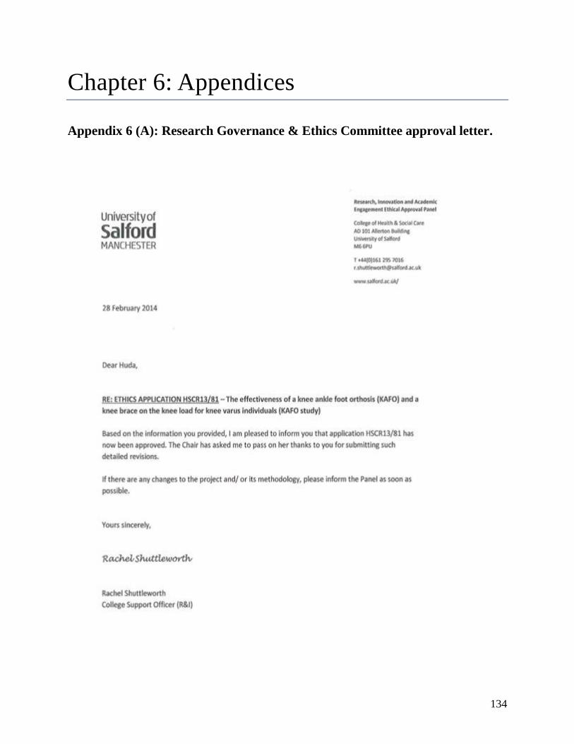
134
Chapter 6: Appendices
Appendix 6 (A): Research Governance & Ethics Committee approval letter.

135
Appendix 6 (B): Demographic and examination sheet.
Demographic collection sheet (KAFO study):
Participant's ID.: ............................... Gender: Male / Female.
Age:...................................................... Height (M):........................
Mass (Kg):......................................... Knee varus degree: ...............
KAFO: left / right. Knee joint type:..................
Any comments:
.................................................................................................................................................................
.................................................................................................................................................................
.................................................................................................................................................................
.................................................................................................................................................................

136
Appendix 6 (C): The study poster.

137
Appendix 6 (D): Consent Form.
Principal investigator: Huda Alfatafta.
INFORMED CONSENT FORM
Study title: The effect of a knee ankle foot orthosis on lower limb kinematics and kinetics in an
individual with varus knee alignment (KAFO study).
1. I confirm that I have read and understand the participant information sheet dated (V 1.1 Dated
22.1.2014) for the above study and have had the opportunity to ask any questions.
2. I understand that my participation is voluntary and that I am free to withdraw at any time, without
giving any reason and without my medical care or legal rights being affected.
3. Should I withdraw I understand that the data acquired from my participation may still be used unless I
specify otherwise in writing.
4. I agree for the research team to keep my details on record for future research they are performing in
conation related to osteoarthritis and I am happy to be contacted to discuss potential participation in the
study.
5. I understand that the information about me and the data generated from the study is to be kept and
may be used in future studies.
6. I agree to take part in the above study.
Name of Participant Date Signature
Name of person taking consent Date Signature
(If different from Researcher)
Researcher Date Signature
1 copy for participant; 1 for researcher to be kept with study file
Please initial
Each box

138
Appendix 6 (E): Abbreviations.
AC Ascending Cycle
ACL Anterior Cruciate Ligament
AMTI Advanced Mechanical Technology Incorporation
ANOVA Analysis Of Variance
AS Ascending
ASIS Anterior Superior Iliac Spine
BMI Body Mass Index
CAST Calibration Anatomical System Technique
CP Center of Pressure
CVs Coefficients of Variations
De Descending
DC Descending Cycle
DIP Distal Interphalangeal
EKAM External Knee Adduction Moment
IC Initial Contact
ICC Intraclass Correlation Coefficients
KAAI Knee Adduction Angular Impulse
KAFO Knee Ankle Foot Orthosis
KL Kellgren and Lawrence
LLL Lower limb length
MCL Medial Collateral Ligament
MTP Metatarsophalangeal
NSAIDs Non-Steroidal Anti-Inflammatory Drugs
OA Osteoarthritis
OTS Off The Shelf
PIP Proximal Interphalangeal
QTM Qualisys Track Manger
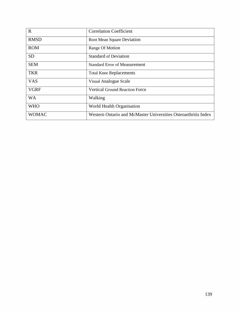
139
R Correlation Coefficient
RMSD Root Mean Square Deviation
ROM Range Of Motion
SD Standard of Deviation
SEM Standard Error of Measurement
TKR Total Knee Replacements
VAS Visual Analogue Scale
VGRF Vertical Ground Reaction Force
WA Walking
WHO World Health Organisation
WOMAC Western Ontario and McMaster Universities Osteoarthritis Index
140
References
1. Abdallah, A. A., & Radwan, A. Y. (2011). Biomechanical changes accompanying
unilateral and bilateral use of laterally wedged insoles with medial arch supports in
patients with medial knee osteoarthritis. Clinical Biomechanics, 26(7), 783-789.
2. Ahlbäck, S. (1968). Osteoarthrosis of the knee. A radiographic investigation. Acta
radiologica: diagnosis, Suppl-277.
3. Alshawabka, A. Z., Liu, A., Tyson, S. F., & Jones, R. K. (2014). The use of a lateral
wedge insole to reduce knee loading when ascending and descending stairs in medial
knee osteoarthritis patients. Clinical Biomechanics, 29(6), 650-6.
4. Altman, R. D. (1991a, June). Classification of disease: osteoarthritis. InSeminars in
arthritis and rheumatism (Vol. 20, No. 6, pp. 40-47). WB Saunders.
5. Altman, R., Alarcon, G., Appelrouth, D., Bloch, D., Borenstein, D., Brandt, K., ... &
Wolfe, F. (1990). The American College of Rheumatology criteria for the classification
and reporting of osteoarthritis of the hand. Arthritis & Rheumatism, 33(11), 1601-1610.
6. Altman, R., Alarcon, G., Appelrouth, D., Bloch, D., Borenstein, D., Brandt, K., ... &
Wolfe, F. (1991b). The American College of Rheumatology criteria for the
classification and reporting of osteoarthritis of the hip. Arthritis & Rheumatism, 34(5),
505-514.
7. Altman, R., Asch, E., Bloch, D., Bole, G., Borenstein, D., Brandt, K., Christy, W.,
Cooke, T. D., Greenwald, R. , Hochberg, M. (1986). Development of criteria for the
classification and reporting of osteoarthritis: Classification of osteoarthritis of the knee.
Arthritis & Rheumatism, 29(8), 1039-1049.

141
8. Al-Zahrani, K.S., & Bakheit, A.M. (2002). A study of gait characteristic of patients
with chronic osteoarthritis of the knee. Disabil Rehabil., 24, 275-280.
9. Al-Zahrani, Y. (2014). Effectiveness of a valgus knee brace on biomechanical and
clinical outcomes during walking and stair climbing in individuals with knee
osteoarthritis. PhD thesis. University of Salford. Available at:
http://usir.salford.ac.uk/32833/1/Yousef_Al-zahrani_Thesis_%28Sep-2014%29.pdf
10. Anderson, J. J. & Felson, D. T. (1988). Factors associated with osteoarthritis of the
knee in the first national health and nutrition examination survey (hanes i). American
Journal of Epidemiology, 128(1), 179-189.
11. Andriacchi, T.P. (1994). Dynamics of knee malalignment. Orthop Clin North Am,
25(3), 395-403.
12. Andriacchi, T. P., Andersson, G. B., Fermier, R. W., Stern, D., & Galante, J. O. (1980).
A study of lower-limb mechanics during stair-climbing. J Bone Joint Surg Am, 62(5),
749-57.
13. Andriacchi, T. P., Briant, P. L., Bevill, S. L., & Koo, S. (2006). Rotational changes at
the knee after ACL injury cause cartilage thinning. Clinical orthopaedics and related
research, 442, 39-44.
14. Andriacchi, T. P., Mündermann, A., Smith, R. L., Alexander, E. J., Dyrby, C. O., &
Koo, S. (2004). A framework for the in vivo pathomechanics of osteoarthritis at the
knee. Annals of biomedical engineering, 32(3), 447-457.
15. Andriacchi, T. P., Ogle, J. A., & Galante, J. O. (1977). Walking speed as a basis for
normal and abnormal gait measurements. Journal of biomechanics, 10(4), 261-268.
142
16. Angst, F., Aeschlimann, A., Steiner, W., & Stucki, G. (2001). Responsiveness of the
WOMAC osteoarthritis index as compared to the SF-36 in patients with osteoarthritis
of the legs undergoing a comprehensive rehabilitation intervention. Annals of the
rheumatic diseases, 60(9), 834-840.
17. Arazpour, M., Bani, M. A., Maleki, M., Ghomshe, F. T., Kashani, R. V., & Hutchins,
S. W. (2013). Comparison of the efficacy of laterally wedged insoles and bespoke
unloader knee orthoses in treating medial compartment knee osteoarthritis. Prosthetics
and Orthotics International, 37(1), 50-57.
18. Arokoski, J. P. A., Jurvelin, J. S., Väätäinen, U., & Helminen, H. J. (2000). Normal and
pathological adaptations of articular cartilage to joint loading. Scandinavian journal of
medicine & science in sports, 10(4), 186-198.
19. Arthritis care (2004) OA nation. www.Arthritiscare.Org.Uk.
20. Arthritis research UK (2013). http://www.arthritisresearchuk.org.
21. Astephen, J. L., Deluzio, K. J., Caldwell, G. E., & Dunbar, M. J. (2008a).
Biomechanical changes at the hip, knee, and ankle joints during gait are associated with
knee osteoarthritis severity. Journal of Orthopaedic Research, 26(3), 332-341.
22. Astephen, J. L., Deluzio, K. J., Caldwell, G. E., Dunbar, M. J., & Hubley-Kozey, C. L.
(2008b). Gait and neuromuscular pattern changes are associated with differences in
knee osteoarthritis severity levels. Journal of Biomechanics, 41(4), 868-876.
23. Baker, K., Goggins, J., Xie, H., Szumowski, K., LaValley, M., Hunter, D.J., Felson,
D.T. (2007). A randomized crossover trial of a wedged insole for treatment of knee
osteoarthritis. Arthritis Rheum, 56(4):1198-203.
143
24. Baker, K., Xu, L., & Zhang, Y. (2005). Quadriceps weakness and its relationship to
tibiofemoral and patellofemoral knee osteoarthritis in Chinese. Br J Sports Med, 39,
50-51.
25. Baker, R. (2013). Gait graphs for beginners. http://wwrichard.net/2013/11/08/gait-
graphs-for-beginners/.
26. Baliunas, A.J., Hurwitz, D.E., Ryals, A.B., Karrar, A., Case, J.P., Block, J.A., &
Andriacchi, T.P. (2002). Increased knee joint loads during walking are presented in
subjected with knee osteoarthritis. Osteaoarthritis Cartalidge, 10, 573-579.
27. Bashaw, R. T., & Tingstad, E. M. (2005). Rehabilitation of the osteoarthritic patient:
focus on the knee. Clinics in sports medicine, 24(1), 101-131.
28. Beaupre, G., Stevens, S., Carter, D. (2000). Mechanobiology in the development,
maintenance, and degeneration of articular cartilage. Journal of Rehabilitation
Research and Development, 37(2), 145—151.
29. Benjamin, A., & Trickey, E. L. (1969). Double osteotomy for the painful knee in
rheumatoid arthritis and osteoarthritis. J Bone Joint Surg Br, 51(4), 694-699.
30. Bennell, K., Bowles, K. A., Payne, C., Cicuttini, F., Osborne, R., Harris, A., &
Hinman, R. (2007). Effects of laterally wedged insoles on symptoms and disease
progression in medial knee osteoarthritis: a protocol for a randomised, double-blind,
placebo controlled trial. BMC musculoskeletal disorders, 8(1), 96.
31. Bennell, K.L, Bowles, K.A, Payne, C., Cicuttini, F., Williamson, E., Forbes, A., Hanna,
F., Davies-Tuck, M., Harris A., Hinman, R.S. (2011a). Lateral wedge insoles for
medial knee osteoarthritis: 12 month randomised controlled trial. BMJ. 18, 342.
144
32. Bennell, K. L., Bowles, K. A., Wang, Y., Cicuttini, F., Davies-Tuck, M., & Hinman, R.
S. (2011b). Higher dynamic medial knee load predicts greater cartilage loss over 12
months in medial knee osteoarthritis. Annals of the rheumatic diseases, 70(10), 1770-
1774.
33. Bennell, K. L., Kean, C. O., Wrigley, T. V., & Hinman, R. S. (2013). Effects of a
modified shoe on knee load in people with and those without knee
osteoarthritis. Arthritis & heumatism, 65(3), 701-709.
34. Bennell, K. L., Hinman, R. S., Metcalf, B. R., Buchbinder, R., McConnell, J., McColl,
G., ... & Crossley, K. M. (2005). Efficacy of physiotherapy management of knee joint
osteoarthritis: a randomised, double blind, placebo controlled trial. Annals of the
Rheumatic Diseases, 64(6), 906-912.
35. Bennell, K. L., Hunt, M. A., Wrigley, T. V., Hunter, D. J., McManus, F. J., Hodges, P.
W., & Hinman, R. S. (2010). Hip strengthening reduces symptoms but not knee load in
people with medial knee osteoarthritis and varus malalignment: a randomised
controlled trial. Osteoarthritis and Cartilage, 18(5), 621-628.
36. Bergland A, Jarnlo GB, Laake K. (2003). Predictors of falls in the elderly by location.
Aging Clin Exp Res, 15(1), 43–50.
37. Billings, A., Scott, D. F., Camargo, M. P., & Hofmann, A. A. (2000). High Tibial
Osteotomy with a Calibrated Osteotomy Guide, Rigid Internal Fixation, and Early
Motion. Long-Term Follow-up. The Journal of Bone & Joint Surgery, 82(1), 70-9.
38. Bolen, J., Schieb, L., Hootman, J. M., Helmick, C. G., Theis, K., Murphy, L. B., &
Langmaid, G. (2010). Peer Reviewed: Differences in the Prevalence and Impact of
Arthritis Among Racial/Ethnic Groups in the United States, National Health Interview
Survey, 2002, 2003, and 2006. Preventing chronic disease, 7(3), 10_0035a.

145
39. Bowker, P. (1993). The biomechanics of orthoses. Biomechanical Basis of Orthotic
Management. Oxford, England: Butterworth-Heinemann
40. Brater, D. C. (1988). Clinical pharmacology of NSAIDs. The Journal of Clinical
Pharmacology, 28(6), 518-523.
41. Brehm, M. A., Beelen, A., Doorenbosch, C. A., Harlaar, J., & Nollet, F. (2007). Effect
of carbon-composite knee-ankle-foot orthoses on walking efficiency and gait in former
polio patients. Journal of Rehabilitation Medicine, 39(8), 651-657.
42. Briem, K., & Snyder‐Mackler, L. (2009). Proximal gait adaptations in medial knee
OA. Journal of Orthopaedic Research, 27(1), 78-83.
43. Brooks, P., & Hochberg, M. (2001). Outcome measures and classification criteria for
the rheumatic diseases. A compilation of data from OMERACT (Outcome Measures
for Arthritis Clinical Trials), ILAR (International League of Associations for
Rheumatology), regional leagues and other groups.Rheumatology, 40(8), 896-906.
44. Bruton, A., Conway, J. H., & Holgate, S. T. (2000). Reliability: what is it, and how is it
measured?. Physiotherapy, 86(2), 94-99.
45. Buckwalter, J.A., Saltzman, C., Brown, T. (2004). The impact of osteoarthritis:
implications for research. Clin Orthop Rel Res., 427(Suppl), S6–S15.
46. Carter, D.R., Beaupré ,G.S., Wong ,M., Smith, R.L., Andriacchi, T.P., Schurman, D.J.
(2004). The mechanobiology of articular cartilage development and degeneration. Clin
Orthop Relat Res, (427 Suppl), S69-77.
47. Cappozzo, A., Catani, F., Della Croce, U., & Leardini, A. (1995). Position and
orientation in space of bones during movement: anatomical frame definition and
determination. Clinical Biomechanics, 10(4), 171-178.
146
48. Chang, A., Hayes, K., Dunlop, D., Song, J., Hurwitz, D., Cahue, S., & Sharma, L.
(2005). Hip abduction moment and protection against medial tibiofemoral osteoarthritis
progression. Arthritis & Rheumatism, 52(11), 3515-3519.
49. Chang, A., Hayes, K., Dunlop, D., Hurwitz, D., Song, J., Cahue, S., & Sharma, L.
(2004). Thrust during ambulation and the progression of knee osteoarthritis. Arthritis &
Rheumatism, 50(12), 3897-3903.
50. Chang, C.B., Yoo, J.H., Koh, I.J., Kang, Y.G., Seong, S.C., & Kim, T.K. (2010). Key
factors in determining surgical timing of total knee arthroplasty in osteoarthritic
patients: age, radiographic severity, and symptomatic severity. J.Orthop. Traumatol, 11
(1), 21-27.
51. Chinn, S. (1991). Statistics in respiratory medicine. 2. Repeatability and method
comparison. Thorax, 46(6), 454-456.
52. Cooke, T. D. V., Scudamore, A., & Greer, W. (2003). Varus knee osteoarthritis:
whence the varus?. Journal of rheumatology, 30(12), 2521-2523.
53. Cooper, C., Snow, S., McAlindon, T. E., Kellingray, S., Stuart, B., Coggon, D., &
Dieppe, P. A. (2000). Risk factors for the incidence and progression of radiographic
knee osteoarthritis. Arthritis & Rheumatism, 43(5), 995-1000.
54. Costa, R. A., Oliveira, L. M. D., Watanabe, S. H., Jones, A., & Natour, J. (2010).
Isokinetic assessment of the hip muscles in patients with osteoarthritis of the
knee. Clinics, 65(12), 1253-9.
55. D’Ambrosia, R.D. (2005). Epidemiology of osteoarthritis. Orthopedics, 28(2 Suppl),
s201–205.
147
56. Davies, A. P., Calder, D. A., Marshall, T., & Glasgow, M. M. S. (1999). Plain
radiography in the degenerate knee A CASE FOR CHANGE. Journal of Bone & Joint
Surgery, British Volume, 81(4), 632-635.
57. Davies-Tuck, M. L., Wluka, A. E., Teichtahl, A. J., Martel-Pelletier, J., Pelletier, J. P.,
Jones, G., ... & Cicuttini, F. M. (2008). Association between meniscal tears and the
peak external knee adduction moment and foot rotation during level walking in
postmenopausal women without knee osteoarthritis: a cross-sectional study. Arthritis
Research and Therapy, 10(3), R58.
58. Dawson, J., Linsell, L., Zondervan, K., Rose, P., Randall, T., Carr, A., & Fitzpatrick,
R. (2004). Epidemiology of hip and knee pain and its impact on overall health status in
older adults. Rheumatology, 43(4), 497-504.
59. Dayal, N., Chang, A., Dunlop, D., Hayes, K., Chang, R., Cahue, S., ... & Sharma, L.
(2005). The natural history of anteroposterior laxity and its role in knee osteoarthritis
progression. Arthritis & Rheumatism, 52(8), 2343-2349.
60. Deie, M., Shibuya, H., Toriyama, M., Kumahashi, N., Uchio, Y., Sasashige, Y., &
Ochi, M. (2013). The Effects of an Unloading Knee Brace and Insole with Subtalar
Strapping for Medial Osteoarthritis of the Knee. International Journal of Clinical
Medicine, 4, 6.
61. Della Croce, U., & Bonato, P. (2007). A novel design for an instrumented
stairway. Journal of biomechanics, 40(3), 702-704
62. Dempster, W. T. (1955). Space Requirements of the Seated Operator: Geometric,
Kinematic, and Mechanical Aspects of the Body with Special Reference to Limbs.
NTIS.

148
63. DeVita, P., & Hortobagyi, T. (2000). Age causes a redistribution of joint torques and
powers during gait. Journal of applied physiology, 88(5), 1804-1811.
64. Dhawan, A., Mather III, R. C., Karas, V., Ellman, M. B., Young, B. B., Bach Jr, B. R.,
& Cole, B. J. (2014). An Epidemiologic Analysis of Clinical Practice Guidelines for
Non-Arthroplasty Treatment of Osteoarthritis of the Knee. Arthroscopy: The Journal of
Arthroscopic & Related Surgery, 30(1), 65-71.
65. Dieppe, P., Lohmander, L., (2005). Pathogenesis and management of pain in
osteoarthritis. Lancet, 365 (9463), 965-73.
66. Dillon, C.F., Rasch, E.K., Gu, Q., Hirsch, R. (2006). Prevalence of knee osteoarthritis
in the United States: arthritis data from the Third National Health and Nutrition
Examination Survey 1991–94. J Rheumatol, 33(11), 2271–9.
67. Divine, J. G., & Hewett, T. E. (2005). Valgus bracing for degenerative knee
osteoarthritis: relieving pain, improving gait, and increasing activity. The Physician
and sports medicine, 33(2), 40-46.
68. D'Lima, D.D., Fregly, B.J., Patil, S., Steklov, N., Colwell, C.W. (2012). Knee joint
forces: prediction, measurement, and significance. Proc Inst Mech Eng H, 226(22), 95-
102.
69. Draganich, L., Reider, B., Rimington, T., Piotrowski, G., Mallik, K., & Nasson, S.
(2006). The effectiveness of self-adjustable custom and off-the-shelf bracing in the
treatment of varus gonarthrosis. The Journal of Bone & Joint Surgery, 88(12), 2645-
2652.
70. Draper, E., Cable, J., Sanchez-Ballester, J., Hunt, N., Robinson, J., & Strachan, R.
(2000). Improvement in function after valgus bracing of the knee an analysis of gait
symmetry. Journal of Bone & Joint Surgery, British Volume, 82(7), 1001-1005.
149
71. Dreinhöfer, K. E., Dieppe, P., Stürmer, T., Gröber-Grätz, D., Flören, M., Günther, K.
P., ... & Brenner, H. (2006). Indications for total hip replacement: comparison of
assessments of orthopaedic surgeons and referring physicians. Annals of the rheumatic
diseases, 65(10), 1346-1350.
72. Duncan, R.C., Hay, E.M., Saklatvala, J., et al. (2006). Prevalence of radiographic
osteoarthritis – it all depends on your point of view. Rheumatology, 45(6), 757–60.
73. Eckstein, F., Faber S., M¨uhlbauer R., Hohe J., Englmeier K., Reiser M., Putz R.
(2002). Functional adaptation of human joints to mechanical stimuli. Osteoarthritis
Cartilage, 10, 44–50.
74. Emrani, P. S., Katz, J. N., Kessler, C. L., Reichmann, W. M., Wright, E. A.,
McAlindon, T. E., & Losina, E. (2008). Joint space narrowing and Kellgren–Lawrence
progression in knee osteoarthritis: an analytic literature synthesis.Osteoarthritis and
Cartilage, 16(8), 873-882.
75. Erhart‐Hledik, J. C., Elspas, B., Giori, N. J., & Andriacchi, T. P. (2012). Effect of
variable‐stiffness walking shoes on knee adduction moment, pain, and function in
subjects with medial compartment knee osteoarthritis after 1 year. Journal of
Orthopaedic Research, 30(4), 514-521.
76. Erhart, J. C., Mündermann, A., Elspas, B., Giori, N. J., & Andriacchi, T. P. (2008). A
variable-stiffness shoe lowers the knee adduction moment in subjects with symptoms
of medial compartment knee osteoarthritis. Journal of biomechanics, 41(12), 2720-
2725.
77. Erhart, J. C., Mündermann, A., Elspas, B., Giori, N. J., & Andriacchi, T. P. (2010).
Changes in knee adduction moment, pain, and functionality with a variable‐stiffness
walking shoe after 6 months. Journal of Orthopaedic Research, 28(7), 873-879.

150
78. Fallah Yakhdani, H. R., Bafghi, H. A., Meijer, O. G., Bruijn, S. M., Dikkenberg, N. V.
D., Stibbe, A. B., ... & van Dieën, J. H. (2010). Stability and variability of knee
kinematics during gait in knee osteoarthritis before and after replacement
surgery. Clinical biomechanics, 25(3), 230-236.
79. Fantini Pagani, C. H., Böhle, C., Potthast, W., & Brüggemann, G. P. (2010a). Short-
term effects of a dedicated knee orthosis on knee adduction moment, pain, and function
in patients with osteoarthritis. Archives of physical medicine and rehabilitation, 91(12),
1936-1941.
80. Fantini Pagani, C. H., Hinrichs, M., & Brüggemann, G. P. (2012). Kinetic and
kinematic changes with the use of knee valgus brace and lateral wedge insoles in
patients with medial knee osteoarthritis. Journal of Orthopaedic Research, 30(7), 1125-
1132.
81. Fantini Pagani, C. H., Potthast, W., & Brüggemann, G. P. (2010b). The effect of valgus
bracing on the knee adduction moment during gait and running in male subjects with
varus alignment. Clinical Biomechanics, 25(1), 70-76.
82. Felson, D.T., Lawrence, R.C., Dieppe P.A., et al. (2000). Osteoarthritis: new insights.
Part 1: the disease and its risk factors. Ann Intern Med. 133(8), 635-46.
83. Felson, D.T., Naimark, A., Anderson, J., Kazis, L., Castelli, W, Meenan, R.F. (1987).
The prevalence of knee osteoarthritis in the elderly: the Framingham Osteoarthritis
Study. Arthritis Rheum, 30(8), 914– 918.
84. Felson, D. T., Nevitt, M. C., Zhang, Y., Aliabadi, P., Baumer, B., Gale, D., ... & Xu, L.
(2002). High prevalence of lateral knee osteoarthritis in Beijing Chinese compared to
Framingham Caucasian subjects. Arthritis & Rheumatism, 46(5), 1217-1222.

151
85. Felson, D. T., Zhang, Y. (1998). An update on the epidemiology of knee and hip
osteoarthritis with a view to prevention. Arthritis & Rheumatism, 41(8), 1343-1355.
86. Fitzgerald, G. K., Piva, S. R., & Irrgang, J. J. (2004). Reports of joint instability in knee
osteoarthritis: its prevalence and relationship to physical function. Arthritis Care &
Research, 51(6), 941-946.
87. Flegal, K. M., Graubard, B. I., Williamson, D. F., & Gail, M. H. (2005). Excess deaths
associated with underweight, overweight, and obesity. JAMA: the journal of the
American Medical Association, 293(15), 1861-1867.
88. Fleiss, J. L. ( 1 9 86 ) . The Design and Analysis of Clinical Experiments. New York,
NY: Wiley.
89. Gaasbeek, R. D., Groen, B. E., Hampsink, B., Van Heerwaarden, R. J., & Duysens, J.
(2007). Valgus bracing in patients with medial compartment osteoarthritis of the knee:
A gait analysis study of a new brace. Gait & posture, 26(1), 3-10.
90. Giori, NJ. (2004). Load-shifting brace treatment for osteoarthritis of the knee: a
minimum 2 1/2-year follow-up study. J Rehabil Res, 41(2), 187–93.
91. Gök, H., Ergin, S., & Yavuzer, G. (2002). Kinetic and kinematic characteristics of gait
in patients with medial knee arthrosis. Acta Orthopaedica, 73(6), 647-652.
92. Gombatto, S. P., Collins, D. R., Sahrmann, S. A., Engsberg, J. R., & Van Dillen, L. R.
(2007). Patterns of lumbar region movement during trunk lateral bending in 2
subgroups of people with low back pain. Physical Therapy, 87(4), 441-454.
93. Goldin, R., McAdam, L., Louie, J., Gold, R., Bluestone, R. (1976). Clinical and
radiological survey of the incidence of osteoarthrosis among obese patients. Annals of
the rheumatic diseases, 35(4), 349-353.

152
94. Griffin, M. R., Piper, J. M., Daugherty, J. R., Snowden, M., & Ray, W. A. (1991).
Nonsteroidal anti-inflammatory drug use and increased risk for peptic ulcer disease in
elderly persons. Annals of Internal Medicine, 114(4), 257-263.
95. Griffin, T. M., & Guilak, F. (2005). The role of mechanical loading in the onset and
progression of osteoarthritis. Exercise and sport sciences reviews, 33(4), 195-200.
96. Grood, E. S., & Suntay, W. J. (1983). A joint coordinate system for the clinical
description of three-dimensional motions: application to the knee. Journal of
biomechanical engineering, 105(2), 136-144.
97. Guccione, A. A., Felson, D. T., Anderson, J. J., Anthony, J. M., Zhang, Y., Wilson, P.
W., ... & Kannel, W. B. (1994). The effects of specific medical conditions on the
functional limitations of elders in the Framingham Study.American Journal of Public
Health, 84(3), 351-358.
98. Guillemin, F., Rat, A.C., Mazieres, B., Pouchot, J., Fautre,l B., Euller-Ziegler,
L., Fardellone, P., Morvan, J., Roux, C.H., Verrouil, E., Saraux, A., Coste, J.
3000 Osteoarthritis group. (2011).Prevalence of symptomatic hip and knee
osteoarthritis: a two-phase population-based survey. Osteoarthritis Cartilage, 19(11),
1314-22.
99. Guo, M., Axe, M. J., & Manal, K. (2007). The influence of foot progression angle on
the knee adduction moment during walking and stair climbing in pain free individuals
with knee osteoarthritis. Gait & posture, 26(3), 436-441.
100. Haim, A., Rozen, N., Dekel, S., Halperin, N., & Wolf, A. (2008). Control of knee
coronal plane moment via modulation of centre of pressure: a prospective gait analysis
study. Journal of biomechanics, 41(14), 3010-3016.
153
101. Hamel, K. A., & Cavanagh, P. R. (2004). Stair performance in people aged 75 and
older. Journal of the American Geriatrics Society, 52(4), 563-567.
102. Haq, I., Murphy, E., Darce, J. (2003). Osteoarthritis. Postgrad Med J, 79 (933), 377-
383.
103. Hayashi, D., Englund, M., Roemer, F. W., Niu, J., Sharma, L., Felson, D. T., ... &
Guermazi, A. (2012). Knee malalignment is associated with an increased risk for
incident and enlarging bone marrow lesions in the more loaded compartments: the
MOST study. Osteoarthritis and Cartilage, 20(11), 1227-1233.
104. Hebert, J. S., & Liggins, A. B. (2005). Gait evaluation of an automatic stance-control
knee orthosis in a patient with postpoliomyelitis. Archives of physical medicine and
rehabilitation, 86(8), 1676-1680.
105. Hewett, T. E., Noyes, F. R., Barber-Westin, S. D., & Heckmann, T. P. (1998).
Decrease in knee joint pain and increase in function in patients with medial
compartment arthrosis: a prospective analysis of valgus bracing. Orthopedics, 21(2),
131-140.
106. Hicks-Little, C. A., Peindl, R. D., Fehring, T. K., Odum, S. M., Hubbard, T. J., &
Cordova, M. L. (2012). Temporal-Spatial Gait Adaptations during Stair Ascent and
Descent in Patients With Knee Osteoarthritis. The Journal of Arthroplasty, 27(6),
1183-1189.
107. Hicks-Little, C. A., Peindl, R. D., Hubbard, T. J., Scannell, B. P., Springer, B. D.,
Odum, S. M., ... & Cordova, M. L. (2011). Lower extremity joint kinematics during
stair climbing in knee osteoarthritis. Medicine and science in sports and
exercise, 43(3), 516-524.

154
108. Hinman, R. S., Bowles, K. A., Payne, C., & Bennell, K. L. (2008). Effect of length
on laterally‐wedged insoles in knee osteoarthritis. Arthritis Care & Research, 59(1),
144-147.
109. Hinman, R. S., Crossley, K. M., McConnell, J., & Bennell, K. L. (2003). Efficacy of
knee tape in the management of osteoarthritis of the knee: blinded randomised
controlled trial. Bmj, 327(7407), 135.
110. Hunt, M.A., Hinman, R.S., Metcalf, B.R., Lim, B.W., Wrigley, T.V., Bowles, K.A.,
Kemp, G., Bennell, K.L. (2010). Quadriceps strength is not related to gait impact
loading in knee osteoarthritis. Knee, 17(4), 296-302.
111. Hunter, D.J., McDougall, J.J., Keefe, F.J. (2008). The symptoms of osteoarthritis and
the genesis of pain. Rheum Dis Clin North Am, 34(3), 623-43
112. Hurwitz, D.E., Ryals, A.R., Block, J.A., Sharma, L., Schnitzer, T.J., & Andriacchi,
T.P. (2000). Knee pain and joint loading in subjects with oasteoarthritis of the knee. J
Orthop Res., 18, 572-579.
113. Hurwitz, D. E., Hulet, C. H., Andriacchi, T. P., Rosenberg, A. G., & Galante, J. O.
(1997). Gait compensations in patients with osteoarthritis of the hip and their
relationship to pain and passive hip motion. Journal of orthopaedic research, 15(4),
629-635.
114. Hurwitz, D. E., Ryals, A. B., Case, J. P., Block, J. A., & Andriacchi, T. P. (2002).
The knee adduction moment during gait in subjects with knee osteoarthritis is more
closely correlated with static alignment than radiographic disease severity, toe out
angle and pain. Journal of Orthopaedic Research, 20(1), 101-107.
115. Jacobson, L. T. (1996). Definitions of osteoarthritis in the knee and hand.Annals of
the rheumatic diseases, 55(9), 656.

155
116. Järvenpää, J., Kettunen, J., Kröger, H., & Miettinen, H. (2010). Obesity may impair
the early outcome of total knee arthroplasty. Scandinavian journal of surgery: SJS:
official organ for the Finnish Surgical Society and the Scandinavian Surgical
Society, 99(1), 45.
117. Jenkyna, T., Hunta, M., Jonesa, I., Giffina, J., Birmingham, T. (2008). Toe-out gait in
patients with knee osteoarthritis partially transforms external knee adduction moment
into flexion moment during early stance phase of gait: A tri-planar kinetic mechanism.
Journal of Biomechanics, 41, 276–283.
118. Jevsevar, D. S., Riley, P. O., Hodge, W. A., & Krebs, D. E. (1993). Knee kinematics
and kinetics during locomotor activities of daily living in subjects with knee
arthroplasty and in healthy control subjects. Physical Therapy, 73(4), 229-239.
119. Johnson, F., Leitl, S., & Waugh, W. (1980). The distribution of load across the knee.
A comparison of static and dynamic measurements. Journal of Bone & Joint Surgery,
British Volume, 62(3), 346-349.
120. Johnson, G. R., Ferrarin, M., Harrington, M., Hermens, H., Jonkers, I., Mak, P., &
Stallard, J. (2004). Performance specification for lower limb orthotic devices. Clinical
Biomechanics, 19(7), 711-718.
121. Jones, R. K., Chapman, G. J., Parkes, M. J., Forsythe, L., & Felson, D. T. (2014).
Foot and ankle biomechanics play a role in biomechanical response to lateral wedge
insoles. Journal of Foot and Ankle Research, 7(Suppl 1), A39.
122. Jones, R. K., Nester, C. J., Richards, J. D., Kim, W. Y., Johnson, D. S., Jari, S., ... &
Tyson, S. F. (2013). A comparison of the biomechanical effects of knee valgus braces
and lateral wedged insoles in patients with knee osteoarthritis. Gait & Posture.37 (3),
368-72.
156
123. Jordan, K., Arden, N., Do herty, M., Bannwarth, B., Bijlsma, J., Dieppe P., et al.,
(2003). Standing Committee for International Clinical Studies Including Therapeutic
Trials ESCISIT: EULAR Recommendations 2003: an evidence based approach to the
management of knee osteoarthritis: Report of a Task Force of the Standing Committee
for International Clinical Studies Including Therapeutic Trials (ESCISIT). Ann Rheum
Dis, 62(12), 1145-1155.
124. Julin, J., Jämsen, E., Puolakka, T., Konttinen, Y. T., & Moilanen, T. (2010). Younger
age increases the risk of early prosthesis failure following primary total knee
replacement for osteoarthritis: a follow-up study of 32,019 total knee replacements in
the Finnish arthroplasty register. Acta orthopaedica, 81(4), 413-419.
125. Kakihana, W., Akai, M., Nakazawa, K., Takashima,T., Naito,K., Torii,S. (2005).
Effects of laterally wedge insole on knee and subtalar joint moments. Arch Phys Med
Rehabil, 86(7):1465-71.
126. Kakihana, W., Akai, M., Yamasaki, N., Takashima, T., & Nakazawa, K. (2004).
Changes of joint moments in the gait of normal subjects wearing laterally wedged
insoles. American journal of physical medicine & rehabilitation, 83(4), 273-278.
127. Kaufman, K. R., Hughes, C., Morrey, B. F., Morrey, M., & An, K. N. (2001). Gait
characteristics of patients with knee osteoarthritis. Journal of biomechanics, 34(7),
907-915.
128. Kean, C. O., Hinman, R. S., Bowles, K. A., Cicuttini, F., Davies-Tuck, M., &
Bennell, K. L. (2012). Comparison of peak knee adduction moment and knee adduction
moment impulse in distinguishing between severities of knee osteoarthritis. Clinical
Biomechanics, 27(5), 520-523.
129. Kellgren, J. H., Jeffrey, M. R., & Ball, J. (1963). Atlas of standard radiographs of
arthritis. Vol II. The epidemiology of chronic rheumatism. Oxford Blackwell Scientific.

157
130. Kellgren, J., Lawrence, J. (1958), Osteoarthritis and disk generation in an urban
population. Annals of Rheumatic disease, 17(4), 388-397.
131. Kellgren, J., Lawrence, J. (1957). Radiological assessment of osteo-arthrosis. Ann
Rheum Dis, 16(4), 494-502.
132. Kellgren, J. H., & Lawrence, J. S. (1952). Rheumatism in miners. Part II: X-ray
study. British Journal of Industrial Medicine, 9(3), 197.
133. Kerrigan, D. C., Johansson, J. L., Bryant, M. G., Boxer, J. A., Della Croce, U., &
Riley, P. O. (2005). Moderate-heeled shoes and knee joint torques relevant to the
development and progression of knee osteoarthritis. Archives of physical medicine and
rehabilitation, 86(5), 871-875.
134. Kerrigan, D. C., Lelas, J. L., Goggins, J., Merriman, G. J., Kaplan, R. J., & Felson,
D. T. (2002). Effectiveness of a lateral-wedge insole on knee varus torque in patients
with knee osteoarthritis. Archives of physical medicine and rehabilitation, 83(7), 889-
893.
135. Kerrigan, D. C, Todd, M. K, Riley, P. O. (1998). Knee osteoarthritis and high-heeled
shoes. Lancet, 351 (9113), 1399–401.
136. Kijowski, R., Blankenbaker, D., Stanton P., Fine, J., De Smet, A. (2006). Arthroscopic
validation of radiographic grading scales of osteoarthritis of the tibiofemoral joint. AJR
Am J Roentgenol, 187(3), 794-9.
137. King, L., Birmingham, T., Kean, C., Jones, I., Bryant, D., & Giffin, J. R. (2008).
Resistance training for medial compartment knee osteoarthritis and
malalignment. Medicine and Science in Sports and Exercise, 40(8), 1376.

158
138. Kirkley, A., Birmingham, T. B., Litchfield, R. B., Giffin, J. R., Willits, K. R., Wong,
C. J., ... & Fowler, P. J. (2008). A randomized trial of arthroscopic surgery for
osteoarthritis of the knee. New England Journal of Medicine, 359(11), 1097-1107.
139. Ko, S. U., Ling, S. M., Schreiber, C., Nesbitt, M., & Ferrucci, L. (2011). Gait
patterns during different walking conditions in older adults with and without knee
osteoarthritis—Results from the Baltimore Longitudinal Study of Aging. Gait &
posture, 33(2), 205-210.
140. Koca, B., Öz, B., Ölmez, N., & Memiş, A. (2009). Effect of lateral wedge shoe
insoles on pain and function in patients with knee osteoarthritis. Türk Fiz Tıp Rehab
Derg, 55, 158-62.
141. Kowalk, D. L., Duncan, J. A., & Vaughan, C. L. (1996). Abduction-adduction
moments at the knee during stair ascent and descent. Journal of biomechanics, 29(3),
383-388.
142. Landry, S. C., McKean, K. A., Hubley-Kozey, C. L., Stanish, W. D., & Deluzio, K.
J. (2007). Knee biomechanics of moderate OA patients measured during gait at a self-
selected and fast walking speed. Journal of biomechanics, 40(8), 1754-1761.
143. Lane, N. E., Gore, L. R., Cummings, S. R., Hochberg, M. C., Scott, J. C., Williams,
E. N., & Nevitt, M. C. (1999). Serum vitamin D levels and incident changes of
radiographic hip osteoarthritis: a longitudinal study. Arthritis & Rheumatism, 42(5),
854-860.
144. Lawrence, R.C., Helmick, C.G., Arnett, F.C., Deyo, R.A., Felson, D.T., Giannini,
E.H., et al. (1998). Estimates of the prevalence of arthritis and selected musculoskeletal
disorders in the United States. Arthritis & Rheumatism, 41(5), 778–799.

159
145. Leitner, M., Schmid, S., Hilfiker, R., & Radlinger, L. (2011). Test–retest reliability
of vertical ground reaction forces during stair climbing in the elderly population. Gait
& Posture, 34(3), 421-425.
146. Leufkens, H. G., Ameling, C. B., Hekster, Y. A., & Bakker, A. (1990). Utilization
patterns of non-steroidal anti-inflammatory drugs in an open Dutch
population. Pharmaceutisch Weekblad, 12(3), 97-103.
147. Lindenfeld, T. N., Hewett, T. E., & Andriacchi, T. P. (1997). Joint loading with
valgus bracing in patients with varus gonarthrosis. Clinical orthopaedics and related
research, 344, 290-297.
148. Linley, H. S., Sled, E. A., Culham, E. G., & Deluzio, K. J. (2010). A biomechanical
analysis of trunk and pelvis motion during gait in subjects with knee osteoarthritis
compared to control subjects. Clinical Biomechanics, 25(10), 1003-1010.
149. Lippert, L. (2006). Knee (4rd edition). Clinical Kinesiology and Anatomy (pp. 251-
258). Philadelphia: FA.Davis.
150. Liu, K., Lao, L., Asami, T., Itoh, Y., & Watanabe, H. (1998). Clinical care of
osteoarthritic knee with knee orthoses. Fukuoka igaku zasshi= Hukuoka acta
medica, 89(10), 298.
151. Manninen, P., Riihimäki, H., Heliövaara, M., & Suomalainen, O. (2001). Physical
exercise and risk of severe knee osteoarthritis requiring arthroplasty.
Rheumatology, 40(4), 432-437.
152. Matsuno, H., Kadowaki, K. M., & Tsuji, H. (1997). Generation II knee bracing for
severe medial compartment osteoarthritis of the knee. Archives of physical medicine
and rehabilitation, 78(7), 745-749.
160
153. McAlindon, T. E., Felson, D. T., Zhang, Y., Hannan, M. T., Aliabadi, P., Weissman,
B., ... & Jacques, P. (1996a). Relation of dietary intake and serum levels of vitamin D
to progression of osteoarthritis of the knee among participants in the Framingham
Study. Annals of Internal Medicine, 125(5), 353-359.
154. McAlindon, T. E., Jacques, P., Zhang, Y., Hannan, M. T., Aliabadi, P., Weissman,
B., ... & Felson, D. T. (1996b). Do antioxidant micronutrients protect against the
development and progression of knee osteoarthritis?. Arthritis & Rheumatism, 39(4),
648-656.
155. McAlindon, T. E., Snow, S., Cooper, C., & Dieppe, P. A. (1992). Radiographic
patterns of osteoarthritis of the knee joint in the community: the importance of the
patellofemoral joint. Annals of the rheumatic diseases, 51(7), 844-849.
156. McFadyen, B. J., & Winter, D. A. (1988). An integrated biomechanical analysis of
normal stair ascent and descent. Journal of biomechanics, 21(9), 733-744.
157. Mcneill, T., Warwick, D., Andersson, G., & Schultz, A. (1980). Trunk strengths in
attempted flexion, extension, and lateral bending in healthy subjects and patients with
low-back disorders. Spine, 5(6), 529-538.
158. Meng, X. L., Zhang, Z. Q., Sun, S. Y., Wu, J. K., & Wong, W. C. (2012).
Biomechanical model-based displacement estimation in micro-sensor motion
capture. Measurement Science and Technology, 23(5), 055101.
159. Messier, S. P., Loeser, R. F., Miller, G. D., Morgan, T. M., Rejeski, W. J., Sevick,
M. A., ... & Williamson, J. D. (2004). Exercise and dietary weight loss in overweight
and obese older adults with knee osteoarthritis: the Arthritis, Diet, and Activity
Promotion Trial. Arthritis & Rheumatism, 50(5), 1501-1510.

161
160. Mills, O. S., & Hull, M. L. (1991). Rotational flexibility of the human knee due to
varus/valgus and axial moments in vivo. Journal of biomechanics, 24(8), 673-690.
161. Miyazaki, T., Wada, M., Kawahara, H., Sato, M., Baba H., Shimada, S. (2002).
Dynamic load at baseline can predict radiographic disease progression in medial
compartment knee osteoarthritis. Ann Rheum Dis, 61, 617–622.
162. Moreland, J. R., Bassett, L. & Hanker, G. 1987. Radiographic analysis of the axial
alignment of the lower extremity. The Journal of Bone and Joint Surgery, 69 (5), 745-
749.
163. Morrison, J.B., 1969. Function of the knee joint in various activities. Biomedical
Engineering, 4(12), 473–580.
164. Moyer, R. F., Birmingham, T. B., Dombroski, C. E., Walsh, R. F., Leitch, K. M.,
Jenkyn, T. R., & Giffin, J. R. (2013). Combined effects of a valgus knee brace and
lateral wedge foot orthotic on the external knee adduction moment in patients with
varus gonarthrosis. Archives of physical medicine and rehabilitation, 94(1), 103-112.
165. Mündermann, A., Asay, J. L., Mündermann, L., & Andriacchi, T. P. (2008a).
Implications of increased medio-lateral trunk sway for ambulatory mechanics.Journal
of biomechanics, 41(1), 165-170.
166. Mündermann, A., Dyrby, C. O., & Andriacchi, T. P. (2008b). A comparison of
measuring mechanical axis alignment using three-dimensional position capture with
skin markers and radiographic measurements in patients with bilateral medial
compartment knee osteoarthritis. The Knee, 15(6), 480-485.
167. Mündermann, A., Dyrby, C. O., & Andriacchi, T. P. (2005). Secondary gait changes
in patients with medial compartment knee osteoarthritis: increased load at the ankle,
knee, and hip during walking. Arthritis & Rheumatism, 52(9), 2835-2844.

162
168. Mündermann, A., Dyrby, C. O., D'Lima, D. D., Colwell, C. W., & Andriacchi, T. P.
(2008c). In vivo knee loading characteristics during activities of daily living as
measured by an instrumented total knee replacement. Journal of Orthopaedic
Research, 26(9), 1167-1172.
169. Mündermann, A., Dyrby, C.O., Hurwitz, D.E., Sharma, L., Andriacchi, T.P. (2004).
Potential strategies to reduce medial compartment loading in patients with knee
osteoarthritis of varying severity: reduced walking speed. Arthritis Rheum, 50(4), 1172-
8.
170. Murphy, L., Schwartz, T. A., Helmick, C. G., Renner, J. B., Tudor, G., Koch, G., ... &
Jordan, J. M. (2008). Lifetime risk of symptomatic knee osteoarthritis. Arthritis Care &
Research, 59(9), 1207-1213.
171. Nagura, T., Dyrby, C. O., Alexander, E. J., & Andriacchi, T. P. (2002). Mechanical
loads at the knee joint during deep flexion. Journal of Orthopaedic Research, 20(4),
881-886.
172. National Collaborating Centre for Chronic Conditions (Grande Bretagne). (2008).
Osteoarthritis: National clinical guideline for care and management in adults. Royal
College of Physicians. London.
173. Nevitt, M. C., Cummings, S. R., Lane, N. E., Hochberg, M. C., Scott, J. C.,
Pressman, A. R., ... & Cauley, J. A. (1996). Association of estrogen replacement
therapy with the risk of osteoarthritis of the hip in elderly white women. Archives of
Internal Medicine, 156(18), 2073.
174. Nicholas, H.Y., Paul, K.C., Hamid, N.H. (2010). The effect of the frontal plane
tibiofemoral angle and varus knee moment on the contact stress and strain at the knee
cartilage. Journal of Applied Biomechanics, 26, 432-443.

163
175. Nietert, M., (1975). Untersuchungen zur Kinematik des menschlichen Kniegelenks
im Hinblick auf ihre Approximation in der Prothetik. Dissertation, Berlin.
176. Novak, A. C., Reid, S. M., Costigan, P. A., & Brouwer, B. (2010). Stair negotiation
alters stability in older adults. Lower Extremity Review, 2(10), 47-51.
177. Noyes, F. R., Schipplein, O. D., Andriacchi, T. P., Saddemi, S. R., & Weise, M.
(1992). The anterior cruciate ligament-deficient knee with varus alignment An analysis
of gait adaptations and dynamic joint loadings. The American journal of sports
medicine, 20(6), 707-716.
178. Nussbaumer, S., Leunig, M., Glatthorn, J. F., Stauffacher, S., Gerber, H., &
Maffiuletti, N. A. (2010). Validity and test-retest reliability of manual goniometers for
measuring passive hip range of motion in femoroacetabular impingement
patients. BMC musculoskeletal disorders, 11(1), 194.
179. Oliveria, S. A., Felson, D. T., Reed, J. I., Cirillo, P. A. & Walker, A. M. (1995).
Incidence of symptomatic hand, hip, and knee osteoarthritis among patients in a health
maintenance organization. Arthritis & Rheumatism, 38(8), 1134-1141.
180. O’Reilly, S. C., Jones, A., Muir, K. R., & Doherty, M. (1998). Quadriceps weakness
in knee osteoarthritis: the effect on pain and disability. Annals of the rheumatic
diseases, 57(10), 588-594.
181. Parkes, M., Maricar, N., Lunt, M., LaValley M., Jones, R., Segal N., Takahashi-
Narita, K., Felson, D. (2013). Lateral Wedge Insoles as a Conservative Treatment for
Pain in Patients With Medial Knee Osteoarthritis A Meta-analysis. JAMA, 310(7):722-
730.
164
182. Peat, G., Thomas, E., Duncan, R., Wood, L., Hay, E., & Croft, P. (2006). Clinical
classification criteria for knee osteoarthritis: performance in the general population and
primary care. Annals of the rheumatic diseases, 65(10), 1363-1367.
183. Perneger, T. V. (1998). What’s wrong with Bonferroni adjustments. BMJ: British
Medical Journal, 316(7139), 1236.
184. Perry, J. (1992). Gait analysis: Normal and pathological function, SLACK
incorporated. Journal of Pediatric Orthopaedics, 12, 815.
185. Petersson, I. F., Jacobsson, L. T. (2002). Osteoarthritis of the peripheral joints. Best
Practice & Research Clinical Rheumatology, 16(5), 741-760.
186. Pincus, T., Koch, G. G., Sokka, T., Lefkowith, J., Wolfe, F., Jordan, J. M., ... & Fort,
J. (2001). A randomized, double-blind, crossover clinical trial of diclofenac plus
misoprostol versus acetaminophen in patients with osteoarthritis of the hip or
knee. Arthritis & Rheumatism, 44(7), 1587-1598.
187. Pinto, R. Z., Souza, T. R., Trede, R. G., Kirkwood, R. N., Figueiredo, E. M., &
Fonseca, S. T. (2008). Bilateral and unilateral increases in calcaneal eversion affect
pelvic alignment in standing position. Manual therapy, 13(6), 513-519.
188. Pollo, F. E., Otis, J. C., Backus, S. I., Warren, R. F., & Wickiewicz, T. L. (2002).
Reduction of medial compartment loads with valgus bracing of the osteoarthritic
knee. The American Journal of Sports Medicine, 30(3), 414-421.
189. Prodromos, C. C., Andriacchi, T. P., & Galante, J. O. (1985). A relationship between
gait and clinical changes following high tibial osteotomy. The Journal of Bone & Joint
Surgery, 67(8), 1188-1194.

165
190. Protopapadaki, A., Drechsler, W. I., Cramp, M. C., Coutts, F. J., & Scott, O. M.
(2007). Hip, knee, ankle kinematics and kinetics during stair ascent and descent in
healthy young individuals. Clinical Biomechanics, 22(2), 203-210.
191. Ramsey, D. K., Briem, K., Axe, M. J., & Snyder-Mackler, L. (2007). A mechanical
theory for the effectiveness of bracing for medial compartment osteoarthritis of the
knee. The Journal of Bone & Joint Surgery, 89(11), 2398-2407.
192. Redford, J. B., Basmajian, J. V., & Trautman, P. (1995). Orthotics: clinical practice
and rehabilitation technology. Churchill Livingstone.
193. Reginster, J. Y. (2002). The prevalence and burden of arthritis. Rheumatology,
41(suppl 1), 3-6.
194. Reid, D., Kennedy, N., Smith, M. A., Nicoll, J., Brown, N., Tothill, P., & Nuki, G.
(1986).Bone loss in rheumatoid arthritis and primary generalized osteoarthrosis:
effects of corticosteroids, suppressiveantirheumatic drugs and calcium supplements.
Rheumatology, 25(3), 253-259.
195. Richards, J. D., Sanchez-Ballester, J., Jones, R. K., Darke, N., & Livingstone, B. N.
(2005). A comparison of knee braces during walking for the treatment of osteoarthritis
of the medial compartment of the knee. Journal of Bone & Joint Surgery, British
Volume, 87(7), 937-939.
196. Richette, P., Poitou, C., Garnero, P., Vicaut, E., Bouillot, J. L., Lacorte, J. M., ... &
Chevalier, X. (2011). Benefits of massive weight loss on symptoms, systemic
inflammation and cartilage turnover in obese patients with knee osteoarthritis. Annals
of the rheumatic diseases, 70(1), 139-144.

166
197. Richmond, J., Hunter, D., Irrgang, J., Jones, M. H., Levy, B., Marx, R., ... &
McGowan, R. (2009). Treatment of osteoarthritis of the knee (nonarthroplasty).Journal
of the American Academy of Orthopaedic Surgeons, 17(9), 591-600.
198. Richy, F., Bruyère, O., Ethgen, O., Rabenda, V., Bouvenot, G., Audran, M., ... &
Reginster, J. Y. (2004). Time dependent risk of gastrointestinal complications induced
by non-steroidal anti-inflammatory drug use: a consensus statement using a meta-
analytic approach. Annals of the rheumatic diseases, 63(7), 759-766.
199. Roos, E. M. (2005). Joint injury causes knee osteoarthritis in young adults. Current
opinion in rheumatology, 17(2), 195-200.
200. Robbins, S. M., & Maly, M. R. (2009). The effect of gait speed on the knee adduction
moment depends on waveform summary measures. Gait & posture, 30(4), 543-546.
201. Roos, H., Laurén, M., Adalberth, T., Roos, E. M., Jonsson, K., & Lohmander, L. S.
(1998). Knee osteoarthritis after meniscectomy: prevalence of radiographic changes
after twenty‐one years, compared to matched controls. Arthritis & Rheumatism, 41(4),
687-693.
202. Ryu, J. H., Lee, A., Huh, M. S., Chu, J., Kim, K., Kim, B. S., ... & Youn, I. (2012).
Measurement of mmp activity in synovial fluid in cases of osteoarthritis and acute
inflammatory conditions of the knee joints using a fluorogenic peptide probe-
immobilized diagnostic kit. Theranostics, 2(2), 198.
203. Sasaki, K., & Neptune, R. R. (2010). Individual muscle contributions to the axial
knee joint contact force during normal walking. Journal of biomechanics, 43(14),
2780-2784.

167
204. Sasaki, T., & Yasuda, K. (1987). Clinical evaluation of the treatment of osteoarthritic
knees using a newly designed wedged insole. Clinical orthopaedics and related
research, 221, 181-187.
205. Sattari, S., & Ashraf, A. (2011). Comparison the Effect of 3 Point Valgus Stress
Knee Support and Lateral Wedge Insoles in Medial Compartment Knee
Osteoarthritis. Iranian Red Crescent Medical Journal, 13(9), 624.
206. Sattin, R. W. (1992). Falls among older persons: a public health perspective. Annual
review of public health, 13(1), 489-508.
207. Self, B., Greenwald, R., & Pflaste, D. (2000). A biomechanical analysis of a medial
unloading brace for osteoarthritis in the knee. Arthritis Care & Research, 13(4), 191-
197.
208. Schmalz, T., Knopf, E., Drewitz, H., & Blumentritt, S. (2010). Analysis of
biomechanical effectiveness of valgus-inducing knee brace for osteoarthritis of knee. J
Rehabil Res Dev, 47(5), 419-429.
209. Schnitzer, T. J., Popovich, J. M., Andersson, G. B., & Andriacchi, T. P. (1993). Effect
of piroxicam on gait in patients with osteoarthritis of the knee. Arthritis &
Rheumatism, 36(9), 1207-1213.
210. Shakoor, N., Lidtke, R. H., Sengupta, M., Fogg, L. F., & Block, J. A. (2008). Effects
of specialized footwear on joint loads in osteoarthritis of the knee.Arthritis Care &
Research, 59(9), 1214-1220.
211. Sharma, L., Song, J., Felson, D. T., Cahue, S., Shamiyeh, E., & Dunlop, D. D.
(2001). The role of knee alignment in disease progression and functional decline in
knee osteoarthritis. JAMA: the journal of the American Medical Association, 286(2),
188-195.
168
212. Shelburne, K., Torry M., Steadman J., Pandy, G. (2008). Effects of foot orthoses and
valgus bracing on the knee adduction moment and medial joint load during gait.
Clinical Biomechanics, 23, 814–821.
213. Shimada, S., Kobayashi, S., Wada, M., Uchida, K., Sasaki, S., Kawahara, H., ... &
Baba, H. (2006). Effects of disease severity on response to lateral wedged shoe insole
for medial compartment knee osteoarthritis. Archives of physical medicine and
rehabilitation, 87(11), 1436-1441.
214. Shiomi, T. (1994). Effects of different patterns of stair climbing on physiological
cost and motor efficiency. Journal of human ergology, 23(2), 111-1120.
215. Shull, P. B., Shultz, R., Silder, A., Dragoo, J. L., Besier, T. F., Cutkosky, M. R., &
Delp, S. L. (2013). Toe-in gait reduces the first peak knee adduction moment in
patients with medial compartment knee osteoarthritis. Journal of biomechanics, 46(1),
122-128.
216. Skou, S. T., Hojgaard, L., & Simonsen, O. H. (2013). Customized foot insoles have a
positive effect on pain, function, and quality of life in patients with medial knee
osteoarthritis. Journal of the American Podiatric Medical Association, 103(1), 50-55.
217. Silva, M. P., & Ambrósio, J. A. C. (2002). Kinematic data consistency in the inverse
dynamic analysis of biomechanical systems. Multibody System Dynamics, 8(2), 219-
239.
218. Simmonds, M. J., Kumar, S., & Lechelt, E. (1996). Psychosocial factors in disabling
low back pain: causes or consequences?. Disability & Rehabilitation, 18(4), 161-168.
169
219. Slemenda, C., Brandt, K. D., Heilman, D. K., Mazzuca, S., Braunstein, E. M., Katz,
B. P., & Wolinsky, F. D. (1997). Quadriceps weakness and osteoarthritis of the
knee. Annals of internal medicine, 127(2), 97-104.
220. Spector, T. D., Cicuttini, F., Baker, J., Loughlin, J., & Hart, D. (1996). Genetic
influences on osteoarthritis in women: a twin study. BMJ: British Medical
Journal, 312(7036), 940-3.
221. Spector, T., Hart, D. (1992), How serious is knee osteoarthritis?. Annals of
Rheumatic disease, 51(10), 1105-1106.
222. Spector, T. D., Hart, D. J., & Doyle, D. V. (1994). Incidence and progression of
osteoarthritis in women with unilateral knee disease in the general population: the
effect of obesity. Annals of the rheumatic diseases, 53(9), 565-568.
223. Squyer, E., Stamper, D. L., Hamilton, D. T., Sabin, J. A., & Leopold, S. S. (2013).
Unloader knee braces for osteoarthritis: do patients actually wear them?. Clinical
Orthopaedics and Related Research, 471(6), 1982-1991.
224. Stacoff, A., Diezi, C., Luder, G., Stüssi, E., & Kramers-de Quervain, I. A. (2005).
Ground reaction forces on stairs: effects of stair inclination and age. Gait &
Posture, 21(1), 24-38.
225. Stamenović, D., Kojić, M., Stojanović, B., & Hunter, D. (2008). A finite element
analysis of an osteoarthritis knee brace. Journal of the Serbian Society for
Computational Mechanics/Vol, 2(2), 29-41.
226. Stauffer, R. N., Chao, E. Y., & Györy, A. N. (1977). Biomechanical gait analysis of
the diseased knee joint. Clinical orthopaedics and related research, 126, 246-255.

170
227. Stratford, P. W., & Goldsmith, C. H. (1997). Use of the standard error as a reliability
index of interest: an applied example using elbow flexor strength data. Physical
therapy, 77(7), 745-750.
228. Svensson, W., & Holmberg, U. (2007). Stair gait classification from kinematic
sensors. Available at: http://cdn.intechopen.com/pdfs-wm/575.pdf.
229. Swift, A. (2012). A clinical study exploring hip and knee osteoarthritis pain
transmission using cerebrospinal fluid. Doctoral dissertation, University of
Birmingham.
230. Taghi Karimi, M., Esrafilian, A., & Amiri, P. (2012). Design and Evaluation of a
New Type of Knee Orthosis to Align the Mediolateral Angle of the Knee Joint with
Osteoarthritis. International Proceedings of Chemical, Biological & Environmental
Engineering, 34. doi: 10.1155/2012/104927.
231. Takacs, J., Hunt, M.A. (2012). The effect of contralateral pelvic drop and trunk lean
on frontal plane knee biomechanics during single limb standing. J Biomech, 45, 2791–
6.
232. Taktak, D. M., & Bowker, P. (1995). Lightweight, modular knee-ankle-foot orthosis
for Duchenne muscular dystrophy: design, development, and evaluation. Archives of
physical medicine and rehabilitation, 76(12), 1156-1162.
233. Tanaka, K., Miyashita, K., Urabe, Y., Ijiri, T., Takemoto, Y., Ishii, Y., & Ochi, M.
(2008). Characteristics of trunk lean motion during walking in patients with
symptomatic knee osteoarthritis. The Knee, 15(2), 134-138.
234. Toda, Y., Tsukimura, N., & Segal, N. (2005). An optimal duration of daily wear for
an insole with subtalar strapping in patients with varus deformity osteoarthritis of the
knee. Osteoarthritis and cartilage, 13(4), 353-360.
171
235. Thomas, P. A., Philipp, L. L., Eugene, J. A., Debra, E. H. (2000). Methods for
evaluating the progression of osteoarthritis. J Rehabil Res Dev, 37, 163 -170.
236. Thorp, L. E., Sumner, D. R., Block, J. A., Moisio, K. C., Shott, S., & Wimmer, M. A.
(2006). Knee joint loading differs in individuals with mild compared to moderate
medial knee osteoarthritis. Arthritis & Rheumatism, 54(12), 3842-3849.
237. Thorstensson, C. A., Henriksson, M., Von Porat, A., Sjödahl, C., & Roos, E. M.
(2007). The effect of eight weeks of exercise on knee adduction moment in early knee
osteoarthritis–a pilot study. Osteoarthritis and Cartilage, 15(10), 1163-1170.
238. Toriyama, M., Deie, M., Shimada, N., Otani, T., Shidahara, H., Maejima, H., ... &
Ochi, M. (2011). Effects of unloading bracing on knee and hip joints for patients with
medial compartment knee osteoarthritis. Clinical Biomechanics, 26(5), 497-503.
239. Trepczynski, A., Kutzner, I., Bergmann G., Taylor WR., Heller MO. Modular of the
relation between external knee adduction moments and medial joint contact forces
across subjects and activities. Arthritis Rheumatol, 66(5), 1218-27.
240. Update, C. S. M. (1986). Non-steroidal anti-inflammatory drugs and serious
gastrointestinal adverse reactions-1. Br Med J, 292(614), 614.
241. Valente, G., Taddei, F., & Jonkers, I. (2013). Influence of weak hip abductor
muscles on joint contact forces during normal walking: probabilistic modeling
analysis. Journal of biomechanics, 46, 2186-2193.
242. Van der Esch, M., Steultjens, M. P., Harlaar, J., van den Noort, J. C., Knol, D. L., &
Dekker, J. (2011). Lateral Trunk Motion and Knee Pain in Osteoarthritis of the Knee: a
cross-sectional study. BMC musculoskeletal disorders, 12(1), 141.

172
243. Van Saase, J., Van Romunde, L., Cats, A., Vandenbroucke, J., & Valkenburg, H.
(1989). Epidemiology of osteoarthritis: Zoetermeer survey. Comparison of radiological
osteoarthritis in a Dutch population with that in 10 other populations. Annals of the
rheumatic diseases, 48(4), 271-280.
244. Walker, C. R. C., Myles, C., Nutton, R., & Rowe, P. (2001). Movement of the knee
in osteoarthritis The Use Of Electrogoniometry to Assess Function.Journal of Bone &
Joint Surgery, British Volume, 83(2), 195-198.
245. Walter, J., D'Lima, D. D., Colwell, C. W., & Fregly, B. J. (2010). Decreased knee
adduction moment does not guarantee decreased medial contact force during
gait. Journal of Orthopaedic Research, 28(10), 1348-1354.
246. Whatling, Gemma Marie, Jones, L., Holt, Catherine Avril and Evans, Samuel
Lewin (2007). A study of stair climbing for patients with advanced knee osteoarthritis
[Abstract]. Gait & Posture 26 (S1), S117. 10.1016/S0966-6362(07)80010-5.
247. WHO Scientific Group, (1992). “Rheumatic diseases,” Tech. Rep., WHO, Geneva,
Switzerland.
248. Whiteside J., (2011). Improved Outcomes Using a New Free Motion KAFO for
Treatment of Blounts Disease vs. Traditional Locked Knee Designs. Available from:
http://www.anatomicalconceptsinc.com/sites/default/files/images/knee-ankle-foot-
orthoses/KAFO%20blounts/White%20Paper-
Treat%20Blounts%20Using%20Free%20Motion%20KAFO.pdf.
249. Williamson, J.D., Fried, L.P. (1996). Characterization of older adults who attribute
functional decrements to “old age”. J Am Geriatr Soc, 44(12):1429-1434.

173
250. Winby, C.R., Lloyd, D.G., Besier, T.F., Kirk, T.B. (2009). Muscle and external load
contribution to knee joint contact loads during normal gait. J Biomech, 42(14), 2294–
300.
251. Wright, J. M., Crockett, H. C., Slawski, D. P., Madsen, M. W., & Windsor, R. E.
(2005). High tibial osteotomy. Journal of the American Academy of Orthopaedic
Surgeons, 13(4), 279-289.
252. Yakimovich, T., Lemaire, E. D., & Kofman, J. (2006). Preliminary kinematic
evaluation of a new stance-control knee–ankle–foot orthosis. Clinical
Biomechanics, 21(10), 1081-1089.
253. Yu, B., Kienbacher, T., Growney, E. S., Johnson, M. E., & An, K. N. (1997a).
Reproducibility of the kinematics and kinetics of the lower extremity during normal
stair‐climbing. Journal of orthopaedic research, 15(3), 348-352.
254. Yu, B., Stuart, M. J., Kienbacher, T., Growney, E. S., & An, K. N. (1997b). Valgus-
varus motion of the knee in normal level walking and stair climbing. Clinical
Biomechanics, 12(5), 286-293.
255. Zachazewski, J. E., Riley, P. O., & Krebs, D. E. (1993). Biomechanical analysis of
body mass transfer during stair ascent and descent of healthy subjects. Journal of
rehabilitation research and development, 30, 412-412.
256. Zeni, J. A., & Higginson, J. S. (2009). Differences in gait parameters between
healthy subjects and persons with moderate and severe knee osteoarthritis: a result of
altered walking speed?. Clinical Biomechanics, 24(4), 372-378.
257. Zeni, J. A., & Higginson, J. S. (2011). Knee osteoarthritis affects the distribution of
joint moments during gait. The Knee, 18(3), 156-159.
174
258. Zhang, Y., Hannan, M.T., Chaisson, C.E., McAlindon, T.E., Evans, S.R., Aliabadi,
P., et al. (2000). Bone mineral density and risk of incident and progressive
radiographic knee osteoarthritis in women: the Framingham Study. J Rheumatol. 27
(4), 1032-7.
259. Zhang, Y., Niu, J., Kelly-Hayes, M., Chaisson, C.E., Aliabadi, P., Felson, D.T.
(2002). Prevalence of symptomatic hand osteoarthritis and its impact on functional
status among the elderly: The Framingham Study. American Journal of Epidemiology,
156(11), 1021–1027.