Infection Control Universal Precautions Immune/Lymphatic Systems

RESEARCH ARTICLE Open Access
The early immune response to infection ofchickens with Infectious Bronchitis Virus(IBV) in susceptible and resistant birdsJacqueline Smith1*†, Jean-Remy Sadeyen2†, David Cavanagh2, Pete Kaiser1 and David W. Burt1
Abstract
Background: Infectious Bronchitis is a highly contagious respiratory disease which causes tracheal lesions and alsoaffects the reproductive tract and is responsible for large economic losses to the poultry industry every year. This isdue to both mortality (either directly provoked by IBV itself or due to subsequent bacterial infection) and lost eggproduction. The virus is difficult to control by vaccination, so new methods to curb the impact of the disease needto be sought. Here, we seek to identify genes conferring resistance to this coronavirus, which could help inselective breeding programs to rear chickens which do not succumb to the effects of this disease.
Methods: Whole genome gene expression microarrays were used to analyse the gene expression differences,which occur upon infection of birds with Infectious Bronchitis Virus (IBV). Tracheal tissue was examined from controland infected birds at 2, 3 and 4 days post-infection in birds known to be either susceptible or resistant to the virus.The host innate immune response was evaluated over these 3 days and differences between the susceptible andresistant lines examined.
Results: Genes and biological pathways involved in the early host response to IBV infection were determinedandgene expression differences between susceptible and resistant birds were identified. Potential candidate genesfor resistance to IBV are highlighted.
Conclusions: The early host response to IBV is analysed and potential candidate genes for disease resistance areidentified. These putative resistance genes can be used as targets for future genetic and functional studies to provea causative link with resistance to IBV.
Keywords: Chicken, Infectious bronchitis virus, Microarray, Disease resistance, Candidate gene
BackgroundInfectious bronchitis (IB) is a highly contagious respiratorydisease of chickens first described in the USA in the1930’s [1–3]. Clinical signs include: coughing, sneezing,rales and nasal discharge. The disease can also affect thereproductive organs, which leads to a decrease in eggquality and production, thus making it a major cause ofeconomic losses within the poultry industry [4]. Thecausative virus, Infectious Bronchitis Virus (IBV) is a cor-onavirus, which is an enveloped virus with a singlepositive-stranded RNA genome, which replicates in the
host cell cytoplasm [5]. Proteins encoded by IBV includethe viral RNA polymerase, structural spike proteins, mem-brane and nucleocapsid and various other regulatory pro-teins. The spike glycoprotein mediates cell attachmentand plays a significant role in host cell specificity [6].The existence of many different IBV serotypes, which
are not cross-protective means that control of IB, is verydifficult. Mortality is usually fairly low (~5 %), howeversome strains of the virus can also cause nephritis meaningthat, depending on strain, mortality can be greater than50 % [7, 8] or even up to 80 % with some Australian iso-lates [9]. IBV infection leaves birds more susceptible tocolibacillosis [10] and subsequent bacterial infections canalso lead to a high level of mortality [11]. Currently, atten-uated live vaccines are used in broilers and pullets, and
* Correspondence: [email protected]†Equal contributors1The Roslin Institute & R(D)SVS, University of Edinburgh, Easter Bush,Midlothian EH25 9RG, UKFull list of author information is available at the end of the article
© 2015 Smith et al. Open Access This article is distributed under the terms of the Creative Commons Attribution 4.0International License (http://creativecommons.org/licenses/by/4.0/), which permits unrestricted use, distribution, andreproduction in any medium, provided you give appropriate credit to the original author(s) and the source, provide a link tothe Creative Commons license, and indicate if changes were made. The Creative Commons Public Domain Dedication waiver(http://creativecommons.org/publicdomain/zero/1.0/) applies to the data made available in this article, unless otherwise stated.
Smith et al. BMC Veterinary Research (2015) 11:256 DOI 10.1186/s12917-015-0575-6

killed vaccines are used in layers and breeders [12]. How-ever, virus control is very difficult, as there are only a fewvaccine types and many different strains of IBV. The virusalso continues to mutate rapidly, generating more virulentstrains of the disease [13–15]. Coronaviruses have nowalso been detected in other avian species such as turkey,duck, goose, pheasant, guinea fowl, teal, pigeon, peafowland partridge [4].The extent to which the virus affects the host is highly
dependent on the chicken breed [4] and the MHC Blocus is known to play a role in susceptibility to the virus[16]. In this study we attempt to identify non-MHCgenes, which may be involved in resistance to IBV. Nogenetic analyses have thus far been undertaken in orderto try and do this and no quantitative trait loci or genesassociated with resistance have been determined, so far.Based on differential gene expression in susceptible andresistant lines of chickens, we identify potential candi-date genes for disease resistance towards IBV (virulentM41 strain). Building on the previous work by Dar et al.[17] and Wang et al. [18] we used Affymetrix whole-genome chicken microarrays to examine the trachealgene expression profiles of a line of birds known to besusceptible to IBV infection (line 15I) and a line knownto show resistance (line N). We determined the earlyhost response to infection and propose possible candi-date genes for involvement in disease resistance towardsIBV. Understanding how coronaviruses infect the hostand identifying genes involved in resistance is importantnot only for the poultry industry but also has importantimplications for human health, as diseases such as SARSare also caused by coronaviruses [19, 20].
MethodsEthics statementAll animal work was conducted according to UK HomeOffice guidelines and approved by the Roslin InstituteAnimal Welfare and Ethical Review Body.
Experimental animalsThe lines used in these experiments are an IBV suscep-tible line – line 15I (inbred White Leghorn strain) [21]and an IBV resistant line – line N (non-inbred Cornellstrain). Line 15I was developed at East Lansing in theUSA in the 1940s [22] and Line N at Cornell, USA inthe 1960s [23]. The lines have since been maintained atthe Institute for Animal Health in Compton, UK. Two-week-old chicks from each line (15I and N) were sepa-rated into two experimental rooms, with ad libitum ac-cess to food and water. In one room, 54 birds (27 fromeach line) were infected with 4 log10 CID50 (104 CID50)of virulent IBV-M41 strain in a total of 100 μl of 0.2 %BSA in PBS equally by intra nasal and ocular routes. Inthe other room, 54 control birds (27 from each line)
received 100ul PBS via the same route. Trachea samples(upper half ) were collected at 2, 3 and 4 days post-infection (9 individual birds from each line at each timepoint). The trachea of infected and control birds fromeach line were analysed for viral load using Taqmanreal-time quantitative RT-PCR assays.
RNA preparationTissue samples (~30 mg) were stabilized in RNAlater(Ambion, Life Technologies, Paisley, UK) and disruptedusing a bead mill (Retsch MM 300, Retsch, Haan,Germany) at 20 Hz for 4 min. Total RNA was preparedusing an RNeasy kit (Qiagen, Crawley, UK) extractionmethod as per the manufacturer’s protocol. Sampleswere resuspended in a final volume of 50 μl of RNAse-free water. Concentrations of the samples were calculatedby measuring OD260 and OD280 on a spectrophotometer(Nanodrop, Thermo Scientific, Paisley, UK). Quality of theRNA was checked on a bioanalyser (Agilent Technologies,South Queensferry, UK). An RNA integrity number(RIN) > 8 proved the integrity of the RNA.
Whole genome gene expression microarray hybridizationBiotinylated fragmented cRNA was hybridized to theAffymetrix Chicken Genome Array. This array containscomprehensive coverage of 32,773 transcripts corre-sponding to over 28,000 chicken genes. The ChickenGenome Array also contains 689 probe sets for detecting684 transcripts from 17 avian viruses. For each experi-mental group (control and infected birds in each of thetwo lines at each of 2, 3 and 4 dpi), three biological repli-cates (3 RNA pools from 3 birds) were hybridized. Thus,36 arrays were used in total. Hybridization was performedat 45 °C for 16 hours in a hybridization oven with constantrotation (60 rpm). The microarrays were then automatic-ally washed and stained with streptavidin-phycoerythrinconjugate (SAPE; Invitrogen, Paisley, UK) in a GenechipFluidics Station (Affymetrix, Santa Clara, CA). Fluores-cence intensities were scanned with a GeneArray Scanner3000 (Affymetrix, Santa Clara, CA). The scanned imageswere inspected and analyzed using established qualitycontrol measures. Array data have been submitted toArray Express (http://www.ebi.ac.uk/arrayexpress/) underthe Accession Number E-TABM-1128.
Statistical analysisGene expression data generated from the GeneChip Op-erating Software (GCOS) was normalised using thePLIER (probe logarithmic intensity error) method [24]within the Affymetrix Expression Console software pack-age. This normalised data was then analysed using thelimma and FARMS [25] packages within R in Biocon-ductor [26]. Probes with a False Discovery Rate (FDR)
Smith et al. BMC Veterinary Research (2015) 11:256 Page 2 of 14

Table 1 Primers used in QRT-PCR analysis
Gene Forward primer Reverse primer Probe primer Opt. primer conc. Probe (μM) GenBank
28S GGCGAAGCCAGAGGAAACT GAC GACCGATTTGCACGTC AGGACCGCTACGGACCTCCACCA 0.6 5 FM165415
C1S GCGCAAAGGCTGGAAAATAC TCAAGAACAGAATTGGGAGTGACA TACTATGCTGAACCCATAACCTGTCTCCCG 0.6 5 NM_001030777
CCL13 CAGAGCCTGGCCCAGAGA TGTCCATTTTGATTCTTCTGGTATG CTGTGCCTGACAAGTGCTGCTTCAACTT 0.2 5 XM_415779
CCLi7 (ah221) CACAACCTGCTGCTTCTCCTATG TGTAGGCGGAGGCAATGAG TCAACGTCCCGTCCCACGCA 0.2 5 AY037860
CD38 GCTTGATGGGCTTTCATGGT CACATTCACTCCATTTTGGACAA ACCCCTCAGCTCCAGGAATCAACTATGAA 0.6 5 XM_420774
CLU TGAGTCAGAATCCCGTAACTTCAG GCAGTCCACAGCCAAGATCTC AGATCCGGCGCAACTCGGCC 0.2 5 NM_204900
COX11 TGGGATCTCCACCTACAACGTA CAGCCACTGTTCTTCAAAACAAA TGCCCTTCGAAGCAGGACAGTACTTCA 0.4 5 XM_001233972
DDT GGCCCCGAGCGGATT CATGACTGTTCTGTTCTTGCCAAT CATTCGCTTTTACCCGCTGGAGCC 0.8 5 NM_001030667
FK-506-BP51 CGGAGGATCAAGAGGAAAGGA CAGAACCCCTCCAGGTGAATT AAGGCTATTCCAACCCCAACGAAGGTG 0.1 5 NM_001005431
HSC20 GGAAATCATGGAAATCAATGAGAAA CACCTCTTTGGTCAGTTCTTCTTG CAGAGCCCGAGAACGACGAGATCC 0.8 5 XR_026662
IFNAR2 TGGTCACTGCATCTCTAAATAAACATT CTGCAATTGTGATGCCATAATAATC CATCCCATCAGCCTGGAAATGCATAACT 0.4 5 NM_204858
IGFBP5 GAAGAGCAGCCAGAGGATGGT GCTTGCACTGCTTCCTCTTGT CACCTCCCCAACTGCGACCGAAA 0.1 5 XM_422069
IRF-7 ACCCGGACCGCCGTAT GCCCAGGCCTTGAAGATCTC CATCCCTTGGAAGCACAACGCCA 0.6 5 NM_205372
MAFK GCGATGATGAACTCGTGTCAA TTCAGACGGATGACCTCCTCTT TCCGTACGGGAGCTGAACCAGCAC 0.4 5 NM_204756
MAP4K4 TGCTATTGAAATGGCTGAAGGA TCCGTGGGATGAGGAAGAGT TCCTCCCCTGTGTGACATGCACCC 0.2 5 NM_001031126
MMD2 TGCCACGCACGCATTCT GGTCATCGGAGAGGACGTAGAG TCCTGCCCAGCATCCTCGGC 0.4 5 XM_414787
MX1 TGGACTTCTGCAACGAATTGTC ATCCAGAAGAGTGCTGAAATGTTTG TTCACCTCCGCAATCCAGCAAGAGA 0.6 5 NM_204609
SRI TACTATCAGGGCGGGTATGGA AGCAAAATAACCATACAGAGGATCCT CAGCTCCAGGAGGCCCATCATTCC 0.1 5 NM_001080865
SUCLG2 AGCTTCCCGGCTGTTCAAT CATGGTCTGCCATCAGCTTTT AACCCCTAGACGATGGCTGAATCTGCA 0.1 5 NM_001006141
TLR3 ATCCATGGTGCAGGAAGTTTAAG GATGGAGTCTCGACTTTGCTCAATA TGCATCATGCTTTACAGC 1.0 5 JF273967.1
TNFAIP1 GTTGTGGGAAGCACTTTGGAA TCAACTCCTTGATCTCCTGTCTGT CCGAGATGACACAATTGCACTTCCAAAA 0.2 5 NM_001030726
TP-D53 TGGCCAAGCTAGAGGATGAAA TCAGGCTCATGCCAAGTTTTT ACTAGCAGCCAAAGAAAAGCACCTGATTGA 0.2 5 NM_204215
Smith
etal.BM
CVeterinary
Research (2015) 11:256
Page3of
14
value <0.05 and a fold change ≥1.5 were deemed to bebiologically significant.
Analysis of differentially-expressed genesIn order to determine which biological pathways are in-volved in the responses to viral infection, we analysedour differentially-expressed (DE) genes using PathwayExpress [27, 28] which uses KEGG pathways [29] to pic-torially display up/down regulation of genes. (NB. Thesediagrams are based on the human pathways and so arenot completely representative of the chicken pathways).Genes differentially expressed during the host response(FDR <0.05) were analysed against a reference backgroundconsisting of all genes expressed in the experiment. Fac-tors considered by Pathway Express include the magni-tude of a gene’s expression change and its position andinteractions in any given pathway, thus including an ‘im-pact factor’ when calculating statistically significant path-ways. Anything with a p-value <0.25 is deemed significantwhen using this software. Use of the Ingenuity PathwayAnalysis (IPA) program [30] revealed which canonicalpathways are being switched on by IBV infection in thehost (with Benjamini-Hochberg multiple testing correc-tion) and allowed us to analyze the gene interaction net-works involved in the host response. Genes were clusteredby similar expression pattern and analysed for enrichedGO-terms and transcription factor binding sites (TFBS)using Expander (v5.2) [31]. Normalised expression datafrom control samples were compared with infected sam-ples to examine the host response to IBV infection. En-richment analysis of particular GO terms or TFBS withinclusters was done using the TANGO and PRIMA func-tions, respectively, within the Expander package.
Viral quantification and specific gene expression analysisby quantitative real-time PCRTaqman real-time quantitative RT-PCR (qRT-PCR) wasused to quantify viral RNA levels and for confirmationof the microarray results for the mRNA levels of selectedgenes. This was performed on 3 replicate pools of 3samples (9 birds). Primers (Sigma) and probe (PE Ap-plied Biosystems, Warrington, UK) (Table 1) were de-signed using Primer Express (PE Applied Biosystems).Briefly, the assays were performed using 2 μL of totalRNA and the Taqman FAST Universal PCR Master Mixand one-step RT-PCR mastermix reagents (PE AppliedBiosystems) in a 10 μL reaction. Amplification and de-tection of specific products were performed using theApplied Biosystems 7500 Fast Real-Time PCR Systemwith the following cycle profile: one cycle at 48 °C for30 min and 95 °C for 20 sec, followed by 40 cycles at95 °C for 3 sec and 60 °C for 30 sec. Data are expressedin terms of the cycle threshold (Ct) value, normalised foreach sample using the Ct value of 28S rRNA product for
the same sample, as well described previously [32–34].Final results are shown as 40-Ct using the normalisedvalue, or as fold-change from uninfected controls.
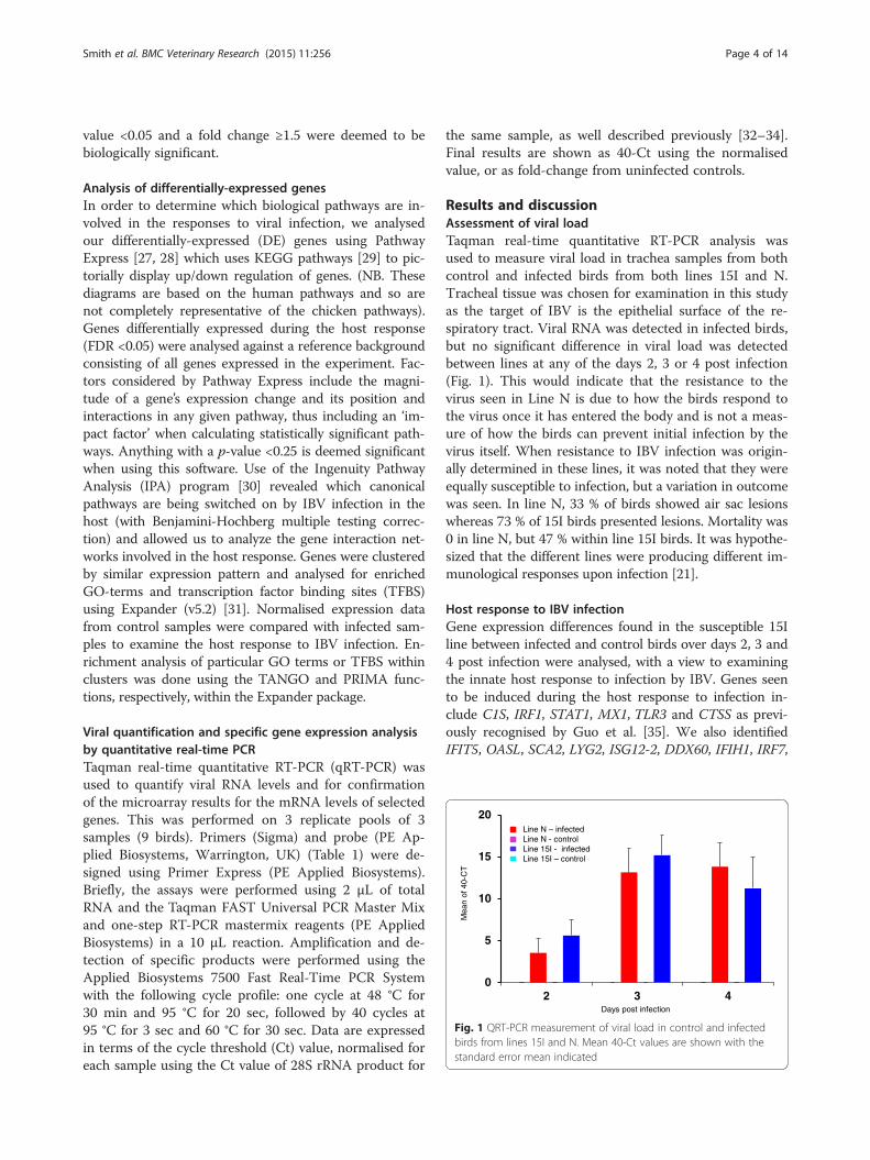
Results and discussionAssessment of viral loadTaqman real-time quantitative RT-PCR analysis wasused to measure viral load in trachea samples from bothcontrol and infected birds from both lines 15I and N.Tracheal tissue was chosen for examination in this studyas the target of IBV is the epithelial surface of the re-spiratory tract. Viral RNA was detected in infected birds,but no significant difference in viral load was detectedbetween lines at any of the days 2, 3 or 4 post infection(Fig. 1). This would indicate that the resistance to thevirus seen in Line N is due to how the birds respond tothe virus once it has entered the body and is not a meas-ure of how the birds can prevent initial infection by thevirus itself. When resistance to IBV infection was origin-ally determined in these lines, it was noted that they wereequally susceptible to infection, but a variation in outcomewas seen. In line N, 33 % of birds showed air sac lesionswhereas 73 % of 15I birds presented lesions. Mortality was0 in line N, but 47 % within line 15I birds. It was hypothe-sized that the different lines were producing different im-munological responses upon infection [21].
Host response to IBV infectionGene expression differences found in the susceptible 15Iline between infected and control birds over days 2, 3 and4 post infection were analysed, with a view to examiningthe innate host response to infection by IBV. Genes seento be induced during the host response to infection in-clude C1S, IRF1, STAT1, MX1, TLR3 and CTSS as previ-ously recognised by Guo et al. [35]. We also identifiedIFIT5, OASL, SCA2, LYG2, ISG12-2, DDX60, IFIH1, IRF7,
0
5
10
15
20
2 3 4Days post infection
Mea
n of
40-
CT
Line N – infectedLine N - controlLine 15I - infectedLine 15I – control
Fig. 1 QRT-PCR measurement of viral load in control and infectedbirds from lines 15I and N. Mean 40-Ct values are shown with thestandard error mean indicated
Smith et al. BMC Veterinary Research (2015) 11:256 Page 4 of 14

ZC3HAV1, DHX58, CCli7, IFITM1 and IFITM3 as beingup-regulated in response to IBV infection. Few genesare seen to be down-regulated during the early stage
of the host response, but those which are includeCHAC1 (pro-apoptotic), HBB (implicated in inflammationregulation) and PDK4 (glucose regulation). For a full list
Fig. 2 Pathway Express analysis of the host response to IBV infection in the trachea of susceptible birds (Line 15I). Many genes involved inantigen processing and presentation (a) and in the Toll-like receptor pathway (b) can be seen to be up-regulated (shown in red)
Smith et al. BMC Veterinary Research (2015) 11:256 Page 5 of 14

of the genes involved in the tracheal immune response(133 DE probes), see Additional file 1: Table S1.To elucidate which biological pathways are being per-
turbed during the host response to IBV infection, we ana-lysed our data using Pathway Express [36]. The resultingpathway diagrams are extremely useful in establishing
which gene networks are involved in a particular experi-mental response. As seen in Fig. 2, genes involved in anti-gen presentation and the Toll-like receptor (TLR)pathway are up-regulated. TLRs identify pathogen associ-ated molecular patterns (PAMPs) and are crucial to theinnate immune system. In this study TLR3 is shown to be
Table 2 Pathway Express analysis of the host response to IBV infection
Rank Pathway name Impact Factor Input genes/Genes in Pathway Corrected gamma p-value
1 Antigen processing and presentation 14.244 6/89 9.93E-06
2 Toll-like receptor signaling pathway 7.927 5/102 0.003221463
3 Notch signaling pathway 7.073 3/47 0.006843371
4 Pancreatic cancer 4.448 3/72 0.06375221
5 Maturity onset diabetes of the young 3.952 1/24 0.095158775
6 Phosphatidylinositol signaling system 3.884 2/76 0.100456001
7 Complement and coagulation cascades 3.841 3/69 0.103946526
8 mTOR signaling pathway 3.652 2/52 0.120669151
9 Acute myeloid leukemia 3.448 2/59 0.141487282
10 Systemic lupus erythematosus 3.332 2/144 0.15474593
11 VEGF signaling pathway 3.126 2/74 0.181102595
a
bDevelopment and differentiation of T-cells Fibroblast
response
Quantity of lymphocytes
Activation of leukocytes, dendritic cells, T-cells
Quantity of IgG
Abnormal uterus morphology
Fig. 3 Ingenuity Pathway Analysis (IPA) of genes responding to IBV infection. a Canonical biological pathways which are activated in the hostupon IBV infection (p < 0.05). The line represents the ratio of genes represented within each pathway. b The most highly represented (p < 0.05)physiological functions of genes differentially expressed during the host response to IBV (in the trachea in susceptible birds (Line 15I)). Specificfunctions within groups are highlighted
Smith et al. BMC Veterinary Research (2015) 11:256 Page 6 of 14

Fig. 4 Gene expression cluster analysis of the host response in susceptible birds (Line 15I) using the Expander program (http://acgt.cs.tau.ac.il/expander/expander.html). a The expression profile of genes up-regulated during the response to virus. b The GO biological process terms whichare significantly enriched during the host response to infection. The frequency of genes of a functional class within the examined set is describedas a percentage. c Binding sites for the transcription factors IRF7 and ISRE are seen to be significantly over-represented in genes up-regulatedduring the host response to IBV infection. The frequency ratio (frequency in set divided by frequency in background) is shown
Smith et al. BMC Veterinary Research (2015) 11:256 Page 7 of 14

induced at 3 dpi. TLR3 recognizes double-stranded RNAintermediates produced during viral replication and haspreviously been shown to be induced in the trachea at thistime after IBV infection [37]. Another pathway involved isthe phosphatidylinositol signalling pathway (Table 2).Phosphatidylinositol kinases are known to play an import-ant role in the viral life cycle after infection of the hostand PI4KB is known to be exploited by coronaviruses forviral entry. The product of PI4KB catalysis is phos-phatidylinositol 4-phosphate (PI4P) and coronavirus entryinto the host is mediated by the PI4P lipid microenviron-ment [38]. Genes involved in the complement system arealso highlighted as being up-regulated in response to IBVinfection. Complement-mediated lysis of viruses is an im-portant facet of the host innate immune system and itsrole in defence against viral infection [39] – as reflected inthe induction of these genes in this study.Use of Ingenuity Pathway Analysis (IPA) software also
allowed us to determine which biological systems are ac-tive during the host response. Up-regulated genes areseen to be part of the canonical biological pathwaysshown in Fig. 3a. Biological processes involving patternrecognition receptors and interferon signalling featureheavily. The interferon response is a powerful antiviralmechanism, which has previously been shown to be in-volved in the host response after IBV infection. A veryearly induction of IFN-γ has been reported in spleno-cytes [40], and in peripheral blood mononuclear cells(PBMCs) and lung leukocytes [41]. IFNB expression hasalso been reported in trachea between 1 and 2 dpi [42].We do not see this increase in expression of interferongenes (due to the absence of data earlier than 2 dpi), butwe do see the downstream effects, with increased ex-pression of many interferon-induced genes. Specificphysiological processes activated upon IBV infection canalso be seen in Fig. 3b. The stimulation of various differentimmune cells is seen along with the indication of repro-ductive abnormality, which would reflect the problemsseen with egg-laying upon IBV infection.
In order to cluster genes seen to be involved in thehost response to infectious bronchitis into groups withsimilar expression profiles and probably sharing similarfunctions or gene regulatory pathways, we utilised theCLICK algorithm within the Expander program [43].Figure 4a shows the expression profile of genes up-regulated during the response to virus. The Expanderprogram was also used to analyse the Gene Ontology(GO) functional annotations of the genes being differ-entially expressed. Figure 4b shows the biologicalprocess terms, which are significantly enriched in thegenes responding during the host response to infec-tion. As would be expected, these include terms like‘innate immune response’ and ‘antigen processing andpresentation’. ‘NAD + ADP-ribosyltransferase activity’and ‘phosphoinositide binding’ are also highlighted.Transcription factor binding sites present in DE geneswhich are significantly over-represented were alsopredicted. Figure 4c shows that genes up-regulatedduring the host response have a high proportion ofIRF7 and ISRE binding sites. IRF7 is a transcriptionalactivator, which binds to the interferon-stimulated re-sponse element (ISRE) in IFN promoters and functions asa molecular switch for antiviral activity.
Differences between susceptible and resistant linesAnalysis of the gene expression differences between in-fected and control birds across the two lines has pro-vided us with information on how these lines differ intheir response to infection. Examination of the gene ex-pression profiles in the control birds of the two differentlines also allowed us to identify genes, which are inher-ently different between the susceptible and resistantbirds. It can be seen that there are numerous genes,which show large expression differences between thetwo lines, even before infection. Dramatic differences ingene expression of certain genes, including DDT, SRI,BLB1, HSCB, BF1, BF2, SUCLG2, MX1 and SRI, whichare more highly expressed in the resistant N line
Fig. 5 Ingenuity Pathway Analysis (IPA) of genes showing inherent differential expression between susceptible and resistant control birds. Thisgraph shows the most highly represented (p < 0.05) molecular functions of DE genes
Smith et al. BMC Veterinary Research (2015) 11:256 Page 8 of 14

Fig. 6 Ingenuity Pathway Analysis (IPA) of genes showing differential expression between susceptible and resistant lines during the host responseto IBV infection. a The most highly represented (p < 0.05) molecular functions of DE genes. b This biological network shows genes associatedwith connective tissue disorders. Genes shown in red are more highly expressed in the resistant line and those in green have higher expressionin the susceptible line
Smith et al. BMC Veterinary Research (2015) 11:256 Page 9 of 14
compared to the susceptible 15I line are noted (Add-itional file 2: Table S2 shows all 1930 DE probes) So, itcan be seen that these are genes which have inherentlydifferent expression levels between susceptible and re-sistant birds, even before infection occurs. We thereforepostulate that some of these genes may play an im-portant role in disease resistance. The potential inter-actome of IBV has recently been investigated bystable isotope labelling with amino acids in cell cul-ture (SILAC) coupled to a green fluorescent protein-nanotrap pull-down methodology [44]. Host proteins,which bind to the IBV N protein were identified,some of the genes for which, we see as being inher-ently expressed at higher levels in susceptible birds inthis study. These genes include MYH9, CAPRIN1,DHX57, HNRNPH3, RPL27A, FMR1, C22orf28, HNRPDL,SFRS3, RPL31, NPM1 and RPSA. This may therefore beone of the reasons why Line 15I is more susceptible toIBV infection – there are more host proteins to which thevirus binds, compared with the resistant Line N.Upon infection, differences in response are also seen be-
tween the two lines. Interestingly, apart from CD38 andCD4 at 3 dpi and FKBP5 at 4 dpi, all other differentialgene expression between the lines is seen at 2 dpi in thisstudy (Additional file 3: Table S3). CD38 is a glycoproteinfound on the surface of many immune cells including
CD4+, CD8+, B lymphocytes and natural killer cells and isa marker of cell activation. It functions in cell adhesion,signal transduction and calcium signalling. CD4 is foundon the surface of immune cells such as T helper cells,monocytes, macrophages and dendritic cells. It is a mem-brane glycoprotein which interacts with MHCII antigens.The protein functions to initiate or augment the earlyphase of T-cell activation. The protein encoded by FKBP5is a member of the immunophilin protein family, whichplay a role in immuno-regulation and basic cellular pro-cesses involving protein folding and trafficking. Early de-fence by the host is a key mechanism for combatting viralinfection, and induction of IFNB and other innate genesin response to IBV infection has been shown to peakaround 18–36 hr post infection [42].In this study, genes more highly expressed (or less
down-regulated) in the resistant N line at 2 dpi include anumber of collagen genes (COL3A1, COL1A2, COL9A1,COL9A2, COL6A1 and COL4A1) and other genes such asACAN, FSTL1, COMP, EIF3A, STAT3 and IGFBP5. Genesseen to be more highly expressed (or less down-regulated)in the susceptible 15I line include RBM39, MAFB, NNK2,CCN1, MGAT5 and THRAP3. One consequence of IBVinfection is the production of poor quality, misshapen eggsby infected birds [45]. Some of the genes previouslyidentified as being important for the creation of a
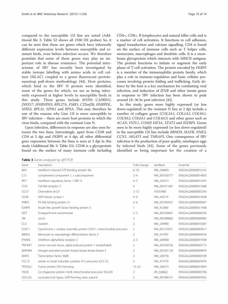
Table 3 Genes analyzed by qRT-PCR
Gene Description Fold change GenBank Ensembl
MX1 Interferon-induced GTP-binding protein Mx 6–20 NM_204609 ENSGALG00000016142
C1S complement component 1, s subcomponent 2–4 NM_001030777 ENSGALG00000014603
IRF7 Interferon regulatory factor 7 (IRF-7) 4–5 NM_205372 ENSGALG00000014297
TLR3 Toll-like receptor 3 4 NM_001011691 ENSGALG00000013468
CCLi7 Chemokine ah221 2–12 AY037860 ENSGALG00000002343
CD38 ADP-ribosyl cyclase 1 4–9 XM_420774 ENSGALG00000014508
FKBP5 FK-506 binding protein 51 5–6 NM_001005431 ENSGALG00000000947
IGFBP5 Insulin-like growth factor binding protein 5 1 XM_422069 ENSGALG00000011468
DDT D-dopachrome tautomerase 2–3 NM_001030667 ENSGALG00000006350
SRI sorcin 2 NM_001080865 ENSGALG00000008985
CLU clusterin 2 NM_204900 ENSGALG00000016587
COX11 Cytochrome c oxidase assembly protein COX11, mitochondrial precursor 2 XM_001233972 ENSGALG00000003017
MMD2 Monocyte to macrophage differentiation factor 2 2 XM_414787 ENSGALG00000004530
IFNAR2 interferon alpha/beta receptor 2 2–3 NM_204858 ENSGALG00000015938
TNFAIP1 tumor necrosis factor, alpha-induced protein 1 (endothelial) 2 NM_001030726 ENSGALG00000005715
MAP4K4 mitogen-activated protein kinase kinase kinase kinase 4 2 NM_001031126 ENSGALG00000008970
MAFK Transcription factor MafK. 2 NM_204756 ENSGALG00000004189
CCL13 similar to Small inducible cytokine A13 precursor (CCL13) 3 XM_415779 ENSGALG00000024470
TPD52L1 Tumor protein D53 homolog 13–23 NM_204215 ENSGALG00000014843
HSCB Co-chaperone protein HscB, mitochondrial precursor (Hsc20) 2 XR_026662 ENSGALG00000005706
SUCLG2 succinate-CoA ligase, GDP-forming, beta subunit 2 NM_001006141 ENSGALG00000007652
Smith et al. BMC Veterinary Research (2015) 11:256 Page 10 of 14

healthy eggshell are seen to be more highly expressedby the resistant N line birds after infection in this study.These genes include COL1A2, CRELD2, HSP90B1,P4HB and ERP29 [46]. For a full list of genes differen-tially expressed between the two lines in trachea (409DE probes) see Additional file 3: Table S3.IPA analysis of genes showing different inherent ex-
pression between lines 15I and N shows that the mo-lecular functions of these genes is primarily concernedwith their involvement in cell death and cell adhesion(Fig. 5), two processes previously shown to be significantin infected kidneys [47]. When the differential host re-sponses to infection are examined, it is seen that genesinvolved in proliferation of T-lymphocytes and genesconcerned with cell attachment and cytoplasmicorganization are more highly expressed in the resistantline N. Other processes significantly involved are apop-tosis and necrosis (Fig. 6a), which have been previouslydocumented in IBV-infected Vero cells by Liu et al. [48].
One of the most perturbed biological networks noted inthis analysis is that involving genes related to connectivetissue disorders and involve many collagen genes. Thesegenes are more highly expressed in susceptible line 15Ibirds compared to resistant line N birds (Fig. 6b) sug-gesting that IBV infection might cause more disorder ofeggshell formation in this line [49]. The production ofpoor quality eggs by IBV infected birds may, in part be areflection of the expression of these kinds of gene net-works compared to that seen in resistant birds.
Confirmation of differential gene expression byquantitative real-time PCRTwenty-one genes were selected for qRT-PCR validation(Table 3). These genes were chosen based on their in-volvement in the host response and whether they weredifferentially expressed between the susceptible and re-sistant lines (either inherently or during the course of in-fection). Of the 21 genes tested, 19 showed comparable
Table 4 Potential candidate genes for involvement in resistance to IBV
Gene Description Fold change GenBank Ensembl
MX1 Interferon-induced GTP-binding protein Mx. 4–24a NM_204609 ENSGALG00000016142
C1S Complement component 1, s subcomponent 2a NM_001030777 ENSGALG00000014603
IRF7 Interferon regulatory factor 7 (IRF-7). 6a NM_205372 ENSGALG00000014297
TLR3 Toll-like receptor 3 4a NM_001011691 ENSGALG00000013468
C1R Complement component 1, r subcomponent 2a XM_416518 ENSGALG00000014659
CCLi7 Chemokine ah221 8a AY037860 ENSGALG00000002343
ISG12-2 Interferon stimulated gene 12-2 16–18a NM_001001296 ENSGALG00000013575
IFITM3 Interferon induced transmembrane protein 3 4–5a KC876032 ENSGALG00000004243
CD38 ADP-ribosyl cyclase 1 4b XM_420774 ENSGALG00000014508
CD4 CD4 protein 4b NM_204649 ENSGALG00000014477
FKBP5 FK-506 binding protein 51 2b NM_001005431 ENSGALG00000000947
STAT3 Signal transducer and activator of transcription 3 (acute-phase response factor) 3b NM_001030931 ENSGALG00000003267
OASL 2'-5'-oligoadenylate synthetase-like 4c NM_205041 ENSGALG00000013723
DDT D-dopachrome tautomerase 62–72c NM_001030667 ENSGALG00000006350
SRI Sorcin 65–89c NM_001080865 ENSGALG00000008985
BLB1 MHC Class II beta 1 and 2 domains 10–22c NM_001044694 ENSGALG00000000141
IFNAR2 Interferon alpha/beta receptor 2 2–3c NM_204858 ENSGALG00000015938
TPD52L1 Tumor protein D53 homolog 12–14c NM_204215 ENSGALG00000014843
BCL2L1 BCL2-like - apoptosis regulator 1.7c NM_001025304 ENSGALG00000006211
FAIM2 Fas apoptotic inhibitory molecule 2 1.7c XM_004950568 ENSGALG00000027555
CIAPIN1 Cytokine Induced Apoptosis Inhibitor 11 1.7c NM_001005834 ENSGALG00000005706
HSCB Co-chaperone protein HscB, mitochondrial precursor (Hsc20). 8–10c XR_026662 ENSGALG00000005706
BF1 MHC class I antigen B-F minor heavy chain 13–27c NM_001097530 ENSGALG00000000178
BF2 Major class I glycoprotein precursor 7–8c NM_001031338 ENSGALG00000000178
SUCLG2 succinate-CoA ligase, GDP-forming, beta subunit 16–32c NM_001006141 ENSGALG00000007652aUpregulated in response to infection in the susceptible linebHigher expression in response to infection in the resistant than in the susceptible linecInherently higher expression in the resistant line
Smith et al. BMC Veterinary Research (2015) 11:256 Page 11 of 14

differential expression to that determined by the arrays.However, the results for IFNAR2 and IGFBP5 were notconfirmed (Additional file 4: Figure S1).
Potential candidate genes for IBV resistanceBesides knowing that the MHC B locus has a bearing ondisease resistance, the lack of any genetic information oridentified QTL meant that we had to rely upon the geneexpression differences we saw between susceptible andresistant lines to give us clues as to genes potentially in-volved in resistance to IBV infection. Identifying geneswhich were expressed at different levels in the two linesof birds highlighted B-locus genes (BLB1, BF1, BF2, B-G)as well as bringing to our attention various othernon-MHC genes which, due to their known biology,could be candidates for being involved in resistanceto IBV infection (Table 4).MX1, C1S, IRF7,TLR3, C1R, CCLi7, ISG12-2 and IFITM3
are all strongly induced during the host response to IBV in-fection. They are all innate immune genes which could po-tentially have a role in determining susceptibility to thevirus. MX1 and IFITM3 are already established as anti-viralmolecules [50–52]. CD38, CD4, FKBP5 and STAT3 all showa higher level of expression during the host response in theresistant birds compared to that of the susceptible birds, in-dicating their involvement in the host defence mechanism.CD38 and CD4, with their role as receptors on immunecells, as described above, are obvious candidates, along withFKBP5 as an immune-regulator. STAT3 is activated by vari-ous cytokines and growth factors and functions in cellularprocesses such as cell growth and apoptosis.Even before infection, many genes are seen to be highly
differentially expressed between lines 15I and N. OASL isan interferon-induced molecule known to have anti-viralactivity against certain viruses such as hepatitis C virus.DDT is highly homologous to the macrophage migrationinhibition factor, MIF. We have also shown it to be highlydifferentially expressed in other chicken lines, which aresusceptible or resistant to Marek’s Disease virus [53].IFNAR2 is an obvious candidate prediction, as the inter-feron response is central to the host’s defence against IBVinfection. TPD52L1, BCL2L1, FAIM2 and CIAPIN1 are allknown to be involved in regulation of apoptosis, a processseen to be important during IBV infection. HSCB, SRI, andSUCLG2, although not having an obvious potential bio-logical role in disease resistance, are highly differentiallyexpressed between susceptible and resistant lines andshould thus be considered as potential candidates.
ConclusionsResistance to IBV infection is brought about by the im-mune response after the virus has entered the host andis not due to prevention of initial viral infection. Thereis a small initial innate response at 2 dpi, with much
more gene expression seen at 3 and 4 dpi. Analysis ofgenes being activated or inhibited upon infection showsthat the biological pathways primarily affected duringIBV infection include MAPK signalling, those involvedin the interferon response and those involving patternrecognition receptors.Susceptible and resistant lines show a differential host
response mostly at 2 dpi. There are also genes which areinherently different between the two lines studied, includ-ing many genes, which control the apoptotic potential ofthe host. These differences seen in gene expression levels,allow us to postulate on many candidate genes for diseaseresistance. Some potential candidates for involvement indisease resistance include genes already known to conferresistance to other viral infections (MHC-B locus genes,MX1, OASL and IFITM3), genes involved in apoptoticprocesses (TPD52L1, BCL2L1, FAIM2 and CIAPIN1) andothers which could be potential candidates due to theirknown biology (e.g. DDT and CD4).
Availability of supporting dataArray data have been submitted to Array Express(http://www.ebi.ac.uk/arrayexpress/) under the accessionnumber E-TABM-1128.
Additional files
Additional file 1: Table S1. Gene expression seen during the hostresponse to IBV infection in the trachea of susceptible birds. (XLSX 24 kb)
Additional file 2: Table S2. Gene expression differences found to beinherent between susceptible and resistant lines in the trachea. (XLS 386 kb)
Additional file 3: Table S3. Genes found to be differentially expressedbetween susceptible and resistant lines in response to IBV infection inthe trachea. (XLS 107 kb)
Additional file 4: Figure S1. qRT-PCR analysis of 21 genes differentiallyexpressed during IBV infection. (A). MX1 (B). C1S (C). IRF7 (D). TLR3 (E).CCLi7 (F). DDT (G). SRI (H). CLU (I). COX11 (J). IFNAR2 (K). TNFAIP1 (L).TP-D53 (M). MAP4K4 (N). MAFK (O). CCL13 (P). HSC20 (Q). SUCLG2 (R).MMD2 (S). CD38 (T). FK506-BP51 (U). IGFBP5. Graphs A-E show expressionchanges during the host response in the susceptible line. Graphs F-Rshow inherent differences in gene expression between susceptible andresistant control birds, while graphs S-U indicate differential gene expressionin susceptible and resistant lines during the host response. (PDF 97 kb)
AbbreviationsBSA: Bovine serum albumin; CID50: mean chicken infectious dose;DE: Differentially expressed; FARMS: Factor Analysis for Robust MicroarraySummarization; FDR: False discovery rate; GCOS: GeneChip OperatingSoftware; GO: Gene ontology; IB: Infectious bronchitis; IBV: Infectiousbronchitis virus; KEGG: Kyoto Encyclopaedia of Genes and Genomes;MHC: Major histocompatibility complex; OD: Optical density; PBS: Phosphatebuffered saline; PLIER: Probe Logarithmic Error Intensity Estimate; qRT-PCR: Quantitative reverse transcription polymerase chain reaction;QTL: Quantitative trait loci; RIN: RNA integrity number; SARS: Severe acuterespiratory syndrome; SILAC: Stable isotope labelling with amino acids in cellculture; TFBS: Transcription factor binding site.
Competing interestsThe authors declare no conflicts of interest and no competing financialinterests.
Smith et al. BMC Veterinary Research (2015) 11:256 Page 12 of 14

Authors’ contributionsJS performed the arrays, analysed the results and wrote the manuscript; DCcarried out challenge experiments, J-RS prepared RNA, measured viral loadand performed qRT-PCR; DB and PK conceived and supervised the projectand revised the manuscript. All authors read and approved the finalmanuscript.
AcknowledgementsThis work was supported by the Biotechnology and Biological ScienceResearch Council (BBSRC), as part of grant numbers BB/D013704/1, BB/D013704/2 and BB/D010705/1. The authors would like to thank AlisonDowning (Edinburgh Genomics, Edinburgh, UK) for excellent technicalassistance with the Affymetrix microarray experiments.
Author details1The Roslin Institute & R(D)SVS, University of Edinburgh, Easter Bush,Midlothian EH25 9RG, UK. 2The Pirbright Institute, Compton Laboratory,Compton, Berkshire RG20 7NN, UK.
Received: 1 July 2015 Accepted: 5 October 2015
References1. Schalk AF, Hawn MC. An apparently new respiratory disease in baby chicks.
J Am Vet Med Assoc. 1931;78:413–22.2. Beach JR, Schalm OW. Studies of infectious coryza of chickens with special
reference to its etiology. Poult Sci. 1936;15:199–206.3. Beaudette FR, Hudson CB. Cultivation of the virus of infectious bronchitis.
J Am Vet Med Assoc. 1937;90:51–8.4. Cavanagh D. Coronavirus avian infectious bronchitis virus. Vet Res.
2007;38:281–97.5. Enjuanes L, Sola I, Zúñiga S, Almazán F. Coronavirus Replication and
Interaction with Host. In: Mettenleiter TC, Sobrino F, editors. Animal Viruses:Molecular Biology. Norfolk: Caister Academic Press; 2008. p. 149–202.
6. Casais R, Dove B, Cavanagh D, Britton P. Recombinant avian infectiousbronchitis virus expressing a heterologous spike gene demonstrates thatthe spike protein is a determinant of cell tropism. J Virol. 2003;77:9084–9.
7. Asif M, Lowenthal JW, Ford ME, Schat KA, Kimpton WG, Bean AG.Interleukin-6 expression after infectious bronchitis virus infection inchickens. Viral Immunol. 2007;20:479–86.
8. Jackwood MW. Review of infectious bronchitis virus around the world.Avian Dis. 2012;56:634–41.
9. Meulemans G, Van den Berg TP. Nephropathogenic avian infectiousbronchitis viruses. World Poult Sci. 1998;54:145–53.
10. Ariaans MP, Matthijs MG, van Haarlem D, van de Haar P, van Eck JH, HensenEJ, et al. The role of phagocytic cells in enhanced susceptibility of broilersto colibacillosis after Infectious Bronchitis Virus infection. Vet ImmunolImmunopathol. 2008;123:240–50.
11. Matthijs MG, van Eck JH, Landman WJ, Stegeman JA. Ability ofMassachusetts-type infectious bronchitis virus to increase colibacillosissusceptibility in commercial broilers: a comparison between vaccine andvirulent field virus. Avian Pathol. 2003;32:473–81.
12. Jackwood MW, de Wit S. Infectious Bronchitis. In: Swayne DE, Glisson JR,McDougald LR, Nolan LK, Suarez DL, Nair VL, editors. Diseases of Poultry.13th ed. UK: Wiley-Blackwell; 2013. p. 139–60.
13. Worthington KJ, Currie RJW, Jones RC. A reverse transcriptase–polymerasechain reaction survey of infectious IBV infection bronchitis virus genotypesin Western Europe from 2002 to 2006. Avian Pathol. 2008;37:247–57.
14. Ignjatovic J, Gould G, Sapats S. Isolation of a variant infectious bronchitisvirus in Australia that further illustrates diversity among emerging strains.Arch Virol. 2006;151:1567–85.
15. Mase M, Kawanishi N, Ootani Y, Murayama K, Karino A, Inoue T. A novelgenotype of avian infectious bronchitis virus isolated in Japan in 2009.J Vet Med Sci. 2010;72:1265–8.
16. Banat GR, Tkalcic S, Dzielawa JA, Jackwood MW, Saggese MD, Yates L, et al.Association of the chicken MHC B haplotypes with resistance to aviancoronavirus. Dev Comp Immunol. 2013;39:430–7.
17. Dar A, Munir S, Vishwanathan S, Manuja A, Griebel P, Tikoo S, et al.Transcriptional analysis of avian embryonic tissues following infection withavian infectious bronchitis virus. Virus Res. 2005;110:41–55.
18. Wang X, Rosa AJM, Oliverira HN, Rosa GJM, Guo X, Travnicek M, et al.Transcriptome of Local Innate and Adaptive Immunity during Early Phase ofInfectious Bronchitis Viral Infection. Viral Immunol. 2006;19:768–74.
19. Drosten C, Günther S, Preiser W, van der Werf S, Brodt HR, Becker S, et al.Identification of a novel coronavirus in patients with severe acuterespiratory syndrome. N Engl J Med. 2003;348:1967–76.
20. Ksiazek TG, Erdman D, Goldsmith CS, Zaki SR, Peret T, Emery S, et al. A novelcoronavirus associated with severe acute respiratory syndrome. N Engl JMed. 2003;348:1953–66.
21. Cook J, Otsuki K, Huggins M, Bumstead N. Investigations into resistance ofchicken lines to infection with infectious bronchitis virus. Adv Exp Med Biol.1990;276:491–6.
22. Stone HA. Use of highly inbred chickens in research. USDA AgriculturalResearch Service Technical Bulletin. 1975, No.1514, Washington, DC.
23. Cole RK. Studies on genetic resistance to Marek's disease. Avian Dis.1968;12:9–28.
24. Affymetrix. Guide to Probe Logarithmic Intensity Error (PLIER) Estimation.Santa Clara: Affymetrix I; 2005.
25. Talloen W, Clevert DA, Hochreiter S, Amaratunga D, Bijnens L, Kass S, et al.I/NI-calls for the exclusion of non-informative genes: a highly effectivefiltering tool for microarray data. Bioinformatics. 2007;23:2897–902.
26. R Development Core Team. R: A language and environment for statisticalcomputing. Vienna: R Foundation for Statistical Computing; 2007.http://www.R-project.org. ISBN 3-900051-07-0.
27. Khatri P, Sellamuthu S, Malhotra P, Amin K, Done A, Draghici S. Recentadditions and improvements to the Onto-Tools. Nucleic Acids Res.2005;33(Web Server issue):W762–5.
28. Pathway Express. [http://vortex.cs.wayne.edu/projects.htm]29. Kanehisa M, Goto S. KEGG: kyoto encyclopedia of genes and genomes.
Nucleic Acids Res. 2000;28:27–30.30. Ingenuity Pathway Analysis. [Ingenuity® Systems. www.ingenuity.com]31. Expander. [http://acgt.cs.tau.ac.il/expander/expander.html]32. Eldaghayes I, Rothwell L, Williams A, Withers D, Balu S, Davison F, et al.
Infectious bursal disease virus: strains that differ in virulence differentiallymodulate the innate immune response to infection in the chicken bursa.Viral Immunol. 2006;19:83–91.
33. Poh TY, Pease J, Young JR, Bumstead N, Kaiser P. Re-evaluation of chickenCXCR1 determines the true gene structure: CXCLi1 (K60) and CXCLi2 (CAF/interleukin-8) are ligands for this receptor. J Biol Chem. 2008;283:16408–15.
34. Rothwell L, Hu T, Wu Z, Kaiser P. Chicken interleukin-21 is costimulatory for T cellsand blocks maturation of dendritic cells. Dev Comp Immunol. 2012;36:475–82.
35. Guo X, Rosa AJM, Chen D-G, Wang X. Molecular mechanisms of primaryand secondary mucosal immunity using avian infectious bronchitis virus asa model system. Vet Immunol Immunopathol. 2008;121:332–43.
36. Draghici S, Khatri P, Tarca AL, Amin K, Done A, Voichita C, et al. A systemsbiology approach for pathway level analysis. Genome Res. 2007;17:1537–45.
37. Kameka AM, Haddadi S, Kim DS, Cork SC, Abdul-Careem MF. Induction ofinnate immune response following infectious bronchitis corona virusinfection in the respiratory tract of chickens. Virology. 2014;450–451:114–21.
38. Yang N, Ma P, Lang J, Zhang Y, Deng J, JU X, et al. Phosphatidylinositol 4-Kinase IIIβ Is Required for Severe Acute Respiratory Syndrome CoronavirusSpike-mediated Cell Entry. J Biol Chem. 2012;287:8457–67.
39. Favoreel HW, Van de Walle GR, Nauwynck HJ, Pensaert MB. Viruscomplement evasion strategies. J Gen Virol. 2003;84:1–15.
40. Ariaans MP, van de Haar PM, Hensen EJ, Vervelde L. Infectious BronchitisVirus induces acute interferon-gamma production through polyclonalstimulation of chicken leukocytes. Virology. 2009;385:68–73.
41. Vervelde L, Matthijs MG, van Haarlem DA, de Wit JJ, Jansen CA. Rapid NK-cell activation in chicken after infection with infectious bronchitis virus M41.Vet Immunol Immunopathol. 2013;151:337–41.
42. Kint J, Fernandez-Gutierrez M, Maier HJ, Britton P, Langereis MA, Koumans J,et al. Activation of the chicken type I IFN response by infectious bronchitiscoronavirus. J Virol. 2015;89:1156–67.
43. Sharan R, Maron-Katz A, Shamir R. CLICK and EXPANDER: a system for clusteringand visualizing gene expression data. Bioinformatics. 2003;19:1787–99.
44. Emmott E, Munday D, Bickerton E, Britton P, Rodgers MA, Whitehouse A, etal. The cellular interactome of the coronavirus infectious bronchitis virusnucleocapsid protein and functional implications for virus biology. J Virol.2013;87:9486–500.
45. Crinion RA. Egg quality and production following infectious bronchitis virusexposure at one day old. Poult Sci. 1972;51:582–5.
Smith et al. BMC Veterinary Research (2015) 11:256 Page 13 of 14
46. Brionne A, Nys Y, Hennequet-Antier C, Gautron J. Hen uterine geneexpression profiling during eggshell formation reveals putative proteinsinvolved in the supply of minerals or in the shell mineralization process.BMC Genomics. 2014;15:220.
47. Cong F, Liu X, Han Z, Shao Y, Kong X, Liu S. Transcriptome analysis ofchicken kidney tissues following coronavirus avian infectious bronchitis virusinfection. BMC Genomics. 2013;14:743.
48. Liu C, Xu HY, Liu DX. Induction of caspase-dependent apoptosis in culturedcells by the avian coronavirus infectious bronchitis virus. J Virol.2001;75:6402–9.
49. Nii T, Isobe N, Yoshimura Y. Effects of avian infectious bronchitis virusantigen on eggshell formation and immunoreaction in hen oviduct.Theriogenology. 2014;81:1129–38.
50. Goodbourn S, Didcock L, Randall RE. Interferons: cell signalling, immunemodulation, antiviral response and virus countermeasures. J Gen Virol.2000;81:2341–64.
51. Haller O, Frese M, Kochs G. Mx proteins: Mediators of innate resistance toRNA viruses. Rev Sci Technol. 1998;17:220–30.
52. Perreira JM, Chin CR, Feeley EM, Brass AL. IFITMs restrict the replication ofmultiple pathogenic viruses. J Mol Biol. 2013;425:4937–55.
53. Smith J, Sadeyen JR, Paton IR, Hocking PM, Salmon N, Fife M, et al. Systemsanalysis of immune responses in Marek's disease virus-infected chickensidentifies a gene involved in susceptibility and highlights a possible novelpathogenicity mechanism. J Virol. 2011;85:11146–58.
Submit your next manuscript to BioMed Centraland take full advantage of:
• Convenient online submission
• Thorough peer review
• No space constraints or color figure charges
• Immediate publication on acceptance
• Inclusion in PubMed, CAS, Scopus and Google Scholar
• Research which is freely available for redistribution
Submit your manuscript at www.biomedcentral.com/submit
Smith et al. BMC Veterinary Research (2015) 11:256 Page 14 of 14