The Dual Amylin‐ and Calcitonin‐Receptor Agonist KBP… · KBP-042 led to impaired glucose ......
12
General rights Copyright and moral rights for the publications made accessible in the public portal are retained by the authors and/or other copyright owners and it is a condition of accessing publications that users recognise and abide by the legal requirements associated with these rights. • Users may download and print one copy of any publication from the public portal for the purpose of private study or research. • You may not further distribute the material or use it for any profit-making activity or commercial gain • You may freely distribute the URL identifying the publication in the public portal If you believe that this document breaches copyright please contact us providing details, and we will remove access to the work immediately and investigate your claim. Downloaded from orbit.dtu.dk on: Dec 18, 2017 The Dual Amylin- and Calcitonin-Receptor Agonist KBP-042 Increases Insulin Sensitivity and Induces Weight Loss in Rats with Obesity Hjuler, Sara Toftegaard; Gydesen, Sofie; Andreassen, Kim Vietz; Pedersen, Steffen Lund Kjær; Hellgren, Lars; Karsdal, Morten Asser; Henriksen, Kim Published in: Obesity Link to article, DOI: 10.1002/oby.21563 Publication date: 2016 Document Version Publisher's PDF, also known as Version of record Link back to DTU Orbit Citation (APA): Hjuler, S. T., Gydesen, S., Andreassen, K. V., Pedersen, S. L. K., Hellgren, L. I., Karsdal, M. A., & Henriksen, K. (2016). The Dual Amylin- and Calcitonin-Receptor Agonist KBP-042 Increases Insulin Sensitivity and Induces Weight Loss in Rats with Obesity. Obesity, 24(8), 1712-1722. DOI: 10.1002/oby.21563
Transcript of The Dual Amylin‐ and Calcitonin‐Receptor Agonist KBP… · KBP-042 led to impaired glucose ......
General rights Copyright and moral rights for the publications made accessible in the public portal are retained by the authors and/or other copyright owners and it is a condition of accessing publications that users recognise and abide by the legal requirements associated with these rights.
• Users may download and print one copy of any publication from the public portal for the purpose of private study or research. • You may not further distribute the material or use it for any profit-making activity or commercial gain • You may freely distribute the URL identifying the publication in the public portal
If you believe that this document breaches copyright please contact us providing details, and we will remove access to the work immediately and investigate your claim.
Downloaded from orbit.dtu.dk on: Dec 18, 2017
The Dual Amylin- and Calcitonin-Receptor Agonist KBP-042 Increases InsulinSensitivity and Induces Weight Loss in Rats with Obesity
Hjuler, Sara Toftegaard; Gydesen, Sofie; Andreassen, Kim Vietz; Pedersen, Steffen Lund Kjær; Hellgren,Lars; Karsdal, Morten Asser; Henriksen, KimPublished in:Obesity
Link to article, DOI:10.1002/oby.21563
Publication date:2016
Document VersionPublisher's PDF, also known as Version of record
Link back to DTU Orbit
Citation (APA):Hjuler, S. T., Gydesen, S., Andreassen, K. V., Pedersen, S. L. K., Hellgren, L. I., Karsdal, M. A., & Henriksen, K.(2016). The Dual Amylin- and Calcitonin-Receptor Agonist KBP-042 Increases Insulin Sensitivity and InducesWeight Loss in Rats with Obesity. Obesity, 24(8), 1712-1722. DOI: 10.1002/oby.21563
The Dual Amylin- and Calcitonin-Receptor Agonist KBP-042Increases Insulin Sensitivity and Induces Weight Lossin Rats with ObesitySara Toftegaard Hjuler1, Sofie Gydesen1, Kim Vietz Andreassen1, Steffen Lund Kjær Pedersen1, Lars I. Hellgren2,Morten Asser Karsdal1, and Kim Henriksen1
Objective: In this study, KBP-042, a dual amylin- and calcitonin-receptor agonist, was investigated as a
treatment of obesity and insulin resistance in five different doses (0.625 mg/kg–10 mg/kg) compared with
saline-treated and pair-fed controls.
Methods: Rats with obesity received daily s.c. administrations for 56 days, and glucose tolerance was
assessed after one acute injection, 3 weeks of treatment, and again after 7 weeks of treatment. To
assess the effect on insulin sensitivity, rats received 5 mg/kg KBP-042 for 21 days before hyperinsuline-
mic–euglycemic clamp.
Results: KBP-042 induced a sustained weight loss of up to 20% without any significant weight reduction
in the pair-fed groups. Decreases in adipose tissues and lipid deposition in the liver were observed, while
plasma adiponectin was increased and plasma leptin levels were decreased. Acute administration of
KBP-042 led to impaired glucose tolerance and increased plasma lactate, while this diabetogenic effect
was reversed by chronic treatment. Finally, assessment of insulin sensitivity using the hyperinsulinemic–
euglycemic clamp showed that KBP-042 increased the glucose infusion rate.
Conclusions: The study indicates that KBP-042 combines two highly relevant features, namely weight
loss and insulin sensitivity, and is thus an excellent candidate for chronic treatment of obesity and insulin
resistance.
Obesity (2016) 24, 1712–1722. doi:10.1002/oby.21563
IntroductionObesity is one of the greatest public health challenges of the 21st
century (1). Obesity can lead to insulin resistance and type 2 diabe-
tes (2), which are associated with a range of metabolic dysfunctions
(3,4). Weight loss, improved glycemic control, and increased insulin
action to reduce strain on the b cells are key points for improving
disease status. This can be achieved by different interventions (exer-
cise, diet, medication, surgery) which all cause improvements in
metabolic profiles and increase of insulin sensitivity and b-cell func-
tion (5,6). However, as lifestyle changes often result in only minor
weight reductions followed by a rapid regain of weight (7), there is
a need for treatments targeting multiple factors of the obesity-related
diseases. These include insulin resistance and b-cell failure to avoid
development of type 2 diabetes, as well as diabetic complications.
Activation of amylin receptors has already been linked with reduc-
tion of food intake (8), increased responsiveness to leptin (9-11),
weight loss (12,13), and indications of increased energy expenditure
(11,13-16). However, amylin is a short-lasting agonist in vivo, and
there is a need for improved ligands. KBP-042 is a dual amylin- and
calcitonin-receptor agonist with highly potent antiobesity and anti-
diabetic effects (17), although a long-term chronic treatment has not
yet been tested.
In this study, KBP-042 was tested in a long-term treatment of predia-
betic rats with obesity, in order to evaluate KBP-042’s potential as a
chronic treatment of obesity. We further examined whether the bene-
ficial effects on glucose homeostasis were maintained throughout the
study, and finally we investigated whether treatment with KBP-042
could increase insulin sensitivity and reduce hepatic steatosis.
1 Nordic Bioscience, Herlev, Denmark. Correspondence: Sara Toftegaard Hjuler ([email protected]) 2 Department of Systems Biology, TechnicalUniversity of Denmark, Denmark.
Funding agencies: Danish Agency for Science, Technology and Innovation and the Danish Research Foundation as well as the Technical University of Denmark.
Disclosure: MAK and KH own stock in Nordic Bioscience. All other authors declared no conflict of interest.
Author contributions: STH designed and performed the animal studies, analyzed data, and wrote the manuscript. SG assisted in animal studies and performed analyses.
KVA, SLKP, LIH performed analyses on liver. MAK assisted with the study design. KH assisted with the study design and data interpretation as well as manuscript writing.
All authors approved the final version of the manuscript.
Additional Supporting Information may be found in the online version of this article.
Received: 11 January 2016; Accepted: 13 April 2016; Published online 14 June 2016. doi:10.1002/oby.21563
1712 Obesity | VOLUME 24 | NUMBER 8 | AUGUST 2016 www.obesityjournal.org
Original ArticleOBESITY BIOLOGY AND INTEGRATED PHYSIOLOGY
Obesity
MethodsPeptide therapyRecombinant KBP-042 peptide (Unigene Laboratories, Boonton, NJ)
was dissolved in saline for subcutaneous (s.c.) delivery. The doses
for KBP-042 administration in the studies were based on previous
studies in animal models of obesity and type 2 diabetes and ranged
from 10 mg/kg to 0.625 mg/kg (�2.87–0.18 nmol/kg/day) (17,18).
Animal experimentsAll animal procedures were performed in accordance with guidelines
from the Animal Welfare Division of the Danish Ministry of Justice
under the institutional license issued to Nordic Bioscience (2012-15-
2934-00094). All male Sprague Dawley rats were obtained at 6
weeks of age and housed under controlled temperature (208C 6 2)
on a normal 12-h light–dark cycle with ad libitum access to water
and food. Normal diet control rats (ND) were fed rodent chow
(5002, LabDiet, St. Louis, MO) and high-fat diet (HFD) rats a 60%
fat kcal diet (#D12495, Research Diets Inc., NJ). After 10 weeks of
high-fat feeding rats were assigned into groups (n 5 10) and con-
trolled for equal mean body weight.
Acute study. Food was removed in the afternoon (4 p.m.). After
16 to 18 h of fasting an oral glucose tolerance test (OGTT) was per-
formed. Rats received a single dose of saline (vehicle) or peptide
(10 mg/kg, 5 mg/kg, 2.5 mg/kg, 1.25 mg/kg, 0.625 mg/kg). After 30
min, a glucose bolus (2 g/kg, Sigma-Aldrich, Copenhagen, Den-
mark) was administered by oral gavage. Blood glucose was moni-
tored by Accu-CheckVR Avia monitoring system (Roche Diagnostics,
Rotkreuz, Switzerland) and EDTA-plasma was obtained from the
lateral tail vein at t 5 0, 15, 30, 60, and 120 min.
Pica test. Fasted animals were administered s.c. with 5, 10, or 50
mg/kg KBP-042 or vehicle (saline). After dosing, animals had free
access to normal chow or kaolin pellets (5TBP, Test diet, MO) and
food and kaolin intake was monitored after 4 and 24 h.
Chronic study. Each rat was dosed once daily with either saline
(vehicle, pair-fed 5 mg/kg, pair-fed 10 mg/kg) or KBP-042 (10 mg/kg,
5 mg/kg, 2.5 mg/kg, 1.25 mg/kg, 0.625 mg/kg) in the afternoon for 8
weeks. The two pair-fed groups were food restricted to match the daily
food intake of their corresponding treatment groups (5 mg/kg or 10 mg/
kg). Pair-fed animals received an average of the daily intake of their
treated paired group every day in the afternoon. Food intake and body
weight were monitored daily for the first 6 days, then weekly. OGTT,
performed as in the acute study and intravenous glucose tolerance tests
(IVGTT) were performed after 3 and 7 weeks of treatment. IVGTT
was performed in the morning after 18 h of fasting. Each rat received
a single dose of either saline (vehicle, pair-fed 5 mg/kg, pair-fed 10
mg/kg) or peptide (10 mg/kg, 5 mg/kg, 2.5 mg/kg, 1.25 mg/kg, 0.625 mg/
kg), after 30 min glucose (0.5 g/kg, Sigma-Aldrich, Copenhagen, Den-
mark) was administered in the lateral tail vein and blood glucose was
monitored and EDTA-plasma was obtained at t 5 0, 5, 15, 30, 60, and
120 min, as described above. To assess effect on gastric emptying,
overnight-fasted rats received s.c. KBP-042 injection, were adminis-
tered 40 mg/kg acetaminophen by oral gavage (4 mL/kg) after 30 min
and the appearance of acetaminophen in plasma was monitored (19).
Blood was collected 30 min after administration from the tail vein and
acetaminophen levels were measured in EDTA-plasma (Acetamino-
phen Direct ELISA Kit, Immuneanalysis, Pomona, CA). Gastric emp-
tying was calculated as % change relative to ND rats.
After 8 weeks, EDTA-Aprotinin plasma samples were collected for
hormonal analyses after 3 h fasting. Animals were euthanized under
isoflurane inhalation followed by exsanguination. Excised tissue was
snap-frozen in liquid nitrogen and stored at 2808C, and plasma was
stored at 2208C samples until further analysis.
Hyperinsulinemic–euglycemic clampInsulin-mediated whole body glucose uptake was estimated in rats fed
either HFD or ND (as described above). The HFD rats were stratified
into HFD vehicle or HFD-KBP-042 groups (n 5 5–7). ND vehicle
and HFD vehicle rats received saline injections while HFD-KBP-042
received 5 mg/kg of KBP-042 s.c. for 21 days. After the treatment
period, animals were subjected to a hyperinsulinemic–euglycemic
clamp experiment explained in details in the Supporting Information.
Plasma analysisPlasma levels of lactate (L-lactate colorimetric assay, Abcam, Cam-
bridge, UK), insulin (Mercodia Rat Insulin ELISA, Mercodia AB,
Uppsala, Sweden), leptin (Rat Leptin ELISA, Millipore Corporation,
Billerica, MA), glucose-dependent insulinotropic peptide (GIP) (Rat/
Mouse GIP (Total) ELISA, Merck Millipore, Billerica, MA), and
adiponectin (Rat Adiponectin ELISA, Millipore Corporation, Biller-
ica, MA) were analyzed according to manufacturer�s instruction.
Tissue analysisLipids were extracted from liver samples with addition of internal
standards and triacylglycerol (TAG) was isolated from the total lipid
extract using aminopropyl solid-phase extraction cartridges, trans-
methylated, and quantified using Gas Chromatography–Flame Ioni-
zation Detector as previously described (20).
Statistical analysisData were statistically analyzed by one-way ANOVA multiple com-
parison followed by Tukey’s test. In Supporting Information Table
S1, ND controls were compared with HFD vehicle using Student’s
t-test. Values of P < 0.05 were considered to be significant.
ResultsKBP-042 mediated substantial and sustainedreductions in body weightThe baseline characteristics of HFD rats and lean controls confirmed
the obese and prediabetic status of the HFD rats (Supporting Infor-
mation Table S1).
After treatment with KBP-042 for 8 weeks, a dose-dependent and sus-
tained reduction of body weight was observed. A large weight loss
was observed in the initial phase of the study (Figure 1A, B) in the
three highest treatment groups (2.5 mg/kg, 5 mg/kg, and 10 mg/kg), as
well as the two corresponding pair-fed groups (pair-fed 5 mg/kg and
pair-fed 10 mg/kg). This corresponds well with the large reduction in
food intake in the first 6 days of treatment (Figure 1D). Due to the
drastic reduction in food intake, pica behavior was tested as a surro-
gate for nausea in rats. The two highest doses, 5 and 10 mg/kg KBP-
042 did not give rise to kaolin intake whereas a high dose of KBP-
042 not used in this study (50 mg/kg) provoked pica behavior
Original Article ObesityOBESITY BIOLOGY AND INTEGRATED PHYSIOLOGY
www.obesityjournal.org Obesity | VOLUME 24 | NUMBER 8 | AUGUST 2016 1713
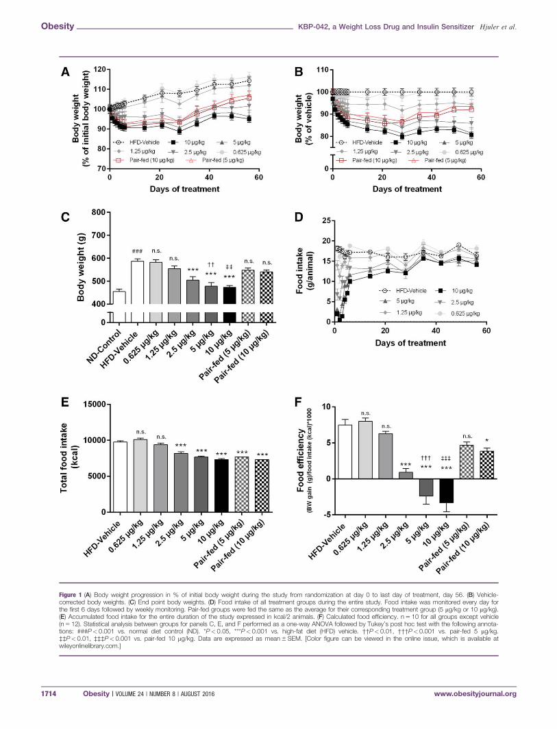
Figure 1 (A) Body weight progression in % of initial body weight during the study from randomization at day 0 to last day of treatment, day 56. (B) Vehicle-corrected body weights. (C) End point body weights. (D) Food intake of all treatment groups during the entire study. Food intake was monitored every day forthe first 6 days followed by weekly monitoring. Pair-fed groups were fed the same as the average for their corresponding treatment group (5 mg/kg or 10 mg/kg).(E) Accumulated food intake for the entire duration of the study expressed in kcal/2 animals. (F) Calculated food efficiency. n 5 10 for all groups except vehicle(n 5 12). Statistical analysis between groups for panels C, E, and F performed as a one-way ANOVA followed by Tukey’s post hoc test with the following annota-tions: ###P< 0.001 vs. normal diet control (ND). *P< 0.05, ***P< 0.001 vs. high-fat diet (HFD) vehicle. ††P< 0.01, †††P< 0.001 vs. pair-fed 5 mg/kg.‡‡P< 0.01, ‡‡‡P< 0.001 vs. pair-fed 10 mg/kg. Data are expressed as mean 6 SEM. [Color figure can be viewed in the online issue, which is available atwileyonlinelibrary.com.]
Obesity KBP-042, a Weight Loss Drug and Insulin Sensitizer Hjuler et al.
1714 Obesity | VOLUME 24 | NUMBER 8 | AUGUST 2016 www.obesityjournal.org
(Supporting Information Figure S1). After the transient reduction in
feeding, food intake increased during the study. The pair-fed groups
gained weight again after feeding increased; inversely, treatment with
KBP-042 sustained the initial weight reduction throughout the 56 days,
with significant reductions in the 2.5 mg/kg, 5 mg/kg, and 10 mg/kg
groups compared with the HFD vehicle (Figure 1C). The accumulated
food intake corresponds well with the weight change for the three high-
est treatment groups (2.5 mg/kg, 5 mg/kg, and 10 mg/kg) (Figure 1E),
although the pair-fed groups which received the same amount of food as
their corresponding treatment group did not lose significant weight.
Accordingly, treatment with 2.5, 5, and 10 mg/kg KBP-042 resulted in
drastic and significant reduction in food efficiency compared with pair-
fed (Figure 1F), suggesting increased energy expenditure.
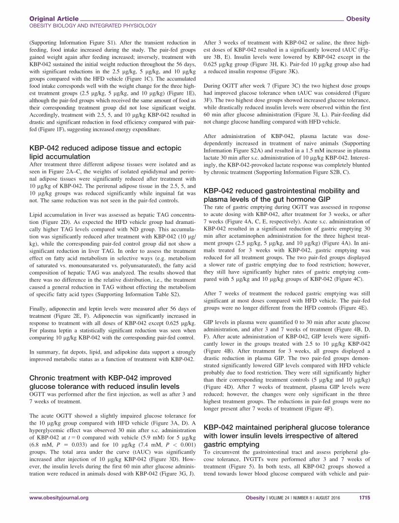
KBP-042 reduced adipose tissue and ectopiclipid accumulationAfter treatment three different adipose tissues were isolated and as
seen in Figure 2A–C, the weights of isolated epididymal and perire-
nal adipose tissues were significantly reduced after treatment with
10 mg/kg of KBP-042. The perirenal adipose tissue in the 2.5, 5, and
10 mg/kg groups was reduced significantly while inguinal fat was
not. The same reduction was not seen in the pair-fed controls.
Lipid accumulation in liver was assessed as hepatic TAG concentra-
tion (Figure 2D). As expected the HFD vehicle group had dramati-
cally higher TAG levels compared with ND group. This accumula-
tion was significantly reduced after treatment with KBP-042 (10 mg/
kg), while the corresponding pair-fed control group did not show a
significant reduction in liver TAG. In order to assess the treatment
effect on fatty acid metabolism in selective ways (e.g. metabolism
of saturated vs. monounsaturated vs. polyunsaturated), the fatty acid
composition of hepatic TAG was analyzed. The results showed that
there was no difference in the relative distribution, i.e., the treatment
caused a general reduction in TAG without effecting the metabolism
of specific fatty acid types (Supporting Information Table S2).
Finally, adiponectin and leptin levels were measured after 56 days of
treatment (Figure 2E, F). Adiponectin was significantly increased in
response to treatment with all doses of KBP-042 except 0.625 mg/kg.
For plasma leptin a statistically significant reduction was seen when
comparing 10 mg/kg KBP-042 with the corresponding pair-fed control.
In summary, fat depots, lipid, and adipokine data support a strongly
improved metabolic status as a function of treatment with KBP-042.
Chronic treatment with KBP-042 improvedglucose tolerance with reduced insulin levelsOGTT was performed after the first injection, as well as after 3 and
7 weeks of treatment.
The acute OGTT showed a slightly impaired glucose tolerance for
the 10 mg/kg group compared with HFD vehicle (Figure 3A, D). A
hyperglycemic effect was observed 30 min after s.c. administration
of KBP-042 at t 5 0 compared with vehicle (5.9 mM) for 5 mg/kg
(6.8 mM, P 5 0.033) and for 10 mg/kg (7.4 mM, P < 0.001)
groups. The total area under the curve (tAUC) was significantly
increased after injection of 10 mg/kg KBP-042 (Figure 3D). How-
ever, the insulin levels during the first 60 min after glucose adminis-
tration were reduced in animals dosed with KBP-042 (Figure 3G, J).
After 3 weeks of treatment with KBP-042 or saline, the three high-
est doses of KBP-042 resulted in a significantly lowered tAUC (Fig-
ure 3B, E). Insulin levels were lowered by KBP-042 except in the
0.625 mg/kg group (Figure 3H, K). Pair-fed 10 mg/kg group also had
a reduced insulin response (Figure 3K).
During OGTT after week 7 (Figure 3C) the two highest dose groups
had improved glucose tolerance when tAUC was considered (Figure
3F). The two highest dose groups showed increased glucose tolerance,
while drastically reduced insulin levels were observed within the first
60 min after glucose administration (Figure 3I, L). Pair-feeding did
not change glucose handling compared with HFD vehicle.
After administration of KBP-042, plasma lactate was dose-
dependently increased in treatment of naive animals (Supporting
Information Figure S2A) and resulted in a 1.5 mM increase in plasma
lactate 30 min after s.c. administration of 10 mg/kg KBP-042. Interest-
ingly, the KBP-042-provoked lactate response was completely blunted
by chronic treatment (Supporting Information Figure S2B, C).
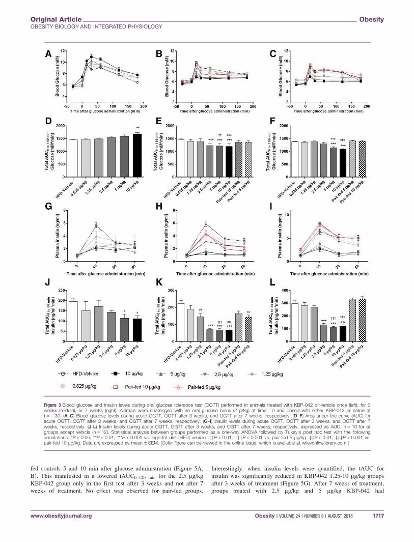
KBP-042 reduced gastrointestinal mobility andplasma levels of the gut hormone GIPThe rate of gastric emptying during OGTT was assessed in response
to acute dosing with KBP-042, after treatment for 3 weeks, or after
7 weeks (Figure 4A, C, E, respectively). Acute s.c. administration of
KBP-042 resulted in a significant reduction of gastric emptying 30
min after acetaminophen administration for the three highest treat-
ment groups (2.5 mg/kg, 5 mg/kg, and 10 mg/kg) (Figure 4A). In ani-
mals treated for 3 weeks with KBP-042, gastric emptying was
reduced for all treatment groups. The two pair-fed groups displayed
a slower rate of gastric emptying due to food restriction; however,
they still have significantly higher rates of gastric emptying com-
pared with 5 mg/kg and 10 mg/kg groups of KBP-042 (Figure 4C).
After 7 weeks of treatment the reduced gastric emptying was still
significant at most doses compared with HFD vehicle. The pair-fed
groups were no longer different from the HFD controls (Figure 4E).
GIP levels in plasma were quantified 0 to 30 min after acute glucose
administration, and after 3 and 7 weeks of treatment (Figure 4B, D,
F). After acute administration of KBP-042, GIP levels were signifi-
cantly lower in the groups treated with 2.5 to 10 mg/kg KBP-042
(Figure 4B). After treatment for 3 weeks, all groups displayed a
drastic reduction in plasma GIP. The two pair-fed groups demon-
strated significantly lowered GIP levels compared with HFD vehicle
probably due to food restriction. They were still significantly higher
than their corresponding treatment controls (5 mg/kg and 10 mg/kg)
(Figure 4D). After 7 weeks of treatment, plasma GIP levels were
reduced; however, the changes were only significant in the three
highest treatment groups. The reductions in pair-fed groups were no
longer present after 7 weeks of treatment (Figure 4F).
KBP-042 maintained peripheral glucose tolerancewith lower insulin levels irrespective of alteredgastric emptyingTo circumvent the gastrointestinal tract and assess peripheral glu-
cose tolerance, IVGTTs were performed after 3 and 7 weeks of
treatment (Figure 5). In both tests, all KBP-042 groups showed a
trend towards lower blood glucose compared with vehicle and pair-
Original Article ObesityOBESITY BIOLOGY AND INTEGRATED PHYSIOLOGY
www.obesityjournal.org Obesity | VOLUME 24 | NUMBER 8 | AUGUST 2016 1715
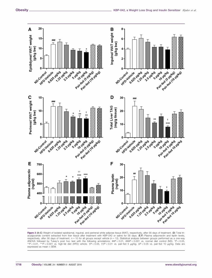
Figure 2 (A–C) Weight of isolated epididymal, inguinal, and perirenal white adipose tissue (WAT), respectively, after 56 days of treatment. (D) Total tri-acylglyceride content extracted from liver tissue after treatment with KBP-042 or saline for 56 days. (E,F) Plasma adiponectin and leptin levels,respectively, after 56 days of treatment. n 5 10 for all groups except vehicle (n 5 12). Statistical analysis between groups performed as a one-wayANOVA followed by Tukey’s post hoc test with the following annotations: ##P< 0.01, ###P< 0.001 vs. normal diet control (ND). *P< 0.05,**P< 0.01, ***P< 0.001 vs. high-fat diet (HFD) vehicle. †P< 0.05, ††P< 0.01 vs. pair-fed 5 mg/kg. ‡P< 0.05 vs. pair-fed 10 mg/kg. Data areexpressed as mean 6 SEM.
Obesity KBP-042, a Weight Loss Drug and Insulin Sensitizer Hjuler et al.
1716 Obesity | VOLUME 24 | NUMBER 8 | AUGUST 2016 www.obesityjournal.org
fed controls 5 and 10 min after glucose administration (Figure 5A,
B). This manifested in a lowered tAUC0–120 min for the 2.5 mg/kg
KBP-042 group only in the first test after 3 weeks and not after 7
weeks of treatment. No effect was observed for pair-fed groups.
Interestingly, when insulin levels were quantified, the tAUC for
insulin was significantly reduced in KBP-042 1.25-10 mg/kg groups
after 3 weeks of treatment (Figure 5G). After 7 weeks of treatment,
groups treated with 2.5 mg/kg and 5 mg/kg KBP-042 had
Figure 3 Blood glucose and insulin levels during oral glucose tolerance test (OGTT) performed in animals treated with KBP-042 or vehicle once (left), for 3weeks (middle), or 7 weeks (right). Animals were challenged with an oral glucose bolus (2 g/kg) at time 5 0 and dosed with either KBP-042 or saline att 5 230. (A–C) Blood glucose levels during acute OGTT, OGTT after 3 weeks, and OGTT after 7 weeks, respectively. (D–F) Area under the curve (AUC) foracute OGTT, OGTT after 3 weeks, and OGTT after 7 weeks, respectively. (G–I) Insulin levels during acute OGTT, OGTT after 3 weeks, and OGTT after 7weeks, respectively. (J-L) Insulin levels during acute OGTT, OGTT after 3 weeks, and OGTT after 7 weeks, respectively, expressed as AUC. n 5 10 for allgroups except vehicle (n 5 12). Statistical analysis between groups performed as a one-way ANOVA followed by Tukey’s post hoc test with the followingannotations: *P< 0.05, **P< 0.01, ***P< 0.001 vs. high-fat diet (HFD) vehicle. ††P< 0.01, †††P<0.001 vs. pair-fed 5 mg/kg. ‡‡P< 0.01, ‡‡‡P< 0.001 vs.pair-fed 10 mg/kg. Data are expressed as mean 6 SEM. [Color figure can be viewed in the online issue, which is available at wileyonlinelibrary.com.]
Original Article ObesityOBESITY BIOLOGY AND INTEGRATED PHYSIOLOGY
www.obesityjournal.org Obesity | VOLUME 24 | NUMBER 8 | AUGUST 2016 1717
Figure 4 (A) Relative rates of gastric emptying measured 30 min after glucose challenge in the oral glucose tolerance test (OGTT) performed in treat-ment naive animals. (B) Area under the curve (AUC) of plasma levels of glucose-dependent insulinotropic peptide (GIP) during OGTT in treatment naiveanimals up to 30 min after glucose challenge. (C) Relative rates of gastric emptying measured 30 min after glucose challenge in the OGTT performedanimals treated with KBP-042 for 3 weeks. (D) AUC of plasma levels of GIP during OGTT in animals treated for 3 weeks, up to 30 min after glucosechallenge. (E) Relative rates of gastric emptying measured 30 min after glucose challenge in the OGTT performed animals treated with KBP-042 for 7weeks. (F) AUC of plasma levels of GIP during OGTT in animals treated for 7 weeks, up to 30 min after glucose challenge. n 5 10 for all groupsexcept high-fat diet (HFD) vehicle (n 5 12). Statistical analysis between groups performed as a one-way ANOVA followed by Tukey’s post hoc testwith the following annotations: *P< 0.05, **P< 0.01, ***P< 0.001 vs. HFD vehicle. ††P< 0.01, †††P< 0.001 vs. pair-fed 5 mg/kg. ‡‡P< 0.01,‡‡‡P< 0.001 vs. pair-fed 10 mg/kg. Data are expressed as mean 6 SEM.
Obesity KBP-042, a Weight Loss Drug and Insulin Sensitizer Hjuler et al.
1718 Obesity | VOLUME 24 | NUMBER 8 | AUGUST 2016 www.obesityjournal.org
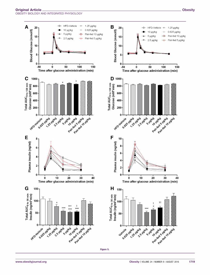
Figure 5.
Original Article ObesityOBESITY BIOLOGY AND INTEGRATED PHYSIOLOGY
www.obesityjournal.org Obesity | VOLUME 24 | NUMBER 8 | AUGUST 2016 1719
significantly reduced insulin levels while maintaining glucose toler-
ance (Figure 5H).
KBP-042 improved whole body insulin sensitivityin the hyperinsulinemic–euglycemic clampA hyperinsulinemic–euglycemic clamp study was performed to
address the effect of KBP-042 on insulin sensitivity. For this study,
ND rats were compared with insulin-resistant HFD rats and 5 mg/kg
KBP-042 treated HFD rats. Figure 6A shows GIR reduced by �30%
(P 5 0.057) in the HFD group compared with ND. The treatment
with KBP-042 led to a significant increase in GIR (82%, P <0.001) compared with HFD vehicle. When KBP-042 treatment is
compared with ND, GIR is increased with 27% (P < 0.05). As
expected, body weight was increased after HFD for 10 weeks as
compared with ND (Figure 6B), but treatment with KBP-042 for 21
days reduced weight with �18%, and the body weight was not sig-
nificantly different from the ND rats at the end of the study.
DiscussionIn this study, KBP-042 induced a significant weight loss over a
period of 8 weeks, albeit with dramatic reductions in food intake
initially. Kaolin consumption was, however, only stimulated in a
higher dose than used in this study, thus indicating the reduction in
food intake was not due to illness. However, minor nausea in the
rats cannot be excluded. The highest KBP-042 groups sustain the
weight loss (up to 20% compared with HFD vehicle) throughout the
study, a phenomenon not seen in the pair-fed groups. The decreased
food efficiency of the KBP-042-treated rats (2.5 mg/kg–10 mg/kg)
and the large weight difference between treated and pair-fed rats,
indicate increased energy expenditure. In general, amylin agonism
blunts the reduction of energy expenditure that is normally caused
by food restriction and weight loss, as well as changing RER
(11,21), an indicator of fat utilization. Interestingly, amylin only
increases energy expenditure when given as chronic infusion s.c. or
i.c.v. (15,16,22), a finding likely related to short-lived activity of
amylin (23). KBP-042 has a longer and more potent activation pro-
file (17), despite a fast disappearance from plasma (<120 min) (18).
However, energy expenditure, as well as potential fecal energy
losses have to be formally assessed in future studies.
KBP-042 was able to significantly reduce TAG accumulation in the
liver at both 2.5 mg/kg and 10 mg/kg. The reduction did not reach a
significant level at 1.25 mg/kg and 5.0 mg/kg due to the relatively
large individual variations in the hepatic TAG levels, but there is a
tendency towards reduced hepatic TAG in these groups. Since
ectopic deposition of lipids in the liver is related to increased insulin
resistance, reducing the hepatic lipid-load could improve hepatic
insulin sensitivity, hereby reducing gluconeogenesis in the fatty liver
and increasing glucose tolerance (24). As of today, weight loss is
the only remedy for ectopic lipid deposition, and KBP-042 serves as
an excellent drug candidate to mediate this in an efficient manner.
However, the extent to which a similar effect could be obtained by
matching the weight loss remains to be explored. Importantly, the
Figure 5 (A,B) Intravenous glucose tolerance test (IVGTT) performed in animals treated for 3 weeks and 7 weeks, respectively, with either KBP-042 orsaline. Animals were dosed s.c. at t 5 230 and received i.v. glucose challenge at t 5 0. (C) Area under the curve (AUC) 0 to 120 min for the IVGTT inpanel A performed after treatment with KBP-042 for 3 weeks. (D) AUC 0 to 120 min for the IVGTT in panel B performed after treatment with KBP-042 for7 weeks. (E) Plasma insulin levels during the IVGTT performed after 3 weeks of treatment. (F) Plasma insulin levels during the IVGTT performed after 7weeks of treatment (legends as for panel A). (G) AUC for plasma insulin levels 0 to 30 min after glucose challenge in the IVGTT in panel A performed aftertreatment with KBP-042 for 3 weeks (legends as for panel B). (H) AUC for plasma insulin levels 0 to 30 min after glucose challenge in the IVGTT in panelB performed after treatment with KBP-042 for 7 weeks. n 5 10 for all groups except high-fat diet (HFD) vehicle (n 5 12). Statistical analysis betweengroups performed as a one-way ANOVA followed by Tukey’s post hoc test with the following annotations: *P< 0.05, ***P< 0.001 vs. HFD vehicle.†P< 0.05, †††P< 0.001 vs. pair-fed 5 mg/kg. ‡P< 0.05, ‡‡P< 0.01 vs. pair-fed 10 mg/kg. Data are expressed as mean 6 SEM. [Color figure can beviewed in the online issue, which is available at wileyonlinelibrary.com.]
Figure 6 (A) Glucose infusion rate (GIR) at steady state during hyperinsulinemic–euglycemic clamp when blood glucose was clampedat basal levels after 21 days of treatment. (B) Body weight at hyperinsulinemic–euglycemic clamp experiment day after 21 days oftreatment. Statistical analysis between groups performed as a one-way ANOVA followed by Tukey’s post hoc test with the followingannotations: *P< 0.05, **P< 0.01, ***P< 0.001. Data are expressed as mean 6 SEM.
Obesity KBP-042, a Weight Loss Drug and Insulin Sensitizer Hjuler et al.
1720 Obesity | VOLUME 24 | NUMBER 8 | AUGUST 2016 www.obesityjournal.org
analysis of the fatty acid composition of TAG further suggests that
the fatty acid metabolism in the liver is unaltered, and the changes
are an overall TAG reduction.
During acute OGTT, increases in plasma lactate and blood glucose
were seen 30 min after administration of KBP-042, corresponding to
previous studies showing acute hyperglycemia following acute admin-
istration of salmon calcitonin or rat amylin (25). This is likely
explained by inhibition of insulin secretion, but also increased plasma
lactate as seen in this study. This manifested as a tendency towards
impaired glucose tolerance. Interestingly, the increase in plasma lac-
tate was not present in animals treated chronically. In fact, chronic
treatment led to improved oral glucose tolerance compared with both
vehicle and pair-fed groups. Importantly, the improved glucose clear-
ance was achieved with significantly lower plasma insulin levels, indi-
cating improved insulin action. The improved glucose tolerance
together with reduced liver TAG supports a general improved metabo-
lism and insulin sensitivity. This is further supported by the reduction
in adiposity, as plasma adiponectin is reduced in subjects who have
obesity and related to for example, inflammation, insulin resistance,
and energy metabolism (26,27), as well as type of phenotype in differ-
ent fat depots (28). The observed increase in adiponectin is in align-
ment with the improvement in both glucose tolerance and insulin
action as well as fatty acid removal from liver that KBP-042 induces
(26,29-31). The reduced adiposity also manifested in lowering of
plasma leptin, which corresponds well with previous demonstrations
that KBP-042 increases the sensitivity towards leptin (18), a finding
also seen with amylin (14,32).
IVGTT was performed to assess peripheral glucose homeostasis
while circumventing the gastrointestinal system, which is obviously
very affected by amylin agonism such as KBP-042 (33,34). Rats
treated with KBP-042 maintained glucose tolerance with reduced
insulin levels hence implying improved insulin sensitivity, albeitwith an effect markedly lower than in the OGTT. This corroborates
that KBP-042 has gastric emptying-independent effects on glucose
tolerance. The reduced insulin levels both during IVGTT and OGTT
could be explained by a direct KBP-042-mediated inhibition of both
insulin and glucagon secretion directly in the islets of Langerhans
(17), but maintaining or improving glycemia, glucose disposal rate,
and insulin action after a significant weight loss is also well
described in humans (5).
Plasma GIP levels and gastric emptying was assessed during the
OGTT, and the rate of gastric emptying correlated to the GIP levels.
In summary, KBP-042 reduces plasma incretin levels during OGTT,
directly inhibits insulin and glucagon release from the islets of
Langerhans (17), and reduces gastric emptying. These effects can
also explain the reduced insulin levels in the OGTT, but not in the
IVGTT. The reduced gastric emptying can mediate a beneficial
effect on postprandial glucose levels, which along with fasting
plasma glucose levels are very important factors in the reduction of
risks related to hyperglycemia.
To formally assess the suggested increase in insulin action we per-
formed a hyperinsulinemic–euglycemic clamp study. The reduced
GIR seen in the HFD group compared with ND was expected since
obesity is negatively correlated to insulin sensitivity and GIR (2).
The large increase in GIR after treatment with KBP-042 illustrated
the increase in insulin sensitivity. The KBP-042-induced weight loss
could explain a large increase in GIR. However, here the rats treated
with KBP-042 had similar body weight to the ND, but with a signif-
icantly increased GIR. This could suggest that insulin sensitivity is
increased beyond what would be expected from weight loss,
although this has to be further tested in weight-matched animals
receiving the same diet.
In conclusion, KBP-042 induced a sustained weight loss over 8
weeks in obese prediabetic rats but not in pair-fed animals, leading
to reduction in adipose tissues, ectopic TAG deposition, improved
glucose tolerance, and improved insulin action. The combination of
a weight-reducing and insulin-sensitizing agent is to our knowledge
unique. KBP-042 thus shows great promise for the treatment of type
2 diabetes and obesity due to its multiple beneficial effects on sev-
eral aspects of the metabolic syndrome.O
AcknowledgmentsWe thank Jannie Felskov Agersten for skillful technical assistance.
VC 2016 The Obesity Society
References1. WHO. A Comprehensive Global Monitoring Framework, Including Indicators, and
a Set of Voluntary Global Targets for the Prevention and Control ofNoncommunicable Diseases. Geneva: WHO; 2012.
2. Kahn SE, Hull RL, Utzschneider KM. Mechanisms linking obesity to insulinresistance and type 2 diabetes. Nature 2006;444:840-846.
3. Guh DP, Zhang W, Bansback N, Amarsi Z, Birmingham CL, Anis AH. Theincidence of co-morbidities related to obesity and overweight: a systematic reviewand meta-analysis. BMC Public Health 2009;9:88.
4. Haslam DW, James WP. Obesity. Lancet 2005;366:1197-1209.
5. Bradley D, Conte C, Mittendorfer B, et al. Gastric bypass and banding equallyimprove insulin sensitivity and beta cell function. J Clin Invest 2012;122:4667-4674.
6. Li G, Zhang P, Wang J, et al. Cardiovascular mortality, all-cause mortality, anddiabetes incidence after lifestyle intervention for people with impaired glucosetolerance in the Da Qing Diabetes Prevention Study: a 23-year follow-up study.Lancet Diabetes Endocrinol 2014;2:474-480.
7. Dombrowski SU, Knittle K, Avenell A, raujo-Soares V, Sniehotta FF. Long termmaintenance of weight loss with non-surgical interventions in obese adults:systematic review and meta-analyses of randomised controlled trials. BMJ 2014;348:g2646.
8. Lutz TA. Effects of amylin on eating and adiposity. Handb Exp Pharmacol 2012;231-250.
9. Roth JD, Roland BL, Cole RL, et al. Leptin responsiveness restored by amylinagonism in diet-induced obesity: evidence from nonclinical and clinical studies.Proc Natl Acad Sci USA 2008;105:7257-7262.
10. Trevaskis JL, Parkes DG, Roth JD. Insights into amylin-leptin synergy. TrendsEndocrinol Metab 2010;21:473-479.
11. Trevaskis JL, Coffey T, Cole R, et al. Amylin-mediated restoration of leptinresponsiveness in diet-induced obesity: magnitude and mechanisms. Endocrinology2008;149:5679-5687.
12. Trevaskis JL, Lei C, Koda JE, Weyer C, Parkes DG, Roth JD. Interaction of leptinand amylin in the long-term maintenance of weight loss in diet-induced obese rats.Obesity (Silver Spring) 2010;18:21-26.
13. Roth JD, Hughes H, Kendall E, Baron AD, Anderson CM. Antiobesity effects ofthe beta-cell hormone amylin in diet-induced obese rats: effects on food intake,body weight, composition, energy expenditure, and gene expression. Endocrinology2006;147:5855-5864.
14. Kusakabe T, Ebihara K, Sakai T, et al. Amylin improves the effect of leptin oninsulin sensitivity in leptin-resistant diet-induced obese mice. Am J PhysiolEndocrinol Metab 2012;302:E924-E931.
15. Wielinga PY, Lowenstein C, Muff S, Munz M, Woods SC, Lutz TA. Centralamylin acts as an adiposity signal to control body weight and energy expenditure.Physiol Behav 2010;101:45-52.
16. Fernandes-Santos C, Zhang Z, Morgan DA, Guo DF, Russo AF, Rahmouni K.Amylin acts in the central nervous system to increase sympathetic nerve activity.Endocrinology 2013;154:2481-2488.
17. Andreassen KV, Feigh MM, Hjuler ST, et al. A novel oral dual amylin andcalcitonin receptor agonist (KBP-042) exerts anti-obesity and anti-diabetic effects inrats. Am J Physiol Endocrinol Metab 2014;307:E24-33.
Original Article ObesityOBESITY BIOLOGY AND INTEGRATED PHYSIOLOGY
www.obesityjournal.org Obesity | VOLUME 24 | NUMBER 8 | AUGUST 2016 1721
18. Hjuler ST, Andreassen KV, Gydesen S, Karsdal MA, Henriksen K. KBP-042improves bodyweight and glucose homeostasis with indices of increased insulinsensitivity irrespective of route of administration. Eur J Pharmacol 2015;762:229-238.
19. Hatanaka S, Kondoh M, Kawarabayashi K, Furuhama K. The measurement ofgastric emptying in conscious rats by monitoring serial changes in serumacetaminophen level. J Pharmacol Toxicol Methods 1994;31:161-165.
20. Ingvorsen C, Thysen AH, Fernandez-Twinn D, et al. Effects of pregnancy onobesity-induced inflammation in a mouse model of fetal programming. Int J Obes(Lond) 2014;38:1282-1289.
21. Trevaskis JL, Turek VF, Wittmer C, et al. Enhanced amylin-mediated body weightloss in estradiol-deficient diet-induced obese rats. Endocrinology 2010;151:5657-5668.
22. Mack C, Wilson J, Athanacio J, et al. Pharmacological actions of the peptidehormone amylin in the long-term regulation of food intake, food preference, andbody weight. Am J Physiol Regul Integr Comp Physiol 2007;293:R1855-R1863.
23. Mack CM, Soares CJ, Wilson JK, et al. Davalintide (AC2307), a novel amylin-mimetic peptide: enhanced pharmacological properties over native amylin to reducefood intake and body weight. Int J Obes (Lond) 2010;34:385-395.
24. Yu H, Jia W, Guo Z. Reducing liver fat by low carbohydrate caloric restrictiontargets hepatic glucose production in non-diabetic obese adults with non-alcoholicfatty liver disease. J Clin Med 2014;3:1050-1063.
25. Young AA, Wang MW, Gedulin B, Rink TJ, Pittner R, Beaumont K. Diabetogeniceffects of salmon calcitonin are attributable to amylin-like activity. Metabolism1995;44:1581-1589.
26. Nigro E, Scudiero O, Monaco ML, et al. New insight into adiponectin role inobesity and obesity-related diseases. Biomed Res Int 2014;2014:658913.
27. De RA, Monaco ML, Capasso M, et al. Adiponectin oligomers as potentialindicators of adipose tissue improvement in obese subjects. Eur J Endocrinol 2013;169:37-43.
28. Drolet R, Belanger C, Fortier M, et al. Fat depot-specific impact of visceralobesity on adipocyte adiponectin release in women. Obesity (SilverSpring) 2009;17:424-430.
29. Ghoshal K, Bhattacharyya M. Adiponectin: probe of the molecular paradigmassociating diabetes and obesity. World J Diabetes 2015;6:151-166.
30. Tilg H, Moschen AR. Adipocytokines: mediators linking adipose tissue,inflammation and immunity. Nat Rev Immunol 2006;6:772-783.
31. Shklyaev S, Aslanidi G, Tennant M, et al. Sustained peripheral expression oftransgene adiponectin offsets the development of diet-induced obesity in rats. ProcNatl Acad Sci USA 2003;100:14217-14222.
32. Moon HS, Chamberland JP, Diakopoulos KN, et al. Leptin and amylin actin an additive manner to activate overlapping signaling pathways inperipheral tissues: in vitro and ex vivo studies in humans. Diabetes Care 2011;34:132-138.
33. Young A. Inhibition of gastric emptying. Adv Pharmacol 2005;52:99-121.
34. Young AA, Gedulin B, Vine W, Percy A, Rink TJ. Gastric emptying is acceleratedin diabetic BB rats and is slowed by subcutaneous injections of amylin.Diabetologia 1995;38:642-648.
Obesity KBP-042, a Weight Loss Drug and Insulin Sensitizer Hjuler et al.
1722 Obesity | VOLUME 24 | NUMBER 8 | AUGUST 2016 www.obesityjournal.org