Taxonomy Bootcamp 2011 Avoiding the Autobiographical Taxonomy: Creating the Right Taxonomy
Upload
dinhkhuongCategory
view
219download
1
Fungal Diversity
The diversity and taxonomy of anaerobic gut fungi
J * 2 • 3Y.W. Ho , N. Abdullah and S. Jalaludm
I Institute of Bioscience, 2 Department of Biochemistry and Microbiology and 3Department ofAnimal Science, Universiti Putra Malaysia, 43400 UPM Serdang, Selangor, Malaysia; * email: [email protected]
Ho, Y.W., Abdullah, N. and Jalaludin, S. (2000). The diversity and taxonomy of anaerobic gutfungi. Fungal Diversity 4: 37-51.
Anaerobic gut fungi have been isolated from the gut of a wide range of herbivores and theirdistribution is world-wide. Many characteristics such as life cycle, reproductive structures,zoospore ultrastructures, thallus morphology and development, molecular affinities andisozyme profiles of the anaerobic gut fungi show that they are more closely related to theChytridiomycetes than to any other group of fungi. Thus, they are assigned to the classChytridiomycetes, but a new order, Neocallimastigales, and a new family,Neoca/limastigaceae, were erected to accommodate them. This review describes thedistribution, life cycle, morphological diversity, taxonomy and phylogeny of the anaerobic gutfungi.
Key words: anaerobic fungi, diversity, gut fungi, phylogeny, taxonomy.
IntroductionFor a long time, the rumen microbial population was believed to be
composed of a diverse population consisting of anaerobic bacteria andprotozoa. It was only in 1975 that the anaerobic rumen fungi were identifiedand recognised as an integral part of the rumen microbiota (Orpin, 1975). Thepresence of chitin in their cell walls (Orpin, 1977a), their morphology and lifecycle confirmed that they are true fungi. The significance and possible role ofthe rumen fungi in fibre digestion was recognised when extensive colonisationof fibrous plant materials by the fungi was observed in the rumen of sheep andcattle (Bauchop, 1979a,b). The fungi preferentially colonise the thick-walledsclerenchyma and vascular tissues (Akin et al., 1983; Ho et al., 1988a,b, 1991,1996) (Fig. 1) and are able to contribute to the overall digestion of variousforages and wheat straw (Akin et al., 1983; Gordon and Ashes, 1984).Anaerobic fungi have now been found to occur in the guts of a wide range ofherbivores and are now commonly known as anaerobic gut fungi. Given theircommon occurrence and abundance, particularly in animals fed fibrous diets, itseems amazing that the anaerobic fungi in the rumen remained unrecognisedfor so long whilst the other anaerobic rumen microorganisms have been
37
discovered and studied for about a hundred years. One of the reasons for this isprobably because most studies on rumen microorganisms are by using rumenfluid of filtered rumen contents instead of the solid rumen digesta that isusually discarded. It is in these rumen digesta that the fungal thalli are attached.Hence, the fungal zoospores swimming freely in the rumen fluid are often thestructures observed by early rumen microbiologists who thought that they wereflagellated protozoa (Liebetanz, 1910; Braune, 1913).
DistributionThe distribution of anaerobic gut fungi is worldwide. They are commonly
found in many herbivores of tropical as well as temperate regions. Anaerobicgut fungi seem to be confined to the alimentary tract of herbivores and have notbeen isolated in other anaerobic terrestrial or aquatic habitats (Orpin andJoblin, 1988; Bauchop, 1989). Table 1 shows the range of ruminants and nonruminants from which anaerobic gut fungi have been isolated.
Herbivores where anaerobic fungi have not been found are the giant panda(Ai/uropoda melanoleuca: Milne et al., 1989), rabbit (Oryctolagus cuniculatus:Bauchop, 1989), possum (Trichosurus vulpecula: Bauchop, 1989), giraffe(Giraffa camelopardalis: Milne et al., 1989), blackbuck (Anti/ope cervicapra:Milne et al., 1989), okapi (Okapia johnstonii: Milne et al., 1989), Braziliantapir (Tapirus terrestris: Milne et al., 1989), lesser mouse deer (Tragulusjavanicus: Y.W. Ho, unpublished) and larger mouse deer (Tragulus napu:Y.W. Ho, unpublished).
The distribution of anaerobic fungi in the alimentary tract of a cattle hadbeen studied by Davies et al. (1993). They found fungi throughout the wholedigestive tract, including the faeces. The highest number of fungi was in thedigesta contents from the reticulo-rumen and omasum (Davies et al., 1993).
The fungi in the reticulo-rumen accounted for about 89% of the total fungi inthe whole digestive tract. Fungal populations were considerably less in theabomasum, small intestine and hindgut. Fungal populations in the hindgutranged from 0.5-2.3% of the total fungal population of the digestive tract(Davies et al., 1993). It is rather surprising that anaerobic fungi are present inthe abomasum where gastric digestion takes place and the digesta contents areacidic with pH values ranging from 3.3 to 4.9. Besides the digestive tract andfaeces, anaerobic fungi have also been isolated from saliva of a sheep (Lowe etal., 1987a). Although anaerobic fungi have been isolated from all parts of thealimentary tract of the cattle, there is little evidence to indicate that growthtakes place in any organ except the rumen. Fungi found in other parts of thealimentary tract probably originated from the rumen (Davis et al., 1993; Wonget al., 1995).
38
Fungal Diversity
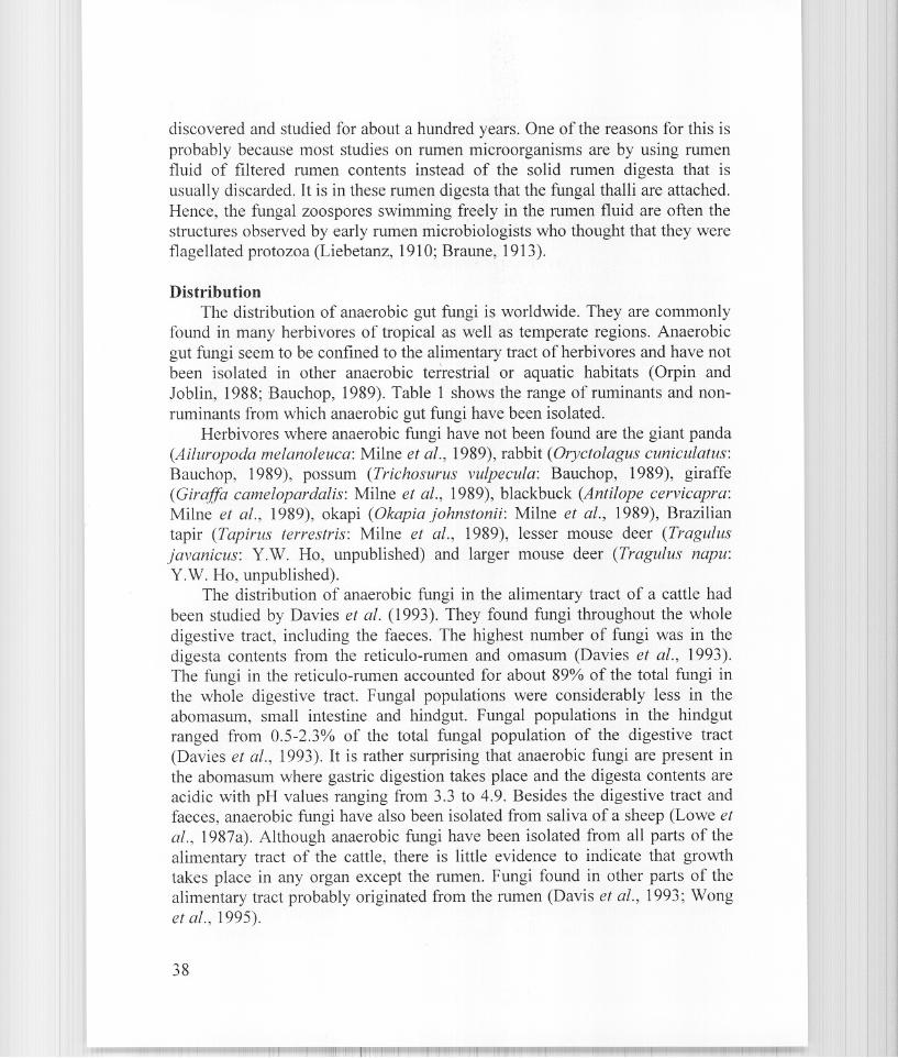
Figs. 1-5. Scanning electron micrograph and light micrographs. 1. Fungi colonising the thickwalled sclerenchyma and vascular tissues of a rice straw fragment in the rumen of a goat. 2. Aunitlagellated zoospore (P. minutus). 3. A polytlagellated zoospore (N. frontalis). 4. Ananaerobic rumen fungus (N. frontalis) with filamentous myceloid rhizoids. 5. An anaerobicrumen fungus (c. communis) with bulbous rhizoids. Bars: 1 = 25 Ilm; 2-5 = 20 Ilm.
39
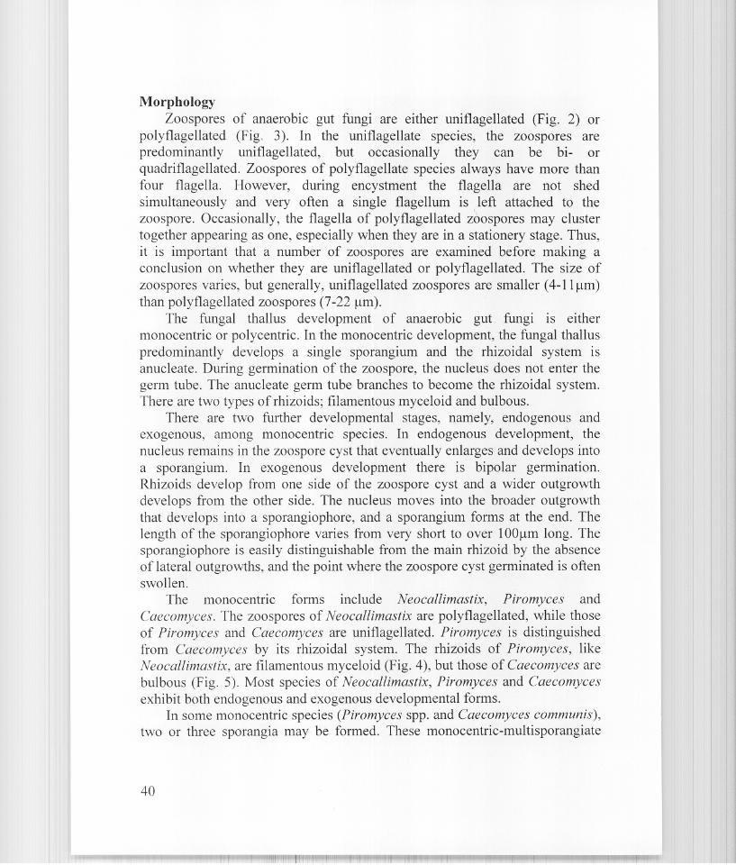
MorphologyZoospores of anaerobic gut fungi are either uniflagellated (Fig. 2) or
polyflagellated (Fig. 3). In the uniflagellate species, the zoospores arepredominantly uniflagellated, but occasionally they can be bi- orquadriflagellated. Zoospores of poly flagellate species always have more thanfour flagella. However, during encystment the flagella are not shedsimultaneously and very often a single flagellum is left attached to thezoospore. Occasionally, the flagella of polyflagellated z60spores may clustertogether appearing as one, especially when they are in a stationery stage. Thus,it is important that a number of zoospores are examined before making aconclusion on whether they are uniflagellated or polyflagellated. The size ofzoospores varies, but generally, uniflagellated zoospores are smaller (4-11 /lm)
than polyflagellated zoospores (7-22 /lm).
The fungal thallus development of anaerobic gut fungi is eithermonocentric or polycentric. In the monocentric development, the fungal thalluspredominantly develops a single sporangium and the rhizoidal system isanucleate. During germination of the zoospore, the nucleus does not enter thegerm tube. The anucleate germ tube branches to become the rhizoidal system.There are two types of rhizoids; filamentous myceloid and bulbous.
There are two further developmental stages, namely, endogenous andexogenous, among monocentric species. In endogenous development, thenucleus remains in the zoospore cyst that eventually enlarges and develops intoa sporangium. In exogenous development there is bipolar germination.Rhizoids develop from one side of the zoospore cyst and a wider outgrowthdevelops from the other side. The nucleus moves into the broader outgrowththat develops into a sporangiophore, and a sporangium forms at the end. Thelength of the sporangiophore varies from very short to over 100/lm long. Thesporangiophore is easily distinguishable from the main rhizoid by the absenceof lateral outgrowths, and the point where the zoospore cyst germinated is oftenswollen.
The monocentric forms include Neocallimastix, Piromyces andCaecomyces. The zoospores of Neocallimastix are polyflagellated, while thoseof Piromyces and Caecomyces are uniflagellated. Piromyces is distinguishedfrom Caecomyces by its rhizoidal system. The rhizoids of Piromyces, likeNeocallimastix, are filamentous myceloid (Fig. 4), but those of Caecomyces arebulbous (Fig. 5). Most species of Neocallimastix, Piromyces and Caecomycesexhibit both endogenous and exogenous developmental forms.
In some monocentric species (Piromyces spp. and Caecomyces communis),two or three sporangia may be formed. These monocentric-multisporangiate
40
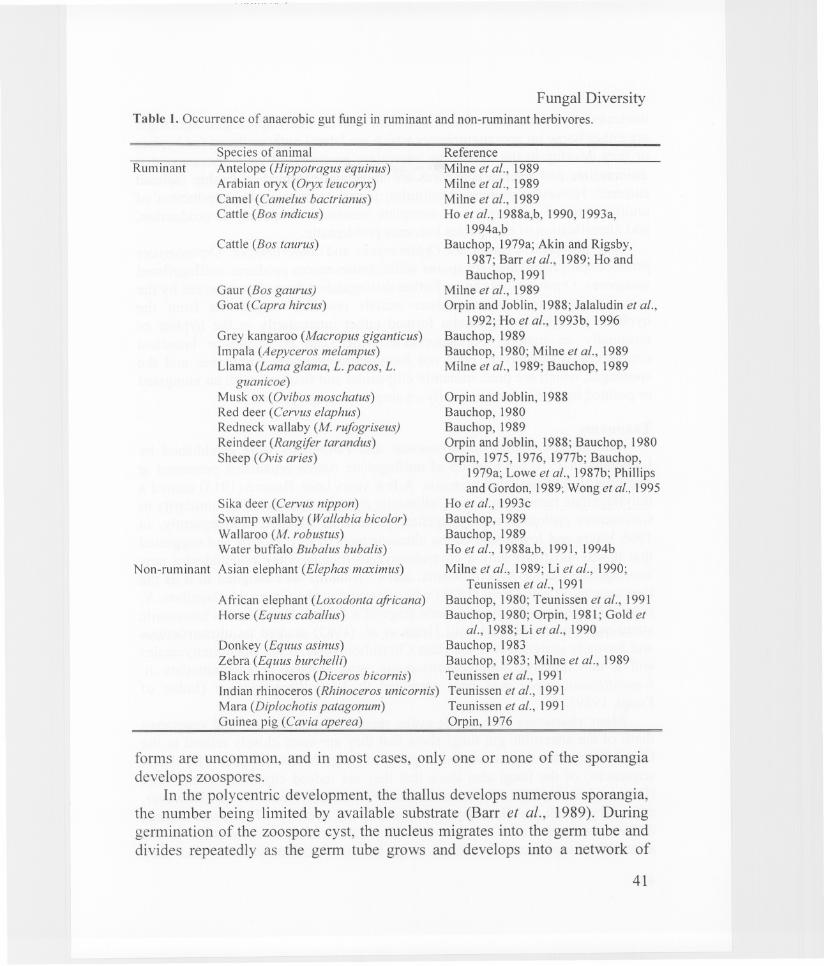
Fungal DiversityTable 1. Occurrence of anaerobic gut fungi in ruminant and non-ruminant herbivores.
Reference
Milne et al., 1989Milne et aI., 1989Mi1ne et al., 1989Ho et al., 1988a,b, 1990, 1993a,
1994a,bBauchop, 1979a; Akin and Rigsby,
1987; Barr et al., 1989; Ho andBauchop, 1991
Milne et al., 1989Orpin and Joblin, 1988; Jala1udin et al.,
1992; Ho et al., 1993 b, 1996Bauchop, 1989Bauchop, 1980; Milne et al., 1989Milne et al., 1989; Bauchop, 1989
African elephant (Loxodonta africana)Horse (Equus cabal/us)
Orpin and Joblin, 1988Bauchop, 1980Bauchop, 1989Orpin and Joblin, 1988; Bauchop, 1980Orpin, 1975, 1976, 1977b; Bauchop,
1979a; Lowe et al., 1987b; Phillipsand Gordon, 1989; Wong et aI., 1995
Ho et aI., 1993cBauchop, 1989Bauchop, 1989Ho et al., 1988a,b, 1991, 1994b
Milne et al., 1989; Li et al., 1990;Teunissen et al., 1991
Bauchop, 1980; Teunissen et al., 1991Bauchop, 1980; Orpin, 1981; Gold et
al., 1988; Li et al., 1990
Donkey (Equus asinus) Bauchop, 1983Zebra (Equus burchelli) Bauchop, 1983; Milne et al., 1989Black rhinoceros (Diceros bicornis) Teunissen et aI., 1991Indian rhinoceros (Rhinoceros unicornis) Teunissen et al., 1991Mara (Diplochotis patagonum) Teunissen et al., 1991Guinea pig (Cavia aperea) Orpin, 1976
Sika deer (Cervus nippon)Swamp wallaby (Wallabia bicolor)Wallaroo (M. robustus)Water buffalo Bubalus bubalis)
Asian elephant (Elephas maximus)
Cattle (Bos taurus)
Gaur (Bos gaurus)Goat (Capra hircus)
Grey kangaroo (Macropus giganticus)1mpala (Aepyceros melampus)Llama (Lama glama, L. pacos, L.
guanicoe)Musk ox (Ovibos moschatus)Red deer (Cervus elaphus)Redneck wallaby (M. rufogriseus)Reindeer (Rangifer tarandus)Sheep (Ovis aries)
Species of animalAntelope (Hippotragus equinus)Arabian oryx (Oryx leucoryx)Camel (Camelus bactrianus)Cattle (Bos indicus)
Non-rum inant
Ruminant
forms are uncommon, and in most cases, only one or none of the sporangiadevelops zoospores.
In the polycentric development, the thallus develops numerous sporangia,the number being limited by available substrate (Barr et al., 1989). Duringgermination of the zoospore cyst, the nucleus migrates into the germ tube anddivides repeatedly as the germ tube grows and develops into a network of
41
nucleated mycelium (rhizomycelium) which forms many sporangia. Sporangiaare either borne on sporangiophores which are lateral outgrowths of the hyphaeor they develop in the hyphae as intercalary sporangia. Viable sporangia thatdifferentiate and release zoospores are abundantly formed in freshly isolatedcultures. However, prolonged culturing usually results in the production ofundifferentiated sporangia or a complete cessation of sporangial production,and identification of the species becomes problematic.
The polycentric forms are Orpinomyces and Anaeromyces. Orpinomycesproduces polyflagellated zoospores while Anaeromyces produces uniflagellatedzoospores. Orpinomyces can be further distinguished from Anaeromyces by thepresence of many sporangiophore initials (swollen outgrowths from thehyphae) and globose sporangia formed either intercalarily in the hyphae orterminally on mature sporangiophores which can be single or branchedcomplexes. Anaeromyces does not form sporangiophore complexes and thesporangia, which are predominantly ellipsoidal and fusiform with an elongatedor pointed tip, are borne terminally on single sporangiophores.
TaxonomyThe generic names Sphaeromonas and Piromonas were established by
Liebetanz in 1910 for a group of uniflagellate rumen organisms presumed atthat time to be flagellated protozoa. A few years later, Braune (1913) named apolyflagellate rumen organism Callimastix frontalis because of its similarity toCallimastix cyclops is, a polyflagellate parasite of copepod. Subsequently, in1966 Vavra and Joyon studied the ultrastructures of C. cyclops is and suggestedthat it was most probably a chytridiomycete fungus. They established a newzooflagellate genus, Neocallimastix, and C. frontalis was assigned to it as thetype species. It was Orpin (1975) who showed that the rumen zoo flagellate, N.Fontalis, was actually the zoosporic stage of a fungus. However, its taxonomicstatus was not established until Heath et al. (1983) studied its ultrastructuresand formally assigned it to the class Chytridiomycete, order Spizellomycetalesand a new family, Neocallimasticaceae, was erected to accommodate it.Neocallimasticaceae was later corrected to Neocallimastigaceae (Index ofFungi, 1989).
Many characters such as life cycle, reproductive structures and vegetativethalli of the anaerobic gut fungi show that they are more closely related to theChytridiomycetes than to any other group of fungi. Molecular data (18S rRNAsequences) of the fungi also show that they are indeed chytridiomycete fungi(Dore and Stahl, 1991; Bowman et al., 1992; Li and Heath, 1992). However,studies by Heath and Bauchop (1985) and Munn et al. (1987) showed distinctdifferences in some of the mitotic and ultrastructural characteristics of the
anaerobic fungi from the other fungi in the Spizellomycetales and they
42
Fungal Diversity
suggested the establishment of a new order for the anaerobic fungi. Later, Li etal. (1993) using cladistic analysis of structural data found the anaerobic gutfungi to be distinct from both the Spizellomycetales and the Chytridales and anew order, Neocallimastigales (= Neocallimasticales), was established forthem.
At present, anaerobic gut fungi are classified and placed in:Class ChytridiomycetesOrder NeocallimastigalesFamily Neocallimastigaceae
So far, five genera have been established for the anaerobic gut fungi. Theyare Neocallimastix (Vavra and Joyon, 1966), Piromyces (Gold et al., 1988),Caecomyces (Gold et al., 1988), Orpinomyces (Barr et al., 1989) andAnaeromyces (Breton et al., 1990). The classification of the genera is basedmainly on the number of flagella present in zoospores, fungal thallusmorphology and development, and ultrastructures of the zoospores. A total ofseventeen species have been formally named and described. Neocallimastix
joyonii (Breton et al., 1989) was later transferred to Orpinomyces and O. boviswas placed in synonymy with O. joyoni (Li et al., 1991). Later, Ho and Barr(1995) revised the classification of anaerobic gut fungi and placed N.patriciarum (Orpin and Munn, 1986) and N. variabilis (Ho et al., 1993a) insynonymy with N. frontalis, thus reducing the number of species to fourteen.They also provided a detailed description and a key for all the genera andspecies in their revised classification.
The identification of anaerobic gut fungal species is complicated anddifficult as natural morphological variation occurs within a species. At timesthere is sufficient morphological variation that the fungal thallus of one speciesmay look like another. Artificial culture media probably cause the mostvariations. In rich medium such as glucose medium, sporangia may becomeabnormally large and they usually abort before zoospores are differentiated.Thus, it is important that conclusions on the identification of species are madeonly after examination of sufficient materials.
Although ultrastructures of zoospores provide valuable and essentialinformation on the classification of anaerobic gut fungi, it is totally impracticaland unnecessary to use them for their identification. Species of anaerobic gutfungi can be identified solely on characters seen in the light microscope.
PhylogenyDore and Stahl (1991), Li and Heath (1992) and Li et al. (1993) assessed
the phylogenetic relationships among the anaerobic gut fungi and theirrelationships with other eukaryotes using 18S rRNA sequences and cladistic
43
analysis of structural data and concluded that the anaerobic gut fungi aremonophyletic and form a closely related group in the Chytridiomycetes. Theisozymatic characteristics (Ho et al., 1994b) and the G+C contents (Brownlee,1989) of the anaerobic gut fungi also indicate that they are a monophyleticgroup. Anaerobic gut fungi are thought to have derived from free-livingchytridiomycetes which have evolved the ability to survive the anaerobicconditions in the digestive tract of their herbivorous hosts (Heath, 1988). Theabsence of mitochondria and lipid droplets and presence of hydrogenosomesare undoubtedly adaptations to anaerobiosis; the unique surface layer of thezoospores is probably an adaptation to the harsh chemical environment of therumen (Heath et al., 1983).
Based on zoospore ultrastructures, thallus morphology and development,molecular affinities and isozymatic characteristics, the anaerobic gut fungi aremore closely related to the chytridiomycete fungi in the orderSpizellomycetales than those in the other orders (Barr, 1988, Bowman et al.,1992; Ho et al., 1994b). Despite this close relationship, the anaerobic gut fungi,nevertheless, show distinct differences and form a separate cluster (ondendrograms showing phylogenetic relationships) from the Spizellomycetales(Li et al., 1993; Ho et al., 1994b). As mentioned earlier, a new order, theNeocallimastigales has been established for the anaerobic gut fungi.
At the generic level, the phylogenetic relationships among Neocallimastix,Piromyces, Caecomyces and Orpinomyces are still unclear. Ho et al. (1994b)using Nei's genetic identity calculated from the isozymatic characteristics ofthe anaerobic fungi found that Piromyces was more closely related toCaecomyces than to Neocallimastix. In contrast, Dore and Stahl (1991) using18S rRNA sequences found Piromyces to be more closely related toNeocallimastix than to Caecomyces (c. communis = Sphaeromonas comminis,Gold et al., 1988). Li et al. (1993), on the other hand, using cladistic analysisof structural data found that P. dumbonica and C. equi were more closelyrelated, forming a cluster but C. communis was consistently isolated and wascloser to Neocallimastix. Matrix of genetic similarity and distance coefficientsshowed that Orpinomyces is more related to Piromyces and Caecomyces thanto Neocallimastix (Ho et al., 1994b). 18S rRNA sequences also showedOrpinomyces (N. joyonii = O. joyonii; Li et al., 1991) and Piromyces to beclosely related but they are closer to Neocallimastix than to Caecomyces.Cladistic analysis of rRNA sequences and cladistic analysis of structural datashowed that the relationships between Orpinomyces, Piromyces andNeocallimastix are not clear and depending on the algorithms or set of dataused or outgroup chosen, Orpinomyces can be close to Piromyces and remotefrom Neocallimastix or close to Neocallimastix and remote from Piromyces (Li
44
Fungal Diversity
and Heath, 1992; Li et al., 1993). To resolve the relationships among these fourgenera, further studies and analyses need to be done. In the case ofAnaeromyces, it is found to be more distantly related to Orpinomyces,Piromyces and Neocallimastix than these three genera are to one another (Liand Heath, 1992).
Life cyclesAll anaerobic gut fungi have very similar life cycles. Basically, it consists
of a motile flagellated zoospore stage swimming freely in the fluid contentsand a non-motile, vegetative and reproductive stage attached to the digestamaterials. The life cycle of monocentric anaerobic fungi is about 24-32 h(Bauchop, 1981; Lowe et al., 1987b). The polycentric anaerobic fungi, besidesproducing zoospores for reproduction, are also capable of vegetativereproduction by fragmentation of their mycelium.
Zoospores of Neocallimastix show very sensitive chemotactic response tosoluble carbohydrates and they probably locate freshly ingested plant fragments by migrating up a soluble-carbohydrate gradient to stomata and damagedareas (Orpin and Bountiff, 1978). Four chemoreceptors, sensing fructose,glucose, mannitol and mannose, have been identified and mixtures of lowconcentrations of glucose, sucrose and fructose caused synergistic responsewhich could be responsible for location of specific plant tissues by the zoospores (Orpin and Bountiff, 1978). Zoospores of N. frontalis, P. communis, O.joyonii (= O. bovis) and Anaeromyces sp. (= Ruminomyces) also showchemotactic response to phenolic acids such as p-coumaric acid, ferulic acidsand syringic acid which are often found in the lignified tissues of plants(Wubah and Kim, 1996).
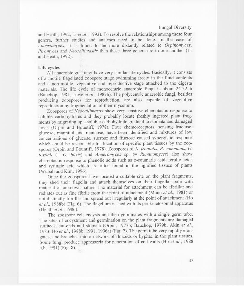
Once the zoospores have located a suitable site on the plant fragments,they shed their flagella and attach themselves on their flagellar pole withmaterial of unknown nature. The material for attachment can be fibrillar and
radiates out as fine fibrils from the point of attachment (Munn et al., 1981) ornot distinctly fibrillar and spread out irregularly at the point of attachment (Hoet al., 1988b) (Fig. 6). The flagellum is shed with its perikinetosomal apparatus(Heath et al., 1986).
The zoospore cell encysts and then germinates with a single germ tube.The sites of encystment and germination on the plant fragments are damagedsurfaces, cut-ends and stomata (Orpin, 1977b; Bauchop, 1979b; Akin et al.,1983; Ho et al., 1988b, 1991, 1996a) (Fig. 7). The germ tube very rapidly elongates, and branches into a network of rhizoids or hyphae in the plant tissues.Some fungi produce appressoria for penetration of cell walls (Ho et al., 1988a,b, 1991) (Fig. 8).
45
Figs. 6-8. Scanning electron micrographs. 6. An attached zoospore. 7. Germinated zoospores ina stoma of a grass fragment in the rumen of buffalo. 8. An appressorium (A) with a penetrationpeg (P) for penetration of the plant cell wall. Bars: 6, 8 = 1 Ilm; 7 = 5 Ilm.
Zoosporogenesis has been reported to be induced by a water-solublecomponent in the diet of the host animal (Orpin, 1977c). One of thecomponents is haem (Orpin and Greenwood, 1986). For N.frontalis, maximumproduction of zoospores occurs 15-30 min after the onset of feeding by the hostanimal (Orpin, 1975) but for P. communis and C. communis, maximumproduction occurs about 1 h after feeding (Orpin, 1976, 1977b). Several haemcontaining compounds have also been found to induce zoosporogenesis and ithas been proposed that haem in synergism with other compounds in the dietsinduced abundant zoosporogenesis in the rumen (Orpin and Greenwood, 1986).
Zoospores are discharged from the sporangia in several ways. It can bethrough an apical pore in the sporangium (N. hurleyensis, Webb andTheodorou, 1991; P. rhizinflatus, Breton et al., 1991), through the dissolutionof a wide apical portion of the sporangium wall (P. minutus, Ho et al., 1993c;C. communis, Ho and Barr, 1995), through rupture and dissolution ofthe apicalpart of the sporangium followed by collapse and dissolution of the sporangialwall (N. frontalis, Heath et al., 1983; O. joyonii, P. comminis, Ho and Barr,1995; O. intercalaris, Ho et al., 1994a), through rapid dissolution of the whole
46
Fungal Diversity
sporangial wall (P. spiralis, Ho et al., 1993b) or through dissolution of papillae(P. mae, Li et al., 1990) .
The ability of the anaerobic fungi in faeces to survive long periods ofdesiccation, reduced temperature and exposure to air (Davies, et al., 1993) hasled to the suggestion that an additional survival or resistant stage be included inthe life cycle (Davies et al., 1993). This stage occurs when conditions becomeunfavourable for vegetative growth, and survival structures are formed towithstand the adverse conditions. However, the survival structures have not yet
been positively identified. Wubah et al. (1991) have reported that aNeocallimastix sp. produced dark brown resistant sporangia as the culture grewolder. These resistant sporangia had melanised cell walls and nuclear DNAlevels of two to four times that of normal sporangia. However, none of theseresistant sporangia could germinate.
ConclusionsIncreased interest and research activities in the anaerobic gut fungi in the
last decade or so have provided much information on their biology, taxonomy,physiology and biochemistry. However, at present, there is still very littleinformation on the range and diversity of fungal species inhabiting differenthost species, different substrates and different parts of the alimentary tract. It isnot known whether the gut fungi are host- or substrate-specific. Further work isneeded to elucidate this.
The taxonomic position of some of the established species and thephylogenetic relatedness among species also need to be resolved. Moreexacting technique will be required for this and it is likely that it lies withmolecular technology.
References
Akin, D.E. and Rigsby, L.L. (1987). Mixed fungal population and lignocellulosic tissuedegradation in the bovine rumen. Applied and Environmental Microbiology 53: 19871995.
Akin, D.E., Gordon, G.L.R. and Hogan, J.P. (1983). Rumen bacterial and fungal degradation ofDigitaria pentzii grown with or without sulphur. Applied and EnvironmentalMicrobiology 46: 738-748.
Barr, DJ.S. (1988). How modem systematics relates to the rumen fungi. BioSystem 21: 351-356.
Barr, DJ.S., Kudo, H., Jaboker, K.D. and Cheng, K.J. (1989). Morphology and development ofrumen fungi: Neocallimastix sp., Piromyces communis and Orpinomyces bovis gen. nov.,sp. novo Canadian Journal of Botany 67: 2815-2824.
Bauchop, T. (1979a). Rumen anaerobic fungi of cattle and sheep. Applied and EnvironmentalMicrobiology 38: 148-158.
47
Bauchop, T. (1979b). The rumen anaerobic fungi: colonizers of plant fibre. Annales deRecherches Veterinaires 10: 246-248.
Bauchop, T. (1980). Scanning electron microscopy in the study of microbial digestion of plantfragments in the gut. In: Contemporary Microbial Ecology (eds. D.J. ElIwood, J.M.Hedger, MJ. Latham, J.M. Lynch and J.H. Slater). Academic Press, London and NewYork: 305-326.
Bauchop, T. (1981). The anaerobic fungi in rumen fibre digestion. Agriculture andEnvironment 6: 339-348.
Bauchop, T. (1983). The gut anaerobic fungi: co Ionizers of dietary fibre. In: Fibre in Humanand Animal Nutrition (eds. C. Wallace and L. Bell). Royal Society of New Zealand,Wellington, New Zealand: 143-148.
Bauchop, T. (1989). Biology of gut anaerobic fungi. BioSystem 23: 53-64.Bowman, B.H., Taylor, J.W., Brownlee, A.G., Lee, J., Lu, S-D. and White, TJ. (1992).
Molecular evolution of the fungi: relationship of the Basidiomycetes, Ascomycetes andChytridiomycetes. Molecular Biology and Evolution 9: 285-296.
Braune, R. (1913). Untersuchungen uber die Wiederkauemagen vorkommenden protozoen.Archiv fur Protistenkunde 32: 111-170.
Breton, A., Bernalier, A., Bonnemoy, F., Fonty, G., Gaillard-Martinie, B. and Gouet, Ph.(1989). Morphology and metabolic characterisation of a new species of strictly anaerobicrumen fungus: Neocallimastix joyonii. FEMS Microbiology Letters 58: 309-314.
Breton, A., Bernalier, A., Dusser, M., Fonty, G., Gaillard-Martinie, B. and Guillot, J. (1990).Anaeromyces mucronatus novo gen., novo sp. A new strictly anaerobic rumen fungus withpolycentric thallus. FEMS Microbiology Letters 70: 177-182.
Breton, A.A., Dusser, M., Gaillard-Martinie, B., Guillot, J., Millet, L. and Prensier, G. (1991).Piromyces rhizinjlata novo sp., a strictly anaerobic fungus from faeces of the Saharaianass: a morphological, metabolic and ultrastructural study. FEMS Microbiology Letters 82:1-8.
Brownlee, A.G. (J 989). Remarkably AT-rich genomic DNA from the anaerobic fungusNeocallimastix. Nucleic Acids Research 17: 1327-1335
Davies, D.R., Theodorou, M.K., Lawrence, M.I. and Trinci, A.PJ. (J 993). Distribution ofanaerobic fungi in the digestive tract of cattle and their survival in faeces. Journal ofGeneral Microbiology 139: 1395-1400.
Dore, J. and Stahl, G .A. (1991). Phylogeny of anaerobic rumen Chytridiomycetes inferred fromsmall subunit ribosomal RNA sequence comparison. Canadian Journal of Botany 69:1964-1971.
Gold, J.J., Heath, LB. and Bauchop, T. (J 988). Ultrastructure description of a new chytridgenus of caecum anaerobes, Caecomyces equi gen. nov., sp. nov., assigned to theNeocallimasticaceae. BioSystem 21: 403-415.
Gordon, G .L.R. and Ashes, J.R. (J 984). In vitro digestion of wheat straw by different rumenanaerobic fungi. Canadian Journal of Animal Science 64: 156-157.
Heath, LB. (1988). Gut fungi. Trends in Ecology and Evoution 3: 167-171.Heath, LB. and Bauchop, T. (1985). Mitosis and the phylogeny of the genus Neocallimastix.
Canadian Journal of Botany 63: 1595-1604.Heath, l.B., Bauchop, T. and Skipp, R.A. (1983). Assignment of the rumen anaerobe
Neocallimastix frontalis to the Spizellomycetales (Chytridiomycetes) on the basis of itspolyflagellate zoospore ultrastructure. Canadian Journal of Botany 61: 295-307.
Heath, I.B., Kaminsky, S.G.W. and Bauchop, T. (1986). Basal body loss during fungalzoospore encystment: evidence against centriole autonomy. Journal of Cell Science 83:135-140.
48
Fungal Diversity
Ho, Y.W. and Barr, D.J.S. (1995). Classification of anaerobic gut fungi from herbivores withemphasis on rumen fungi from Malaysia. Mycologia 87: 655-677.
Ho, Y.W. and Bauchop, T. (1991). Morphology of three polycentric rumen fungi anddescription of a procedure for the induction of zoosporogenesis and release of zoosporesin cultures. Journal of General Microbiology 137: 213-217.
Ho, Y.W., Abdullah, N. and Jalaludin, S. (1988a). Penetrating structures of anaerobic rumenfungi in cattle and swamp buffalo. Journal of General Microbiology 134: 177-181.
Ho, Y.W., Abdullah, N. and Jalaludin, S. (1988b). Colonization of guinea grass by anaerobicrumen fungi in swamp buffalo and cattle. Animal Feed Science and Technology 22: 161172.
Ho, Y.W., Abdullah, N. and Jalaludin, S. (1991). Fungal colonization of rice straw and palmpress fibre in the rumen of cattle and buffalo. Animal Feed Science and Technology 34:311-321.
Ho, Y.W., Abdullah, N. and Jalaludin, S. (1994a). Orpinomyces intercalaris, a new species ofpolycentric anaerobic rumen fungus from cattle. Mycotaxon 50: 139-150.
Ho, Y.W., Abdullah, N. and Jalaludin, S. (1996). Microbial colonisation and degradation ofsome fibrous crop residues in the rumen of goats. Asian-Australasian Journal of AnimalScience 9: 519-524.
Ho, Y.W., Bauchop, T., Abdullah, N. and Jalaludin, S. (1990). Ruminomyces elegans gen. etsp. nov., a polycentric anaerobic fungus from cattle. Mycotaxon 38: 397-405.
Ho, Y.W., Barr, D.J.S., Abdullah, N., Jalaludin, S. and Kudo, H. (1993a). Neocallimastixvariabilis, a new species of anaerobic rumen fungus from cattle. Mycotaxon 46: 241-258.
Ho, Y.W., Barr, DJ.S., Abdullah, N., Jalaludin, S. and Kudo, H. (1993b). Piromyces spiralis, anew species of anaerobic fungus from the rumen of goat. Mycotaxon 48: 59-68.
Ho, Y.W., Barr, DJ.S., Abdullah, N., Jalaludin, S. and Kudo, H. (1993c). A new species ofPiromyces from the rumen of deer in Malaysia. Mycotaxon 47: 285-293.
Ho, Y.W., Khoo, I.Y.S., Tan, S.G., Abdullah, N., Jalaludin, S. and Kudo, H. (1994b). Isozymeanalysis of anaerobic rumen fungi and their relationship to aerobic chytrids. Microbiology140: 1495-1504.
Index of Fungi. (1989). Supplement, family names. C.A.B. International, Walligforf, U.K.Jalaludin, S., Ho, Y.W., Abdullah, N. and Kudo, H. (1992). Use of scanning electron
microscopy in the study of microbial colonisation of some fibrous feed materials in therumen of goats. In: Electron Microscopy 11, Proceedings of the 5th Asia-Pacific ElectronMicroscopy Conference (eds. K.H. Kudo and Z.H. Zhai). Beijing, China: 114-115.
Li, J. and Heath, I.B. (1992). The phylogenetic relationships of anaerobic chytridiomycetousgut fungi (Neocallimasticaeae) and the Chytridiomycota. I. Cladistic analysis of rRNAsequences. Canadian Journal of Botany 70: 1738-1746.
Li, J., Heath, LB. and Bauchop, T. (1990). Piromyces mae and Piromyces dumbonica, two newspecies of uniflagellate anaerobic chytridiomycete fungi from the hind-gut of the horseand elephant. Canadian Journal of Botany 68: 1021-1033.
Li, J., Heath, I.B. and Cheng, K, J. (1991). The development and zoospore ultrastructure of apolycentric chytridiomycete gut fungus, Orpinomyces joyonii comb. novo CanadianJournal of Botany 60: 580-589.
Li, J., Heath, I.B. and Pecker, L. (1993). The phylogenetic relationships of the anaerobicchytridiomycetous gut fungi (Neocallimasticaceae) and the Chytridiomycota. 11.Cladisticanalysis of structural data and description of Neocallimasticales ord. novo CanadianJournal of Botany 71: 393-407.
Liebetanz, E. (1910). Die parasitischen Protozoen des Widenkauermagens. Archiv furProtistenkunde 19: 19-80.
49
Lowe, S.E., Theodorou, M.K. and Trinci, A.PJ. (1987a). Isolation of anaerobic fungi fromsaliva and faeces of sheep. Journal of General Microbiology 133: 1829-1834.
Lowe, S.E., Griffith, G.G., Milne, A., Theodorou, M.K. and Trinci, A.PJ. (l987b). Life cycleand growth kinetics of an anaerobic rumen fungus. Journal of General Microbiology 133:18]5-1827.
Milne, A., Theodorou, M.K., Jordon, M.G.C., King-Spooner, C. and Trinci, A.PJ. (1989).Survival of anaerobic fungi in faeces, in saliva and in pure culture. ExperimentalMycology 13: 27-37.
Munn, E.A., Greemwood, C.A. and Orpin, e.G. (1987). Organization of the kinetosomes andassociated structures of zoospores of the rumen chytridiomycete Neocallimastixpatriciarum. Canadian Journal of Botany 65: 456-465.
Munn, E.A., Orpin, e.G. and Hall, FJ. (1981). Ultrastructural studies of the free zoospore ofthe rumen phycomycete Neocallimastix frontalis. Journal of General Microbiology 125:311-323.
Orpin, e.G. (1975). Studies on the rumen flagellate Neocallimastix frontalis. Journal ofGeneral Microbiology 91: 249-262.
Orpin, e.G. (1976). Studies on the rumen flagellate Sphaeromonas communis. Journal ofGeneral Microbiology 94: 270-280.
Orpin, e.G. (I 977a). The occurrence of chitin in the cell walls of the rumen organismsNeocallimastix frontalis, Piromonas communis and Sphaeromonas communis. Journal ofGeneral Microbiology 99: 215-218.
Orpin, C.G. (]977b). The rumen flagellate Piromonas communis: its life-history and invasionof plant materials in the rumen. Journal of General Microbiology 99: 107-117.
Orpin, C.G. (I 977c). On the induction of zoosporogenesis in the rumen phycomycetesNeocallimastix frontalis, Piromonas communis and Sphaeromonas communis. Journal ofGeneral Microbiology 101: 181-218.
Orpin, e.G. (1981). Isolation of cellulolytic phycomycete fungi from the caecum of the horse.Journal of General Microbiology 123: 287-296.
Orpin, e.G. and Bountiff, L. (1978). Zoospore chemotaxis in the rumen phycomyceteNeocallimastix frontalis. Journal of General Microbio]ogy 104: 113-122.
Orpin, e.G. and Greenwood, Y. (] 986). Effects of haems and related compounds on growthand zoosporogenesis of the rumen phycomycete Neocallimastix frontalis H8. Journal ofGeneral Microbiology 132: 2179-2185.
Orpin, e.G. and Joblin, K.N. (1988). The rumen anaerobic fungi. In: The Rumen MicrobialEcosystem (ed. P.N. Hobson). Elsevier Applied Science, London: 129-150.
Orpin, G.e. and Munn, E.A. (1986). Neocallimastix patriciarum sp. nov., a new member of theNeocallimasticaceae inhibiting the rumen of sheep. Transactions of British MycologicalSociety 86: 178-181.
Phillips, M.W. and Gordon, G.L.R. (]989). Growth characteristics on cellobiose of threedifferent anaerobic fungi isolated from the ovine rumen. Applied and EnvironmentalMicrobiology 55: 1695-1702.
Teunissen, M.J., Op den Camp, HJ.M., Orpin, e.G., Huis, J.H. and Vogels, G.D. (1991).Comparison of growth characteristics of anaerobic fungi isolated from ruminant and nonruminant herbivores during cultivation in a novel defined medium. Journal of GeneralMicrobiology ]37: ]401-]408.
Vavra, 1. and Joyon, L. (1966). Etude sur la morphologie, le cycle evo]utif et al positionsystematique de Callimastix cyclops is Weissenberg 1912. Protistologica 2: 15-16.
We bb, 1. and Theodorou, M.K. (1991). Neocallimastix hurleyensis sp. novo an anaerobicfungus from the ovine rumen. Canadian Journal of Botany 69: 1220-1224.
50
Fungal DiversityWong, M.V.L., Ho, Y.W., Tan, S.G., Abdullah, N. and Jalaludin, S. (1995). Isozyme and
morphological characteristics of the anaerobic fungus Piromyces mae isolated from theduodenum, rumen and faeces of sheep. FEMS Microbio]ogy Letters 134: 9-14.
Wubah, D.A., Fuller, M.S. and Akin, D.E. (1991). Resistant body Iormation in Neocallimastixsp., an anaerobic fungus from the rumen ofa cow. Mycologia 83: 40-47.
Wubah, D.A. and Kim, S.H. (1996). Chemoattraction of anaerobic rumina] fungi zoospores toselected phenolic acids. Microbio]ogical Research 151: 257-262.
(Received 28 Aug. 1999, accepted ]2 Jan. 2000)
51


![Biology and Taxonomy Chapter 1 The Biology of … pathway has recently been identified in S. aureus[9]. Pigment is not produced under anaerobic Historical perspective of the isolation](https://static.fdocuments.in/doc/165x107/5d390da388c99366578d817f/biology-and-taxonomy-chapter-1-the-biology-of-pathway-has-recently-been-identied.jpg)