Management of the Difficult Airway Alternative Airway Techni
Upload
rolf-morrisCategory
view
218download
1
The Difficult or Failed Airway
Pat Melanson, MD

The Difficult Airway
• Must be able to assess or anticipate the degree of difficulty
• Then select method most likely to succeed
• If properly assessed and felt to be intubatable without significant difficulty– 1-4 /1000 will be impossible intubations (O.R.)
– 1 / 280 obstetrical patients
– 1 /10,000 impossible to intubate or ventilate(O.R.)
– 1-2 % cricothyroidotomy rate in ED

Definitions• Failed intubation
– inability to place an ETT
• Difficult intubation– requires more than 3 attempts or 10 minutes
• Difficult laryngosopy– Cormack and Lehane grade III (epiglottis only) or
grade IV view (soft palate only)
• Difficult mask ventilation• Failed airway
– can’t intubate, can’t ventilate
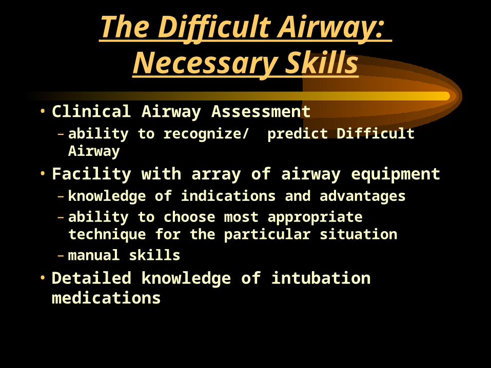
The Difficult Airway: Necessary Skills
• Clinical Airway Assessment– ability to recognize/ predict Difficult Airway
• Facility with array of airway equipment– knowledge of indications and advantages – ability to choose most appropriate technique for
the particular situation– manual skills
• Detailed knowledge of intubation medications

The Difficult Airway
• Not all airway management failures are avoidable or predictable
• Attempt to minimize failures
• Have several definite back-up plans ready for the “Failed Airway”

Prediction of the Difficult Airway
• Historical features ( prior AW difficulty)– Anesthesia record in old chart– Medic alert bracelet– Cric or tracheotomy scar
• Anatomic features

Prediction of the Difficult Airway
• C-spine mobility
• External dimensions ( 3-3-2 rule)– Mouth opening 3 fingers (TMJ)– Mandible large enough to accommodate
tongue - 3 fingers from tip of chin to hyoid– Length of neck/position of larynx - 2 fingers
between top of thyroid and floor of jaw
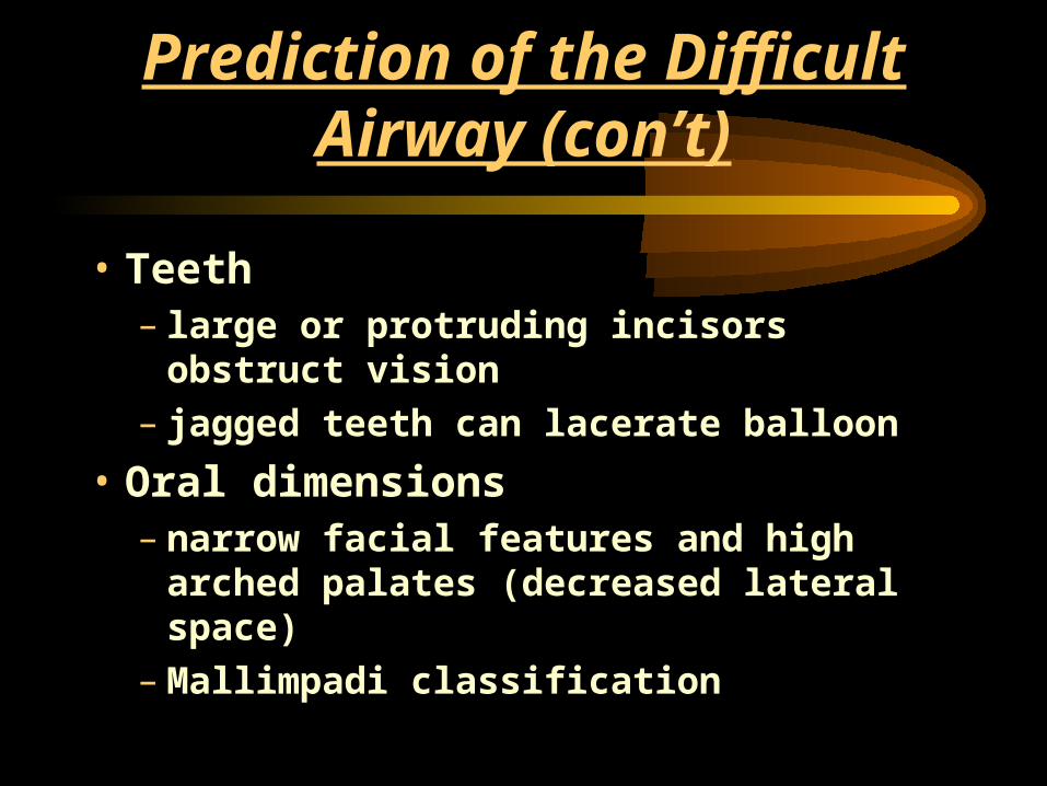
Prediction of the Difficult Airway (con’t)
• Teeth– large or protruding incisors obstruct vision– jagged teeth can lacerate balloon
• Oral dimensions– narrow facial features and high arched
palates (decreased lateral space)– Mallimpadi classification

Mallimpadi Classification (Tongue to Pharyngeal Size)
• I - soft palate, uvula, tonsillar pillars visible– 99 % have grade I laryngoscopic view
• II - soft palate, uvula visible• III - soft palate, base of uvula• IV - soft palate not visible
– 100% grade III or grade IV views• *** this exam is seldom possible in an emergency situation

Predictors of Difficult Laryngoscopy
• Short,thick, muscular neck
• Receding mandible
• Protruding maxillary incisors– “Buck teeth”
• Poor TMJ mobility/ limited jaw opening
• Limited head and neck movement – ( including trauma )
• High, arched palate

Difficult Airway : Laryngoscopy
• Tumor, abscess or hematoma
• Burns
• Angioneurotic edema
• Blunt or penetrating trauma
• Rheumatoid arthritis, ankylosing spondylitis
• Congenital syndromes
• Neck surgery or radiation

Plan B : Response to Unanticipated
Difficulty
• Difficult laryngoscopy and intubation– Can’t intubate but Can ventilate– Can’t intubate and Can’t ventilate
• Difficult Mask Ventilation

Unsuccessful Intubation : Plan B• Bag the patient• Maximize neck flex/ head ex• Move tongue out of line of site• Maximize mouth opening• ID landmarks and adjust blade• BURP maneuver
– (Backwards Upwards Rightwards Pressure on Thyroid Cartilage)
• Increasing lifting force• Consider Miller blade• Bag the patient

Unsuccessful Intubation : Plan B
• An optimal or best attempt at difficult laryngoscopy should consist of :– use of optimal sniffing position– no significant muscle tone– use of optimum external laryngeal
manipulation (BURP)– one change in length of blade– one change in type of blade– a reasonably experienced laryngoscopist

Unsuccessful Intubation : Plan B
• Remember, the first response to failure to intubate should always be to Bag-Mask-Ventilate the patient
• The first response to failure of bag-mask-ventilation is always better bag-mask-ventilation

Algorithm for Difficulty “Bagging”
• Remove FB - Magill forceps
• Triple maneuver if c-spine clear
– Head tilt, jaw lift, mouth opening
• Nasal or oropharyngeal airways
• two-person, four-hand technique
• Do not abandon bagging unless it is impossible with two people and both an OP and NP airway

The Failed Intubation: Definition
• Three failed attempts to intubate– by an experienced intubator
• Inability to ventilate with BVM
• Inability to oxygenate

The Failed Intubation
• If can’t intubate but can ventilate with BVM have time to consider options– Light guided technique (Lighted stylet)– Combitube– LMA– Fiberoptic techniques– Retrograde intubation– Cricothyrotomy

The Failed Intubation
• If can’t intubate, can’t ventilate , must act immediately– Cricothyrotomy– Percutaneous Transtracheal Jet Ventilation– Combitube– LMA– The last three are temporizing measures and
not definitive airway management

Clinical Approach to the Difficult Airway
Is a difficult airway predicted?“nothing should be taken away from the
patient that the airway manager can’t replace”
Bag-Mask predicted to be successful? Intubation deemed reasonably likely ? Do I have the ability to rescue the airway
if “can’t intubate, can’t ventilate”?
Awake Oral Intubation
• Consider for anticipated can’t intubate, can’t ventilate situation
• distorted upper airway anatomy • (i.e., penetrating neck trauma)
• Avoids ‘burning bridges”• maintains ventilation • maintains patient’s ability to protect airway
• May use to take quick look to assure that you can see enough for RSI

Awake Oral Intubation
• Prepare patient psychologically
• Pre-oxygenate
• Topical anesthesia if time permits
• Titrated sedation - avoid obtundation
• Reassure patient throughout procedure

Difficult Airway Kit• Multiple blades and ETTs
• ETT guides (stylets, bougé, light wand)
• Emergency nonsurgical ventilation ( LMA, Combitube, TTJV )
• Emergency surgical airway access ( cricothyroidotomy kit, cricotomes )
• ETT placement verification
• Fiberoptic and retrograde intubation

Techniques for Difficult Intubation
• Alternative laryngoscope blades• Awake intubation• Blind oral or nasal intubation• Fiberoptic intubation• Gum Elastic Bougé• Light wand• Retrograde intubation• Surgical airway

Techniques for Difficult Ventilation
• Combitube
• Laryngeal Mask Airway
• Oral and nasopharyngeal airways
• Two person mask ventilation
• Transtracheal jet ventilation
• Surgical airway
Difficult Airway Maxims
• The first response to failure of Bag-Mask Ventilation is always better BVM– optimize airway position– place both OP and NP airways– two-handed, two-person technique – try lifting head off pillow to open airway– Generate as much positive pressure as
possible without inflating the stomach

Difficult Airway Maxims
• Use judicious sedation and topical airway anesthesia to have a quick look in doubtful cases
• In certain situations a paralytic agent and RSI may still be the best choice

Difficult Airway Maxims
• “It is preferable to use superior judgement -- to avoid having to use superior skill”.