The diagnosis and management of complicated twins
11
4/7/21 1 The diagnosis and management of complicated twins Joanne Stone MD Director, Maternal Fetal Medicine Mount Sinai Health System Professor OBGYN at Icahn School of Medicine at Mount Sinai 1 • I have no financial disclosures 2 3 Dizyotic (2/3): Maternal age (FSH) Genetics ART Monozygotic (1/3): 0.4 – 0.45% following non- stimulated in vivo conception MZ twinning increased after ART: <1% 75% 25% 66% - 75% MC 4 Unique Risks of Monochorionic Twins • Fetal loss • Fetal anomalies – 2-4X increased risk • Twin Twin Transfusion Syndrome (TTTS) • Unequal placental sharing • Discordant twin growth • Selective Intrauterine Growth Restriction (sFGR) • Twin Anemia Polycythemia Syndrome (TAPS) • Twin Reversed Arterial Perfusion (TRAP) • Monoamniotic Monochorionic Twins (MA/MC, MoMo) • Conjoined twins • Death of one twin 5 Other important facts about MC twins • 100% have vascular connections • Cerebral Palsy rate is 1.5% • White matter injury incidence 25% in MC twins after co-twin death 6
Transcript of The diagnosis and management of complicated twins

4/7/21
1
The diagnosis and management of complicated
twinsJoanne Stone MD
Director, Maternal Fetal Medicine Mount Sinai Health SystemProfessor OBGYN at Icahn School of Medicine at Mount Sinai
1
• I have no financial disclosures
2
3
Dizyotic(2/3):Maternal age (FSH)GeneticsART
Monozygotic (1/3):0.4 – 0.45% following non-stimulated in vivo conceptionMZ twinning increased after ART: <1%
75%
25%
66% - 75% MC
4
Unique Risks of Monochorionic Twins• Fetal loss• Fetal anomalies – 2-4X increased risk• Twin Twin Transfusion Syndrome (TTTS)• Unequal placental sharing• Discordant twin growth• Selective Intrauterine Growth Restriction (sFGR)
• Twin Anemia Polycythemia Syndrome (TAPS)• Twin Reversed Arterial Perfusion (TRAP)• Monoamniotic Monochorionic Twins (MA/MC, MoMo)• Conjoined twins• Death of one twin
5
Other important facts about MC twins
• 100% have vascular connections• Cerebral Palsy rate is 1.5%• White matter injury incidence 25% in MC twins after co-twin death
6

4/7/21
2
• Establishing Chorionicity• Diagnosis best in 1st trimester• 98% accurate • Single placenta, T sign, membrane
thickness < 1.5-2 mm
7
Dating a twin pregnancy
• Use known date of conception if ART used• Ideally date CRL at 11+0 – 13+6 weeks• For spontaneous twins, larger CRL should be used
ISUOG guidelines Ultrasound Obstet gGnecol 2016
8
Follow-up after diagnosing MC twins
• MFM consultation• US every 1-2 weeks• MVP (maximum vertical pocket) to assess amniotic fluid• Bladder• Umbilical artery and ductus venosus dopplers as appropriate• MCA (middle cerebral artery dopplers)• Early and routine anatomy survey• Fetal echocardiograms
9 10
Survival rates in MC twins
•When both fetuses alive at 12 weeks, chance of delivering at least one or two live-borns:• DC twins: 98% or 96%•MC/DI twins: 92% or 86%•MC/MA twins: 67% or 67%
Kristiansen MK et al BJOG 2015
11
Predictors of adverse outcome
• NT, CRL, EFW discordance
• Velamentous cord insertion in 1 or both twins increases risk adverse outcome and TTTS
Couck et al Ultrasound Obstet Gynecol 2018
12

4/7/21
3
• CRL discordance ≧ 10% or NT ≧ 20% perform detailed US and karyotype • For MCDA: increased NT found in 25% MC twins and risk early intra-
uterine demise or development TTTS > 30% but poor PPV and NPV
13
Fetal Anomalies• 3-5X increase risk congenital anomalies in MZ
twins• 20% concordant – especially CHD and NTDs•MZ twins are NOT “IDENTICAL”• Post-zygotic genetic/epigenetic and
environmental events• Unequal blastomere allocation • Genetic/epigenetic discordance
14
Pathophysiology of complications of MC twins
15
MC twins: single placenta• ”chorio-angio-pagus” - (placenta-
vascular-conjoined)• Angioarchitecture explains
pathophysiology behind unique complications and reasoning for management• Explains how both are affected by
complications
16
Arteries override veins
A-A anastom osis
A-V anastom osis
Num erous A-V anastom osisbeneath surface
17
Complications of MC twins
•Majority are uncomplicated• 10 - 15% have TTTS• 3% have spontaneous TAPS• 15% have sFGR• Unbalanced division of the placenta -> sFGR
• Can have combination of unbalanced intertwin blood flow and unequal placental share
18

4/7/21
4
Diseases associated with unbalanced intertwin blood flow
• Twin-twin transfusion syndrome (TTTS)• Twin anemia polycythemia sequence (TAPS)
19
Diseases association with unequal partitioning of placenta
• Selective FGR or discordant fetal growth• Can have both TTTS and sFGR co-existing
FGR twin
AGA twin
20
Unbalanced AV anastomoses and partitioning
TTTS TAPS
TTTS and TAPS
sFGR
TTTS and sFGR
TTTS and sFGR TTTS and sFGR and TAPS
Adapted from Dr Stephen Emery NAFNET
21
TTTS
• Complicates 8-10% of MCDA twins• Untreated TTTS has 70-100% loss rate – esp early severe disease• High neurologic morbidity in survivors (10-30%)
Lewi L AJOG 2008, Berghella V JRM 2001,van Hetern CF, Obstet Gynecol 1998
22
Angioarchitecture in TTTS
• Intertwin transfusion• Unequal sharing of blood flow due to unbalanced deep AV anastomoses• Changes in cardiac function
23
Imbalance of A-V anastomoses in one direction – donor “transfuses” volume to recipient
Dx: twin poly-oligo ( MVP ≤ 2cm, ≥ 8cm)
Recipient: polyuria, hypervolemia, distended bladder, HTN, cardiac hypertrophy and failure, abnormal arterial and/or venous Dopplers
Donor:hypovolemia, oliguria, collapsed
bladder, IUGR, abnormal umbilical artery Dopplers
24

4/7/21
5
Ultrasound findings in TTTS
• 1st trimester• CRL discordance• NT > 95th %ile or discordance >20%• Reversal or absence of ductus venosus A wave
• 2nd trimester findings• Abdominal circumference discordance• Membrane folding• Velamentous placental cord insertion in donor
25
TTTS Quintero Staging
Quintero RA. J Perinatol 1999, Stamillo AJOG 2010, Simpson L. AJOG Jan 2013
Some centers incorporate fetal echocardiography (recipient cardiomyopathy) into staging
26
TTTS Management
• Delivery• Expectant management• majority stage I remain stable or regress• High perinatal mortality in stage III or higher
• Serial amnioreduction• Laser photocoagulation• Superior to AR in RCT• Treatment of choice for dual survival
• Selective termination of one fetus• Pregnancy termination
Senat MV NEJM 2004
27 28
Management options in TTTS
• Pregnancy termination• Early or advanced stage TTTS• Complete termination or umbilical cord occlusion
• Amnioreduction• > 26 weeks• Declines/unavailable fetoscopic laser therapy
• Laser photocoagulation of communicating vessels• 18 – 26 weeks• Generally more advanced stage TTTS
• Delivery for late presentation
29
TTTS outcomes with laser therapy
• 85% chance 1 survivor• 65% chance 2 survivors• 10% chance of no survivors• 54% chance donor demise in Stage III with
abnormal Dopplers and sFGR• Mean GA delivery – 33 weeks• 10-20% chance neurologic morbidity of survivors
by age 2
Senat MF NEJM 2005, Chmiat RH AJOG 2011, Simpson et al ACOG. 2011
30
4/7/21
6
NAFNet: what to do about stage I
• Multicenter retrospective observational study• 124 cases stage I TTTS• Expectant mgmt. vs. AR vs Laser• Risk factors for progression• Outcome data• Good: 2 survivors ≧ 30 weeks• Mixed: Single survivor or delivery 26-29.9 weeks• Poor: Double fetal demise or delivery < 26 weeks
AJOG 2016
31
Regress/stable:Good outcome
Emery, NAFTNet AJOG 2016
Stage I TTTS: data from North American Fetal Therapy Network
32
Expectant management group
• Average of 11 days until change in status (regress, progress, termination, etc)• No factors at diagnosis predictive of disease
outcome• AR or laser protected against no survivors• Laser protected against poor outcome
33
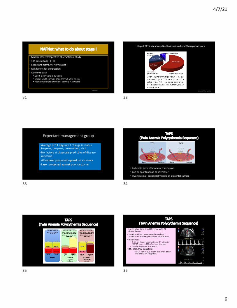
TAPS(Twin Anemia Polycythemia Sequence)
• A chronic form of feto-fetal transfusion• Can be spontaneous or after laser• Involves small peripheral vessels on placental surface
34
TAPS(Twin Anemia Polycythemia Sequence)
35
TAPS(Twin Anemia Polycythemia Sequence)
• Large inter-twin Hb difference w/o AF discordance• Small unidirectional unbalanced AV
anastomoses near perimeter of placenta• Incidence:
• 3-6% previously uncomplicated 3rd trimester MC/DA twins vs 13% after laser therapy
• Usually diagnosed > 26 weeks• DX: MCA-PSV dopplers:
• MCA PSV > 1.5 MoM in donor and < 0.8 MoM in recipient
36
4/7/21
7
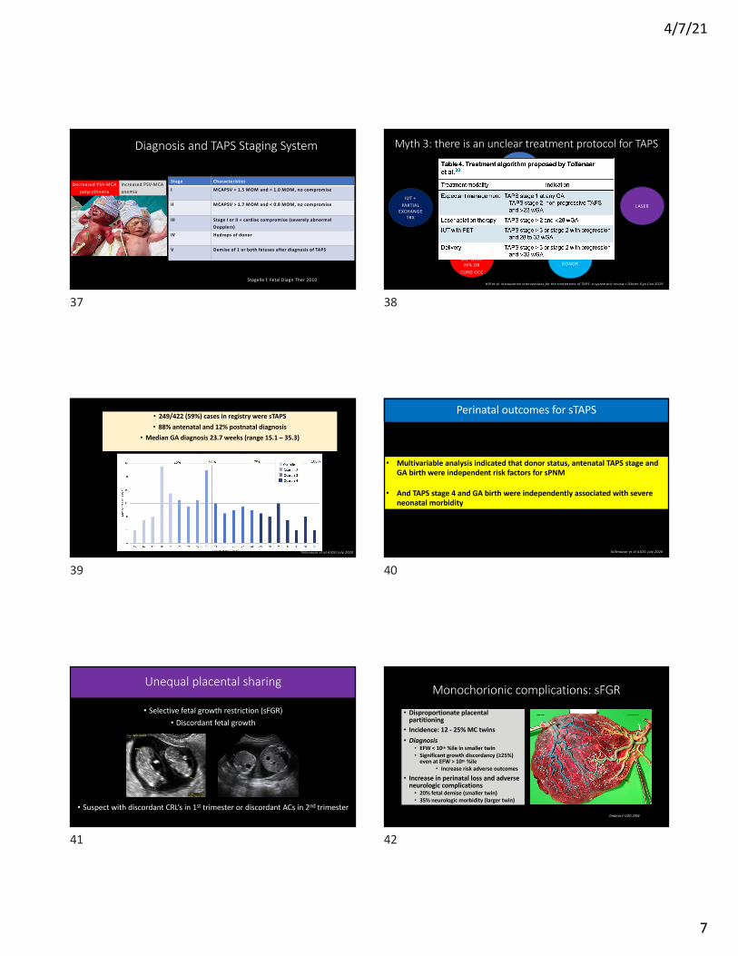
Diagnosis and TAPS Staging System
Stage Characteristics
I MCAPSV > 1.5 MOM and < 1.0 MOM, no compromise
II MCAPSV > 1.7 MOM and < 0.8 MOM, no compromise
III Stage I or II + cardiac compromise (severely abnormal Dopplers)
IV Hydrops of donor
V Demise of 1 or both fetuses after diagnosis of TAPS
Stagelle f. Fetal Diagn Ther 2010
Decreased PSV-MCApolycythemia
Increased PSV-MCAanemia
37
Myth 3: there is an unclear treatment protocol for TAPS
EXPECTANT
LASER
IUT OF DONOR
IUT + PARTIAL
EXCHANGE TRX
SELECTIVE TERMINATION WITH
RFA OR CORD OCC
Data limited by small case series/report, only 1 small comparative studySuggest laser may have lower mortality
Consider treatment algorithm proposed by Tollenar:
Hill et al Intrauterine interventions for the treatm ents of TAPS: a system atic review J Obstet Gyn Can 2019
38
• 249/422 (59%) cases in registry were sTAPS• 88% antenatal and 12% postnatal diagnosis
• Median GA diagnosis 23.7 weeks (range 15.1 – 35.3)
Tollenaaar et al AJOG July 2020
39
Tollenaaar et al AJOG July 2020
Perinatal outcomes for sTAPS
• Multivariable analysis indicated that donor status, antenatal TAPS stage and GA birth were independent risk factors for sPNM
• And TAPS stage 4 and GA birth were independently associated with severe neonatal morbidity
40
Unequal placental sharing
• Selective fetal growth restriction (sFGR)• Discordant fetal growth
• Suspect with discordant CRL’s in 1st trimester or discordant ACs in 2nd trimester
41
Monochorionic complications: sFGR
• Disproportionate placental partitioning• Incidence: 12 - 25% MC twins• Diagnosis
• EFW < 10th %ile in smaller twin• Significant growth discordancy (≥25%)
even at EFW > 10th %ile• Increase risk adverse outcomes
• Increase in perinatal loss and adverse neurologic complications• 20% fetal demise (smaller twin)• 35% neurologic morbidity (larger twin)
Graticos E UOG 2004
42

4/7/21
8
Classification, Outcomes and Management of sFGR
Type Umbilical arteryDopplers
Un-expected IUFD of either
Neurologic complic.
In-uterodeterioration
GA delivery
monitor
I Pos DF 2-4%unpredictable
<5% rare 35w WeeklyDopplers
II PersistentAEDF orREDF
0 – 30%More
predictable
14% 90% 32w DopplersSel. TermPTD, laser
?
III IntermittentAEDF orREDF
10-20%
Unstable-Large AAAs
15-40% 11% 31-32w DopplersSel term
Laser?
Gratacos Ultrasound Obstet and Gynecol 2007, Ishti et al Fetal Diag 2009, Valsky et al Sem Fetal and Neon Med 2010, Johnson, A personal communication
43
Management options in sFGR
• Demise of MCDA twin – 10-15% risk of death of co-twin and 20-25% risk of neurologic morbidity• Considerations include severity, stage, gestational age, parental
wishes and technical considerations• Selective reduction by cord occlusion• Survival > 80% for non-reduced twin with normal neurologic outcom in >90%
• Laser• Technically challenging – no polyhydramnios, large AAAs• High risk for demise of sFGR twin (65-75%)
Perra =Cerdero 2015, Chalouhi 2013, Valskey 2010, Quintero 2001
44
Intra-uterine dem ise
Neonatal death
Intact survival
Type I sFGR expectant 3.1% 97.9%
Type I sFGR laser 16.7%
Type I sFGR selective red 0% 100%
Type II sFGR expectant 16.6% 6.4% 89.3%
Type II sFGR laser 44.3% 100%
Type II sFGR selective red 5% 3.7% 90.6%
Type III sFGR expectant 13.2% 6.8% 61.9%
Type III sFGR laser 32.9% 100%
Type III sFGR selective red 0% 5.2% 98.8%
Exp mgmt. best
Laser or SR may be better at previableGA in severe cases to protect survivingtwin from demise or neurologicimpairment
Townsend et al Ultasound Obstet Gynecol 2019
45
TRAP(Twin Reversed Arterial Perfusion)
• Occurs in 1/100 MC twins (3/4 MCDA, ¼ MOMA)• Early vascular disruption results in abnormal AAA between twins• Acardiac twin depends on retrograde arterial supply of deoxygenated blood from pump twin• Diagnosis: Doppler ultrasound of acardiac fetus’ umbilical cord shows arterial blood flowing
toward the acardiac twin
46
• Early loss of 1 of a mc twin pair with patent anastomoses perfusing other?
• Pump twin at risk of • Hemodynamic compromise (30%)
with cardiac dysfunction and hydrops• Anomalies • Aneuploidy• 2 vessel cord
47
Management Considerations• Sonographic markers for poor prognosis• Ratio of acardiac twin to pump twin > 50%• Polyhydramnios – 60%• Cardiac failure – 30%• PTD – 90%
• Pump twin with cardiac failure with abnormal Dopplers• Increase in size of pump twin (AC of
acardiac/pump >1.0)
• How do you estimate size of acardiac twin• L x W x H x 0.52 (formula for a sphere)
48

4/7/21
9
Management• Expectant
• 30% loss rate between 1st trimester diagnosis and 2nd trimester intervention
• Consider intervention• Acardiac/pump ration> 50%• Rapid growth acardiac twin• Hemodynamic compromise of pump twin
• Intervention• Occlusion of vascular connections (RFA,
laser)• RFA survival 80-90%
49
Monoamniotic (MA) twins
• 1 in 10,000 pregnancies
• 1% of all monozygotic twins
• Occurs when split occurs around days 8-13
50
Monoamniotic (MA) twins
• Diagnosis• Lack of intertwin membrane• Single placenta with both cord
insertions close to each other• Cord entanglement
51
Monoamniotic (MA) twins: management• Consider CVS or amniocentesis• Anatomy ultrasound and fetal echo• Growth ultrasounds• Consider hospital admission around 24-
28 weeks when patient would intervene• Antepartum surveillance• BPP• NSTs or continuous monitoring
• Delivery by cesarean around 32-34 weeks• Survival in anatomically normal fetuses
is > 90%Ultra Obstet Gyn 2000;16(3):223, Acta Obstet Gyn Scand 2005;84(5):432, ltra Obstet Gyn 2006;28:681 Prefumo et al Pren Dx 2015
52
• Multinational cohort study 2010-2017• Non-anomalous uncomplicated MO/MO twins with 2 live fetus at 26 weeks
included• 10 centers inpatient, 12 centers outpatient• Primary outcome IUFD• 195 women (290 fetuses)• Results
• Overall perinatal loss rate 10.8%• 4 women (5/3%) inpt and 15 women (12.5%) outpt IUFD• Peak fetal death rate 4.3% occurring at 29 weeks• From 32 – 36 +6 weeks no fetal/neonatal deaths• No difference in in-patient or out-patient groups
53
Conjoined twins• Very rare: 10.2/million births• 18% prenatally-diagnosed
fetuses survive• Increase rate of structural
anomalies• Outcomes depend on which
organs are shared
54

4/7/21
10
Discordant anomalies
• Structural anomalies more common in MC twins (6-8%)• Only 20% are concordant for anomaly• Monozygotic twins are NOT identical• Post-zygotic mutation• Variations in gene expression• Asymmetric x-chromosome inactivation• Parental imprinting• Discordant gene methylation• Vascular accidents
55
Options
• Expectant• Termination• Umbilical cord occlusion• Bipolar cord coagulation• Radiofrequency ablation (RFA)
56
RFA outcomes – for various etiologies
• About 15% PPROM (up to 25%)• Miscarriage survivor about 5%• Neurologic morbidity survivor about 5%• Live birth rate about 80%• Mean GA delivery 33-36 weeks
Kumar et al AJOG 2014, Lee et al Fetal Diagn Ther
57
Death of one twin
• Bleeding of surviving twin into demise twin• Hypotension, hypovolemia, anemia, hypoxia, acidosis• 15% risk demise of co-twin• 25-35% risk severe neurologic morbidity in survivor
• Management• Immediate deliver after unwitnessed twin death – no benefit• Expectant management• Fetal brain MRI’s of survivor
58
Demise of co-twin
• Retrospective observational study at UCSF• 21 MC twins (none had laser/RFA)• Mean GA demise: 19 6/7 w (12 4/7 – 26 6/7)• Interval to MRI: 4 3/7 w (0-12 1/7)• 41% associated with TTTS• Abnormal findings in 7 cases (33%)• Majority had normal ultrasound
Jelin et al AJOG 2008
59
Take home messages
• Establish chorionicity early• Every 1-2 week surveillance• Anatomy surveys and echocardiography• Options for therapy for TTTS, TAPS, anomalies, RFA• Deliver uncomplicated MC twins around 36 weeks
60

4/7/21
11
Thank you
61