The De˚nition and Classi˚cation of Dry Eye Disease and... · tor nerves that connect them.17...
18
THE OCULAR SURF ACE / APRIL 2007, VOL. 5, NO. 2 / www.theocularsurface.com 75 The Definition and Classification of Dry Eye Disease: Report of the Definition and Classification Subcommittee of the International Dry E y e W ork Shop (2 0 0 7 ) DEWS Definition and Classification ©2007 Ethis Communications, Inc. The Ocular Surface ISSN: 1542- 0124. (No authors listed ). T he d efi nition and classifi cation of d ry eye d isease: report of the Defi nition and Classifi cation Sub committee of the International Dry Eye W orkShop (2007). 2007;5(2):75-92. ABSTRACT The aim of the DEWS Definition and Classifica- tion Subcommittee was to provide a contemporary definition of dry eye disease, supported within a comprehensive clas- sification framework. A new definition of dry eye was devel- oped to reflect current understanding of the disease, and the committee recommended a three-part classification system. The first part is etiopathogenic and illustrates the multiple causes of dry eye. The second is mechanistic and shows how each cause of dry eye may act through a common pathway. It is stressed that any form of dry eye can interact with and exacerbate other forms of dry eye, as part of a vicious circle. Finally, a scheme is presented, based on the severity of the dry eye disease, which is expected to provide a rational basis for therapy. These guidelines are not intended to override the clinical assessment and judgment of an expert clinician in individual cases, but they should prove helpful in the conduct of clinical practice and research. KEYWORDS definition, DEWS, dry eye disease, Dry Eye WorkShop, etiopathogenesis, mechanism, severity grading I. INTRODUCTION he Definition and Classification Subcommittee reviewed previous definitions and classification schemes for dry eye, as well as the current clinical and basic science literature that has increased and clarified knowledge of the factors that characteriz e and contribute to dry eye. Based on its findings, the Subcommittee presents herein an updated definition of dry eye and classifications based on etiology, mechanisms, and severity of disease. II. GOALS OF THE DEFINITION AND CLASSIFICATION SUBCOMMITTEE The goals of the DEWS Definition and Classification Subcommittee were to develop a contemporary definition of dry eye disease and to develop a three-part classification of dry eye, based on etiology, mechanisms, and disease stage. The manner of working of the committee is outlined in the introduction to this issue of The O cular S urface. Further details are published on the TFOS-DEWS web-site (www. tearfilm.org). III. DEFINITION OF DRY EYE DISEASE The committee reviewed the definition and classifica- tion presented at the 19 9 5 National Eye Institute (NEI)/In- dustry Dry Eye Workshop, which was: Dry eye is a disorder of the tear film due to tear deficiency or excessive evaporation, which causes damage to the interpalpebral ocular surface and is associated with symptoms of ocular discomfort. 1 The committee agreed that the definition could be improved in the light of new knowledge about the roles of tear hyperosmolarity and ocular surface inflammation in dry eye and the effects of dry eye on visual function. Initially two definitions were developed and presented to members of the workshop. These “general” and “operational” defini- tions overlapped to some ex tent, and, therefore, in this final report, these versions have been combined to produce the following definition: Dry eye is a multifactorial disease of the tears and ocu- lar surface that results in symptoms of discomfort, 2-4 visual disturbance, 5-7 and tear film instability 8-10 with potential damage to the ocular surface. It is accompa- nied by increased osmolarity of the tear film 11-14 and inflammation of the ocular surface. 15,16 T Accepted for publication January 2007. Definition and Classfication Subcommittee members: M ic hae l A . L em p , M D (Chair); Christophe Baudouin, MD, PhD; Jules Baum, MD; Murat Dogru, MD; Gary N. Foulks, MD; Shigeru Kinoshita, MD; Peter Laibson, MD; James McCulley, MD; Juan Murube, MD, PhD; Stephen C. Pflugfelder, MD; Maurizio Rolando, MD; Ikuko Toda, MD. The Subcommittee is indebted to Professors A.J. Bron and G.N. Foulks for their invaluable contributions to the writing of this report. Proprietary interests of Subcommittee members are disclosed on pages 202 and 204. Reprints are not available. Articles can be accessed at: www.tearfilm.org Correspondence in regard to the this chapter should be addressed to Michael A. Lemp, MD, 4000 Cathedral Avenue NW, Apt 828B, Washington, DC 20016 (Email: [email protected]. Tel: 202-338-6424)
-
Upload
hoangnguyet -
Category
Documents
-
view
212 -
download
0
Transcript of The De˚nition and Classi˚cation of Dry Eye Disease and... · tor nerves that connect them.17...
THE OCULAR SURF ACE / APRIL 2007, VOL. 5, NO. 2 / www.theocularsurface.com 75
The Definition and Classification of Dry Eye Disease:Report of the Definition and Classification Subcommittee of
the International Dry E y e W ork Shop (2 0 0 7 )
DEWS Definition and Classification
©2007 Ethis Communications, Inc. The Ocular Surface ISSN: 1542-
0124. (No authors listed ). T he d efi nition and classifi cation of d ry eye
d isease: report of the Defi nition and Classifi cation Sub committee of
the International Dry Eye W orkShop (2007). 2007;5(2):75-92.
ABSTRACT The aim of the DEWS Definition and Classifica-
tion Subcommittee was to provide a contemporary definition
of dry eye disease, supported within a comprehensive clas-
sification framework. A new definition of dry eye was devel-
oped to reflect current understanding of the disease, and the
committee recommended a three-part classification system.
The first part is etiopathogenic and illustrates the multiple
causes of dry eye. The second is mechanistic and shows how
each cause of dry eye may act through a common pathway.
It is stressed that any form of dry eye can interact with and
exacerbate other forms of dry eye, as part of a vicious circle.
Finally, a scheme is presented, based on the severity of the
dry eye disease, which is expected to provide a rational basis
for therapy. These guidelines are not intended to override the
clinical assessment and judgment of an expert clinician in
individual cases, but they should prove helpful in the conduct
of clinical practice and research.
KEYWORDS definition, DEWS, dry eye disease, Dry Eye
WorkShop, etiopathogenesis, mechanism, severity grading
I. INTRODUCTION
he Definition and Classification Subcommittee reviewed previous definitions and classification schemes for dry eye, as well as the current clinical
and basic science literature that has increased and clarified knowledge of the factors that characteriz e and contribute to dry eye. Based on its findings, the Subcommittee presents herein an updated definition of dry eye and classifications based on etiology, mechanisms, and severity of disease.
II. GOALS OF THE DEFINITION AND
CLASSIFICATION SUBCOMMITTEE
The goals of the DEWS Definition and Classification Subcommittee were to develop a contemporary definition of dry eye disease and to develop a three-part classification of dry eye, based on etiology, mechanisms, and disease stage.
The manner of working of the committee is outlined in the introduction to this issue of The O cular S urface. Further details are published on the TFOS-DEWS web-site (www.tearfilm.org).
III. DEFINITION OF DRY EYE DISEASE
The committee reviewed the definition and classifica-tion presented at the 19 9 5 National Eye Institute (N E I)/In-dustry Dry Eye Workshop, which was: Dry eye is a disorderof the tear film due to tear deficiency or excessive evaporation,which causes damage to the interpalpebral ocular surface andis associated with symptoms of ocular discomfort.1
The committee agreed that the definition could be improved in the light of new knowledge about the roles of tear hyperosmolarity and ocular surface inflammation in dry eye and the effects of dry eye on visual function. Initially two definitions were developed and presented to members of the workshop. These “general” and “operational” defini-tions overlapped to some ex tent, and, therefore, in this final report, these versions have been combined to produce the following definition:
Dry eye is a multifactorial disease of the tears and ocu-lar surface that results in symptoms of discomfort,2-4
visual disturbance,5-7 and tear film instability8-10 withpotential damage to the ocular surface. It is accompa-nied by increased osmolarity of the tear film11-14 andinflammation of the ocular surface.15,16
T
Accepted for publication January 2007.
Definition and Classfication Subcommittee members: M ic hae l A . L e m p , M D (Chair); Christophe Baudouin, MD, PhD; Jules Baum, MD; Murat Dogru, MD; Gary N. Foulks, MD; Shigeru Kinoshita, MD; Peter Laibson, MD; James McCulley, MD; Juan Murube, MD, PhD; Stephen C. Pflugfelder, MD; Mauriz io Rolando, MD; Ikuko Toda, MD.
The Subcommittee is indebted to Professors A.J. Bron and G.N. Foulks for their invaluable contributions to the writing of this report.
Proprietary interests of Subcommittee members are disclosed on pages 202 and 204.
Reprints are not available. Articles can be accessed at: www.tearfilm.org
Correspondence in regard to the this chapter should be addressed to Michael A. Lemp, MD, 4000 Cathedral Avenue NW, Apt 828B, Washington, DC 20016 (Email: [email protected]. Tel: 202-338-6424)
THE OCULAR SURFACE / APRIL 2007, VOL. 5, NO. 2 / www.theocularsurface.com76
Dry eye is recognized as a disturbance of the LacrimalF unctional U nit (LFU), an integrated system comprising the lacrimal glands, ocular surface (cornea, conjunctiva and meibomian glands) and lids, and the sensory and mo-tor nerves that connect them.17 Trigeminal sensory fibers arising from the ocular surface run to the superior salivary nucleus in the pons, from whence efferent fibers pass, in the nervus intermedius, to the pterygopalatine ganglion. Here, postganglionic fibers arise, which terminate in the lacrimal gland, nasopharynx, and vessels of the orbit. Another neural pathway controls the blink reflex, via trigeminal afferents and the somatic efferent fibers of the seventh cranial nerve. Higher centers feed into the brainstem nuclei, and there is a rich sympathetic supply to the epithelia and vasculature of the glands and ocular surface.
This functional unit controls the major components of the tear film in a regulated fashion and responds to environmental, endocrinological, and cortical influences. Its overall function is to preserve the integrity of the tear
film, the transparency of the cornea, and the quality of the image projected onto the retina.17-20 At the 2007 Dry Eye WorkShop, it was noted that the corneal and conjunctival epithelia are in continuity, through ductal epithelia, with the acinar epithelia of the main and accessory lacrimal glands and the meibomian glands, which themselves arise as specialized invaginations from the ocular surface. Also, these epithelia have the same embryological derivation. This broader concept, which has additional features, has been termed the Ocular Surface System and is discussed further in the “Research” chapter of this issue.21
An important aspect of the unit is the part played by sensory impulses, which arise from the ocular surface, in the maintenance of resting tear flow. Currently, it is considered that waking tear flow is a reflex response to afferent im-pulses deriving particularly, but not entirely, from the ocular surface.22 Sensory input from the nasal mucosa also makes a contribution.23 Disease or damage to any component of the LFU (the afferent sensory nerves, the efferent autonomic and motor nerves, and the tear-secreting glands) can desta-bilize the tear film and lead to ocular surface disease that expresses itself as dry eye. Tear film stability, a hallmark of the normal eye, is threatened when the interactions between stabilizing tear film constituents are compromised by de-creased tear secretion, delayed clearance, and altered tear composition. Ocular surface inflammation is a secondary consequence. Reflex tear secretion in response to ocular irritation is envisioned as the initial compensatory mecha-nism, but, with time, inflammation accompanying chronic secretory dysfunction and a decrease in corneal sensation eventually compromises the reflex response and results in even greater tear film instability. Perturbation of the LFU is considered to play an important role in the evolution of different forms of dry eye.
The distinctions aq ueous-deficient dry eye and evaporativedry eye were removed from the definition, but are retained in the etiopathogenic classification.
IV. CLASSIFICATION OF DRY EYE DISEASE
A. Background
V itali, writing about the harmonized classification crite-ria for Sjogren syndrome (SS) remarked that classification criteria are not necessarily appropriate for use in diagnosis and may lead to misclassification of a disease, particularly in its early stages.24 In an individual patient, a classification scheme can provide a guide, but an expert clinician, apply-ing appropriate diagnostic criteria, is needed to establish a diagnosis.
Although the NEI/Industry Workshop classification1 has served as a useful and durable scheme for over a decade, it does not reflect newer knowledge on pathophysiological mechanisms, effects on vision, and the utility of an assess-ment of severity of disease. Recently, two new classification schemes were published, and these were used as source documents by the committee. These include: the Triple Classification25,26 and the report of the Delphi panel.27
The Triple Classification evolved from reports presented
OUTLINE
I. Introduction
II. Goals of the Definition and Classification Subcommittee
III. Definition of dry eye disease
IV . Classification of dry eye disease
A. Background
B. Etiopathogenic classification of dry eye disease
1. Aqueous tear-deficient dry eye
a. Sjogren syndrome dry eye
b. Non-Sjogren syndrome dry eye
1) Primary lacrimal gland deficiencies
2) Secondary lacrimal gland deficiencies
3) Obstruction of the lacrimal gland ducts
4) Reflex hyposecretion
a) Reflex sensory block
b) Reflex motor block
2. Evaporative dry eye
a. Intrinsic causes
1) Meibomian gland dysfunction
2) Disorders of lid aperature and lid/globe congruity or dynamics
3) Low blink rate
b. Extrinsic causes
1) Ocular surface disorders
2) Contact lens wear
3) Ocular surface disease
4) Allergic conjunctivitis
C. The causative mechanisms of dry eye
1. Tear hyperosmolarity
2. Tear film instability
D. The basis for symptoms in dry eye
E. Classification of dry eye based on severity
DEWS DEFINITION AND CLASSIFICATION
THE OCULAR SURFACE / APRIL 2007, VOL. 5, NO. 2 / www.theocularsurface.com 77
DRY EYE
E� ect of theEnvironment
Milieu Interieur
Low blink rate
behavior, VTU,
microscopy
Wide lid aperture
gaze position
Aging
Low androgen pool
Systemic Drugs:
antihistamines,
beta-blockers,
antispasmodics,
diuretics, and
some psychotropic
drugs
Milieu Exterieur
Low relative humidity
High wind velocity
Occupational
environment
Aqueous-de�cient
SjogrenSyndrome
Dry Eye
Primary
Secondary
Non-SjogrenDry Eye
LacrimalDe�ciency
Re�ex Block
LacrimalGland Duct
Obstruction
SystemicDrugs
Evaporative
Extrinsic
Meibomian OilDe�ciency
Intrinsic
Vitamin A-De�ciency
Topical DrugsPreservatives
Low BlinkRate
Drug ActionAccutane
Contact LensWear
Disordersof Lid
Aperture
Ocular SurfaceDisease
eg, Allergy
at the 14th Congress of the European Society of Ophthal-mology.25 After further clinical experience, an updated ver-sion was published in 2005, which presented three separate schemes: one based on etiopathogenesis; one based on the glands and tissues targeted in dry eye; and one based on disease severity.26
The committee felt that the concept of three different schemes serving different purposes was attractive, but it was noted that evidence-based referencing was limited. For this reason, the scheme as a whole was not adopted, but many conceptual aspects were incorporated into the committee’s final schemes.
The Delphi Panel was a consensus group that met to review the classification of dry eye.27 The panel proposed changing the name of dry eye disease to dysfunctional tear syn-drome, suggesting that the name more accurately reflected pathophysiological events in dry eye. However, although the committee felt that the term embraced the essential
features of the disease, they concluded that retention of the name dry eye had much to recommend it and that its use was embedded in the literature. The committee also rejected a subdivision based on the presence or absence of lid dis-ease, because it is frequently difficult to identify the relative contribution of lid disease to a particular case of dry eye.
The majority of the Definition and Classification Sub-committee was in favor of adopting a severity grading based on the report of the Delphi Panel, recognizing it as a com-prehensive approach that could form the basis of therapy according to severity of the disease. As noted above, the Triple Classification also presented a severity grading.
B. Etiopathogenic Classification of Dry Eye Disease
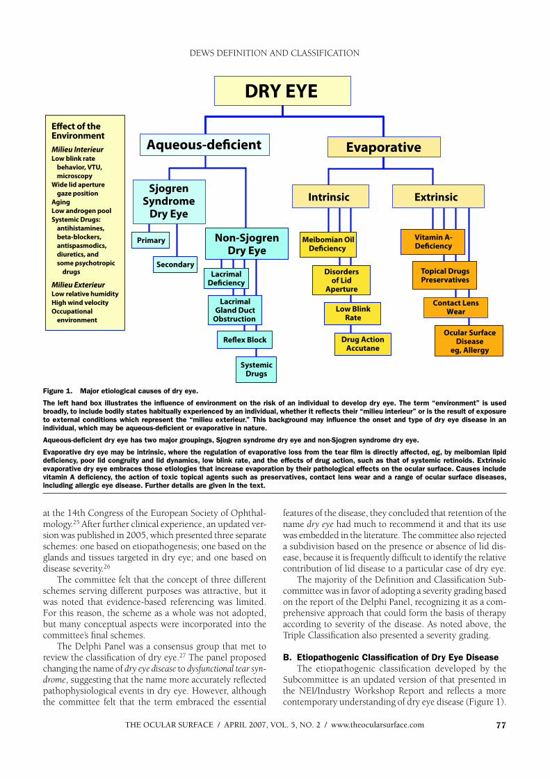
The etiopathogenic classification developed by the Subcommittee is an updated version of that presented in the NEI/Industry Workshop Report and reflects a more contemporary understanding of dry eye disease (Figure 1).
Figure 1. Major etiological causes of dry eye.
The left hand box illustrates the influence of environment on the risk of an individual to develop dry eye. The term “environment” is used
broadly, to include bodily states habitually experienced by an individual, whether it reflects their “milieu interieur” or is the result of exposure
to external conditions which represent the “milieu exterieur.” This background may influence the onset and type of dry eye disease in an
individual, which may be aqueous-deficient or evaporative in nature.
Aqueous-deficient dry eye has two major groupings, Sjogren syndrome dry eye and non-Sjogren syndrome dry eye.
Evaporative dry eye may be intrinsic, where the regulation of evaporative loss from the tear film is directly affected, eg, by meibomian lipid
deficiency, poor lid congruity and lid dynamics, low blink rate, and the effects of drug action, such as that of systemic retinoids. Extrinsic
evaporative dry eye embraces those etiologies that increase evaporation by their pathological effects on the ocular surface. Causes include
vitamin A deficiency, the action of toxic topical agents such as preservatives, contact lens wear and a range of ocular surface diseases,
including allergic eye disease. Further details are given in the text.
DEWS DEFINITION AND CLASSIFICATION
THE OCULAR SURFACE / APRIL 2007, VOL. 5, NO. 2 / www.theocularsurface.com78
As in the 1995 report, the term dry eye is regarded as syn-onymous with the term keratoconjunctivitis sicca (KCS).
The classification has the following features: The left hand box in Figure 1 illustrates the influence of
environment on an individual’s risk of developing dry eye. The term environment is used broadly to include physiologi-cal variation between individuals (their milieu interieur), as well as the ambient conditions that they encounter (their milieu exterieur).
The milieu interieur implies physiological conditions particular to an individual that could influence their risk of dry eye. For instance, a normal subject may have a low natural blink rate, or the blink rate may be slowed for be-havioral or psychological reasons.28 Slowing of the blink rate increases the blink interval and increases the period of evaporative loss between each blink.29
Similarly, the natural height of the palpebral aperture in the primary position varies between individuals and between ethnic groups.30 The aperture is also wider in upgaze than downgaze.31 Evaporative loss per eye increases with increas-ing palpebral width and is, therefore, increased in upgaze.32
Extensive evidence supports a role for the sex hormones in the etiology of dry eye33 with the generalization that low levels of androgens and high estrogen levels are risk factors for dry eye. Biologically active, androgens promote lacrimal and meibomian gland function.33 Androgen deficiency is associated with dry eye34 and may be prevented by topical or systemic androgen therapy.35-38 Dry eye occurs in patients exposed to anti-androgens in the treatment of prostatic cancer,39,40 and women with complete androgen insensitiv-ity syndrome show an increase in the signs and symptoms of dry eye, associated with evidence of meibomian gland and goblet cell dysfunction.41-43 A significantly depleted androgen pool in “non-autoimmune” dry eye associated with meibomian gland dysfunction (MGD) has been re-ported.44 Also, as noted elsewhere in this issue,45 female sex and postmenopausal estrogen therapy are important risk factors for dry eye,46,47 and women with premature ovarian failure suffer from the symptoms and signs of dry eye, although their tear production is not affected.48
Lacrimal tear secretion is reduced by a number of systemic drugs, and these effects may be looked upon as disturbances of the milieu interieur. Their details are dis-cussed later in this report. Aging is associated with physi-ological changes that may predispose to dry eye, including decreased tear volume and flow, increased osmolarity,49
decreased tear film stability,50 and alterations in the com-position of the meibomian lipids.51
The milieu exterieur involves the occupational and external environments, which may represent risk factors for the development of dry eye. Evaporative water loss from the eye is increased in conditions of low relative humidity, occurring either as part of natural variation at different geographic locations or in special circumstances created by air-conditioning, air travel, or other artificial environments.52 Similarly, tear evaporation is increased by exposure to high wind velocity, and this mechanism has
been incorporated into some of the newer experimental models of dry eye.
Occupational factors may cause a slow blink rate, repre-senting a risk for dry eye in those working with video dis-play terminals.53 Other activities associated with decreased blinking and an increase in palpebral width, including that associated with upgaze, have been reported to carry a risk for the development of dry eye symptoms.
The major classes of dry eye, as in the 1995 workshop,1
are still held to be aqueous tear-deficient dry eye (ADDE)and evaporative dry eye (EDE). The category ADDE refers chiefly to a failure of lacrimal secretion, and this approach is retained. However, it should be recognized that a failure of water secretion by the conjunctiva could also contribute to aqueous tear deficiency. The class EDE has been subdivided to distinguish those causes that are dependent on intrinsic conditions of the lids and ocular surface and those that arise from extrinsic influences.
Dry eye can be initiated in any of these classes, but they are not mutually exclusive. It is recognized that disease initi-ated in one major subgroup may coexist with or even lead to events that cause dry eye by another major mechanism. This is part of a vicious circle of interactions that can amplify the severity of dry eye. An example might be that all forms of dry eye cause goblet cell loss and that this, in turn, will contribute to loss of tear film stability, to surface damage and evaporative water loss, and to symptoms resulting from a loss of lubrication and surface inflammatory events.
The major classes and subclasses of dry eye are de-scribed below.
1. Aqueous Tear-Deficient Dry Eye (Tear Deficient Dry Eye; Lacrimal Tear Deficiency) Aqueous tear-deficient dry eye implies that dry eye is
due to a failure of lacrimal tear secretion. In any form of dry eye due to lacrimal acinar destruction or dysfunction, dryness results from reduced lacrimal tear secretion and volume.54,55 This causes tear hyperosmolarity, because, although the water evaporates from the ocular surface at normal rates, it is from a reduced aqueous tear pool. Tear film hyperosmolarity causes hyperosmolarity of the ocular surface epithelial cells and stimulates a cascade of inflam-matory events involving MAP kinases and NFkB signalling pathways56,57 and the generation of inflammatory cytokines (interleukin (IL)-1 ; -1 ; tumor necrosis factor (TNF)- )and matrix metalloproteinases (MMP-9).58 When lacrimal dysfunction is due to lacrimal gland infiltration and inflam-mation, inflammatory mediators generated in the gland are assumed to find their way into the tears and be delivered to the ocular surface. However, when such mediators are detected in the tears, it is not usually possible to know whether they derive from the lacrimal gland itself or from the ocular surface (conjunctiva and cornea).
It is uncertain whether evaporation is reduced59 or in-creased59-64 in ADDE. It is possible that this is determined by the stage of the disease. Some studies suggest that the reservoir of lid oil is larger in non-Sjogren syndrome dry
DEWS DEFINITION AND CLASSIFICATION
THE OCULAR SURFACE / APRIL 2007, VOL. 5, NO. 2 / www.theocularsurface.com 79
eye (NSSDE)65 and that the tear film lipid layer is thicker,66
but dynamic studies of the tear film lipid layer in ADDE have shown that spreading of the lipid layer is delayed in the interblink.67,68 Additionally, in severe ADDE, spread-ing may be undetectable by interferometry, suggesting a major defect in the tear film lipid layer. Delayed or absent spreading of the tear film could lead to an increase in water loss from the eye.
ADDE has two major subclasses, SS dry eye (SSDE)and non-SS dry eye.
a. Sjogren Syndrome Dr y EyeSjogren syndrome is an exocrinopathy in which the
lacrimal and salivary glands are targeted by an autoimmune process; other organs are also affected. The lacrimal and salivary glands are infiltrated by activated T-cells, which cause acinar and ductular cell death and hyposecretion of the tears or saliva. Inflammatory activation within the glands leads to the expression of autoantigens at the surface of epithelial cells (eg, fodrin, Ro and La)69 and the retention of tissue-specific CD4 and CD8 T-cells.70 Hyposecretion is amplified by a potentially reversible neurosecretory block, due to the effects of locally released inflammatory cytokines or to the presence of circulating antibodies (eg, anti-M3
antibody) directed against muscarinic receptors with-in the glands.71-73
There are two forms of SS, and classification criteria have recently been harmonized in a European-American collaboration.74
Primary SS consists of the occurrence of ADDE in combination with symp-toms of dry mouth, in the presence of autoantibod-ies, evidence of reduced salivary secretion and with a positive focus score on minor salivary gland bi-opsy.75,76 Details of the cri-teria are presented in Table 1. Secondary SS consists of the features of primary SS together with the features of an overt autoimmune connective disease, such as rheumatoid arthritis, which is the most common, or systemic lupus erythema-tosis, polyarteritis nodosa, Wegener’s granulomatosis, systemic sclerosis, primary biliary sclerosis, or mixed connective tissue disease. Diagnostic criteria for each
of these connective tissue disorders have been published.77
The precise triggers leading to autoimmune acinar damage are not known in full, but risk factors include genetic profile,78 androgen status79 (a low androgen pool favoring an inflammatory environment within the target tissues), and exposure to environmental agents, ranging from viral infections affecting the lacrimal gland to polluted environments. A nutritional deficiency in omega-3- and other unsaturated fatty acids and unsupplemented intake of vitamin C has also been reported in patients with SS.80
It is generally accepted that environmental factors leading to increased evaporative water loss from the eye (eg, low humidity, high wind velocity, and increased exposure of the ocular surface) may act as a trigger by invoking inflamma-tory events at the ocular surface through a hyperosmolar mechanism (see Section V).
The ocular dryness in SSDE is due to lacrimal hypose-cretion and the accompanying characteristic inflammatory changes in the lacrimal gland, together with the presence of inflammatory mediators in the tears and within the conjunctiva.81 It is not known whether the conjunctival changes are due to an autoimmune targeting of this tissue or whether they are due to the effect of inflammatory media-tors released from the lacrimal glands into the tears.
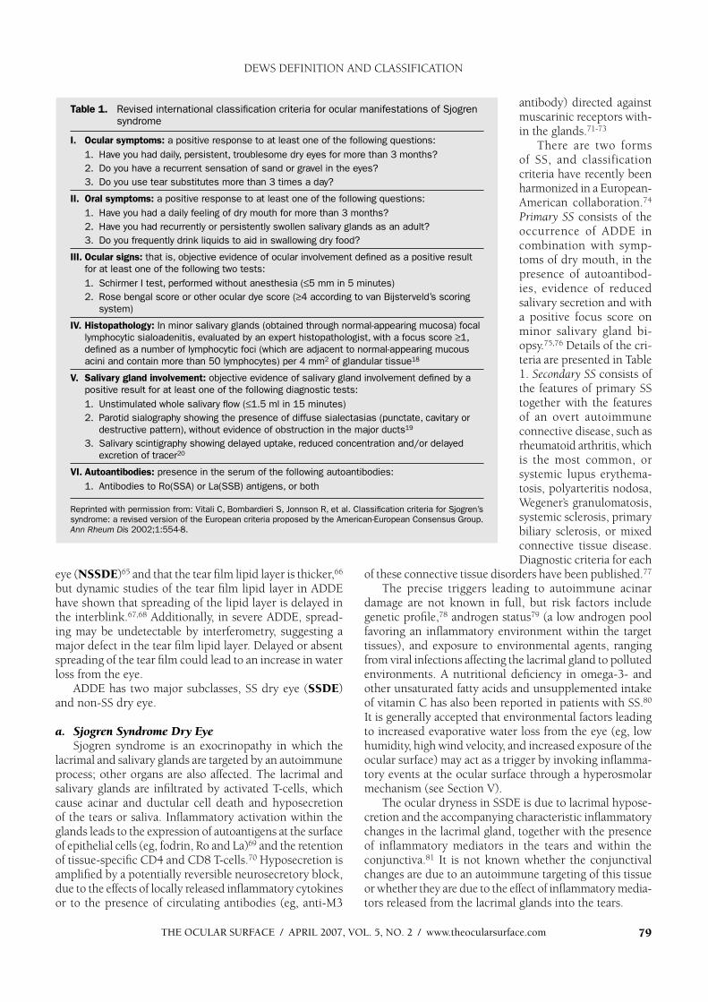
Table 1. Revised international classification criteria for ocular manifestations of Sjogren syndrome
I. Ocular symptoms: a positive response to at least one of the following questions:
1. H ave you had daily, persistent, troublesome dry eyes for more than 3 months?
2. Do you have a recurrent sensation of sand or gravel in the eyes?
3. Do you use tear substitutes more than 3 times a day?
II. Oral symptoms: a positive response to at least one of the following questions:
1. H ave you had a daily feeling of dry mouth for more than 3 months?
2. H ave you had recurrently or persistently swollen salivary glands as an adult?
3. Do you frequently drink liquids to aid in swallowing dry food?
III. Ocular signs: that is, objective evidence of ocular involvement defined as a positive result
for at least one of the following two tests:
1. Schirmer I test, performed without anesthesia ( 5 mm in 5 minutes)
2. Rose bengal score or other ocular dye score ( 4 according to van Bijsterveld’s scoring
system)
IV. Histopathology: In minor salivary glands (obtained through normal-appearing mucosa) focal
lymphocytic sialoadenitis, evaluated by an expert histopathologist, with a focus score 1,
defined as a number of lymphocytic foci (which are adjacent to normal-appearing mucous
acini and contain more than 50 lymphocytes) per 4 mm2 of glandular tissue18
V. Salivary gland involvement: objective evidence of salivary gland involvement defined by a
positive result for at least one of the following diagnostic tests:
1. U nstimulated whole salivary flow ( 1.5 ml in 15 minutes)
2. Parotid sialography showing the presence of diffuse sialectasias (punctate, cavitary or
destructive pattern), without evidence of obstruction in the major ducts19
3. Salivary scintigraphy showing delayed uptake, reduced concentration and/or delayed
excretion of tracer20
VI. Autoantibodies: presence in the serum of the following autoantibodies:
1. Antibodies to Ro(SSA) or La(SSB) antigens, or both
Reprinted with permission from: Vitali C, Bombardieri S, Jonnson R, et al. Classification criteria for Sjogren’s
syndrome: a revised version of the European criteria proposed by the American-European Consensus Group.
Ann Rheum Dis 2002;1:554-8.
DEWS DEFINITION AND CLASSIFICATION
THE OCULAR SURFACE / APRIL 2007, VOL. 5, NO. 2 / www.theocularsurface.com80
The frequency of MGD is higher in patients with SS than in the normal population; thus, a defective tear film lipid layer may contribute to dry eye by leading to excess evaporation.82
b. Non-Sjogren Syndrome Dry Eye Non-Sjogren syndrome dry eye is a form of ADDE due
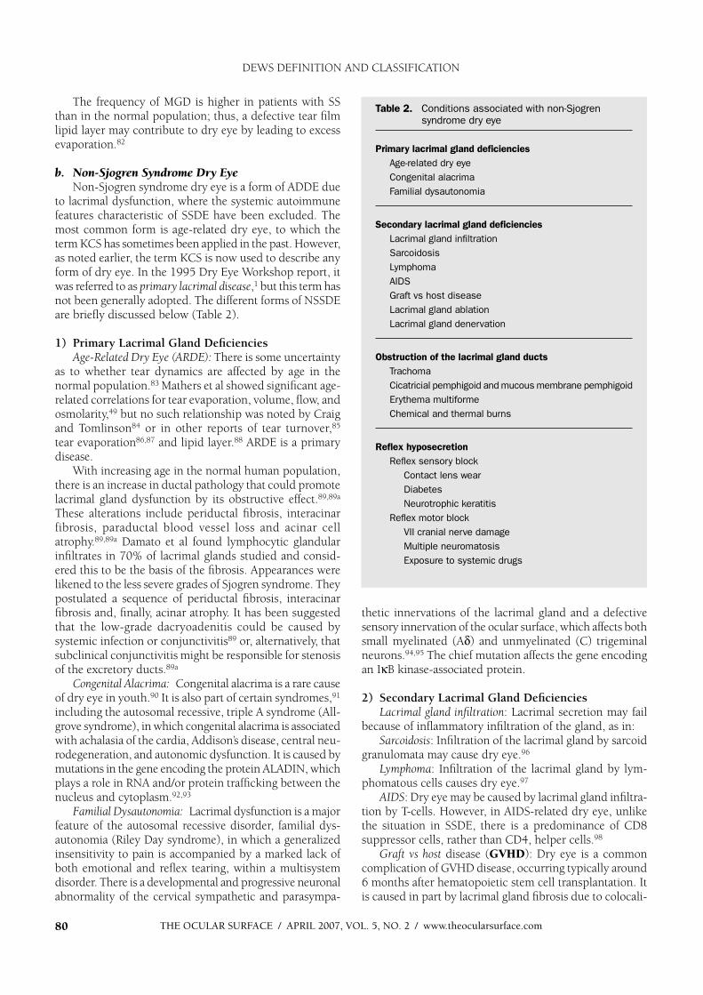
to lacrimal dysfunction, where the systemic autoimmune features characteristic of SSDE have been excluded. The most common form is age-related dry eye, to which the term KCS has sometimes been applied in the past. However, as noted earlier, the term KCS is now used to describe any form of dry eye. In the 1995 Dry Eye Workshop report, it was referred to as primary lacrimal disease,1 but this term has not been generally adopted. The different forms of NSSDE are briefly discussed below (Table 2).
1) Primary Lacrimal Gland DeficienciesAge-Related Dry Eye (ARDE): There is some uncertainty
as to whether tear dynamics are affected by age in the normal population.83 Mathers et al showed significant age-related correlations for tear evaporation, volume, flow, and osmolarity,49 but no such relationship was noted by Craig and Tomlinson84 or in other reports of tear turnover,85
tear evaporation86,87 and lipid layer.88 ARDE is a primary disease.
With increasing age in the normal human population, there is an increase in ductal pathology that could promote lacrimal gland dysfunction by its obstructive effect.89,89a
These alterations include periductal fibrosis, interacinar fibrosis, paraductal blood vessel loss and acinar cell atrophy.89,89a Damato et al found lymphocytic glandular infiltrates in 70% of lacrimal glands studied and consid-ered this to be the basis of the fibrosis. Appearances were likened to the less severe grades of Sjogren syndrome. They postulated a sequence of periductal fibrosis, interacinar fibrosis and, finally, acinar atrophy. It has been suggested that the low-grade dacryoadenitis could be caused by systemic infection or conjunctivitis89 or, alternatively, that subclinical conjunctivitis might be responsible for stenosis of the excretory ducts.89a
C ongenital Alacrima: Congenital alacrima is a rare cause of dry eye in youth.90 It is also part of certain syndromes,91
including the autosomal recessive, triple A syndrome (All-grove syndrome), in which congenital alacrima is associated with achalasia of the cardia, Addison’s disease, central neu-rodegeneration, and autonomic dysfunction. It is caused by mutations in the gene encoding the protein ALADIN, which plays a role in RNA and/or protein trafficking between the nucleus and cytoplasm.92,93
Familial Dysautonomia: Lacrimal dysfunction is a major feature of the autosomal recessive disorder, familial dys-autonomia (Riley Day syndrome), in which a generalized insensitivity to pain is accompanied by a marked lack of both emotional and reflex tearing, within a multisystem disorder. There is a developmental and progressive neuronal abnormality of the cervical sympathetic and parasympa-
thetic innervations of the lacrimal gland and a defective sensory innervation of the ocular surface, which affects both small myelinated (A ) and unmyelinated (C) trigeminal neurons.94,95 The chief mutation affects the gene encoding an I B kinase-associated protein.
2) Secondary Lacrimal Gland DeficienciesLacrimal gland infiltration: Lacrimal secretion may fail
because of inflammatory infiltration of the gland, as in: Sarcoidosis: Infiltration of the lacrimal gland by sarcoid
granulomata may cause dry eye.96
Lymphoma: Infiltration of the lacrimal gland by lym-phomatous cells causes dry eye.97
AIDS: Dry eye may be caused by lacrimal gland infiltra-tion by T-cells. However, in AIDS-related dry eye, unlike the situation in SSDE, there is a predominance of CD8 suppressor cells, rather than CD4, helper cells.98
Graft vs host disease (GVHD): Dry eye is a common complication of GVHD disease, occurring typically around 6 months after hematopoietic stem cell transplantation. It is caused in part by lacrimal gland fibrosis due to colocali-
Table 2 . Conditions associated with non-Sjogren syndrome dry eye
Primary lacrimal gland deficiencies
Age-related dry eye
Congenital alacrima
Familial dysautonomia
Secondary lacrimal gland deficiencies
Lacrimal gland infiltration
Sarcoidosis
Lymphoma
AIDS
Graft vs host disease
Lacrimal gland ablation
Lacrimal gland denervation
Obstruction of the lacrimal gland ducts
Trachoma
Cicatricial pemphigoid and mucous membrane pemphigoid
Erythema multiforme
Chemical and thermal burns
Reflex hyposecretion
Reflex sensory block
Contact lens wear
Diabetes
Neurotrophic keratitis
Reflex motor block
VII cranial nerve damage
Multiple neuromatosis
Exposure to systemic drugs
DEWS DEFINITION AND CLASSIFICATION
THE OCULAR SURFACE / APRIL 2007, VOL. 5, NO. 2 / www.theocularsurface.com 81
zation of periductal T-lymphocytes (CD4 and CD8) with antigen-presenting fibroblasts.99,100
Lacrimal gland ablation: The ducts of the main lacrimal gland pass through its palpebral part, so that excision of the palpebral part will be expected to have the same effect as excision of the main gland. Dry eye may be caused by partial or complete ablation of the lacrimal gland at any age, but is not an obligatory consequence, presumably because accessory gland and conjunctival secretion may compensate in some cases.55 It is, therefore, of interest that ablation of the main lacrimal gland in squirrel monkeys, while reducing both basal and reflex tear secretion, does not in itself lead to dry eye in that species.101
Lacrimal gland denervation: Parasympathetic denerva-tion of the human lacrimal gland may cause dry eye,102
and, experimentally in the rat, it causes reduced tear flow and lacrimal protein secretion and activates inflammatory changes in the gland.103 The accessory glands are innervated similarly to the main and palpebral lacrimal glands104 and are assumed to be under similar reflex control; however, evidence for this is lacking.
3) Obstruction of the Lacrimal Gland Ducts Obstruction of the ducts of the main palpebral and ac-
cessory lacrimal glands leads to aqueous-deficient dry eye and may be caused by any form of cicatrising conjunctivitis (Table 2). In these disorders, it is not uncommon for con-junctival scarring to cause a cicatricial obstructive MGD. In addition, lid deformity influences tear film spreading by affecting lid apposition and dynamics. Specific conditions are discussed below.
Trachoma: Trachoma is a cause of blindness on a global scale, in which corneal opacity and blindness are caused by a combination of tarsal and conjunctival scarring, trichiasis and a cicatrizing meibomian gland obstruction. Dry eye is part of the overall picture, resulting from lacrimal duct obstruction, lid malapposition, and a deficient tear film lipid layer.105
Cicatricial pemphigoid and mucous membrane pemphi-goid: Cicatricial and mucous membrane pemphigoid are mucocutaneous disorders characterized by blistering of the skin and mucous membranes, leading to severe and progressive conjunctival scarring. Dry eye may be caused by lacrimal obstruction, cicatricial MGD, and/or poor lid apposition.106-108
Erythema multiforme: This is an acute, self-limited muco-cutaneous disorder usually precipitated by drugs, infection or malignancy. Conjunctival scarring can lead to dry eye in the manner outlined above.109
Chemical and thermal burns: Diffuse burns may cause sufficient scarring to cause dry eye.110
4) Reflex Hyposecretiona) Reflex Sensory Block (T ables 2 and 3)
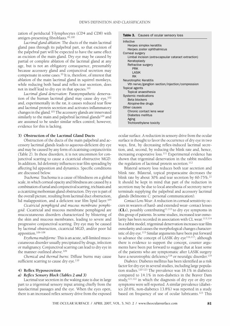
Lacrimal tear secretion in the waking state is due in large part to a trigeminal sensory input arising chiefly from the nasolacrimal passages and the eye. When the eyes open, there is an increased reflex sensory drive from the exposed
ocular surface. A reduction in sensory drive from the ocular surface is thought to favor the occurrence of dry eye in two ways, first, by decreasing reflex-induced lacrimal secre-tion, and, second, by reducing the blink rate and, hence, increasing evaporative loss.111 Experimental evidence has shown that trigeminal denervation in the rabbit modifies the regulation of lacrimal protein secretion.112
Bilateral sensory loss reduces both tear secretion and blink rate. Bilateral, topical proparacaine decreases the blink rate by about 30% and tear secretion by 60-75%.22
It should be kept in mind that part of the reduction in secretion may be due to local anesthesia of secretory nerve terminals supplying the palpebral and accessory lacrimal glands (Belmonte C: personal communication).
Contact Lens W ear: A reduction in corneal sensitivity oc-curs in wearers of hard- and extended wear- contact lenses (CLs), possibly contributing11,113 to dry eye symptoms in this group of patients. In some studies, increased tear osmo-larity has been recorded in association with CL wear.113,114
In a rabbit model, trigeminal denervation increases tear film osmolarity and causes the morphological changes character-istic of dry eye.115 Similar arguments have been put forward to advance the concept of LASIK dry eye116,117; although there is evidence to support the concept, counter argu-ments have been put forward to suggest that at least some of the patients who are symptomatic after LASIK surgery have a neurotrophic deficiency118 or neuralgic disorder.119
Diabetes: Diabetes mellitus has been identified as a risk factor for dry eye in several studies, including large popula-tion studies.120-123 The prevalence was 18.1% in diabetics compared to 14.1% in non-diabetics in the Beaver Dam study,121,122 in which the diagnosis of dry eye or dry eye symptoms were self-reported. A similar prevalence (diabet-ics 20.6%, non-diabetics 13.8%) was reported in a study based on frequency of use of ocular lubricants.123 This
Table 3 . Causes of ocular sensory loss
Infective
Herpes simplex keratitis
Herpes zoster ophthalmicus
Corneal surgery
Limbal incision (extra-capsular cataract extraction)
Keratoplasty
Refractive surgery
PRK
LASIK
RK
Neurotrophic Keratitis
Vth nerve/ganglion section/injection/compression
Topical agents
Topical anaesthesia
Systemic medications
Beta blockers
Atropine-like drugs
Other causes
Chronic contact lens wear
Diabetes mellitus
Aging
Trichlorethylene toxicity
DEWS DEFINITION AND CLASSIFICATION

THE OCULAR SURFACE / APRIL 2007, VOL. 5, NO. 2 / www.theocularsurface.com82 82
study also noted an associa-tion between poor glycemic control (as indicated by serum HbA1C) and fre-quency of drop use. Goeb-bels124 found a reduction in reflex tearing (Schirmer test) in insulin-dependent diabetics, but no differ-ence in tear film breakup time or basal tear flow by fluorophotometry.
It has been suggested that the association may be due to diabetic sensory or autonomic neuropathy, or to the occurrence of microvas-cular changes in the lacrimal gland.123
Neurotrophic keratitis: Ex-tensive sensory denervation of the anterior segment, involv-ing the cornea and the bulbar and palpebral conjunctiva, as a component of herpes zoster ophthalmicus or induced by trigeminal nerve section, injection, or compression or toxicity, can lead to neuro-trophic keratitis. This condi-tion is characterized by fea-tures of dry eye, such as tear instability, diffuse punctate keratitis, and goblet cell loss, and also, most importantly, the occurrence of an indolent or ulcerative keratitis, which may lead to perforation.115,125
The sensory loss results in a reduction of lacrimal se-cretion126 and a reduction in blink rate. In addition, it is envisaged that there is a loss of trophic support to the ocular surface125 after sensory denervation, due to a deficient release of substance-P or expression of nerve growth factor.127-131
b) Reflex Motor BlockCentral damage to the VII cranial nerve, involving the
nervus intermedius, leads to dry eye due to loss of lacrimal secretomotor function. The nervus intermedius carries postganglionic, parasympathetic nerve fibers (of pterygo-palatine ganglion origin) to the lacrimal gland. Dry eye is due to lacrimal hyposecretion in addition to incomplete lid closure (lagophthalmos). Multiple neuromatosis has also been reported as a cause of dry eye.132
An association between systemic drug use and dry eye has been noted in several studies, with decreased lacrimal secretion being the likely mechanism. Responsible agents include: antihistamines, beta blockers, antispasmodics, and diuretics, and, with less certainty, tricyclic antidepressants,
selective serotonin reuptake inhibitors, and other psycho-tropic drugs.122 Additional associations with drying medica-tions were reported by Schein et al, unrelated to the disease for which they were used.133 Use of ACE (angiotensin converting enzyme) inhibitors was associated with a lower incidence of dry eye, and no relationship was found with calcium channel blockers or cholesterol-lowering drugs.122
2. Evaporative Dry EyeEvaporative dry eye is due to excessive water loss from
the exposed ocular surface in the presence of normal lac-rimal secretory function. Its causes have been described as intrinsic, where they are due to intrinsic disease affecting lid structures or dynamics, or extrinsic, where ocular surface disease occurs due to some extrinsic exposure. The bound-ary between these two categories is inevitably blurred.
a. Intrinsic Causes1) Meibomian Gland Dysfunction
Meibomian gland dysfunction, or posterior blepharitis, is a condition of meibomian gland obstruction and is the
Table 4 . Meibomian gland diseases causing evaporative dry eye
Category Disease References
Reduced number Congenital deficiency
Acquired—MGD Bron et al137
Replacement Dystichiasis Bron et al137
Dystichiasis lymphedema syndrome Brooks et al138
Kiederman et al139
Metaplasia
Meibomian Gland Dysfunction
Hypersecretory Meibomian seborrhoea Gifford140
Cowper141
Hyposecretory MGD Retinoid therapy Mathers et al142
Obstructive MGD Primary or secondary Bron et al143
Focal or diffuse Bron et al143
Simple or cicatricial Foulks and Bron134
Atrophic or inflammatory—
note association with dermatoses Pflugfelder et al144
Simple MGD: Primary, or Secondary to:
Local disease Anterior blepharitis
Systemic disease Acne rosacea; seborrhoeic dermatitis; McCulley Dougherty145
atopy; icthyosis; psoriasis; McCulley146
Syndromes Anhydrotic ectodermal dysplasia; Baum et al147
ectrodactyly syndrome; Turner syndrome Mondino et al148
Systemic toxicity 13-cis retinoic acid Mathers et al142
Lambert and Smith149,150
Polychlorinated biphenyls Ikui151
Ohnishi et al152,153
Epinephrine (rabbit) Jester et al154
Cicatricial MGD: Primary, or Secondary to:
Local disease Chemical burns; trachoma; pemphigoid;
erythema multiforme; acne rosacea;
VKC and AKC
DEWS DEFINITION AND CLASSIFICATION

THE OCULAR SURFACE / APRIL 2007, VOL. 5, NO. 2 / www.theocularsurface.com 83 83
most common cause of evaporative dry eye.134-136 Its multi-ple causes and associations are listed in Table 4 and include dermatoses, such as acne rosacea, seborrhoeic dermatitis, and atopic dermatitis. Less common but important associa-tions include the treatment of acne vulgaris with isotretin-oin, which leads to a reversible meibomian gland atrophy, loss of acinar density on meibography, and reduced volume and increased viscosity of expressed excreta.142 Additionally, exposure to polychlorinated biphenyls, through ingestion of contaminated cooking oils, causes a chronic disorder with gross and extensive acneiform skin changes, meibomian seborrhoea with thick excreta and glandular cyst forma-tion. Other organs are affected.152,153,155 Meibomian duct keratinization occurs in the experimental model.149,150
MGD can be primary or secondary, simple or cicatricial. In simple MGD, the gland orifices remain located in the skin of the lid, anterior to the mucocutaneous junction. In cicatricial MGD, the duct orifices are drawn posteriorly onto the lid and tarsal mucosa and, hence, are unable to deliver oil to the surface of the tear film. Diagnosis is based on morphologic features of the gland acini and duct orifices, presence of orifice plugging, and thickening or absence of expressed excreta. Methods exist to grade the degree of MGD,143 measure the degree of gland dropout (meibogra-phy),156,157 and the amount of oil in the lid margin reservoir (meibometry).65,158 Evidence from several sources suggests that MGD of sufficient extent and degree is associated with a deficient tear film lipid layer, an increase in tear evapora-tion, and the occurrence of an evaporative dry eye.
It is important to recognize the effect of lid commensal organisms on meibomian lipid composition and its poten-tial effect on tear film lipid layer stability. Shine and McCul-ley have shown that constitutional differences in meibomian lipid composition exist in different individuals.159,160 They identified one group of subjects with low levels of choles-terol esters and esters of unsaturated fatty acids (ie, the ”normal-cholesterol absent” group: N[CA]), and another group with high levels of these fractions (”normal-choles-terol present”’ group: N[CP]). In the latter group, esterases and lipases produced by normal lid commensals (coagulase-negative staphylococci [CoNS], Propionobacterium acnes and S aureus) can release fatty acids and mono- and diglycerides into the tear film, which may be a source of irritation or of soap formation, said to be responsible for producing ”meibomian foam.”161 It should also be noted that S. aureusgrowth can be stimulated by the presence of cholesterol and that, in a study by Shine and McCulley, there were twice as many staphylococcal strains on the lid margins of those normal subjects whose meibomian lipid was cholesterol-rich, than in the cholesterol-poor group.160 Factors such as these may influence the microbial load and type on normal lid margins and influence the development of blepharitis.
2) Disorders of Lid Aperture and Lid/Globe Congruity or DynamicAn increase in the exposed evaporative surface of the
eye occurs in craniostenosis, endocrine and other forms of
proptosis, and in high myopia. Endocrine exophthalmos and, specifically, increased palpebral fissure width, is as-sociated with ocular drying and tear hyperosmolarity.162
Increasing palpebral fissure width correlates with increased tear film evaporation.61 Increased ocular surface exposure also occurs in particular gaze positions, such as upgaze,163
and in activities that induce upgaze, such as playing pool, where, while aiming, the head is inclined downward and the eyes are in extreme upgaze.
Drying of the ocular surface due to poor lid apposition or to lid deformity, leading to exposure or poor tear film re-surfacing, are accepted causes of ocular surface drying, but they have received little formal study.164 Dry eye problems may be caused by problems of lid congruity after plastic surgery of the lids.165
3) Low Blink RateDrying of the ocular surface may be caused by a reduced
blink rate, which lengthens the period during which the ocular surface is exposed to water loss before the next blink.166 Methods have been developed to record the blink rate and to relate this to the development of dry eye.163 This may occur as a physiological phenomenon during perfor-mance of certain tasks of concentration, eg, working at video terminals167 or microscopes, or it may be a feature of an extrapyramidal disorder, such as Parkinson disease (PD).
The reduced blink rate in PD is due to a decrease in the dopaminergic neuron pool of the substantia nigra and is proportional to disease severity.168 Reduced blink rate is regarded by some authors as the basis of dry eye in PD.169
Biousse et al found blink rate and tear film breakup time (TFBUT) to be significantly reduced in untreated, early-onset PD patients with a significantly increased frequency of dry eye symptoms, whereas the Schirmer test and rose bengal staining measurements were no different in PD pa-tients than in controls.170 However, other authors report a reduced lacrimal secretion in PD,171-173 and abnormalities of tear film stability, fluorescein and rose bengal staining, tear meniscus height, and meibomian gland function.173
Tamer et al reported dry eye symptoms in 87.5% of PD patients versus 20.6% of age-matched controls, with a mean total number of abnormal dry eye tests of 3.10 1.8in PD, versus 0.35 0.9 in controls. (P < 0.001). Each test was significantly abnormal in PD patients versus controls, and all the tear tests (except meibomian gland function and meniscus height) showed a significant correlation with a PD severity index. The overall number of abnormal tests in PD patients was inversely related to the blink rate.
On the basis of these findings, Tamer et al postulated several mechanisms by which PD may induce dry eye. 1) Reduced blink rate and impaired meibomian oil delivery to the tear film can increase evaporative loss. They also suggest that a reduced blink rate could impair the clear-ance of lipid-contaminated mucin.174 2) Experimentally, androgens are required for the normal functioning of both the lacrimal175,176 and meibomian glands, 177,178 and there is clinical evidence that dry eye symptoms are promoted by
DEWS DEFINITION AND CLASSIFICATION

THE OCULAR SURFACE / APRIL 2007, VOL. 5, NO. 2 / www.theocularsurface.com84 84
blockade of androgen receptors.43 The levels of circulating androgens are low in a large proportion of PD patients,179
and it is suggested that this may contribute to lacrimal and meibomian dysfunction. 3) In addition, decreased reflex tearing in PD has been attributed to autonomic dysfunction, reflecting the presence of Lewy bodies in the substantia nigra, sympathetic and peripheral parasympathetic gan-glia.180 Magalhaes et al found evidence of autonomic failure in about a third of patients with PD.
In conclusion, it is possible that dry eye disease in PD has multiple causes.
b. Extrinsic Causes1) Ocular Surface Disorders
Disease of the exposed ocular surface may lead to imperfect surface wetting, early tear film breakup, tear hyperosmolarity, and dry eye. Causes include vitamin A deficiency and the effects of chronically applied topical anesthetics and preservatives.
Vitamin A Deficiency: Vitamin A deficiency may cause dry eye (xerophthalmia) by two distinct mechanisms.Vitamin A is essential for the development of goblet cells in mucous membranes and the expression of glycocalyx mucins.181,182 These are deficient in xerophthalmia, lead-ing to an unstable tear film characterized by early tear film break up. Vitamin A deficiency can cause lacrimal acinar damage, and, therefore, some patients with xerophthalmia may have a lacrimal, aqueous tear-deficient dry eye.183
Topical Drugs and Preservatives: Many components of eye drop formulations can induce a toxic response from the ocular surface. Of these, the most common offenders are preservatives, such as benzalkonium chloride (BAC),which cause surface epithelial cell damage and punctate epithelial keratitis, which interferes with surface wettability. Use of preserved drops is an important cause of dry eye signs and symptoms in glaucoma patients, and it is usually reversible on switching to nonpreserved preparations.184
Therefore, frequent applications of preserved artificial tear preparations should be avoided.
Topical anesthesia causes drying in two ways. It re-duces lacrimal secretion by reducing sensory drive to the lacrimal gland and also reduces the blink rate. It has also been suggested that anesthesia of those lacrimal secretory nerve terminals close to the surface of the upper fornix (innervating the palpebral and accessory portions of the lacrimal gland) may also be blocked by topical anaesthetics (Belmonte C: personal communication).
Chronic use of topical anesthetics can cause a neuro-trophic keratitis leading to corneal perforation.185,186
2) Contact Lens W earContact lens wear is prevalent in the developed world,
with 35 million wearers cited in the USA in the year 2000.187 The causes of CL-related symptoms and of lens intolerance are, therefore, of personal and general economic importance. The primary reasons for CL intolerance are discomfort and dryness.188,189 In recent years, a number
of questionnaires have been developed to identify dry eye symptoms in CL wearers.45,190-192 Use of such question-naires has indicated that about 50% of CL wearers report dry eye symptoms.191-194 CL wearers are 12 times more likely than emmetropes and five times more likely than spectacle-wearers to report dry eye symptoms.195
In a large cross-sectional study of CL wearers (91% hy-drogel and 9% gas permeable lenses), several factors were found to be associated with dry eye diagnosed using the Contact Lens Dry Eye Questionnaire (CLDEQ). Pre-lens tear film (PLTF) thinning time was most strongly associated with dry eye (dry eye: 8.23 5.67 seconds; non-dry eye: 11.03 8.63 seconds. [P = 0.0006]), followed by nominal CL water content and refractive index.114
The pre-lens lipid layer thickness was less in dry eye subjects and correlated well with the pre-lens tear film thin-ning time. This, together with poor lens wettability, could be a basis for a higher evaporative loss during lens wear and was attributed to potential changes in tear film lipid compo-sition, rather than to a loss of meibomian gland oil delivery.
Patients wearing high water-content hydrogel lenses were more likely to report dry eye. This is a controversial area in the literature. In a study of the effects of five hydrogel lenses on tear film physiology, Thai et al found that all the examined soft CL materials increased the evaporation rate and decreased the tear film thinning time.196 The surface wetting ability of the CL materials was the same, regardless of special surface lens treatments. Efron et al found that patients wearing low water CLs, which maintained their hydration, were free from symptoms.197 However, other studies reported no correla-tion between CL hydration and dry eye symptoms189 and no relationship between lens hydration and tear film thinning time and dry eye symptoms198 or evaporative water loss.199
Dry eye was associated with a higher tear osmolarity, but not in the range normally associated with dry eye tear hyperos-molarity. The authors commented that this lower value might have been caused by reflex tearing at the time of sampling.114
Women were found to report dry eye more frequently than men, with 40% of the menand 62% of the women clas-sified as having dry eye (P < 0.0001).114 The reasons for this were not explored, but potential contributing factors were considered to be hormone fluctuations during the menstrual cycle or after menopause and use of oral contraceptives or hormone replacement therapy. It was also noted that symp-tom reporting by women, in general, tends to be higher than that by men.200 Some studies show no effect of oral contra-ceptives or hormone levels on a range of tear parameters.201
Glasson et al202 showed that intolerance to hydrogel lenses in normals correlates with a shorter blink interval, noninvasive TFBUT and phenol red thread test length and a lower tear meniscus height and area; this has had predictive power in people presenting for CL fitting. A formula linking symptoms (using the McMonnies Dry Eye Questionnaire), non-invasive tear break up time (NITFBUT), and tear me-niscus height predicted potential intolerant subjects with a sensitivity of 100%, specificity of 57%, and accuracy of 78%. Intolerance was also associated with an increase in degraded
DEWS DEFINITION AND CLASSIFICATION
THE OCULAR SURFACE / APRIL 2007, VOL. 5, NO. 2 / www.theocularsurface.com 85 85
lipid products, phospholipase A2, and lipocalin in tear sam-ples.203 These studies suggest that features compatible with a dry eye state may predispose an individual to CL intolerance.
The variations in visual performance with soft CLs may be due to light scattering produced by changes in the hydration levels of the lens or changes in the tear film over the lens.204,205
Decreases in retinal image quality have been inferred from the modulation transfer function induced by the drying tear film and observed with the Schack-Hartman aberrometer.206
Contrast sensitivity in soft CL wearers is significantly re-duced in the middle-to-high spatial frequencies, when the precorneal lens tear film dries and causing breakup. This
DEWS DEFINITION AND CLASSIFICATION
Table 4
CORE
mechanisms
Systemic drugs
inhibit flow
Refractive Surgery
CL wear
Topical anesthesia
Activate
epithelial
MAPK +
NF B +
Tear
Film
Instability
High
Evaporation
Low
Lacrimal
Flow
Goblet cell,
glycocalyx mucin loss
epithelial damage- apoptosis
Inflammatory
lacrimal damageSSDE; NSDE;
Lacrimal
obstruction
EnvironmentHigh Air Speed
Low Humidity
Blepharitis
Lid flora
lipases esterases
detergents
nerve
injury
Hyperosmolarity
Tear
Deficient or
unstable TF
lipid Layer
MGD Xerophthalmia
Ocular allergy
Preservatives
CL wear?
Low androgens
Aging
IL-1+
TNF +
MMPs
– –
–Lacrimal
Gland
– –
–
– –
–
++
+
initial lacrimal stimulation
increased
reflex drive
nerve
stimulation
neurogenic
inflammation
neurosecretoryblock
Reflex
block
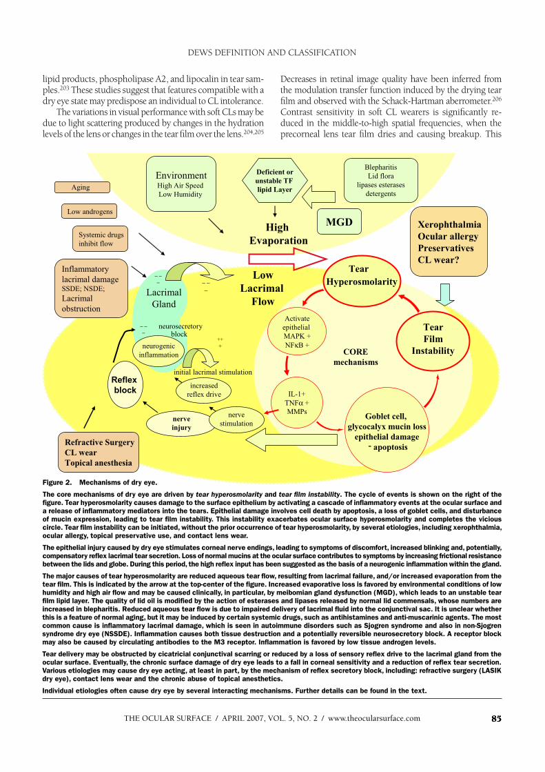
Figure 2. Mechanisms of dry eye.
The core mechanisms of dry eye are driven by tear hyperosmolarity and tear film instability. The cycle of events is shown on the right of the
figure. Tear hyperosmolarity causes damage to the surface epithelium by activating a cascade of inflammatory events at the ocular surface and
a release of inflammatory mediators into the tears. Epithelial damage involves cell death by apoptosis, a loss of goblet cells, and disturbance
of mucin expression, leading to tear film instability. This instability exacerbates ocular surface hyperosmolarity and completes the vicious
circle. Tear film instability can be initiated, without the prior occurrence of tear hyperosmolarity, by several etiologies, including xerophthalmia,
ocular allergy, topical preservative use, and contact lens wear.
The epithelial injury caused by dry eye stimulates corneal nerve endings, leading to symptoms of discomfort, increased blinking and, potentially,
compensatory reflex lacrimal tear secretion. Loss of normal mucins at the ocular surface contributes to symptoms by increasing frictional resistance
between the lids and globe. During this period, the high reflex input has been suggested as the basis of a neurogenic inflammation within the gland.
The major causes of tear hyperosmolarity are reduced aqueous tear flow, resulting from lacrimal failure, and/or increased evaporation from the
tear film. This is indicated by the arrow at the top-center of the figure. Increased evaporative loss is favored by environmental conditions of low
humidity and high air flow and may be caused clinically, in particular, by meibomian gland dysfunction (MGD), which leads to an unstable tear
film lipid layer. The quality of lid oil is modified by the action of esterases and lipases released by normal lid commensals, whose numbers are
increased in blepharitis. Reduced aqueous tear flow is due to impaired delivery of lacrimal fluid into the conjunctival sac. It is unclear whether
this is a feature of normal aging, but it may be induced by certain systemic drugs, such as antihistamines and anti-muscarinic agents. The most
common cause is inflammatory lacrimal damage, which is seen in autoimmune disorders such as Sjogren syndrome and also in non-Sjogren
syndrome dry eye (NSSDE). Inflammation causes both tissue destruction and a potentially reversible neurosecretory block. A receptor block
may also be caused by circulating antibodies to the M3 receptor. Inflammation is favored by low tissue androgen levels.
Tear delivery may be obstructed by cicatricial conjunctival scarring or reduced by a loss of sensory reflex drive to the lacrimal gland from the
ocular surface. Eventually, the chronic surface damage of dry eye leads to a fall in corneal sensitivity and a reduction of reflex tear secretion.
Various etiologies may cause dry eye acting, at least in part, by the mechanism of reflex secretory block, including: refractive surgery (LASIK
dry eye), contact lens wear and the chronic abuse of topical anesthetics.
Individual etiologies often cause dry eye by several interacting mechanisms. Further details can be found in the text.
THE OCULAR SURF ACE / APRIL 2007, VOL. 5, NO. 2 / www.theocularsurface.com86
could account for complaints of intermittent blurred vision in some CL wearers and may provide a stimulus to blink.207
3) Ocular Surface DiseaseThere is evidence that various forms of chronic ocular sur-
face disease result in destabilization of the tear film and add a dry eye component to the ocular surface disease. Allergic eye disease offers a well-studied example.208 Also, any form of dry eye, whatever its origins, may cause at least a loss of goblet cell numbers, so that an ocular surface element is added.209
4) Allergic ConjunctivitisAllergic conjunctivitis takes several forms, which in-
clude seasonal allergic conjunctivitis, vernal keratoconjunc-tivitis, and atopic keratoconjunctivitis. The general mecha-nism leading to disease is that exposure to antigen leads to degranulation of IgE-primed mast cells, with the release of inflammatory cytokines. A Th2 response is activated at the ocular surface, initially in the conjunctival and, later, in the corneal epithelium, subsequently leading to submucosal changes. There is stimulation of goblet cell secretion and loss of surface membrane mucins.210 Surface epithelial cell death occurs, affecting conjunctival and corneal epithelium (punctate keratoconjunctivitis). Surface damage and the release of inflammatory mediators leads to allergic symp-toms and to reflex stimulation of the normal lacrimal gland.
Surface irregularities on the cornea (punctate epithelial keratitis and shield ulcer) and conjunctiva can lead to tear film instability and, hence, to a local drying component to the allergic eye disease. In chronic disease, there may be meibomian gland dysfunction, which could exacerbate surface drying by interfering with the tear film lipid layer. Lid swelling, eg, in vernal catarrh and atopic keratocon-junctivitis, can interfere with lid apposition and tear film spreading, thus exacerbating the dry eye.
Ocular allergy was noted to be a risk factor for dry eye in the Beaver Dam study, although the concomitant use of sys-temic medications, such as antihistamines, was recognized as a potential contributor.122 Factors leading to a dry eye state in allergic eye disease are discussed by Fujishima et al.211
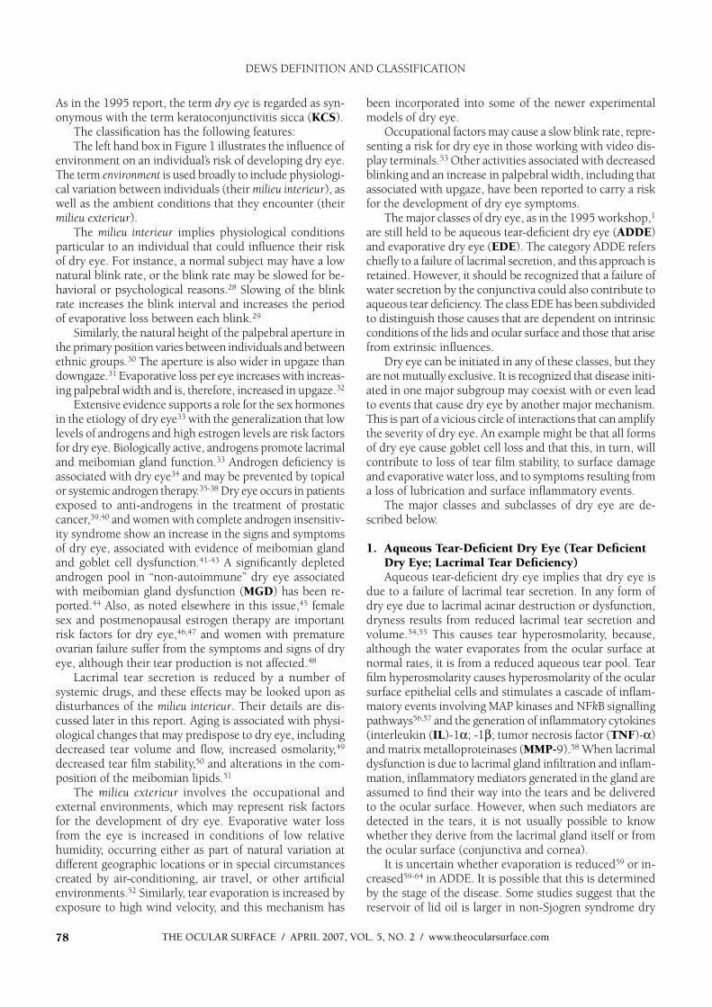
C. The Causative Mechanisms of Dry Eye
From the above discussion, it can be seen that certain core mechanisms are envisaged at the center of the dry eye process that can initiate, amplify, and potentially change the character of dry eye over time. These are tear hyperosmolar-ity and tear film instability. This section is intended to show how the several subclasses of dry eye activate these core mechanisms and explain the features of various forms of dry eye. The interactions of various etiologies with these core mechanisms are summarized in Figure 2.
It should be noted that an attractive mechanistic schema for dry eye has been presented in detail by Baudouin.212 In this concept, two levels of involvement are identified. The first level includes the known risk factors or causes of dry eye that ultimately lead to a series of secondary biological cascades, resulting in breakdown of the tear film and ocular
surface. This pathbreaking conceptual approach describes the relationship of early disparate events to biological re-sponses common to all forms of dry eye, many of which are mutually reinforcing. This leads to a vicious circle or loop. It is thought that early therapeutic intervention may disrupt this loop. The schema in Figure 2, developed from the discussion of our Subcommittee, emphasizes the core biological mechanisms described in this text.
1. Tear HyperosmolarityTear hyperosmolarity is regarded as the central mecha-
nism causing ocular surface inflammation, damage, and symptoms, and the initiation of compensatory events in dry eye. Tear hyperosmolarity arises as a result of water evaporation from the exposed ocular surface, in situations of a low aqueous tear flow, or as a result of excessive evapo-ration, or a combination of these events. Nichols et al have demonstrated the wide variation of tear film thinning rates in normal subjects, and it is reasonable to conclude that, for a given initial film thickness, subjects with the fastest thin-ning rates would experience a greater tear film osmolarity than those with the slowest rates.114 R apid thinning may be hypothesized as a risk factor for tear hyperosmolarity.
Since the lacrimal fluid is secreted as a slightly hypoton-ic fluid, it will always be expected that tear osmolarity will be higher in the tear film than in other tear compartments. There are also reasons to believe that osmolarity is higher in the tear film itself than in the neighboring menisci. One reason for this is that the ratio of area to volume (which determines the relative concentrating effect of evaporation) is higher in the film than the menisci.213
Hyperosmolarity stimulates a cascade of inflammatory events in the epithelial surface cells, involving MAP kinases and NFkB signalling pathways56 and the generation of inflammatory cytokines (IL-1 ; -1 ; TNF- ) and MMPs (MMP9),58 which arise from or activate inflammatory cells at the ocular surface.214 These concepts are supported by studies of desiccating stress in the experimental model,215
which have demonstrated the evolution of inflammatory cytokine release and MMP activation.57 There is evidence that these inflammatory events lead to apoptotic death of surface epithelial cells, including goblet cells216; thus, goblet cell loss may be seen to be directly related to the effects of chronic inflammation.217,218 Goblet cell loss is a feature of every form of dry eye, and consistent with this is the dem-onstration of reduced levels of the gel mucin MUC5AC in dry eye.219,220 With the evolution of dry eye, other factors are likely to amplify these initiating inflammatory events, and the contribution of direct autoimmune targeting of the ocular surface cannot be excluded.
In the initial stages of dry eye, it is considered that ocular surface damage caused by osmotic, inflammatory or me-chanical stresses (loss of surface lubrication) results in reflex stimulation of the lacrimal gland. R eflex trigeminal activity is thought to be responsible for an increased blink rate and a compensatory response, increased lacrimal secretion. In the case of lacrimal gland insufficiency (SSDE or NSSDE), the
DEWS DEFINITION AND CLASSIFICATION

THE OCULAR SURFACE / APRIL 2007, VOL. 5, NO. 2 / www.theocularsurface.com 87
reflex secretory response will be insufficient to fully compen-sate for the tear film hyperosmolarity, and in the steady state, this form of dry eye will be characterized by a hyperosmolar-ity state with low tear volume and flow. In evaporative dry eye (eg, caused by MGD), it can be hypothesized that, since the lacrimal gland is initially healthy in this situation, lacrimal secretory compensation is at first able to compensate for tear film hyperosmolarity. Ultimately it would be expected that in the steady state, dry eye would be a condition of hyper-osmolarity with a tear volume and flow greater than normal. This possibility of a high volume dry eye is supported by the increased tear secretion (based on the Schirmer I test) in patients with MGD compared to normals,221 although this evidence requires support by studies using more so-phisticated tests of tear flow. In the study of Shimazaki et al, despite the increased tear flow, particularly in the gland dropout group, there was a shorter TFBUT and greater degree of dye staining in those with MGD than in those without.
Excessive reflex stimulation of the lacrimal gland experimentally may induce a neurogenic inflammatory cy-tokine response within the gland, leading to the sequence of glandular autoantigen expression, T-cell targeting, and the release of inflammatory mediators into the tears.20,222
It has also been considered to induce a state of ”lacrimal exhaustion” due to excessive reflex stimulation of the lac-rimal gland.223,224 However, these provocative hypotheses await experimental support.
Knowledge is insufficient regarding the natural history of different forms of dry eye in relation to ocular surface sensitivity. Most reports,144,225,226 but not all,119 suggest that corneal sensitivity is impaired in chronic dry eye disease, suggesting that an initial period of increased reflex sensory activity is followed by a chronic period of reduced sensory input. This is likely to be the result of the longterm effects of inflammatory mediators on sensory nerve terminals supply-ing the ocular surface, and there is evidence of morphologi-cal changes in the sub-basal nerve plexus.227 At this stage of dry eye, the reflex sensory drive to lacrimal secretion becomes reduced, which would reverse any compensatory drive to lacrimal secretion that is postulated for the earlier phase of the disease. This would be expected to reduce the lacrimal secretory response, regardless of the etiology of the dry eye, and would therefore exacerbate both ADDE and EDE by reinforcing the low volume state in ADDE and converting a potentially high volume state in MGD-based EDE to a normal or low volume state due to an added lac-rimal deficiency. The sensory drive to the blink reflex might be expected to be similarly affected, although there is no evidence to this effect and this area requires further study.
The above proposal may explain why a clear clinical separation between ADDE and EDE may at times be difficult to support on the basis of substantive tests. Thus, while there are studies that indicate, as expected, that tear ev aporationrate is increased in MGD,62,63,82,83,221,228 or where there is an incomplete or absent tear film lipid layer229 in some groups of MGD, evaporation rate may be normal.221 Similarly, an increased evaporation rate has been reported by some authors
in ADDE,59-63and a decreased rate by others.59 Again, whereas a reduction in tear flow is the hallmark of ADDE,63,83,124 a reduction in flow has also been reported with MGD.63,83
These findings appear contradictory, but may simply highlight our ignorance of the natural history of the pri-mary disorders. Thus, there is evidence that spreading of the tear film lipid layer is retarded in severe ADDE, which has been attributed to the effect of the thinned aqueous phase of the tear film. Conversely, as noted earlier, it may be conceived that a loss of corneal sensitivity in EDE could reduce the reflex drive to tear secretion and, hence, result in a combined form of dry eye. These postulated interactions, occurring over time, may explain the overlap of findings in these two disorders and fit in to the general concept of a vicious circle in which widely varying influences combine to cause dry eye with a complex profile.
2. Tear Film InstabilityIn some forms of dry eye, tear film instability may be the
initiating event, unrelated to prior tear hyperosmolarity. 1) While frank tear film instability in the form of early tear
film break up may readily be accepted as a component of dry eye, more subtle degrees of tear film instability may also pre-dispose to dry eye complications in response to ocular surface stress. Thus, Goto et al reported that in a group of patients undergoing LASIK surgery and showing no features of dry eye by standard tests, those who showed tear film instability by the tear film analysis system (TMS) showed a greater decrease in tear film stability and more severe symptoms and dry eye signs, including punctate keratitis, postoperatively.10
2) Where the TFBUT is less than the blink interval, it is implied that tear film breakup in that individual is occurring normally in the waking state. (This state is expressed by the Ocular Protection Index, which is the ratio of the TFBUT divided by the blink interval.230 (See relevant template website [www.tearfilm.org]). When this value is less than 1, then tear film breakup occurs in the waking, open-eye condition. If the TFBUT is greater than the blink interval but less than 10 seconds, then this TFBUT value is still currently regarded as an index of tear film instability. Where tear film instability represents tear film breakup occurring within the blink interval, it is assumed to give rise to local drying and hyperosmolarity of the exposed surface, to surface epithelial damage, and to a disturbance of glycocalyx and goblet cell mucins. The latter consequently exacerbates the tear film instability as part of a vicious circle of events.
Two examples of this clinical sequence, where tear film instability is due to a disturbance of ocular surface mucins, are xerophthalmia231 and allergic eye disease. 211 The initial loss of tear stability in vitamin A deficiency results from a re-duced expression of mucins at the ocular surface and a loss of goblet cells.183,232 In seasonal allergic conjunctivitis or vernal keratoconjunctivitis, a disturbance of mucin expression at the surface of the eye is due, initially, to an IgE-mediated type I hypersensitivity mechanism, leading to the release of inflam-matory mediators in response to allergen challenge.
Other examples include the actions of topical agents, in par-ticular, preservatives such as BAC, which excite the expression of
DEWS DEFINITION AND CLASSIFICATION

THE OCULAR SURFACE / APRIL 2007, VOL. 5, NO. 2 / www.theocularsurface.com88
inflammatory cell markers at the ocular surface, causing epithelial cell damage, cell death by apoptosis, and a decrease in goblet cell density.233 There is both clinical and experimental evidence to support such events.234-238 In a study of patients treated for glau-coma for at least one year, flow cytometry demonstrated a greater expression of inflammatory markers (HLA-DR and ICAM-1) in those receiving preserved drops (BAC) than in normals or those receiving unpreserved drops. Use of preservative was associated with a lower expression of MUC5AC and the lowest MUC5AC levels were associated with the highest ICAM-1 and HLA-DR levels.239 This negative correlation suggested inflammation as a possible basis for the decreased mucin expression, in addition to any direct effect of BAC on goblet cells themselves.
Considering the possible relationship between these find-ings and dry eye, Pisella et al, in an unmasked study of 4107 glaucoma patients, found that the frequency of ocular surface changes was twice as high in those receiving preserved drops than in those receiving unpreserved drops, and the frequency of signs and symptoms was dose-related.184
CL wear may also provide a route of entry into the dry eye mechanism, a route in addition to reduced corneal sensitivity. For a considerable time, CL wear has been recognized to cause changes to the ocular surface epithelia. Knop and Brewitt dem-onstrated surface epithelial metaplasia and a reduced goblet cell density with hydrogel lens wear.240,241 Other studies have shown an increase in goblet cell density evolving over a period of 6 months in subjects wearing polymacon, galyfilcon, and silicone hydrogel lenses.242,243 In another study, no change in goblet cell density was found after 6 months wear of a daily
disposable lens with a 2-week wearing schedule, and further studies suggest that the goblet cell responses may differ be-tween hard and soft CLs.244
A recent study combining impression cytology with flow cytometry demonstrated an increase in inflammatory markers (HLA-DR and ICAM-1) at the ocular surface and a nonsig-nificant trend toward a decrease in the expression of mucin markers (MUC5AC) in patients with a history of chronic CL wear.245 A later study has shown no difference between CL wearers and non-CL wearers in mucin expression (MUC5AC and the carbohydrate epitope H185, a marker for MUC 16) in tears or impression cytology samples.182 In summary, it ap-pears that CL wear may activate proinflammatory markers and stimulate the ocular surface epithelia to a variable degree. It is not yet possible to say whether these changes alone predispose individuals to the occurrence of dry eye with CL wear.
D. The Basis for Symptoms in Dry Eye
The basis for symptoms in dry eye is not truly known but may be surmised from a consideration of the etiologies, mechanisms, and responses of dry eye to therapy.246 The oc-currence of symptoms implies the activation of sensory nerves subserving nociception at the ocular surface.247,248 Candidates include tear and ocular surface hyperosmolarity – including tear film break-up in the interblink, shear-stress between the lids and globe in response to reduced tear volume, and/or the reduced expression of mucins at the ocular surface, the pres-ence of inflammatory mediators at the surface of the eye, and, finally, hypersensitivity of the nociceptive sensory nerves.
Table 5 . Dry eye severity grading scheme
Dry Eye Severity
Level 1 2 3 4 *
Discomfort, severity
& freq u ency
M ild and/or ep isodic;
occu rs u nder
environmental stress
M oderate ep isodic or
chronic, stress or no
stress
Severe freq u ent or
constant w ithou t
stress
Severe and/or
disab ling and constant
V isu al symp tomsN one or ep isodic mild
fatigu e
A nnoying and/or
activity-limiting
ep isodic
A nnoying, chronic
and/or constant,
limiting activity
C onstant and/or
p ossib ly disab ling
C onju nctival injection N one to mild N one to mild +/– +/++
C onju nctival staining N one to mild V ariab le M oderate to mark ed M ark ed
C orneal staining
(severity/location)N one to mild V ariab le M ark ed central
Severe p u nctate
erosions
C orneal/tear signs N one to mild M ild deb ris, meniscu s
F ilamentary k eratitis,
mu cu s clu mp ing,
tear deb ris
F ilamentary k eratitis,
mu cu s clu mp ing,
tear deb ris, u lceration
L id/meib omian glands M G D variab ly p resent M G D variab ly p resent F req u entTrichiasis, k eratinization,
symb lep haron
T F B U T (sec) V ariab le 1 0 5 Immediate
Schirmer score
(mm/5 min)V ariab le 1 0 5 2
* M u st have signs A N D symp toms. T B U T : fl u orescein tear b reak -u p time. M G D: meib omian gland disease
R ep rinted w ith p ermission from B ehrens A , Doyle JJ, Stern L , et al. Dysfu nctional tear syndrome. A Delp hi ap p roach to treatment recommendations.
Cornea 2006 ;25:9 0-7
DEWS DEFINITION AND CLASSIFICATION
THE OCULAR SURFACE / APRIL 2007, VOL. 5, NO. 2 / www.theocularsurface.com 89
E. Classification of Dry Eye on the Basis of Severity
The Subcommittee considered that there was consider-able clinical utility to adopting a classification of disease based on severity. The basic scheme of the Delphi Panel Report was adopted and modified to produce the third component of the recommendation (Table 5).
REFERENCES1. Lemp MA. Rep ort of the National Eye Institute/Industry W orkshop on
Clinical Trials in Dry Eye. CLAO J 1995;21:221-322. B egley CG, Chalmers RL, Ab etz L, et al. The relationship b etween hab itual
p atient-rep orted symp toms and clinical signs among p atients with dry eye of v arying sev erity. Inv est Op h th alm o l V is S ci 2003;44:4753-61
3. Adatia FA, Michaeli-Cohen A, Naor J, et al. Correlation b etween corneal sensitiv ity, sub jectiv e dry eye symp toms and corneal staining in Sjogren’s syndrome. Can J Op h th alm o l 2004;39:767-71
4. Vitale S, Goodman LA, Reed GF, Smith JA. Comp arison of the NEI-VFQand OSDI q uestionnaires in p atients with Sjogren’s syndrome-related dry eye. H ealth Q u al Ou tco m es 20041;2:44
5. Rieger G. The imp ortance of the p recorneal tear fi lm for the q uality of op tical imaging. B r J Op h th alm o l 1992;76:157-8
6. Liu Z , Pfl ugfelder SC. Corneal surface irregularity and the effect of artifi cial tears in aq ueous tear defi ciency. Op h th alm o lo g y 1999;106:936-43
7. Goto E, Y agi Y , Matsumoto Y , Tsub ota K. Imp aired functional v isual acuity of dry eye p atients. Am J Op h th alm o l 2002;133:181-6
8. Holly F, Lemp MA. Formation and rup ture of the tear fi lm. E x p E ye R es1973;15:515-25
9. B ron AJ. Diagnosis of dry eye. S u r v Op h th alm o l 2001;45 Sup p l 2:S221-610. Goto T, Z heng X , Klyce SD, et al. A new method for tear fi lm stab ility
using v ideokeratograp hy. Am J Op h th alm o l 2003;135:607-1211. Farris RL, Stuchell RN, Mandel ID Tear osmolarity v ariation in the dry
eye. T r ans Am Op h th alm o l S o c 1986;84:250-6812. Gilb ard JP. Human tear fi lm electrolyte concentrations in health and dry-
eye disease. Int Op h th alm o l Clin 1994 W inter;34:27-3613. Murub e J. Tear osmolarity. Ocu l S u r f 2006;1;62-7314. Tomlinson A, Khanal S, Ramaesh K, et al. Tear fi lm osmolarity: determination
of a referent for dry eye diagnosis. Inv est Op h th alm o l V is S ci 2006;47:4309-1515. Pfl ugfelder SC, Jones D, Ji Z , et al. Altered cytokine b alance in the tear fl uid
and conjunctiv a of p atients with Sjogren’s syndrome keratoconjunctiv itis sicca. Cu r r E ye R es 1999;19:201-11
16. Tsub ota K, Fujihara T, Saito K, Takeuchi T. Conjunctiv al ep ithelium ex-p ression of HLA-DR in dry eye p atients. Op h th alm o lo g ica 1999;213:16-9
17. Stern ME, B euerman RW , Fox RI, et al. The p athology of dry eye; the interac-tion b etween the ocular surface and lacrimal glands. Co rnea 1998;17:584-9
18. Pfl ugfelder SC, Solomon A, Stern ME. The diagnosis and management of dry eye:a twenty-fi v e year rev iew. Co rnea 2000;19:644-9
19. B euerman RW , Mircheff A, Pfl ugfelder SC, Stern ME. The lacrimal func-tional unit, in Pfl ugfelder SC, B euerman RW , Stern ME (eds). D ry eye and o cu lar su r face diso rder s. New Y ork, Marcel Dekker, 2004
20. Stern ME, Gao J, Siemarko KF, et al. The role of the lacrimal functional unit in the p athop hysiology of dry eye. E x p E ye R es 2004;78:409-6
21. (No authors listed). Research in dry eye: Rep ort of the Research Sub com-mittee of the 2007 Dry Eye W orkShop (DEW S). Ocu l S u r f 2007;5:179-193
22. Jordan A, B aum J. B asic tear fl ow. Does it exist? Op h th alm o lo g y 1980;87:920.23. Gup ta A, Heigle T, Pfl ugfelder SC. Nasolacrimal stimulation of aq ueous
tear p roduction. Co rnea 1997;16:645-824. Vitali C. Classifi cation criteria for Sjogren’s syndrome. Ann R h eu m D is
2003;62:94-525. Murub e J, B enitez del Castillo JM, Chenghou L, et al. The Madrid trip le
classifi cation system. Arch S o c E sp Oftalm o l 2003;78;587-9326. Murub e J, Nemeth J, Hoh H, et al. The trip le classifi cation of dry eye for
p ractical clinical use. E u r J Op h th alm o l 2005;15:660-727. B ehrens A, Doyle JJ, Stern L, et al. Dysfunctional tear syndrome: A Delp hi
ap p roach to treatment recommendations. Co rnea 2006;25:900-728. Doughty MJ, Naase T. Further analysis of the human sp ontaneous eye b link
rate b y a cluster analysis-b ased ap p roach to categoriz e indiv iduals with ‘normal’ v ersus ‘freq uent’ eye b link activ ity. E ye Co ntact Lens 2006;32:294-9
29. Nakamori K, Odawara M, Nakajima T, et al. B linking is controlled p rimar-ily b y ocular surface conditions. Am J Op h th alm o l 1997;124:24-30
30. Mangub at L, Luague S. Normal measurements of the p alp eb ral fi ssure and the interp alp eb ral distance among Filip inos. P h ilip p J S u rg S u rg S p ec 1966;21:304-6
31. Stoller SH, Meyer DR. Quantitating the change in up p er lid p osition during
downgaz e. Op h th alm o lo g y 1994;101:1604-732. Cho P, Sheng C, Chan C, et al. B aseline b link rates and the effect of v isual
task diffi culty and p osition of gaz e. Cu r r E ye R es 2000;20: 64-7033. Sulliv an DA. Sex and sex steroid infl uences on the dry eye syndrome, in
Pfl ugfelder SC, B euerman RW , Stern ME (eds). D ry eye and o cu lar su r face diso rder s. New Y ork, Marcel Dekker, 2004
34. Sulliv an DA. Androgen defi ciency and dry eye syndromes. Arch S o c E sp ano la Oftalm o lo g ia 2004;79:49-50
35. Sulliv an DA. Tearful relationship s? Sex, hormones and aq ueous-defi cient dry eye. Ocu l S u r f 2004;2:92-123
36. W orda C, Nep p J, Hub er JC, Sator MO. Treatment of keratoconjunctiv itis sicca with top ical androgen. M atu r itas 2001;37, 209-12
37. Z eligs MA. Dehydroep iandrosterone therap y for the treatment of dry eye disorders. Int P atent Ap p licatio n. W O 94/04155, 1994
38. Schiffman RM, B radford R, B unnell B , et al. A multi-center, doub le-masked, randomiz ed, v ehicle-controlled, p arallel group study to ev aluate the safety and effi cacy of testosterone op hthalmic solution in p atients with meib o-mian gland dysfunction. ARVO e-ab stract # 5608, 2006 (www.arv o.org)
39. Krenz er KL, Dana MR, Ullman MD, et al. Effect of androgen defi ciency on the human meib omian gland and ocular surface. J Clin E ndo cr M etab2000;85:4874-82
40. Sulliv an B D, Ev ans JE, Krenz er KL, et al. Imp act of antiandrogen treat-ment on the fatty acid p rofi le of neutral lip ids in human meib omian gland secretions. J Clin E ndo cr M etab 2000;85:4866-73
41. Sulliv an B D, Ev ans JE, Cermak JM, et al. Comp lete androgen insensitiv ity syndrome: Effect on human meib omian gland secretions. Arch Op h th alm o l 2002;120:1689-99
42. Cermak JM, Krenz er KL, Sulliv an RM, et al. Is comp lete androgen insen-sitiv ity syndrome associated with alterations in the meib omian gland and ocular surface? Co rnea 2003;22:516-21
43. Mantelli F, Moretti C, Micera A, B onini S. Conjunctiv al mucin defi ciency in comp lete androgen insensitiv ity syndrome (CAIS). G r aefes Arch ClinE x p Op h th alm o l 2006 Nov 2; [Ep ub ahead of p rint]
44. Tamer C, Oksuz H, Sogut S. Androgen status of the nonautoimmune dry eye sub typ es. Op h th alm ic R es 2006;38:280-6
45. (No authors listed). Ep idemiology of dry eye: Rep ort of the Ep idemiol-ogy Sub committee of the 2007 Dry Eye W orkShop (DEW S). Ocu l S u r f2007;5:93-107
46. Schaumb erg DA, B uring JE, Sulliv an DA, Dana MR. Hormone rep lacement therap y and dry eye syndrome. JAM A 2001;286:2114-9
47. Schaumb erg DA, Sulliv an DA, B uring JE, Dana MR. Prev alence of dry eye syndrome among US women. Am J Op h th alm o l 2003;136:318-26
48. Smith JA, Vitale S, Reed GF, et al. Dry eye signs and symp toms in women with p remature ov arian failure. Arch Op h th alm o l 2004;122:151-6
49. Mathers W D, Lane JA, Z immerman MB . Tear fi lm changes associated with normal aging. Co rnea 1996;15: 229-34
50. Patel S, Farrell JC. Age-related changes in p recorneal tear fi lm stab ility. Op to m V is S ci 1989;66:175-8
51. Sulliv an B D, Ev ans JE, Dana MR, Sulliv an DA. Infl uence of aging on the p olar and neutral lip id p rofi les in human meib omian gland secretions. Arch Op h th alm o l 2006;124: 286-92
52. Paschides CA, Stefaniotou M, Pap ageorgiou J, et al. Ocular surface and env ironmental changes. Acta Op h th alm o l S cand 1998;876:74-7
53. Tsub ota K, Nakamori K. Dry eyes and v ideo disp lay terminals N E ng l J M ed 1993;328:584
54. Mishima S, Gasset A, Klyce S, B aum J. Determination of tear v olume and tear fl ow. Inv estig ativ e Op h th alm o lo g y 1966; 5: 264-76
55. Scherz W , Dohlman CH. Is the lacrimal gland disp ensab le? Keratoconjunc-tiv itis sicca after lacrimal gland remov al. Arch Op h th alm o l 1975;93: 81-3
56. Li DQ, Chen Z , Song X J, et al. Stimulation of matrix metallop roteinases b y hyp erosmolarity v ia a JNK p athway in human corneal ep ithelial cells. Inv est Op h th alm o l V is S ci 2004;45:4302-11
57. Luo L, Li DQ, Corrales RM, Pfl ugfelder SC. Hyp erosmolar saline is a p roinfl am-matory stress on the mouse ocular surface. E ye Co ntact Lens 2005;31:186-93
58. De Paiv a CS, Corrales RM, Villarreal AL, et al. Corticosteroid and doxycycline sup p ress MMP-9 and infl ammatory cytokine exp ression, MAPK activ ation in the corneal ep ithelium in exp erimental dry eye. E x p E ye R es 2006;83:526-35
59. Tsub ota K, Y amada M. Tear ev ap oration from the ocular surface. Inv estOp h th alm o l V is S ci 1992;33:2942-50
60. Rolando M, Refojo MF, Kenyon KR. Increased tear ev ap oration in eyes with keratoconjunctiv itis sicca. Arch Op h th alm o l 1983;101:557-8
61. Rolando M, Refojo MF, Kenyon KR. Tear water ev ap oration and eye surface diseases. Op h th alm o lo g ica 1985;190:147-9
62. Mathers W D. Ocular ev ap oration in meib omian gland dysfunction and dry eye. Op h th alm o lo g y 1993;100:347-51
DEWS DEFINITION AND CLASSIFICATION
THE OCULAR SURFACE / JULY 2006, VOL. 4, NO. 3 / www.theocularsurface.com90
63. Mathers WD, Lane JA, Sutphin JE, Zimmerman MB. Model for ocular tear film function. Cornea 1996;15:110-9
64. Mathers WD, Daley TE. Tear flow and evaporation in patients with and without dry eye. Ophthalmology 1996;103:664-9
65. Yokoi N, Mossa F, Tiffany JM, Bron A J. Assessment of meibomian gland function in dry eye using meibometry. Arch Ophthalmol 1999;117:723-9
66. Yokoi N, Takehisa Y, Kinoshita S. Correlation of tear lipid layer interfer-ence patterns with the diagnosis and severity of dry eye. Am J Ophthalmol1996;122:818-24
67. Owens H, Phillips JR. Tear spreading rates: post-blink. Adv Exp Med Biol 2002;506(Pt B):1201-4
68. Goto E, Tseng SC. Differentiation of lipid tear deficiency dry eye by kinetic analysis of tear interference images. Arch Ophthalmol 2003;121:173-80
69. Nakamura H, Kawakamu A, Eguchi K. Mechanisms of autoantibody production and the relationship between autoantibodies and the clinical manifestations in Sjogren’s syndrome. Trans Res 2006;148(6):281-8
70. Hayashi Y, Arakaki R, Ishimaru N. The role of caspase cascade on the development of primary Sjogren’s syndrome. J Med Invest 2003;50:32-8
71. Zoukhri D. Effect of inflammation on lacrimal gland function. Exp Eye Res 2006;82:885-98. [Epub 2005 Nov 23]
72. Dawson L, Tobin A, Smith P, Gordon T. Antimuscarinic antibodies in Sjogren’s syndrome: where are we, and where are we going? Arthritis Rheum 2005;52:2984-95
73. Dawson LJ, Stanbury J, Venn N, et al. Antimuscarinic antibodies in primary Sjogren’s syndrome reversibly inhibit the mechanism of fluid secretion by hu-man submandibular salivary acinar cells. Arthritis Rheum 2006;54:1165-73
74. Vitali C, Bombardieri S, Jonsson R, et al. Classification criteria for Sjogren’s syndrome: a revised version of the European criteria proposed by the American-European Consensus Group. Ann Rheum Dis 2002;61:554-8
75. Vitali C, Bombardieri S, Moutsopoulos HM, et al. Preliminary criteria for the classification of Sjogren’s syndrome. Results of a prospective concerted ac-tion supported by the European Community. Arthritis Rheum 1993;36:340-7
76. Fox RI, Robinson CA, Curd JG, et al. Sjogren’s syndrome. Proposed criteria for classification. Arthritis Rheum 1986;29:477-585
77. Wiik A, Cervera R, Haass M, et al. European attempts to set guidelines for improv-ing diagnostics of autoimmune rheumatic disorders. Lupus 2006;15:391-6
78. Moutsopoulos HM, Manoussakis MN. Immunopathogenesis of Sjogren syndrome, “ facts and fancy.” Autoimmunity 1989;5:17-24
79. Sullivan DA, Belanger A, Cermak JM, et al. Are women with Sjogren’s syndrome androgen deficient? J Rheumatology 2003;30:2413-9
80. Cermak JM, Papas AS, Sullivan RM, et al: Nutrient intake in women with primary and secondary Sjogren’s syndrome. Eur J Clin Nutr 2003; 57:328-34
81. Jones DT, Monroy D, Ji Z, et al. Sjogren’s syndrome: cytokine and Epstein-Barr viral gene expression within the conjunctival epithelium. Invest Ophthalmol Vis Sci 1994;35:3493-504
82. Shimazaki J, Goto E, Ono M, et al. Meibomian gland dysfunction in patients with Sjogren syndrome. Ophthalmology 1998;105:1485-8
83. Tomlinson A, Khanal S. Assessment of tear film dynamics: quantification approach. Ocul Surf 2005;3:81-95
84. Craig JP, Tomlinson A. Age and gender effects on the normal tear film. Adv Exp Med Biol 1998; 438:411-5.
85. Sahlin S, Chen E. Evaluation of the lacrimal drainage function by the drop test. Am J Ophthalmol 1996;122:701-8
86. Rolando M, Refojo MF. Tear evaporimeter for measuring water evaporation rate from the tear film under controlleded conditions in humans. Exp Eye Res 1983;36:25-33
87. Tomlinson A, Geisbrecht J. The aging tear film. Br J Contact Lens Assoc1993;16;67-9
88. Norn MS. Semiquantitative interference study of the fatty layer of the pre-corneal film. Acta Ophthalmol (Copenh) 1979;57:766-74
89. Damato BE, Allan D, Murray SB, Lee WR. Senile atrophy of the human lacrimal gland: the contribution of chronic inflammatory disease. Br J Ophthalmol 1984;68:674-80
89a. Obata H, Yamamoto S, Horiuchi H, Machinami R. Histopathologic study of human lacrimal gland. Statistical analysis with special reference to ag-ing. Ophthalmology 1995;102:678-86
90. Davidoff E, Friedman AH. Congenital alacrima. Surv Ophthalmol1977;22:113-9
91. Arya SK, Chaudhuri Z, Jain R, et al. Congenital alacrima in Pierre Robin sequence. Cornea. 2004; 23:632-4
92. Brooks BP, Kleta R, Stuart C, et al. Genotypic heterogeneity and clinical phenotype in triple A syndrome: a review of the NIH experience 2000-2005. Clin Genet 2005;68:215-21
93. Krumbholz M, Koehler K, Huebner A. Cellular localization of 17 natural mutant variants of ALADIN protein in triple A syndrome— shedding light
on an unexpected splice mutation. Biochem Cell Biol 2006;84:243-994. Axelrod FB, Chelimsky GG, Weese-Mayer DE. Pediatric autonomic
disorders. Pediatrics 2006;118: 309-2195. Gold-von Simson G, Axelrod FB. Familial dysautonomia: update and
recent advances. Curr Probl Pediatr Adolesc Health Care 2006;36:218-37 96. James DG, Anderson R, Langley D, Ainslie D. Ocular sarcoidosis. Br J
Ophthalmol 1964;48:461-7097. Heath P. Ocular lymphomas. Trans Am Ophthalmol Soc 1948;46:385-9898. Itescu S, Brancato LJ, Buxbaum J, et al. A diffuse infiltrative CD8 lymphocy-
tosis syndrome in human immunodeficiency virus (HIV) infection: a host immune response associated with HLA-DR5. Ann Intern Med1990;112:3-10
99. Ogawa Y, Okamoto S, Wakui M, et al. Dry eye after haematopoietic stem cell transplantation. Br J Ophthalmol 1999;83:1125-30
100. Ogawa Y, Kuwana M, Yamazaki K, et al. Periductal area as the primary site for T-cell activation in lacrimal gland chronic graft- versus-host disease. Invest Ophthalmol Vis Sci 2003;44:1888-96
101. Maitchouk DY, Beuerman RW, Ohta T, et al. Tear production after unilateral removal of the main lacrimal gland in squirrel monkeys. Arch Ophthalmol2000;118:246-52
102. Whitwell J. Denervation of the lacrimal gland. Br J Ophthalmol 1958;42:518-25103. Maitchouk DY, Beuerman RW, Ohta T, et al. Tear production after unilateral
removal of the main lacrimal gland in squirrel monkeys. Arch Ophthalmol2000;118:246-52
103. Nguyen DH, Vadlamudi V, Toshida H, Beuerman RW. Loss of parasym-pathetic innervation leads to sustained expression of pro-inflammatory genes in the rat lacrimal gland. Auton Neurosci 2006;124:81-9
104. Seifert P, Stuppi S, Spitznas M. Distribution pattern of nervous tissue and pepti-dergic nerve fibers in accessory lacrimal glands. Curr Eye Res 1997;16: 298-302
105. Guzey M, Ozardali I, Basar E, et al. A survey of trachoma: the histopa-thology and the mechanism of progressive cicatrization of eyelid tissues. Ophthalmologica 2000;214: 277-84
106. Dart J. Cicatricial pemphigoid and dry eye. S eminO phthalmol 2005;20: 95-100107. Eschle-Meniconi ME, Ahmad SR, Foster CS. Mucous membrane pem-
phigoid: an update. Curr Opin Ophthalmol 2005;16:303-7108. Hingorani M, Lightman S. Ocular cicatricial pemphigoid. Curr Opin Allergy
Clin Immunol 2006;6: 373-8109. Power WJ, Ghoraishi M, Merayo-Lloves J, et al. Analysis of the acute ophthalmic
manifestations of the erythema multiforme/Stevens-Johnson syndrome/tox-ic epidermal necrolysis disease spectrum. Ophthalmology 1995;102:1669-76
110. Lemp MA. Basic principles and classification of dry eye disorders, in Lemp MA, Marquandt R (eds). The dry eye: a comprehensive guide. New York, Springer,1992,pp 101-31
111. Battat L, Macri A, Dursun D, Pflugfelder SC. Effects of laser in situ keratomileusis on tear production, clearance, and the ocular surface. Ophthalmology 2001;108:1230-5.
112. Nguyen DH, Beuerman RW, Toshida H. The effects of sensory and parasym-pathetic denervation on the kinases and initiation factors controlling pro-tein synthesis in the lacrimal gland. Adv Exp Med Biol 2002;506(PtA):65-70
113. Gilbard JP, Gray KL, Rossi SR. A proposed mechanism for increased tear-film osmolarity in contact lens wearers. Am J Ophthalmology 1986;102:505-7
114. Nichols JJ, Sinnott LT. Tear film, contact lens, and patient-related factors associated with contact lens-related dry eye. Invest Ophthalmol Vis Sci2006;47:1319-28
115. Gilbard JP, Rossi SR. Tear film and ocular surface changes in a rabbit model of neurotrophic keratitis. Ophthalmology 1990;97:308-12
116. Albietz JM, Lenton LM, McLennan SG. Chronic dry eye and regression after laser in situ keratomileusis for myopia. J Cataract Refract Surg 2004;30:675-84
117. Albietz JM, Lenton LM, McLennan SG. Dry eye after LASIK: comparison of outcomes for Asian and Caucasian eyes. Clin Exp Optom 2005;88:89-96
118. Wilson SE, Ambrosio R Jr. Laser in-situ keratomileusis-induced neuro-tophic epitheliopathy. Am J Ophthalmol 2001;132:405
119. De Paiva CS, Pflugfelder SC. Corneal epitheliopathy of dry eye induces hyper-esthesia to mechanical air jet stimulation. Am J Ophthalmol 2004;137: 109-15
120. Seifart U, Strempel I. The dry eye and diabetes mellitus. Ophthalmologe1994;91:235-9
121. Moss SE, Klein R, K lein BE. Prevalence of and risk factors for dry eye syn-drome. Arch O phthalmol 2000;118:1264-8
122. Moss SE, Klein R, Klein BE. Incidence of dry eye in an older population. Arch Ophthalmol 2004; 122: 369-73
123. Kaiserman I, Kaiserman N, Nakar S, Vinker S. Dry eye in diabetic patients. Am J Ophthalmol 2005;139: 498-503
124. Goebbels M. Tear secretion and tear film function in insulin dependent diabetics. Br J Ophthalmol 2000;84:19-21
125. Cavanagh HD, Colley AM. The molecular basis of neurotrophic k eratitis. Acta O phthalmol S uppl 1989;192:115-34
DEWS DEFINITION AND CLASSIFICATION

THE OCULAR SURFACE / JULY 2006, VOL. 4, NO. 3 / www.theocularsurface.com 91
126. Heigle TJ, Pflugfelder SC. Aqueous tear production in patients with neurotrophic keratitis. Cornea 1996;15:135-8
127. Nishida T, Nakamura M, Konma T, et al. [Neurotrophic keratopathy--studies on substance P and the clinical significance of corneal sensation]. Nippon Ganka Gakkai Z asshi 1997;101: 948-74
128. Lambiase A, Rama P, Bonini S, et al. Topical treatment with nerve growth factor for corneal neurotrophic ulcers. N Engl J Med 1998;338:1174-80
129. Bonini S, Rama P, Olzi D, Lambiase A. Neurotrophic keratitis. Eye2003;17:989-95
130. Yamada N, Yanai R, Inui M, Nishida T. Sensitizing effect of substance P on corneal epithelial migration induced by IGF-1, fibronectin, or interleukin-6. Invest Ophthalmol Vis Sci 2005;46:833-9
131. Yamada N, Yanai R, Kawamoto K, et al. Promotion of corneal epithelial wound healing by a tetrapeptide (SSSR) derived from IGF-1. Invest Oph-thalmol Vis Sci 2006;47:3286-92
132. Baum JL, Adler ME. Pheochromocytoma, medullary thyroid carcinoma, multiple mucosal neuroma. A variant of the syndrome. Arch Ophthalmol1972;87: 74-84
133. Schein OD, Hochberg MC, Munoz B, et al. Dry eye and dry mouth in the elderly: a population-based assessment. Arch Int Med 1999;159: 1359-63
134. Foulks G, Bron AJ. A clinical description of meibomian gland dysfunction. Ocul Surf 2003:107-26
135. Bron AJ, Tiffany JM. The contribution of Meibomian disease to dry eye.Cornea 2004;2:149-64
136. Bron AJ, Tiffany JM, Gouveia SM, et al. Functional aspects of the tear film lipid layer. Exp Eye Res 2004;78 347-60
137. Bron AJ, Mengher LS. Congenital deficiency of meibomian glands. Br J Ophthalmol 1987;71:312-4
138. Kriederman BM, Myloyde TL, Witte MH, et al. FOXC2 haploinsufficient mice are a model for human autosomal dominant lymphedema-distichiasis syndrome. Hum Mol Genet 2003;12:1179-85
139. Gifford SR. The etiology of chronic meibomitis. A mJOphthalmol 1921;4: 566-70140. Brooks BP, Dagenais SI, Nelson CC, et al. Mutation of the FOXC2 gene
in familial distichiasis. J AAPOS 2003;7:354-7141. Cowper HW. Meibomian seborrhoea. Am J Ophthalmol 1922;5:25-30142. Mathers WD, Shields WJ, Sachdev MS, et al. Meibomian gland morphology
and tear osmolarity changes with Acutane therapy.Cornea 1991;10:286-90143. Bron AJ, Benjamin L, Snibson GR. Meibomian gland disease. Classification
and grading of lid changes. Eye 1991;5:395-411144. Pflugfelder SC,Tseng, SC, Sanabria O, et al. Evaluation of subjective as-
sessments and objective diagnostic tests for diagnosing tear-film disorders known to cause ocular irritation. Cornea 1998;17:38-56
145. McCulley JP, Dougherty JM. Blepharitis associated with acne rosacea and seborrheic dermatitis. Int Ophthalmol Clin 1985;25:159-72
146. McCulley JP: Meibomitis, in Kaufman HE, Barron BA, McDonald MB, et al (eds). The cornea. New York, London and Edinburgh, Churchill Livingstone, 1988, pp125-37
147. Baum JL, Bull MJ. Ocular manifestations of the ectrodactyly, ectodermal dysplasia, cleft lip-palate syndrome. Am J Ophthalmol 1974;78:211-6
148. Mondino BJ, Bath PE, Foos RY, et al. Ocular manifestations of the ectro-dactyly, ectodermal dysplasia, cleft lip-palate syndrome. Am J Ophthalmol1984;97:496-500
149. Lambert R, Smith RE. Hyperkeratinization in a rabbit model of meibomian gland dysfunction. Am J Ophthalmol 1988;105:703-5
150. Lambert RW, Smith RE. Effects of 13-cis-retinoic acid on the hamster meibomian gland. J Invest Dermatol 1989;92:321-5
151. Ikui H, Sugi K, Uga S. Ocular signs of chronic chlorobiphenyl poisoning (Yusho). F ukuoka Igaku Z asshi. F ukuoka Acta Medica 1969;60:432
152. Ohnishi Y, Ikui S, Kurimoto S, Kawashima K. Further ophthalmic studies of patients with chronic chlorobiphenyls poisoning. F ukuoka Igaku Z asshi. F ukuoka Acta Medica 1975;66:640
153. Ohnishi Y, Kohno T. Polychlorinated biphenyls poisoning in monkey eye. Invest Ophthalmol Vis Sci 1979;18:981-4
154. Jester JV, Nicolaides N, Kiss-Palvolgyi I, Smith RE. Meibomian gland dysfunction. II. The role of keratinization in a rabbit model of MGD. Invest Ophthalmol Vis Sci 1989b;30:936-45
155. Fu YA. Ocular manifestation of polychlorinated biphenyls intoxication. Am J Ind Med 1984;5:127-32
156. Robin JB, Jester JV, Nobe J, et al. In vivo transillumination biomicroscopy and photography of meibomian gland dysfunction. A clinical study. Oph-thalmology 1985;92:1423-6
157. Mathers W, Shields W, Sachdev M, et al. Meibomian gland dysfunction in chronic blepharitis. Cornea 1991;10: 277-85
158. Chew CKS, Hykin PG, Jansweijer C, et al. The casual level of meibomian lipids in humans. Curr Eye Res 1993;12:255-9
159. Shine WE, McCulley JP. The role of cholesterol in chronic blepharitis.Invest Ophthalmol Vis Sci 1991;32: 2272-80
160. Shine WE, Silvany R, McCulley JP. Relation of cholesterol-stimulated Staphylococcus aureus growth to chronic blepharitis. Invest Ophthalmol Vis Sci 1993;34:2291-6
161. Dougherty JM, McCulley JP. Bacterial lipases and chronic blepharitis. Invest Ophthalmol Vis Sci 1986;27:486-91
162. Gilbard JP, Farris RL. Ocular surface drying and tear film osmolarity in thyroid eye disease. Acta Ophthalmol 1983;61:108-16
163. Tsubota K, Nakamori K. Effects of ocular surface area and blink rate on tear dynamics. Arch Ophthalmol 1995;113:155-8
164. Lemp MA. Surfacing abnormalities in the preocular tear film and dry eye syndromes. Int Ophthalmol Clin 1973;13:191-9
165. Rees TD, Jelks GW. Blepharoplasty and the dry eye syndrome: guidelines for surgery? Plast Reconstr. Surg 1981;68:249-52
166. Abelson MB, Ousler GW III, Nally LA, et al. Alternative reference values for tear film break up time in normal and dry eye populations. Adv Exp Med Biol 2002;506(Pt B):121-5
167. Nakamori K, Odawara M, Nakajima T, et al. Blinking is controlled primar-ily by ocular surface conditions. Am J Ophthalmol 1997;124:24-30
168. Lawrence MS, Redmond DE Jr, Elsworth JD, et al. The D1 receptor antagonist, SCH23390, induces signs of Parkinsonism in African green monkeys. Life Sci 1991;49:PL229-34
169. Karson CN, Burns RS, LeWitt PA, et al. Blink rates and disorders of move-ment. Neurology 1984;34:677-8
170. Biousse V, Skibell BC, Watts RL, et al. Ophthalmologic features of Parkinson’s disease. Neurology 2004;62:177-80
171. Kwon OY, Kim SH, Kim JH, et al. Schirmer test in Parkinson’s disease. J Korean Med Sci 1994;9:239-42
172. Bagheri H, Berlan M, Senard JM, et al. Lacrimation in Parkinson’s disease. Clin Neuropharmacol 1994;17:89-91.
173. Tamer C, Melek IM, Duman T, Oksuz H. Tear film tests in Parkinson’s disease patients. Ophthalmology 2005;112:1795
174. Driver PJ, Lemp MA. Meibomian gland dysfunction. Surv Ophthalmol 1996;40: 43-67
175. Sullivan DA, Krenzer KL, Sullivan BD, et al. Does androgen insufficiency cause lacrimal gland inflammation and aqueous tear deficiency? Invest Ophthalmol Vis Sci 1999;40:1261-5
176. Sullivan DA, Wickham LA, Rocha EM, et al. Androgens and dry eye in Sjogren’s syndrome. Ann N Y Acad Sci 1999;876:312-24
177. Sullivan DA, Sullivan BD, Evans JE, et al. Androgen deficiency, Meibomian gland dysfunction, and evaporative dry eye. Ann N Y Acad Sci 2002;966:211-22
178. Sullivan DA, Schaumberg DA, Suzuki T, et al. Sex steroids, meibomian gland dysfunction and evaporative dry eye in Sjogren’s syndrome. Lupus 2002;11:667
179. Okun MS, Walter BL, McDonald WM, et al. Beneficial effects of testoster-one replacement for the nonmotor symptoms of Parkinson disease. A rchNeurol 2002;59:1750-3
180. Magalhaes M, Wenning GK, Daniel SE, Quinn NP. Autonomic dysfunc-tion in pathologically confirmed multiple system atrophy and idiopathic Parkinson’s disease—a retrospective comparison. Acta Neurol Scand1995;91:98-102
181. Tei M, Spurr-Michaud SJ, Tisdale AS, Gipson IK. Vitamin A deficiency alters the expression of mucin genes by the rat ocular surface epithelium.Invest Ophthalmol Vis Sci 2000;41:82-8
182. Hori Y, Spurr-Michaud S, Russo CL, et al. Differential regulation of mem-brane-associated mucins in the human ocular surface epithelium. Invest Ophthalmol Vis Sci 2004;45:114-22
183. Sommer A, Emran N. Tear production in a vitamin A responsive xeroph-thalmia. Am J Ophthalmol 1982;93:84-7
184. Pisella PJ, Pouliquen P, Baudouin C. Prevalence of ocular symptoms and signs with preserved and preservative free glaucoma medication. Br J Ophthalmol 2002;86:418-23
185. Pharmakakis NM, Katsimpris JM, Melachrinou MP, Koliopoulos JX. Cor-neal complications following abuse of topical anesthetics. Eur J Ophthalmol2002;12:373-8
186. Chen HT, Chen KH, Hsu WM. Toxic keratopathy associated with abuse of low-dose anesthetic: a case report. Cornea 2004;23:527-9
187. McMahon TT, Zadnik K. Twenty-five years of contact lenses: the impact on the cornea and ophthalmic practice. Cornea 2000;19:730-40
188. Schlanger JL. A study of contact lens failures. J Am Optom Assoc 1993;64:220– 4189. Pritchard N, Fonn D. Dehydration, lens movement and dryness ratings
of hydrogel contact lenses. Ophthalmic Physiol Opt 1995;15:281-6190. (No authors cited). Methodologies to diagnose and monitor dry eye.
Report of the Diagnostic Subcommittee of the International Dry Eye WorkShop (2007). Ocul Surf 2007;5:108-152
DEWS DEFINITION AND CLASSIFICATION
THE OCULAR SURFACE / APRIL 2007, VOL. 5, NO. 2 / www.theocularsurface.com92
191. Doughty MJ, Fonn D, Richter D, et al. A patient questionnaire approach to estimating the prevalence of dry eye symptoms in patients presenting to optometric practices across Canada. Optom Vis Sci 1997;74:624–31
192. Nichols JJ, Mitchell GL, Nichols KK, et al. The performance of the Contact Lens Dry Eye Questionnaire as a screening survey for contact lens-related dry eye. Cornea 2002;21:469–75
193. Begley CG, Caffery B, Nichols KK, Chalmers R. Responses of contact lens wearers to a dry eye survey. Optom Vis Sci 2000;77:40-6
194. Begley CG, Chalmers RL, Mitchell GL, et al. Characterization of ocular surface symptoms from optometric practices in North America. Cornea 2001;20:610-8
195. Nichols JJ, Ziegler C, Mitchell GL, Nichols KK. Self-reported dry eye dis-ease across refractive modalities. Invest Ophthalmol Vis Sci 2005;46:1911–4
196. Thai LC, Tomlinson A, Doane MG. Effect of refractive contact lens materi-als on tear physiology. Optom Vis Sci 2004;81:194-204
197. Efron N, Brennan NA. A survey of wearers of low water content hydrogel contact lenses. Clin Exp Optom 1988;71:86–90
198. Fonn D, Situ P, Simpson T. Hydrogel lens dehydration and subjective comfort and dryness ratings in symptomatic and asymptomatic contact lens wearers. Optom Vis Sci 1999;76:700–4
199. Cedarstaff TH, Tomlinson A. A comparative study of tear evaporation rates and water content on soft contact lenses. Am J Optom Physiol Opt 1983;60:167-74
200. Ladwig KH, Marten-Mittag B, Formanek B, Dammann G. Gender dif-ferences of symptom reporting and medical health care utilization in the German population. Eur J Epidemiol 2000;16:511–8
201. Tomlinson A, Pearce EI, Simmons PA, Blades K. Effect of oral contracep-tives on tear physiology. Ophthal Physiol Opt 2001;21:9-6
202. Glasson MJ, Keay L, Sweeney DF, et al. Tolerant and intolerant contact lens wearers show differences in clinical parameters and tear film volume. Invest Ophthalmol Vis Sci 2003;44:5116-24
203. Glasson MJ, Stapleton F, Willcox MD. Lipid, lipase and lipocalin differ-ences between tolerant and intolerant contact lens wearers. Curr Eye Res2002;25:227-35
204. Timberlake GT, Doane MG, Bertera GH. Short term, low contrast visual acuity reduction associated with in vivo contact lens dry eye. Optom Vis Sci 1992;69:755-60
205. Lohmann CT, Fitzke F, O’Brart D, et al. Corneal light scattering and visual performance in myopic individuals with spectacles, contact lens or excel-sior laser filter refractive keratectomy. Am J Ophthalmol 1993;115:444-53
206. Thibos LN, Cheng X, Bradley A. Design principles and limitations of wave-front guided contact lenses. Eye Contact Lens 2003;29(1 Suppl):S167-70
207. Thai LC, Tomlinson A, Ridder WH. Contact lens drying and visual perfor-mance: the vision cycle for contact lenses. Optom Vis Sci 2002;79: 81-8
208. Abelson MB, Smith L, Chapin M. Ocular allergic disease: Mechanisms, disease subtypes, treatment. Ocul Surf 2003;1:127-49
209. Ralph RA. Conjunctival goblet cell density in normal subjects and in dry eye syndromes. Invest Ophthalmol 1975;14:299-302
210. Kunert KS, Keane-Myers AM, Spurr-Michaud S, et al. Alteration in goblet cell numbers and mucin gene expression in a mouse model of allergic conjunctivitis. Invest Ophthalmol Vis Sci 2001;42:2483-9
211. Fujishima H, Toda I, Shimazaki J, Tsubota K. Allergic conjunctivitis and dry eye. Br J Ophthalmol 1996;80:994-7
212. Baudouin C. [The vicious circle in dry eye syndrome: a mechanistic ap-proach.] J Fr Ophtalmol 2007;30:239-46
213. Bron AJ, Tiffany JM, Yokoi N, Gouveia SM. Using osmolarity to diagnose dry eye: a compartmental hypothesis and review of our assumptions. Adv Exp Med Biol 2002;506(PtB):1087-95
214. Baudouin C. The pathology of dry eye. Surv Ophthalmol 2001;45 Suppl 2:S211-20
215. Niederkorn JY, Stern ME, Pflugfelder SC, et al. Dessicating stress induces T-cell mediated Sjogren’s syndrome-like lacrimal keratoconjunctivitis sicca. J Immunol 2006;176:3950-7
216. Yeh S, Song XJ, Farley W, et al. Apoptosis of ocular surface cells in experi-mentally induced dry eye. Invest Ophthalmol Vis Sci 2003;44:124-9
217. Kunert KS, Tisdale AS, Gipson IK. Goblet cell numbers and epithelial proliferation in the conjunctiva of patients with dry eye syndrome treated with cyclosporine. Arch Ophthalmol 2002;120:330-7
218. Brignole F, Pisella PJ, Goldchild M, et al. Flow cytometric analysis of inflammatory markers in conjunctival epithelial cells of patients with dry eyes. Invest Ophthalmol Vis Sci 2000;41:1356-63
219. Zhao H, Jumblatt JE, Wood TO, Jumblatt MM. Quantification of MUC-S5AC protein in human tears. Cornea 2001;20:873-7
220. Argueso P, Balaram M, Spurr-Michaud S, et al. Decreased levels of the goblet cell mucin MUC5AC in tears of Sjogren’s syndrome patients. Invest Ophthalmol Vis Sci 2002;43:1004-1011
221. Shimazaki J, Sakata M, Tsubota K. Ocular surface changes and discom-fort in patients with meibomian gland dysfunction. Arch Ophthalmol 1995;113:1266-70
222. Tsubota K. Tear dynamics and dry eye. P rog R etin E ye R es 1998;17:565-96223. Qian L, Xie J, Rose CM, et al. Altered traffic to the lysozyme in an ex vivo
lacrimal acinar cell model for chronic muscarinic receptor stimulation. Exp Ey Res 2004;79:665-75
224. Tang NE, Zuure PL, Pardo RD, et al. Reflex lacrimation in patients with glaucoma and healthy control subjects by fluorophotometry. Invest Oph-thalmol Vis Sci 2000;41:709-14
225. Xu KP, Yagi Y, Tsubota K. Decrease in corneal sensitivity and change in tear function in dry eye. C ornea 1995;15:235-9
226. Bourcier. Decreased corneal sensitivity in patients with dry eye. Invest Ophthalmol Vis Sci 2005;46:2341-5
227. Benitez-Del-Castillo JM, Acosta MC, Wassfi MA, et al. Relation between corneal innervation with confocal microscopy and corneal sensitivity with noncontact esthesiometry in patients with dry eye. Invest Ophthalmol Vis Sci 2007;48:173-81
228. Goto E, Endo K, Suzuki A, et al. Tear evaporation dynamics in normal subjects and subjects with obstructive meibomian gland dysfunction. Invest Ophthalmol Vis Sci 2003;44:533-9
229. Craig JP, Tomlinson A. Importance of the lipid layer in human tear film stability and evaporation. Optom Vis Sci 1997;74:8-13
230. Ousler GW, Emory TB, Welch D, Abelson MB. Factors that influence the inter-blink interval (IBI) as measured by the ocular protection index (OPI). Poster presentation, ARVO, 2002
231. Sommer A: Nutritional blindness. X erophthalmia and keratomalacia.Oxford, Oxford University Press, 1982
232. Sommer A, Green WR. Goblet cell response to vitamin A treatment for corneal xerophthalmia. Am J Ophthalmol 1982;94:213-5
233. Rolando M, Brezzo G, Giordano P, et al, in Van Bijsterweld OP, Lemp MA, Spi-nelli D (eds). The effect of different benzalkonium chloride concentrations on human normal ocular surface. A controlled prospective impression cytol-ogy study. The lacrimal system. Amsterdam, Kugler & Ghedini, 1991:89-91
234. Becquet F, Goldschild M, Moldovan MS, et al. Histopathological effects of topical ophthalmic preservatives on rat corneoconjunctival surface. Curr Eye Res 1998;17:419-25
235. De Saint Jean M, Brignole F, Bringuier AF, et al. Effects of benzalkonium chloride on growth and survival of Chang conjunctival cells. Invest Oph-thalmol Vis Sci 1999;40:619-30
236. Ishibashi T, Yokoi N, Kinoshita S. Comparison of the short-term effects on the human corneal surface of topical timolol maleate with and without benzalkonium chloride. J Glaucoma 2003;12:486-90
237. Pisella PJ, Debbasch C, Hamard P, et al. Conjunctival proinflammatory and proapoptotic effects of latanoprost and preserved and unpreserved timolol: an ex vivo and in vitro study. Invest Ophthalmol Vis Sci 2004;45:1360-8
238. De Saint Jean M, Debbasch C, Brignole F, et al. Toxicity of preserved and unpreserved antiglaucoma topical drugs in an in vitro model of conjunc-tival cells. Curr Eye Res 2000;20:85-94
239. Baudouin C, Pisella PJ, Fillacier K, et al. Ocular surface inflammatory changes induced by topical antiglaucoma drugs: human and animal studies. Ophthalmology 1999;106:556-3
240. Knop E, Brewitt H. Conjunctival cytology in asymptomatic wearers of soft contact lenses. Graefes Arch Clin Exp Ophthalmol 1992;230:340-7
241. Knop E, Brewitt H. Induction of conjunctival epithelial alterations by contact lens wearing. A prospective study. Ger J Ophthalmol 1992;1:125-34
242. Connor CG, Campbell JB, Steel SA, Burke JH. The effects of daily wear contact lenses on goblet cell density. J Am Optom Assoc 1994;65:792-4
243. Lievens CW, Connor CG, Murphy H. Comparing goblet cell densities in pa-tients wearing disposable hydrogel contact lenses versus silicone hydrogel contact lenses in an extended-wear modality. Eye Contact Lens 2003;29:241-4
244. Aragona P, Ferreri G, Micali A, Puzzolo D . Morphological changes of the conjunctival epithelium in contact lens wearers evaluated by impression cytometry. Eye 1998;12:461-6
245. Pisella PJ, Malet F, Lejeune S, et al. Ocular surface changes induced by contact lens wear. Cornea 2001;20:820-5
246. Afonso A, Monroy D, Stern M, et al. Correlation of tear fluorescein clear-ance and Schirmer test scores with ocular irritation symptoms. Ophthal-mology 1999;106:803-10
247. Belmonte C, Tervo T. Pain in and around the eye, in McMahon S, Koltzen-burg M (eds). W all and Melz ack’s Textbook of Pain, 5th Edition. London, Elsevier Science, 2005
248. Belmonte C, Acosta MC, Gallar J. Neural basis of sensation in intact and injured corneas. Exp Eye Res 2004;78:513-25
DEWS DEFINITION AND CLASSIFICATION