The degradation of cellulose and the production of ...
60
STUDIA FORESTALIA SUECICA The degradation of cellulose and the production of cellulase, xylanase, mannanase and amylase by wood - attacking microfungi Cellulosanedbrytning och produktion av cellulas, xylanas, mannanas och arnylas hos vedangripande mikrosvampar. THOMAS NILSSON Department of Forest Products SKOGSHOGSKOLAN ROYAL COLLEGE OF FORESTRY STOCKHOLM
Transcript of The degradation of cellulose and the production of ...

STUDIA FORESTALIA SUECICA
The degradation of cellulose and the production of cellulase, xylanase, mannanase and amylase by wood - attacking microfungi
Cellulosanedbrytning och produktion av cellulas, xylanas, mannanas och arnylas hos vedangripande mikrosvampar.
THOMAS NILSSON Department of Forest Products
S K O G S H O G S K O L A N
ROYAL COLLEGE OF FORESTRY
STOCKHOLM

Abstract
Thirty-six species o f wood-inhabiting microfungi have been assayed for cellulase, xylanase, mannanase and amylase activity by the use o f different test methods. The wood-degrading capabilities of the test organisms were previously known. Three o f the species were unable t o degrade birch wood. The other species represented three different degradation patterns, viz. I ) formation of soft rot cavities (Type 1 attack); 2) erosion of the wood cell walls (Type 2 attack);and 3) simultaneous Type 1 and Type 2 attack.
'With one exception, all species were able t o degrade starch. The production of the enzymes cellulase, xylanase and mannanase was demonstrated for twenty o f the wood-degrading species. Five of the wood-degrading species, which all produced soft rot cavities in birch wood, failed t o exhibit any of the enzyme activities. The remaining wood-degrading species could be shown to produce one or two o f the enzymes. One of the species which was unable t o degrade birch wood produced xylanase. Cellulase and mannanase were not produced by any of these species.
Twelve of the wood-degrading species appeared unable to degrade pure cellulose substrates when grown on agar media or in liquid cultures. Their only exhibition o f cellulolytic activity was found in solid birch wood, where they formed soft rot cavities. These species have been referred t o as "non-cellulolytic" soft rot fungi. Various explanations of the anomalous behaviour o f these species are discussed.
Ms received 27th February, 1974
Allmanna Forlaget
ISBN 91-38-01913-2
Tryck: Tryckindustri AB, Solna, 1974

Contents
1 Introduction . . . . . . . . . . . 5
2 Materials and methods . . . . . . . 6 2.1 Organisms . . . . . . . . . . . . 6 2.2 Substrates . . . . . . . . . . . . 6 2.3 Assay of cellulase, xylanase, mannanase and
amylase with the RautelaCowling technique 6 2.4 Cultivation on cellulose agar plates and
assay of the cellulase produced . . . . 7 2.5 Assay of cellulase and xylanase present in
wood blocks attacked by the test organisms 9 2.6 Cultivation in liquid media. Weight losses
of cellulosic substrates and assays of cellu- lase and xylanase . . . . . . . . . . 9
3 Results . . . . . . . . . . . . . 3.1 Type of attack produced in birch wood . 3.2 Clearing produced in agar columns with
cellulose, xylan, glucomannan and starch (Rautela-Cowling technique) . . . . .
3.3 Growth and cellulase production on cellu- lose agar plates . . . . . . . . . .
3.4 Presence of cellulase and xylanase in birch wood attacked by the test organisms . .
3.5 Growth onmedium EP and B-VII-L in liquid cultures with glucose as source of carbon
3.6 Degradation of cellulose N , Avicel and cot- ton and the production of cellulase and xy- lanase on these substrates in liquid cultures
3.7 Degradation of birch wood meal and the production of cellulase and xylanase on this substrate in liquid cultures . . .
3.8 Degradation of cotton and jute fibres and the production of cellulase and xylanase on these substrates in liquid cultures . .
3.9 Effect of various amounts of glucose added to medium EP on the production of cellulase and xylanase . . . . . . . .
3.10 Comparison of the results obtained with different test methods . . . . . . .
4 Discussion . . . . . . . . . . . . . 22 4.1 Wood degradation and enzyme production 22 4.2 Test methods and results . . . . . . . 24 4.3 The anomalous behaviour of the "non-
cellulolytic" soft rot fungi . . . . . . 31
Summary . . . . . . . . . . . . . . . 3 5
Acknowledgements . . . . . . . . . . . 37
Sammanfattning . . . . . . . . . . . . 3 8
References . . . . . . . . . . . . . . 40
Tables . . . . . . . . . . . . . . . . 43

1 Introduction
In a previous paper (Nilsson 1973) the celluloly tic activity of 160 species of micro- fungi was compared with their ability t o degrade birch wood (Betula verrucosa Ehrh.). The Rautela and Cowling (1966) method was used for assays of celluloly- tic activity. With this method cellulolytic activity is measured as the depth of clearing of cellulose beneath fungal cultures growing on the top of agar columns in test tubes. The ability t o degrade birch wood was deter- mined by microscopic observations on sec- tions from wood which had been attacked. Loss in weight of the birch wood was also determined for several of the species.
When using the decay method employed (Nilsson 1973), i t was found that the micro- fungi could be classified into four groups with respect to their type of attack in birch wood: 1) fungi producing n o attack, o r producing n o other attack than small bore holes through the cell walls; 2) fungi produ- cing only soft rot cavities (Type 1 attack); 3 ) fungi producing only a type of erosion of the cell walls (Type 2 attack); and 4) fungi producing both soft rot cavities and erosion.
A comparison of the results from the assays of cellulolytic activity and the results from the wood decay tests showed that there was a good correlation between the actual clear- ing of cellulose and the ability t o degrade birch wood. Of the 109 species which produced clearing of cellulose, 105 (96.3 %) were able t o degrade birch wood. However, several of the species tested (9.4 %), which were able t o produce soft rot cavities in the wood, failed t o produce any clearing of cellulose. They would thus have been consi- dered as non-cellulolytic if only the assay
method for cellulolytic activity mentioned above had been used.
In order t o investigate whether this anoma- lous behaviour is a consistent feature of this group of fungi or whether it is an effect due t o the test method used for assaying cellulo- lytic activity, further studies have been carried out with other assay methods. The studies have also been extended t o assays of xylanase, mannanase and amylase. It is likely that xylanase and mannanase are involved in the degradation of wood and it was intended t o examine whether fungi which had failed t o show cellulolytic activity also would fail t o exhibit xylanase and mannanase activities. Amylase activity was assayed only for the purposes of comparison.
Thirty-six different species of microfungi were selected from our culture collections for these studies. Their type of wood attack was known and the species were selected so that representatives from all of the four groups of the fungi mentioned above were included.
The objects of the present investigation were two; 1) t o demonstrate the production of cellulase, xylanase and mannanase, enzymes which a priori were assumed t o be produced by the wood-degrading species, and 2) t o obtain information about the conditions under which these enzymes are produced in order t o achieve a better understanding of the mechanisms of wood degradation. No specific studies of the activities of the various enzymes were attempted. The pur- pose was merely t o demonstrate the presen- ce or absence of the various enzymes by the use of different test methods.

2 Material and methods
2.1 Organisms
Thirty-six different species of microfungi were studied. All species are listed in Table 1. Data on isolation, type of wood attack, weight losses of wood and cellulolytic activity can be obtained from a previous paper (Nilsson 1973) for all but four of the species, viz. Ceratocystis albida (Mathiesen- Kaarik) Hunt , Ceratocystis stenoceras (Ro- bak) C. Moreau, an unidentified Cladospo- rium species, tentatively called Cladospo- rium sp. A, and an unidentified imperfect fungus called Fungus D. Fungus D is very common in Sweden in chip piles and preser- vative treated poles. The strain used here was isolated 1968 from spruce pulpwood chips. Ceratocystis albida (strain B-23) was isola- ted 1952 by Dr. A. Kaarik from galleries of Pissodes pini in a pine log. Ceratocystis stenoceras (strain B-104) was obtained from "Centraalbureau voor Schimmelcultu- res" in Baarn, and Cladosporium sp. A (strain SP78-4) was isolated 1971 from a pine foundation pile.
2.2 Substrates
Avicel. A microcrystalline cellulose prepara- tion. Average particle size 38 ,U (Kebo AB).
Walseth cellulose. Cellulose swollen in 85 percent o-phosphoric acid. Prepared from cellulose powder (Whatman CF 11) accor- ding t o the description by Rautela and Cowling (1966).
Ball-milled cellulose. This cellulose was ob- tained from Dr. H.O.W. Eggins and Bernard King at the Biodeterioration Information Centre, University of Aston in Birmingham.
HCI-cellulose. Cellulose powder (Whatman CF 11) treated with concentrated hydro-
chloric acid according to a method described by Bose ( 1963).
Cotton wool. Chemically pure (Kebo AB).
Cellulose N (Nattraby cellstoff). This cellulo- se is a commercial product prepared from equal amounts of bleached pine sulphate pulp and bleached pine sulphite pulp. Accor- ding t o the manufacturer (Molnlycke AB) this cellulose contains only small amounts of hemicellulose.
Jute fibres. From Corchorus olitorius (red jute). Received from Dr. N.J. Poole at the School of Agriculture, Aberdeen.
Birch wood meal. 8 0 mesh. Prepared from Betula verrucosa.
Larch xylan. (Koch-Light Laboratories Ltd.).
Glucomannan. Prepared from Pinus silvestris at Swedish Forest Products Research Labo- ratory. The preparation contained 78.2 per- cent mannose, 17.8 percent glucose, 1.4 percent galactose, 1.4 percent xylose and 1.2 percent arabinose.
Starch. Soluble starch. ( E . Merck, Darm- stadt).
2.3 Assay of cellulase, xylanase, mannanase and amylase with the Rautela-Cowling technique
The technique employed by Rautela and Cowling (1966) for testing the cellulolytic activity of fungi was adopted. Vertical agar columns (approx. height 40 mm), containing the various substrates were prepared in test tubes (18 m m diam.). The two media descri- bed in a previous study (Nilsson 1973) were

also used here. The media had the following compositons:
R-C medium (after Rautela and Cowling 1966) NHqH2P04 2.0 g, KH2P04 0.6 g, K2HP04 0.4 g, MgS04' 7 H 2 0 0.89 g, thiamine HC1 100 pg, yeast extract 0.5 g, adenine 4.0 mg, adenosine 8.0 mg, agar 17 g and distilled water t o make 1 liter.
B-VII medium (slight modification of Brav- ery's (1968) medium VII): (NH4)2S04 0.543 g, KH2P04 1.0 g, KC1 0.5 g, MgS04 7 H 2 0 0.2 g, Ca C12 0.1 g, thiamine HC 1 1 mg, agar 1 5 g and 1000 ml of deionized water.
In addition t o these media, two further media were employed in test tubes with cellulose. These media had the following composition :
Medium A (NH4)2S04 2.5 g, KH2PO4 1.0 g, KC1 0.1 g, MgS04' 7 H 2 0 0.5 g, Ca C12 0.1 g, FeS04 ' 7 H 2 0 10 mg, CuS04 ' 5 H 2 0 10 mg, yeast extract 0.5 g, agar 15 g and 1000 ml of deionized water.
Medium B The same as medium A but 3.0 g KNO3 instead of (NH4)2S04.
The media R-C, A and B contain yeast extract which might be used as a carbon source by certain fungi. Medium B-VII contains n o yeast extract. The media A and B have a higher nitrogen content than media R-C and B-VII. If the amount of nitrogen is calculated from the nitrogen containing salts, medium A contains 530 mg, medium B 41 5 mg, medium R-C 243 mg and medium B-VII 11 5 mg N per liter.
The different carbohydrates to be tested were added t o the four media in the follow- ing combinations:
Carbohydrate Concentration Medium percent ( W l v )
AviceI 0.125 R-C Avicel 0.125 B-VII
Walseth cellulose 0.25 Walseth cellulose 0.125 Walseth cellulose 0.25 Walseth cellulose 0.125 Ball-milled cellulose 0.2 Ball-milled cellulose 0.2 Ball-milled cellulose 0.2 HC 1-cellulose 0.125 Xylan 0.125 Glucomannan 0.1 Starch 0.2
R-C R-C B-VII B-VII B-VII A B B-VII B-VII B-VII B-VII
The test tubes were fitted with cotton plugs and sterilized by autoclaving. The tubes were agitated by hand during the cooling of the agar in order t o keep the carbohydrates uniformly suspended. The tubes were left in upright position when the agar solidified in order to obtain vertical columns. Opaque agar columns were obtained since the sub- strates used were insoluble, or only partly soluble, in water.
Most of the test tubes were inoculated with small pieces (approx. 2x2 mm) of mycelium and agar taken from actively growing cultu- res on malt extract (2.5 percent) agar. The test tubes with Walseth cellulose and ball- milled cellulose in B-VII medium were, however, inoculated with spores o r aerial mycelium in order t o avoid the addition of malt extract from the agar plates. Each fungus was inoculated in two replicate tubes.
All test tubes were incubated, standing perpendicular, at the ambient room tempera- ture (23 - 2 5 ' ~ ) . Some of the fungi were also incubated at 15 and 3 0 ' ~ . The cotton plugs were covered with an aluminium foil t o prevent the agar from drying out.
The depth of clearing was measured every week for up t o twelve weeks. If clearing had occurred after three or six weeks, no further measurements were taken. The depth of clearing was measured in millimeters from the top of the agar to the front of the clear zone.
2.4 Cultivation on cellulose agar plates and assay of the cellulase produced.
Cellulose agar plates were prepared in 90

mm plastic Petri dishes with the following two media:
F6A Avicel 10 g, asparagine 1.0 g, N H 4 N 0 3 1.0 g, KH2P04 1.0 g, MgS04 7 H 2 0 0.5 g, FeS04 ' 7 H z 0 1 0 mg, .ZnSOq 7 H 2 0 10 mg, glucose 2.5 g, yeast extract 0.5 g, agar 15 g and 1000 ml of deionized water.
This is the same cellulose agar medium which was used in a previous study (Nilsson 1973) on the wood attack produced by micro fungi.
B- VII cellulose agar 10 grams of Avicel was added to 1 liter of the medium B-VII described above.
The media were autoclaved and the plates were poured when the agar had cooled t o about 4 5 O ~ . The media were agitated when pouring in order t o suspend the cellulose particles in the agar.
The agar plates with medium F6A were inoculated with small pieces of mycelium and agar taken from actively growing cultu- res on malt extract (2.5 percent) agar. The agar plates with B-VII medium were inocu- lated with spores or pieces of aerial mycelia. Three agar plates were inoculated with each fungus and each medium. For growth com- parisons each fungus was also inoculated on three plates with malt extract (2.5 percent) agar.
The agar plates were placed in perforated plastic bags and incubated at the ambient room temperature for periods of three and six weeks. Some of the fungi growing on B-VII cellulose agar were incubated for fifteen weeks.
After incubation a visual estimation of the growth on the cellulose agar plates was made. The growth was compared with the growth on malt extract agar.
The cellulose agar plates were also examined for any visible clearing under or around the fungal colonies.
The agar-diffusion test described by Savory
e t al. (1967) was used t o assay the cellulase produced in the cellulose agar media. This method employs the transfer of mycelium- agar plugs from the cellulose agar plates where the fungi have been growing. The agar plugs are cut with a cork-borer and transfer- red t o a hole in a test agar plate which contains suspended cellulose. The cellulase present in the transferred plug will diffuse out into the surrounding agar and produce a clear zone. Savory et al. used an addition of sodium azide (0.005 mole / l ) t o the test agar in order t o prevent growth of the fungi.
Two types of test agar plates were used in this study. One was prepared with Rautela- Cowling medium which was poured on plates instead of into the test tubes. The concentration of Walseth cellulose was 0.25 percent. The other plates contained the same medium and amount of Walseth cellulose, but sodium azide (0.3 g/l) was also added.
Mycelium-agar plugs were cut with a cork- borer (10 m m diam). from the plates with F6A and B-VII cellulose agar on which the fungi had grown for three, six o r fifteen weeks. Two plugs were taken from each plate; one plug was taken close t o the centre of the colony while the other was taken immediately behind the margin of the colo- ny. The agar plugs were transferred t o holes cut with the same cork-borer in the test agar plates. The plugs were inserted in the holes with the fungal growth uppermost. Approxi- mately ten agar plugs were placed in each test plate.
The test plates without s o d k m azide were incubated for two days at 40 C. This tempe- rature was employed in order t o prevent fungal growth without the use of any poiso- nous additives. It is also known that cellula- ses generally have a higher activity at 40' than at 23-25', which was the room tempe- rature here. The test plates with sodium azide were incubated for two days at room temperature.
After incubation the test plates were exami- ned for clearing around the agar plugs. No measurements of the width of the clear zones were made, only the presence or absence of clearing being noted.

2.5 Assay of cellulase and xylanase present in wood blocks attacked b y the test organisms.
Decay tests were carried out with birch sap wood (Betula verrucosa) on cellulose and malt extract agar slopes in test tubes accor- ding t o the methods described in a previous paper (Nilsson 1973). Each fungus was cultivated on the medium which had previ- ously been found t o support the heaviest attack.
After varying periods of time, usually after approximately 2 0 , 4 0 or 60 days, the attack- ed wood blocks were removed from the test tubes and immediately sectioned with a razor blade. Both transverse and longitudinal sections were cut. The sections were 0.5 t o 1 mm thick and the sides were 3 t o 5 mm.
The sections were placed on two types of test plates immediately after cutting. The test plates were prepared according t o Stranks and Bieniada (1971) in 9 0 mm plastic Petri dishes.
The test plates were prepared as a twin agar where the bot tom layer contained agar (1 percent), sodium azide (0.005 percent) and 3 0 ml of 0.2 M sodium acetate buffer pH 5.5 per 100 ml. The top layer contained the same ingredients but the agar concentration was reduced t o 0.5 percent. The substrate t o be tested, in this case cellulose or xylan, was added t o the top layer. When the agar had solidified, the substrate formed a thin mono- layer between the two agar layers. The top layer in the test plates used here for assay of cellulase activity contained 0.25 percent Walseth cellulose instead of the recommen- ded 2 percent. The top layer in the test plates for the assay of xylanase activity contained 2 percent larch xylan.
Stranks and Bieniada suggested the use of these plates for detection of cellulase and hemicellulases in culture filtrates. The for- mation of clear zones around small droplets of culture filtrate added t o the test plates indicate cellulase or hemicellulase activity. In the present study, sections from attacked wood were used instead of culture filtrate. Tests showed that the fungi did not continue
t o grow in the wood sections since the test agar was poisoned with sodium azide. The enzymes present in the wood at the time of sectioning will diffuse out, if diffusible, in the agar and produce clear zones in the substrate (see Fig. 3). Henningsson et al. (1972) used this method t o demonstrate the presence of cellulase and xylanase in birch wood chips attacked by a white rot fungus. This method is extremely sensitive, at least for the detection of cellulase, as is evident from dilution tests with a cellulase-contain- ing solution (see section 2.6). The test agar plates were incubated for up t o five days at 4 0 ' ~ . After incubation the test plates were examined for clearing under or around the wood sections.
2.6 Cultivation in liquid media. Weight los- ses of cellulosic substrates and assays of cellulase and xylanase.
The two following liquid media were used:
Medium EP (NH4)2 SO4 0.5 g, DL-asparagine 0.5 g, KH2P04 1.0 g, KC1 0.5 g, MgS04 7 H 2 0 0.2 g, CaCI2 0.1 g, yeast extract 0.5 g and deionized water 1000 ml.
After autoclaving together with 100 mg cellulose N the pH was 5.4.
Medium EP contained the same ingredients as the medium described by Eggins and Pugh (1962), except for the agar and ball-milled cellulose present in the Eggins and Pugh medium.
Medium B- VII-L As medium B-VII described previously, but since it was a liquid medium, n o agar was added. In addition t o thiamine, medium B-VII-L also contained 0.2 mg biotin and 0.2 mg pyridoxin per 1000 ml.
The pH was 4.6 after autoclaving together with 100 mg cellulose N.
The fungi were cultured in 100 ml Erlen- meyer flasks. In most experiments 20 ml medium was added t o each flask together with 100 mg of the cellulose substrate

(weighed air-dry). The flasks were sterilized by autoclaving. Each flask was inoculated with 2 ml of a spore or mycelial suspension. Three or four replicate flasks were used in each test. Both shake and stationary cultures were used. The shake cultures were placed in a rotary shaker at 125 revolutions per minute. All cultures were incubated at am- bient room temperature.
Growth on medium EP and B-VII-L with glucose as carbon source
All fungi which had failed to produce clearing of cellulose in the tests with the Rautela-Cowling technique and some of the fungi which had produced clearing only on B-VII medium were tested on the liquid media EP and B-VII-L in order t o see if these media could support growth of the fungi. For this purpose glucose, 20 g/liter, was added to 1000 ml of each of the media. As a control, some of the fungi were grown on medium EP without glucose. Only statio- nary cultures were employed. The flasks were harvested after 14 or 15, 21 and 28 days, except for flasks containing medium B-VII-L which were harvested after 21 days. The mycelia were filtered off in glass crucibles and were dried and weighed t o ascertain the dry weights of mycelium. pH was measured in the culture filtrates.
Determination of weight losses o f cellulose N, Avicel and cotton produced by the test fungi. Assay o f cellulase and xylanase in culture filtrates.
20 ml of medium was added t o each flask together with 100 mg of the cellulose substrate. All of the fungi were tested on medium EP with cellulose N as substrate, some of them were also tested on Avicel and cotton. All of the species which failed t o show activity on the EP medium were tested on B-VII-L medium with cellulose N as substrate. Some of these species were also tested on B-VII-L medium with Avicel or cotton as substrate. A number of the species which were active on EP medium were also cultivated on B-VII-L medium for compar- ison. Both shake and stationary cultures were employed. Non-inoculated flasks with
EP medium and the cellulose substrates were used as controls.
After incubation the mycelium and remain- ing cellulose were filtered off in pre-weigh- ed glass crucibles and washed several times with distilled watoer. The crucibles were dried overnight at 105 C and weighed. The weight loss of the cellulose substrates was calculated as the difference between the added amount of cellulose (100 mg) and the dry weight of the remaining cellulose plus mycelium. The weight losses reported were not corrected for the weight of mycelium, nor were they corrected by the values obtained from the non-inoculated control flasks.
The pH of the culture filtrates was measured and is reported as the final pH. The culture filtrates were also used for assays of cellulase and xylanase. The test agar plates recom- mended by Stranks and Bieniada (19711, but slightly modified as described in the pre- ceeding section (2.5), were used for the assays. 8 0 pl of culture filtrate was added t o a test plate with Walseth cellulose and 80 pl of the same filtrate was added t o a test plate with larch xylan. Up t o twenty droplets could be put on the same test plate if the enzyme activity was low. If enzyme activity was high, a maximum of six droplets could be added t o each plate.
The test plates were incubated at 4 0 ' ~ for two days and they were then examined for clear zones. This incubation time was chosen since some of the filtrates showed such low activity that n o clearing was perceptible after only one day. In the beginning of the experiments, some plates were kept for up t o seven days but n o case was found where clearing was perceptible first after three or more days. Some measurements were made of the width of the clear zones, but since exact measurements were difficult due t o diffuse clearing and diffuse zone fronts, only the presence or absence of activity is report- ed in the results presented.
The sensitivity of the test method was studied here with a solution of cellulase from Sporotrichum pu2verulentum Novobra- nova obtained from Dr. B. Pettersson at the Swedish Forest Products Research Laborato-

ry. This cellulase solution released 1.6 mg reducing sugar per ml of an Avicel suspen- sion (1 %) after 4 hours at 3 0 ' ~ . Several dilutions were prepared of the cellulase solution with distilled water. 80 p l of each dilution was added t o a test plate containing Walseth cellulose. Perceptible clearing was still obtained at a 400-fold dilution. This shows that the method is very sensitive for a detection of cellulase.
Growth in liquid cultures with birch wood meal. Assay o f cellulase and xylanase.
A few of the fungi were tested in medium EP and medium B-VII-L with birch wood meal as the substrate. 100 mg wood meal (dried overnight at 1 0 5 ' ~ ) was added t o each flask plus 20 ml of the medium. Both shake and stationary cultures were employ- ed. Weight loss determinations and assays of cellulase and xylanase were performed as described above.
Determination o f weight losses of cotton and jute fibres
Five of the test fungi, Bispora betulina, Ceratocystis albida, Phialophora sp. A, War- domyces inflatus and Xylogone sphaero- spora were grown in medium EP on cotton and jute fibres in order t o compare the degradation of non-lignified and lignified fibres.
500 mg of substrates (weighed air-dry) was added t o each 100 ml Erlenmeyer flask together with 5 ml of medium EP. The flasks were inoculated and incubated as stationary cultures. Weight loss determinations, pH measurements and assays of cellulase and xylanase were carried out as described abo- ve. Non-inoculated flasks were used as cont- rols.
Some microscopic studies of the exposed fibres were carried out after weighing. Small amounts of the fibres were spread on a glass slide, mounted in a glycerol-water mixture (1 :1) and viewed under a light microscope using polarized light.
Effect of various amounts o f glucose on the production o f cellulase and xylanase
Bispora betulina, Ceratocystis albida, Cla- dorrhinum sp. A, Phialocephala sp. A, Xylo- gone sphaerospora and Fungus A were grown in stationary cultures in flasks con- taining 20 ml of medium EP t o which varying amounts of glucose had been added. Approximately 100 mg of cellulose N added t o each flask served as cellulose substrate. The flasks were harvested after the incuba- tion times given in Table 9. The mycelium and cellulose were filtered off and discarded. pH was measured in the culture filtrates and assays of cellulase and xylanase in the filtrates were made on test plates as describ- ed above.

3 Results
3.1 Type of attack produced in birch wood
The type of attack produced by the fungi in birch wood is shown in Table 2. These data are taken from a previous paper (Nilsson 1973), except for the four species mention- ed in Materials and methods. Their decay patterns were studied and it was found that Ceratocystis albida and Ceratocystis steno- ceras only produced soft rot cavities and no erosion in birch wood. Cladosporium sp. A and Fungus D produced a weak erosion of the cell walls but n o cavities.
3.2 Clearing produced in agar columns with cellulose, xylan, glucomannan and starch (Rautela-Cowling technique).
The average depth of clearing in two repli- cate tubes after 21 and 42 days on the various substrates is given in Table 2 , Walseth cellulose was used in 0.25 percent concen- tration. The concentrations of the other substrates are given in Materials and me- thods. The depth of clearing of ball-milled cellulose on media A and B is not shown in
the table since clearing was similar t o that occurring o n medium B-VII.
The depth of clearing varied considerably among individual species. It was also depen- dent on time of incubation, type of medium and type of substrate. Fig. 1 shows the amount of clearing produced by Xylogone sphaerospora, Cordana pauciseptata, Bispora betulina and Cladosporium resinae in test tubes with B-VII medium containing 0.2 percent ball-milled cellulose.
The greatest clearing zones in the agar columns with cellulose as the substrate were obtained with Walseth cellulose. Smaller clearing zones were obtained on ball-milled cellulose and these zones tended t o be more diffuse. Only a few of the fungi were able t o produce any measurable clearing on Avicel and HC 1 -treated cellulose.
If clearing of Walseth cellulose and Avicel on the two media, R-C and B-VII, is compar- ed, it is obvious that greater clearing zones were always obtained on medium B-VII. All of the species which produced clearing of
Figure 1. Amount of clearing after 42 days in test tubes containing 0.2 percent ball-milled cellu- lose in B-VII medium. A. Xylogone sphaerospora, B . Cordana pauciseptata, C . Bi- spora betulina, and D. Clado- sporium resinae. Distinct clearing is evident in tubes A and B. No clearing occurred in tubes C and D although a fair amount of growth was produced by B. betulina and C. resinae, which can be seen as a dark colouration in the agar columns.

Walseth cellulose on R-C medium also produced clearing of Walseth cellulose and ball-milled cellulose on B-VII medium. The difference in the amount of clearing on these media was, however, considerable for some of the fungi. Acremonium atro-griseum, Cla- dorrhinum sp. A, Dictyosporium elegans, Phialophora fastigiata and Wardomyces infla- tus, which all produced less than a 1 mm clear zone of Walseth cellulose on R-C medium, produced clear zones of 9 t o 13 mm on B-VII medium with the same substrate.
Chrysosporium pannorum was the only species which produced clearing of Avicel on R-C medium. When B-VII medium was used, ten species produced some clearing of Avicel, and twelve species produced clearing of the HC 1 cellulose. The amount of clearing was, however, considerably less than that produced on Walseth cellulose.
Cladosporium sp. A, Humicola alopallonella, Phialocephala dim orphospora, Scy talidium sp. B and Fungus D all failed t o produce any clearing of cellulose on R-C medium, even if the concentration of Walseth cellulose was reduced t o 0.125 percent. Clearing was, however, obtained on B-VII medium with 0.25 percent of Walseth cellulose. Phialoce- phala dimorphospora failed to produce clear- ing of ball-milled cellulose on B-VII med- ium, while the remaining four species pro- duced clearing. Of these five fungi, only H. alopa1lonella was able to produce a slight clearing of HC 1 cellulose.
All of the species which produced clearing of Walseth cellulose on R-C medium also produced clearing of ball-milled cellulose on media A and B. The depth of clearing was very similar between medium A and medium B. Almost equally large zones were formed in ball-milled cellulose on medium B-VII as on media A and B. Humicola alopallonella, Phialocephala dimorphospora, Scytalidium sp. B and Fungus D failed, however, t o produce any clearing on media A and B.
It is evident from Table 2 that all of the species which produced an erosion-type at- tack (Type 2) in the birch wood also produced at least some clearing of the
cellulose. But Acremonium atro-griseum, Humicola alopallonella, Petriellidium boydii and Phialocephala dimorphospora which also produced clearing of the cellulose, have been found t o be unable t o cause any detectable erosion-type attack in birch wood. These species only form soft rot cavities in the birch wood (Type 1 attack). Similar results were already obtained in a previous study (Nilsson 1973).
The following sixteen species failed t o pro- duce clearing on any of the cellulose agars tested: Bispora betulina, Catenularia heimii, Ceratocystis albida, Ceratocystis olivacea, Ceratocystis stenoceras, Cladosporium resi- nae, Gonatobotrys sp. A, Graphium sp. A, Mollisia sp. A, Phialocephala sp. A, Phialo- cephala sp. C, Phialophora verrucosa, Phialo- phora sp. A, Rhinocladiella sp. A, Fungus A and Fungus B.
These fungi were incubated for up t o 12 weeks on Walseth cellulose with R-C and B-VII medium, but even after this time no clearing had occurred. Tests on cellulose agar with a lower concentration of Walseth cellu- lose, 0.125 percent instead of 0.25 percent, also gave negative results. The sixteen spe- cies were also tested on B-VII medium with 0.125 percent Walseth cellulose at 15 and 3 0 ' ~ for up to six weeks, but still no clearing was obtained.
All of these fungi produced at least some growth on the cellulose agars. As can be seen in Fig. 1, some of them produced a fair amount of growth without being able t o form any clearing zones. To ensure that clearing was not obscured by the mycelia which penetrated into the agar, microscopic studies of the top layers of the agar columns were carried out as described in a previous paper (Nilsson 1973). However, n o evidence of degradation of the cellulose was noted for any of the species mentioned above.
Only three of the sixteen species, viz. Ceratocystis olivacea, Cladosporium resinae and Phialophora verrucosa, are unable t o produce other attack in birch wood than small bore holes through the cell walls. The remaining thirteen species all produce soft rot cavities in birch wood (see Table 2).

All species which produced clearing of cellu- lose, except Humicola alopallonella and Phialocephala dimorphospora, also produced clearing of xylan and glucomannan. H. alo- pallonella produced no clearing of glucoman- nan and P. dimorphospora produced no clearing of xylan.
Of the sixteen species which failed to produce clearing of any of the cellulose substrates, the following formed clear zones in both xylan and glucomannan agar: Phialo- phora sp. A, Fungus A and Fungus B. Five of the species, Bispora betulina, Cladospo-
rium resinae, Graphium sp. A, Mollisia sp. A and Phialocephala sp. A produced clearing of xylan, but not of glucomannan. The remain- ing eight species: Catenularia heimii, Cerato- cystis albida, Ceratocystis olivacea, Cerato- cystisstenoceras, Gonatobotrys sp. A, Phialo- cephala sp. C, Phialophora verrucosa and Rhinocladiella sp. A failed t o produce clear- ing of both xylan and glucomannan.
Phialophora verrucosa was the only species which failed t o produce clearing of starch. All of the other species already produced appreciable clearing after 21 days.
Figure 2. Comparison of growth of Petriellidium boydii(A),Phia- lophora sp. A (B) and Rhinocladzella sp. A ( C ) on malt extract (2.5 %) agar (right) and F6A cellulose agar (left).

3.3 Growth and cellulase production on cellulose agar plates
All results from these experiments are shown in Table 3.
There were great variations in the amount of growth produced on the three types of agar media employed. Growth was generally best on malt extract agar, followed by F6A cellulose agar and less on B-VII cellulose agar. Fungi such as Cladorrhinurn sp. A, Coniothyrium fuckelii var. sporulosum, Hu- micola grisea, Petriellidum boydii, Phialo- phora fastigiata, Phialophora hoffmannii, Scytalidium lignicola, Wardomyces inflatus and Xylogone sphaerospora produced equal o r better growth on F6A cellulose agar when compared with malt extract agar. All of these species also produced clearing of cellu- lose with the Rautela-Cowling technique (see Table 2). Although Cordana pauciseptata, Rhinocladiella anceps and Scy talidium sp. B also produced clearing of cellulose with the same technique their growth was sparse on F6A cellulose agar. The remainder of the tested species showed sparse o r moderate growth o n F6A cellulose agar. Fig. 2 shows the growth of three species on F6A cellulose agar as compared with growth on malt extract agar.
All species, except Scytalidium sp. B, pro- duced much less growth on B-VII cellulose agar as compared with malt extract agar and in most cases also when compared with F6A cellulose agar. Humicola alopallonella, Scyta- lidium sp. B and Fungus D were the only species which produced equal or increased growth on B-VII than on F6A cellulose agar. Scytalidium sp. B grew almost equally well on B-VII cellulose agar as on malt extract agar.
The truly non-celluloytic fungi Ceratocystis olivacea, Cladosporium resinae and Phialo- phora verrucosa produced equal or better growth on both of the two cellulose agar media than several of the species which were able t o produce soft rot in birch wood but which had failed t o produce clearing in cellulose agar columns. Such species were Catenularia heimii, Ceratocystis albida, C. stenoceras, Gonatobotrys sp. A, Phialoce- phala sp. C and Rhinocladiella sp. A.
It was difficult, due t o obscuring mycelia, to observe any clearing of the cellulose agar on which the fungi grew. However, the follow- ing species produced visible clearing in F6A cellulose agar: Cladorrhinurn sp. A, Conio- thyrium fuckelii var. sporulosum, Humicola grisea, Petriellidium boydii, Phialophora hoffmannii, Scytalidium lignicola and Xylo- gone sphaerospora. Cladosporium sp. A, Scytalidium sp. B and Fungus D produced visible clearing on B-VII but not on F6A medium.
No differences in cellulolytic activity were shown if the test plates were incubated at 4 0 ' ~ or if the test agar was poisoned with sodium azide. Thus, separate results with the two types of test plates are not shown in Table 3. Cladorrhinum sp. A and Petrielli- dium boydii produced zome growth on the plates incubated at 40 C but this did not obscure the clearing.
All of the species which had produced clearing of cellulose with the Rautela-Cow- ling technique on R-C medium, except for Rhinocladiella anceps, were found to produ- ce cellulase when growing on F6A cellulose agar. But Humicola alopallonella, Scytali- dium sp. B and Fungus D only produced cellulase when they grew o n B-VII cellulose agar. Irrespective of media, n o celluloytic activity was found for Phialocephala dimor- phospora or Rhinocladiella anceps, nor for any of the sixteen species which had failed t o produce clearing of cellulose with the Rautela-Cowling technique (see section 3.2), even if the incubation time was extended t o fifteen weeks.
3.4 Presence of cellulase and xylanase in birch wood attacked by the test orga- nisms
Thirty of the test fungi were used in experiments t o demonstrate the presence of cellulase and xylanase in attacked birch wood. No cellulolytic activity had been found for fourteen of these species when using the two methods described previously, and six of the species had failed t o produce clearing of xylan with the Rautela-Cowling technique.
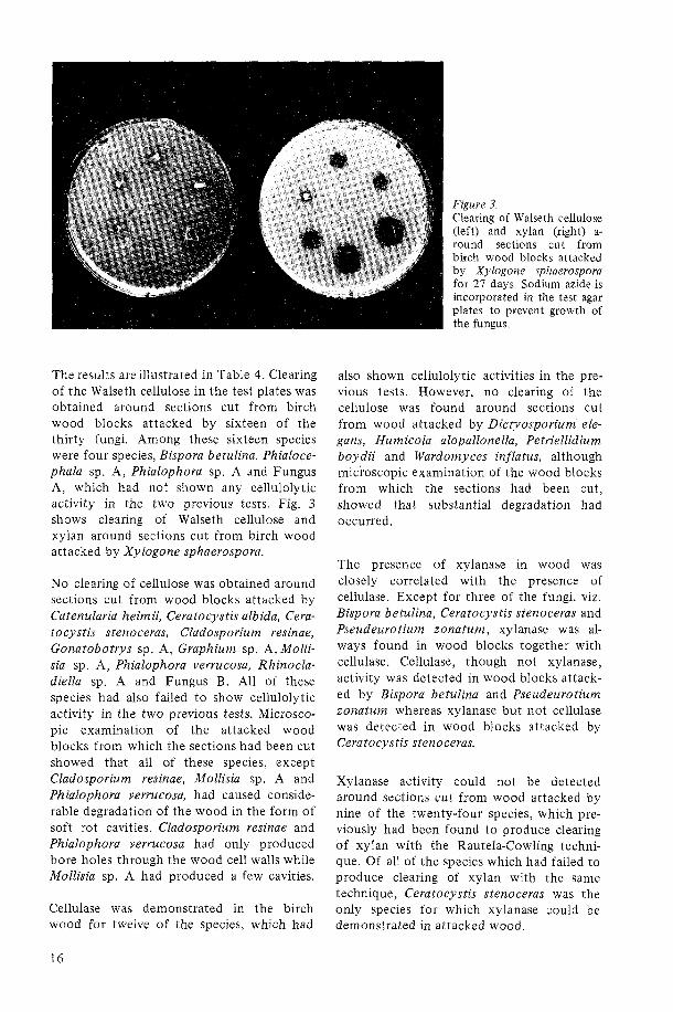
The results areillustrated in Table 4. Clearing of the Walseth cellulose in the test plates was obtained around sections cut from birch wood blocks attacked by sixteen of the thirty fungi. Among these sixteen species were four species, Bispora betulina. Phialoce- phala sp. A, Phialophora sp. A and Fungus A, which had not shown any cellulolytic activity in the two previous tests. Fig. 3 shows clearing of Walseth cellulose and xylan around sections cut from birch wood attacked by Xylogone sphaerospora.
No clearing of cellulose was obtained around sections cut from wood blocks attacked by Catenularia heimii, Ceratocystis albida, Cera- tocystis stenoceras, Cladosporium resinae, Gonatobotrys sp. A, Graphium sp. A, Molli- sia sp. A, Phialophora verrucosa, Rhinocla- diella sp. A and Fungus B. All of these species had also failed t o show cellulolytic activity in the two previous tests. Microsco- pic examination of the attacked wood blocks from which the sections had been cut showed that all of these species, except Cladosporium resinae, Mollisia sp. A and Phialophora verrucosa, had caused conside- rable degradation of the wood in the form of soft rot cavities. Cladosporium resinae and Phialophora verrucosa had only produced bore holes through the wood cell walls while Mollisia sp. A had produced a few cavities.
Cellulase was demonstrated in the birch wood for twelve of the species, which had
Figure 3. Clearing of Walseth cellulose (left) and xylan (right) a- round sections cut from birch wood blocks attacked by Xylogone sphaerospora for 27 days. Sodium azide is incorporated in the test agar plates to prevent growth of the fungus.
also shown cellulolytic activities in the pre- vious tests. However, n o clearing of the cellulose was found around sections cut from wood attacked by Dictyosporium ele- guns, Humicola alopallonella, Petriellidium boydii and Wardomyces inflatus, although micioscopic examination of the wood blocks from which the sections had been cut, showed that substantial degradation had occurred.
The presence of xylanase in wood was closely correlated with the presence of cellulase. Except for three of the fungi. viz. Bispora betulina, Ceratocystis stenoceras and Pseudeurotium zonatum, xylanase was al- ways found in wood blocks together with cellulase. Cellulase, though not xylanase, activity was detected in wood blocks attack- ed by Bispora betulina and Pseudeurotium zonatum whereas xylanase but not cellulase was detected in wood blocks attacked by Ceratocystis stenoceras.
Xylanase activity could not be detected around sections cut from wood attacked by nine of the twenty-four species, which pre- viously had been found t o produce clearing of xylan with the Rautela-Cowling techni- que. Of all of the species which had failed to produce clearing of xylan with the same technique, Ceratocystis stenoceras was the only species for which xylanase could be demonstrated in attacked wood.

3.5 Growth on medium EP and B-VII-L in liquid cultures with glucose as the sour- ce of carbon
The results illustrated in Table 5 show that all of the tested fungi could grow on the two media, EP and B-VII-L, with glucose as the source of carbon. No growth or very slight growth occurred on medium EP without glucose.
Phialocephala sp. C and Rhinocladiella sp. A showed slightly better growth on B-VII-L than on EP medium. All of the other species produced equal or, in some cases, consider- ably more growth on EP medium.
All of the species lowered the pH of both of the media during growth on glucose. A rise in pH was noted in the flasks with medium EP without glucose.
Differences in growth rates between the test species grown on EP medium can be noted in the table. Some species had evidently reached their maximal mycelial weights al- ready after 14 days, while others still show- ed an increase in mycelial weight up t o 28 days.
3.6 Degradation of cellulose N, Avicel and cotton and the production of cellulase and xylanase o n these substrates in liquid cultures
The results with medium EP are shown in Table 6 and the results with medium B-VII -L are illustrated in Table 7. The weight losses reported represent the average of three or four flasks.
The non-inoculated controls with EP as the medium showed weight losses of the cellu- lose substrates which ranged from 0.8 to 1.0 percent in shake cultures and from 2.1 percent to 5.1 percent in stationary cultures. Since great errors are involved in the proce- dure of determining weight losses of the cellulose substrates, only weight losses above 10 percent are regarded as significant.
The results show a pattern similar t o that of the results of the clearing tests using the
Rautela-Cowling technique. Most of the species which had produced clearing of cellulose were also able t o cause weight losses of cellulose and cellulase and xylanase could be found in the culture filtrates.
The weight losses were generally higher on medium EP than on medium B-VII-L. Exceptions were found for Acremonium a t ro-griseum and Cordana pauciseptata which produced higher weight losses of cotton on medium B-VII-L than on EP medium. Their weight losses of Avicel and cellulose N were, however, higher on EP than on B-VII-L medium. Rhinocladiella anceps and Scytalidium sp. B were also exceptional in causing higher weight losses of cellulose N on B-VII-L medium than on EP medium. The last two fungi produced no weight losses of cellulose N on EP medium after 42 days whereas the weight losses on B-VII-L medium were 26.8 and 46.0 per- cent respectively after 2 1 days.
On medium B-VII-L only very few of the fungi produced cellulase and xylanase which could be detected in the culture filtrates. This also applies t o species which caused significant weight losses on B-VII-L me- dium.
Differences were noted between shake and stationary cultures. Coniothyrium fuckelii var. sporulosum, Pseudeurotium zonatum, Scytalidium lignicola and Fungus D caused higher weight losses on medium EP in shake cultures than on the same medium in statio- nary cultures, while the reverse was found for Dictyosporium elegans, Humicola alo- pallonella, Phialophora fastigiata and Fungus A (see Table 6). Especially great differences were noted for Phialophora fastigiata grow- ing on cotton in EP medium. After 42 days the weight loss of cotton in stationary cultures was 38 percent while only 1 percent was lost in shake cultures. Fungus A behaved similarily on cellulose N on EP medium. The weight loss was nearly 46 percent after 42 days in stationary cultures while no weight losses occurred in shake cultures.
If the weight losses obtained in medium EP for the different celluloses are compared, it appears that cellulose N in most cases was

more rapidly degraded than Avicel, while cotton was the most resistant substrate. But individual variations were found, Chryso- sporium pannorum, for instance, caused a higher weight loss of Avicel than of cellulose N. Some of the fungi showed considerable variations in their ability t o degrade the different cellulose substrates. Acremonium atro-griseum caused weight losses of 3 5.0 and 48.4 percent of Avicel and cellulose N respectively, while the weight loss of cotton was only 4.1 percent. When medium B-VII -L was used, the fungus produced during the same time a weight loss of 33.2 percent of cotton while Avicel and cellulose N lost only 15.2 and 13.5 percent respectively.
Dictyosporium elegans and Fungus A caused weight losses only in cellulose N. Avicel and cotton were not attacked. Phialophora fasti- giata did not degrade Avicel but caused high weight losses of both cotton and cellulose N.
Among the species which were tested on medium EP, the following produced cellu- lase and xylanase that could be detected in the culture filtrates, both in shake and stationary cultures: Coniothyrium fuckelii var, sporulosum, Phialophora fastigiata, Scy- talidium lignicola and Fungus D. Dictyo- sporium elegans and Pseudeurotium zonatum produced both cellulase and xylanase in stationary cultures but in culture filtrates from shake cultures n o xylanase could be detected. The reverse was found for Petrielli- dium boydii. Humicola alopallonella and Fungus A did not produce any of the two enzymes in shake cultures, both produced cellulase in stationary cultures and Fungus A also produced xylanase. Mollisia sp. A pro- duced xylanase but not cellulase in shake cultures. None of the enzymes were found in stationary cultures.
On medium B-VII-L comparisons can only be made for Phialophora sp. A and Fungus D. Cellulase and xylanase were detected in stationary cultures of Fungus D, while n o xylanase was found in shake cultures. Phialo- phora sp. A showed xylanase activity on Avicel after 42 days. No cellulase activity was found on any of the substrates in stationary cultures. None of the enzymes were found in shake cultures.
If weight loss is used as a criterion for cellulolytic activity, the following fungi, which caused more than 10 percent weight loss, may be considered cellulolytic: Acre- monium atro-griseum, Chrysosporium pan- norum, Cladorrhinum sp. A, Coniothyrium fuckelii var. sporulosum, Cordana paucisep- tata, Dicty osporium elegans, Humicola alo- pallonella, Humicola grisea, Petriellidium boydii, Phialophora fastigiata, Phialophora hoffmannii, Pseudeurotium zonatum, Rhino- cladiella anceps (only on medium B-VII -L), Scytalidium lignicola, Scytalidium sp. B (only on medium B-VII-L), Wardomyces inflatus, Xylogone sphaerospora, Fungus A and Fungus D.
Both cellulase and xylanase were detected in culture filtrates from all the species mention- ed above, except for the culture filtrates from Humicola alopallonella and Scytali- dium sp. B in which no xylanase was detected. Among the remainder of the tested species Cladosporium sp. A was found t o produce cellulase and Mollisia sp. A and Phialophora sp. A were found t o produce xylanase. None of these enzymes was detec- ted in culture filtrates from the rest of the fungi.
The pH after cultivation was higher than the pH of the non-inoculated controls on me- dium EP. The highest pH values were found in culture filtrates from flasks where n o cellulose degradation had occurred. On me- dium B-VII-L, the pH was the same or lower than the pH of the medium after autoclaving. Especially low pHs were mea- sured in culture filtrates from flasks with medium B-VII-L where significant degrad- ation of the cellulose had occurred.
3.7 Degradation of birch wood meal and production of cellulase and xylanase on this substrate in liquid cultures
Chrysosporium pannorum, Bispora betulina, Phialophora sp. A and Phialocephala sp. A were grown in liquid cultures on birch wood meal as described in Materials and methods (section 2.6). The incubation was extended t o 42 days but even after this time n o significant weight losses were obtained, nor
was cellulase and xylanase detected in any of the culture filtrates.
3.8 Degradation of cot ton and jute fibres and production of cellulase and xylana- se o n these substrates in liquid cultures
Table 8 shows the results obtained from liquid cultures with cotton and jute fibres as substrates.
Bispora betulina and Phialophora sp. A did not cause any significant weight losses of cotton and jute fibres, nor could cellulase and xylanase be detected in the culture filtrates. No degradation of any of the two fibres was observed in the microscope.
Judging from the small and insignificant weight losses, no attack was caused by Ceratocystis albida on cotton fibres. Neither could any degradation of the cotton fibres be seen in the microscope. The pH of the culture filtrates was considerably higher than that of the non-inoculated controls. Signifi- cant weight loss was, however, obtained in the jute fibres, 30.8 percent after 42 days. Microscopic examination of the jute fibres revealed numerous soft rot cavities. Cellulase and xylanase could not, however be detected in the culture filtrates. The pH of the filtrates was lower than that of the controls.
Wardomyces inflatus was capable of attack- ing both cotton and jute. After 42 days the weight losses were 12.9 and 19.8 percent respectively. Microscopical examination re- vealed erosion in the cotton fibres and erosion and soft rot cavities in the jute fibres. Both cellulase and xylanase were detected in the culture filtrates from the flasks with cotton after 42 days, though neither of these enzymes appeared in the jute cultures. The pH of the culture filtrates from both of the substrates was higher than the pH in the control flasks.
Xylogone sphaerospora was the only species which produced a higher weight loss of cotton than of jute, 59.5 and 27.2 percent respectively, after 42 days. Cellulase and
xylanase were detected in filtrates from jute fibre cultures, whereas only cellulase was detected in the cultures with cotton. Only an erosion type of attack was seen under the microscope in both types of fibres. The pH of the culture filtrates was lower, especially the pH of filtrates from cultures with cot- ton, than the pH of the controls.
3.9 Effect of various amounts of glucose added t o medium EP o n the production of cellulase and xylanase.
Table 9 shows the influence of the addition of various amounts of glucose t o the EP medium on the production of cellulase and xy lanase.
The addition of up t o 0.1 percent glucose as "start-glucose" did not stimulate Bispora betulina, Ceratocystis albida or Phialoce- phala sp. A to produce cellulase o r xylanase. Table 9 shows that no cellulase or xylanase could be detected in any of the culture filtrates. The pH was higher than that of the autoclaved EP medium before inoculation.
It can be noted in Table 9 that increasing amounts of glucose retarded the production of cellulase and xylanase by Cladorrhinum sp. A, Xylogone sphaerospora and Fungus A. The addition of glucose had a greater in- fluence on the production of xylanase than on that of cellulase. 0.25 percent glucose prevented the formation of detectable amounts of xylanase by Cladorrhinum sp. A and Xylogone sphaerospora. The latter spe- cies produced detectable cellulase at 0.5 percent glucose, whereas cellulase could not be found for Cladorrhinum sp. A at this glucose concentration.
Fungus A produced cellulase after seven days in cultures with 0.005 and 0.01 percent glucose, but none in cultures with 0.025 percent. However, after 14 days cellulase was found in cultures with 0.1 percent glucose, which was the highest concentration tested with this fungus. Xylanase was also found after 14 days in cultures with 0.1 percent glucose.

The pH of the culture filtrates from Clador- rhinum sp. A, Xylogone sphaerospora and Fungus A were in most cases lower than the start pH, especially if increasing amounts of glucose had been added.
3.10 Comparison of the results obtained by the different methods
Table 10 shows which of the enzymes cellulase, xylanase, mannanase and amylase could be demonstrated for each of the species by using the different methods. The results should be compared with the wood- degrading ability of the different species as given in Table 2. The results of tests of the growth on cellulose agar plates and the clearing of cellulose in these plates are not included in Table 10.
Cellulase could be demonstrated for twenty- four of the thirty-six species. Of these twenty-four species, twenty were found to be active when the Rautela-Cowling tech- nique was employed. Twenty species pro- duced detectable amounts of cellulase in liquid cultures. The weight loss test with a cellulose substrate (cellulose N) resulted in nineteen positive results and the agar-plug method yielded eighteen positive results. All species were not tested by the wood block method but when this method was used, sixteen out of thirty species were found to be cellulolytic. Thirteen of the wood-degrad- ing species failed to show cellulolytic acti- vity on pure cellulose substrates. All of them were able t o produce soft rot cavities within the birch wood. They will subsequently be referred to as "non-cellulolytic7' soft rot fungi.
The highest number of positive results for xylanase activity was obtained with the Rautela-Cowling technique. When this tech- nique was used, twenty-seven of the thirty- six species were found to produce xylanase. Eighteen species produced detectable amounts of xylanase in liquid cultures and when the wood block method was used, xylanase activity could be demonstrated for fourteen of the thirty species tested by this
method. Tests for mannanase and amylase were carried out only with the Rautela- Cowling technique so no comparisons can be made with the other methods.
All species except Phialophora verrucosa produced amylase. The following twenty species were found to also produce all of the three other enzymes: Acremonium atro- griseum, Chrysosporium pannorum, Clador- rhinum sp. A, Cladosporium sp. A, Coniothy- rium fuckelii var. sporulosum, Cordana pau- ciseptata, Dictyosporium elegans, Humicola grisea, Petriellidium boydii, Phialophora fasti- giata, Phialophora hoffmannii, Phialophora sp. A, Pseudeurotium zonatum, Rhinocla- diella anceps, Scytalidium lignicola, Scytali- dium sp. B , Wardomyces inflatus, Xylogone sphaerospora, Fungus A and Fungus D.
Ten of these species exhibited both cellulase and xylanase activity in all the tests used. One of these ten species, Fungus D, was not tested by the wood block method. It pro- duced, however, positive results in all the other tests.
Nine of the twenty species mentioned above failed to show cellulase or xylanase activity in one or more of the tests. Cladosporium sp. A did not cause any weight losses of cellulose N and xylanase could not be detected in culture filtrates. This species and Scytalidium sp. B were not tested by the wood block method. Scytalidium sp. B did not produce detectable amounts of xylanase in liquid cultures. Dictysporium elegans, Petriellidium boydii and Wardomyces infla- tus did not show cellulase and xylanase activity with the wood block method. No xylanase activity was found when employing the same method for Pseudeurotium zona- turn. Cellulolytic activity could be demon- strated only by the wood block method for Phialophora sp. A, whereas xylanase activity was found with all the other three test methods used. Fungus A did not produce clearing of cellulose with the Rautela-Cow- ling technique nor did it produce cellulase when grown on cellulose agar plates. But this species caused high weight losses of cellulose N in liquid cultures with EP medium and cellulase was detected in the culture filtrates.

Two of the cell wall degrading enzymes could be demonstrated for the following species: Bispora betulina (cellulase and xylanase), Humicola alopallonella (cellulase and xyla- nase), Phialocephala dimorphospora (cellu- lase and mannanase), Phialocephala sp. A (cellulase and xylanase) and Fungus B (xyla- nase and mannanase). Bispora betulina and Phialocephala sp. A did not show cellulolytic activity in any other test than the wood block test. Bispora betulina failed t o show xylanase activity in this test. Both the species produced clearing of xylan with the RautelaCowling technique but none of them was able t o produce detectable amounts of xylanase in liquid cultures. Humicola alopal- lonella failed t o show cellulase and xylanase activity in the wood block test, nor was any xylanase produced in liquid cultures.
Phialocephala dimorphospora produced clearing of cellulose with the Rautela-Cow- ling technique but did not produce cellulase when growing on cellulose agar plates. Nor did this species produce any weight losses of cellulose N in liquid cultures and cellulase could not be detected in the culture filtrates. This species was not tested with the wood block method. Fungus B exhibited xylanase activity only in test with the Rautela-Cowling technique.
Only one cell wall degrading enzyme, viz. xylanase, could be demonstrated for Cerato- cystis stenoceras, Cladosporium resinae, Graphium sp. A and Mollisia sp. A.
If the results given in Table 10 are compared with the wood-degrading ability of the diffe- rent species shown in Table 2, i t is obvious that most of the wood-degrading fungi pro- duced at least one of the three cell wall degrading enzymes studied. All the sixteen species which produced an erosion-type attack (Type 2 attack) in birch wood could be shown t o produce all three enzymes.
With the test methods used, five of the wood-degrading species failed t o show cellu- lase, xylanase and mannanase activity. These fungi were: Catenulari heimii, Ceratocystis albida, Gonatobotrys sp. A, Phialocephala sp. C and Rhinocladiella sp. A. Table 2 shows that these species are able t o produce soft rot cavities (Type 1 attack) but no erosion-type attack (Type 2 attack) in birch wood.
Of the three species Ceratocystis olivacea, Cladosporium resinae and Phialophora verru- cosa, all of which were incapable of degrad- ing birch wood, only C. resinae showed xylanase activity. Cellulase and mannanase could not be demonstrated for any of them.

4 Discussion
4.1 Wood degradation and enzyme produc- tion
Wood is degraded by a great number of different species of fungi belonging t o the Basidiomycetes, Ascomycetes and Fungi im- perfecti. The Basidiomycetes cause white or brown rot while the wood-attacking Asco- mycetes and Fungi imperfecti cause soft rot. The term "soft rot", coined by Savory (1954) for the attack by microfungi and characterised by cavity formation in the secondary walls of the wood cells, has in later years been used for all types of wood degradation caused by microfungi, irrespec- tive of whether cavities are formed or not.
The main constituents of wood are cellulose, hemicelluloses and lignin. The white rot fungi decompose all three components, whe- reas the brown rot fungi leave most of the lignin as a residue. Few studies have been made on the changes in chemical composi- tion of wood attacked by pure cultures of microfungi. Only one species, Chaetomium globosum, has been studied in detail. These studies were carried out by Savory and Pinion (1958), Levi and Preston (1965) and Seifert (1966). Their results and scattered data from other investigations (Merrill e t al. 1965, Bergman & Nilsson 1967 and Lund- strom 1973) indicate that the main targets for the wood-degrading microfungi are cellu- lose and hemicelluloses, although small re- ductions in the lignin content have been reported.
A prerequisite for fungi capable of degrading wood is the production of extra-cellular enzymes that catalyze the dissolution of the polymeric wood components. So far as the author is aware, no fungus has proved capable of removing only one of the three main components of wood. The removal of only one component is probably physically blocked by the other components. Removal
of two of the components is, however, possible as shown by the brown rot and soft rot fungi. These fungi which degrade the cellulose and hemicelluloses of the wood must produce extra-cellular cellulases and hemicellulases. The main constituents of hemicelluloses are xylan in hardwoods and mannan in softwoods. The brown rot and soft rot fungi would consequently be expect- ed to produce cellulase, xylanase and man- nanase. A simultaneous production of xyla- nase and mannanase is most probable since production of only one of these enzymes would restrict the degradation ability t o either hardwoods or softwoods.
It is interesting t o note that many fungi which have proved t o be cellulolytic have also been found t o produce xylanase and mannanase. This also applies t o fungi whose wood-degrading abilities are unknown (cf. Lyr & Novak 1961).
Lyr (1963) showed that the wood-rotting Basidiomycetes, Schizophyllum commune, Trametes versicolor, Phellinus igniarius, Col- lybia velutipes, Fomes marginatus, Conio- phora cerebella, Piptoporus betulinus and Gloeophyllum saepiarium produced both xylanase and mannanase. He also found that both of these enzymes were produced by the cellulolytic mould Trichoderma viride and the well-known soft rot fungus Chaetomium globosum. Xylanase production by C. globo- sum has also been found by Ssrensen (1952 and 1957) and Fuller (1970).
Ahlgren and Eriksson (1967) showed that three wood-rotting fungi, Chrysosporium lig- noruml), Fomes annosus and Stereum san- guinolentum produced cellulase, xylanase
l ) ~ h e fungus called Chrysosporium lignorum is, according to "Centraalbureau voor Schimmelcul- tures" in Baarn, identical with Sporotrichum pul- verulentum Novobranova.

and mannanase. Lyr and Novak (1961), who studied the production of these enzymes among ten species of Fungi imperfecti, found that the three enzymes were produced by all of the species. Gascoigne and Gascoig- ne (1960) found xylanase activity among several species of fungi which are also known t o be cellulolytic. In a study of a soil fungus population, Domsch and Gams (1 969) found that Cephalosporium (= Acremonium) fur- catum, Chrysosporium pannorum, Coniothy- rium fuckelii var. sporulosum, Cylindrocar- pon didymum, Cylindrocarpon magnusia- num, Doratomyces microsporus, Epicoccum nigrum, Gliomastix (= Acremonium) muro- rum, Margarinomyces (= Phialophora) luteo- viridis, Oidiodendron echinulatum, species in the Phialophora fastigiata group, Phoma eupyrena, Pseudogymnoasus roseus, Pseudeu- rotium zonatum. Trichocladium opacum and Verticillium nigrescens could decompose xylan. Nilsson (1973) found that all of the mentioned species were cellulolytic and all of them, except Oidiodendron echinulatum, were able t o degrade birch wood.
Thus, it is obvious that there is a connection between the ability t o produce cellulase and the ability t o produce xylanase and manna- nase. This is also shown by the fact that xylanase and mannanase are often produced even if the fungus is cultivated on pure cellulose. Eriksson and Goodell (in press), who prepared a number of cellulase-less mutants of the wood-rotting fungus Polypo- rus adustus, found that most of the mutants lacked xylanase and mannanase as well. One of the cellulase-less mutants was mutageni- zed and cellulose-degrading revertants were selected. These revertants were not only found t o be capable of degrading cellulose, but they also degraded xylan and mannan. On the basis of their results, Eriksson and Goodell suggested that the induction of cellulase, xylanase and mannanase is control- led by a single regulator gene.
Table 10, which summarizes the results of all experiments in the present investigation, shows that all three enzymes could be demonstrated for twenty of the thirty-three wood-degrading species. Five species were shown t o produce two of the enzymes and for three of the species only one enzyme was
demonstrated. Five species, all producing soft rot cavities in birch wood, seemed t o lack cellulase as well as xylanase and manna- nase. The apparent lack of one, two or all three of the enzymes by the wood-degrading species is most likely due t o imperfect test methods and not t o a real incapability t o produce these enzymes. This will be discuss- ed later in more detail.
Of the three species which were unable t o degrade wood, viz. Ceratocystis olivacea, Cladosporium resinae and Phialophora verru- cosa, only C. resinae produced xylanase. Cellulase and mannanase were not produced by any of the species.
It was observed in a previous study (Nilsson 1973) that all species which were able t o produce an erosion-type attack (Type 2 attack) on the cell walls of birch wood were also able t o produce clearing of Walseth cellulose. The same was found in the present investigation and, furthermore, it was ascer- tained that such species could also be shown t o produce the enzymes xylanase and man- nanase. It is obvious that hyphae which are growing in the cell lumina and which act upon the surface of the cell wall must produce more or less diffusible enzymes which accomplish the degradation of the components of the cell wall by means of erosion.
In the investigation mentioned above, it was also observed that a few of the species which were able to produce clearing of Walseth cellulose did not produce- erosion-type at- tack in birch wood. I t was suggested that this failure could be due t o a lack of wall-degrading enzymes like xylanase and mannanase. Another explanation was that the S3 layer of the wood fibres represented a resistant barrier which could not be de- graded by these species and through which their enzymes could not diffuse. Acremo- nium atro-griseum and Petriellidium boydii are such species which failed to produce erosion of the cell walls of birch wood although they were able t o produce clearing of Walseth cellulose. The birch wood was, however, degraded by these species through cavity formation. As may be seen in Tables 2 and 10, these species are able t o produce

diffusible xylanase and mannanase as well as diffusible cellulase. Thus, it is evident that their failure to produce an erosion type of attack is not due t o a lack of these enzymes but is more probably due to their inability t o degrade the S3 layer.
A literature survey covering the degradation of cell wall components by those species in the present study which are fully identified has been made. For several of the species no information about cellulolytic activity or degradation of xylan and mannan could be found. Some of the data collected is found below. The data presented by the author in a previous paper (Nilsson 1973) are not in- cluded.
Ceratocystis albida (= Ophiostoma albidum). Kaarik (1960) found that cellulose was a very poor source of carbon for this species, as well as for other Ceratocystis species. However, she stated that all species were "able t o make a starvation type of growth on it".
Chrysosporium pannorum. Degradation of filter paper by this species was found by Schaefer (1957). Domsch and Gams (1969) found that the fungus degraded xylan and CMC.
Cladosporium resinae. Some variation seems to exist among different strains. Parbery (1969) found that one out of five isolates was able t o degrade cellulose.
Coniothyrium fuckelii var. sporulosum. Domsch and Gams (1969) found that this fungus degraded xylan and CMC.
Humicola alopallonella. Meyers and Rey- nolds (1 959) found that this species was cellulolytic.
Humicola grisea. Traaen ( 1 9 14) already showed that this fungus decomposed cellu- lose. Later, several investigations have prov- ed the cellulolytic activity of this species (cf. Domsch & Gams 1970). Poole and Taylor (1973) found production of cellulase and xylanase.
Phialophora fastigiata. Reese et al. ( 1 950) showed that P. fastigiata reduced tensile
strength of cotton duck. Domsch and Gams (1969) found that this species degraded xylan and CMC. Fuller (1970) found that production of cellulase in liquid cultures but no xylanase was found. King and Eggins (1973) showed that the fungus reduced the strength of filter paper.
Phialophora verrucosa. Reese et al. (1950) found that this species was unable t o reduce the strength of cotton textile.
Pseudeurotium zonatum. Domsch and Gams (1969) showed that this species degraded xylan and CMC.
Scytalidium lignicola. King and Eggins (1973) showed that this fungus could de- grade cellulose.
If the above data are compared with the results obtained in the present study, it is evident that there is good agreement. Un- fortunately, n o studies seem to have been done earlier with the most interesting species in this study, i.e. those which degrade wood but fail t o show production of the necessary enzymes. The only exception is the study of Ceratocystis albida by Kaarik (1960).
Production of amylase is not necessary for the degradation of wood but might be beneficial for fungi colonizing wood sub- strates since starch is an important reserve nutrient in many trees. The present investi- gation shows that the ability to degrade starch is common among wood-inhabiting microfungi. This was also found by King and Eggins (1973) who found amylolytic activity in all of the thirty-three different mould and staining fungi which were tested.
4.2 Test methods and results
In the following the results obtained with the different test methods will be discussed.
Numerous methods have been used for assays of cellulolytic activity among micro- organisms. Eriksson (1969) gives a list of various assay methods used for determining cellulolytic activity. His list contains the following methods:

loss in weight of insoluble substrates decrease in mechanical properties of fibres or films change in turbidity of cellulose suspen- sion increase in reducing ends groups decrease of viscosity of cellulose deriva- tives colorimetric determination of dissolved decomposed products of cellulose measurements of clearance zones in cellu- lose agar
To this list may be added: 8) microscopic observations of morphologi-
cal changes in a cellulose substrate (like fibres or cellophane)
9) growth on cellulose agar
Except for method 9 , these methods can be used both for determining the cellulolytic activity of growing microorganisms and the activity of isolated cellulases.
Methods 3 and 7 are very much of the same type. These assay methods, which aim at a clearing of cellulose substrates, have been extensively used in this study. Methods 1 , 8 and 9 have also been used. All of the methods referred t o may be considered to be indicative of a C1 enzyme sensu, Reese, Siu and Levinson (1950).
Measurements of the decrease of viscosity of cellulose derivatives (method 5) have been widely used for determining the cellulolytic activity of microorganisms. Most of the studies have been carried out with carboxy- methyl cellulose (CMC) as substrate. Yet Reese and Levinson (1 952) already found several non-cellulolytic organisms that pro- duced CMC-degrading enzymes. These enzy- mes are usually referred t o as C, enzymes. The production of C, enzymes by non-cellu- lolytic fungi has later been demonstrated in other investigations. Wood (1969) re- ferred t o these fungi as "pseudo-cellulolytic" species. The aforementioned method has applications in the studies of the compo- nents of the cellulase system, but it should not be used for assays of the cellulolytic activity of microorganisms.
Reese, Siu and Levinson (1950) suggested that
truly cellulolytic fungi possess an additional enzyme, C1, which enables them to degrade native cellulose. King and Vessal (1 969) also defined the C1 enzyme as an enzyme which is required for the hydrolysis of highly oriented solid cellulose, like cotton and Avicel, by 0 - 1 + 4 glucanases (=C,). Since wood cellulose also is a highly oriented solid cellulose, it follows from the definition that all fungi which are able to degrade wood must produce C1 enzymes. Thus, the inabil- ity of certain fungi to degrade cotton or Avicel does not necessarily mean that they lack C1 enzymes. It may possibly be that the culture conditions are unsuitable for the production of this enzyme.
Whether the degradation of a modified cellulose such as Walseth cellulose indicates C1 activity or merely C, activity has already been discussed in a previous paper (Nilsson 1973). The findings, that most of the fungi which degraded Walseth cellulose were also able to degrade wood cellulose, and the fact that CMC-degrading fungi failed t o degrade Walseth cellulose, support the former theory.
As can be seen in Table 10, a high number of positive results were obtained by means of the Rautela-Cowling technique. This techni- que has several advantages. It is simple t o carry out and, as pointed out by Rautela and Cowling (1966), cellulolytic activity is "determined directly on a continuous, cumulative basis". Other substrates than cellulose, e.g. hemicelluloses and starch, can also be tested with this technique, as has been done in the present study. This method also seems t o be quite sensitive and appears t o give positive results even for species with weak cellulolytic activity.
It was already shown in a previous paper, Nilsson (1973), that cultivation on medium VII suggested by Bravery (1968) yielded a higher number of positive results for cellu- lolytic activity than cultivation on the me- dium formulated by Rautela and Cowling (1966). Several species which failed t o pro- duce clearing on R-C medium did clear the cellulose on B-VII medium. This was also confirmed in the present study in the case of two additional species, viz. Cladosporium sp. A and Fungus D. This phenomenon might be

explained by the presence of an alternative carbon source (yeast extract) in the R-C medium, since it was demonstrated by Bravery (1968) that small amounts of alter- native carbon sources, like asparagine and yeast extract, inhibited clearing of cellulose agar by certain soft rot fungi. The decreased amount of clearing of cellulose produced by all active species on medium R-C compared with the clearing that occurred on B-VII medium (see Table 2) might also be due t o inhibition by the presence of yeast extract in the former medium. Alternative carbon sources like glucose, asparagine and yeast extract appear, however, t o exert a varying influence on the cellulolytic activity de- pending on the species of fungi and also on the culture conditions. As can be seen from the results of the tests with cellulose in liquid cultures (Tables 6 and 7), the presence of asparagine and yeast extract in the EP medium appeared to have no adverse effects on the cellulolytic activity of the tested fungi. Park (1973), who made a study similar t o that of Bravery (1968), tested several modifications of the Eggins and Pugh (1962) medium. He reached the conclusion that the presence of asparagine might have disadvantages, whereas yeast extract seems t o have no disadvantages. Dennis (1972), who studied the clearing of cellulose agar by yeasts (Trichosporon spp.) found that small amounts of yeast extract and asparagine stimulated the activity of some of the isolates, but reduced the activity in others. Norkrans and Aschan (1953), who cultivated different strains of Collybia velutipes on a weak yeast-glucose-cellulose medium, found that the cellulolytic activity did not change much when cultivated on a richer yeast extract medium. If the strains were culti- vated on a glucose-free medium, the activity was the same or decreased.
I t is obvious from the results presented in Table 2 that the amount of clearing of cellulose with the Rautela-Cowling techni- que depends very much on the type of cellulose substrate. The greatest amount of clearing was obtained on Walseth cellulose and ball-milled cellulose. The preparation of these two types of celluloses involves treat- ments that increase the proportion of amorphus cellulose and also the intrinsic
porosity. Both are factors which will in- crease the susceptibility t o hydrolysis (cf. Norkrans 1950, Walseth 1952 and Stone et al. 1969).
Table 3 shows the results of three different assay methods of cellulolytic activity; 1) growth on cellulose agar, 2) clearing of cellulose underneath or around fungal colo- nies growing on cellulose agar plates and 3) clearing of cellulose in test plates around transferred mycelium-agar plugs. The only difference between method 2 and the Rautela-Cowling technique is that the former is carried out in petri dishes while the latter employs test tubes.
It is evident from the results that growth on cellulose agar is a very uncertain criterion for cellulolytic activity. Visual estimation of the amount of growth of fungi on agar is difficult since the fungi might produce dif- ferent types of colonies. Some fungi produce a dense mycelium on a restricted area on a certain medium, while on other media the same species will produce a widely spreading but thin mycelium. The estimation of growth and non-growth on cellulose agar media of the types used in the present investigation seems impossible since the truly non-cellu- lolytic fungi also produced some growth on the B-VII medium which contains no source of carbon other than cellulose. It is possible that these fungi are able t o make some growth on impurities containing car- bon, which may be present in the Avicel that was used as cellulose substrate. The other cellulose agar medium used, F6A, contains both glucose and yeast extract. This explains why all of the tested species were able t o grow on this medium. In fact, two of the non-cellulolytic species grew better on F6A celluloses agar than some of the cellulolytic species. The species which had failed t o produce clearing of cellulose with the Rautela-Cowling technique (see Table 2) made sparse or moderate growth even on the F6A cellulose agar. They did not appear to be able to utilize the cellulose in the medium, since no clearing of the cellulose could be detected in the agar nor was any cellulase detected with the mycelium-agar plug technique of Savory e t al. (1 967).
A rather paradoxical conclusion which can

be drawn from the results obtained in the present study is that cellulose agar substrates are not suitable for the isolation of certain cellulolytic fungi.
The two agar media used for these studies contained Avicel, a cellulose which is more resistant to hydrolysis than Walseth cellulose and ball-milled cellulose. This might explain the failure of most of the species t o produce visible clearing in the cellulose agar on which they grew. I t was already shown in Table 2 that clearing of Avicel was very weak, as compared with the clearing of the two other celluloses. But the obscuring by dense mycelia, which made it difficult t o detect any clearing probably also contributed t o the low number of positive results.
The agar-diffusion method employing myce- lium-agar plugs adopted after Savory et al. (1967) had n o advantages over the Rautela- Cowling technique. The agar-diffusion method is more time consuming and the assays of cellulolytic activity are made after certain arbitrarily chosen times. The number of positive results was lower with this method than with the Rautela-Cowling technique (see Table 10). Cellulolytic activity could not be detected with the mycelium- agar plug technique for two of the fungi, Phialocephala dirnorphospora and Rhinocla- diella anceps, which had produced clearing of cellulose with the Rautela-Cowling tech- nique.
If the production of cellulase on the cellu- lose agars F6A and B-VII is compared for the fungi which produced clearing of cellu- lose only on medium B-VII when the Rautela-Cowling technique was used (see Table 2), it is evident that the glucose, and possibly also the yeast extract, present in the cellulose agar F6A did inhibit the cellulo- lytic activity of these fungi. Of these fungi only Cladosporiurn sp. A was able t o pro- duce detectable amounts of cellulase on F6A agar.
The test method described in Materials and methods (section 2.5) which employs the detection of cellulase and xylanase present in wood blocks attacked by the test organ- isms, is based on different premises than the other test methods used in the present
investigation. With the other test methods nothing is known about the formation of cellulase on the substrate used. But when solid wood is used as substrate, the degrada- tion of the wood cell walls in the form of erosion or cavity formation indicates that cellulase is present. The degradation of the cellulose in the cell walls is easily detected by microscopic studies using polarized light and is in itself a demonstration of cellulo- lytic activity. By means of the wood block method it could be shown that the cellulase present in the wood could degrade a pure cellulose such as Walseth cellulose (see Table 4).
The wood block method was the only means by which cellulase could be demonstrated for Bispora betulina, Phialocephala sp. A and Phialophora sp. A. The xylanase activity of Ceratocystis stenoceras was demonstrated only by this method. Fungus A, which had failed t o demonstrate cellulolytic activity in the previous tests discussed, was found to be cellulolytic when the wood block method was used and also when cultured on cellulose in liquid media.
The species Catenularia heimii, Ceratocystis albida, Ceratocystis stenoceras, Gonato- botrys sp. A, Graphiurn sp. A, Mollisia sp. A, Rhinocladiella sp. A and Fungus B, all of which failed t o show cellulolytic activity with the other test methods used, also failed with the wood block method. However, microscopic studies showed that numerous cavities were present in all the wood blocks attacked by these species,. with the excep- tion of wood blocks attacked by Mollisia sp. A in which only a few cavities were found. It was further ascertained that several species which showed celluiolytic activity in other tests failed in the wood block method. Two explanations can be given for the failures in the wood block method: 1 ) the enzymes are produced in only minute amounts in the wood, adequate for wood degradation but not sufficient for diffusion out into the test agar; 2) the enzymes are firmly bound either t o the wood substrate and/or the mycelium and are thus not able t o diffuse freely out into the test agar.
The method in which the fungi were cul-

tured on cellulosic substrates in liquid media provided two criteria for cellulolytic activ- ity. The first was weight loss of the substrate and the other detection of cellulase in the culture filtrate. A weight loss of the cellu- losic substrate is not necessarily correlated with detectable amounts of cellulase in the culture filtrate. This can be seen in Table 7 where, for example, Acremonium atro- griseum, Cordana pauciseptata and Scytali- dium lignicola is shown t o have caused high weight losses although no cellulase could be detected in the culture filtrates. This might be explained by binding of the enzyme to the mycelium and/or t o the substrate. The same fungi, however, produced detectable cellulase on medium EP. Medium B-VII-L is a rather poor medium compared with medium EP. Medium EP contains yeast extract and 2 12 mg N/liter while medium B-VII-L lacks yeast extract and contains only 115 mg N/liter. When growing on medium B-VII-L the fungi must use their enzymes more economically than when growing on the richer medium. Free en- zymes in the solution can be regarded as a wastage of available resources, especially nitrogen.
The fact that cellulolytic enzymes are de- tected in the culture filtrates is not always correlated with significant weight losses of the substrate. Table 6 shows that cellulase was detected in culture filtrates from Cla- dosporium sp. A on cellulose N, Dictyospo- rium elegans on Avicel and cotton and Phialophora fastigiata on Avicel while n o significant weight losses occurred. Thus the use of two methods for determining cellu- lolytic activity increased the number of positive results.
The differences in nitrogen content between the two media might explain why the weight losses were generally lower on the B-VII-L medium. However, two species, Acrerno- nium atro-griseurn and Cordana paucisep- tata, caused higher weight losses of one of the substrates, viz. cotton, on medium B-VII-L than on medium EP. It is difficult t o explain why the weight losses of the other two substrates, Avicel and cellulose N, were lower on the B-VII-L medium than on medium EP. The results show that the
susceptibility of a certain cellulose substrate not only depends on the structure of the substrate but also on the medium in which the tests are carried out. The order of increasing susceptibility of different types of cellulose under one condition might there- fore change drastically under other condi- tions.
Differences in the distribution of the cellu- lose substrates within the nutrient solution are also likely t o influence the results. Cotton, for example, is partly floating and partly immersed in the solution, while the Avicel particles are completely immersed. These particles will sink t o the bottom of the flasks if stationary cultures are used. Thus, apparent differences in the susceptibil- i ty of the cellulose substrates as found here when growing fungi in liquid cultures must not be considered t o reflect an absolute indication of the susceptibility of these substrates t o fungal degradation.
It was expected that the species which produced clearing on B-VII but failed t o produce clearing of cellulose on the R-C medium (see Table 2) would give the best results on B-VII-L medium since this medium lacks alternative carbon sources. But of these species both Cladosporiurn sp. A and Humicola alopallonella produced better results on the EP medium. Only Scytalidium sp. B showed a positive response t o medium B-VII-L. The same positive response was found for Rhinocladiella anceps, although this species was also able t o produce clearing of cellulose on the R-C medium.
If the results from the experiments with liquid cultures are compared with the results obtained with the Rautela-Cowling tech- nique, it is found that essentially the same fungi have shown positive results by the two test methods. There are only two excep- tions: one is Phialocephala dimorphospora which failed t o give any indications of cellulolytic activity when cultured in liquid media; the other was Fungus A which had failed t o produce clearing of cellulose with the Rautela-Cowling technique. This fungus produced significant weight losses in liquid cultures and cellulase was found in the

culture filtrates. No explanation has been discovered for the behaviour of Fungus A, but the physiological conditions are probab- ly not suitable for the production of cellu- lase when the fungus grows on agar.
With the exception of Fungus A, all soft rot fungi which failed t o produce clearing of cellulose with the Rautela-Cowling tech- nique also failed when cultured in liquid media on cellulose substrates. The con- clusion which can be drawn is that these species did not produce cellulase under the prevailing conditions. It might be argued that cellulases were not detected in the culture filtrates because these species may have only cell-bound enzymes. But even if the enzymes are cell-bound, attack on the cellulose would still be possible and this would lead t o a weight loss of the substrate. However, n o weight losses were recorded for the species mentioned.
The high numbers of positive results for cellulolytic activity obtained from the tests on cellulose in liquid cultures, shown in Table 10, were achieved only by the use of two nutrient media of different composi- tion. If only medium EP had been used the number of positive results from the weight loss determinations would have been 17 instead of 19 and cellulase would have been detected in the culture filtrates for 18 instead of 20 species. The number of posi- tive results would have been far less if only medium B-VII-L had been used. When the Rautela-Cowling technique was used, all positive results could be obtained on a single medium, viz. medium B-VII. It is obvious that this technique is preferable t o tests on cellulose in liquid media if the objective is a survey of the cellulolytic activity of micro- fungi.
Birch wood meal in liquid cultures appears t o be an unsuitable substrate for tests of cellulolytic activity since n o activity was obtained, even with the highly cellulolytic species Chrysosporium pannorum. Degrada- tion of the substrate did not occur, neither could cellulase nor xylanase be detected in the culture filtrates. As can be seen in Table 6, the same fungus caused an extensive degradation of Avicel and cellulose N in
liquid cultures and both cellulase and xyla- nase were found in the culture filtrates. The purpose of using birch wood meal as a substrate was that it contains all the consti- tuents of solid birch wood, in which several of the test organisms made their only mani- festation of cellulolytic activity in the form of cavities. But even the cavity-forming species tested were unable t o degrade the birch wood meal and t o produce cellulase and xylanase.
It is a common observation that lignified substrates are less susceptible t o degradation than non-lignified substrates. This seems especially t o be the case when isolated cellulase systems are used. However, Table 8 clearly shows that some species of micro- fungi are able t o degrade a lignified fibre more readily than a non-lignified fibre. Ceratocystis albida is an exemple of such a species. This fungus failed t o degrade the cotton fibres while the jute fibres were degraded t o a significant degree although jute fibres contain about 11 percent lignin (Roelofsen 1959). Wardomyces inflatus was able t o degrade cotton but the weight losses of jute were higher. Only Xylogone sphae- rospora caused higher weight losses of cot- ton than of jute. Of the five species used in this experiment, all except X. sphaerospora were able to produce soft rot cavities in birch wood. Under the conditions employ- ed here, Ceratocystis albida and Wardomyces inflatus were also able t o produce cavities in the lignified jute fibres but not in the non-lignified cotton fibres.
C. albida, which evidently is unable t o produce cellulases in liquid cultures on pure cellulose substrates (cf. Tables 6 and 7 ) , could thus not degrade the cotton fibres. Wardomyces inflatus which is able t o pro- duce cellulases under the same conditions (cf. Tables 6 and 7) degraded the cotton fibres in this experiment but the degradation of the jute fibres was more extensive due to pro- minent cavity formation in these fibres.
Bispora betulina and Phialophora sp. A are able, as will be shown in a later paper, t o produce cavities in jute fibres. However, cavities are not formed by these species in fibres which are immersed in a nutrient
29

solution as they were in the present inves- tigation. Both of the species are also unable t o produce cavities in cotton fibres in liquid cultures. Cellulase was not produced when they were cultured in liquid cultures on pure cellulose substrates or on jute fibres (cf. Tables 6, 7 and 8). These facts explain why the two fungi failed t o degrade the cotton and the jute fibres.
Both the cotton and jute fibres were degrad- ed by Xylogone sphaerospora by a type of erosion attack. No soft rot cavities were formed in any of the fibres. The erosion is caused by enzymes which are released into the liquid medium by the fungus. The breakdown of the jute fibres under these circumstances appears t o be limited by the presence of lignin. The fungus consequently caused the highest weight losses on the non-lignified cotton fibres.
The observations made in the experiments discussed above might also explain the results obtained by Chakravarty et al. (1962). They compared the degradation of jute fibres with degradation of filter paper. All species tested except one, Sordaria hypo- coproides, caused the highest weight losses when cultured on filter paper. S. hypoco- proides, however, caused higher weight loss of jute fibres than of filter paper. It might be possible that this species is also able' t o produce cavities within the jute fibres and that the amount of cellulase released into the nutrient medium is small. But so far as the present author is aware, n o studies of the cavity-forming ability of this fungus have been carried out.
Simpson and Marsh (1964) discovered that Aspergillus niger and certain other Asper- gillus species caused a strength loss of cotton fabric only when a small amount of glucose was added t o the test medium. Similarly, Basu and Ghose (1 960) found that additions of sugar, especially xylose, stimulated the production of cellulase by certain species of fungi which had been found t o be unable t o produce cellulase when growing on pure cellulose. The addition of small amounts of glucose as "start-glucose" t o medium EP had, however, n o positive effect on the production of cellulase by Bispora betulina,
Ceratocystis albida and Phialocephala sp. A (see Table 9). Thus, the initial lack of an easily available carbon source does not ex- plain why these species failed t o show cellulolytic activity.
The addition of 0.1 percent glucose t o medium EP delayed cellulase production of Fungus A only slightly. Since the EP medi- um also contains small amounts of aspara- gine and yeast extract it is evident that this species is not prevented from producing cellulase by small amounts of alternative carbon sources.
The cellulase production of Cladorrhinum sp. A and Xylogone sphaerospora was delay- ed by glucose additions and Cladorrhinum sp. A produced no cellulase at a glucose concentration of 0.5 percent. It has been shown by Bemiller e t al. (1969) and others that the cellulase production does not start until the glucose in a nutrient solution has been consumed. The more sugar added, the longer time it will take for the consumption of glucose and the initiation of cellulase production. This fact will also explain the delay in cellulase production observed in the experiments carried out here.
The conclusion drawn from the experiments presented here and from the preceeding discussion is that there is no single simple method for the assay of the cellulolytic activity fo microfungi. This does not apply solely t o fungi isolated from wood, since fungi with an apparent lack of cellulolytic activity but which are able t o produce cavities in various cellulosic substrates, might also be isolated from other sources, for example soil and compost heaps. It has been shown by Baker (1939) and by Nilsson (1974) that in nature cavity formation of the soft rot type is not restricted to wood but might also occur in other materials containing cellulose fibres.
If a total survey of the cellulolytic activity is objective, the following procedure is recom- mended:
1) All the isolated species should be tested by means of the Rautela-Cowling tech- nique, employing Walseth cellulose as the

substrate. 0.25 percent appears t o be a suitable concentration of the cellulose. Ball-milled cellulose might also be used but the number of positive results is likely t o decrease. Avicel and similar types of celluloses should be avoided. The use of medium B-VII is recommended since it yielded a greater number of positive results than the medium used by Rautela and Cowling (1966). All of the species which produce clearing of the cellulose can safely be regarded as cellulo- lytic but obviously nothing is known about the cellulolytic activity of the remaining species since certain soft rot fungi of the type described in the present study will not produce any clearing.
2) All of the species which have failed t o produce clearing of cellulose are tested on birch wood (Betula species) for the abili- ty t o form soft rot cavities. Some other hardwood species might equally well be used, such as aspen (Populus species) or beech (Fagus silvatica), but it must be ascertained that the wood used is not very resistant t o attack by soft rot fungi. The decay method used by Nilsson (1973) and which employs agar slopes in test tubes is recommended. The time required for cavity formation varies among the species. Most of the species tested here have formed numerous cavi- ties in birch wood already within 2-4 weeks. The species which produce soft rot cavites must also be regarded as cellulolytic and be included in the total number of cellulolytic species. It is highly probable that most of the remaining species are non-cellulolytic, though it cannot be totally excluded that some of these species are able t o degrade cellu- lose under certain special conditions. The procedure described above will, nevertheless, increase the number of cel- lulolytic species, even if not all of the cellulolytic species are detected.
The recommended procedures are restricted t o assays of the cellulolytic activity of microfungi. Problems concerning the assays of the cellulolytic activity of the basidio- mycete fungi will be discussed in a later paper.
The test method described above may appear tedious and time-consuming but until new methods for assaying cellulolytic activi- ty are found, it is a procedure which will give reliable information of the cellulolytic activity of species of microfungi.
4.3 The anomalous behaviour of the "non- cellulolytic" soft rot fungi
It was previously suggested that the wood- degrading species would produce all the three cell wall degrading enzymes cellulase, xylanase and mannanase. This assumption was also confirmed for about half of the number of the species tested. But some wood-degrading species failed t o exhibit one or more of these enzymes when assayed with the test methods employed. Various expla- nations of this behaviour are possible. The lack of activity against xylan and mannan could be due t o alterations of these substra- tes during their preparation from wood so that their structure differs too greatly from the natural. It is also possible, in fact, that some fungi lack these enzymes. It is more difficult t o explain the failure of certain species to degrade the pure cellulose sub- strates. This phenomenon and others con- cerning the production of cellulase will now be discussed in more detail.
Cellulase is the only enzyme which is with certainty known t o be produced by the wood-degrading species since degradation of cellulose occurred in the birch wood fibres. This degradation was easily detected in the microscope using polarized light. Since n o chemical analysis of birch decayed by the test organisms was made, nothing is really known about the degradation of xylan. It may, however, be assumed t o be very prob- able that the xylan was also decomposed.
The discussion will be focused on the follow- ing organisms, all of which produced soft rot cavities in the birch wood but failed t o exhibit cellulolytic activity on pure cellulose substrates: Bispora betulina, Catenularia hei- mii, Ceratocystis albida, Ceratocystis steno- ceras, Gonatobotrys sp. A, Graphium sp. A, Mollisia sp. A, Phialocephala sp. A, Phialoce- phala sp. C, Phialophora sp. A, Rhinocladiel-

la sp. A and Fungus B. These fungi have previously been referred t o as "non-cellulo- lytic" soft rot fungi. The presence of cellula- se in attacked birch wood blocks could be demonstrated for some of these species. The experiments carried out using the Rautela- Cowling technique and the cultivation of the fungi on pure cellulose substrates in liquid media showed that n o cellulase was produc- ed under the prevailing conditions. These findings suggest that cellulase of the men- tioned species is an inducible enzyme and that the induction is regulated by factors which are so far unknown.
Various explanations may be advanced for the failure of the "non-cellulolytic" soft rot fungi t o degrade pure cellulose substrates under the conditions prevailing in the pre- sent investigation.
1) the "non-cellulolytic" soft rot fungi have a very weak cellulolytic activity.
There have been some false opinions regard- ing the cellulase activity of the soft rot fungi (cf. Lyr 1960, Levi 1964 and Wilhelmsen 1965). It has been claimed that these fungi produce less of these enzymes than the white and brown rot fungi. Such generalisa- tions should not be drawn unless they are based on a large number of observations. Seifert (1966) could, in fact, show that in the early stages, cellulose degradation of beech wood by the soft rot fungus Chaeto- mium globosum was greater than the degra- dation caused by one white and one brown rot fungus.
In fact, the soft rot species tested here can be regarded t o have a rather high cellulase activity when growing on birch wood. If a typical soft rot cavity is observed, only a single hypha is present in each cavity. The cavity may be very large in relation t o the size of the hypha. This indicates a high enzyme activity per mycelium unit.
The opinion that the soft rot fungi have a low cellulolytic activity probably is a result of the fact that several of these fungi cause comparatively small weight losses of the wood blocks used in decay tests. It has been shown by Lundstrom (1973) that wood
blocks in laboratory tests are most heavily degraded in the outer layers. Weight losses reported from laboratory tests refer to the weight loss in the whole blocks. Thus, a thin outer layer of the wood blocks might be very highly degraded even if the total loss in weight is small. Microscopic observations of the soft rot attack on birch wood blocks by the "non-cellulolytic" soft rot fungus Cera- tocystis albida (Nilsson, not published) have shown that the wood fibres in the outer layers of the birch wood blocks may already be degraded to 50 - 9 0 percent after three weeks. Thus the low weight losses caused by some soft rot fungi seem t o a higher degree t o be due t o a restriction of the attack t o the outer layers of the wood than t o a low cellulolytic activity.
2) The cellulase production is temperature- dependent.
It has been demonstrated that the cellulolytic activity of some fungi is quite sensitive t o temperature. Marsh et al. (1949) and Reese and Levinson (1952) have shown that Cla- dosporium herbarum has cellulolytic activity at 2 5 ' ~ but not at 3 0 ' ~ . Hirsch (1954) found that Neurospora crassao had a very weak cellulolytic activity at 25 C compared with the activity at 3 5 ' ~ . The wood-degrad- ing species in the present investigation, which had failed t o produce clearing of cellulose at the ambient room temperature (23-25') were also incubated at 1 5 ' ~ and 3 0 ' ~ . Since n o clearing of the cellulose occurred even after incubation for six weeks at these temperatures, it must be regarded as unlikely that the failure t o produce clearing zones in the cellulose was due t o a tempera- ture factor.
3) Some growth factors are lacking in the media used.
The "non-cellulolytic" soft rot fungi produc- ed at least some growth on the cellulose agar media used. All species also grew in the liquid media EP and B-VII-L when glucose was added. Thus, it is not likely that their failure t o show cellulolytic activity is due a lack of growth factors, unless certain growth factors are required specially for the pro- duction of cellulase.

4) The "non-cellulolytic" soft rot fungi ini- tially require an easily available carbon source ("start-glucose")
This hypothesis was tested (see Table 9) and it was found that n o stimulation of the cellulase production could be ascertained when glucose was added in small amounts.
5) The cellulase is cell-bound.
According t o Cowling and Brown (1969), the cavities formed in the wood cell walls by soft rot fungi are an example of a degrada- tion pattern produced by cell-bound enzy- mes. However, the mere fact that the degra- dation is restricted t o localized areas within the cell walls does not prove that the enzymes are cell-bound. The restriction of the attack is more probably due t o the compact structure of the cell wall in which the enzymes are formed. If the enzymes are unable t o diffuse through this compact structure they will migrate only with the simultaneous dissolution of the components of the cell wall.
In the present study it has been found that several of the fungi which produce soft rot cavities also produce diffusible cellulases as indicated by the clear zones formed in cellulose agar beneath the growing fungi. Several of these species also release cellulase into the nutrient solution when grown in liquid media. Most, but not all, of these species also cause an erosion of the wood cell walls. However, i t might be possible that such species produce two different types of cellulases; one which is diffusible and pro- duced by luminal hyphae, while the other is cell-bound and produced by hyphae within the cavities in the wood cell walls. The "non-cellulolytic" species which were all found t o be unable t o cause an erosion of the wood cell walls would thus only produce the cell-bound enzyme.
As stated previously, the possession of only cell-bound cellulases would still allow the "non-cellulolytic" species t o degrade the celluloses used as substrates in the liquid cultures as well as the cellulose used in the agar media. Since none of these species were able t o degrade cellulose under the condi-
tions used, it is unlikely that cell-bound enzymes is an explanation of the anomalous behaviour of these fungi. Furthermore, the clear zones obtained around wood sections cut from wood blocks decayed by some of the "non-cellulolytic" species indicate that the cellulases of these species are not cell- bound.
6) Cellulase is not produced on pure cellulo- se.
The cellulose substrates which were used, viz. Avicel, cotton, cellulose N, Walseth cellulose and ball-milled cellulose, can be regarded as pure celluloses containing only small amounts of other substances. Wood, on the other hand, contains hemicelluloses and lignin in addition t o cellulose. It might be possible that the hemicelluloses and/or lignin in some way stimulate the production of cellulase. Basu and Ghose (1960), have, for example, found that several species of microfungi were able t o produce cellulase on jute holocellulose (which contains hemicellu- loses) but not when grown on a pure cellulose like filter paper.
In the present study some of the "non-cellu- lolytic" soft rots were grown on birch wood meal and jute fibres. As mentioned previous- ly, birch wood meal was not degraded by any of the "non-cellulolytic" soft rot fungi and the jute fibres were degraded only by one species, viz. Ceratocystis albida. This would seem t o indicate that C. albida is able to degrade cellulose only when it is associa- ted with hemicellulose and lignin. This is, however, not the case as will be shown in a later paper. As stated earlier, the use of wood meal and jute fibres in liquid media is an unsuitable method for assays of enzyma- tic activity since even a cellulolytic species like Chrysosporium pannorum failed t o show any activity on birch wood meal. Thus, the experiments carried out in the present investigation are not suitable for studies of the influence of associated substances on the production of cellulase. These questions will be discussed more in detail in a later paper.
7) Cellulase production is regulated by the physical structure of the wood.

The formation of soft rot cavities in wood must be regarded as a rather unique type of degradation. The enzyme-producing hyphae of white and brown rot fungi and the hyphae of microfungi which cause erosion of the wood cell walls, are more or less random- ly distributed within the wood. The distri- bution of the cavity-forming hyphae of the soft rot fungi are, however, closely related t o the structure of the wood cell wall. These hyphae always appear t o grow parallel with the direction of the cellulose microfibrils within the cell walls (cf. Bailey & Vestal 1937 and Nilsson 1974).
It was suggested in a previous paper concern- ing the formation of soft rot cavities in various cellulose fibres by Humicola alopal- lonella (Nilsson 1974) that the cellulase production was stimulated when the hyphae grow parallel t o the cellulose microfibrils inside the wood cell wall. This idea was supported by the following observation: The fungus, when offered a thin transverse sec- tion of birch wood, failed t o produce an erosion attack on the exposed secondary cell walls. This indicated a low cellulase activity of the hyphae which grew over the wood section. The only hyphae which exhibited cellulolytic activity were those which pene- trated the secondary cell walls in the longi- tudinal direction of the wood fibres. These hyphae appeared t o follow the direction of the cellulose microfibrils and they formed typical soft rot cavities in the cell walls.
The only exhibition of cellulolytic activity of the "non-cellulolytic" soft rot fungi which was found was the formation of soft rot cavities in the secondary cell walls of birch wood. The hyphae within the cell walls, which secrete cell wall degrading enzymes and give rise t o the cavities, appear t o follow closely the direction of the cellulo- se microfibrils in the same manner as Humi- cola alopallonella. Thus, there is a strong relationship between the enzyme-producing (= cavity-forming) hyphae and the physical structure of the wood cell walls. Based on these observations, the following hypotheti- cal explanation is suggested for the anoma- lous behaviour of the "non-cellulolytic" soft
rot fungi: Theses species are induced t o produce cellulase when their hyphae are growing parallel t o the cellulose microfibrils whithin the wood cell walls.
8) The physiological conditions of cultiva- tion have not been suitable for cellulase production.
A common feature for all types of experi- ments carried out here is that the cellulose substrates have had a very high moisture content. The cellulose has either been im- mersed in liquid media or suspended in agar substrates. Since no experiments with cellu- lose substrates with lower moisture content have been carried out in the present study, it is impossible t o discuss the influence of the moisture content at the present stage. It will, however, be shown in a later paper that the moisture content of the cellulose substrates has a decisive influence on the degradation of these substrates by certain soft rot fungi.
If the last part of the discussion is summari- zed,, the following conclusions can be drawn: a) The inability of the "non-cellulolytic"
soft rot fungi t o degrade pure cellulose is not due t o 1) weak cellulolytic activity; 2) temperature-dependent cellulase in the sense that this enzyme is produced only within a certain temperature range; 3) lack of growth factors; 4) lack of "start-glucose"; 5) cell-bound enzymes.
b) The failure of the "non-cellulolytic" soft rot fungi t o degrade pure cellulose might be due to : 1) The absence of substances such as hemicellulase and lignin which are associated with the cellulose in wood; 2) specific regulation of the cellulase production by the physical structure of the wood; 3) unsuitable physiological conditions.
The data from the experiments carried out in the present investigation are evidently insufficient t o provide a solution of the problem discussed. It is obvious that much work remains t o be done before the decay mechanisms of the soft rot fungi can be understood.

Summary
The production of the enzymes cellulase, xylanase, mannanase and amylase have been studied for thirty-six different species of wood-attacking microfungi. The ability of the fungi t o degrade wood was already known. Three of the species lacked the ability t o degrade birch wood while the remainder gave rise t o the following three patterns of attack in birch wood: 1) soft rot cavities in the secondary cell walls (= Type 1 attack); 2) erosion of the cell walls caused by fungal hyphae in the cell lumina (= Type 2 attack); and 3) simultaneous attacks of Type 1 and Type 2.
It was presumed a priori that the wood- degrading microfungi produced all of the three cell wall degrading enzymes, cellulase, xylanase and mannanase. No effort was made t o obtain an absolute measurement of the various enzyme activities. The investigations were therefore concentrated on an endea- vour t o demonstrate the presence of the different enzymes.
The enzyme activity of the fungi was studied by means of the following methods: 1) mea- suring the clearing zones under fungal cultu- res growing on various substrates on agar columns in test tubes, Rautela-Cowling tech- nique (cellulase, xylanase, mannanase and amylase activity); 2) the growth, formation of clearing zones and production of cellulase in fungal cultures on cellulose agar plates; 3) determination of the occurrence of cellu- lase and xylanase in birch wood which had been attacked by the test organisms; 4) de- termination of the weight losses in various cellulose substrates during culture in nutri- ent solutions and 5) determination of the occurrence of cellulase and xylanase in culture filtrates from cultures on cellulose substrates in liquid solutions.
The influence of various factors on the cellulase activity of the fungi was studied.
Among other factors, studies were carried out t o determine the effects of the composi- tion of different media, additives of small amounts of glucose (as "start-glucose"), the type of cellulose substrate, different cultiva- tion conditions such as shake and stationary cultures and different incubation tempera- tures. The results showed that the composi- tion of the cultivation media was of decisive importance for certain fungi. Small amounts of yeast extract had an inhibiting effect on cellulase activity during cultivation on cellu- lose agar media. This effect was not notice- able during cultivation on a cellulose sub- strate in liquid solutions. The largest clearing zones were obtained on Walseth cellulose and ball-milled cellulose. Significantly smal- ler clearing zones were obtained on Avicel and o n cellulose treated with hydrochloric acid. The different species of fungi reacted individually when cultivated in shake or stationary cultures. Some species caused the greatest losses in weight during cultivation in shake cultures while others showed greater activity during cultivation in stationary cul- tures.
All of the fungi studied, with the exception of Phialophora verrucosa, could degrade starch. Among the wood-degrading fungi, all of the three cell wall degrading enzymes, cellulase, xylanase and mannanase, could be determined in twenty species. Four of the twenty species only gave rise to Type 1 attacks (cavities), while the remainder caus- ed some attack of Type 2 (erosion). In five of the wood-degrading fungi, all of which gave rise t o attacks of Type 1, none of the enzymes mentioned could be found. Only one or two of the enzymes could be noted in the remainder of the wood-degrading fungi. Among the fungi which lacked the ability t o degrade birch wood, xylanase, could be found in connection with one species while cellulase and mannanase could not be dis- covered in relation t o either of these fungi.

The fact that one or more of the three cell wall degrading enzymes could not be detec- ted in all of the wood-degrading fungi is considered t o be a result of deficiencies in the test methods employed and not t o the inability of the fungi t o produce the enzy- me.
Twelve of the wood-degrading fungi appear- ed t o be totally unable t o degrade pure cellulose substrate under the conditions pre- vailing in the tests described. All of these fungi caused attacks of Type 1 in birch wood. This attack was the only expression of cellulase activity which could be demon- strated. These fungi were therefore called "non-cellulolytic" soft rot fungi. The fungi could not degrade any of the pure cellulose substrates which were tested in agar media or in liquid cultures. Some of the species were cultivated on birch wood meal in liquid cultures but no degrading of this substrate was obtained. The degradation of cotton and jute fibres in liquid cultures was studied in a smaller test. It proved that one of the "non-cellulolytic" soft rot fungi, Ceratocys- tis albida, achieved a significant degradation of jute fibres while cotton was not attacked. The fungus formed soft rot cavities in the jute fibres but evidently lacked the ability t o accomplish this in cotton fibres.
During cultivation on a pure cellulose sub- strate in a liquid culture, small amounts of glucose (as "start-glucose") had n o stimula- ting effect on the cellulase production of the "non-cellulolytic" soft rot fungi.
Various explanations of the anomalous be- havior of "non-cellulolytic" soft rot fungi are discussed. The results obtained demon- strate that their inability of degrade pure cellulose substrate is probably not due t o 1) weak cellulolytic activity, 2 ) tempera- ture factors, 3 ) the lack of growth factors, 4) the requirement of "start-glucose" or
5) a cell-bound enzyme. Possible explana- tions may be 1) that the production of cellulase is induced by substances such as hemicellulase and lignin, which are associat- ed with the cellulose in wood; 2 ) that the production of cellulase is specifically regu- lated by the physical structure of the wood; 3 ) that the cultivation conditions employed are unsuitable for the production of cellu- lase.
A compilation of the results obtained by the various test methods is shown in Table 10. The advantages and disadvantages of the different methods for the determination of cellulase acitivity in microfungi are discuss- ed. Among other things, it is shown that growth on a cellulose agar medium is an uncertain criterium for cellulase activity. The method employing the Rautela-Cowling technique appears t o be superior t o the other methods.
The present study shows that it is not possible to prove the existance of cellulase in certain species of cellulolytic microfungi by means of several generally employed me- thods. In studies of a group of microfungi by means of conventional methods it may therefore be anticipated that a number of cellulolytic species will not be detected. This work proposes a procedure, consisting of two methods, for the determination of cellulase activity in microfungi. The ability of the fungi t o produce clearing zones in cellulose agar is studied first with the Raute- la-Cowling technique. All species which do not show cellulolytic activity with this meth- od are then tested on birch wood for the ability t o form soft rot cavities. Since the wood cellulose is degraded during the forma- tion of cavities, all species which are able to form soft rot cavities in the birch wood must be regarded as cellulolytic. The mentioned procedure significantly increases the possibi- lities of discovering cellulolytic species.

Acknowledgements
The present study was carried out at the Department of Forest Products, Royal Colle- ge of Forestry, Stockholm.
I wish t o express my warm gratitude t o Professor Per Nylinder and Professor Bjorn Henningsson for their valuable support and encouragement in the course of this investi- gation. I also wish t o thank Professor Aino Kaarik and Dr. Hans Lundstrom for stimula- ting discussions and valuable advice.
For the supply of various substrates, I wish t o thank Dr. H.O.W. Eggins and Mr. Bernard
King at the Biodeterioration Information Centre in Birmingham, Dr. Nigel Poole at the School of Agriculture in Aberdeen and Dr. K.-E. Eriksson and Mr. Paul Ander at the Swedish Forest Products Research Labora- tory. Thanks are also due t o Mr. Bert Pettersson at the Swedish Forest Products Research Laboratory for the supply of a cellulase preparation.
The excellent technical assistance of Ylva Andersson and Inger Nordh is greatly appre- ciated.
Sammanfattning
Produktionen av enzymerna cellulas, xyla- nas, mannanas och amylas har studerats hos trettiosex olika arter av vedangripande mik- rosvampar. Svamparnas vednedbrytande for- miga var tidigare kand. Tre av arterna saknade formiga at t bryta ned bjorkved medan de ovriga fororsakade foljande tre angreppsmonster i bjorkved: 1) soft rot (mogelrota) kaviteter i de sekundara cell- vaggarna (= angrepp av Typ 1); 2) erosion av cellvaggarna fororsakade av svamphyfer i cellumina (= angrepp av Typ 2); och 3) sam- tidigt angrepp av Typ 1 och Typ 2.
Det forutsattes a priori att de vednedbrytan- de mikrosvamparna bildade samtliga av de tre cellvaggsnedbrytande enzymerna, cellu- las, xylanas och mannanas. Nigra absoluta m i t t p i de olika enzymaktiviteterna efter- stravades inte. Undersokningarna inriktades darfor p i att forsoka pivisa narvaron av de olika enzymerna.
Svamparnas enzymaktivitet studerades med foljande metoder: 1) Matning av klarnings- zoner i olika substrat under svampkulturer vaxande p i agarpelare i provror, Rautela- Cowling teknik (cellulas-, xylanas-, manna- nas- och amylsaktivitet), 2) tillvaxt, bildning av klarningszoner och produktion av cellulas vid odling av svamparna p i cellulosa-agar- plattor, 3) bestamning av forekomsten av cellulas och xylanas i bjorkved som angripits av testorganismerna, 4) bestamning av vikts- forluster hos olika cellulosasubstrat vid od- ling i narlosningar, och 5) bestamning av forekomsten av cellulas och xylanas i kultur- filtrat f r in odlingar p i cellulosasubstrat i narlosningar.
Inverkan av olika faktorer p i svamparnas cellulasaktivitet undersoktes. Bland annat studerades effekten av sammansattningen av olika media, tillsatser av s m i mangder glukos (som "start-glukos"), typ av cellulosasub- strat, olikartade odlingsbetingelser som skak-
eller stationara kulturer och olika inkube- ringstemperaturer. Resultaten visade att od- lingsmediets sammansattning var av avgoran- de betydelse for vissa svampar. Vid odling p i cellulosaagarmedia hade s m i mangder av jastextrakt en hammande effekt p i cellulas- aktiviteten. Denna effekt var ej markbar vid odling p i cellulosasubstrat i narlosningar. De storsta klarningszonerna erholls p i Walseth cellulosa och s k "ball-milled" cellulosa. Be- tydligt mindre klarningszoner erholls p i Avi- cel och saltsyrabehandlad cellulosa. De olika svamparterna reagerade individuellt vid od- ling i skak- eller stationar kultur. Nigra arter fororsakade de hogsta viktsforlusterna vid odling i skakkultur medan andra uppvisa- de storre aktivitet vid odling i stationara kulturer.
Samtliga undersokta svampar, med undantag av Phialophora verrucosa, kunde bryta ned starkelse. Bland de vednedbrytande svampar- na kunde samtliga av de tre cellvaggsned- brytande enzymerna, cellulas, xylanas och mannanas pivisas hos tjugo arter. Fyra av de tjugo arterna fororsakade angrepp endast av Typ 1 (kaviteter), medan ovriga fororsakade nigot angrepp av Typ 2 (erosion). Hos fem vednedbrytande svampar, vilka samtliga for- orsakade angrepp av Typ 1, kunde ingetdera av de namnda enzymerna pivisas. Hos resten av de vednedbrytande svamparna kunde en- dast e t t eller t v i av enzymerna pivisas. Bland de svampar som saknade formiga at t bryta ned bjorkved, kunde xylanas pivisas hos en art, medan cellulas och mannanas inte kunde pivisas hos nigondera av dessa svampar.
Det forhillandet att e t t eller flera av de tre cellvaggsnedbrytande enzymerna inte kunde pivisas hos samtliga vednedbrytande svam- par, anses bero p i bristfalligheter i de anvan- da testmetoderna och inte p i att svamparna saknar formiga att producera enzymen.
Tolv av de vednedbrytande svamparna tyck-

tes helt oforrnogna att bryta ned rena cellulosasubstrat under de betingelser som ridde i de beskrivna forsoken. Sarntliga dessa svampar fororsakade angrepp av Typ 1 i bjorkved. Detta angrepp var den enda yttring av cellulasaktivitet sorn kunde pivisas. Dessa svarnpar kallades darfor "icke-cellulolytiska" soft rot svampar. Svamparna kunde ej bryta ned nigon av de rena cellulosasubstraten som provades i agarmedia eller i narlosning- ar. Nigra arter odlades p i rnald bjorkved i narlosningar men nigon nedbrytning av det- ta substrat erholls ej. I e t t mindre forsok jarnfordes nedbrytningen av bornull och jute- fibrer vid odling i narlosningar. Det visade sig at t en av de "icke-cellulolytiska" soft rot svamparna, Ceratocystis albida, istadkom en kraftig nedbrytning av jutefibrerna rnedan bomullen ej angreps. Svampen bildade soft rot kvaiteter i jutefibrerna men saknade tydligen formiga at t gora detta i bomulls- fibrerna. Vid odling p i e t t rent cellulosa- substrat i narlosning, hade smb mangder glukos (sorn "start-glukos") ingen stirnule- rande effekt p i cellulasproduktionen hos dessa svampar.
Olika forklaringar till fenomenet rned de "icke-cellulolytiska" soft rot svamparna dis- kuteras. De resultat som erhillits visar att deras oformiga at t bryta ned rena cellulosa- substrat troligen inte beror p i 1) svag cellu- lasaktivitet, 2) ternperaturfaktorer, 3) av- saknad av tillvaxtfaktorer, 4) krav p i "start -glukos" eller 5) cellbundna enzym. Mojliga forklaringar kan vara 1) att cellulasproduk- tionen induceras av substanser sisom herni- cellulosa och lignin, vilka ar associerade rned cellulosan i ved, 2) att cellulasbild-
ningen specifikt regleras av vedens fysikalis- ka struktur, eller 3 ) att de odlingsbetingelser sorn anvants ar olampliga for produktion av cellulas.
En sammanstallning av de resultat sorn er- hillits rned de olika testmetoderna ges i Tabell 10. For- och nackdelar rned de olika metoderna for att bestamma cellulasaktivitet hos mikrosvarnpar diskuteras. Bland annat visas at t tillvaxt p i e t t cellulosaagarmedium ar e t t osakert kriteriurn p i cellulasaktivitet. Den metod dar Rautela-Cowling tekniken utnyttjas frarnst5r som overlagsen de andra rnetoderna.
Foreliggande arbete visar, att man rned flera allrnant anvanda rnetoder for bestamning av cellulasaktivitet hos svarnpar, inte kan pivisa cellulas hos vissa arter av cellulolytiska rnik- rosvarnpar. Vid undersokningar av en grupp rnikrosvarnpar rned konventionella metoder, kan det darfor befaras att ett antal celluloly- tiska arter undgir att upptackas. I detta arbete foreslis e t t forfarande, bestiende av t v i olika metoder, for bestamning av cellu- lasaktiviteten hos mikrosvampar. Svarnpar- nas forrniga att istadkomrna klarningszoner i cellulosaagar studeras forst rned Rautela- Cowling tekniken. De svampar, sorn ej upp- visar nigon aktivitet rned denna rnetod, undersokes sedan rned avseende p i formigan att bilda soft rot kaviteter i bjorkved. Efter- sorn vedens cellulosa brytes ned vid bildan- det av dessa kaviteter, miste alla arter som har formiga at t bilda kaviteter i bjorkved betraktas som cellulolytiska. Detta forfaran- de okar avsevart rnojligheterna at t upptacka cellulolytiska arter.

References
Ahlgren, E. and Eriksson, K.-E. 1967 : Characteriza- tion of cellulases and related enzymes by isoelectric focusing, gel filtration and zone elec- trophoresis. I1 Studies on Stereum sanguino- lentum, Fomes annosus and Chrysosporium liznorum enzymes. Acta Chem. Scand. 21, 1193-1200. -
Bailev. I.W. and Vestal. M.R. 1937: The significan- . T
ce of certain wood-destroying fungi in the study of the enzymatic hydr61y& of cellulose. J. Arnold Arbor. 18, 196-205.
Baker, F. 1939: Role of fungi and actinomycetes in the decomposition of cellulose. Nature (Lond.) 143,522-523.
Basu, S.N. and Ghose, S.N. 1960: The production of cellulase by fungi on mixed cellulosic sub- strates. Can. J. Microbiol. 6, 265-282.
Bemiller, J.N., Tegtmeier, D.O. and Pappelis, A.J. 1969: Constitutive cellulolytic enzymes of Dip- lodia zeae. In Cellulases and Their Applications. Advances in Chemistry Series. No. 95. Ameri- can Chemical Society, pp. 188-195.
Bergman, 0. and Nilsson, T. 1967: Studier over utomhuslagring av aspvedsflis vid Hornefors sulfitfabrik. (On outside storage of aspen chips at Hornefors' Sulphite Mill). Research Note R55, Dep. of Forest Prod., Royal College of Forestry, Stockholm.
Bose, R.G. 1963: A modified cellulosic medium for the isolation of cellulolytic fungi from infected materials and soils. Nature (Lond.) 198, 505-506.
Bravery, A.F. 1968: Microbiological breakdown of cellulose in the presence of alternative carbon sources. J. Sci. Food Agric. 19, 133-135.
Chakravarty, T., Bose, R.G. and Basu, S.N. 1962: Fungi growing on jute fabrics deteriorating under weather exposure and in storage. Appl. Microbiol. 10, 441-447.
Cowling, E.B. and Brown, W. 1969: Structural features of cellulosic materials in relation to enzymatic hydrolysis. In Cellulases and Their Applications. Advances in Chemistry Series. No. 95. American Chemical Society, pp. 152- 186.
Dennis, C. 1972: Breakdown of cellulose by yeast species. J. Gen. Microbiol. 7 1,409-41 1.
Domsch, K.H. and Gams, W. 1969: Variability and potential of a soil fungus population to decom- pose pectin, xylan and carboxymethyl-cellulo- se. Soil Biol. Biochem. l ,29-36.
Domsch, K.H. and Gams, W. 1970: Pilze aus Agrarbijden. Gustav Fischer Verlag, Stuttgart.
Eggins, H.O.W. and Pugh, G.J.F. 1962: Isolation of cellulose decomposing fungi from soil. Nature (Lond.) 193,94-95.
Eriksson, K.-E. 1969: New methods for the investi- gation of cellulases. In Cellulases and Their Applications. Advances in Chemistry Series No. 95. American Chemical Society, pp. 83-104.
Eriksson, K.-E. and Goodell, E.W.: Pleiotropic mutants of the wood-rotting fungus Polyporus adustus lacking cellulase, mannanase and xyla- nase. (In press).
Fuller, D.B. 1970: Cellulolytic enzymes of some wood attacking fungi. Ph. D. Thesis, University of London.
Gascoigne, J.A. and Gascoigne, M.M. 1960: The xylanases of Fusarium roseum. J. Gen. Micro- biol. 22, 242-248.
Henningsson, B., Henningsson, M. and Nilsson, T. 1972: Defibration of wood by use of a white- rot fungus. Research Note R78, Dep. of Forest Prod., Royal College of Forestry, Stockholm.
Hirsch, H.M. 1954: Temperature-dependent cellu- lase production by Neurospora crassa and its ecological implications. Experientia 10 (4), 180-182.
King, B. and Eggins, H.O.W. 1973: Decay mecha- nisms of microfungi which might produce an enhanced permeability in wood. Int. Biodetn. Bull. 9 (1-2), 35-43.
King, K.W. and Vessal, M.I. 1969: Enzymes of the cellulase complex. In Cellulases and Their App- lications. Advances in Chemistry Series No. 95. American Chemical Society, pp. 7-25.
Kaarik, A. 1960: Growth and sporulation of Ophiostoma. Symb. Bot. Upsal. 16: 3.
Levi, M.P. 1964: The fungal degradation of wood. J. Inst. Wood Sci. No. 12, 56-66.
Levi, M.P. and Preston, R.D. 1965: A chemical and microscopic examination of the action of the soft-rot fungus Chaetomium globosum on beechwood (Fagus sylv.) Holzforschung 19 (6), 183-190.
Lundstrom, H. 1973: Studies on the wood-decay- ing capacity of the soft rot fungi Allescheria terrestris, Phialophora (Margarinomyces) luteo- viridis and Phialophora richardsiae. Research Note R87, Dep. of Forest Prod., Royal College of Forestry, Stockholm.
Lyr, H. 1960: Die Bildung von Ektoenzymen durch holzzerstorende und holzbewohnende Pilze auf verschiedenen Nahrboden. V. Ein komplexes Medium als C-Quelle. Arch. Mikrobiol. 35, 258-278.
Lyr, H. 1963: Uber das Vorkommen von Manna- nase bei Pilzen. 2. Allg. Mikrobiol. 3, 25-36.
Lyr, H and Novak, E. 1961: Vergleichende Unter- suchungen uber die Bildung von Cellulasen und Hemicellulasen bei einigen Pilzen. 2. Allg. Mikrobiol. 2 (2), 86-98.

Marsh, P.B., BoUenbacher, K., Butler, M.L. and Raper, K.B. 1949: The fungi concerned in fiber deterioration. 11. Their ability to decompose cellulose. Text. Res. J. 19, 462-484.
Merrill, W., French, D.W. and Hossfeld, R.L. 1965 : Effects of common molds on physical and chemical properties of wood fiberboard. Tappi 48, (8), 470-474.
Meyers, S.P. and Reynolds, E.S. 1959: Growth and cellulolytic activity of lignicolous deuteromyce- tes from marine localities. Can. J. Microbiol. 5, 493-503.
Nilsson, T. 1973: Studies on wood degradation and cellulolytic activity of microfungi. Stud, for. suec. 104.
Nilsson, T. 1974: Formation of soft rot cavities in various cellulose fibres by Humicola alopallo- nella Meyers & Moore. Stud. for. suec. 112.
Norkrans, B. 1950: Influence of cellulolytic en- zymes from hymenomycetes on cellulose prepa- rations of different crystallinity. Physiol. Plant. 3, 75-87.
Norkrans, B. and Aschan, K. 1953: A study in the cellulolytic variation for wildtypes and mutants of Collybia velutipes. 11. Relations to different nutrient media. Physiol. Plant. 6, 829-836.
Parbery, D.G. 1969: The natural occurrence of Cladosporium resinae. Trans. Brit. mycol. Soc. 53, 15-23.
Park, D. 1973: A modified medium for isolation and enumeration of cellulose-decomposing fungi. Trans. Brit. mycol. Soc. 60 (I) , 148-151.
Poole, NJ . and Taylor, D.E. 1973: Degradation of plant fibres. Report from Division of Agricultu- ral Bacteriology, Department of Agriculture, University of Aberdeen.
Rautela, G.S. and Cowling, E.B. 1966: Simple cultural test for relative cellulolytic activity of fungi. Appl. Microbiol. 14 (6), 892-898.
Reese, E.T. and Levinson, H.S. 1952: A compara- tive study of the breakdown of cellulose by microorganisms. Physiol. Plant. 5 , 345-366.
Reese, E.T., Levinson, H.S., Downing, M.H. and White, W.L. 1950: Quartermaster culture col- lection. Farlowia 4 ( I ) , 45-86.
Reese, E.T., Siu, R.G.H. and Levinson, H.S. 1950: The biological degradation of soluble cellulose derivatives and its relationship to the mechanism of cellulose hydrolysis. J. Bacteriol. 59, 485-497.
Roelofsen, P.A. 1959: The Plant Cell Wall. In Encyclopedia of Plant Anatomy. III:4. Edited by W. Zimmerman and P.G. Ozenda, Gebruder Borntraeger. Berlin-Nikolassee.
Savory, J.G. 1954: Breakdown of timber by Ascomycetes and Fungi Imperfecti. Ann.app1. Biol. 41, 336-347.
Savory, J.G. and Pinion, L.C. 1958: Chemical aspects of decay of beech wood by Chaeto- mium globosum. Holzforschung 12, 99-103.
Savory, J.G., Mather, B., Maitland, C.C. and Selby, K. 1967 : Assay of fungi for cellulolytic activity. Chem. Ind. (Lond.) 4, 153-154.
Schaefer, A.C. 1957: uber die Bildung und Wirkung eines zellulosespaltenden Fermentes aus Schim- melpilzen. Ber. schweiz. bot. Ges., 67, 218-270.
Seifert, K. 1966: Die chemische Veranderung der Buchenholz-Zellwand durch Moderf'aule (Chae- tomium globosum Kunze). Holz Roh- u. Werk- stoff, 24, 185-189.
Simpson, M.E. and Marsh, P.B. 1964: Cellulose decomposition by the Aspergil1i.U.S.D.A. Ag- ric. Res. Sew. Technical Bullentin. No 1303.
Smensen, H. 1952: On the specificity and products of action of xylanase from Chaetomium globo- sum Kunze. Physiol. Plant. 5 , 183-198.
Sorensen, H. 1957: Microbial decomposition of xylan. Acta Agric. Scand. Supplement 1, 1-86.
Stone, J.E., Scallan, A.M., Donefer, E. and Ahl- gren, E. 1969: Digestibility as a simple function of a molecule of similar size to a cellulase enzyme. In Cellulases and Their Applications. Advances in Chemistry Series No. 95. American Chemical Society, pp. 219-238.
Stranks, D.W. and Bieniada, J. 1971: A rapid test for cellulolytic activity. Int. Biodetn. Bull. 7 (31, 109-111.
Traaen, A.E. 1914: Untersuchungen iiber Boden- pilze aus Nonvegen. Nytt Mag. Naturvidensk. 52,20-121.
Walseth, C.S. 1952: Occurrence of cellulases in enzyme preparations from microorganisms. Tappi 35, 228-233.
Wilhelmsen, G. 1965 : Mikromorfologiske og bio- kjemiske forandringer i trevirke ved enzymatisk nedbrytning. Norsk Skogsind. 19, 187-193.
Wood, T.M.. 1969: The relationship between cellu- lolytic and pseudo-cellulolytic microorganisms. Biochim. Biophys. Acta 192,531-534.


Table 1. List of organisms
Acremonium atro-griseum (Panasenko) W. Gams Bispora betulina (Corda) Hughes Catenularia heimii Mangenot Ceratocystis albida (Mathiesen-Kaarik) Hunt Ceratocystis olivacea (Mathiesen) Hunt
Ceratocystis stenoceras (Robak) C. Moreau Chrysosporium pannorum (Link) Hughes Cladorrhinum sp. A Cladosporium resinae (Lindau) de Vries Cladosporium sp. A
Coniothyrium fuckelii Sacc. var. sporulosum W. Gams & Domsch Cordana pauciseptata Preuss Dictyosporium elegans Corda Gonatobotrys sp. A Graphium sp. A
Humicola alopallonella Meyers & Moore Humicola grisea Traaen Mollisia sp. A Petriellidium boydii (Shear) Malloch Phialocephala dirnorphospora Kendrick
Phialocephala sp. A Phialocephala sp. C 1) Phialophora fastigiata (Lagerb. & Melin) Conant Phialophora hoffmannii (van Beyma) Schol-Schwarz Phialophora verrucosa Medlar
Phialophora sp. A Pseudeurotium zonatum van Beyma Rhinocladiella anceps (Sacc. & Ellis) Hughes Rhinocladiella sp. A Scytalidium lignicola Pesante
Scytalidium sp. B Wardomyces inflatus (March.) Hennebert Xylogone sphaerospora v. Arx & Nilsson Fungus A Fungus B
Fungus D
Strain
SP35-7 (=CBS 981.70) P175-26 CBS 141.53 B-23 B-57
B-I04 SP47-16 600-2 13385 SP78-4
CBS 218.68 B63-A-25 SP5-16 (= CBS 946.70) SP37-6 B68-A-16
CBS 207.60 SP37-22 T694C SP3 1-4 CBS 300.62
P152-55 (=CBS 390.71) 71257-13 731-1-3b SP33-4 P152-8 (= CBS 839.69)
SP35-1 (= CBS 882.73) SP17-2 SP35-15 P160-14 H97 - 1
721009-2 P180-62 (= CBS 412.68) E7-2 (= CBS 186.69) A40-1 SP3-10
B77-A-23
Strain number within parenthesis refer to species which have been included in the culture collection at "Centraalbureau voor Schirnmel-cultures" in Baarn.
1) Recently identified as Phialocephala fusca Kendrick.

Table 2. Depth of clearing ( m m ) of Walseth cellulose, ball-milled cellulose, Avicel, HC1-treated cellulose, larch xylan, pine glucomannan and starch in agar columns (Raute laCowling technique).
Species Type of attack Walseth cellulose on birch wood 1)
Ball-milled cellu- lose
R-C medium B-VII medium B-VII medium
21 days 42 days 21 days 42 days 21 days 42 days
Acremonium atro-griseum Bispora betulina Catenularia heimii Ceratocystis albida Ceratocystis olivacea
Ceratocystis stenoceras Chrysosporium pannorum Cladorrhinum sp. A Cladosporium resinae Cladosporium sp. A
Coniothyrium fuckelii var. sporulosum Cordana pauciseptata Dictyosporium elegans Gonatobotrys sp. A Graphium sp. A
Humicola alopallonella Humicola grisea Mollisia sp. A Petriellidium boydii Phialocephala dimorphospo~
Phialocephala sp. A Phialocephala sp. C Phialophora fastigiata Phialophora hoffmannii Phialophora verrucosa
Phialophora sp. A Pseudeurotium zonatum Rhinocladiella anceps Rhinocladiella sp. A Scytalidium lignicola
Scytalidium sp. B Wardomyces inflatus Xylogone sphaerospora Fungus A Fungus B
Fungus D -
Notes 1) Data from Nilsson (1973) except for Ceratocystis albida, Ceratocystis stenoceras, Cladosporium sp. A and
Fungus D. These fungi were tested for type of wood attack according to the methods described previously. 2) Cellulose powder (Whatman CF 11) treated with concentrated hydrochloric acid according to Bose (1963).
Type o f attack 0 no attack, 1 Type 1 attack (cavities), 2 Type 2 attack (erosion).
Cellulolytic activity + definite clearing, but measurements not possible due to diffuse zonal fronts. D diffuse clearing.

S u b s t r a t e
Avicel HC1-treated Xylan Glucomannan S t a c h cellulose 2)
R-C medium B-VII medium B-VII medium B-VJI medium B-VII medium B-VII medium
21 days 42 days 21 days 42 days 21 days 42 days 21 days 42 days 21 days 42 days 21 days 42 days

Table 3. Relative growth o n t w o cellulose agar media, clearing of cellulose in agar plates and demonstration of cellulolytic activity by the use of P a an agar-plate diffusion technique wi th mycelium-agar plugs.
Clearing of cellulose in the agar Clearin of cellulose agar around transferred agar
Species Time Relative growth on cellulose agar 1) test plates 2) plugs 27
(days) Medium F6A Medium B-VII Medium F6A Medium B-VII Plugs from medium F6A Plugs from medium B-VII
Acremonium atro-griseum 21 2 - + Bispora betulina 21 2 1 - - - -
42 2 1 - - - -
Catenularia heimii 21 1 1 - - - -
42 1 1 - - - -
Ceratocystis albida 21 1 1 - - - - 42 1 1 - - - -
Ceratocystis olivacea 21 1 1 - - - -
42 1 1 - - - -
Ceratocystis stenoceras 21 1 1 - - - - 42 1 1 - - - -
Chrysosporium pannorum 21 2 - + Cladorrhinum sp. A 2 1 3 + + Cladosporium resinae 21 2 - -
42 2 - -
Cladosporium sp. A 21 2 - -
42 2 - + Coniothyrium fuckelii var. sporulosum 21 3 + + Cordana pauciseptata 21 1 - + Dictyosporium elegans 21 2 - + Gonatobotrys sp. A 21 1 - -
42 1 - -
Graphium sp. A 21 1 - -
42 1 - -
Humicola alopallonella 21 2 - -
42 2 - -
Humicola grisea 21 3 + + Mollisia sp. A 2 1 2 - -
4 2 2 - -


Table 4. Cellulase and xylanase activity in birch wood blocks attacked b y 30 species of t he test fungi.
Species Cellulase Xylanase Agar medium (1)
Acremonium atro-griseurn Bispora betulina Catenularia heimii Ceratocystis albida Ceratocystis stenoceras
Chrysosporium pannorum Cladorrhinum sp. A Cladosporium resinae Coniothyrium fuckelii var. sporulosum Cordana pauciseptata
Dictyosporium elegans Gonatobotrys sp. A Graphium sp. A Hurnicola alopallonella Humicola grisea
Mollisia sp. A Petriellidium boydii Phialocephala sp. A Phialophora fastigiata Phialophora hoffmannii
Phialophora verrucosa Phialophora sp. A Pseudeurotium zonatum Rhinocladiella anceps Rhinocladiella sp. A
Scytalidium lignicola Wardomyces inflatus Xylogone sphaerospora Fungus A Fungus B
Notes I ) The agar medium used in the decay test C = Cellulose agar F6A M = 2.5 percent malt extract agar

Table 5. Growth o n the nutrient solutions EP and B-VII-L wi th added glucose
EP (Control. No nlucose added) EP+ glucose (20 g/l) B-VII-L+ glucose (20 g/l)
Species
Bispora betulina
Catcnularia heimii
Ceratocystis albida
Ceratocystis olivacea
Ceratocystis stenoceras
Cladosporium resinae
Cladosporium sp. A
Gonatobotrys sp. A
Graphium sp. A
Mollisia sp. A
Phialocephala dimorphospora
Time Weight of (days) mycelium (mg) Final pH
Weight of mycelium (mg) Final pH
Weight of mycelium (mg) Final pH

g Table 5. (Continued).
EP (Control. No glucose added) EP +glucose (20 g/l) B-VII-L+ glucose (20 g/l)
Species Time Weight of Weight of Weight of (days) mycelium (mg) Final pH mycelium (mg) Final pH mycelium (mg) Final pH
Phialocephala sp. A 14 97.2 2.6 21 112.4 2.6 32.0 2.5 28 120.1 2 .7
Phialocephala sp. C 14 13.2 3.7 21 20.2 4.9 44.2 2.5 28 28.4 6.2
Phialophora verrucosa 14 42.4 3.6 21 56.8 3.8 22.6 2.6 28 75.1 3.0
Phialophora sp. A 14 49.0 3.4 21 93.0 3.0 32.2 2.6 28 126.3 2.7
Rhinocladiella sp. A 14 16.5 3.1 21 28.4 2.9 43.9 2.5 28 39.2 2.8
Scytalidium sp. B 14 102.9 2.9 21 129.9 2.6 82.9 2.4 28 105.2 2.9
Fungus A 7 19.3 3.6 15.6 3 .0 14 52.9 3.5 30.4 2.7 21 84.1 3.5 62.5 2.4 2 8 122.4 3.0 94.0 2.4
Fungus B 14 29.9 3.5 21 97.9 3.4 32.6 2.7 28 107.8 3.1
Fungus D 14 47.0 3.2 21 68.4 2.9 30.4 2.5 28 96.1 2.7

Table 6. Degradation of cellulose N, Avicel and co t ton , and the product ion of cellulase and xylanase in liquid cultures wi th medium EP.
Culture conditions
Species Substrate Time Shake cultures Stationary cultures
(days) Weieht loss (%) Cellulase Xvlanase Final DH Weieht loss (%) Cellulase Xvlanase Final DH
Control (not inoculated) Avicel Cotton Cellulose N
Acremonium atro-griseum Avicel Cotton Cellulose N
Bispora betulina Avicel
Cotton
Cellulase N
Catenularia heimii Cellulose N
Ceratoc ystis albida Cellulose N
Ceratocystis olivacea Cellulose N
Ceratocystis stenoceras Cellulose N
Chrysosporium pannorum Avicel Cotton Cellulose N
Cladorrhinum sp. A Avicel Cellulose N
Cladosporium resinae Cellulose N
Cladosporium sp. A Cellulose N
V, + Coniothyrium fuckelii Avicel
var. sporulosum Cellulose N

Table 6 . (Continued)
Culture conditions
Species Substrate Time Shake cultures
(days) Weight loss (%) Cellulasc
Stationary cultures
Final DH Weieht loss (%) Cellulase Xvlanase Final VB --
Xylanasc
Cordana pauciseptata
Dictyosporium elegans
Avicel Cotton Cellulose N
Avicel
Cotton
Cellulose N
Avicel
Cotton
Cellulose
Avicel Cotton Cellulose N
Avicel
Cellulose N
Avicel Cotton Cellulose N
Avicel
Cotton
Cellulose N
Gonatobotrys sp. A
Graphium sp. A
Humicola alopallonella
Humicola grisea
Mollisia sp. A

3 Q ' 9 9 0 9 0 9 ' ? Q ! m l ' ? ' ? 9 U ? t ' 9 ' ? ' ? ? \ 4 1"9 '?" ' ? N 9 r ? G w r - t - w w r - w r - w w w w w w w w w w w r-t- t-t- r-t- r - w
I I l l I I + + I + + I I I I I I l I 1 I I
I I l l I 1 I I + + + + + I I I I 1 I I 1 1 I I + +

Table 6 . (Continued)
P
Species
Rhinocladiella anceps
Rhinocladiella sp. A
Sytalidium lignicola
Sytalidium sp. B
Wardomyces inflatus
Xylogone sphaerospora
Fungus A
Fungus B
Fungus D
Substrate
-
Avicel
Cotton
Cellulose N
Avicel Cotton
Cellulose N
Avicel Cellulose N
Cellulose N
Avicel Cotton Cellulose N
Avicel Cotton Cellulose N
Avicel
Cotton
Cellulose N
Cellulose N
Cellulose N
Culture conditions
Time Shake cultures Stationary cultures
(days) Weight loss (%) Cellulase Xylanase Final pH Weight loss (%) Cellulase Xylanase Final pH

Table 7. Degradation of cellulose N, Avicel and cot ton, and the product ion of cellulase and xylanase in liquid cultures wi th medium B-VII-L.
Culture conditions
Shake cultures Stationary cultures
Weight loss (%) Cellulase Xvlanase Final pH Weight loss (%) Cellulase Xylanase Final pH
Species Substrate Time (days) -
21 21 21
21 4 2
21 42
21 42
2 1 4 2
2 1 42
21 42
21 4 2
21 4 2
21 21 2 1
21 21 21
2 1 4 2
2 1 4 2
2 1 4 2
Acremonium atro-griseum Avicel Cotton Cellulose N
Avicel Bispora betulina
Cellulose N
Catenularia heimii
Ceratocystis albida
Ceratocystis olivacea
Ceratocystis stenoceras
Cladosporium resinae
Cladosporium sp. A
Cordana pauciseptata
Cellulose N
Cellulose N
Cellulose N
Cellulose N
Cellulose N
Cellulose N
Avicel Cotton Cellulose N
Avicel Cotton Cellulose N
Cellulose N
Dictyosporium elegans
Gonatobotrys sp. A
Graphium sp. A
vl vl Humicola alopallonella
Cellulose N
Cellulose N

Table 7. (Continued).
Culture conditions
Species Substrate Time Shake cultures Stationary cultures
(days) Weirht loss (%) Cellulase Xylanase Final pH Weight loss (%) Cellulase Xylanase Final pH
Humicola grisea
Mollisia sp. A
Phialocephala dimorphospora
Phialocephala sp. A
Phialocephala sp. C
Phialophora verrucosa
Phialophora sp. A
Rhinocladiella anceps
Rhinocladiella sp. A
Scytalidium lignicola
Scytalidium sp. B
Avicel Cotton Cellulose N
Cellulose N
Cellulose N
Avicel
Cellulose N
Cellulose N
Cellulose N
Avicel
Cotton
Cellulose N
Avicel Cellulose N
Cellulose N
Cellulose N
Cellulose N
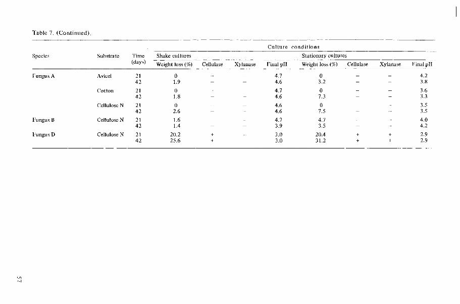
Table 7. (Continued).
Species Substrate Time (days)
Fungus A
Fungus B
Fungus D
Avicel 2 1 4 2
Cotton 2 1 42
Cellulose N 2 1 42
Cellulose N 21 4 2
Cellulose N 2 1 42
Culture conditions
Shake cultures Stationary cultures - -
Wei~ht loss (%) Cellulase Xylanase Final pH Weight loss (%) Cellulase Xylanase Final pH

Table 8. Degradation of cotton and jute fibres, and the production of cellulase and xylanase in liquid cultures with medium EP.
Cot ton Jute Species Time (days) Weight loss % Cellulase Xylanase Final pH Weight loss % Cellulase
Control (not inoculated)
Bispora betulina
Ceratocystis albida
Phialophora sp. A
Wardomyces inflatus
Xylogone sphaerospora
Xylanase Final pH
4.4
4.2
4.2

r- 9 1 w t - t -
I I I
I I 1
2 : 2 1 "
I
-! a C? r- t- t-
I I I
I
2 2 2 I I I
I l l
2 2 2 ! ! I
I I I
' ? L ? C ? t- t- t-
I I I
I l l
t - d - - N
a .- - C Q D m s Q 2
CQ * L? m o r n
I + +
I + +
" N u ! w w m
I + +
I + +
a ? ? W W d
I + +
I + +
z z 2 I + +
+ + +
2 2 ; I + +
+ + +
Y C ? " w w w
I + +
+ + +
t - d m - N
4 M
2 I
Lr,

Table 10. Summat ion of t h e results obtained wi th t h e different test methods.
Species
Acremonium atro-griseum Bispora betulina Catenularia heimii Ceratocystis albida Ceratocystis olivacea
Ceratocystis stenoceras Chrysosporium pannorum Cladorrhinum sp. A Cladosporium resinae Cladosporium sp. A
Coniothyrium fuckelii var. sporulosum Cordana pauciseptata Dictyosporium elegans Gonatobotrys sp. A Graphium sp. A
Humicola alopallonella Humicola grisea Mollisia sp. A Petriellidium boydii Phialocephala dimorphospora
Phialocephala sp. A Phialocephala sp. C Phialophora fastigiata Phialophora hoffmannii Phialophora verrucosa
Phialophora sp. A Pseudeurotium zonatum Rhinocladiella anceps Rhinocladiella sp. A Scytalidium lignicola
Cellulase Xylanase Mannanase Amylase
rest tubes Mycelium- agar plugs Wood blocks
Liquid cultures Weight lossl) Culture filtrate Test tubes
Liquid cultures Wood blocks Culture filtrate Test tubes Test tubes
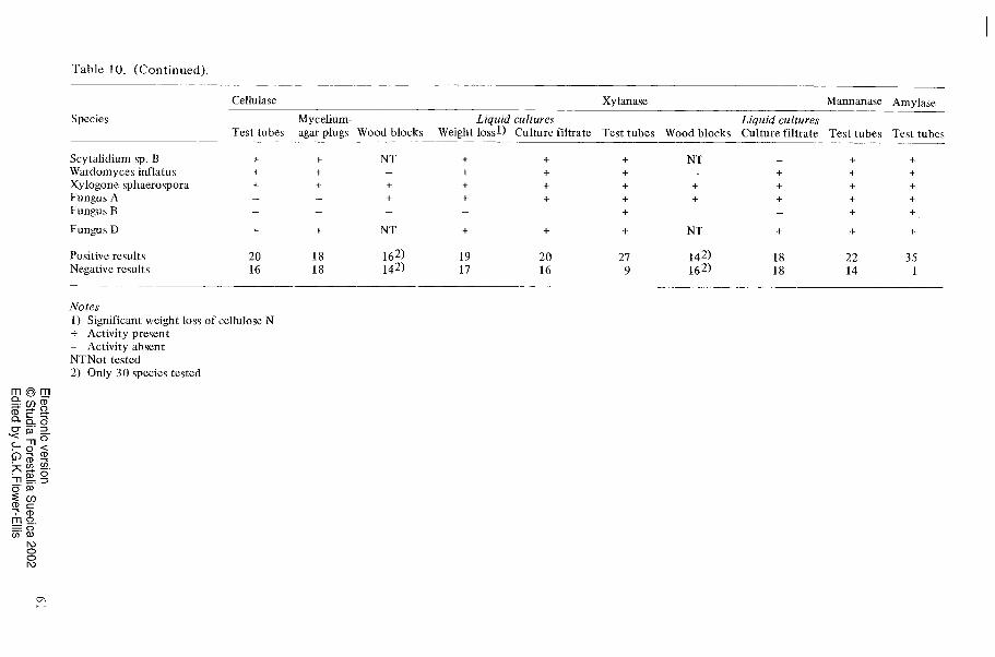
Table 10. (Continued).
Species
Cellulase Xylanase Mannanase Amylase
Mycelium- Liquid cultures Liquid cultures Test tubes agar plugs Wood blocks Weight lossl) Culture filtrate Test tubes Wood blocks Culture filtrate Test tubes Test tubes
Scytalidiurn sp. B Wardornyces inflatus Xylogone sphaerospora Fungus A Fungus B
Fungus D
Positive results Negative results
Notes 1) Significant weight loss of cellulose N + Activity present - Activity absent NTNot tested 2 ) Only 30 species tested