
The Crystallization Behavior of Porous PLA Prepared by ...
10
The Crystallization Behavior of Porous PLA Prepared by Modified Solvent Casting/Particulate Leaching Technique for Potential Use of Tissue Engineering Scaffold Ran Huang a,* , Xiaomin Zhu a , Haiyan Tu a , Ajun Wan a,b,** a School of Chemistry and Chemical Engineering, Shanghai JiaoTong University, Shanghai 200240, China b State Key Laboratory of Pollution Control and Resources Reuse, National Engineering Research Center of Facilities Agriculture, Tongji University, Shanghai 200092, China Abstract The porous PLA foams potential for tissue engineering usage are prepared by a modified solvent casting/particulate leaching method with different crys- tallinity. Since in typical method the porogens are solved in the solution and flow with the polymers during the casting and the crystallinity behavior of PLA chains in the limited space cannot be tracked, in this work the process- ing is modified by diffusing the PLA solution into a steady salt stack. With a thermal treatment before leaching while maintaining the stable structure of the porogens stack, the crystallinity of porous foams is made possible to con- trol. The characterizations indicate the crystallization of porous foams is in a manner of lower crystallibility than the bulk materials. Pores and caves of around 250μm size are obtained in samples with different crystallinity. The macro-structures are not much impaired by the crystallization nevertheless the morphological effect of the heating process is still obvious. Keywords: Porous materials, Biomaterials, Poly(Lactic Acid), Tissue Engineering Scaffold, Crystallization * Corresponding author ** Principal corresponding author Email addresses: [email protected] (Ran Huang ), [email protected] (Xiaomin Zhu), [email protected] (Haiyan Tu), [email protected] (Ajun Wan ) Preprint submitted to Materials Letter October 4, 2018 arXiv:1404.3930v1 [cond-mat.mtrl-sci] 14 Apr 2014
Transcript of The Crystallization Behavior of Porous PLA Prepared by ...
The Crystallization Behavior of Porous PLA Prepared
by Modified Solvent Casting/Particulate Leaching
Technique for Potential Use of Tissue Engineering
Scaffold
Ran Huanga,∗, Xiaomin Zhua, Haiyan Tua, Ajun Wana,b,∗∗
aSchool of Chemistry and Chemical Engineering, Shanghai JiaoTong University,Shanghai 200240, China
bState Key Laboratory of Pollution Control and Resources Reuse, National EngineeringResearch Center of Facilities Agriculture, Tongji University, Shanghai 200092, China
Abstract
The porous PLA foams potential for tissue engineering usage are prepared bya modified solvent casting/particulate leaching method with different crys-tallinity. Since in typical method the porogens are solved in the solution andflow with the polymers during the casting and the crystallinity behavior ofPLA chains in the limited space cannot be tracked, in this work the process-ing is modified by diffusing the PLA solution into a steady salt stack. With athermal treatment before leaching while maintaining the stable structure ofthe porogens stack, the crystallinity of porous foams is made possible to con-trol. The characterizations indicate the crystallization of porous foams is ina manner of lower crystallibility than the bulk materials. Pores and caves ofaround 250µm size are obtained in samples with different crystallinity. Themacro-structures are not much impaired by the crystallization neverthelessthe morphological effect of the heating process is still obvious.
Keywords: Porous materials, Biomaterials, Poly(Lactic Acid), TissueEngineering Scaffold, Crystallization
∗Corresponding author∗∗Principal corresponding authorEmail addresses: [email protected] (Ran Huang ), [email protected]
(Xiaomin Zhu), [email protected] (Haiyan Tu), [email protected] (Ajun Wan )
Preprint submitted to Materials Letter October 4, 2018
arX
iv:1
404.
3930
v1 [
cond
-mat
.mtr
l-sc
i] 1
4 A
pr 2
014

1. INTRODUCTION
Porous poly(lactic acid) (PLA) has been developed for tissue engineeringscaffolds for decades [1-3]. Including poly(L-lactic acid) (PLLA), poly(D-lactic acid) (PDLA) and PLA-based copolymers like poly(lactic-co-glycolic)acid (PLGA), these bio-based resins have been proved to be a successful can-didate of scaffold materials with excellent biocompatibility and biodegrad-ablity. The tissue engineering requires sufficient interconnecting inner spacein the scaffold for biofactor delivery, tissue growth, and the scaffold shouldbe degradable after tissue’s growth meanwhile providing proper mechanicalstrength to support the tissue engineering system. Therefore the balance ofgrowth space, degradation behavior and mechanical properties is the mainconcern of constructing a scaffold. With the respect to the particular re-quirements of certain tissue engineering, nowadays designed preparation andmodification techniques of porous PLA scaffold materials become an inten-sive interests-drawing subject [4-13], which requires more understanding ofthe basic principles of physical and chemical properties, particular in theform of scaffold.
The crystallization of PLA plays an important role in its mechanical prop-erties and degradability. Generally the crystallized polymers have higherstrength and mechanical modulus [14]. In the case of PLA, the crystallinityalso significantly affects the degradability, with the general behavior thatthe degradation time is longer with higher crystallinity, as the crystal seg-ments are more stable than amorphous area and prevent water permeationinto it. For example, it was reported that PLLA takes more than 5 yearsfor total degradation, whereas only about 1 year for the amorphous PLA orPDLLA [15]. However unlike the crystallinity control for the inorganic com-ponents in the tissue engineering scaffold [16], very rare reports concernedthe crystallinity of the polymer scaffold materials. One probable reason isthat the preparation of porous scaffolds is a delicately process, where thecontrol of crystallinity is usually difficult or unavailable. Except the solventcasting/particle leaching method, in other widely used preparation methodssuch as electrospun fiber, phase separation, membrane lamination and gasfoaming, the polymers are not able to experience a thermal treating step,i.e. the most common way to control the crystallinity [1]. In some works thecrystallinity is controlled by the raw materials itself, i.e. selecting raw ma-terials of different molecular weight associated with different crystallizationbehaviors, or a particular processing procedure for chain cleavage to control
2

the crystallinity [17]. And in some methods, even polymer with high crys-tallinity cannot be served as the raw materials to prepare the scaffold, forexample the gas foaming technique reported by David J. Mooney et al. [18].Nevertheless, the study of the crystallization of the scaffold should hold con-siderable practical merits in tissue engineering, for example to fit the scaffolddegradation time with the expected tissue growth time by the control of crys-tallinity (if possible). Also, the scaffold structure may vary with crystallinityand influent the biological behavior of living tissue leaning on it. Park et al.reported a research on the sustained release of human growth hormone fromsemi-crystalline poly(l-lactic acid) and amorphous poly(d,l-lactic-co-glycolicacid) microspheres, which reveals that the morphological effect is importanton protein release [19].
Crystallinity can be tailored in solvent casting/particulate leaching tech-nique. However in this method the salt as porogens are solved in the solutionand flow with the polymers during the casting, therefore without the immo-bilization of porogens the crystallinity behavior in the space-limited gap can-not be tracked [1]. In this work, we modified the solvent casting/particulateleaching technique by diffusing the PLA solution into a steady salt stack in-stead of solving the porogens. The control of crystallinity was made possibleby inserting a thermal treating step before leaching, while maintaining thestable structure of salt stack. We have investigated the morphological effectof limited space on the crystallization of PLA, and the porous structure withdifferent crystallinity under thermal treatments.
2. Experimental
2.1. Materials
The PLA of label 4032D is purchased from Natureworksr, with L/Dratios from 24:1 to 30:1. The porogens is NaCl of analytical grade. The 1:1mixture of dichloromethane and chloroform is served as solvent.
2.2. Porous sponge preparation
The PLA pellets are solved into dichloromethane and chloroform (1:1)with the concentration of 0.2g/l. The NaCl powder is thoroughly groundedand sieved with 109µm then 300µm sieve to screen the particles of sizes inbetween, and paved onto a petri dish where it forms a ˜1.5mm thick disc. ThePLA solution is very slowly poured into the dish at the edge. The pouringis as slow as that the solution diffuses inside the salt stack instead of flowing
3

over the surface, also the slow diffusing guarantees the salt particles are notconsiderably moved by the liquid flowing to keep the inner structure of thesalt stack stable. After pouring, the salt-PLA solution composite is thenplaced in vacuum for 12h for drying out the solvent. The product is a soliddry PLA-glued salt composite ready for thermal treatment. The compositeis placed in water for 48h to leach out the salts after the thermal treatmentfor recrystallization. The leached samples are freeze-dried for 2h and storedin vacuum ready for characterizations.
2.3. Recrystallization
The recrystallization of composite is processed by the heating and coolingprocess. Four samples were made to have different crystallinity. One samplewas kept as the original composite without thermal treatment for reference(sample R). The other three composites are heated in oven at 165◦C for 0.5h,then one composite (sample A) was immediately quenched in liquid nitrogen;sample B was linearly cooled down at the rate 10◦C/30 min.; sample C waslinearly cooled at 10◦C/30 min till 105◦C, kept at 105◦C for 24h, then cooleddown with the same rate to room temperature. For comparison we also madetwo bulk PLA samples with the same crystallization process of sample B andC and they were labeled as sample B’ and C’.
2.4. Characterization
The crystallinity of samples were characterized by X-ray Diffraction (XRD)(D/max-2200/PC, Rigaku Corporation). The porous structure of sampleswere revealed by Scanning Electron Microscope (SEM) (Nova NanoSEM 450,FEI).
3. Results and Discussion
3.1. The crystallization behavior
The XRD results firstly dispel the doubt about the possibility of crys-tallization in confined geometry. Fig.1(1) shows the XRD of three thermaltreated samples A, B and C (Note that the sample R is not included becauseits XRD curve almost overlap with the sample A, i.e. amorphous), and thebulk samples of B’ and C’ are shown in Fig.1(2). With the comparison ofthe bulk behavior, clear crystal peaks at 2θ = 16.6◦ and other minor peaksindicate that the heat treatment makes the crystallization possible but wecan see significant impact of confined geometry, the crystallization of PLA
4

chains in porogens slits is harder and the crystallinity is lower than the bulksample under the same treatment condition. The crystallinity is calculatedto be 17.11% and 17.43% in B and C, comparing to 23.86% and 25.35% inB’ and C’. It is clear that with the same thermal treatment the bulk samplesare much easier to crystallize with higher crystallinity. This agrees with ourexpectation that the crystallization is more difficult in confined geometry,even the size of fibers and walls confining the pores is still in the magnitudeof micrometers (see the SEM paragraph) which far overweighted the diame-ter of chain segments, the chain movement and rearrangement is obstructedby the limited space.
Another evidence that confined space impairs the crystallibility is thecomparable crystallinity of B and C. The small difference is trivial due tomany factors such as sample preparation or the baseline selection. It impliesthat 7h linear cooling process may reach the upper limit crystallinity for theporogens confined environment, while for bulk sample, the effects of stayingat 105◦C for longer time is significant on crystallinity.
Although the samples are leached for 48h there are unavoidable porogensresidues left in the sample. The peaks on 2θ = 31.7◦ and 45.5◦ are NaClcrystals [20], whereas these two peaks do not exist in the bulk samples B’and C’.
3.2. The porous macro-structure
The SEM results in Fig.2 show the macro-structures of unheated originalcasting sample R and three samples with heat treatment A, B and C. Ageneral observation of macro-structure confirms that the thermal treatmentdoes not impressively affect the porous structure forming. The pores andcaves structure in each sample can be clearly observed with the pore sizeof around 250µm, which accords to the 109˜300µm sieving process. Nev-ertheless the morphological effect of heat treatment is obvious. In Fig.2(2),(3) and (4) the reheated samples present the features of thinner pore walls,branches and fragments, while in the reference sample the pore wall is thickerwith rod-like branches. Regardless of the crystallinity, heating the samplesto 165◦C (the melting temperature of PLA) enables the polymer chains toremobilize and diffuse into the slits between salt particles where the solvedchains had not diffused into and occupied. The thinner pore wall indicatesthat the chain remobilization also moves the porogens and makes narrowerspace among them. Although we employ the steady salt stack to confine the
5
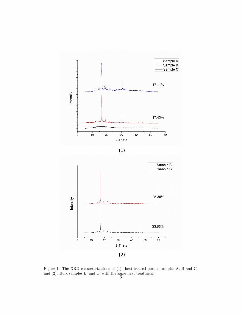
Figure 1: The XRD characterizations of (1): heat-treated porous samples A, B and C,and (2): Bulk samples B’ and C’ with the same heat treatment.
6

Figure 2: The SEM pictures of four porous samples: (1) The reference sample R withoutheat treatment; (2) Sample A with quenching; (3) Sample B with linear cooling; (4) SampleC with linear cooling besides being kept at 105◦C for 24 hours. The scale bar in the firstgraph is 250µm. All the photos have the same scale magnitude.
7

recrystallization within the limited space, the porogens are only relatively”stable” comparing to typical solvent casting technique.
The quenching sample A has more fragmental structures than B and C,it is clear to understand the phenomenon that in sample A the polymer meltdiffused into thinner slits is quenched to solidify its diffusing state of frag-mental features. For sample B and C, the recrystallization process offerssufficient time for the diffused chains to mobilize and rearrange themselvesto be more ordered, crystal structure. This rearrangement provides a lessfragmental structure on the macro-scale. No obvious difference of macro-structures between sample B and C is observed, i.e. 7 hours linear coolingis sufficient for recrystallization to achieve this structural effect, longer re-crystallization time plays very little more effects on the crystallinity and themacro-structure, and this observation also agrees to the crystallinity resultsof B and C as indicated above.
4. Conclusion
The PLA porous matrix for potential use in tissue engineering have beenprepared by modified salt casting and particulate leaching technique. ThePLA solution is diffused into relative stable salt stack, instead of solving saltswith polymers in typical method. Because the raw salt casting solidifies thesalt-PLA composite, we are able to insert a step of thermal treatment torecrystallize the polymer matrix before leaching process. In this way we areable to: 1) investigate the crystallization behavior of PLA confined in limitedspace; 2) develop an available crystallinity control option in porous PLAscaffold preparation. The XRD results indicate the crystallization of porousfoams, in a manner of lower crystallibility than the bulk materials. Themarco-structure of porous samples are observed by SEM, by obtaining thepores of around 250µm, it is revealed that the polymer foam may crystallizewithout significant structure damage. The features of thinner pore walls,branches and fragments confirmed the effect of heating treatment. BothXRD and SEM results of sample B and C indicate that 7 hours linear coolingis sufficient to achieve certain crystallinity and marco-structure.
References
[1] KF Leong, CM Cheah, CK Chua. Biomaterials 24 (2003) 2363–2378.
8

[2] K Rezwana, QZ Chena, JJ Blakera, AR Boccaccinia. Biomaterials 27(2006) 3413–3431.
[3] I Armentano, M Dottori, E Fortunati, S Mattiolia, JM Kenny. PolymerDegradation and Stability 95 (2010) 2126-2146.
[4] Vacanti JP, Morse MA, Saltzman WM, Domb AJ, Peter-Atayde A,Langer R. J Pediatr Surg 1988;23(1):3–9.
[5] Mikos AG, Sarakinos G, Leite SM, Vacanti JP, Langer R. Biomaterials1993;14(5):323–30.
[6] Lu L, Mikos AG. MRS Bull 1996;21(11):28–32.
[7] Thomson RC, Shung AK, Yaszemski MJ, Mikos AG. Polymer scaffoldprocessing. In: Lanza RP, Langer R, Vacanti JP, editors. Principles oftissue engineering, 2nd ed. San Diego: Academic Press, 2000. p. 251–62[Chapter 21].
[8] Yang SF, Leong KF, Du ZH, Chua CK. Tissue Eng 2001;7(6):679–89.
[9] Roether JA, Boccaccini AR, Hench LL, Maquet V, Gautier S, Jerome,R. Biomaterials 2002;23:3871–8.
[10] Ma PX, Zhang R. J Biomed Mater Res 2001;56:469–77.
[11] Xiong Z, Yan YN, Wang SG, Zhang RJ, Zhang C. Scr Mater2002;46:771–6.
[12] Taboas JM, Maddox RD, Krebsbach PH, Hollister SJ. Biomaterials2003;24:181–94.
[13] Lu HH, El-Amin SF, Scott KD, Laurencin CT. J Biomed Mater Res A2003;64A:465–74.
[14] M. Rubinstein, Ralph H. Colby. 2003, Polymer Physics, 1st Edition,Oxford University Press Inc., New York.
[15] Rich J, Jaakkola T, Tirri T, Narhi T, Yli-Urpo A, Seppala J. In vitroevaluation of poly([var epsilon]-caprolactone-co-DL-lactide)/bioactiveglass composites. Biomaterials 2002;23:2143–50.
9

[16] Chihiro Mochizuki, Yuji Sasaki, Hiroki Hara, Mitsunobu Sato, TohruHayakawa, Fei Yang, Xixue Hu, Hong Shen, Shenguo Wang. Journalof Biomedical Materials Research Part B: Applied Biomaterials Volume90B, Issue 1, pages 290–301, July 2009.
[17] Sundararajan V. Madihally, Howard W.T. Matthew. Biomaterials 20(1999) 1133-1142.
[18] David J. Mooney, Daniel F. Baldwin, Nam P. Suh, Joseph P. Vacanti,Robert Larger. Biomaterials 17 (1996) 1417-1422.
[19] Hong Kee Kim, Tae Gwan Park. Journal of Controlled Release 98 (2004)115 – 125
[20] NaCl powder X-ray diffraction pattern, Retrieved fromhttp://www.uiowa.edu/˜c004206/hand9.pdf (April 8, 2014).
10


![Interface and bonding mechanisms of plant fibre composites ... · crystallization of starch owing to the strong interaction between the fibre and starch[35-38]. Polylactic acid (PLA)](https://static.fdocuments.in/doc/165x107/5f3bf9326aec45194a507b17/interface-and-bonding-mechanisms-of-plant-fibre-composites-crystallization-of.jpg)


