The CRISPR/Cas9-created MDM2 T309G enhances vitreous ... · activator specificity protein (Sp)1,...
24
MDM2 T309G enhances vitreous-induced cell survival 1 THE CRISPR/CAS9-CREATED MDM2 T309G ENHANCES VITREOUS- INDUCED EXPRESSION OF MDM2 AND PROLIFERATION AND SURVIVAL OF CELLS Yajian Duan 1,2,3,6 *, Gaoen Ma 1,2,3 *, Xionggao Huang 1,2,3 , Patricia A. D’Amore 1,2,3,4 , Feng Zhang 5 , Hetian Lei 1,2,3 ** From 1 Schepens Eye Research Institute; 2 Massachusetts Eye & Ear; 3 Department of Ophthalmology, and 4 Department of Pathology, Harvard Medical School, Boston, Massachusetts, USA; 5 Broad Institute of Massachusetts Institute of Technology and Harvard University, Cambridge, Massachusetts, USA; 6 Shanxi Eye Hospital, Taiyuan, Shanxi, China Running title: MDM2 T309G enhances vitreous-induced cell survival * Both authors made equal contributions to this work. ** To whom correspondence should be addressed: Hetian Lei, Ph.D., Schepens Eye Research Institute of Massachusetts Eye and Ear, 20 Staniford Street, Boston, MA. 02114 Telephone: 617-912-2521 (office); Fax: 617-912-0101; email: [email protected] Keywords: CRISPR, Cas9, MDM2 T309G, vitreous, proliferation, survival ABSTRACT The 309G allele of single nucleotide polymorphisms (SNPs) in the mouse double minute (MDM2) promoter locus is associated with a higher risk of cancer and proliferative vitreoretinopathy (PVR), but as to whether this SNP G309 contributes to the pathogenesis of PVR is to-date unknown. The clustered, regularly interspaced, short palindromic repeats (CRISPR)-associated endonuclease (Cas)9 from Streptococcus pyogenes (SpCas9) can be harnessed to manipulate a single or multiple nucleotides in mammalian cells. Here, we delivered SpCas9 and guide RNAs (SpGuides) using dual adeno- associated viral (AAV)-derived vectors to target the MDM2 genomic locus together with a homologous repair template for creating the mutation of MDM2 T309G in human primary retinal pigment epithelial (hPRPE) cells, whose genotype is MDM2 T309T. The next generation sequencing results indicated that there was 42.51% MDM2 G309 in the edited hPRPE cells using the AAV-CRISPR/Cas9. Our data showed that vitreous induced an increase in MDM2 and subsequent attenuation of p53 expression in the MDM2 T309G hPRPE cells. Furthermore, our experimental results demonstrated that the MDM2 T309G in the hPRPE cells enhanced vitreous-induced cell proliferation and survival, suggesting that this SNP contributes to the pathogenesis of PVR. Proliferative vitreoretinopathy (PVR) is a vision-threatening disease resulting from surgical correction of rhegmatogenous retinal detachment (RRD) and open ocular injury (1), and it is characterized by the formation of preretinal or epiretinal membranes (ERMs) (2). The ERMs consist of extracellular http://www.jbc.org/cgi/doi/10.1074/jbc.M116.729467 The latest version is at JBC Papers in Press. Published on May 31, 2016 as Manuscript M116.729467 Copyright 2016 by The American Society for Biochemistry and Molecular Biology, Inc. by guest on June 8, 2019 http://www.jbc.org/ Downloaded from
Transcript of The CRISPR/Cas9-created MDM2 T309G enhances vitreous ... · activator specificity protein (Sp)1,...

MDM2 T309G enhances vitreous-induced cell survival
1
THE CRISPR/CAS9-CREATED MDM2 T309G ENHANCES VITREOUS-INDUCED EXPRESSION OF MDM2 AND PROLIFERATION AND SURVIVAL OF CELLS
Yajian Duan1,2,3,6*, Gaoen Ma1,2,3*, Xionggao Huang1,2,3, Patricia A. D’Amore1,2,3,4, Feng Zhang5, Hetian Lei1,2,3**
From 1Schepens Eye Research Institute; 2Massachusetts Eye & Ear; 3Department of Ophthalmology, and 4Department of Pathology, Harvard Medical School, Boston, Massachusetts, USA; 5Broad Institute of Massachusetts Institute of Technology and Harvard University, Cambridge, Massachusetts, USA; 6Shanxi Eye Hospital, Taiyuan, Shanxi, China
Running title: MDM2 T309G enhances vitreous-induced cell survival
*Both authors made equal contributions to this work. ** To whom correspondence should be addressed: Hetian Lei, Ph.D., Schepens Eye Research Institute of Massachusetts Eye and Ear, 20 Staniford Street, Boston, MA. 02114 Telephone: 617-912-2521 (office); Fax: 617-912-0101; email: [email protected] Keywords: CRISPR, Cas9, MDM2 T309G, vitreous, proliferation, survival ABSTRACT
The 309G allele of single nucleotide polymorphisms (SNPs) in the mouse double minute (MDM2) promoter locus is associated with a higher risk of cancer and proliferative vitreoretinopathy (PVR), but as to whether this SNP G309 contributes to the pathogenesis of PVR is to-date unknown. The clustered, regularly interspaced, short palindromic repeats (CRISPR)-associated endonuclease (Cas)9 from Streptococcus pyogenes (SpCas9)
can be harnessed to manipulate a single or multiple nucleotides in mammalian cells. Here, we delivered SpCas9 and guide RNAs (SpGuides) using dual adeno-associated viral (AAV)-derived vectors to target the MDM2 genomic locus together with a homologous repair template for creating the mutation of MDM2 T309G in human primary retinal pigment epithelial (hPRPE) cells, whose genotype is MDM2 T309T. The next generation sequencing
results indicated that there was 42.51% MDM2 G309 in the edited hPRPE cells using the AAV-CRISPR/Cas9. Our data showed that vitreous induced an increase in MDM2 and subsequent attenuation of p53 expression in the MDM2 T309G hPRPE cells. Furthermore, our experimental results demonstrated that the MDM2 T309G in the hPRPE cells enhanced vitreous-induced cell proliferation and survival, suggesting that this SNP contributes to the pathogenesis of PVR.
Proliferative vitreoretinopathy (PVR) is a vision-threatening disease resulting from surgical correction of rhegmatogenous retinal detachment (RRD) and open ocular injury (1), and it is characterized by the formation of preretinal or epiretinal membranes (ERMs) (2). The ERMs consist of extracellular
http://www.jbc.org/cgi/doi/10.1074/jbc.M116.729467The latest version is at JBC Papers in Press. Published on May 31, 2016 as Manuscript M116.729467
Copyright 2016 by The American Society for Biochemistry and Molecular Biology, Inc.
by guest on June 8, 2019http://w
ww
.jbc.org/D
ownloaded from

MDM2 T309G enhances vitreous-induced cell survival
2
matrix proteins and cells including retinal pigment epithelial (RPE) cells, retinal glial cells, fibroblasts, and macrophages. PVR occurs in 8-10% of patients who have undergone a surgical repair of RRD, and accounts for approximately 75% of all primary failures following the surgery (2-8).
The oncogene protein murine double minute 2 (MDM2), an E3 ubiquitin-protein ligase, whose human homologue (also called Hdm2) is an important negative regulator of the p53 tumor suppressor (9-11); the phenotype of murine embryonic lethality of MDM2 null can be prevented by knocking out the p53 gene (12,13). Vitreous from experimental rabbits preferentially activates platelet-derived growth factor receptor α (PDGFRα). This activation in turn triggers the downstream signaling pathway of phosphoinositide 3 kinase (PI3K)/Akt, which phosphorylates MDM2, thereby enhancing p53 degradation (14). Blocking MDM2 binding to p53 with a small molecule Nutlin-3 protects rabbits against retinal detachment in a PVR rabbit model (3).
Intriguingly, the G allele of single nucleotide polymorphisms (SNPs) (rs2279744) in the MDM2 promoter locus has subsequently been found to be associated with a higher risk of PVR for RRD patients (2,15). This SNP is also associated with an increased risk of carcinogenesis (15-21). The SNP T309G (a T to G change at the 309th nucleotide) at the MDM2 first-intron promoter locus enhances the affinity of the transcriptional activator specificity protein (Sp)1, leading to a heightened expression of MDM2 and the subsequent attenuation of p53 expression in cancer cells (15). However, whether or not this SNP contributes to the pathogenesis of PVR has not been explored.
The system of clustered regularly interspaced short palindromic repeats (CRISPR) and CRISPR-associated nucleases (Cas) in bacteria and archaea provides adaptive immunity against viruses and plasmids when their CRISPR RNAs (crRNAs) are used to guide the Cas cleavage of the foreign nucleic acids (22-24). In Streptococcus pyogenes (Sp) the Cas9 (SpCas9) contains two nuclease domains, RuvC and HNH, each of which can cleave one strand of the double-stranded target DNA when directed by the crRNA and transactivating crRNA (tracrRNA) (24,25). This SpCas9 can be reprogrammed to target specific genomic loci in mammalian cells using the processed single guide (sg) RNAs that consist of the crRNA and tracrRNA(24). The double-stranded DNA breaks (DSBs), at the specific genomic loci produced by the CRISPR/Cas9, can be repaired by endogenous repair machinery for either non-homologous end-joining (NHEJ) or homology-directed repair (HDR), which depends on the cell state and presence of a repair template (26,27). NHEJ and HDR are two distinct competent repair pathways in the cells. NHEJ can introduce unpredict-able insertions and deletions (indels), and it may repair the lesion by simply rejoining the two DSB ends (26); HDR can use an exogenous single- or double-stranded DNA template with the desired changes to make mutations in the genomic loci (26). However, HDR is less frequently used than NHEJ because it occurs only during S and G2 phases, whereas NHEJ can be found throughout the cell cycle (26,28). The CRISPR/Cas9 technology has recently been used in a variety of genome-editing applications in eukaryotic cells and mice (26,29-32), and it provides a unique opportunity to demonstrate whether the MDM2 T309G contributes to the pathogenesis of PVR.
by guest on June 8, 2019http://w
ww
.jbc.org/D
ownloaded from

MDM2 T309G enhances vitreous-induced cell survival
3
Here, we generated the mutation of T309G in the MDM2 genomic locus, in the human primary retina pigment epithelial (hPRPE) cells using CRISPR/Cas9 technology. We demonstrated that the vitreous from experimental rabbits (RV) increased expression of MDM2 and subsequent attenuation of p53 expression in the hPRPE cells with the MDM2 T309G; furthermore, we found that the MDM2 T309G in the hPRPE cells promoted RV-induced cell proliferation and survival, which are intrinsic to the development of PVR. RESULTS
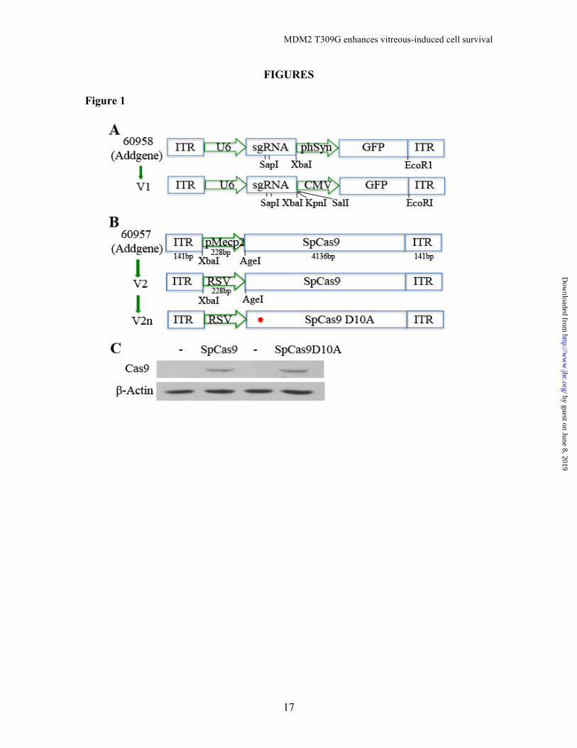
Creation of MDM2 T309G in the genomic locus using CRISPR/Cas9 − The SNP MDM2 T309G is associated with a higher risk of PVR (2)(2); however, it is not known whether this SNP contributes to PVR. Because RPE cells are believed to be the major cell type in the PVR membranes that cause retinal detachment in the development of PVR (2,5-7), we attempted to create this SNP in hPRPE cells using CRISPR/Cas9 technology. Since the ultimate goal of this research is to explore a novel therapeutic approach to PVR, and AAVs do not cause any disease (33), we chose AAV-derived viral vectors to deliver CRISPR/Cas9 into our target cells. However, due to the packaging size limitation of the AAV-derived vectors, we had to adapt a dual-vector system, which packages SpCas9 and sgRNA expression cassettes (SpGuide) in two separate viral vectors, pAAV-SpCas9 and pAAV-SpGuide, respectively (30). To separate hPRPE cells transduced by pAAV-SpGuide, we replaced GFP promoter hSyn (30) with the promoter of CMV (Fig. 1A). There are four types of cells in the PVR membrane, including RPE cells; therefore, we substituted the promoter pMecp2 (30)
for the promoter of the RSV to drive expression of SpCas9 (34) (Fig. 1B).
The SpCas9 contains two conserved nuclease domains, HNH and RuvC, which cleave the target DNA strand complementary and non-complementary to the guide RNA, respectively. A mutation of aspartate-to-alanine (D10A) in the RuvC catalytic domain can convert SpCas9 into the DNA nickase (SpCas9D10A). Two SpCas9 D10A-nicking enzymes directed by a pair of sgRNAs targeting opposite strands of a target locus can mediate double DNA strand breaks while minimizing off-target activity, because single-strand nicks are preferentially repaired by the high-fidelity base excision repair pathway (35). Thus, the SpCas9 in the AAV vector was mutated to SpCas9 D10A with an in situ mutagenesis kit (Fig. 1B). To test the effectiveness of the DNA constructs, we transfected these two vectors separately into 293T cells. Western blot analysis showed that the SpCas9 and SpCas9 D10A were successfully expressed in the transfected 293T cells (Fig. 1C).
We next sought to test the efficiency of SpCas9-mediated editing of the genomic MDM2 locus around the SNP in the RPE cells. To identify RPE cells that were suitable for our experimental purpose, we isolated the genomic DNA from hPRPE cells and PCR amplified a region around the MDM2 SNP for Sanger DNA sequencing, and found that there was T309T in the MDM2 SNP in the hPRPE cells as shown in Fig. 2A. These cells were then used to introduce MDM2 T309G in the genomic locus.
In synthesizing sgRNAs the two 20-nt targeted sequences (30,36-38) (Fig. 2B) were cloned into the pAAV-SpGuide backbone (Fig. 1A), and the clones were verified by DNA sequencing.
by guest on June 8, 2019http://w
ww
.jbc.org/D
ownloaded from

MDM2 T309G enhances vitreous-induced cell survival
4
To edit the genomic MDM2 locus, we transfected hPRPE cells with the dual vectors of pAAV-SpCas9 plus pAAV-MDM2-sgRNA 1 or 2 using electroporation. pAAV-LacZ-sgRNA was used as a control. The transduction efficiency was about 60%, as estimated by immunofluorescence. To determine if any indels were mediated by the CRISPR/Cas9 system, we isolated genomic DNA from the transfected hPRPEs and amplified the region around the MDM2 SNP using a high fidelity Herculase II fusion polymerase. The amplified DNA fragments were then subjected to Sanger DNA sequencing (39). As shown in Fig. 2C there were mutations in front of PAMs (MDM2-sgNRA1: CGG; MDM2-sgRNA2: AGG) from the PCR products derived from the transduced hPRPE cells with SpCas9 plus MDM2-sgRNA 1 or 2, but not from those with LacZ-sgRNA. These results demonstrate that the two MDM2 sgRNAs efficiently guided the SpCas9 to induce indels in the hPRPE cells.
To create the SNP MDM2 T309G in the genome of the hPRPE cells, we chose to use SpCas9D10A nickase activity, as there was no off-target DNA sequence found for the pair of sgRNA1 +2, based on the double nickase design tool (crispr.mit.edu). Thus, the U6-sgRNA2 was PCR amplified and cloned into the pAAV-U6-sgRNA1 vector. The pAAV-SpCas9D10A, pAAV-MDM2-sgRNA 1 and 2 and ssHRT (Fig. 3A) together were transfected into the hPRPE cells by electroporation. The HDR donor template that consisted of a single strand, 96 base pair (bp) genomic sequence homologous to a region encompassing the SNP with a G309 replacement of T309. However, the mutagenesis efficiency using this HDR strategy is very low (only 0.5–20%) (26,28,40,41). To increase the efficiency of HDR-mediated genome editing, we
immediately treated the post-transfected cells with 0.5 µM Scr7, which can inhibit the NHEJ, a competent HDR pathway (26). On the third day after the transfection, the GFP-expressing hPRPE cells were sorted by FACS, and the genomic DNA fragments around the SNP from some of the sorted cells were amplified by PCR using high fidelity Herculase II DNA polymerases. The Surveyor nuclease assay of the PCR products indicated that there was a DNA fragment (about 100 bp) released (Fig. 3B) in the MDM2-sgRNA transfected cells, but there was none in the control LacZ-sgRNA transfected cells, suggesting that there were indels in the middle of the PCR fragment (about 200 bp). Sanger DNA sequencing (Fig. 3C) confirmed that there were heterozygous MDM2 T309 and G309 in the selected hPRPE cells transduced with the SpCas9 and MDM2-sgRNA. NGS analysis indicated that there were 42.51% MDM2 G309 and 57.19% MDM2 T309 in the GFP-positive hPRPE cells transfected with SpCas9D10A, MDM2-sgRNA1+2 and the HDR template (Fig. 3D). Some of the transduced cells were sorted into PCR tubes (a cell per tube), and the genomic DNA from single cells was subjected to PCR amplification of the fragment around the SNP for Sanger DNA sequencing. The sequencing results indicate that in the 309 position there were 20% (2/10) cells containing the T/T, 70% (7/10) cells with the T/G, 10% (1/10) cells with the G/G (Fig. 3E). These results demonstrate that the genomic MDM2 T309G was successfully created in the hPRPE cells using the CRISPR/Cas9 technology. MDM2 T309G promotion of vitreous-stimulated an increase in MDM2 and a decrease in p53 − The MDM2 intron promoter region SNP T309G (rs2279744) has been shown to increase the affinity of
by guest on June 8, 2019http://w
ww
.jbc.org/D
ownloaded from

MDM2 T309G enhances vitreous-induced cell survival
5
the transcriptional activator Sp1, resulting in elevated expression of MDM2 in some cancer cell lines (15). In addition, this SNP is associated with a higher risk of PVR (2). Therefore, we tested whether the CRISPR/Cas9-created MDM2 T309G would elevate expression of MDM2 in the hPRPE cells and whether RV would influence this change in MDM2 and p53 between the hPRPE cells with the MDM2 T309G SNP and WT T309T (WT). Unexpectedly, the SNP failed to enhance MDM2 expression in the MDM2 T309G hRPE cells in comparison with that in the WT cells (Fig. 4A); however, the vitreous enhanced expression of MDM2 and effected a decrease in p53 in the hPRPE cells with the SNP, as compared to the WT (Fig. 4A). These results suggested that RV induced Sp1 association with the MDM2 promoter motif with the SNP and thus enhanced expression of MDM2 and subsequent suppression of p53. If this prediction was correct, blocking the association between MDM2 and p53 should recover the p53 loss. Thus, we treated the hPRPE cells for 16 hours with both RV and Nutlin-3. Western blot analysis of the treated cell lysates (Fig. 4A) indicated that Nutlin-3 prevented RV-induced reduction in p53 in the MDM2 T309G hPRPE cell. This result indicates that the vitreous-induced decrease in p53 was due to vitreous-stimulated interaction between MDM2 and p53.
To examine whether the vitreous-induced increase in MDM2 was because of the vitreous-stimulated association of Sp1 with the MDM2 GC-rich promoter, we treated the hPRPE cells with a Sp1 inhibitor mithramycin A, an aurelic antibiotic that has been shown to selectively inhibit Sp1 transcription factor-mediated transcriptional activation by blocking Sp1 binding to the GC-rich promoter motif. As shown in Fig. 4B,
treatment with mithramycin A blunted a RV-stimulated increase in MDM2 and a decrease in p53 in the hPRPE cells with the MDM2 T309G, suggesting that RV enhanced Sp1 association with the MDM2 GC-rich promoter.
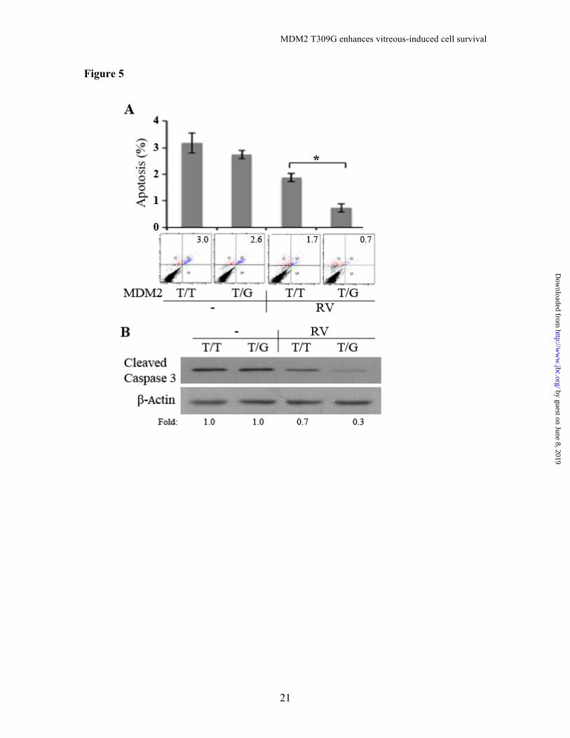
Next, we asked how the vitreous could induce the Sp1-dependent increase in MDM2. Since Erk can phosphorylate Sp1 at Thr 936 (42), thereby enhancing its transcriptional activity, we hypothesized that vitreous could induce Erk phosphorylation of Sp1, which would increase the Sp1 association with the GC-rich motif and promote its target expression. As predicted, treatment of hPRPE cells with an Erk inhibitor PD98059 for 16 hours blocked the vitreous-stimulated phosphorylation of Erk and Sp1 and prevented the vitreous-induced increase in MDM2 in the PRPE cells with the SNP (Fig. 4B). These results demonstrate that the MDM2 T309G in the PRPE cells leads to an enhanced RV-induced Erk/Sp1 dependent increase in MDM2 and a decrease in p53. These biochemical reactions could promote vitreous-induced cellular responses intrinsic to PVR. MDM2 T309G enhancement of RV-induced cell proliferation and survival − Increased MDM2 leads to a decrease in p53 and thus promotes cell proliferation and survival against apoptosis (15,43). RV stimulated an increase in MDM2 in the hPRPE cells with the MDM2 T309G but not with the WT (Fig. 5). Therefore, we suspected that after surgical attachment of the retina, RPE cells enter the vitreous, and that the RPE cells with the T309G or G309G will survive better than those with the WT cells in the MDM2 intron promoter locus. To test this, we treated hPRPE cells with RV for three days and analyzed them for apoptosis. As shown in Fig. 5, with RV
by guest on June 8, 2019http://w
ww
.jbc.org/D
ownloaded from

MDM2 T309G enhances vitreous-induced cell survival
6
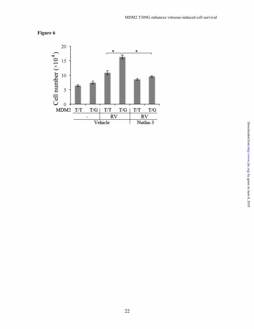
treatment, the hPRPE cells with the MDM2 T309G underwent less apoptosis than those with the WT; in addition, hPRPE cells with the SNP proliferated more in response to vitreous than did those with the WT cells (Fig. 6).
DISCUSSION
In this article, we demonstrate that MDM2 T309G in the hPRPE cells enhances a RV-stimulated increase in MDM2 concomitant with a decrease in p53, as well as cellular responses intrinsic to PVR compared to the WT. These results are consistent with a two hit hypothesis (44). While the polymorphisms of T or G alleles (rs2279744) in the MDM2 309 position did not influence MDM2 expression, they did lead to a differential response to the vitreous stimulation. Notably, MDM2 G309 enhances Sp1-dependent expression of MDM2 in cancer cells in comparison to MDM2 T309 (15). Taken together, our results and clinical findings of MDM2 SNP association with PVR(2) suggest that this SNP contributes to the development of PVR.
We speculate that MDM2 G309 RPE cells respond to vitreous with increased survival, proliferation, and elevated expression of fibrotic proteins (e.g., fibronectin and collagen). We found that the inhibitors of Sp1 and Erk did not completely block vitreous-induced p53 decrease, suggesting that vitreous may activate another pathway to suppress p53 expression. The vitreous could activate Akt (Fig. 7A) (14), which could phosphorylate MDM2, enhancing its association with p53 and promoting its degradation (Fig. 7) (45,46).
In this study, our efforts in raising the percentage of the hPRPE cells with the mutant MDM2 T309G in the whole population were made in two ways. One was the treatment of the AAV-
CRISPR/Cas9 transduced cells with the drug Scr7, which targets the DNA binding domain of DNA ligase IV for inhibiting the ligase IV–dependent NHEJ and enhancing the frequency of HDR (26). The CRISPR/Cas9 system we used is a reprogrammed one from microbial type II CRISPR systems and has been harnessed to facilitate facile genetic manipulations in a variety of cell types and organisms (29). The reprogrammed Cas9 can be guided to generate targeted-DSBs that stimulate genome editing via one of the two DNA damage repair pathways: NHEJ, resulting in insertions and deletions, or HDR, resulting in precise sequence substitution in the presence of a repair template (26). SpCas9 D10A, the mutant form of SpCas9, has been previously shown to facilitate HDR at on-target sites (47), but its efficiency is substantially lower than that of WT Cas9. In our research, we have utilized the double-nicking strategy, which maintains high on-target efficiency while reducing off-target modifications to background levels. Even so, the HDR efficiency might be still low. Thus the drug Scr7 was subjected to our research for increasing the HDR rate. The other strategy was to sort out the transduced hPRPE cells by FACS because in the pAAV-SpGuide vector there was a CMV-GFP expression cassette. Importantly the hPRPE cells could continuously grow and proliferate after cell sorting.
In summary, we have shown that the CRISPR/Cas9-created MDM2 T309G in the hPRPE cells enhanced vitreous-induced expression of MDM2 and cell proliferation and survival. Experimental Procedures Major reagents − The antibodies against Cas9, Erk, phospho (p)-Erk, Sp1, p-Sp1 (T739), Akt, p-Akt (S473), cleaved Caspase 3, p-MDM2 (S166) and p53 were
by guest on June 8, 2019http://w
ww
.jbc.org/D
ownloaded from

MDM2 T309G enhances vitreous-induced cell survival
7
purchased from Cell Signaling Technology (Danvers, MA). The antibody against MDM2 was from Abgent (San Diego, CA). The primary antibodies against β-Actin and the secondary antibodies of the horseradish peroxidase (HRP)-conjugated goat anti-rabbit IgG and anti-mouse IgG were purchased from Santa Cruz Biotechnology (Santa Cruz, CA). Enhanced chemiluminescent substrate for detection of HRP was from Pierce Protein Research Products (Rockford, IL). Carbenicillin and puromycin were purchased from Sigma (St. Louis, MO), and Scr7 was from Xcessbio Biosciences, Inc. (San Diego, CA). Mithramycin A and Nutlin-3 were from Cayman (Ann Arbor, Michigan), and PD98059 was from Cell Signaling Technology. DNA constructs − To generate single guide RNAs (sgRNAs) for SpCas9 targets, we used the CRISPR design tool (http://crispr.mit.edu) to select the two 20-nt target sequences preceding a 5′-NGG of a protospacer adjacent motif (PAM) sequence around the MDM2 SNP309 in the MDM2 genomic locus (Rs2279744, NC_000012.12) (30). Target sequences used were: target 1 (5’- CGGGAGGTCCGGATGATCGC-3’) and target 2 (5’-CGAAGCGGCCCCGCAGC CCC-3’). The control sgRNA sequence was designed to target the LacZ gene from Escherichia coli (target sequence: 5’-TGCGAATACGCCCACGCGATGGG-3’)(30). The pAAV-U6-sgRNA-CMV (cytomegalovirus)-GFP (green fluorescent protein) vector (V1) was originated from
an AAV vector [Addgene (Cat. 60958), Cambridge, MA] (30) by replacing the hSyn-GFP with the PCR (polymerase chain reaction)amplified CMV-GFP from pEGFP-C1 vector (Clontech, Cat. 6084-1, Mountain View, CA) using Xbal/EcoRI. The PCR primers for this amplification were: forward 5’- CGTCTAGAgGGTACC gGGGCCCgGTCGACTAGTTATTAATAGTAATCAATTACGG-3’, and reverse 5’-CAGAATTCGCTGCAGGTTATCGAGA TCTGAGTCCGGACTTGTA-3’. The pAAV-RSV-SpCas9 (V2) was derived from an AAV vector (Addgene, Cat. 60957) by replacing the promoter pMecp2 with the PCR-amplified RSV (Rous Sarcoma Virus) from a pLKO.1-shMDM2 vector (GE Dharmacon, Lafayette, CO) using XbaI/AgeI. The PCR primers for this amplification were: forward 5’- CGGTCTAGAAATGTAGTC TTATG CAATAC -3’, and reverse 5’ –CGGA CCGGTTTTATGTATCGAGCTAGGCA C -3’. The SpCas9 mutation (D10A) was generated in the vector V2 using the following set of mutagenic primers: 5’-CAGCATCGGCCTGGCCATCGGCACC AACT-3’ and its complimentary oligonucleotide, according to instructions provided with a QuickChange XL site-directed mutagenesis kit (Stratagene, La Jolla, CA).
To express SpGuides in the hPRPE cells, the top oligos: 5’-ACCG-20 nucleotide (nt) (target MDM2 DNA sequences 1, 2 or LacZ sgRNA sequence) and bottom oligos: 5’-AAC-20 nt-C (20 nt: complimentary target MDM2 DNA sequences or LacZ sgRNA sequence) were
annealed and cloned into the pAAV vector V1 by SapI. All clones were confirmed by DNA sequencing using a primer 5’-GGACTATCATATGCTTACCG-3’ from the sequence of U6 promoter, which drives expression of sgRNAs.
Both synthesis of primers and
oligos and sequencing of PCR products, clones and mutations were done by Massachusetts General Hospital’s (MGH’s) DNA Core Facility (Cambridge, MA).
by guest on June 8, 2019http://w
ww
.jbc.org/D
ownloaded from

MDM2 T309G enhances vitreous-induced cell survival
8
Cell culture and transfection − hPRPE cells were purchased from Lonza (Walkersville, MD) and cultured in Dulbecco's Modified Eagle Medium (DMEM): nutrient mixture F-12 medium (F12) (Invitrogen, Grand Island, NY) supplemented with 10% fetal bovine serum (FBS) and 1% penicillin/streptomycin. Human embryonic kidney (HEK) 293T cells (293 containing SV40 T-antigen) from Dana-Farber Cancer Institute/Harvard Medical School (Boston, MA) were cultured in DMEM (high glucose, 4.5g/ml) supplemented with 10% FBS. All cells were cultured at 37°C in a humidified incubator with 5% CO2 (4).
The constructs of pAAV-RSV-SpCas9 and pAAV-RSV-SpCas9D10A were respectively transfected into 293T cells using lipofectamine. Briefly, during transfection, lipofectamine 2000 (Invitrogen) (3 µl) were mixed with the plasmid (1 µg) and incubated at room temperature for 30 minutes. The transfection mix was then transferred to 293T cells that were approximately 70% confluent. After 48 hours, the resulting lysates were subjected to western blot analysis using antibodies against Cas9 and β-Actin (4). Western blot analysis − Cells at 90% confluence in either a 24- or 48-well plate were deprived from serum for 24-hour, continuous incubation and were then treated with or without rabbit vitreous (RV, diluted 1:2 in DMEM/F12) for 16 hours. After the cells were washed twice with ice-cold phosphate-buffered saline (PBS), they were lysed in a 1 × sample buffer, which was diluted with extraction buffer (10 mM Tris-HCl, pH 7.4, 5 mM EDTA (ethylenediaminetetraacetic acid), 50 mM NaCl, 50 mM NaF, 1% Triton X-100, 20 µg/mL aprotinin, 2 mM Na3VO4, and 1 mM phenylmethylsulfonyl fluoride)
from the 5 × protein sample buffer [25 mM EDTA (pH=7.0), 10% sodium dodecyl sulfate (SDS), 500 mM dithiothreitol, 50% sucrose, 500 mM Tris HCl (pH=6.8), and 0.5% bromophenol blue]. The samples were boiled for 5 minutes and then centrifuged for 5 minutes at 13,000 ×g. Proteins in the centrifuged and heated samples were separated by 10% SDS-polyacrylamide gel electrophoresis (PAGE), transferred to polyvinylidene difluoride (PVDF) membranes, and subjected to western blot analysis using the appropriate antibodies. Signal intensity was determined by densitometry, using NIH Image J software (4). Transfection of hPRPE cells − The hPRPE cells were transfected with pAAV-SpCas9D10A and pAAV-SpGuide (MDM2-sgRNA or LacZ-sgRNA) supplemented with a single strand homology repair template (ssHRT) (5’-GGAGTTCAGGGTAA AGGTCACG GG GGCCGGGGGCTGCGGGGCCGCTGCGGCGCGGGAGGTCCGGATGATCGCA GGTGCCTGTCGGGTCACTAG-3’) [Integrated DNA Technologies (IDT), Coraville, IA] using electroporation (Amaxa Biosystem). Briefly, 5 ×10⁵ cells were centrifuged at 100 ×g for 10 minutes at room temperature, and the cell pellet was carefully resuspended in a 100 µl, room-temperature Nucleofector solution (Lonza). The cell solution was mixed with 2 µg pAAV-SpCas9D10A, 2 µg pAAV-SpGuide vector and 0.2 µg ssHRT, and then transferred into a certified cuvette placed in the nucleofector cuvette holder. The transfection was then started in a predetermined program. Subsequently, a 500 µl of the pre-equilibrated culture medium was added into the cuvette, and then transfected cells transferred all in one well of a 6-well plate for continuous culture, supplemented with Scr7 (5 µM).
by guest on June 8, 2019http://w
ww
.jbc.org/D
ownloaded from
MDM2 T309G enhances vitreous-induced cell survival
9
At 72 hours post-transfection, the GFP-positive hPRPE cells were sorted by fluorescence-activated cell sorting (FACS) to enrich the population of the transduced cells or into PCR tubes (a cell per tube with 5 µl QuickExtract DNA Extraction Solution (Epicenter Biotechnologyies, Madison, WI) (33). Surveyor nuclease assay and DNA sequencing − During culture some cells were pelleted for genomic DNA extraction using the QuickExtract DNA Extraction Solution (Epicenter), following the manufacturer’s protocol. In brief, the pelleted cells were re-suspended in the QuickExtract DNA Extraction Solution, vortex for 15 seconds, at 65°C for 6 min, vortex for 15 seconds and then at 98°C for 5 min. The genomic region on either side of the MDM2 SNP309 site was PCR amplified with high-fidelity Herculase II DNA polymerases (Agilent Technologies, Santa Clara, CA). The PCR primers (forward: 5’-GGGCGGGATTTCGGACGGC and reverse: 5’-CCACTGAACCGGCCCAA TC) were synthesized by the MGH DNA core facility. The PCR products were separated in 2% agarose gel and purified with a GeneJET Gel Extraction Kit (Thermo Scientific, Carlsbad, CA) for Sanger DNA sequencing, and the next generation sequencing (NGS) that was performed by the MGH DNA core facility and a Surveyor nuclease assay that was performed according to the manufacturer’s instructions (IDT). Briefly, the purified
PCR products (300 ng) from the agarose gel were incubated with the surveyor nuclease and surveyor enhancer S with additional 1/10 MgCl2 (0.15 M) for 30 minutes at 42°C, following the manufacturer’s (IDT’s) recommended protocol (35). Cell proliferation assay − The hPRPE cells with MDM2 T309G or wild-type (WT) cells were seeded into 24-well plates, at a density of 30,000 cells/well in DMEM /F12 (Invitrogen) supplemented with 10% FBS. After the cells had attached to the plates (approximately 8 hours), the medium was aspirated. The cells were then rinsed twice with PBS and cultured in serum-free DMEM/F12 or RV (1:2 dilution in DMEM/F12) with or without Nutlin-3 (10 µM). Next, the cells were counted in a hemocytometer on day 3; and a minimum of three independent experiments were then performed (4). Apoptosis assay − The hPRPE cells with MDM2 T309G or the WT cells were plated into 6 cm dishes at a density of 1×10⁵ cells-per-dish in DMEM/F12 with 10% FBS. After the cells had attached, they were cultured on serum-free DMEM/F12 or RV (1:2 dilution in DMEM/F12) in the dishes. On day 3, the cells were harvested and stained with FITC (fluorescein isothiocyanate)-conjugated Annexin V and propidium iodide (PI) according to the instructions
provided with the apoptosis kit (BD Biosciences, Palo Alto, CA). The cells were analyzed by flow cytometry in a Coulter Beckman XL instrument (4). A minimum of three independent experiments were performed.
Statistics − The data from the three independent experiments were analyzed using an unpaired t test in Prism 6 software. P values of less than 0.05 were considered statistically significant.
by guest on June 8, 2019http://w
ww
.jbc.org/D
ownloaded from
MDM2 T309G enhances vitreous-induced cell survival
10
Acknowledgements: We thank Marie Ortega and Jessica Hoadley for help with the animal studies, Randy Huang for assistance for flow cytometric analysis, Bianai Fan for histological sections and Gale Unger for copyediting this article. Conflict of interest: The authors declare that they have no conflicts of interest with the contents of this article. Author contributions: YD and GM performed most of experiments, analyzed results and both made equal contributions to this paper. XH: performed some experiments. PDA designed experiments and revised the manuscript. FZ designed the experiments. HL conceived and conducted experiments, analyzed data and wrote the manuscript. REFERENCES 1. Morescalchi, F., Duse, S., Gambicorti, E., Romano, M. R., Costagliola, C., and
Semeraro, F. (2013) Proliferative vitreoretinopathy after eye injuries: an overexpression of growth factors and cytokines leading to a retinal keloid. Mediators Inflamm 2013, 269787
2. Pastor-Idoate, S., Rodriguez-Hernandez, I., Rojas, J., Fernandez, I., Garcia-Gutierrez, M. T., Ruiz-Moreno, J. M., Rocha-Sousa, A., Ramkissoon, Y., Harsum, S., MacLaren, R. E., Charteris, D., VanMeurs, J. C., Gonzalez-Sarmiento, R., Pastor, J. C., and Genetics on, P. V. R. S. G. (2013) The T309G MDM2 gene polymorphism is a novel risk factor for proliferative vitreoretinopathy. PloS one 8, e82283
3. Lei, H., Rheaume, M. A., Cui, J., Mukai, S., Maberley, D., Samad, A., Matsubara, J., and Kazlauskas, A. (2012) A novel function of p53: a gatekeeper of retinal detachment. Am J Pathol 181, 866-874
4. Lei, H., Qian, C. X., Lei, J., Haddock, L. J., Mukai, S., and Kazlauskas, A. (2015) RasGAP Promotes Autophagy and Thereby Suppresses Platelet-Derived Growth Factor Receptor-Mediated Signaling Events, Cellular Responses, and Pathology. Mol Cell Biol 35, 1673-1685
5. Casaroli-Marano, R. P., Pagan, R., and Vilaro, S. (1999) Epithelial-mesenchymal transition in proliferative vitreoretinopathy: intermediate filament protein expression in retinal pigment epithelial cells. Invest Ophthalmol Vis Sci 40, 2062-2072
6. Connor, T. B., Jr., Roberts, A. B., Sporn, M. B., Danielpour, D., Dart, L. L., Michels, R. G., de Bustros, S., Enger, C., Kato, H., Lansing, M., and et al. (1989) Correlation of fibrosis and transforming growth factor-beta type 2 levels in the eye. J Clin Invest 83, 1661-1666
7. Leaver, P. K., and Billington, B. M. (1989) Vitrectomy and fluid/silicone-oil exchange for giant retinal tears: 5 years follow-up. Graefes Arch Clin Exp Ophthalmol 227, 323-327
8. Cui, J., Lei, H., Samad, A., Basavanthappa, S., Maberley, D., Matsubara, J., and Kazlauskas, A. (2009) PDGF receptors are activated in human epiretinal membranes. Exp Eye Res 88, 438-444
by guest on June 8, 2019http://w
ww
.jbc.org/D
ownloaded from

MDM2 T309G enhances vitreous-induced cell survival
11
9. Haupt, Y., Maya, R., Kazaz, A., and Oren, M. (1997) Mdm2 promotes the rapid degradation of p53. Nature 387, 296-299
10. Oliner, J. D., Kinzler, K. W., Meltzer, P. S., George, D. L., and Vogelstein, B. (1992) Amplification of a gene encoding a p53-associated protein in human sarcomas. Nature 358, 80-83
11. Vassilev, L. T., Vu, B. T., Graves, B., Carvajal, D., Podlaski, F., Filipovic, Z., Kong, N., Kammlott, U., Lukacs, C., Klein, C., Fotouhi, N., and Liu, E. A. (2004) In vivo activation of the p53 pathway by small-molecule antagonists of MDM2. Science 303, 844-848
12. Jones, S. N., Roe, A. E., Donehower, L. A., and Bradley, A. (1995) Rescue of embryonic lethality in Mdm2-deficient mice by absence of p53. Nature 378, 206-208
13. Montes de Oca Luna, R., Wagner, D. S., and Lozano, G. (1995) Rescue of early embryonic lethality in mdm2-deficient mice by deletion of p53. Nature 378, 203-206
14. Lei, H., Velez, G., and Kazlauskas, A. (2011) Pathological signaling via platelet-derived growth factor receptor {alpha} involves chronic activation of Akt and suppression of p53. Mol Cell Biol 31, 1788-1799
15. Bond, G. L., Hu, W., Bond, E. E., Robins, H., Lutzker, S. G., Arva, N. C., Bargonetti, J., Bartel, F., Taubert, H., Wuerl, P., Onel, K., Yip, L., Hwang, S. J., Strong, L. C., Lozano, G., and Levine, A. J. (2004) A single nucleotide polymorphism in the MDM2 promoter attenuates the p53 tumor suppressor pathway and accelerates tumor formation in humans. Cell 119, 591-602
16. Uhrinova, S., Uhrin, D., Powers, H., Watt, K., Zheleva, D., Fischer, P., McInnes, C., and Barlow, P. N. (2005) Structure of free MDM2 N-terminal domain reveals conformational adjustments that accompany p53-binding. J Mol Biol 350, 587-598
17. Boersma, B. J., Howe, T. M., Goodman, J. E., Yfantis, H. G., Lee, D. H., Chanock, S. J., and Ambs, S. (2006) Association of breast cancer outcome with status of p53 and MDM2 SNP309. J Natl Cancer Inst 98, 911-919
18. Bougeard, G., Baert-Desurmont, S., Tournier, I., Vasseur, S., Martin, C., Brugieres, L., Chompret, A., Bressac-de Paillerets, B., Stoppa-Lyonnet, D., Bonaiti-Pellie, C., and Frebourg, T. (2006) Impact of the MDM2 SNP309 and p53 Arg72Pro polymorphism on age of tumour onset in Li-Fraumeni syndrome. Journal of medical genetics 43, 531-533
19. Yu, H., Huang, Y. J., Liu, Z., Wang, L. E., Li, G., Sturgis, E. M., Johnson, D. G., and Wei, Q. (2011) Effects of MDM2 promoter polymorphisms and p53 codon 72 polymorphism on risk and age at onset of squamous cell carcinoma of the head and neck. Molecular carcinogenesis 50, 697-706
20. Wang, L. H., Wang, X., Xu, W. T., and Hu, Y. L. (2014) MDM2 rs2279744 polymorphism and endometrial cancer: a meta-analysis. Tumour biology : the journal of the International Society for Oncodevelopmental Biology and Medicine 35, 3167-3170
21. Gao, J., Kang, A. J., Lin, S., Dai, Z. J., Zhang, S. Q., Liu, D., Zhao, Y., Yang, P. T., Wang, M., and Wang, X. J. (2014) Association between MDM2 rs 2279744 polymorphism and breast cancer susceptibility: a meta-analysis based on 9,788
by guest on June 8, 2019http://w
ww
.jbc.org/D
ownloaded from

MDM2 T309G enhances vitreous-induced cell survival
12
cases and 11,195 controls. Therapeutics and clinical risk management 10, 269-277
22. Jansen, R., Embden, J. D., Gaastra, W., and Schouls, L. M. (2002) Identification of genes that are associated with DNA repeats in prokaryotes. Molecular microbiology 43, 1565-1575
23. Barrangou, R., Fremaux, C., Deveau, H., Richards, M., Boyaval, P., Moineau, S., Romero, D. A., and Horvath, P. (2007) CRISPR provides acquired resistance against viruses in prokaryotes. Science 315, 1709-1712
24. Jinek, M., Chylinski, K., Fonfara, I., Hauer, M., Doudna, J. A., and Charpentier, E. (2012) A programmable dual-RNA-guided DNA endonuclease in adaptive bacterial immunity. Science 337, 816-821
25. Wang, T., Wei, J. J., Sabatini, D. M., and Lander, E. S. (2014) Genetic screens in human cells using the CRISPR-Cas9 system. Science 343, 80-84
26. Maruyama, T., Dougan, S. K., Truttmann, M. C., Bilate, A. M., Ingram, J. R., and Ploegh, H. L. (2015) Increasing the efficiency of precise genome editing with CRISPR-Cas9 by inhibition of nonhomologous end joining. Nature biotechnology 33, 538-542
27. Sander, J. D., and Joung, J. K. (2014) CRISPR-Cas systems for editing, regulating and targeting genomes. Nature biotechnology 32, 347-355
28. Mali, P., Yang, L., Esvelt, K. M., Aach, J., Guell, M., DiCarlo, J. E., Norville, J. E., and Church, G. M. (2013) RNA-guided human genome engineering via Cas9. Science 339, 823-826
29. Hsu, P. D., Lander, E. S., and Zhang, F. (2014) Development and applications of CRISPR-Cas9 for genome engineering. Cell 157, 1262-1278
30. Swiech, L., Heidenreich, M., Banerjee, A., Habib, N., Li, Y., Trombetta, J., Sur, M., and Zhang, F. (2015) In vivo interrogation of gene function in the mammalian brain using CRISPR-Cas9. Nature biotechnology 33, 102-106
31. Xue, W., Chen, S., Yin, H., Tammela, T., Papagiannakopoulos, T., Joshi, N. S., Cai, W., Yang, G., Bronson, R., Crowley, D. G., Zhang, F., Anderson, D. G., Sharp, P. A., and Jacks, T. (2014) CRISPR-mediated direct mutation of cancer genes in the mouse liver. Nature 514, 380-384
32. Yin, H., Xue, W., Chen, S., Bogorad, R. L., Benedetti, E., Grompe, M., Koteliansky, V., Sharp, P. A., Jacks, T., and Anderson, D. G. (2014) Genome editing with Cas9 in adult mice corrects a disease mutation and phenotype. Nature biotechnology 32, 551-553
33. Maguire, A. M., Simonelli, F., Pierce, E. A., Pugh, E. N., Jr., Mingozzi, F., Bennicelli, J., Banfi, S., Marshall, K. A., Testa, F., Surace, E. M., Rossi, S., Lyubarsky, A., Arruda, V. R., Konkle, B., Stone, E., Sun, J., Jacobs, J., Dell'Osso, L., Hertle, R., Ma, J. X., Redmond, T. M., Zhu, X., Hauck, B., Zelenaia, O., Shindler, K. S., Maguire, M. G., Wright, J. F., Volpe, N. J., McDonnell, J. W., Auricchio, A., High, K. A., and Bennett, J. (2008) Safety and efficacy of gene transfer for Leber's congenital amaurosis. N Engl J Med 358, 2240-2248
34. Zarrin, A. A., Malkin, L., Fong, I., Luk, K. D., Ghose, A., and Berinstein, N. L. (1999) Comparison of CMV, RSV, SV40 viral and Vlambda1 cellular promoters in B and T lymphoid and non-lymphoid cell lines. Biochimica et biophysica acta 1446, 135-139
by guest on June 8, 2019http://w
ww
.jbc.org/D
ownloaded from

MDM2 T309G enhances vitreous-induced cell survival
13
35. Ran, F. A., Hsu, P. D., Lin, C. Y., Gootenberg, J. S., Konermann, S., Trevino, A. E., Scott, D. A., Inoue, A., Matoba, S., Zhang, Y., and Zhang, F. (2013) Double nicking by RNA-guided CRISPR Cas9 for enhanced genome editing specificity. Cell 154, 1380-1389
36. Matthews, W., Jordan, C. T., Gavin, M., Jenkins, N. A., Copeland, N. G., and Lemischka, I. R. (1991) A receptor tyrosine kinase cDNA isolated from a population of enriched primitive hematopoietic cells and exhibiting close genetic linkage to c-kit. Proc Natl Acad Sci U S A 88, 9026-9030
37. Matthews, W., Jordan, C. T., Wiegand, G. W., Pardoll, D., and Lemischka, I. R. (1991) A receptor tyrosine kinase specific to hematopoietic stem and progenitor cell-enriched populations. Cell 65, 1143-1152
38. Hillier, L. W., Graves, T. A., Fulton, R. S., Fulton, L. A., Pepin, K. H., Minx, P., Wagner-McPherson, C., et al. (2005) Generation and annotation of the DNA sequences of human chromosomes 2 and 4. Nature 434, 724-731
39. Ran, F. A., Hsu, P. D., Wright, J., Agarwala, V., Scott, D. A., and Zhang, F. (2013) Genome engineering using the CRISPR-Cas9 system. Nat Protoc 8, 2281-2308
40. Wang, H., Yang, H., Shivalila, C. S., Dawlaty, M. M., Cheng, A. W., Zhang, F., and Jaenisch, R. (2013) One-step generation of mice carrying mutations in multiple genes by CRISPR/Cas-mediated genome engineering. Cell 153, 910-918
41. Yang, H., Wang, H., Shivalila, C. S., Cheng, A. W., Shi, L., and Jaenisch, R. (2013) One-step generation of mice carrying reporter and conditional alleles by CRISPR/Cas-mediated genome engineering. Cell 154, 1370-1379
42. Milanini-Mongiat, J., Pouyssegur, J., and Pages, G. (2002) Identification of two Sp1 phosphorylation sites for p42/p44 mitogen-activated protein kinases: their implication in vascular endothelial growth factor gene transcription. J Biol Chem 277, 20631-20639
43. Guo, A., Salomoni, P., Luo, J., Shih, A., Zhong, S., Gu, W., and Pandolfi, P. P. (2000) The function of PML in p53-dependent apoptosis. Nat Cell Biol 2, 730-736
44. Knudson, A. G., Jr. (1971) Mutation and cancer: statistical study of retinoblastoma. Proc Natl Acad Sci U S A 68, 820-823
45. Ofir-Rosenfeld, Y., Boggs, K., Michael, D., Kastan, M. B., and Oren, M. (2008) Mdm2 regulates p53 mRNA translation through inhibitory interactions with ribosomal protein L26. Mol Cell 32, 180-189
46. Ogawara, Y., Kishishita, S., Obata, T., Isazawa, Y., Suzuki, T., Tanaka, K., Masuyama, N., and Gotoh, Y. (2002) Akt enhances Mdm2-mediated ubiquitination and degradation of p53. J Biol Chem 277, 21843-21850
47. Cong, L., Ran, F. A., Cox, D., Lin, S., Barretto, R., Habib, N., Hsu, P. D., Wu, X., Jiang, W., Marraffini, L. A., and Zhang, F. (2013) Multiplex genome engineering using CRISPR/Cas systems. Science 339, 819-823
FOOTNOTES This work was supported in whole by National Institute of Health, National Eye Institute Grant R01 EY012509 (HL) and in part by NIH National Eye Institute Core Grant P30EY003790.
by guest on June 8, 2019http://w
ww
.jbc.org/D
ownloaded from

MDM2 T309G enhances vitreous-induced cell survival
14
The abbreviations used are: CRISPR/Cas9, clustered regularly interspaced short palindromic repeats (CRISPR)/CRISPR-associated nucleases (Cas)9; MDM2: murine double minute 2; SNP: single nucleotide polymorphism; NHEJ, non-homologous end-joining; HDR, homology-directed repair. FIGURE LEGENDS FIGURE 1. Schematic illustration of AAV-SpGuide and AAV-SpCas9 vectors A. Schematic of the AAV-SpGuide vector. ITR: inverted terminal repeat; U6: polymerase III promoter; sgRNA: single guide RNA; phSyn: human synapsin 1 gene promoter for GFP expression in the vector from Addgene (Catalog No. 60958); GFP: green fluorescent protein; CMV: human cytomegalovirus promoter. SapI: a restriction endonuclease for cloning 20 nucleotide (nt) targeted sequence into the AAV derived vector backbone; CMV-GFP replacement of phSyn-GFP was achieved by XbaI/EcoRI. B. Schematic of the AAV-SpCas9 vector. pMecp2: neuron specific Mecp2 promoter; SpCas9: Cas9 from Streptococcus pyogenes; Rous Sarcoma Virus (RSV) promoter replacement of pMecp2 was accomplished by XbaI/AgeI. The 60957 vector was purchased from Addgene. C. 293T cells were transfected with plasmids of pAAV-SpCas9 or pAAV–SpCas9D10A. After 16-hour transfection, the transfected cells were lysed and the lysates were subjected to western blot analysis with indicated antibodies. Non-transfected 293T cell lysates were used as negative controls. This is representative of three independent experiments. FIGURE 2. Identification of MDM2-sgRNAs A. The genomic DNA was isolated from hPRPE cells and subjected to PCR amplification of a region around the MDM2 SNP 309. The PCR products were purified from a 2% agarose gel for DNA sequencing; AAV, adeno-associated virus; hPRPE: human primary retinal epithelial. B. Graphic representation of the MDM2-targeted loci. 20-nt target 1 (612-631) and target 2 (583-675) in the human genomic MDM2 loci (NC_000012.12) are indicated by a green arrow for nicking at sites (red arrows) by SpCas9D10A. The PAMs were marked in blue. C. Genomic DNA was extracted from transfected cells using the QuickExtract DNA Extraction Solution system, following the manufacturer’s protocol for amplification of a region around the MDM2 SNP 309. The gel-purified PCR products were subjected to DNA sequencing analysis. MDM2-sgRNA1, 2 or LacZ: partial sequencing from the DNA extracted from the transfected cells with pAAV-SpCas9 and pAAV-MDM2-sgRNA1, 2 (or LacZ-sgRNA)-CMV-GFP. Arrows indicate the expected SpCas9 cleavage sites . FIGURE 3. Creation of the genomic MDM2 T309G mutation A. Transfection of hPRPEs with dual-AAV vectors and a single-stranded homologous repair template (ssHRT). The dual vectors of pAAV-SpCas9D10A plus pAAV-MDM2-sgRNA 1+2 or pAAV-LacZ-sgRNA and the ssHRT were transfected into hPRPEs by electroporation as described in Experimental Procedures. Subsequently, the cells were treated with Scr7 (5 µM). After transfection for 16 hours, the cells were photographed under a fluorescence microscope. Scale bar: 200 µm. This is representative of three independent experiments.
by guest on June 8, 2019http://w
ww
.jbc.org/D
ownloaded from

MDM2 T309G enhances vitreous-induced cell survival
15
B. Genomic DNA extracted from the transfected cells was used to amplify the region containing the MDM2 SNP 309. The gel-purified PCR products were subjected to a Surveyor nuclease assay. This is representative of two independent experiments. C. The purified PCR products in B were also subjected to Sanger DNA sequencing. Top and bottom panels were from hPRPE cells transfected with LacZ-sgRNA and MDM2-sgRNA 1+ 2, respectively. D. The purified PCR products from 3B were also analyzed by NGS. The results show that there was 57.19% MDM2 T309 and 42.51% MDM2 G309 in the transduced cells with the SpCas9D10A plus MDM2-sgRNA 1+2. E. Single cells from the transduced cells (3B) were FACS sorted into PCR tubes containing 5 µl of QuickExtract DNA Extraction buffer, and the genomic DNA was isolated for PCR amplification of DNA fragments around the SNP. The purified DNA fragments from the 10 single cells were respectively subjected to Sanger DNA sequencing. There were 2 of the 309T/T cells (20%), 7 of the 309T/G (70%) and 1 of the 309G/G (10%) in the 10 sorted single cells. FIGURE 4. Vitreous induction of an increase in MDM2 in MDM2 T309G hPRPE cells A. hPRPE cells (with MDM2 T309 only: T/T or MDM2 T309 plus G309: T/G) that had been serum-starved overnight were treated with or without RV for 16 hours, and the cell lysates were subjected to western blot analysis with the antibodies indicated. “Fold” was calculated by first normalizing to the level of β-Actin and then calculating the ratio of the stimulated over the basal (i.e., unstimulated) . This is representative of three independent experiments. B. hPRPE cells (described above), serum-starved overnight, were subsequently treated with RV and Nutlin-3 (10 µM) for 16 hours. The cell lysates were then western blotted using the indicated antibodies. “Fold” was calculated by first normalizing to the level of β-Actin and then calculating the ratio of the stimulated over the basal (i.e., un-stimulated cells). This is representative of three independent experiments. C. hPRPE cells (described above), serum-starved overnight, were subsequently treated with RV, mithramycin A (200 nM) or PD98059 (50 µM) for 16 hours. The cell lysates were then subjected to western blot analysis using the antibodies indicated. “Fold” was calculated by first normalizing to the level of either β-Actin or Erk or Sp1 and then calculating the ratio of the stimulated over the basal (i.e., unstimulated). This is representative of three independent experiments. FIGURE 5. MDM2 T309G enhancement of RV-induced cell survival against apoptosis A. Serum-starved hPRPE cells, as described in Fig. 3, were seeded into 60-mm dishes at a density of 100,000 cells per dish in DMEM/F12 plus 10% FBS. After the cells attached to the dishes (about 8 hours), the medium was changed to either DMEM/F12 or RV (rabbit vitreous diluted to 1:2 in DMEM/F12). The media were replaced every day. On day 3, the cells were stained with fluorescein isothiocyanate (FITC)–conjugated annexin V and propidium iodide (PI) in an apoptosis assay kit, following the manufacturer’s instructions. Cells that were stained with annexin V and/or PI were detected and quantified by flow cytometry in a Coulter Beckman XL instrument. The mean ± SD (standard deviation) of three independent experiments is shown in A. One example of the experimental raw data is shown below the bar graphs.
by guest on June 8, 2019http://w
ww
.jbc.org/D
ownloaded from

MDM2 T309G enhances vitreous-induced cell survival
16
B. The lysates of the cells treated as in 5A were subjected to western blotting with the antibodies indicated. “Fold” was calculated by first normalizing to the level of β-Actin and then calculating the ratio of the stimulated over the basal (i.e., unstimulated). This is representative of three independent experiments. FIGURE 6. MDM2 T309G promotion of RV-induced cell proliferation The hPRPE cells were seeded into a 24-well plate at a density of 3 × 10⁴ cells/well in DMEM/F12 plus 10% FBS. After the cells had attached to the plates, the medium was changed to either DMEM/F12 or RV (diluted 1:2 in DMEM/F12) with or without Nutlin-3 (10 µM). The media were replaced every day. Cells were counted with a hemocytometer under a light microscope on day 3. Mean ± SD of three independent experiments is shown. *P <0.05, unpaired t-test. FIGURE 7. Potential RV-induced signaling pathways that trigger cellular responses A. The serum-starved hPRPE cells were treated with RV for 16 hours, and then the cell lysates were subjected to western blot analysis using the antibodies indicated. “Fold” was calculated by first normalizing to the level of either Akt, MDM2, or β-Actin and then calculating the ratio of the stimulated over the basal (i.e., unstimulated). This is representative of three independent experiments. B. Pathway 1 indicates that vitreous stimulation leads to the activation of Erk, which phosphorylates Sp1, enhancing its association with the GC-rich motif in the MDM2 promoter. Pathway 2 indicates that vitreous stimulation leads to the activation of Akt and phosphorylation of MDM2, leading, in turn, to an increased association between MDM2 and p53. Both pathways resulted in degradation of p53, thereby enhancing cell proliferation and survival. by guest on June 8, 2019
http://ww
w.jbc.org/
Dow
nloaded from
MDM2 T309G enhances vitreous-induced cell survival
17
FIGURES Figure 1
by guest on June 8, 2019http://w
ww
.jbc.org/D
ownloaded from

MDM2 T309G enhances vitreous-induced cell survival
18
Figure 2
by guest on June 8, 2019http://w
ww
.jbc.org/D
ownloaded from

MDM2 T309G enhances vitreous-induced cell survival
19
Figure 3
by guest on June 8, 2019http://w
ww
.jbc.org/D
ownloaded from

MDM2 T309G enhances vitreous-induced cell survival
20
Figure 4
by guest on June 8, 2019http://w
ww
.jbc.org/D
ownloaded from
MDM2 T309G enhances vitreous-induced cell survival
21
Figure 5
by guest on June 8, 2019http://w
ww
.jbc.org/D
ownloaded from
MDM2 T309G enhances vitreous-induced cell survival
22
Figure 6
by guest on June 8, 2019http://w
ww
.jbc.org/D
ownloaded from

MDM2 T309G enhances vitreous-induced cell survival
23
Figure 7
by guest on June 8, 2019http://w
ww
.jbc.org/D
ownloaded from

LeiYajian Duan, Gaoen Ma, Xionggao Huang, Patricia A. D'Amore, Feng Zhang and Hetian
MDM2 and proliferation and survival of cellsThe CRISPR/Cas9-created MDM2 T309G enhances vitreous-induced expression of
published online May 31, 2016J. Biol. Chem.
10.1074/jbc.M116.729467Access the most updated version of this article at doi:
Alerts:
When a correction for this article is posted•
When this article is cited•
to choose from all of JBC's e-mail alertsClick here
by guest on June 8, 2019http://w
ww
.jbc.org/D
ownloaded from


![Evolution of the p53-MDM2 pathway1158898/FULLTEXT01.pdf · ation [9]. In vertebrates, MDM2 belongs to a family with two members, MDM2 and MDM4. To date, members of the p53/p63/p73](https://static.fdocuments.in/doc/165x107/5e6a22570899fb6605504c19/evolution-of-the-p53-mdm2-1158898fulltext01pdf-ation-9-in-vertebrates-mdm2.jpg)