
The Creator’s Forum: IOL Power Calculations for ... · The calculation of IOL power in an eye...
8
MAY 2012 CATARACT & REFRACTIVE SURGERY TODAY EUROPE 1 COVER STORY Olsen Formula By Thomas Olsen, MD, PhD The calculation of IOL power in an eye that has undergone corneal refractive surgery is not a straightforward task. However, if you understand why the calculation may go wrong, you will have a fair chance of getting a good result using modern technology. It is important to understand that the calculation errors of post-LASIK eyes are the sum of the traditional errors plus the errors associated with the abnormal corneal shape. The post-LASIK eye exposes some of the weakness- es of the traditional IOL power calculation formulas. ERRORS OF TRADITIONAL FORMULAS Many of the old thin-lens IOL power calculation formulas, such as the Binkhorst, Hoffer Q, Holladay 1, and SRK/T use only the K reading and the AL as input. 1 When these formulas are used in post-LASIK cases, two problems occur. Problem No. 1. The flatness of the K reading makes the formula believe the ELP is more anterior than it really is. With a shallow ELP, the formula will pre- dict a low IOL power, causing a hyperopic surprise. Figure 1 explains Fyodorov’s concept of the corneal height, which is the ELP principle of the SRK/T and the Holladay 1 formulas. Problem No. 2. A simple K reading measures only the curvature of the anterior surface of the cornea. However, most keratometers calculate the power according to the formula K = (1.3375-1)/r, where r is the anterior radius in meters and 1.3375 is the fictitious refractive index of the cornea, as if the cornea were a thin lens. Therefore, in order for the anterior curvature measurement to be translated into the true power of the cornea, the following must be calculated: the curvature of the posterior surface, the cor- neal thickness, and the true refractive index of the cornea (1.376). In normal corneas, it is reasonable to assume that there is a constant ratio (called the Gullstrand ratio from the Gullstrand exact schematic eye) between the anterior and posterior curvatures. For the post-LASIK cornea, how- ever, the Gullstrand ratio is reduced, and the keratometer- reported K reading will overestimate the true corneal power. Again, the formula will think the IOL power should be reduced, and the patient ends up with a hyperopic error. Modern Scheimpflug imaging and other tomography techniques provide more insight into the variation of the Gullstrand ratio in normal and post-LASIK eyes. Figure 2 shows the results of Pentacam (Oculus Optikgeräte GmbH) measurement of the anterior and posterior corneal cur- vatures, giving the distribution of the Gullstrand ratio in a normal population and in a post-LASIK population (average ablation, -7.00 D). The reduction of the Gullstrand ratio is dependent on the amount of laser ablation (Figures 3 and 4). For example, if the Gullstrand ratio is 0.7 (ie, as a result of a 7.00 D ablation; Figure 3), the difference between the true corneal power and the K reading is approximately 2.00 D. However, for the normal cornea (Gullstrand ratio, 0.83), the difference is not nil. This is because the conventional K read- ing carries an inborn overestimation of the corneal power of approximately 1.00 D. The effect of the two sources of error on IOL power prediction with the SRK/T formula is shown in Figure 5. The K reading error and the ELP error are the same mag- nitude, and both tend to give a hyperopic error. The total The Creator’s Forum: IOL Power Calculations for Postrefractive Surgery Eyes eyetube.net
Transcript of The Creator’s Forum: IOL Power Calculations for ... · The calculation of IOL power in an eye...

MAY 2012 CATARACT & REFRACTIVE SURGERY TODAY EUROPE 1
COVER STORY
Olsen FormulaBy Thomas Olsen, MD, PhDThe calculation of IOL power in an eye that has undergone corneal refractive surgery is not a straightforward task. However, if you understand why the calculation may go
wrong, you will have a fair chance of getting a good result using modern technology.
It is important to understand that the calculation errors of post-LASIK eyes are the sum of the traditional errors plus the errors associated with the abnormal corneal shape. The post-LASIK eye exposes some of the weakness-es of the traditional IOL power calculation formulas.
ERRORS OF TRADITIONAL FORMULASMany of the old thin-lens IOL power calculation
formulas, such as the Binkhorst, Hoffer Q, Holladay 1, and SRK/T use only the K reading and the AL as input.1 When these formulas are used in post-LASIK cases, two problems occur.
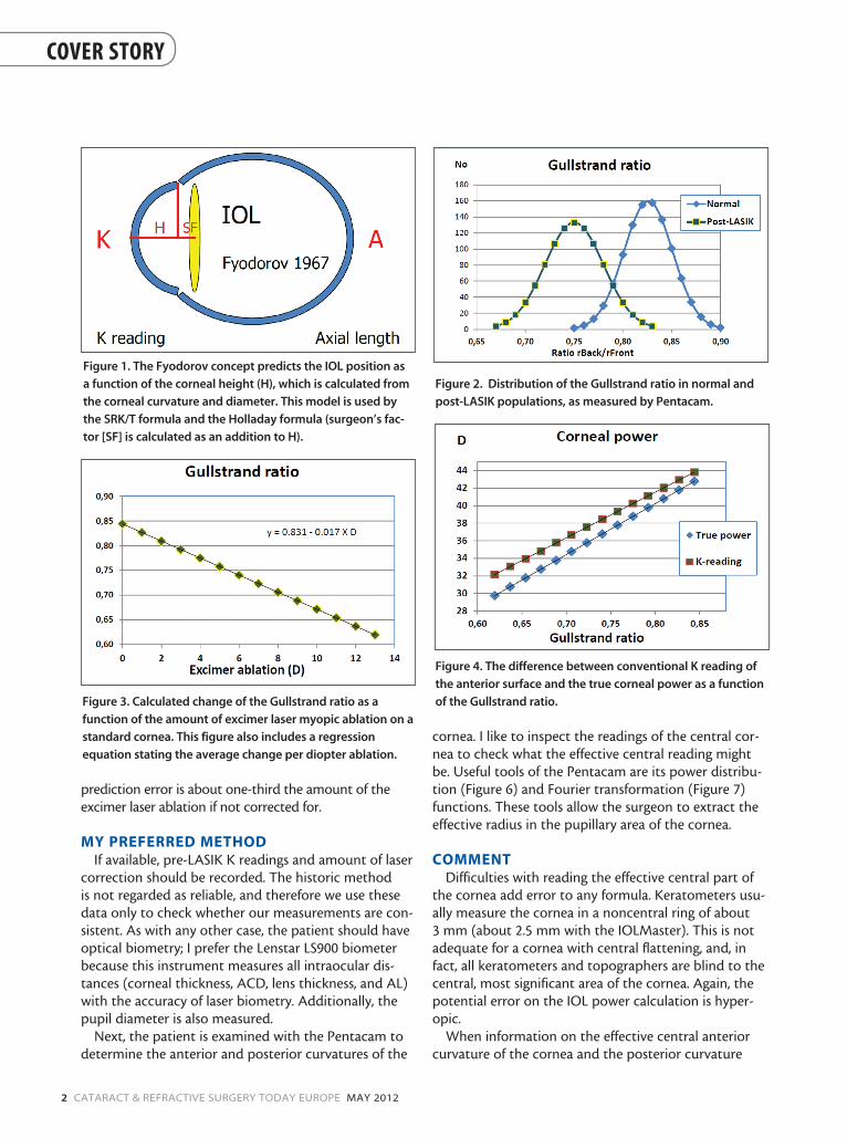
Problem No. 1. The flatness of the K reading makes the formula believe the ELP is more anterior than it really is. With a shallow ELP, the formula will pre-dict a low IOL power, causing a hyperopic surprise. Figure 1 explains Fyodorov’s concept of the corneal height, which is the ELP principle of the SRK/T and the Holladay 1 formulas.
Problem No. 2. A simple K reading measures only the curvature of the anterior surface of the cornea. However, most keratometers calculate the power according to the formula K = (1.3375-1)/r, where r is the anterior radius in meters and 1.3375 is the fictitious refractive index of the
cornea, as if the cornea were a thin lens. Therefore, in order for the anterior curvature measurement to be translated into the true power of the cornea, the following must be calculated: the curvature of the posterior surface, the cor-neal thickness, and the true refractive index of the cornea (1.376). In normal corneas, it is reasonable to assume that there is a constant ratio (called the Gullstrand ratio from the Gullstrand exact schematic eye) between the anterior and posterior curvatures. For the post-LASIK cornea, how-ever, the Gullstrand ratio is reduced, and the keratometer-reported K reading will overestimate the true corneal power. Again, the formula will think the IOL power should be reduced, and the patient ends up with a hyperopic error.
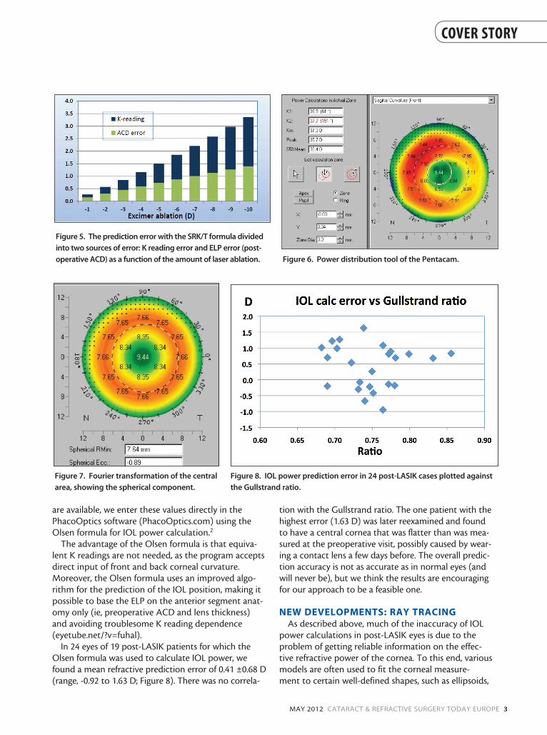
Modern Scheimpflug imaging and other tomography techniques provide more insight into the variation of the Gullstrand ratio in normal and post-LASIK eyes. Figure 2 shows the results of Pentacam (Oculus Optikgeräte GmbH) measurement of the anterior and posterior corneal cur-vatures, giving the distribution of the Gullstrand ratio in a normal population and in a post-LASIK population (average ablation, -7.00 D). The reduction of the Gullstrand ratio is dependent on the amount of laser ablation (Figures 3 and 4). For example, if the Gullstrand ratio is 0.7 (ie, as a result of a 7.00 D ablation; Figure 3), the difference between the true corneal power and the K reading is approximately 2.00 D. However, for the normal cornea (Gullstrand ratio, 0.83), the difference is not nil. This is because the conventional K read-ing carries an inborn overestimation of the corneal power of approximately 1.00 D.
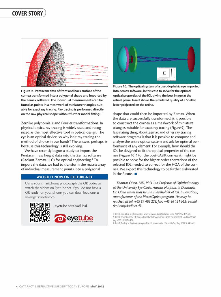
The effect of the two sources of error on IOL power prediction with the SRK/T formula is shown in Figure 5. The K reading error and the ELP error are the same mag-nitude, and both tend to give a hyperopic error. The total
The Creator’s Forum:
IOL Power Calculations for Postrefractive
Surgery Eyes
eyetube.net
2 CATARACT & REFRACTIVE SURGERY TODAY EUROPE MAY 2012
COVER STORY
prediction error is about one-third the amount of the excimer laser ablation if not corrected for.
MY PREFERRED METHODIf available, pre-LASIK K readings and amount of laser
correction should be recorded. The historic method is not regarded as reliable, and therefore we use these data only to check whether our measurements are con-sistent. As with any other case, the patient should have optical biometry; I prefer the Lenstar LS900 biometer because this instrument measures all intraocular dis-tances (corneal thickness, ACD, lens thickness, and AL) with the accuracy of laser biometry. Additionally, the pupil diameter is also measured.
Next, the patient is examined with the Pentacam to determine the anterior and posterior curvatures of the
cornea. I like to inspect the readings of the central cor-nea to check what the effective central reading might be. Useful tools of the Pentacam are its power distribu-tion (Figure 6) and Fourier transformation (Figure 7) functions. These tools allow the surgeon to extract the effective radius in the pupillary area of the cornea.
COMMENTDifficulties with reading the effective central part of
the cornea add error to any formula. Keratometers usu-ally measure the cornea in a noncentral ring of about 3 mm (about 2.5 mm with the IOLMaster). This is not adequate for a cornea with central flattening, and, in fact, all keratometers and topographers are blind to the central, most significant area of the cornea. Again, the potential error on the IOL power calculation is hyper-opic.
When information on the effective central anterior curvature of the cornea and the posterior curvature
Figure 1. The Fyodorov concept predicts the IOL position as
a function of the corneal height (H), which is calculated from
the corneal curvature and diameter. This model is used by
the SRK/T formula and the Holladay formula (surgeon’s fac-
tor [SF] is calculated as an addition to H).
Figure 2. Distribution of the Gullstrand ratio in normal and
post-LASIK populations, as measured by Pentacam.
Figure 3. Calculated change of the Gullstrand ratio as a
function of the amount of excimer laser myopic ablation on a
standard cornea. This figure also includes a regression
equation stating the average change per diopter ablation.
Figure 4. The difference between conventional K reading of
the anterior surface and the true corneal power as a function
of the Gullstrand ratio.
MAY 2012 CATARACT & REFRACTIVE SURGERY TODAY EUROPE 3
COVER STORY
are available, we enter these values directly in the PhacoOptics software (PhacoOptics.com) using the Olsen formula for IOL power calculation.2
The advantage of the Olsen formula is that equiva-lent K readings are not needed, as the program accepts direct input of front and back corneal curvature. Moreover, the Olsen formula uses an improved algo-rithm for the prediction of the IOL position, making it possible to base the ELP on the anterior segment anat-omy only (ie, preoperative ACD and lens thickness) and avoiding troublesome K reading dependence (eyetube.net/?v=fuhal).
In 24 eyes of 19 post-LASIK patients for which the Olsen formula was used to calculate IOL power, we found a mean refractive prediction error of 0.41 ±0.68 D (range, -0.92 to 1.63 D; Figure 8). There was no correla-
tion with the Gullstrand ratio. The one patient with the highest error (1.63 D) was later reexamined and found to have a central cornea that was flatter than was mea-sured at the preoperative visit, possibly caused by wear-ing a contact lens a few days before. The overall predic-tion accuracy is not as accurate as in normal eyes (and will never be), but we think the results are encouraging for our approach to be a feasible one.
NEW DEVELOPMENTS: RAY TRACINGAs described above, much of the inaccuracy of IOL
power calculations in post-LASIK eyes is due to the problem of getting reliable information on the effec-tive refractive power of the cornea. To this end, various models are often used to fit the corneal measure-ment to certain well-defined shapes, such as ellipsoids,
Figure 5. The prediction error with the SRK/T formula divided
into two sources of error: K reading error and ELP error (post-
operative ACD) as a function of the amount of laser ablation. Figure 6. Power distribution tool of the Pentacam.
Figure 7. Fourier transformation of the central
area, showing the spherical component.
Figure 8. IOL power prediction error in 24 post-LASIK cases plotted against
the Gullstrand ratio.
4 CATARACT & REFRACTIVE SURGERY TODAY EUROPE MAY 2012
COVER STORY
Zernike polynomials, and Fourier transformations. In physical optics, ray tracing is widely used and recog-nized as the most effective tool in optical design. The eye is an optical device, so why isn’t ray tracing the method of choice in our hands? The answer, perhaps, is because this technology is still evolving.
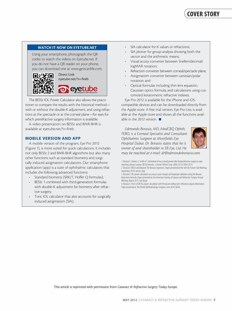
We have recently begun a study to import the Pentacam raw height data into the Zemax software (Radiant Zemax, LLC) for optical engineering.3 To import the data, we had to transform the matrix array of individual measurement points into a polygonal
shape that could then be imported by Zemax. When the data are successfully transformed, it is possible to construct the cornea as a meshwork of miniature triangles, suitable for exact ray tracing (Figure 9). The fascinating thing about Zemax and other ray tracing software programs is that it is possible to compose and analyze the entire optical system and ask for optimal per-formance of any element. For example, how should the IOL be designed to fit the optical properties of the cor-nea (Figure 10)? For the post-LASIK cornea, it might be possible to solve for the higher-order aberrations of the selected IOL needed to correct for the HOA of the cor-nea. We expect this technology to be further elaborated in the future. n
Thomas Olsen, MD, PhD, is a Professor of Ophthalmology at the University Eye Clinic, Aarhus Hospital, in Denmark. Dr. Olsen states that he is a shareholder of IOL Innovations, manufacturer of the PhacoOptics program. He may be reached at tel: +45 89 493 228; fax: +45 86 121 653; e-mail: [email protected].
1. Olsen T. Calculation of intraocular lens power: a review. Acta Ophthalmol Scand. 2007;85(5):472-485.2. Olsen T. Prediction of the effective postoperative (intraocular lens) anterior chamber depth. J Cataract Refract Surg. 2006;32(3):419-424.3. Olsen T, Funding M. Ray tracing analysis of the IOL power in situ. J Cataract Refract Surg. 2012;38:641-647.
Figure 9. Pentacam data of front and back surface of the
cornea transformed into a polygonal shape and imported by
the Zemax software. The individual measurements can be
found as points in a meshwork of miniature triangles, suit-
able for exact ray tracing. Ray tracing is performed directly
on the raw physical shape without further model fitting.
Figure 10. The optical system of a pseudophakic eye imported
into Zemax software, in this case to solve for the optimal
optical properties of the IOL giving the best image at the
retinal plane. Insert shows the simulated quality of a Snellen
letter projected on the retina.
WATCH IT NOW ON EYETUBE.NET
Using your smartphone, photograph the QR codes to watch the videos on Eyetube.net. If you do not have a QR reader on your phone, you can download one at www.getscanlife.com.
eyetube.net/?v=fuhal

MAY 2012 CATARACT & REFRACTIVE SURGERY TODAY EUROPE 5
COVER STORY
These formulas are useful for eyes that have previously undergone myopic or hyperopic laser treatments.
By Edmondo Borasio, MD, MedCBQ Ophth, FEBO
Using standard keratometry and raw data taken from corneal topography to calculate refractive corneal power in postrefractive surgery eyes can result in an inaccurate representation of the true corneal power. In these cases, instead of using a pseudophakic IOL power calculation that incorporates preoperative data, which can result in refractive surprises, I prefer to use direct corneal measurements.
I use two approaches to calculate IOL power follow-ing laser refractive surgery, the first of which, the Borasio Edmondo Smith and Stevens formulas (BESSt and BESSt 2), is based on direct anterior and posterior corneal sur-face measurements and requires the Pentacam (Oculus Optikgeräte GmbH).1 The second approach, the more recently developed Borasio Myopic Regression and Borasio Hyperopic Regression (BMR-BHR) formula, requires direct measurements from the IOLMaster (Carl Zeiss Meditec). These formulas can be used for eyes that have previously undergone myopic or hyperopic excimer laser treatments, even if there is a lack of prere-fractive surgery information.
BESSt AND BESST 2Based on direct anterior and posterior corneal curvature
and corneal thickness measurements using the Pentacam and a modified Gaussian optics formula, the BESSt algo-rithm estimates corneal power in eyes after laser refractive surgery for myopia or hyperopia. It is not possible to use val-ues obtained from the Gaussian optics formula in most cur-rent IOL power formulas because they are calibrated using a standard keratometric index of 1.3375, which does not take true posterior corneal curvature into account. This can therefore yield inaccurate results, especially in postrefractive surgery eyes where the relationship between the anterior and posterior corneal radii is no longer constant. BESSt starts from the true net corneal power as calculated with the Gaussian optics formula, but then it makes crucial adjust-ments based on the actual measured postoperative corneal radii and the altered anterior/posterior radius relationship.
We have found that the BESSt 2, a second iteration of the formula, results in greater prediction accuracy and less risk of refractive surprise after hyperopic treatments (unpublished data). BESSt 2 incorporates the following improvements compared with its predecessor: (1) automatic prediction of
the preoperative anterior corneal radius from postoperative posterior corneal radius measurement, thus allowing auto-matic application of Aramberri’s double-K adjustment for a more accurate prediction of the estimated IOL position, (2) two separate algorithms based on the results of regression analyses, one for myopic and one for hyperopic treatments, (3) automatic application of the BESSt 2-derived corneal power to a modified third-generation formula for the pur-pose of IOL power calculation, and (4) an error-limitation algorithm to prevent serious errors in eyes with extreme axial lengths.
BMR-BHRThe latest formulas I have developed for postrefractive sur-
gery eyes, the BMR and BHR, require the use of the IOLMaster to measure K values and axial length.2-4 From the measured postrefractive surgery K value, a new corneal power value is obtained by using one of these regression formulas. The resulting corneal power values are then automatically entered into the SRK/T formula, yielding a suggested IOL power.
The BMR-BHR formulas are based on linear, logarithmic, and polynomial regression analyses and give similar results in most cases, with no greater than 0.25 D difference between the analyses with the exception of eyes with very steep or very flat corneas, where differences can be more substantial. Further studies are needed to determine which formula is the most accurate, but I tend to use linear regression. Polynomial regression has a smaller error in the majority of cases, but it can also have more serious outliers than linear regression; logarithmic regression lies in between the other two. It is best to use polynomial regression only when the suggested IOL power value is neither too low nor too high.
BMR-BHR is currently available only as part of Eye Pro 2012 for the iPhone/iPad. A desktop version will soon be available as an upgrade to the BESSt 2 IOL Power Calculator.
CONSIDERATIONS AND RESULTSThe same considerations apply to the BESSt, BESSt 2,
and BMR-BHR formulas; they should not be used in the presence of significant corneal haze or scarring or after incisional refractive surgery such as radial keratotomy. Additionally, these formulas should be used with caution in eyes that have undergone astigmatic keratotomy; in eyes that have previously undergone myopic or hyper-opic treatments for severe refractive errors; or in eyes that were operated on a long time ago using small opti-cal zones. Other considerations include the following:
• The BMR-BHR requires K values specifically mea-
THE BESSt AND BMR-BHR FORMULAS

6 CATARACT & REFRACTIVE SURGERY TODAY EUROPE MAY 2012
COVER STORY
sured by the IOLMaster; • I recommended comparing BESSt 2 with BMR-
BHR. If the BESSt 2 formula suggests a very low (or very high) IOL power, I either use the BMR-BHR formula instead or the average of the two. It is always advisable to compare with other methods before proceeding with surgery. Whenever in the doubt, opt for a stronger IOL to target myopia;
• Be suspicious of very low or very high IOL pow-ers resulting from BMR-BHR or BESSt in eyes with very steep or very flat corneas. These cases are at the extreme edges of any regression analysis, and the risk of a refractive surprise is high; and
• The BESSt 2 and the Eye Pro have not been approved by the US Food and Drug Administration (FDA) or received the Conformiteé Europeénne (CE) Mark.
I recently conducted a study in 62 eyes, of which 38 received a myopic treatment and 24 a hyperopic, to compare the accuracy of the BESSt, BESSt 2, and BMR-BHR formulas. In the myopic group, BESSt and BESSt 2 performed similarly (mean error, -0.21 ±0.78 D and -0.02 ±0.81 D, respectively; P>.05), but in the hyperopic group BESSt 2 performed statistically significantly better (-1.10 ±0.90 D and 0.02 ±1.00 D, respectively; P<.05). The pro-portion of eyes within ±0.50 D of the intended target changed negligibly (from 37% to 38%) in the myopic group but improved significantly (from 13% to 38%) in the hyperopic group. The proportion of eyes within ±1.00 D of the target changed from 73% to 76% in the myopic group and from 38% to 75% in the hyperopic group.
Comparing the results with BESSt 2 to those with BMR and BHR, in the myopic group, the mean error was -0.02 ±0.81 D with BESSt 2 and 0.00 ±0.75 D with BMR (P>.05).
In the hyperopic group, the mean error was 0.02 ±1.00 D for BESSt 2 and 0.01 ±0.75 D for BHR (P>.05). Although these differences were not statistically significant, 31% more eyes were ±0.50 D or less from the target refrac-tion with BMR compared with BESSt 1 and 30% more compared with BESSt 2. Similarly, with BHR 50% more eyes were ±0.50 D or less from the target compared with BESSt 1 and 13% more compared with BESSt 2. We believe this represents an improvement.
PC VERSION FOR THE PENTACAMThe BESSt 2 IOL Power Calculator (EB Eye Ltd, UK;
besstformula.com) is available as an optional software add-on for the Pentacam. The main advantage of this cal-culator is that it can be installed on the same computer as the Pentacam. Therefore, calculations can be done directly from the Pentacam program by clicking on the BESSt 2 button, which will export all the required data (anterior and posterior corneal curvature and central pachymetry) to the program for the calculations. The program also keeps a database of calculations for future reference and for recalculation. A standalone version for research pur-poses is also available and can be used on a laptop not physically connected to the Pentacam hardware.
Another feature of the BESSt 2 IOL Power Calculator for PC is real-time IOL power plotting to show the behav-ior of different biometry formulas when parameters are modified for any given axial length. This allows immediate identification of potential biometry artifacts that affect any given formula. Examples include the SRK/T nega-tive square root and cusp phenomena that occur for certain combinations of axial length and K values (most frequently for steep corneas). When not identified, these artifacts can lead to inaccurate IOL power calculations.
Figure 1. Screenshots taken from Eye Pro 2012: (From left to right) BMR-BHR for IOLMaster, mobile version of BESSt 2
myopia, double-angle polar plot showing the average surgically induced astigmatism in a group of eyes, standard
biometry on the go, and IOL power matrix.
MAY 2012 CATARACT & REFRACTIVE SURGERY TODAY EUROPE 7
COVER STORY
The BESSt IOL Power Calculator also allows the practi-tioner to compare the results with the historical method—with or without the double-K adjustment, and using refrac-tions at the spectacle or at the corneal plane—for eyes for which prerefractive surgery information is available.
A video presentation on BESSt and BMR-BHR is available at eyetube.net/?v=fireb.
MOBILE VERSION AND APPA mobile version of the program, Eye Pro 2012
(Figure 1), is more suited for quick calculations. It includes not only BESSt 2 and BMR-BHR algorithms but also many other functions such as standard biometry and surgi-cally induced astigmatism calculations. Our smartphone application (app) is a suite of ophthalmic calculators that includes the following advanced functions:
• Standard biometry (SRK/T, Hoffer Q formulas); • BESSt 1 combined with third-generation formulas
with double-K adjustment for biometry after refrac-tive surgery;
• Toric IOL calculator that also accounts for surgically induced astigmatism (SIA);
• SIA calculator for K values or refractions; • SIA plotter for group analysis showing both the
vector and the arithmetic means;• Visual acuity converter between Snellen/decimal/
logMAR notation; • Refraction converter between corneal/spectacle plane;• Astigmatism converter between cartesian/polar
notation; and• Optical formulas including thin lens equation,
Gaussian optics formula, and calculations using cus-tomized keratometric refractive indexes.
Eye Pro 2012 is available for the iPhone and iOS- compatible devices and can be downloaded directly from the Apple store. A free trial version, Eye Pro Lite, is avail-able at the Apple store and shows all the functions avail-able in the 2012 version. n
Edmondo Borasio, MD, MedCBQ Ophth, FEBO, is a Corneal Specialist and Consultant Ophthalmic Surgeon at Moorfields Eye Hospital Dubai. Dr. Borasio states that he is owner of and shareholder in EB Eye, Ltd. He may be reached at e-mail: [email protected].
1. Borasio E, Stevens J, Smith GT. Estimation of true corneal power after keratorefractive surgery in eyes requiring cataract surgery: BESSt formula. J Cataract Refract Surg. 2006;32(12):2004-2014.2. Borasio E. BESSt and beyond: The Borasio regression. Paper presented at the: 6th IOL Power Club Meeting; September 2010; Venice, Italy.3. Borasio E. IOL power calculation accuracy in post-myopic and hyperopic ablations using the Borasio Regression formula. Paper presented at: the American Society of Cataract and Refractive Surgery Annual Meeting; March 2011; San Diego.4. Borasio E. Post-LASIK IOL power calculation with Pentacam without pre-refractive surgery information. Paper presented at: the World Ophthalmology Congress; June 2010; Berlin.
WATCH IT NOW ON EYETUBE.NET
Using your smartphone, photograph the QR codes to watch the videos on Eyetube.net. If you do not have a QR reader on your phone, you can download one at www.getscanlife.com.
Direct Linkeyetube.net/?v=fireb
This article is reprinted with permission from Cataract & Refractive Surgery Today Europe.