The Citric Acid Cycle
35
The Citric Acid Cycle Oxidizes Two-Carbon Units The conversion of pyruvate into acetyl CoA by the pyruvate dehydrogenase complex is the link between glycolysis and cellular respiration because acetyl CoA is the fuel for the citric acid cycle. The citric acid cycle begins with the condensation of a four-carbon unit, oxaloacetate, and a two-carbon unit, the acetyl group of acetyl CoA. Oxaloacetate reacts with acetyl CoA and H 2 O to yield citrate and CoA. o This reaction, which is an aldol condensation followed by a hydrolysis, is catalyzed by citrate synthase. Oxaloacetate first condenses with acetyl CoA to form citryl CoA, a molecule that is energy rich because it contains the thioester bond that originated in acetyl CoA. The hydrolysis of citryl CoA thioester to citrate and CoA drives the overall reaction far in the direction of the
description
The Citric Acid Cycle
Transcript of The Citric Acid Cycle

The Citric Acid Cycle Oxidizes Two-Carbon Units
The conversion of pyruvate into acetyl CoA by the pyruvate dehydrogenase complex is the link between glycolysis and cellular respiration because acetyl CoA is the fuel for the citric acid cycle
The citric acid cycle begins with the condensation of a four-carbon unit oxaloacetate and a two-carbon unit the acetyl group of acetyl CoA
Oxaloacetate reacts with acetyl CoA and H 2O to yield citrate and CoA
o
This reaction which is an aldol condensation followed by a hydrolysis is catalyzed by citrate synthase
Oxaloacetate first condenses with acetyl CoA to form citryl CoA a molecule that is energy rich because it contains the thioester bond that originated in acetyl CoA
The hydrolysis of citryl CoA thioester to citrate and CoA drives the overall reaction far in the direction of the synthesis of citrate
It is very important that side reactions notably the hydrolysis of acetyl CoA to acetate and CoA be minimized Let us briefly consider how the citrate synthase prevents the wasteful hydrolysis of acetyl CoA
X-ray crystallographic studies of citrate synthase and its complexes with several substrates and inhibitors revealed that the enzyme undergoes large conformational changes in the course of catalysis
Citrate synthase exhibits sequential ordered kinetics oxaloacetate binds first followed by acetyl CoA The reason for the ordered binding is that oxaloacetate induces a major
structural rearrangement leading to the creation of a binding site for acetyl CoA The binding of oxaloacetate converts the open form of the enzyme into a closed form
Small domain rotates 19 degrees relative to the large domain o These structural changes create a binding site for acetyl
CoA Citrate synthase catalyzes the condensation reaction by bringing
the substrates into close proximity orienting them and polarizing certain bonds
The donation and removal of protons transforms acetyl CoA into an enol intermediate
1 The enol attacks oxaloacetate to form a carbonndashcarbon double bond linking acetyl CoA and oxaloacetate
2 The newly formed citryl CoA induces additional structural changes in the enzyme causing the active site to become completely enclosed
3 The enzyme cleaves the citryl CoA thioester by hydrolysis 4 CoA leaves the enzyme followed by citrate and the
enzyme returns to the initial open conformation Citrate synthase is well suited to hydrolyze citryl CoA but not
acetyl CoA 1 First acetyl CoA does not bind to the enzyme until
oxaloacetate is bound and ready for condensation 2 Second the catalytic residues crucial for the hydrolysis of
the thioester linkage are not appropriately positioned until citryl CoA is formed
o
Citrate is isomerized into isocitrate
The hydroxyl group is not properly located in the citrate molecule for the oxidative decarboxylations that follow Thus citrate is isomerized into isocitrate to enable the six-carbon unit to undergo oxidative decarboxylation
The isomerization of citrate is accomplished by a dehydration step followed by a hydration step The result is an interchange of an H and an OH
Aconitase is an ironndashsulfur protein o Its four iron atoms are complexed to four inorganic sulfides
and three cysteine sulfur atoms leaving one iron atom
available to bind citrate through one of its COO- groups and an OH group
o This Fe-S cluster participates in dehydrating and rehydrating the bound substrate
Isocitrate is oxidized and decarboxylated to alpha-ketoglutarate
First of four oxidationndashreduction reactions in the citric acid cycle The oxidative decarboxylation of isocitrate is catalyzed by isocitrate dehydrogenase
The intermediate in this reaction is oxalosuccinate an unstable b-ketoacid While bound to the enzyme it loses CO2 to form a-ketoglutarate
The rate of formation of a-ketoglutarate is important in
determining the overall rate of the cycle This oxidation generates the first high-transfer-potential electron
carrier NADH in the cycle
Succinyl coenzyme A is formed by the oxidative decarboxylation of alpha-ketoglutarate
The conversion of isocitrate into a-ketoglutarate is followed by a second oxidative decarboxylation reaction the formation of succinyl CoA from a-ketoglutarate
This reaction is catalyzed by the 1113090a-ketoglutarate dehydrogenase complex an organized assembly of three kinds of enzymes that is homologous to the pyruvate dehydrogenase complex
Both reactions include the decarboxylation of an a-ketoacid and the subsequent formation of a thioester linkage with CoA that has
a high transfer potential
A compound with high phosphoryl-transfer potential is generated from succinyl coenzyme A
In the citrate synthase reaction the cleavage of the thioester bond powers the synthesis of the six-carbon citrate from the four-carbon oxaloacetate and the two-carbon fragment
The cleavage of the thioester bond of succinyl CoA is coupled to the phosphorylation of a purine nucleoside diphosphate usually ADP This reaction which is readily reversible is catalyzed by succinyl CoA synthetase (succinate thiokinase)
This reaction is the only step in the citric acid cycle that directly yields a compound with high phosphoryl-transfer potential
o In tissues that perform large amounts of cellular respiration such as skeletal and heart muscle the ADP-requiring isozyme predominates
o In tissues that perform many anabolic reactions such as the liver the GDP-requiring enzyme is common
o The GDP-requiring enzyme is believed to work in reverse of the direction observed in the TCA cycle that is GTP is used to power the synthesis of succinyl CoA which is a precursor for heme synthesis
Mechanism Succinyl coenzyme A synthetase transforms types of biochemical energy
The mechanism of this reaction is a clear example of an energy
transformation energy inherent in the thioester molecule is transformed into phosphoryl-group-transfer potential
Oxaloacetate is regenerated by the oxidation of succinate
Reactions of four-carbon compounds constitute the final stage of the citric acid cycle the regeneration of oxaloacetate
A methylene group (CH2) is converted into a carbonyl group (CPO) in three steps an oxidation a hydration and a second oxidation reaction Oxaloacetate is thereby regenerated for another round of the cycle and more energy is extracted in the form of FADH2 and NADH
Succinate is oxidized to fumarate by succinate dehydrogenase
The hydrogen acceptor is FAD rather than NAD+ which is used in the other three oxidation reactions in the cycle FAD is the hydrogen acceptor in this reaction because the free-energy
change is insufficient to reduce NAD+ FAD is nearly always the electron acceptor in oxidations that remove two hydrogen atoms from a substrate
Succinate dehydrogenase like aconitase is an ironndashsulfur protein
In fact succinate dehydrogenase is directly associated with the electron-transport chain the link between the citric acid cycle and ATP formation FADH2 produced by the oxidation of succinate does not dissociate from the enzyme in contrast with NADH produced in other oxidationndashreduction reactions Rather two electrons are transferred from FADH2 directly to ironndashsulfur clusters of the enzyme which in turn passes the electrons to coenzyme Q (CoQ) Coenzyme Q an important member of the electron-transport chain passes electrons to the ultimate acceptor molecular oxygen
The next step is the hydration of fumarate to form L-malate
Fumarase catalyzes a stereospecific trans addition of H+ and
OH- The OH- group adds to only one side of the double bond of fumarate hence only the L isomer of malate is formed
Finally malate is oxidized to form oxaloacetate This reaction is
catalyzed by malate dehydrogenase and NAD+ is again the hydrogen acceptor
The oxidation of malate is driven by the use of the productsmdashoxaloacetate by citrate synthase and NADH by the electron- transport chain
The citric acid cycle produces high-transfer-potential electrons ATP and CO2
The net reaction of the citric acid cycle is o
1 Two carbon atoms enter the cycle in the condensation of an acetyl unit (from acetyl CoA) with oxaloacetate Two carbon atoms leave the cycle in the form of CO2 in the successive decarboxylations catalyzed by isocitrate dehydrogenase and a-ketoglutarate dehydrogenase
2 Four pairs of hydrogen atoms leave the cycle in four oxidation
reactions Two NAD+ molecules are reduced in the oxidative decarboxylations of isocitrate and a-ketoglutarate one FAD molecule is reduced in the oxidation of succinate and one
NAD+ molecule is reduced in the oxidation of malate Recall
also that one NAD+ molecule is reduced in the oxidative decarboxylation of pyruvate to form acetyl CoA
3 One compound with high phosphoryl-transfer potential usually ATP is generated from the cleavage of the thioester linkage in succinyl CoA
4 Two water molecules are consumed one in the synthesis of citrate by the hydrolysis of citryl CoA and the other in the hydration of fumarate
Isotope-labeling studies revealed that the two carbon atoms that enter each cycle are not the ones that leave The two carbon atoms that enter the cycle as the acetyl group are retained during the initial two decarboxylation reactions and then remain incorporated in the four-carbon acids of the cycle
Note that succinate is a symmetric molecule Consequently
the two carbon atoms that enter the cycle can occupy any of the carbon positions in the subsequent metabolism of the four-carbon acids The two carbons that enter the cycle as the acetyl group will be released as CO2 in subsequent trips through the cycle
The electron-transport chain oxidizes the NADH and FADH2
formed in the citric acid cycle The transfer of electrons from these carriers to O2 the ultimate electron acceptor leads to the generation of a proton gradient across the inner mitochondrial membrane This proton-motive force then powers the generation of ATP
Consequently nine high-transfer-potential phosphoryl groups are generated when the electron-transport chain oxidizes 3 NADH molecules and 1 FADH2 molecule and one high-transfer-potential phosphoryl group is directly formed in one round of the citric acid cycle Thus one acetyl unit generates approximately 10 molecules of ATP
Recall that molecular oxygen does not participate directly in the citric acid cycle However the cycle operates only under
aerobic conditions because NAD+ and FAD can be regenerated in the mitochondrion only by the transfer of electrons to molecular oxygen Glycolysis has both an aerobic and an anaerobic mode whereas the citric acid cycle is strictly aerobic
Lipids
Membranes are as diverse in structure as they are in function However they do have in common a number of important attributes 1 Membranes are sheet like structures only two molecules
thick which form closed boundaries between different compartments The thickness of most membranes is between 60 Aring (6 nm) and 100 Aring (10 nm)
2 Membranes consist mainly of lipids and proteins The mass ratio of lipids to proteins ranges from 14 to 41 Membranes also contain carbohydrates that are linked to
lipids and proteins 3 Membrane lipids are small molecules that have both
hydrophilic and hydrophobic moieties These lipids spontaneously form closed bimolecular sheets in aqueous media These lipid bilayers are barriers to the flow of polar molecules
4 Specific proteins mediate distinctive functions of membranes Proteins serve as pumps channels receptors energy transducers and enzymes Membrane proteins are embedded in lipid bilayers which create suitable environments for their action
5 Membranes are noncovalent assemblies The constituent protein and lipid molecules are held together by many noncovalent interactions which act cooperatively
6 Membranes are asymmetric The two faces of biological membranes always differ from each other
7 Membranes are fluid structures Lipid molecules diffuse rapidly in the plane of the membrane as do proteins unless they are anchored by specific interactions In contrast lipid molecules and proteins do not readily rotate across the membrane Membranes can be regarded as two-dimensional solutions of oriented proteins and lipids
8 Most cell membranes are electrically polarized such that the inside is negative [typically 260 millivolts (mV)] Membrane potential plays a key role in transport energy conversion and excitability
The properties of fatty acids and of lipids derived from them are markedly dependent on chain length and degree of saturation
Unsaturated fatty acids have lower melting points than do saturated fatty acids of the same length
Chain length also affects the melting point Thus short chain length and unsaturation enhance the fluidity
of fatty acids and of their derivatives Lipids are water-insoluble biomolecules that are highly
soluble in organic solvents such as chloroform
Lipids have a variety of biological roles o They serve as fuel molecules o Highly concentrated energy stores o Signal molecules o Messengers in signal-transduction pathways and
components of membranes The three major kinds of membrane lipids are phospholipids
glycolipids and cholesterol
Phospholipids are the major class of membrane lipids
A phospholipid molecule is constructed from four components one or more fatty acids a platform to which the fatty acids are attached a phosphate and an alcohol attached to the phosphate
The fatty acid components provide a hydrophobic barrier whereas the remainder of the molecule has hydrophilic properties that enable interaction with the aqueous environment
The platform on which phospholipids are built may be glycerol a three carbon alcohol or sphingosine a more complex alcohol
Phospholipids derived from glycerol are called phosphoglycerides
o A phosphoglyceride consists of a glycerol backbones to which are attached two fatty acid chains and a phosphorylated alcohol
In phosphoglycerides the hydroxyl groups at C-1 and C-2 of glycerol are esterified to the carboxyl groups of the two fatty acid chains
The C-3 hydroxyl group of the glycerol backbone is esterified to phosphoric acid
When no further additions are made the resulting compound is phosphatidate (diacylglycerol 3-phosphate) the simplest phosphoglyceride
Only small amounts of phosphatidate are present in membranes However the molecule is a key intermediate in the biosynthesis of the other phosphoglycerides
The major phosphoglycerides are derived from phosphatidate by the formation of an ester bond between the phosphate group of phosphatidate and the hydroxyl group of one of several alcohols
The common alcohol moieties of phosphoglycerides are the amino acid serine ethanolamine choline glycerol and inositol
The second major class of membrane lipids glycolipids are sugar-containing lipids
The amino group of the sphingosine backbone is acylated by a fatty acid as in sphingomyelin Glycolipids differ rom sphingomyelin in the identity of the unit that is linked to the primary hydroxyl group of the sphingosine backbone In glycolipids one or more sugars (rather than phosphorylcholine) are attached to this group
Cholesterol the third major type of membrane lipid has a structure that is quite different from that of phospholipids It is a steroid built from four linked hydrocarbon rings
A hydrocarbon tail is linked to the steroid at one end and a
hydroxyl group is attached at the other end In membranes the orientation of the molecule is parallel to the fatty acid chains of the phospholipids and the hydroxyl group interacts with the nearby phospholipid head groups
However these lipids possess a critical common structural theme membrane lipids are amphipathic molecules (amphiphilic molecules) A membrane lipid contains both a hydrophilic and a hydrophobic moiety
The two hydrophobic fatty acid chains are approximately parallel to each other whereas the hydrophilic phosphorylcholine moiety points in the opposite direction
The hydrophilic unit also called the polar head group is represented by a circle and the hydrocarbon tails are depicted by straight or wavy lines
Membrane formation is a consequence of the amphipathic nature of the molecules Their polar head groups favor contact with water whereas their hydrocarbon tails interact with one another in preference to water
One way is to form a globular structure called a micelle The polar head groups form the outside surface of the micelle
which is surrounded by water and the hydrocarbon tails are sequestered inside interacting with one another
A lipid bilayer is also called a bimolecular sheet The hydrophobic tails of each individual sheet interact with one
another forming a hydrophobic interior that acts as a permeability barrier
The hydrophilic head groups interact with the aqueous medium on each side of the bilayer
The favored structure for most phospholipids and glycolipids in aqueous media is a bimolecular sheet rather than a micelle
The reason is that the two fatty acid chains of a phospholipid or a glycolipid are too bulky to fit into the interior of a micelle
The formation of bilayers instead of micelles by phospholipids is of critical biological importance A micelle is a limited structure usually less than 200 Aring (20 nm) in diameter
In contrast a bimolecular sheet can extend to macroscopic
dimensions as much as a millimeter (107 Aring or 106 nm) or more Lipid bilayers form spontaneously by a self-assembly process In
other words the structure of a bimolecular sheet is inherent in the structure of the constituent lipid molecules The growth of lipid bilayers from phospholipids is rapid and spontaneous in water
Hydrophobic interactions are the major driving force for the formation of lipid bilayers
Water molecules are released from the hydrocarbon tails of membrane lipids as these tails become sequestered in the nonpolar interior of the bilayer
Furthermore van der Waals attractive forces between the hydrocarbon tails favor close packing of the tails
Finally there are electrostatic and hydrogen-bonding attractions between the polar head groups and water molecules Thus lipid bilayers are stabilized by the full array of forces that mediate molecular interactions in biological systems
Because lipid bilayers are held together by many reinforcing noncovalent interactions (predominantly hydrophobic) they are cooperative structures
These hydrophobic interactions have three significant biological consequences
1 (1) lipid bilayers have an inherent tendency to be extensive 2 (2) lipid bilayers will tend to close on themselves so that
there are no edges with exposed hydrocarbon chains and so they form compartments and
3 (3) lipid bilayers are self-sealing because a hole in a bilayer is energetically unfavorable
Lipid bilayers are highly impermeable to ions and most polar molecules
Lipid bilayer membranes have a very low permeability for ions and most polar molecules
The permeability of small molecules is correlated with their solubility in a nonpolar solvent relative to their solubility in water
This relation suggests that a small molecule might traverse a lipid bilayer membrane in the following way
a First it sheds its solvation shell of water b Then it is dissolved in the hydrocarbon core of the
membrane and c Finally it diffuses through this core to the other side
of the membrane where it becomes resolvated by water
Thermodynamics of Membrane Transport
Sodium ions pass through specific channels in the hydrophobic barrier formed by membrane proteins
This means of crossing the membrane is called facilitated
diffusion because the diffusion across the membrane is facilitated by the channel
It is also called passive transport because the energy driving the ion movement originates from the ion gradient itself without any contribution by the transport system
Protein transporters embedded in the membrane are capable of using an energy source to move the molecule up a concentration gradient
Because an input of energy from another source is required this means of crossing the membrane is called active transport
An unequal distribution of molecules is an energy-rich condition because free energy is minimized when all concentrations are equal
The free-energy change in transporting this species from side 1 where it is present at a concentration of c1 to side 2 where it is present at concentration c2 is
o ∆ G=RT ln (c2c 1)
o where R is the gas constant (8315 3 10-3 kJ mol-1 deg-1 or
1987 3 10-3 kcal mol-1 deg-1) and T is the temperature in kelvins
For a charged species the unequal distribution across the membrane generates an electrical potential that also must be considered because the ions will be repelled by the like charges
Electrochemical potential or membrane potential o ∆ G=RT ln (c2c 1)+ZF ∆V
o In which Z is the electrical charge of the transported species DV is the potential in volts across the membrane
and F is the Faraday constant (965 kJ V-1 mol-1 or 231
kcal V-1 mol-1) A transport process must be active when ∆G is positive It can be passive when ∆G is negative
Oxidative Phosphorylation
We begin our study of oxidative phosphorylation by examining the oxidationndashreduction reactions that allow the flow of electrons
from NADH and FADH2 to oxygen The electron flow takes place in four large protein complexes that
are embedded in the inner mitochondrial membrane together called the respiratory chain or the electron-transport chain
The resulting unequal distribution of protons generates a pH
gradient and a transmembrane electrical potential that creates a proton-motive force ATP is synthesized when protons flow back to the mitochondrial matrix through an enzyme complex
Thus the oxidation of fuels and the phosphorylation of ADP are
coupled by a proton gradient across the inner mitochondrial membrane
Collectively the generation of high-transfer-potential electrons by the citric acid cycle their flow through the respiratory chain and the accompanying synthesis of ATP is called respiration or cellular respiration
The primary function of the citric acid cycle was identified as the generation of NADH and FADH2 by the oxidation of acetyl CoA In oxidative phosphorylation electrons from NADH and FADH2
are used to reduce molecular oxygen to water o Electron-transport chain
The electron-transfer potential of an electron is measured as redox potential
In oxidative phosphorylation the electron-transfer potential of NADH or FADH2 is converted into the phosphoryl-transfer potential of ATP
The measure of phosphoryl-transfer potential is already familiar to us it is given by ∆ G deg for the hydrolysis of the activated phosphoryl compound The corresponding expression for the electron-transfer potential is Ersquo0 the reduction potential (also called the redox potential or oxidationndashreduction potential)
Thus a strong reducing agent (such as NADH) is poised to donate electrons and has a negative reduction potential whereas a strong oxidizing agent (such as O2) is ready to accept electrons and has a positive reduction potential
A 114-volt potential difference between NADH and molecular oxygen drives electron transport through the chain and favors the formation of a proton gradient
The driving force of oxidative phosphorylation is the electron-transfer potential of NADH or FADH2 relative to that of O2
The released energy is initially used to generate a proton gradient that is then used for the synthesis of ATP and the transport of metabolites across the mitochondrial membrane
Electrons are transferred from NADH to O2 through a chain of three large protein complexes called NADH-Q oxidoreductase Q-cytochrome c oxidoreductase and cytochrome c oxidase
Electron flow within these transmembrane complexes leads to the transport of protons across the inner mitochondrial membrane
A fourth large protein complex called succinate-Q reductase contains the succinate dehydrogenase that generates FADH2 in the citric acid cycle
Electrons from this FADH2 enter the electron-transport chain at Q-cytochrome oxidoreductase
Succinate-Q reductase in contrast with the other complexes does not pump protons
NADH-Q oxidoreductase succinate-Q reductase Q-cytochrome c oxidoreductase and cytochrome c oxidase are also called Complex I II III and IV respectively
Two special electron carriers ferry the electrons from one
complex to the next The first is coenzyme Q (Q) also known as ubiquinone because it
is a ubiquitous quinone in biological systems o Ubiquinone is a hydrophobic quinone that diffuses rapidly
within the inner mitochondrial membrane Electrons are carried from NADH-Q oxidoreductase to Q-
cytochrome c oxidoreductase the second complex of the chain by the reduced form of Q
Electrons from the FADH2 generated by the citric acid cycle are transferred first to ubiquinone and then to the Q-cytochrome c oxidoreductase complex
In contrast with Q the second special electron carrier is a protein Cytochrome c a small soluble protein shuttles electrons from Q-cytochrome c oxidoreductase to cytochrome c oxidase the final component in the chain and the one that catalyzes the reduction of O2
The high-potential electrons of NADH enter the respiratory chain at NADH-Q oxidoreductase
The electrons of NADH enter the chain at NADH-Q oxidoreductase (also called Complex I and NADH dehydrogenase)
NADH-Q oxidoreductase is L-shaped with a horizontal arm lying in the membrane and a vertical arm that projects into the matrix
The reaction catalyzed by this enzyme appears to be
o
The initial step is the binding of NADH and the transfer of its two high- potential electrons to the flavin mononucleotide (FMN) prosthetic group of this complex to give the reduced form FMNH2
The electron acceptor of FMN the isoalloxazine ring is identical with that of FAD
Electrons are then transferred from FMNH2 to a series of ironndashsulfur clusters the second type of prosthetic group in NADH-Q oxidoreductase
Fe-S clusters in ironndashsulfur proteins (also called nonheme iron
proteins) play a critical role in a wide range of reduction reactions in biological systems
NADH-Q oxidoreductase contains both 2Fe-2S and 4Fe-4S
clusters Iron ions in these Fe-S complexes cycle between Fe2+
(reduced) and Fe3+ (oxidized) states o Unlike quinones and flavins ironndashsulfur clusters generally
undergo oxidationndashreduction reactions without releasing or binding protons
NADH transfers its two electrons to FMN o These electrons flow through a series of Fe-S centers and
then to coenzyme Q o The flow of two electrons from NADH to coenzyme Q
through NADH-Q oxidoreductase leads to the pumping of four hydrogen ions out of the matrix of the mitochondrion
o In accepting two electrons Q takes up two protons from the matrix as it is reduced to QH2
Ubiquinol is the entry point for electrons from FADH2 of flavoproteins
FADH2 enters the electron-transport chain at the second protein complex of the chain
Succinate dehydrogenase a citric acid cycle enzyme is part of the succinate-Q reductase complex (Complex II) a integral membrane protein of the inner mitochondrial membrane
FADH2 does not leave the complex o Rather its electrons are transferred to Fe-S centers and then
finally to Q to form QH2 which then is ready to transfer electrons further down the electron-transport chain
o The succinate-Q reductase complex in contrast with NADH-Q oxidoreductase does not pump protons from one side of the membrane to the other
Electrons flow from ubiquinol to cytochrome c through Q-cytochrome c oxidoreductase
The electrons from QH2 are passed on to cytochrome c by the second of the three proton pumps in the respiratory chain Q-cytochrome c oxidoreductase (also known as Complex III and as cytochrome reductase)
The function of Q-cytochrome c oxidoreductase is to catalyze the transfer of electrons from QH2 to oxidized cytochrome c (Cyt c)
a water-soluble protein and concomitantly pump protons out of the mitochondrial matrix
The flow of a pair of electrons through this complex leads to the
effective net transport of 2 H+ to the cytoplasmic side half the yield obtained with NADH-Q reductase because of a smaller thermodynamic driving force
Q-cytochrome c oxidoreductase itself contains two types of
cytochromes named b and c1
A cytochrome is an electron-transferring protein that contains a heme prosthetic group The iron ion of a cytochrome alternates between a reduced ferrous (12) state and an oxidized ferric (13) state during electron transport
The Q cycle funnels electrons from a two-electron carrier to a one-electron carrier and pumps protons
QH2 passes two electrons to Q-cytochrome c oxidoreductase but the acceptor of electrons in this complex cytochrome c can accept only one electron
The mechanism for the coupling of electron transfer from Q to cytochrome c to transmembrane proton transport is known as the Q cycle
Two QH2 molecules bind to the complex consecutively each
giving up two electrons and two H+ These protons are released to the cytoplasmic side of the membrane 1 The first QH2 to exit the Q pool binds to the first Q binding
site (Qo) and its two electrons travel through the complex to different destinations a One electron flows first to the Rieske 2Fe-2S cluster b then to cytochrome c1 c and finally to a molecule of oxidized cytochrome c
converting it into its reduced form i The reduced cytochrome c molecule is free to diffuse
away from the enzyme to continue down the respiratory chain
2 The second electron passes through two heme groups of cytochrome b to an oxidized ubiquinone in a second Q binding site (Q i)
3 The now fully oxidized Q leaves the first Q site free to reenter the Q pool
A second molecule of QH2 binds to the Q o site of Q-cytochrome c oxidoreductase and reacts in the same way as the first
The removal of these two protons from the matrix contributes to the formation of the proton gradient
In one Q cycle two QH2 molecules are oxidized to form two
Q molecules and then one Q molecule is reduced to QH2 The cytochrome b component of the reductase is in essence a
recycling device that enables both electrons of QH2 to be used effectively
Cytochrome c oxidase catalyzes the reduction of molecular oxygen to water
The last of the three proton-pumping assemblies of the respiratory chain is cytochrome c oxidase (Complex IV) Cytochrome c oxidase catalyzes the transfer of electrons from the reduced form of cytochrome c to molecular oxygen the final acceptor
Four electrons are funneled to O2 to completely reduce it to
H2O and concomitantly protons are pumped from the matrix to the cytoplasmic side of the inner mitochondrial membrane
As much of this free energy as possible must be captured in the form of a proton gradient for subsequent use in ATP synthesis
Cytochrome c oxidase contains two heme A groups and three copper ions arranged as two copper centers designated A and B One center CuACuA contains two copper ions linked by
two bridging cysteine residues This center initially accepts electrons from reduced cytochrome c The remaining copper ion CuB is coordinated by three histidine residues one of which is modified by covalent linkage to a tyrosine residue
The copper centers alternate between the reduced Cu+
(cuprous) form and the oxidized Cu2+ (cupric) form as they accept and donate electrons
There are two heme A molecules called heme a and heme a3 in cytochrome c oxidase Heme A differs from the heme in cytochrome c and c1 in three ways
a (1) a formyl group replaces a methyl group b (2) a C17 hydrocarbon chain replaces one of the vinyl
groups and c (3) the heme is not covalently attached to the protein
Heme a and heme a3 have distinct redox potentials because they are located in different environments within cytochrome c oxidase An electron flows from cytochrome c to CuACuA to heme a to heme a3 to CuB and finally to O2
Four molecules of cytochrome c bind consecutively to the enzyme and transfer an electron to reduce one molecule of O2
to H2O1 Electrons from two molecules of reduced cytochrome c flow
down an electron-transfer pathway within cytochrome c oxidase one stopping at CuB and the other at heme a3 With both centers in the reduced state they together can now bind an oxygen molecule
2 As molecular oxygen binds it abstracts an electron from each of the nearby ions in the active center to form a peroxide
(O22-) bridge between them
3 Two more molecules of cytochrome c bind and release electrons that travel to the active center The addition of an
electron as well as H+ to each oxygen atom reduces the two
ionndashoxygen groups to CuB2+-OH and Fe3+-OH
4 Reaction with two more H+ ions allows the release of two
molecules of H2O and resets the enzyme to its initial fully oxidized form
a
b The four protons in this reaction come exclusively from the matrix Thus the consumption of these four protons contributes directly to the proton gradient
5 Cytochrome c oxidase uses this energy to pump four additional protons from the matrix to the cytoplasmic side of the membrane in the course of each reaction cycle for a total of eight protons removed from the matrix
o High-energy electrons in the form of NADH and FADH2 are
generated by the citric acid cycle These electrons flow through the respiratory chain which powers proton pumping and results in the reduction of O2
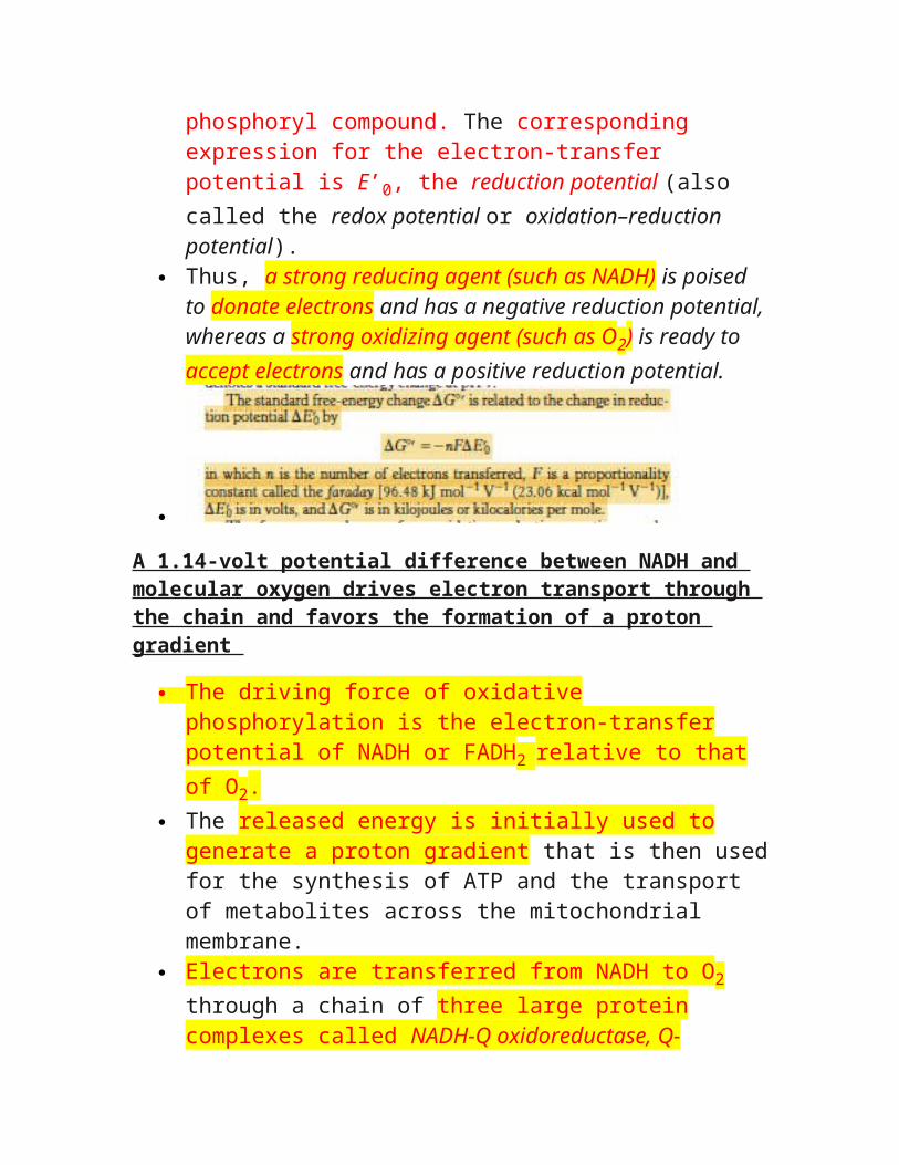