The Ciliary Protein Ftm Is Required for Ventricular Wall...
15
The Ciliary Protein Ftm Is Required for Ventricular Wall and Septal Development Christoph Gerhardt, Johanna M. Lier, Stefanie Kuschel, Ulrich Ru ¨ ther* Institute for Animal Developmental and Molecular Biology, Heinrich Heine University, Du ¨ sseldorf, Germany Abstract Ventricular septal defects (VSDs) are the most common congenital heart defects in humans. Despite several studies of the molecular mechanisms involved in ventricular septum (VS) development, very little is known about VS-forming signaling. We observed perimembranous and muscular VSDs in Fantom (Ftm)-negative mice. Since Ftm is a ciliary protein, we investigated presence and function of cilia in murine hearts. Primary cilia could be detected at distinct positions in atria and ventricles at embryonic days (E) 10.5–12.5. The loss of Ftm leads to shortened cilia and a reduced proliferation in distinct atrial and ventricular ciliary regions at E11.5. Consequently, wall thickness is diminished in these areas. We suggest that ventricular proliferation is regulated by cilia-mediated Sonic hedgehog (Shh) and platelet-derived growth factor receptor a (Pdgfra) signaling. Accordingly, we propose that primary cilia govern the cardiac proliferation which is essential for proper atrial and ventricular wall development and hence for the fully outgrowth of the VS. Thus, our study suggests ciliopathy as a cause of VSDs. Citation: Gerhardt C, Lier JM, Kuschel S, Ru ¨ ther U (2013) The Ciliary Protein Ftm Is Required for Ventricular Wall and Septal Development. PLoS ONE 8(2): e57545. doi:10.1371/journal.pone.0057545 Editor: Robert Dettman, Northwestern University, United States of America Received September 6, 2012; Accepted January 23, 2013; Published February 28, 2013 Copyright: ß 2013 Gerhardt et al. This is an open-access article distributed under the terms of the Creative Commons Attribution License, which permits unrestricted use, distribution, and reproduction in any medium, provided the original author and source are credited. Funding: This work was supported by the Deutsche Forschungsgemeinschaft (Sonderforschungsbereiche 590 and 612) to U.R. The funders had no role in study design, data collection and analysis, decision to publish, or preparation of the manuscript. Competing Interests: The authors have declared that no competing interests exist. * E-mail: [email protected] Introduction One of 100 newborns suffers from a congenital heart defect [1]. Among these human congenital cardiac diseases ventricular septal defects (VSDs) are the most common [2,3] and occur in approximately 1 of 1000 births [4]. The most prevalent VSD subtype is the perimembranous VSD [5,6] which is characterized by the loss of the membranous part of the ventricular septum (VS) and a defect in the development of a second part of the VS - the muscular septum. Interestingly, the membranous VS does not start to grow before the muscular VS generation has been finished [7] indicating that membranous VS development is probably initiated by an interaction of the inlet muscular VS and the atrioventricular endocardial cushion cells (ECCs) [8,9]. The membranous VS arises solely from the ECCs and not from the muscular VS [4]. Although the molecular background of muscular VS development is only poorly understood [10,11], two different hypothesis have been debated for its formation and outgrowth. The first theory describes VS generation as an active process of cell growth in the apical region of the muscular septum [12,13], while the second ascribes muscular septal length gain to a passive process based on the increase of the ventricular cavities. According to this hypothesis, the formation of the muscular septum is carried out by proliferation of cells at distinct regions in the left and right ventricle [14–17], so that it consists of cardiomyocytes with both left-ventricular and right-ventricular identities [11,18,19]. Ftm (alias Rpgrip1l)-negative mice display abnormal heart development particularly a VSD and suffer from a dysfunction of primary cilia [20] indicating a potential relation between heart formation and ciliary action. The Ftm protein is localised at the base of cilia [20] and appears to be present at every cilium. The fact that mutations of FTM in humans were already found in ciliopathies like Meckel-Gruber syndrome, Joubert syndrome and nephronophthisis [21,22] accentuates the importance of this gene in human development. Primary cilia are hairlike, 1–15 mm long protrusions on most vertebrate cells. They function as the cells ‘‘antenna’’ receiving and mediating signals from the environment. These signals, in turn, control important cellular processes like proliferation, apoptosis, migration, differentiation and cell cycle regulation [23]. Consequently, defective primary cilia provoke severe human diseases [24]. Several signaling pathways are thought to be associated with cilia, including Sonic hedgehog (Shh), platelet- derived growth factor receptor a (Pdgfra) as well as canonical and non-canonical Wnt signaling [25–35]. While the connection between cilia and Wnt signaling has been frequently discussed and remains the subject of fierce debate [34–36], it is well-known that Shh and Pdgfra signaling can be mediated by cilia [25– 27,29,30,35]. Shh is a member of the Hedgehog (Hh) family of evolutionary conserved signaling molecules and binds to its receptor Patched (Ptc) which in vertebrates is localized in the ciliary membrane and regulates the activity of Smoothened (Smo), a seven-transmem- brane receptor. Recruited to the cilium active Smo invokes Glioblastoma (Gli) transcription factors. In vertebrates three Gli isoforms exist – Gli1, 2 and 3. They regulate the expression of Shh target genes like for example Ptc1 and thereby cell differentiation, proliferation, survival and growth [37,38]. Gli1 functions as a constitutive activator [39,40], whereas Gli2 and Gli3 have a C- terminal transcriptional activator domain and a N-terminal transcriptional repressor domain [41]. Full-length Gli3 (Gli3- PLOS ONE | www.plosone.org 1 February 2013 | Volume 8 | Issue 2 | e57545
Transcript of The Ciliary Protein Ftm Is Required for Ventricular Wall...
The Ciliary Protein Ftm Is Required for Ventricular Walland Septal DevelopmentChristoph Gerhardt, Johanna M. Lier, Stefanie Kuschel, Ulrich Ruther*
Institute for Animal Developmental and Molecular Biology, Heinrich Heine University, Dusseldorf, Germany
Abstract
Ventricular septal defects (VSDs) are the most common congenital heart defects in humans. Despite several studies of themolecular mechanisms involved in ventricular septum (VS) development, very little is known about VS-forming signaling.We observed perimembranous and muscular VSDs in Fantom (Ftm)-negative mice. Since Ftm is a ciliary protein, weinvestigated presence and function of cilia in murine hearts. Primary cilia could be detected at distinct positions in atria andventricles at embryonic days (E) 10.5–12.5. The loss of Ftm leads to shortened cilia and a reduced proliferation in distinctatrial and ventricular ciliary regions at E11.5. Consequently, wall thickness is diminished in these areas. We suggest thatventricular proliferation is regulated by cilia-mediated Sonic hedgehog (Shh) and platelet-derived growth factor receptor a(Pdgfra) signaling. Accordingly, we propose that primary cilia govern the cardiac proliferation which is essential for properatrial and ventricular wall development and hence for the fully outgrowth of the VS. Thus, our study suggests ciliopathy as acause of VSDs.
Citation: Gerhardt C, Lier JM, Kuschel S, Ruther U (2013) The Ciliary Protein Ftm Is Required for Ventricular Wall and Septal Development. PLoS ONE 8(2): e57545.doi:10.1371/journal.pone.0057545
Editor: Robert Dettman, Northwestern University, United States of America
Received September 6, 2012; Accepted January 23, 2013; Published February 28, 2013
Copyright: � 2013 Gerhardt et al. This is an open-access article distributed under the terms of the Creative Commons Attribution License, which permitsunrestricted use, distribution, and reproduction in any medium, provided the original author and source are credited.
Funding: This work was supported by the Deutsche Forschungsgemeinschaft (Sonderforschungsbereiche 590 and 612) to U.R. The funders had no role in studydesign, data collection and analysis, decision to publish, or preparation of the manuscript.
Competing Interests: The authors have declared that no competing interests exist.
* E-mail: [email protected]
Introduction
One of 100 newborns suffers from a congenital heart defect [1].
Among these human congenital cardiac diseases ventricular septal
defects (VSDs) are the most common [2,3] and occur in
approximately 1 of 1000 births [4]. The most prevalent VSD
subtype is the perimembranous VSD [5,6] which is characterized
by the loss of the membranous part of the ventricular septum (VS)
and a defect in the development of a second part of the VS - the
muscular septum. Interestingly, the membranous VS does not start
to grow before the muscular VS generation has been finished [7]
indicating that membranous VS development is probably initiated
by an interaction of the inlet muscular VS and the atrioventricular
endocardial cushion cells (ECCs) [8,9]. The membranous VS
arises solely from the ECCs and not from the muscular VS [4].
Although the molecular background of muscular VS development
is only poorly understood [10,11], two different hypothesis have
been debated for its formation and outgrowth. The first theory
describes VS generation as an active process of cell growth in the
apical region of the muscular septum [12,13], while the second
ascribes muscular septal length gain to a passive process based on
the increase of the ventricular cavities. According to this
hypothesis, the formation of the muscular septum is carried out
by proliferation of cells at distinct regions in the left and right
ventricle [14–17], so that it consists of cardiomyocytes with both
left-ventricular and right-ventricular identities [11,18,19].
Ftm (alias Rpgrip1l)-negative mice display abnormal heart
development particularly a VSD and suffer from a dysfunction
of primary cilia [20] indicating a potential relation between heart
formation and ciliary action. The Ftm protein is localised at the
base of cilia [20] and appears to be present at every cilium. The
fact that mutations of FTM in humans were already found in
ciliopathies like Meckel-Gruber syndrome, Joubert syndrome and
nephronophthisis [21,22] accentuates the importance of this gene
in human development.
Primary cilia are hairlike, 1–15 mm long protrusions on most
vertebrate cells. They function as the cells ‘‘antenna’’ receiving
and mediating signals from the environment. These signals, in
turn, control important cellular processes like proliferation,
apoptosis, migration, differentiation and cell cycle regulation
[23]. Consequently, defective primary cilia provoke severe human
diseases [24]. Several signaling pathways are thought to be
associated with cilia, including Sonic hedgehog (Shh), platelet-
derived growth factor receptor a (Pdgfra) as well as canonical and
non-canonical Wnt signaling [25–35]. While the connection
between cilia and Wnt signaling has been frequently discussed
and remains the subject of fierce debate [34–36], it is well-known
that Shh and Pdgfra signaling can be mediated by cilia [25–
27,29,30,35].
Shh is a member of the Hedgehog (Hh) family of evolutionary
conserved signaling molecules and binds to its receptor Patched
(Ptc) which in vertebrates is localized in the ciliary membrane and
regulates the activity of Smoothened (Smo), a seven-transmem-
brane receptor. Recruited to the cilium active Smo invokes
Glioblastoma (Gli) transcription factors. In vertebrates three Gli
isoforms exist – Gli1, 2 and 3. They regulate the expression of Shh
target genes like for example Ptc1 and thereby cell differentiation,
proliferation, survival and growth [37,38]. Gli1 functions as a
constitutive activator [39,40], whereas Gli2 and Gli3 have a C-
terminal transcriptional activator domain and a N-terminal
transcriptional repressor domain [41]. Full-length Gli3 (Gli3-
PLOS ONE | www.plosone.org 1 February 2013 | Volume 8 | Issue 2 | e57545
190) protein can be transformed into a transcriptional activator
(Gli3-A) most likely by modifications [42,43]. Importantly, the full-
length protein can be proteolytically processed into a transcrip-
tional repressor (Gli3-R, also known as Gli3-83) [44]. The ratio of
activator and repressor forms controls cellular processes depen-
dend on Shh signaling.
Signaling by Pdgfra relates also to cilia [29]. Pdgfra is localized
to cilia and becomes dimerized and phosphorylated after being
bound by its ligand Pdgf-AA which also functions as a dimer.
Activated Pdgf receptors regulate essential cell processes like
proliferation, anti-apoptosis, migration, differentiation, actin reor-
ganization and cell growth [45–47]. Stimulation of Pdgfra drives
the activation of signal transduction through the Mek1/2-Erk1/2
and Akt/PKB pathways mediated by primary cilia, whereas
Pdgfra signaling gets blocked in the absence of cilia [29].
We used Ftm-deficient mice to investigate whether cardiac cilia
are functionally involved in heart development, especially in VS
formation. Furthermore, we analysed which signals are mediated
by these cilia. We were able to identify components of Shh and
Pdgfra signaling pathways in or at ventricular cilia giving evidence
that these signals are cilia-mediated in embryonic murine hearts.
According to ciliary dysfunction caused by Ftm deficiency
[21,48,49], Shh and Pdfgra signaling are downregulated in Ftm-
negative ventricles. We propose these signaling defects as the cause
of reduced ventricular cell proliferation that in turn results in
diminished ventricular wall thickness and VSDs.
Materials and Methods
Ethics Statement and Animal HusbandryAll mice (Mus musculus) used in this study were on the C3H
background and kept under standard housing conditions with a
12/12 hours dark-light cycle and with food and water ad libitum.
All experiments were performed in accordance with the relevant
national guidelines for the Care and Use of Laboratory Animals,
with approval from the authority for animal work at the Heinrich
Heine University (Permit Number: O18/99). Generation of Ftm
mutant mice was designed and carried out as described [20].
AntibodiesWe used primary antibodies to actin (Sigma #A2066), Arl13b
(Proteintech #17711-1-AP), Gapdh (Sigma #G8795), acetylated
a-tubulin (Sigma #T6793), c-tubulin (Sigma #T6557), detyrosi-
nated tubulin (Millipore #AB3201), BrdU (Developmental Studies
Hybridoma Bank #G3G4), Pdgfra (Santa Cruz #sc-338),
pericentrin (Covance #PRB-432C), pMek1/2 (Cell Signaling
Technology #9121), Gli3 (kindly gift of B. Wang), Gli3 (R&D
systems #AF3690), ErbB3 (Santa Cruz #sc-285), DDR2 (kindly
gift of E.C. Goldsmith) and Tropomyosin (AbD Serotec #9200-
0504). The creation of polyclonal antibodies against Ftm was
delineated formerly [20]. Polyclonal antibodies to Gli3-190 were
generated by immunizing rabbits with a His-Gli3 fusion protein
encompassing the Gli3-C-terminal region (3473–4806 bp) by
Pineda antibody services. Antibodies were affinity-purified with
the antigen coupled to Ni-NTA agarose (Qiagen #30230).
Apoptosis StudiesApoptotic nuclei were labeled in situ by the TdT-mediated
dUTP-biotin nick end labeling (TUNEL) method [50] using Apop
Taq Plus Peroxidase in situ Apoptosis Kit (Millipore #S7101) and
following manufacturer’s instructions.
GenotypingGenotyping of the mice was performed as previously described
[20].
HistochemistryHistochemical stainings were performed as described [20].
Histology and Paraffin EmbeddingEmbryos were dissected and fixed in 4% paraformaldehyde
(PFA) overnight at 4uC. Then they were serially dehydrated using
ethanol, embedded in paraffin and sectioned (12 mm). Afterwards,
sections were stained with hematoxylin and eosin or used for in situ
hybridisation.
ImmunofluorescenceEmbryos were fixed in 4% PFA and incubated in 30% sucrose
(in PBS) overnight at 4uC. Next day they were embedded in
Tissue-Tek O.C.T. compound (Sakura Finetechnical #4583) and
then stored at 280uC. Transverse cryostat sections (7 mm in
thickness) were prepared, washed with PBS and permeabilized
with PBS/0.5% Triton-X-100. Blocking was performed with 10%
FCS in PBS/0.1% Triton-X-100. The sections then were
incubated with the primary antibodies diluted in blocking solution
overnight at 4uC. After three washing steps, they underwent an
incubation in the secondary antibody (diluted in blocking solution)
for 2 hours and then were washed again. Finally, they were
embedded in Mowiol containing DAPI (Merck #1.24653).
In situ HybridisationIn situ hybridisation on paraffin sections were performed as
previously described [51].
Proliferation StudiesMice received an intraperitoneal injection of 10 ml BrdU (Sigma
#B5002-1G) per g body weight 2 hours before they were killed.
After killing embryos were dissected and embedded in Tissue-Tek
O.C.T. compound (Sakura Finetechnical #4583) as described
before. Cryosections were undergone BrdU immunohistochemical
stainings like described before with the exception of two
additionally steps after the first washings: These steps include
incubation in 2 N HCl for 10 minutes at 37uC and then in 50%
formamide/26SSC for 45 minutes at 65uC. Anti-BrdU (Devel-
opmental Studies Hybridoma Bank #G3G4) antibody was used as
primary antibody.
Real-time PCR AnalysisAtrial and ventricular RNA was isolated by using RNeasy Kit
(Qiagen #74104) and RNase-Free DNase Set (Qiagen #79254).
Isolated RNA was converted into cDNA by utilising Expand
Reverse Transcriptase (Roche #11785826001). Quantitative
Real-time PCR was carried out by employing a Step One Real-
Time PCR System Thermal Cycling Block (Applied Biosystems
#4376357) and the TaqMan Universal PCR Master Mix, No
AmpErase UNG (Applied Biosystems #4324020). The following
primer/TaqMan probe sets were used: Gapdh (Assay ID:
Mm99999915_g1), Ptc1 (Assay ID: Mm00970977_m1) and Hif1a(Assay ID: Mm00468878_m1). Real-time PCR was carried out
with 50 ng of cardiac cDNA of each sample in triplicate reactions
in a 20 ml volume containing 100 nM primers and 50 nM probe.
Cycling conditions were 50uC for 2 minutes and 95uC for 10
minutes, followed by a 40-cycle amplification of 95uC for 15
seconds and 60uC for 1 minute. The analysis of real-time data was
performed by using included StepOne Software version 2.0.
Ftm Is Required for Ventricular Development
PLOS ONE | www.plosone.org 2 February 2013 | Volume 8 | Issue 2 | e57545
Semiquantitative PCR AnalysisRNeasy Kit (Qiagen #74104) was used to isolate mRNA from
pooled embryonic hearts (E11.5). Reverse transcription was
carried out by utilising High Capacity RNA-to-cDNA Master
Mix (Applied Biosystems #4390777). The sets of primers were as
following: Hprt: 59-CAC AGG ACT AGA ACA CCT GC
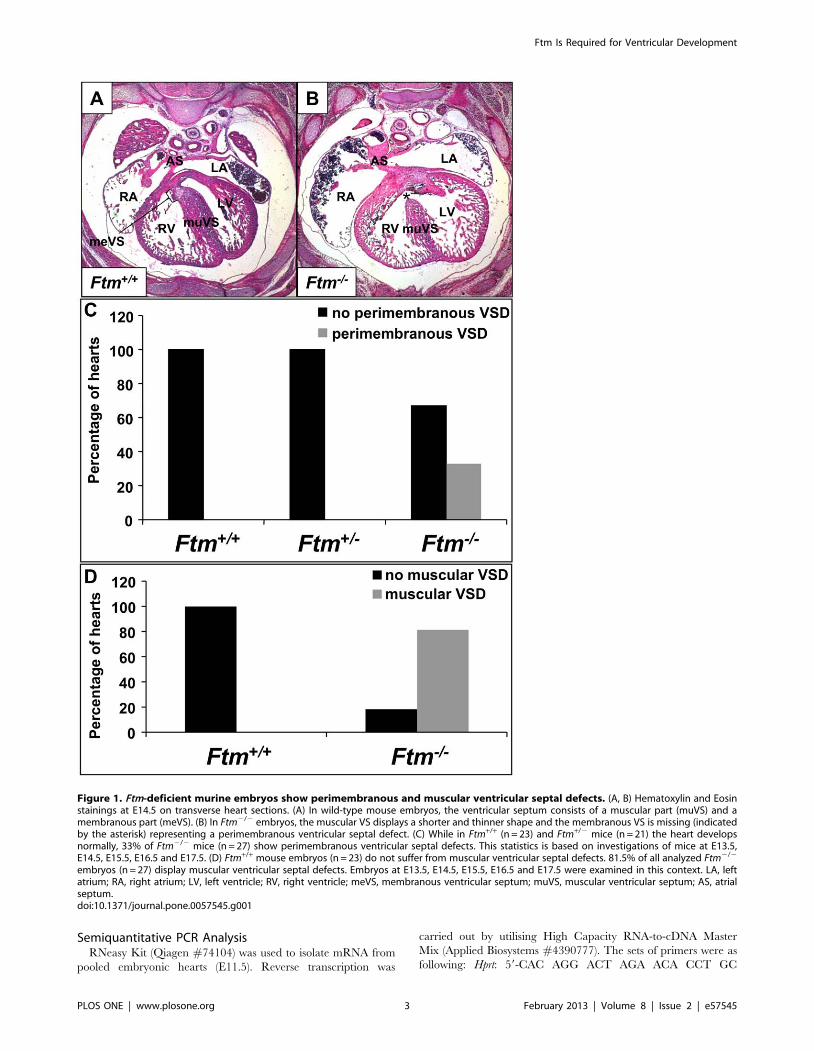
Figure 1. Ftm-deficient murine embryos show perimembranous and muscular ventricular septal defects. (A, B) Hematoxylin and Eosinstainings at E14.5 on transverse heart sections. (A) In wild-type mouse embryos, the ventricular septum consists of a muscular part (muVS) and amembranous part (meVS). (B) In Ftm2/2 embryos, the muscular VS displays a shorter and thinner shape and the membranous VS is missing (indicatedby the asterisk) representing a perimembranous ventricular septal defect. (C) While in Ftm+/+ (n = 23) and Ftm+/2 mice (n = 21) the heart developsnormally, 33% of Ftm2/2 mice (n = 27) show perimembranous ventricular septal defects. This statistics is based on investigations of mice at E13.5,E14.5, E15.5, E16.5 and E17.5. (D) Ftm+/+ mouse embryos (n = 23) do not suffer from muscular ventricular septal defects. 81.5% of all analyzed Ftm2/2
embryos (n = 27) display muscular ventricular septal defects. Embryos at E13.5, E14.5, E15.5, E16.5 and E17.5 were examined in this context. LA, leftatrium; RA, right atrium; LV, left ventricle; RV, right ventricle; meVS, membranous ventricular septum; muVS, muscular ventricular septum; AS, atrialseptum.doi:10.1371/journal.pone.0057545.g001
Ftm Is Required for Ventricular Development
PLOS ONE | www.plosone.org 3 February 2013 | Volume 8 | Issue 2 | e57545
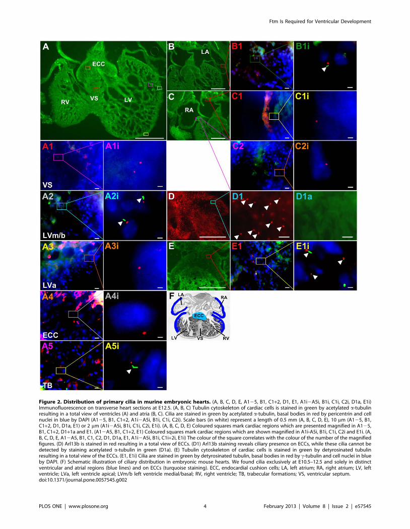
Figure 2. Distribution of primary cilia in murine embryonic hearts. (A, B, C, D, E, A125, B1, C1+2, D1, E1, A1i2A5i, B1i, C1i, C2i, D1a, E1i)Immunofluorescence on transverse heart sections at E12.5. (A, B, C) Tubulin cytoskeleton of cardiac cells is stained in green by acetylated a-tubulinresulting in a total view of ventricles (A) and atria (B, C). Cilia are stained in green by acetylated a-tubulin, basal bodies in red by pericentrin and cellnuclei in blue by DAPI (A125, B1, C1+2, A1i2A5i, B1i, C1i, C2i). Scale bars (in white) represent a length of 0.5 mm (A, B, C, D, E), 10 mm (A125, B1,C1+2, D1, D1a, E1) or 2 mm (A1i2A5i, B1i, C1i, C2i, E1i). (A, B, C, D, E) Coloured squares mark cardiac regions which are presented magnified in A125,B1, C1+2, D1+1a and E1. (A12A5, B1, C1+2, E1) Coloured squares mark cardiac regions which are shown magnified in A1i-A5i, B1i, C1i, C2i and E1i. (A,B, C, D, E, A12A5, B1, C1, C2, D1, D1a, E1, A1i2A5i, B1i, C1i+2i, E1i) The colour of the square correlates with the colour of the number of the magnifiedfigures. (D) Arl13b is stained in red resulting in a total view of ECCs. (D1) Arl13b staining reveals ciliary presence on ECCs, while these cilia cannot bedetected by staining acetylated a-tubulin in green (D1a). (E) Tubulin cytoskeleton of cardiac cells is stained in green by detyrosinated tubulinresulting in a total view of the ECCs. (E1, E1i) Cilia are stained in green by detyrosinated tubulin, basal bodies in red by c-tubulin and cell nuclei in blueby DAPI. (F) Schematic illustration of ciliary distribution in embryonic mouse hearts. We found cilia exclusively at E10.5–12.5 and solely in distinctventricular and atrial regions (blue lines) and on ECCs (turquoise staining). ECC, endocardial cushion cells; LA, left atrium; RA, right atrium; LV, leftventricle; LVa, left ventricle apical; LVm/b left ventricle medial/basal; RV, right ventricle; TB, trabecular formations; VS, ventricular septum.doi:10.1371/journal.pone.0057545.g002
Ftm Is Required for Ventricular Development
PLOS ONE | www.plosone.org 4 February 2013 | Volume 8 | Issue 2 | e57545
(forward), 39-GCT GGT GAA AAG GAC CTC T (reverse); Cyclin
E: 59-CTG GCT GAA TGT TTA TGT CC (forward), 39-TCT
TTG CTT GGG CTT TGT CC (reverse); p27:59-AAC CTC
TTC GGC CCG GTG GAC CAC (forward), 39-GTC TGC
TCC ACA GAA CCG GCA TTT (reverse).
Statistical DataTo compare percentage of proliferating cells and percentage of
apoptotic cells in wild-type and Ftm-mutant hearts, we counted
BrdU or TUNEL marked cells and total number of cells (DAPI-
marked) in distinct regions on ten different, transverse sections per
heart, averaged over them and related them to each other. All
heart chambers could be analysed on every section. Thereby, we
differentiated between ciliary, former ciliary and non-ciliary
regions.
To contrast wild-type with Ftm-negative cardiac wall thickness,
the measurements of wall thickness were performed at distinct
regions on ten different, transverse sections per heart. All four
heart chambers were uncovered on every section. The measured
values per heart were averaged.
Data are presented as mean 6 standard deviation. Student’s t
test was performed to compare percentage of proliferating cells,
cardiac wall thickness, RNA-expression levels and percentage of
apoptotic cells in wild-type and Ftm-mutant hearts by using
Graphpad and Microsoft Excel. A p value ,0.05 was considered
to be statistically significant (one asterisk), a p value ,0.01 was
regarded as statistically very significant (two asterisks) and a p value
,0.001 was accounted statistically high significant (three asterisks).
Western BlottingWestern blot studies were done essentially as described using
anti-Gli3 antibody or anti-pMek1/2 antibody [44]. Anti-actin
antibody and anti-Gapdh antibody were used as control for
loading. Visualising of Gli3, pMek1/2, actin and Gapdh bands
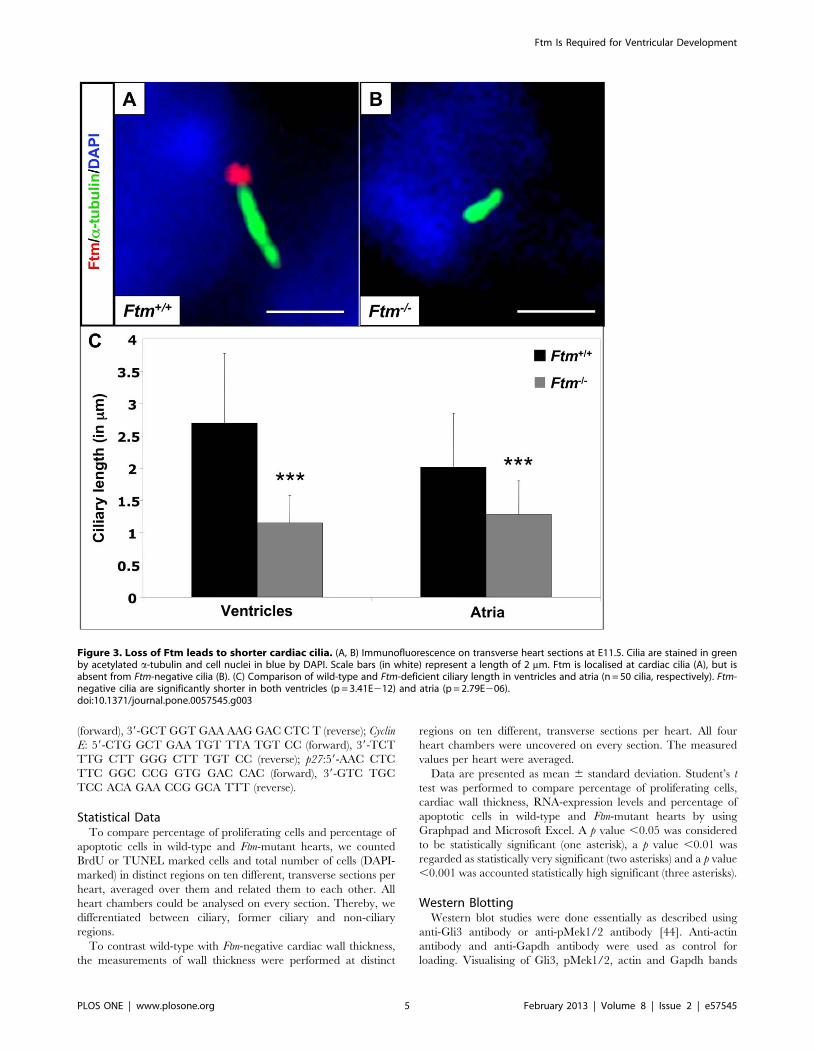
Figure 3. Loss of Ftm leads to shorter cardiac cilia. (A, B) Immunofluorescence on transverse heart sections at E11.5. Cilia are stained in greenby acetylated a-tubulin and cell nuclei in blue by DAPI. Scale bars (in white) represent a length of 2 mm. Ftm is localised at cardiac cilia (A), but isabsent from Ftm-negative cilia (B). (C) Comparison of wild-type and Ftm-deficient ciliary length in ventricles and atria (n = 50 cilia, respectively). Ftm-negative cilia are significantly shorter in both ventricles (p = 3.41E212) and atria (p = 2.79E206).doi:10.1371/journal.pone.0057545.g003
Ftm Is Required for Ventricular Development
PLOS ONE | www.plosone.org 5 February 2013 | Volume 8 | Issue 2 | e57545
Ftm Is Required for Ventricular Development
PLOS ONE | www.plosone.org 6 February 2013 | Volume 8 | Issue 2 | e57545
was realised by LAS-4000 mini (Fujifilm #8692184). Bands were
measured in intensity using Adobe Photoshop 7.0.
Results
Ftm-negative, Murine Embryos Display Muscular andPerimembranous Ventricular Septal Defects
33% of all analysed Ftm-homozygous mutant embryos (9 of 27
embryos) show perimembranous VSDs marked by the combina-
tion of a significantly thinner muscular part of the VS and the
absence of the membranous part of the VS (Figure 1B, C), while
none of the Ftm-heterozygous mutants exhibits an abnormal heart
phenotype (Figure 1C). We measured the length and thickness of
ventricular and atrial septa in Ftm+/+ and Ftm2/2 hearts,
respectively, and found out that the atrial septum (AS) displays
no differences between the wild-type and Ftm-negative state (data
not shown). Furthermore, we did not observe any morphological
AS abnormalities. In contrast to the atria, Ftm-deficient ventricles
display defects, but the length measurements do not reflect a
significant alteration at different embryonic days (Figure S1A, D,
G, J, M). This is due to the fact that the frequency of
perimembranous VSDs in the absence of Ftm is too low during
embryonic development (Figure S1B, E, H, K, N). At E13.5 40%
of all analyzed Ftm2/2 mouse embryos (2 of 5) suffer from
perimembranous VSDs, at E14.5 50% (3 of 6), at E15.5 67% (2 of
3), at E16.5 0% (none of 4) and at E17.5 22% (2 of 9). Compared
to the length measurements, the width of Ftm-negative VS is
significantly reduced (Figure S1A, D, G, J, M) characterizing a
muscular VSD. 81, 5% of all analyzed Ftm2/2 embryos (22 of 27
embryos) suffer from muscular VSDs (Figure 1D). The reduction
of muscular VS width is significant at all analyzed embryonic days
from E13.5 to E17.5 (Figure S1A, D, G, J, M), since the frequency
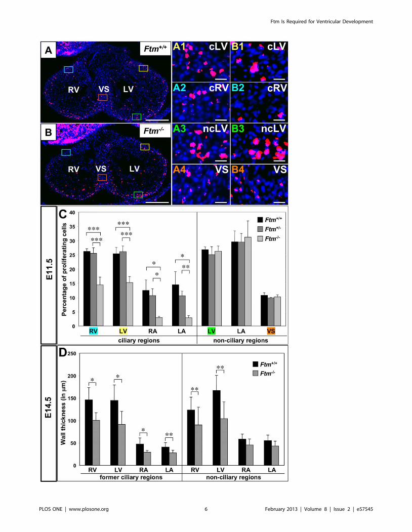
Figure 4. Reduced proliferation in ciliary regions of Ftm-deficient murine hearts and thickness decrease of Ftm-negative walls. (A, B,A1–4, B1–4) Immunofluorescence on transverse ventricular sections at E11.5. Dividing cells (red staining) are marked by BrdU and cell nuclei (bluestaining) by DAPI. Scale bars (in white) represent a length of 0.5 mm (A, B) or 20 mm (A1–4, B1–4). (A, B) Coloured squares mark cardiac regions whichare presented magnified in A1–4 and B1–4, respectively. The colour of the square correlates with the colour of the number of the magnified figures.(C) Proliferation rate is determined by the relation of dividing (BrdU-marked) cells to the number of all cells in this heart region at E11.5 (Ftm+/+: n = 6;Ftm+/2: n = 11; Ftm2/2: n = 5). There is significantly less proliferation in the ciliary regions of ventricles and atria compared to non-ciliary regions. (D)Cardiac wall thickness measurements of wild-type (n = 6) and Ftm-deficient (n = 6) atria and ventricles in former ciliary and non-ciliary regions at E14.5.Walls are significantly thinner in all former ciliary regions. Additionally, ventricular, non-ciliary regions show a reduction in wall thickness, while atrial,non-ciliary regions do not differ significantly. LA, left atrium; RA, right atrium; LV, left ventricle; cLV, ciliary region of the left ventricle; ncLV, non-ciliaryregion of the left ventricle; RV, right ventricle; ciliary region of the right ventricle; VS, ventricular septum.doi:10.1371/journal.pone.0057545.g004
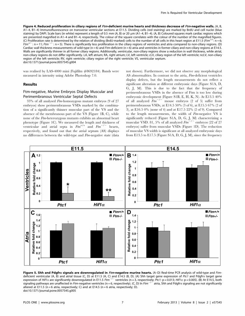
Figure 5. Shh and Pdgfra signals are downregulated in Ftm-negative murine hearts. (A–D) Real-time PCR analysis of wild-type and Ftm-deficient ventricular (A, B) and atrial tissue (C, D) at E11.5 (A, C) and E14.5 (B, D). (A) Shh target gene expression of Ptc1 and Pdgfra target geneexpression of Hif1a are significantly downregulated in E11.5 Ftm2/2 ventricles (n = 3, respectively; Ptc1: p = 0.013; Hif1a: p = 0.005). (B) At E14.5, bothsignaling pathways are unaffected in Ftm-negative ventricles (n = 6, respectively). (C, D) In Ftm2/2 atria, Shh and Pdgfra signaling are not significantlyaltered at E11.5 (n = 6 atria, respectively; C) and at E14.5 (n = 6 atria, respectively; D).doi:10.1371/journal.pone.0057545.g005
Ftm Is Required for Ventricular Development
PLOS ONE | www.plosone.org 7 February 2013 | Volume 8 | Issue 2 | e57545
of muscular VSDs is high in all embryonic stages (Figure S1C, F, I,
L, O). At E13.5 muscular VSDs can be observed in 80% of all
analysed Ftm-negative embryos (4 of 5), at E14.5 in 83% (5 of 6), at
E15.5 in 100% (3 of 3), at E16.5 in 50% (2 of 4) and at E17.5 in
89% (8 of 9). These data indicate that the muscular VS defect
takes place in a high frequency even if the loss of the membranous
VS occurs only in a minority of all Ftm2/2 embryos leading to the
conclusion that muscular VS development is severly disturbed in
most Ftm-deficient embryos.
To test if the appearance of perimembranous VSDs correlates
with other defects of Ftm-deficient mice, we looked for the entire
phenotype of all analyzed mice. Comparing the different
phenotypes, there seems to be no correlation between the
occurrence of perimembranous VSDs and other abnormalities
(Table S1).
Cilia are absent from the VS in E10.5 to E12.5 murine
hearts. Since Ftm is a cilia-associated protein [20], we assumed
that the cause of heart phenotype in Ftm-negative mice could be a
ciliary dysfunction, although never before a ciliopathy was
regarded as the elicitor of VSDs. The pre-condition for this
assumption is the presence of cilia in murine hearts. In previous
studies, cardiac cilia were detected in mice [52,53], but it was not
mentioned if the VS is ciliated. We observed monocilia in a very
distinct spatial distribution from E10.5 to E12.5 (Figure 2), but
could never detect any cilia on VS cells (Figure 2A1, A1i).
Furthermore, we could not demonstrate the presence of cilia on
those ventricular cells which are close to the base of the muscular
VS (Figure 2A3, A3i). Interestingly, cilia on ECCs were hardly
detectable by visualizing acetylated a-tubulin (Figure 2D1a).
Instead, the detection of Arl13b reveals ciliary presence also on
the surface of these cells (Figure 2D1). Since acetylated a-tubulin
serves as a marker for the ciliary axoneme, we tested if ECC cilia
lack axonemes. Therefore, we used an antibody to detyrosinated
tubulin which is another tubulin modification indicative for the
ciliary axoneme [54]. Detyrosinated tubulin was detected on
ECCs (Figure 2E, E1, E1i) demonstrating that ECC cilia exhibit
an axoneme. To proof if cilia can be observed on VS cells by using
other ciliary marker instead of labelling acetylated a-tubulin, we
performed antibody stainings with an anti-Arl13b antibody. But
even by marking Arl13b, we could not detect cilia on VS cells
(Figure S2) or in other non-ciliary regions. Marking different
cardiac cell types, we found cardiac cilia poking out of myocardial
and endocardial cells (Figure S3A, B), but not from cardiac
fibroblasts (Figure S3C).
Cardiac cilia are shortened in Ftm-homozygous mutant
mice. We previously showed that Ftm is present at the base of
cilia in cell culture [20] and others observed Ftm at cilia of murine
eyes and brains [48], but nothing is known about the localisation
of Ftm at cardiac cilia. Consequently, we looked for Ftm in wild-
type hearts. The staining of Ftm and acetylated a-tubulin
(indicative for the ciliary axoneme) in combination with the partly
overlapping staining of Ftm and c-tubulin (basal body marker)
reveals that Ftm is located at the base of atrial and ventricular cilia
(Figure 3A; Figure S4A). Meanwhile, Ftm is completely missing in
Ftm-negative embryos (Figure 3B; Figure S4B). Since the loss of
Ftm in some cilia leads to a change in ciliary morphology (e.g.
nodal cilia; [20]) and since the alteration of ciliary length leads to
ciliary dysfunction [27,55–57], we analysed the length of Ftm-
homozygous mutant, cardiac cilia at E11.5. Cilia of Ftm-deficient
ventricles and atria are clearly shorter than in the wild-type
(Figure 3C) arguing for a possible ciliary dysfunction in those
hearts. Thus, Ftm is necessary for regulating the length of cardiac
cilia.
Less proliferation at ciliary regions in Ftm-deficient
hearts. The VS does not fully grow out in Ftm-negative mice
(Figure 1B). From other tissue and cell culture experiments, it is
known that monocilia mediate proliferative and apoptotic signals
[23]. Since ventricular cilia appear at the time, when the muscular
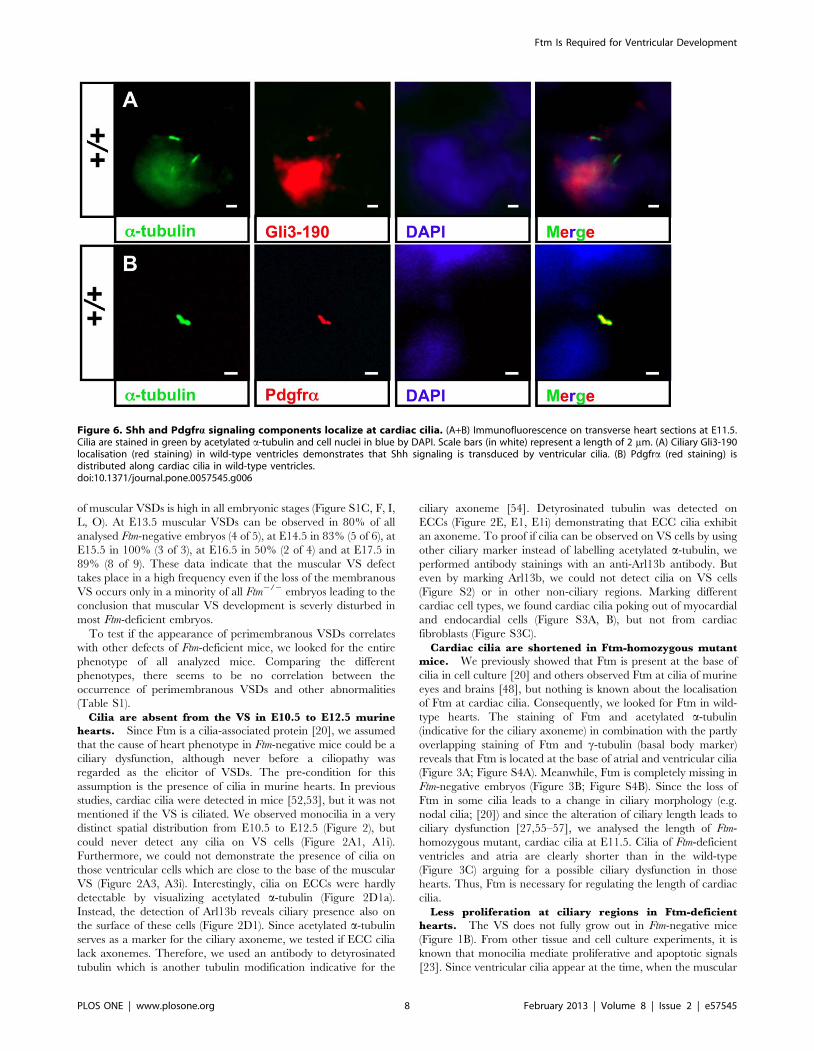
Figure 6. Shh and Pdgfra signaling components localize at cardiac cilia. (A+B) Immunofluorescence on transverse heart sections at E11.5.Cilia are stained in green by acetylated a-tubulin and cell nuclei in blue by DAPI. Scale bars (in white) represent a length of 2 mm. (A) Ciliary Gli3-190localisation (red staining) in wild-type ventricles demonstrates that Shh signaling is transduced by ventricular cilia. (B) Pdgfra (red staining) isdistributed along cardiac cilia in wild-type ventricles.doi:10.1371/journal.pone.0057545.g006
Ftm Is Required for Ventricular Development
PLOS ONE | www.plosone.org 8 February 2013 | Volume 8 | Issue 2 | e57545
VS is growing out [58], and at those regions, where the
proliferation of cells effects the outgrowth of the VS [11], we
investigated proliferation and apoptosis via bromodeoxyuridine
(BrdU) staining to determine the rate of proliferation and by
means of TdT-mediated dUTP-biotin nick end labeling (TUNEL)
staining to look for cell death at E11.5. Whereas the apoptosis
study was inconspicuous (Figure S5A), there were differences in
the proliferation rate between wild-type, Ftm-heterozygous and
Ftm-homozygous mutant hearts. The proliferation in all Ftm-
negative, ciliary areas in ventricles and atria was significantly
diminished, while no proliferation differences could be observed in
non-ciliary regions (Figure 4B1–B4, C). So in embryonic hearts,
cilia seem to be necessary to mediate proliferative signals which in
turn are responsible for a part of cardiac cell proliferation.
Consequently, when cilia are absent at a later point of time, the
rate of proliferation of wild-type, Ftm-heterozygous and Ftm-
homozygous mutant embryos should not differ significantly.
Performing the same proliferation assays in E14.5 hearts, we
found that, indeed, the proliferation in all areas, which were
investigated, was similar in wild-type and Ftm-deficient hearts
(Figure S5B). The diminished proliferation in Ftm-homozygous
mutant hearts is in agreement with the results of semiquantitative
Reverse transcription-PCRs. These experiments uncovered a
change of expression levels of cyclin E and p27 that are involved
in cell cycle regulation and proliferation (Figure S6) substantiating
suspicion of a proliferation defect in Ftm-deficient hearts.
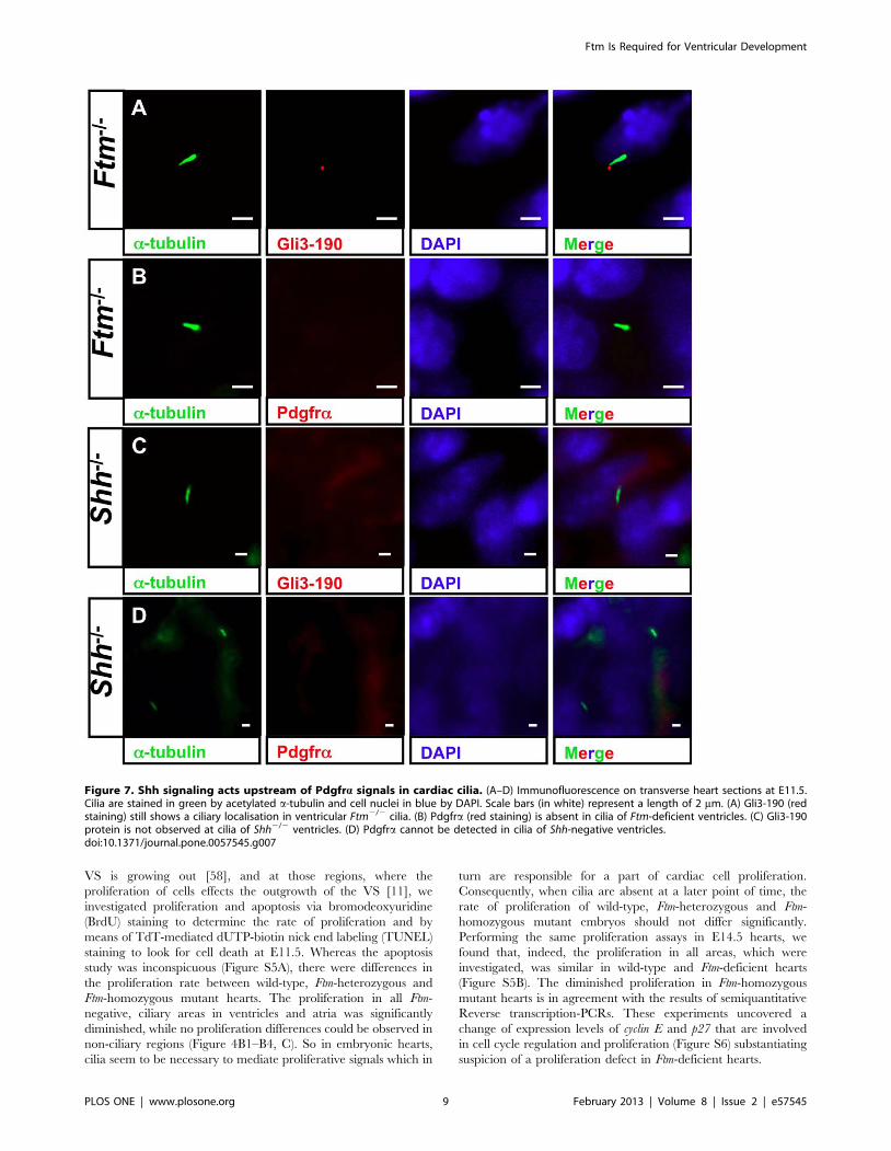
Figure 7. Shh signaling acts upstream of Pdgfra signals in cardiac cilia. (A–D) Immunofluorescence on transverse heart sections at E11.5.Cilia are stained in green by acetylated a-tubulin and cell nuclei in blue by DAPI. Scale bars (in white) represent a length of 2 mm. (A) Gli3-190 (redstaining) still shows a ciliary localisation in ventricular Ftm2/2 cilia. (B) Pdgfra (red staining) is absent in cilia of Ftm-deficient ventricles. (C) Gli3-190protein is not observed at cilia of Shh2/2 ventricles. (D) Pdgfra cannot be detected in cilia of Shh-negative ventricles.doi:10.1371/journal.pone.0057545.g007
Ftm Is Required for Ventricular Development
PLOS ONE | www.plosone.org 9 February 2013 | Volume 8 | Issue 2 | e57545
Reduction of wall thickness in Ftm-negative
hearts. Suggesting ciliary dysfunction is responsible for the
decline of cell number in Ftm-deficient ventricular walls, we
supposed that ventricular walls could be thinner in Ftm-negative
than in wild-type mice. Analysis of ventricular wall sizes reveals a
decrease of wall thickness in all regions of Ftm-negative hearts at
E14.5, where cilia were present at E11.5 (Figure 4D). Further-
more, ventricular walls of Ftm-deficient mice without ciliary
presence at any time are significantly thinner than those of wild-
type mice at E14.5 (Figure 4D). These walls reside close to the base
of the muscular VS. We also detected a reduction of wall thickness
in all ciliary atrial areas, but not in non-ciliary regions of the atria
(Figure 4D). Remarkably, 100% of all analyzed Ftm-negative
embryos (6 of 6) display a decreased wall thickness in atria and
ventricles (data not shown).
Shh and Pdgfra signals are downregulated in Ftm-
deficient hearts. Primary cilia are mediators of signaling
pathways, which activate certain cellular processes. To elucidate,
which signals are indispensable for cilia-controlled, cardiac
proliferation, we looked for target gene expression of signaling
pathways from which is known that they are mediated by cilia
[25,35]. Thereby, Patched1 (Ptc1) is used as target gene of Shh
signaling [59] and Hypoxia-inducible factor 1, a subunit (Hif1a) of
Pdgfra signaling [60]. Gene expression studies were performed at
a ciliary as well as at a non-ciliary period (E11.5 and E14.5,
respectively) and the hearts got subdivided into the ventricular and
the atrial part to differ between ventricular and atrial ciliary signal
mediation. At E11.5, Ftm-deficient ventricles show a significant
downregulation of Shh and Pdgfra signaling (Figure 5A), but these
signaling pathways are unaltered in Ftm-negative atria (Figure 5C).
At the non-ciliary stage E14.5, we do not see expression alterations
of the analysed target genes in the Ftm2/2 state (Figure 5B,D).
Taken together, these results show a downregulation of signaling
pathways in Ftm-homozygous mutant hearts at E11.5, but no
differences at the non-ciliary stage E14.5.
Most likely Shh signaling acts upstream of Pdgfrasignaling in ventricular cilia and is disturbed in Ftm-
deficient hearts owing to a Gli3 processing defect. To
elucidate if these signaling pathways are mediated by cardiac cilia,
we performed immunofluorescence stainings of proteins which are
essential for Shh and Pdgfra signaling, respectively. The Shh
signaling mediator Gli3-190 [41] can be clearly observed at the
base of ventricular cilia (Figure 6A, Figure S7A, B) and Pdgfra is
present all along ventricular cilia (Figure 6B). We could neither
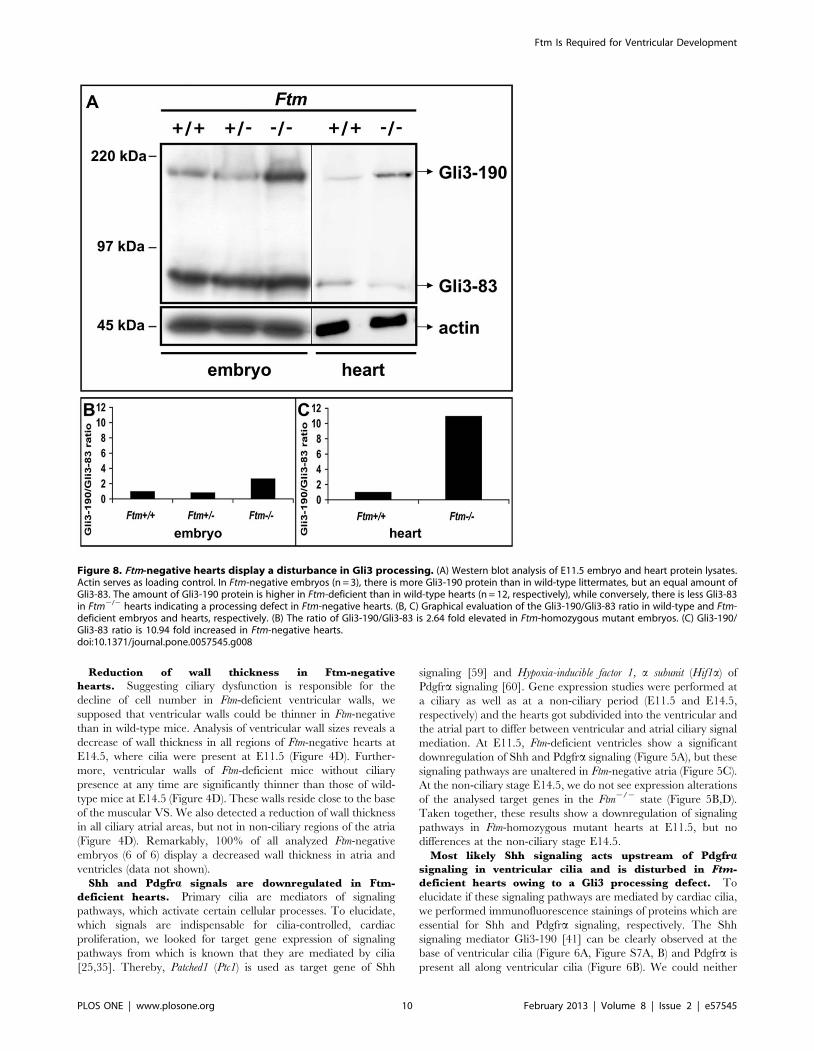
Figure 8. Ftm-negative hearts display a disturbance in Gli3 processing. (A) Western blot analysis of E11.5 embryo and heart protein lysates.Actin serves as loading control. In Ftm-negative embryos (n = 3), there is more Gli3-190 protein than in wild-type littermates, but an equal amount ofGli3-83. The amount of Gli3-190 protein is higher in Ftm-deficient than in wild-type hearts (n = 12, respectively), while conversely, there is less Gli3-83in Ftm2/2 hearts indicating a processing defect in Ftm-negative hearts. (B, C) Graphical evaluation of the Gli3-190/Gli3-83 ratio in wild-type and Ftm-deficient embryos and hearts, respectively. (B) The ratio of Gli3-190/Gli3-83 is 2.64 fold elevated in Ftm-homozygous mutant embryos. (C) Gli3-190/Gli3-83 ratio is 10.94 fold increased in Ftm-negative hearts.doi:10.1371/journal.pone.0057545.g008
Ftm Is Required for Ventricular Development
PLOS ONE | www.plosone.org 10 February 2013 | Volume 8 | Issue 2 | e57545
detect Gli3-190 nor Pdgfra at E11.5 atrial cilia (data not shown).
This indicates that both signaling pathways are mediated by cilia
in ventricles but not in atria at E11.5. Since we already detected a
downregulation of Shh and Pdgfra signaling in Ftm-deficient
hearts via qRT-PCR, we looked for Gli3-190 and Pdgfralocalisation in Ftm-negative cardiac cilia. In these cilia, Gli3-190
is still present (Figure 7A), while Pdgfra gets lost in ventricular cilia
(Figure 7B). These results let assume that Pdgfra signaling acts
downstream of Shh signaling. To confirm this hypothesis, we
investigated Gli3-190 and Pdgfra localisation at Shh-deficient cilia
in the heart. Gli3-190 and Pdgfra are absent in ventricular cilia
(Figure 7C, D) resulting in the conclusion that Pdgfra signaling
functions downstream of Shh signaling in ventricular cilia. The
dependency of ventricular Pdgfra signaling on Shh signaling is
confirmed by a smaller amount of the Pdgfra signaling component
pMek1/2 in Shh2/2 ventricles (Figure S8). This is indicative of a
downregulation of Pdgfra signaling in Shh-negative ventricles.
Since the phenotype of Shh-deficient hearts, which display
atrioventricular septal defects, appears to be much stronger than in
Ftm-negative embryos [61], Ftm functions most likely downstream
of Shh ligand in this pathway. The phenotypes of mice, which are
negative for Ptc1 and Smo, two components of the Shh pathway
downstream of its ligand, are also more severe than the Ftm-
deficient phenotype [57,62], so that we focused on the next players
within this signaling cascade 2 the Gli proteins. We examined
Gli3 processing by western blot analysis, using an antibody against
the N-terminus of Gli3 that detects both the full-length (Gli3-190)
and processed short, repressor (Gli3-83) forms. Previously, we were
able to show that the ratio of Gli3-190/Gli3-83 is higher in Ftm2/
2 whole embryo protein lysates than in wild-type or Ftm-
heterozygous ones (Figure 8A, B) [20]. In Ftm-deficient hearts,
we also detected an increase of the Gli3-190/Gli-83 ratio at E11.5
(Figure 8A,C) confirming our assumption of a Gli3 processing
defect. The ratio of Gli3-190/Gli3-83 in Ftm-negative embryos is
2.64 fold higher than in the wild-type (Figure 8B), while in Ftm-
deficient hearts, the Gli3-190/Gli3-83 ratio is 10.94 fold higher
than in their wild-type counterparts (Figure 8C).
Membranous ventricular septal defects in Ftm-negative
mice are most likely not due to endocardial cushion
defects. An interaction of inlet muscular VS and atrioventric-
ular ECCs seems to be required for the beginning of membranous
VS formation [8,9]. Previously, it was suggested that defective
ciliary function leads to a decreased cellularity of the ECCs [52].
So we looked for endocardial cushion morphology, the expression
of marker genes and ECC proliferation (Figure S9). Morpholog-
ically, ECCs show a normal shape in Ftm2/2 mice (Figure S9B)
and also the marker gene expression of Msh homeobox 1-like protein
(Msx1; Figure S9B) gives no hints indicating an abnormality in
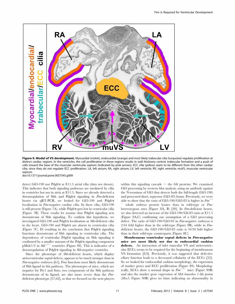
Figure 9. Model of VS development. Myocardial (violett), endocardial (orange) and most likely trabecular cilia (turquoise) regulate proliferation atdistinct cardiac regions. In the ventricles, the cell proliferation in these regions results in wall thickness control, trabecular formation and a push ofcells toward the base of the muscular ventricular septum (indicated by pink arrows). ECC cilia (yellow) seem to be different from the other cardiaccilia, since they do not regulate ECC proliferation. LA, left atrium; RA, right atrium; LV, left ventricle; RV, right ventricle; muVS, muscular ventricularseptum.doi:10.1371/journal.pone.0057545.g009
Ftm Is Required for Ventricular Development
PLOS ONE | www.plosone.org 11 February 2013 | Volume 8 | Issue 2 | e57545
endocardial cushion development. Moreover, the number of
proliferating ECCs is not altered in Ftm-deficient hearts at E11.5
(Figure S9C) and the atrioventricular valves exhibit a normal
shape (data not shown). Since cilia are present on the surface of
ECCs, it is interesting that the loss of Ftm does not seem to affect
ECC proliferation. The proliferation in murine ventricles seems to
be controlled mainly by Shh signals and so we examined ECC
cilia-mediated signaling by analysing the ciliary localisation of
Gli3. Interestingly, we did not detect Gli3 in or at wild-type ECC
cilia (Figure S9D) indicating that these cilia do not transduce Shh
signaling at all. Thus, although Ftm is present at the base of ECC
cilia (Figure S9E), these cilia seem to be different from ventricular
cilia as already suggested by the absence of acetylated a-tubulin
(Figure 2D1). These data let suppose that the origin of
perimembranous VSDs in Ftm-homozygous mutant hearts is not
due to ECC dysfunction, but might be a defective outgrowth of the
muscular VS.
Discussion
VSDs of Ftm-negative Mice are not only a Consequenceof Impaired Left-right (LR) Asymmetry
Until now, the molecular mechanisms underlying VS develop-
ment are largely unknown, but some factors have been elucidated
which lead to the appearance of VSDs. One favoured reason for
the occurrence of these congenital heart defects is the disturbancy
of LR asymmetry. There is a high association between VSDs and
LR asymmetry defects [63,64]. Previously, we published that Ftm-
deficient mice suffer from an impairment of LR asymmetry due to
a dysfunction of nodal cilia [20]. This fact raises the possibility that
the VSDs observed in the absence of Ftm are caused by
randomized heart looping. 19% of Ftm2/2 murine embryos
display an abnormal heart looping [20], while 33% of these mice
exhibit perimembranous VSDs. Thus, this laterality defect cannot
be the exclusive reason for VSDs in Ftm-negative mice.
Furthermore, other studies about hearts from embryos with
abnormal LR development due to paralyzed node cilia show
proper cardiac wall thickness [52], but 100% of all analyzed Ftm2/
2 embryos suffer from reduced wall thickness supporting evidence
for other VSD-causing reasons.
Preliminarily, it was suggested that primary cilia in murine
hearts contribute to proper cardiac development [52]. Since Ftm
deficiency has been shown to result in ciliary dysfunction [20,49],
we examined in this study if Ftm-negative cardiac cilia cause
perimembranous and muscular VSDs. The impact of Ftm absence
on cardiac cilia is obvious, because Ftm-deficient cilia are shorter in
atria and ventricles (Figure 3C). As an alteration of ciliary length
gives a hint on a ciliary dysfunction, we investigated which
molecular signals are mediated by cardiac cilia and if signaling is
defective in the Ftm-negative state.
Do cardiac cilia mediate proliferative and hence
muscular VS-generating signals?. We identified two signal-
ing pathways which might be mediated by murine, ventricular
cilia from E10.5 to E12.5, namely Shh and Pdgfra signaling. The
fact that atrial cilia do not transduce both pathways seems to be
due to a difference in signal transduction of ventricular and atrial
cells. Nevertheless, the correlation between ciliary presence and
proliferation reduction as well as diminished wall thickness within
the atria indicates that atrial cilia are associated with the
proliferation of atrial cells at distinct regions. The control of this
cilia-regulated atrial proliferation might be realized by mediating
other signals than Shh or Pdgfra signaling.
Our data let assume that there is a hierarchy between Shh and
Pdgfra signaling. In wild-type ventricles, Pdgfra is located at cilia
(Figure 6B). Since Pdgfra is missing at Shh-negative, ventricular
cilia (Figure 7D), Shh signaling seems to have an effect on Pdgfrasignaling in cardiac cilia of embryonic ventricles. In some cases, the
loss of Pdgfra alone already leads to VSDs in mice [65]. These
findings provide the indication that the defect in Ftm-negative mice
firstly seems to perturb Shh signaling and then secondly Pdgfrasignaling. Considering the heart phenotypes of Shh- [61], Ptc1- [62]
and Smo-deficient mouse embryos [57], we suggest that the
interruption in Shh signaling appears downstream of Smo, because
the heart defects of these mutants are more severe than the cardiac
phenotype of Ftm2/2 embryos. Since the ratio of Gli3-190 to Gli3-
83 is changed in Ftm-deficient embryonic hearts at E11.5 (Figure 8A,
C), this could be the step in Shh signaling where the disturbance
firstly takes place. The fact that not as much full-length Gli3 is
cleaved to its shorter repressor form as in the wild-type implicates a
defect in proteolytic processing of Gli3. Hence, Ftm-negative hearts
display a higher amount of Gli3-190. Nevertheless, Shh signaling is
downregulated in Ftm-deficient ventricles at E11.5 (Figure 5A). An
explanation for this discrepancy could be a defect in the
transformation of full-length Gli3 to its transcriptional activator
form leading to a reduced activation of Shh target genes.
Since we measured a reduced expression of Shh and Pdgfratarget genes in E11.5 Ftm-deficient ventricles (Figure 5A) indicat-
ing a downregulation of both pathways and detected a reduction
of proliferation at those regions of Ftm2/2 ventricles where cilia
are present, it is possible that cilia control ventricular proliferation
by mediating Shh and Pdgfra signals. We could not detect cardiac
cilia on muscular VS cells (Figure 2A1i) and the proliferation rate
of these cells is not significantly altered (Figure 4B1–B4, C). Hence,
we suggest that VS formation is not based on cell proliferation in
the apical region of the muscular VS.
Remarkably, ECC cilia which are clearly visible by detecting
Arl13b (Figure 2D1) or detyrosinated tubulin (Figure 2E1i), but
not by using an antibody against acetylated a-tubulin
(Figure 2D1a), do not display the presence of Gli3 (Figure S9D)
leading to the assumption that cilia of the ECCs do not mediate
Shh or Pdgfra signaling. Consequently, ECC proliferation is not
significantly affected by Ftm deficiency. Since we did not find any
morphological or molecular ECC alterations in Ftm-negative
hearts (Figure S9), it is unlikely that defective ECCs are the reason
for VSD appearance in Ftm2/2 mice.
Reduced proliferation influences the thickness of cardiac
walls and VS development. We detected a decrease in the
thickness of atrial and ventricular walls at those positions where
cilia previously acted (Figure 4D) in 100% of all Ftm-negative
hearts. Since wall thickness is diminished in all cases, but the
attenuation of muscular VS thickness occurs in 81.5% of all
analysed Ftm-negative hearts and perimembranous VSDs only
appear in 33% of Ftm-deficient hearts, the decline of the wall
thickness seems to be the primary defect of cardiac ciliary
dysfunction, while the VSD is a consequence of it. The entire
phenotype of Ftm-negative mouse embryos is subject to a variation
reaching from embryos with extrinsically mild defects to severly
deformed embryos. Nevertheless, all Ftm2/2 embryos die at latest
around birth. The reason for the phenotype variation is unknown,
but maybe, it is the same, which causes differences in VS
development of Ftm-deficient hearts. Potentially, the number of
functional cilia plays a decisive role in the phenotype variation. If
there is a threshold of cilia-mediated signals determing the severity
of the mutant phenotype, it could be possible that the fewer cilia
are present the stronger shapes the phenotype.
Interestingly, the percentage of perimembranous VSDs is
higher in Ftm2/2 hearts at E13.5 (40%) (Figure S1B) than at
E17.5 (22%) (Figure S1N) suggesting that the defective VS
Ftm Is Required for Ventricular Development
PLOS ONE | www.plosone.org 12 February 2013 | Volume 8 | Issue 2 | e57545
development in the absence of Ftm occurs due to a developmental
delay. Another explanation is that mice displaying a stronger
phenotype and therefore a perimembranous VSD die earlier
within the embryonic development than those with a milder
phenotype. The second possibility is supported by the following
facts: It is obvious that lethality at early embryonic stages (e.g.
E13.5) takes place when Ftm is missing. Ftm-deficient embryos
which suffer from multiple defects die earlier than those displaying
a milder phenotype. Consequently, we observe exclusively milder
mutant phenotypes at late embryonic stages (Table S1). Remark-
ably, some Ftm-negative embryos at late embryonic days which
show mild mutant phenotypes suffer from perimembranous VSDs
meaning that most organs of these embryos develop properly and
indicating that a possible developmental delay only affects heart
development. This argues clearly against a developmental delay.
Moreover, muscular VSDs are detected in a high frequency at late
embryonic days like E17.5 (89%) (Figure S1O) demonstrating that
the observed VSDs are hardly based on a developmental delay.
The analysis of wall thickness in Ftm-negative hearts results in a
clear subdivision appearing in the atria. We observed thinner walls
where cilia had been present and normal wall thickness at those
sites which never showed any cilia. However, ventricular walls
display reduced wall thickness at both ciliary and non-ciliary
regions (Figure 4D). It is known that the muscular VS consists of
cardiomyocytes with both left-ventricular and right-ventricular
identities [11] indicating that the ciliary dysfunction leads to a
decrease of ventricular proliferation and hence to the appearance
of VSDs. In contrary to ventricular development, atrial septal
formation seems to be independent of ciliary function, because the
atrial septum appears to be unaffected in Ftm-negative embryos.
Model of VS formation. Assuming that cardiac cilia regulate
proliferation, our data allow us to propose a model for how the VS
is generated. Ventricular cells at distinct positions assemble
monocilia on their surface. These cardiac cilia contain compo-
nents of Shh and Pdgfra signaling most likely permitting them to
mediate those signals. Thus, target genes of those signaling
pathways are activated in cilia-possessing, ventricular cells.
Interestingly, in ventricular cilia Pdgfra signaling acts downstream
of Shh signaling. In the end, the mediation of these different
signals by cardiac cilia stimulates the cells to proliferate and this
proliferation leads to a push of cells to the base of the muscular VS
(Figure 9). Thus, in Ftm-negative ventricles the wall thickness of non-
ciliary regions near the base of the muscular VS is significantly
thinner (Figure 4D) due to the numeral reduction of cells which are
pushed towards the base of the muscular VS. Both, the pushed cells
and the trabecular formations shape the muscular VS which on its
part grows to a certain point and then interacts molecularly with the
ECCs. In turn, the ECCs start to shape the membranous VS which
then grows towards the muscular VS. When they meet, they fuse
and the development of the VS is finished. So finally, the muscular
VS consists of cells which descend from the left and right ventricular
walls and from the trabecular formations. Thus, our model supports
the idea of muscular septal formation as a product of a passive
process based on proliferation of cells at distinct regions in the left
and right ventricles.
Supporting Information
Figure S1 Defects of VS development in Ftm-negativemice are most likely not due to a developmental delay.Septum length and width was measured as well as the percentage
of murine hearts suffering from perimembranous and muscular
VSDs was determined at E13.5 (A, B, C), at E14.5 (D, E, F), at
E15.5 (G, H, I), at E16.5 (J, K, L) and at E17.5 (M, N, O). (A, D,
G, J, M) Septum measurements of wild-type and Ftm-deficient
ventricles at E13.5 (A), E14.5 (D), E15.5 (G), E16.5 (J) and E17.5
(M). Septum width was measured at different levels of the VS –
apical, medial and basal. The results of all levels together were
used to compile statistics. (A) At E13.5, Ftm-negative VS (n = 5) are
significantly thinner (p = 0.017) than their wild-type counterparts
(n = 5), while the length of Ftm2/2 VS (n = 5) is not significantly
altered in comparison to the wild-type ones (n = 5). (D) At E14.5,
Ftm-negative VS (n = 6) are significantly thinner (p = 0.003) than
their wild-type counterparts (n = 6), while the length of Ftm2/2 VS
(n = 6) is not significantly altered in comparison to the wild-type
ones (n = 6). (G) At E15.5, Ftm-negative VS (n = 3) are significantly
thinner (p = 0.046) than their wild-type counterparts (n = 3), while
the length of Ftm2/2 VS (n = 3) is not significantly altered in
comparison to the wild-type ones (n = 3). (J) At E16.5, Ftm-negative
VS (n = 4) are significantly thinner (p = 0.007) than their wild-type
counterparts (n = 4), while the length of Ftm2/2 VS (n = 4) is not
significantly altered in comparison to the wild-type ones (n = 4).
(M) At E17.5, Ftm-negative VS (n = 9) are significantly thinner
(p = 0.003) than their wild-type counterparts (n = 5), while the
length of Ftm2/2 VS (n = 9) is not significantly altered in
comparison to the wild-type ones (n = 5). Percentages of hearts
affected by perimembranous or muscular VSDs were calculated
from the very same number of embryos used in A, D, G, J and M.
None of the wild-type embryos displays a VSD. (B) At E13.5, 40%
of all analyzed Ftm-deficient embryos exhibit a perimembranous
VSD, (E) at E14.5 50%, (H) at E15.5 67%, (K) at E16.5 0% and at
E17.5 22%. (C) At E13.5, 80% of all analyzed Ftm-deficient
embryos show a muscular VSD, (E) at E14.5 83%, (H) at E15.5
100%, (K) at E16.5 50% and at E17.5 89%. VS, ventricular
septum; VSD, ventricular septal defect.
(TIF)
Figure S2 Cilia are not present on VS cells. Immunoflu-
orescence on transverse heart sections at E12.5. Cilia are stained
in red by marking Arl13b and cell nuclei in blue by the use of
DAPI. Scale bar (in white) represents a length of 100 mm. ECCs
are encircled by a yellow line, VS cells by a green line. White
arrowheads point to cilia which are present on trabecular cells, but
not on VS cells.
(TIF)
Figure S3 Primary cilia are present on myocardial andendocardial cells. (A–C) Immunohistochemistry on transverse
heart sections at E11.5. Cilia are stained in green by acetylated a-
tubulin and cell nuclei in blue by DAPI. Scale bars (in white)
represent a length of 2 mm. (A–C) White arrows point to cilia. (A, B)
Myocardial cells (A; red staining; marked by tropomyosin) and
endocardial cells (B; red staining; marked by ErbB3) possess cilia.
(C) Cardiac fibroblasts (red staining; marked by DDR2) do not show
any cilia. (D) Schematic illustration of ciliary distribution in
embryonic mouse hearts. We found cilia at E10.5–12.5 on
myocardial cells (violett), endocardial cells (orange), ECCs (yellow)
and trabecles (turquoise). LA, left atrium; RA, right atrium; LV, left
ventricle; RV, right ventricle; muVS, muscular ventricular septum.
(TIF)
Figure S4 Co-localisation of Ftm with the basal bodyand centrosome marker c-tubulin. (A, B) Immunohisto-
chemistry on transverse heart sections at E11.5. Centrosomes/
basal bodies are marked in green by c-tubulin and cell nuclei in
blue by DAPI. Scale bars (in white) represent a length of 2 mm. (A)
Ftm staining (red) partially overlaps with the staining of the
centrosome/basal body (green). (B) In Ftm-negative hearts, Ftm is
missing at the centrosome/basal body of cilia.
(TIF)
Ftm Is Required for Ventricular Development
PLOS ONE | www.plosone.org 13 February 2013 | Volume 8 | Issue 2 | e57545
Figure S5 Apoptosis at E11.5 and proliferation rate atE14.5 is unaltered in Ftm-deficient hearts. (A) Apoptosis
studies by TUNEL stainings in E11.5 hearts. No significant
differences can be detected in wild-type (n = 3), Ftm-heterozygous
mutant (n = 3) and Ftm-homozygous mutant (n = 3) heart com-
partments. (B) Proliferation rate is determined by the relation of
dividing (BrdU-marked) cells to the number of all cells in distinct
heart regions at E14.5 (Ftm+/+: n = 3 hearts; Ftm2/2: n = 3 hearts).
In none of the investigated Ftm-negative heart compartments, cell
proliferation is significantly altered.
(TIF)
Figure S6 Expression alterations of genes involved incell cycle progression and proliferation in atria andventricles. Semi-quantitative PCR analysis of wild-type and
Ftm2/2 atrial and ventricular tissue at E11.5. Hprt serves as
loading control. Expression of cyclin E is downregulated and
expression of p27 is upregulated in Ftm-negative atria and
ventricles suggesting a disturbance in cell cycle progression and
proliferation.
(TIF)
Figure S7 Gli3-190 localizes at the base of ventricularcilia. Immunohistochemistry on transverse heart sections at
E11.5. (A) Centrosomes/basal bodies are marked in green by c-
tubulin and cell nuclei in blue by DAPI. Scale bar (in white)
represents a length of 2 mm. Gli3-190 staining (red) partially
overlaps with the staining of the centrosome/basal body (green).
(B) Pericentriolar material at the base of cilia is stained in blue by
pericentrin and the ciliary axoneme in green by acetylated a-
tubulin. Gli3-190 (red staining) co-localizes with pericentrin and
hence is present at the base of ventricular cilia.
(TIF)
Figure S8 pMek1/2, a Pdgfra signaling pathway com-ponent, is downregulated in Shh-negative ventricles.Western blot analysis of E11.5 ventricular protein lysates. Gapdh
serves as loading control. In Shh-negative ventricles (n = 3), there is
less phosphorylated Mek1/2 protein than in wild-type littermates.
(TIF)
Figure S9 Endocardial cushion development is notaltered in Ftm-negative embryos. (A, B) In situ hybridiza-
tions on heart sections at E11.5. Endocardial cushion marker
expression of Msx1 is unchanged in Ftm-deficient, murine hearts
(compare inlets in A and B). (C) Proliferation rate is determined by
the relation of dividing (BrdU-marked) ECCs to the number of all
ECCs in this region at E11.5 (Ftm+/+: n = 3 hearts; Ftm2/2: n = 3
hearts). The number of proliferating ECCs is not significantly
altered in Ftm-negative hearts. (D, E) Immunofluorescence on
transverse heart sections at E11.5. ECC cilia are marked in red by
Arl13b. Scale bars (in white) represent a length of 2 mm. (D) Gli3
(green) is missing at ECC cilia. (E) Ftm (green) is present at ECC
cilia.
(TIF)
Table S1 Phenotypes of all analyzed Ftm-negativeembryos. The phenotypes of all analyzed Ftm-negative embryos
in the developmental stages E13.5 to E17.5 is depicted in this
table. The ‘‘x’’ symbolizes the appearance of the defect. pVSD,
perimembranous ventricular septal defect; mVSD, muscular
ventricular septal defect.
(DOC)
Acknowledgments
The authors thank Drs. Renate Dildrop and Jurgen Schrader for critical
reading of the manuscript; Wioletta Horschken and Peter Sikorski for
technical assistance; Kerstin Rose for generating the Gli3-190 antibody;
and Edie C. Goldsmith and Baolin Wang for providing antibodies. The
antibody against BrdU developed by Dr. Stephen J. Kaufman was
obtained from the Developmental Studies Hybridoma Bank developed
under the auspices of the NICHD and maintained by The University of
Iowa, Department of Biological Sciences, Iowa City, IA 52242.
Author Contributions
Conceived and designed the experiments: CG UR. Performed the
experiments: CG JL SK. Analyzed the data: CG JL. Wrote the paper:
CG JL UR.
References
1. Kovacs AH, Sears SF, Saidi AS (2005) Biopsychosocial experiences of adults
with congenital heart disease: review of the literature. Am Heart J 150: 193–201.
2. Lloyd-Jones D, Adams R, Carnethon M, De Simone G, Ferguson T, et al. (2009)
Heart disease and stroke statistics–2009 update: a report from the American
Heart Association Statistics Committee and Stroke Statistics Subcommittee.
Circulation 119: 480–486.
3. Scully B, Morales D, Zafar F, McKenzie E, Fraser CJ, et al. (2010) Current
expectations for surgical repair of isolated ventricular septal defects. Ann Thorac
Surg 89: 550–551.
4. Komatsu K, Wakatsuki S, Yamada S, Yamamura K, Miyazaki J, et al. (2007)
Meltrin beta expressed in cardiac neural crest cells is required for ventricular
septum formation of the heart. Dev Biol 303: 82–92.
5. Soufflet V, Van de Bruaene A, Troost E, Gewillig M, Moons P, et al. (2010)
Behavior of unrepaired perimembranous ventricular septal defect in young
adults. Am J Cardiol 105: 404–407.
6. Reller MD, Strickland MJ, Riehle-Colarusso T, Mahle WT, Correa A (2008)
Prevalence of congenital heart defects in metropolitan Atlanta, 1998–2005.
J Pediatr 153: 807–813.
7. Icten N, Tetik S (1996) The membranous portion of the interventricular septum
in neonates. An anatomic study in neonatal cadavers. Surg Radiol Anat 18: 97–
101.
8. Meredith M, Hutchins G, Moore G (1979) Role of the left interventricular sulcus
in formation of interventricular septum and crista supraventricularis in normal
human cardiogenesis. Anat Rec 194: 417–428.
9. Lamers W, Moorman A (2002) Cardiac septation: a late contribution of the
embryonic primary myocardium to heart morphogenesis. Circ Res 91: 93–103.
10. Sakata Y, Kamei C, Nakagami H, Bronson R, Liao J, et al. (2002) Ventricular
septal defect and cardiomyopathy in mice lacking the transcription factor
CHF1/Hey2. Proc Natl Acad Sci U S A 99: 16197–16202.
11. Franco D, Meilhac S, Christoffels V, Kispert A, Buckingham M, et al. (2006)
Left and right ventricular contributions to the formation of the interventricular
septum in the mouse heart. Dev Biol 294: 366–375.
12. Patten BM (1964) The heart. Foundation of Embryology, McGraw Hill, New
York (1964): 545–569.
13. Harh JY, Paul MH (1975) Experimental cardiac morphogenesis. I. Development
of the ventricular septum in the chick. J Embryol Exp Morphol 33: 13–28.
14. Goor D, Edwards J, Lillehei C (1970) The development of the interventricular
septum of the human heart; correlative morphogenetic study. Chest 58: 453–
467.
15. Rychter Z, Rychterova V, Lemez L (1979) Formation of the heart loop and
proliferation structure of its wall as a base for ventricular septation. Herz 4: 86–
90.
16. Steding G, Seidl W (1980) Contribution to the development of the heart. Part 1:
normal development. Thorac Cardiovasc Surg 28: 386–409.
17. Van Mierop L, Kutsche L (1985) Development of the ventricular septum of the
heart. Heart Vessels 1: 114–119.
18. Bruneau BG, Logan M, Davis N, Levi T, Tabin CJ, et al. (1999) Chamber-
Specific Cardiac Expression of Tbx5 and Heart Defects in Holt–Oram
Syndrome. Dev Biol 211: 100–108.
19. Takeuchi JK, Ohgi M, Koshiba-Takeuchi K, Shiratori H, Sakaki I, et al. (2003)
Tbx5 specifies the left/right ventricles and ventricular septum position during
cardiogenesis. Development 130: 5953–5964.
20. Vierkotten J, Dildrop R, Peters T, Wang B, Ruther U (2007) Ftm is a novel basal
body protein of cilia involved in Shh signalling. Development 134: 2569–2577.
21. Delous M, Baala L, Salomon R, Laclef C, Vierkotten J, et al. (2007) The ciliary
gene RPGRIP1L is mutated in cerebello-oculo-renal syndrome (Joubert
syndrome type B) and Meckel syndrome. Nat Genet 39: 875–881.
Ftm Is Required for Ventricular Development
PLOS ONE | www.plosone.org 14 February 2013 | Volume 8 | Issue 2 | e57545
22. Wolf M, Saunier S, O’Toole J, Wanner N, Groshong T, et al. (2007) Mutational
analysis of the RPGRIP1L gene in patients with Joubert syndrome andnephronophthisis. Kidney Int 72: 1520–1526.
23. Satir P, Christensen S (2008) Structure and function of mammalian cilia.
Histochem Cell Biol 129: 687–693.24. D’Angelo A, Franco B (2009) The dynamic cilium in human diseases.
PathoGenetics 2: 3.25. Eggenschwiler J, Anderson K (2007) Cilia and developmental signaling. Annu
Rev Cell Dev Biol 23: 345–373.
26. Corbit K, Aanstad P, Singla V, Norman A, Stainier D, et al. (2005) VertebrateSmoothened functions at the primary cilium. Nature 437: 1018–1021.
27. Haycraft C, Banizs B, Aydin-Son Y, Zhang Q, Michaud E, et al. (2005) Gli2 andGli3 localize to cilia and require the intraflagellar transport protein polaris for
processing and function. PLoS Genet 1: e53.28. Ross A, May-Simera H, Eichers E, Kai M, Hill J, et al. (2005) Disruption of
Bardet-Biedl syndrome ciliary proteins perturbs planar cell polarity in
vertebrates. Nat Genet 37: 1135–1140.29. Schneider L, Clement C, Teilmann S, Pazour G, Hoffmann E, et al. (2005)
PDGFRalphaalpha signaling is regulated through the primary cilium infibroblasts. Curr Biol 15: 1861–1866.
30. Rohatgi R, Milenkovic L, Scott M (2007) Patched1 regulates hedgehog signaling
at the primary cilium. Science 317: 372–376.31. Gerdes J, Liu Y, Zaghloul N, Leitch C, Lawson S, et al. (2007) Disruption of the
basal body compromises proteasomal function and perturbs intracellular Wntresponse. Nat Genet 39: 1350–1360.
32. Corbit K, Shyer A, Dowdle W, Gaulden J, Singla V, et al. (2008) Kif3aconstrains beta-catenin-dependent Wnt signalling through dual ciliary and non-
ciliary mechanisms. Nat Cell Biol 10: 70–76.
33. Germino G (2005) Linking cilia to Wnts. Nat Genet 37: 455–457.34. Wallingford J, Mitchell B (2011) Strange as it may seem: the many links between
Wnt signaling, planar cell polarity, and cilia. Genes Dev 25: 201–213.35. Berbari N, O’Connor A, Haycraft C, Yoder B (2009) The primary cilium as a
complex signaling center. Curr Biol 19: R526–535.
36. Lancaster M, Gleeson J (2010) Cystic Kidney Disease: the Role of WntSignaling. Trends Mol Med 16: 349–360.
37. Satir P, Pedersen L, Christensen S (2010) The primary cilium at a glance. J CellSci 123: 499–503.
38. Ruiz i Altaba A, Sanchez P, Dahmane N (2002) Gli and hedgehog in cancer:tumours, embryos and stem cells. Nat Rev Cancer 2: 361–372.
39. Hynes M, Stone D, Dowd M, Pitts-Meek S, Goddard A, et al. (1997) Control of
cell pattern in the neural tube by the zinc finger transcription factor andoncogene Gli-1. Neuron 19: 15–26.
40. Ruiz i Altaba A (1999) The works of GLI and the power of hedgehog. Nat CellBiol 1: E147–148.
41. Sasaki H, Nishizaki Y, Hui C, Nakafuku M, Kondoh H (1999) Regulation of
Gli2 and Gli3 activities by an amino-terminal repression domain: implication ofGli2 and Gli3 as primary mediators of Shh signaling. Development 126: 3915–
3924.42. Chen M, Wilson C, Li Y, Law K, Lu C, et al. (2009) Cilium-independent
regulation of Gli protein function by Sufu in Hedgehog signaling isevolutionarily conserved. Genes Dev 23: 1910–1928.
43. Humke E, Dorn K, Milenkovic L, Scott M, Rohatgi R (2010) The output of
Hedgehog signaling is controlled by the dynamic association between Suppressorof Fused and the Gli proteins. Genes Dev 24: 670–682.
44. Wang B, Fallon J, Beachy P (2000) Hedgehog-regulated processing of Gli3produces an anterior/posterior repressor gradient in the developing vertebrate
limb. Cell 100: 423–434.
45. Christensen S, Pedersen S, Satir P, Veland I, Schneider L (2008) The primarycilium coordinates signaling pathways in cell cycle control and migration during
development and tissue repair. Curr Top Dev Biol 85: 261–301.
46. Yun S, Lee M, Ryu J, Song C, Han H (2009) Role of HIF-1alpha and VEGF inhuman mesenchymal stem cell proliferation by 17beta-estradiol: involvement of
PKC, PI3K/Akt, and MAPKs. Am J Physiol Cell Physiol 296: 317–326.
47. Schild C, Wirth M, Reichert M, Schmid R, Saur D, et al. (2009) PI3K signalingmaintains c-myc expression to regulate transcription of E2F1 in pancreatic
cancer cells. Mol Carcinog 48: 1149–1158.
48. Arts H, Doherty D, van Beersum S, Parisi M, Letteboer S, et al. (2007)Mutations in the gene encoding the basal body protein RPGRIP1L, a
nephrocystin-4 interactor, cause Joubert syndrome. Nat Genet 39: 882–888.
49. Besse L, Neti M, Anselme I, Gerhardt C, Ruther U, et al. (2011) Primary ciliacontrol telencephalic patterning and morphogenesis via Gli3 proteolytic
processing. Development 138: 2079–2088.
50. Gavrieli Y, Sherman Y, Ben-Sasson S (1992) Identification of programmed celldeath in situ via specific labeling of nuclear DNA fragmentation. J Cell Biol 119:
493–501.
51. Moorman A, Houweling A, de Boer P, Christoffels V (2001) Sensitivenonradioactive detection of mRNA in tissue sections: novel application of the
whole-mount in situ hybridization protocol. J Histochem Cytochem 49: 1–8.
52. Slough J, Cooney L, Brueckner M (2008) Monocilia in the embryonic mouse
heart suggest a direct role for cilia in cardiac morphogenesis. Dev Dyn 237:
2304–2314.
53. Clement C, Kristensen S, Møllgard K, Pazour G, Yoder B, et al. (2009) The
primary cilium coordinates early cardiogenesis and hedgehog signaling in
cardiomyocyte differentiation. J Cell Sci 122: 3070–3082.
54. Van der Heiden K, Groenendijk B, Hierck B, Hogers B, Koerten H, et al. (2006)
Monocilia on chicken embryonic endocardium in low shear stress areas. Dev
Dyn 235: 19–28.
55. May S, Ashique A, Karlen M, Wang B, Shen Y, et al. (2005) Loss of the
retrograde motor for IFT disrupts localization of Smo to cilia and prevents the
expression of both activator and repressor functions of Gli. Dev Biol 287: 378–389.
56. Haycraft C, Zhang Q, Song B, Jackson W, Detloff P, et al. (2007) Intraflagellar
transport is essential for endochondral bone formation. Development 134: 307–316.
57. Tran P, Haycraft C, Besschetnova T, Turbe-Doan A, Stottmann R, et al. (2008)
THM1 negatively modulates mouse sonic hedgehog signal transduction andaffects retrograde intraflagellar transport in cilia. Nat Genet 40: 403–410.
58. Henderson D, Copp A (1998) Versican expression is associated with chamber
specification, septation, and valvulogenesis in the developing mouse heart. CircRes 83: 523–532.
59. Marigo V, Tabin C (1996) Regulation of patched by sonic hedgehog in the
developing neural tube. Proc Natl Acad Sci U S A 93: 9346–9351.
60. Nilsson M, Zage P, Zeng L, Xu L, Cascone T, et al. (2010) Multiple receptor
tyrosine kinases regulate HIF-1alpha and HIF-2alpha in normoxia and hypoxiain neuroblastoma: implications for antiangiogenic mechanisms of multikinase
inhibitors. Oncogene 29: 2938–2949.
61. Goddeeris M, Rho S, Petiet A, Davenport C, Johnson G, et al. (2008)Intracardiac septation requires hedgehog-dependent cellular contributions from
outside the heart. Development 135: 1887–1895.
62. Goodrich L, Milenkovic L, Higgins K, Scott M (1997) Altered neural cell fatesand medulloblastoma in mouse patched mutants. Science 277: 1109–1113.
63. Tan S, Rosenthal J, Zhao X, Francis R, Chatterjee B, et al. (2007) Heterotaxy
and complex structural heart defects in a mutant mouse model of primary ciliarydyskinesia. J Clin Invest 117: 3742–3752.
64. Franco D, Icardo J (2001) Molecular characterization of the ventricular
conduction system in the developing mouse heart: topographical correlation innormal and congenitally malformed hearts. Cardiovasc Res 49: 417–429.
65. Richarte A, Mead H, Tallquist M (2007) Cooperation between the PDGF
receptors in cardiac neural crest cell migration. Dev Biol 306: 785–796.
Ftm Is Required for Ventricular Development
PLOS ONE | www.plosone.org 15 February 2013 | Volume 8 | Issue 2 | e57545