The Cell - University of Colorado Boulderpsych.colorado.edu/~carey/hgss2/pdfiles/TheCell.pdfIt is...
16
Chapter 2 The Cell You and I began life as single cells. Now, both of us are composed of several trillion cells. One of these trillion cells, either a sperm or an egg, will join with one of the trillion cells, again a sperm or an egg, of our partner. This cell, now a fertilized egg, will divide into two cells, these two into four, and so on, until another trillion-celled organism develops. This organism, our offspring, will contribute a single cell to a union with his/her partner’s single cell. This fertilized egg divides, and the next trillion-celled organism is our grandchild. Cells beget cells beget cells. And cells have always begotten cells, ever since the first viable cells capable of begetting cells developed on our planet several billion years ago. Looking backwards, you and I began as single fertilized eggs that were the union of two single cells, each coming from the trillion-celled organisms that are our parents. Each of our parents began as a single cell formed from the sperm and eggs of our grandparents. And so on, and so on. The trillions of cells that are you and the trillions that are me are the result of a chain of cellular transmission unbroken over billions of years. You and I share great-great-great-to-a-very-high-power grandparents in some long lost primordial soup. We have also shared grandparents continuously on the way. Sixty million years ago, we had a grandfather who was one of the first mammals and shortly thereafter, a grandmother who led to the first primates. Possibly as recently as 200,000 years ago, our grandfather and grandmother gazed at a sunset on the African savanna and spoke about their love for each other. Because cells beget cells, not only are you and I related, but we are also cousins to chimpanzees, orangutans, cows, snakes, frogs, mosquitoes, and oak trees. Why? Because we all share DNA, because DNA instructs a cell on how to make another cell, and because cells beget cells. To understand genetics, we must first understand the cell. 1
Transcript of The Cell - University of Colorado Boulderpsych.colorado.edu/~carey/hgss2/pdfiles/TheCell.pdfIt is...
Chapter 2
The Cell
You and I began life as single cells. Now, both of us are composed of severaltrillion cells. One of these trillion cells, either a sperm or an egg, will join withone of the trillion cells, again a sperm or an egg, of our partner. This cell,now a fertilized egg, will divide into two cells, these two into four, and so on,until another trillion-celled organism develops. This organism, our offspring,will contribute a single cell to a union with his/her partner’s single cell. Thisfertilized egg divides, and the next trillion-celled organism is our grandchild.
Cells beget cells beget cells. And cells have always begotten cells, ever sincethe first viable cells capable of begetting cells developed on our planet severalbillion years ago. Looking backwards, you and I began as single fertilized eggsthat were the union of two single cells, each coming from the trillion-celledorganisms that are our parents. Each of our parents began as a single cellformed from the sperm and eggs of our grandparents. And so on, and so on.The trillions of cells that are you and the trillions that are me are the resultof a chain of cellular transmission unbroken over billions of years. You andI share great-great-great-to-a-very-high-power grandparents in some long lostprimordial soup.
We have also shared grandparents continuously on the way. Sixty millionyears ago, we had a grandfather who was one of the first mammals and shortlythereafter, a grandmother who led to the first primates. Possibly as recently as200,000 years ago, our grandfather and grandmother gazed at a sunset on theAfrican savanna and spoke about their love for each other.
Because cells beget cells, not only are you and I related, but we are alsocousins to chimpanzees, orangutans, cows, snakes, frogs, mosquitoes, and oaktrees. Why? Because we all share DNA, because DNA instructs a cell on howto make another cell, and because cells beget cells. To understand genetics, wemust first understand the cell.
1

CHAPTER 2. THE CELL 2
Figure 2.1: A schematic of the cell and its important structures.
Ribosomes
EndoplasmicRe/culum
Receptors
Nucleus
Mitochondria
ChromosomesGolgi
Apparatus
PlasmaMembrane
2.1 Structures in the cell
Although cells can have quite complicated structures, there are certain featurescommon to all cells that are important for understanding genetics. A schematicof such a cell is given in Figure X.X. To understand the cell, think of a medievalcastle.
2.1.1 Plasma membrane and receptorsThe first—and very important—structure in the cell is the cell wall, referredto in “biologicalese” as the cell or plasma membrane. It is very incorrect tothink of the plasma membrane as only a physical structure designed to keepthe insides from spilling out, much as a sealed plastic bag contains a cup ofclam chowder. The cell membrane has dynamic properties in addition to itsstructural properties. Embedded in the membrane is a wide range of moleculescollectively called receptors.
Following the castle analogy, receptors on the cell surface have two functions:sentinels and gatekeepers. In a castle, sentinels patrol the wall and then conveymessages from those approaching the castle to the occupants inside. In the cell,sentinels convey messages but they are chemical messages. Unlike the castle

CHAPTER 2. THE CELL 3
where a single sentinel can convey a wide variety of messages, there are a widevariety of chemical sentinels and each usually conveys one and only one message.
The role of a gatekeeper in a castle is to regulate who can and cannot enterand leave the structure. Gatekeeper receptors do the same for the cell. Likethe sentinel receptors, chemical gatekeepers are not generalists. Instead thereare many different types of gates and gatekeepers, each one specific to a certainchemical or class of chemicals.
2.1.2 The nucleusMost medieval castles contain a castle keep. The keep is a castle within a castlethat functions as residence of the royal family and also the last line of defenseagainst marauders. The nucleus is the cell’s castle keep. It is akin to a cellwithin a cell that has its own membrane,
The members of the royal family are the chromosomes. Each chromosomeis a long strand of deoxyribonucleic acid (DNA) packaged in a complicated wayaround proteins. Unlike feudal royalty, there is no hierarchy of authority withinthe chromosomes. Each one acts on its own with full authority.
Also, the chromosomes lack the freedom of royalty. In fact, their are moreakin to slaves than to rulers. They are effectively imprisoned within the nu-cleus. True, they do issue edicts in the form of chemical instructions, but theinstructions are largely a mechanical response to the messages received from thesentinels (receptors) and the substances entering the cell.
2.1.3 VesiclesMuch as you and I might have a special location in the refrigerator for the butterdish, many cells have storage units for certain molecules. These storage unitsare called vesicles and they usually serve two purposes. First, vesicles can storelarge amounts of a molecule in a strategic location so that they can be releaseden mass at a critical time. Second, storage in vesicles can prevent the moleculesfrom being degraded—a fancy term for maiming and mutilation by roving gangsof psychopathic enzymes.
For example, consider the molecule CRH (corticotropin releasing hormone).This is manufactured in the cells of a particular area of the hypothalamus (astructure in our brains) called the paraventricular nucleus. Newly made CRHis transported to a vesicle in these cells until the number of vesicles and amountof CRH is large enough to inhibit the manufacture of new CRH. Within thevesicle, a molecule of CRH has a happy and placid existence relaxing with itsneighboring CRH molecules. That is, until something dreadful occurs. If aperson encounters something that provokes anxiety, stress, or fear, the nerves inthe brain that lie next to the CRH storage cells fire, the CRH containing cellsfire in response, and the CRH is released to enter the bloodstream that carriesit to other cells. There, CRH initiates a cascade of physiological responses. (Wewill learn more about this process in Chapter X.X).

CHAPTER 2. THE CELL 4
2.1.4 Other important structuresJust as a castle contains many specialized structures (e.g., a livery, smith, baker),the cell has numerous organelles–structures that perform specific functions forthe cell. It is not important for our purposes to know all of them, but some ofthem will recur throughout this book, so they deserve mention here.
Snaking throughout the cell, usually in close proximity to the nucleus is theendoplasmic reticulum, a pedantic Latinate term meaning “network within theplasma.” This network is less important in its own right than for the structuresthat tend to congregate on it–the ribosomes. A ribosome is a protein factory.Think of the ribosome as a building that contains all the tools and some of theraw materials for making proteins but lacks a blueprint. Without instructions,such a structure is incapable of manufacturing a protein. The blueprint usedby the ribosomes is a chemical message sent from a section of the chromosomewithin the nucleus. Ribosomes are generalists. A single ribosome can take anymessage and translate it. Hence, one ribosome will produce many different typesof proteins.
Another important structure is the mitochondrion (plural = mitochondria).Mitochondria are little energy packets that help the cell to convert chemicals ef-ficiently. Of equal importance, is the fact that mitochondria contain DNA. Thismitochondrial DNA is abbreviated as mtDNA and is minuscule compared to theamount of DNA in the nucleus. Mitochondrial DNA is maternally transmittedvia the egg. Consequently, all siblings in a family receive their mitochondrialDNA from their mother. The fact of unilateral transmission for mtDNA makesit an excellent system with which to study evolutionary trees.
There is an interesting hypothesis about the origin of mitochondria. It isspeculated that billions of years ago, the ancestors of mitochondria were theirown individual life forms that had a leg up on most other single-celled organismsbecause of their efficient metabolism. The other, much larger organisms wouldtend to engulf and then feed on the smaller mitochondria. During evolution,some of these large unicellular organisms began to devour the ancient mitochon-dria. Instead of digesting the primitive grandparents of mitochondria, the largecells evolved to enslave them. The mitochondria then provided their host withefficient metabolism while the host saved the mitochondria from being eaten toextinction. If this hypothesis is true, then all of us modern life forms owe ourexistence to an ancient form of slavery.1
Keep in mind that the presentation here is very simplified and does notreflect the complexity of life within the cell. There are many other organellesthat are important for the cell but need not concern us. For example, theGolgi complex pictured in Figure 2.1 acts like a post office. It packages proteinsthat are then sent to other parts of the cell. There are waste disposal centersthat break down molecules. The cell also has a miniature skeleton that helps tomaintain structural integrity and also functions as a highway system to transport
1Of course, a different scenario is possible. Perhaps the mitochondria developed their ownmechanism that allowed them to be engulfed, but not digested, by other species in order toescape predation. Like much evolutionary speculation, the true answer may be lost in history.

CHAPTER 2. THE CELL 5
substances from one part of the cell to another.
2.2 Cell division
Cell division is important and must be carried on with a high degree of fidelityto assure that all the genetic material is present in both daughter cells. Theintricacies of the cell cycle and cell division need not concern us here, but twoterms are important.
Mitosis is the ordinary form of cell division that occurs for all of our cellssave sperm and egg. It is depicted in Figure X.X. Normally, the chromosomes inthe nucleus resemble a jumbled ball of string as depicted in Figure 2.1.2 Whenthe cell is ready to divide, each chromosome replicates, making a carbon copy ofitself. The two copies (called sister chromatids) are joined together in a regioncalled the centromere. During this stage, the sister chromatids coil and condenseinto dense bodies, giving the characteristic X-like shape that is visible under themicroscope,
The wall separating the nucleus from the cytoplasm begins to degrade. Ina complicated series of steps, two sets of spindles are constructed, one on theright and the other on the left-hand side of the cell. The spindles then attachthemselves to the chromosomes and pull apart the joined chromatids so thatone is pulled in one direction while its carbon copy is pulled in the oppositedirection. This eventually gives two complete sets of chromosomes, one on theright and the other on the left of the cell. Nuclear membranes form around thetwo sets of chromosomes and a cell wall is constructed down the middle of thecytoplasm. When this process is completed, there are now two cells.
The second major type of cell division is meiosis (the adjectival form ismeiotic) and it refers to the specialized cell division that produces sperm andeggs (see Figure X.X). Obviously, ordinary mitosis cannot be used for theseimportant cells. Otherwise, the number of chromosomes would double eachgeneration.
Meiosis begins with the replication and the chromosomes that takes placein ordinary cell division. But then an important difference occurs—there is aphysical pairing of the chromosome that you received from your father withits counterpart that you received from your mother. At this point a very im-portant phenomenon, termed recombination or crossing over, often occurs. Inrecombination, the maternal and the paternal chromosome exchange DNA witheach other. More information about recombination is given in Chapter 10. Fornow, it is important to recognize that there are four strands of DNA physicallyconnected to one another—the “maternal” chromosome and its “carbon copy”and the “paternal” chromosome and its “carbon copy.” (Quotes are used hereto signify that the terms “maternal” and “paternal” are used loosely becausethe two will have already exchanged DNA. Hence, the “maternal” chromosome
2The “jumble,” however, is far from random. There is a structure to it that will be discussedin Section X.X

CHAPTER 2. THE CELL 6
contains one or more sections of the original paternal chromosome and the “pa-ternal” chromosome contains maternal DNA. The same applies to the “carboncopies” because they too will have exchanged genetic material.)
Figure 2.2: A schematic of enzyme ac-tion.
Spindles appear and separate the“maternal” chromosome and its “car-bon copy” from the “paternal” chro-mosome and its “carbon copy.” Or-dinary cell division then takes place,generating two cells each with thecomplete chromosome complement.That is, each of us humans have 23pairs of chromosomes, so the two cellswill also have 23 pairs of chromo-somes.
The next stage of cell division iscalled reduction division, and it is es-sentially mitosis in the two pre-germcells. Here, spindles appear just asthey do in mitosis and attach them-selves to the chromosomes and their“carbon copies.” The spindles thenpull the chromosomes into the twopoles of the cell and the cell divides.Now each cell will contain 23 chromo-somes instead of 23 pairs of chromo-somes. Hence, the necessary reduc-tion in the number of chromosomes isaccomplished.
2.3 Metabolism
Life inside a cell can be hell. Itis a continual and never ending pro-cess of chemical reactions, termedmetabolism. Few molecules withinthe cell have the luxury of sitting backand relaxing. There is always somechemical ready to chop the moleculeup, grab it and attach it to some other
molecule, or kidnap it by moving it to some other section of the cell or, some-times, entirely out of the cell. A very important class of molecules in thisturbulent scene is the enzyme, a particular type of protein that is responsiblefor a chemical reaction. The suffix “ase” is conventionally used to denote anenzyme, e.g., hydroxylase, decarboxylase, tyrosinase.
There are both a language and a model for the action of enzymes, depicted

CHAPTER 2. THE CELL 7
here in Figure 2.2. A molecule termed the substrate physically binds with aspecific enzyme forming a substrate-enzyme complex. The analogy of a lockand key is used to describe this process. Not every substrate can bind to aparticular enzyme and not every enzyme can bind to a specific substrate. Thesubstrate and enzyme must physically fit together as a particular key opens aspecific lock.3 Thus, one encounters such lingo as “binding site” to refer to thatportion of the enzyme that the substrate recognizes and binds to.
Figure 2.3: Example of a metabolicpathway: synthesis of the neurotrans-mitters dopamine and norepinephrine.
Once a substrate-enzyme complexis formed, a chemical reaction occurs.The reaction will be either a cut orpaste operation. For example, a hy-drogen atom might get lopped off ofthe substrate (a cut operation), ora hydroxyl group (a combination ofhydrogen and oxygen atoms) may beadded to the substrate (a paste oper-ation). The altered substrate is nowcalled a product.
After the enzyme performs its ac-tion, the product and the enzyme dis-sociate. The enzyme goes its merryway hoping to encounter another sub-strate molecule to mutilate, whilethe product usually becomes the sub-strate for a different enzyme. In thisway, a chain of chemical reactions oc-curs until something of importanceis made. This chain of reactions iscalled a metabolic pathway.
Figure 2.3 illustrates the metabolicpathway in the synthesis of dopamineand norepinephrine, two importantneurotransmitters (i.e., a chemicalthat communicates between nerve cells). The process begins with a moleculecalled tyrosine that acts as a substrate for the enzyme tyrosine hydroxylase.Tyrosine binds to tyrosine hydroxylase that converts it into the product di-hydroxyphenylalanine, better known as DOPA. DOPA is then converted intodopamine (DA) by the action of the enzyme DOPA decarboxylase. At thispoint, one of two things can happen to dopamine, depending on the type ofnerve cell in which the metabolic path is operating. In some nerve cells, nofurther chemical conversions will take place, and the DA will be used as aneurotransmitter. In other cells, the enzyme dopamine–b-hydroxylase convertsdopamine into norepinephrine (NE) which will be used as the neurotransmitter.
3Physically, the “lock” and the “key” are three dimensional structures and the binding isdetermined by the electromagnetic charges of the enzyme and substrate.

CHAPTER 2. THE CELL 8
We can now begin to glimpse the important role of genes in this process.DNA contains the blueprint for proteins. Enzymes are a particular class ofproteins. Consequently, DNA has the instructions for the enzymes that areresponsible for the chemical reactions in hundreds of metabolic pathways occur-ring in each and every one of our cells.
2.4 Cell talk
Cells must communicate—and not merely to their immediate neighbors. Whenmy bladder is full, the appropriate cells in my lower gut must get the messageto my eyeballs to look around for a restroom. By far, the most frequently usedmechanism for cell talk is chemical communication.
The mechanism for chemical communication is analogous to the binding lockand key model of enzyme action. One cell sends out a chemical message. An-other cell contains a molecule called a receptor (see Section 2.1.1). Receptorscan reside on the plasma membrane, in which case they are called cell surfaceor membrane receptors, or within the cytoplasm and the nucleus of the cell (in-tracellular receptors). Most receptors are proteins or have a protein embeddedwithin their structure.
Just as the physical conformation of an enzyme is specific for its substrate, sois the physical conformation of the receptor specific to the chemical messenger.The messenger and receptor bind together in the same way that a substratebinds to an enzyme. Just what happens after the messenger-receptor bindingdepends on the particular system—there is a wide array of mechanisms. Theresult, however, is always the same. Some chemical reaction or binding withyet other molecules occurs, informing the cell what has happened and how torespond.
Cell talk can happen between adjacent cells or between very different typesof cells quite far away from each other. Nerve cells communicate using neuro-transmitters, a class of molecules that send a signal from one nerve cell to itsadjacent nerve cells. Hormones are a class of lone distance communicators. Forexample, certain cells in the pituitary gland, located just underneath the brain,send a messenger hormone called ACTH to cells in the adrenal gland, locatedon the top of the kidney. The mode for this type of distance communication isto send the message through the blood. A third type of communicator class isthe cytokine. These are proteins or protein complexes that our immune systemsuse to communicate to other cells.
2.5 The Neuron
One of the most important types of cells for behavior is the nerve cell or neuron.Popular similes and metaphors for the nervous system often invoke electricityand electrical engineering. A psychotic person may be described as having ashort circuit in the brain, but who ever refers to an incontinent person as having

CHAPTER 2. THE CELL 9
Figure 2.4: Schematic of a neuron.consequence
a short circuited kidney? It is indeed true that nerve cells generate electricalimpulses, but it is equally true that they, just like all other cells, are an organizedbundle of chemical reactions. Furthermore, genes play just as important a rolein the chemical reactions of the nerve cell as they do in the kidney cell.
Nerve cells have the same logical structure as other cells. Neurons have a cellmembrane, and there are a host of chemical sentinels and gatekeepers embeddedin the plasma membrane that perform the same function as the receptors onother cells. They announce to the neuron that some messenger is knocking atthe gate, let other messengers in, keep certain ones out, and see to it that theappropriate molecules inside the neuron either stay inside or exit the neuron asneeded. Neurons have mitochondria, an endoplasmic reticulum, and thousandsof ribosomes busily making proteins and enzymes from the DNA blueprint.And, just like other cells, neurons have a nucleus with chromosomes. Like yourbone marrow, muscles, skin, and lungs, the DNA in the nerves of your brain isactively telling your neurons which proteins and enzymes to make and whichproteins and enzymes not to make.
What then, besides the ability to generate an electrical impulse (i.e., depo-larize) distinguishes a nerve cell from other cells? The answer is nothing, really.It is just that most nerve cells look funny.
Although neurons come in all shapes and sizes, the typical neuron, depictedin Figure 2.6, resembles a regular ellipsoid cell extruded from a pasta machinethat was having a bad day. Suppose that you intend to make vermicelli. Instead

CHAPTER 2. THE CELL 10
of a long, very thin cylinder, the pasta dough starts coming out as a frizzledmess, followed by the desired structure for a strand of spaghetti, but finished offa big irregular blob with frizzy ends. That is a neuron. It looks like an octopuswith a neck the size of a giraffe.
There is a very good reason for this structure and for the electrical nature ofthe impulse in neurons. Imagine that you mistakenly sat on an anthill, and thelittle critters, resenting the intrusion, declare war on your bottom. How longwould it take your body to react if those assaulted cells in your gluteus maximushad to chemically communicate this fact to their neighboring cells, those cellsto their own neighbors and so on, until the message finally got to your brain?Then the brain cells would have to chemically communicate the message “Ouch!Get off this stupid anthill!” back down the millions of cells, one cell at a time,until it would prompt movement of the appropriate muscles.
The speed of electrical transmission is on the order of turning on a switchand watching the bulb light up. That is why nerves use electricity. But why thefunny structure? The answer is that one nerve uses chemistry, not electricity, tocommunicate with the next nerve. If nerves were just like other cells, there couldbe a million very tiny neurons between your butt and your brain. The chemicaltransmission between neurons would be painfully slow, even if each individualneuron fired an electrical burst. But with that very long, thin, spaghetti-likestructure in Figure 2.4, only a few neurons are needed to connect your seat toyour central processing unit. The chain of chemical transmission to electricalimpulse to chemical transmission to electrical impulse becomes a very efficientway to send rapid messages. In the case of the anthill, electric impulses alongvery elongated cells permit a speedy retreat and allow you to live to sit anotherday.
Naturally, scientists must come up with fancier names than “spaghetti-likestructure” to refer to the anatomy of the neuron. The large blob that containsthe nucleus is called the cell body. The long vermicelli portion, along which theelectrical impulse is carried, is termed the axon; it is sheathed in an “insulator”called myelin. There are two types of “frizzled ends” in a neuron. Those on theinput side are called dendrites; they receive information from (usually) othernerve cells. At the output side, there are terminals called synaptic buttons thattransmit the information to (usually) other nerve cells.
Do not imagine any of these structures, especially the dendrites and thesynaptic buttons , as being like a copper wire. They are parts of cells and thushave cytoplasm, cell membranes, vesicles, proteins, enzymes, and a host of otherchemical molecules. The neuron is really a chemical complex, not an electronicrelay.
2.5.1 Neuron talkIt is important to place the chemical transmission of neurons under a microscopeto examine the process in more detail. Not only will it give us a better appreci-ation for cell talk, but it will also help us to understand an important focus oftoday’s genetic research on behavior, especially for psychiatric disorders.

CHAPTER 2. THE CELL 11
Figure 2.5: The process of neurotransmission.
Postsynap)c+neuron
Postsynap)c+receptor
Transporter
Neurotransmi3er
Vesicle
Presynap)c+neuron
Enzyme
Presynap)creceptor
The process of neural cell talk is outlined in Figure X.X. Pictured hereare portions of two neurons—the one that fires (the presynaptic neuron) andthe one that responds to the firing of the first one (the postsynaptic neuron).Usually, the two neurons do not physically touch each other. Instead, thereis a physical gap between neurons called the synapse (a.k.a. synaptic cleftor synaptic junction). The adjectival form of this word—synaptic—is used as asuffix for a number of biological terms (e.g., presynaptic receptor—a receptor onthe presynaptic neuron; postsynaptic receptor—a receptor on the postsynapticneuron).
When the first neuron fires, vesicles containing the chemical messenger (akaneurotransmitter) move to the cell wall and release the messenger into thesynapse. This process occurs very rapidly. And with a large number of vesiclesand thousands of neurotransmitter molecules, release resembles a flash floodmore than a trickling stream.
The physical force behind the release pushes the neurotransmitter acrossthe synapse. Sitting on the plasma membrane of the postsynaptic neuron isa host of receptors. The neurotransmitter and receptor bind together in thesame lock-and-key way that substrate and enzyme bind. The binding between

CHAPTER 2. THE CELL 12
neurotransmitter and receptor sparks a chemical reaction in the postsynapticneuron that, in turn, initiates a whole cascade of chemical events that altersthe whole chemical state of the neuron. In some cases, this change of state isexcitatory and makes the postsynaptic neuron more likely to fire. In other cases,the change is inhibitory and decreases the probability of firing.
The final step in the process is really an exercise in tidy housekeeping. Itis important not to let the large mass of neurotransmitter stay in the synapse.Otherwise, the constant binding, unbinding, and rebinding of the neurotrans-mitter with the postsynaptic receptor would keep the postsynaptic neuron in astate of perpetual change. Two major mechanisms take care of the excess neu-rotransmitter. The first is called reuptake. Here, the neurotransmitter binds toa transporter–a specific receptor on the presynaptic neuron–gets “reabsorbed”back into the cell. The second mechanism is enzymatic degradation. One set ofenzymes is lurking around the synapse, just ready to pounce on any waywardneurotransmitter. Another set lies low in the presynaptic neuron, waiting toambush any neurotransmitter that went through the reuptake process but hasnot made it back to the safety of a vesicle.
2.5.2 Consequences of neuronal talkThe fact that thousands of molecules of neurotransmitters flood the synapticcleft and bind to their receptors does not end the story of communication amongneurons. A number of salient events then happen in the post-synaptic neuron.To understand this clearly, we must first realize that neurons are not connectedto each other like links in a chain. Thousands of neurons connect to that singlepost-synaptic neuron depicted in Figure 2.4. Hence, the state of the post-synaptic neuron depends less on what happens in any single pre-synaptic neuronand more on the cacophony of events occurring in all of the pre-synaptic neuronsthat impinge on it.
Still, to understand the end result of a neuronal chemical message it is use-ful to consider the influence of one and only one pre-synaptic neuron. Fordidactic purposes, we can distinguish two types of effects of neurotransmitterbinding—immediate, short-term influences and eventual long-term effects.
The immediate effects are the opening and closing of ion channels illustratedin Figure 2.6. The post-synaptic receptor molecule is often linked to a series ofother molecules, one being a protein or enzyme that influences channels in thepost-synaptic neuron that permit ions (electrically charged atoms) to enter orexit the neuron. Figure 2.6 illustrates the specific case where the binding of aneurotransmitter with its receptor changes the conformation of the receptor topermit positively charged calcium (Ca) and sodium (Na) ions to flow into thepost-synaptic neuron.
In their resting state, neurons have a negative charge. A large influx ofpositive ions and efflux of negative ions will change the polarity of the neuronso that it “fires.” On the other hand, a large influx of negative ions and effluxof positive ions will inhibit the neuron, preventing it from firing.
For our purposes here, the long-term effects of neurotransmitter binding

CHAPTER 2. THE CELL 13
Figure 2.6: Short-term effect of neurotransmission: Ion channel openings andclosings.
!!!!
!!
!!
!!!!!!
!!
+P++##
+#
+P++##
+#
+P++##
+#
+P++##
+#
+P++##
+# +P++##
+#
+P++##
+#
+P++##
+#
+P++##
+#
+P++##
+#
+P++##
+#
+P++##
+#
+P++##
+#
+P++##
+#
+P++##
+#
+P++##
+#+P++##
+#+P++##
+#
+P++##
+#
+P++##
+#+P++##
+#
+P++##
+#
+P++##
+#
Plasma#Membrane#
+#
+#
+#
+#+#
+#
+#
+#+#
+#
+#+#
+#+#
+#
+#
+#
+#+#
+#
+P++##
+#
+P++##
+#
+#
+P++##
+#
+P++##
+#+P++##
+# +#+# +#+#
+#+P++##
+#+P++##
+# +P++##
+# +P++##
+# +#+#
+P++##
+# +P++##
+# +P++##
+#
Postsynap1c#Cytosol#
+P++##
+#
+#+#
+#
1# 2# 3#
Presynap1c#Neuron#
+P++##
+# +P++##
+#
+P++##
+#
+P++##
+#+#
+#
+#+#
+#
+P++##
+#
+P++##
+#
+P++##
+#
+#
Neurotransmi:er#CA++#ion#NA+#ion#
+#+P++##
+#
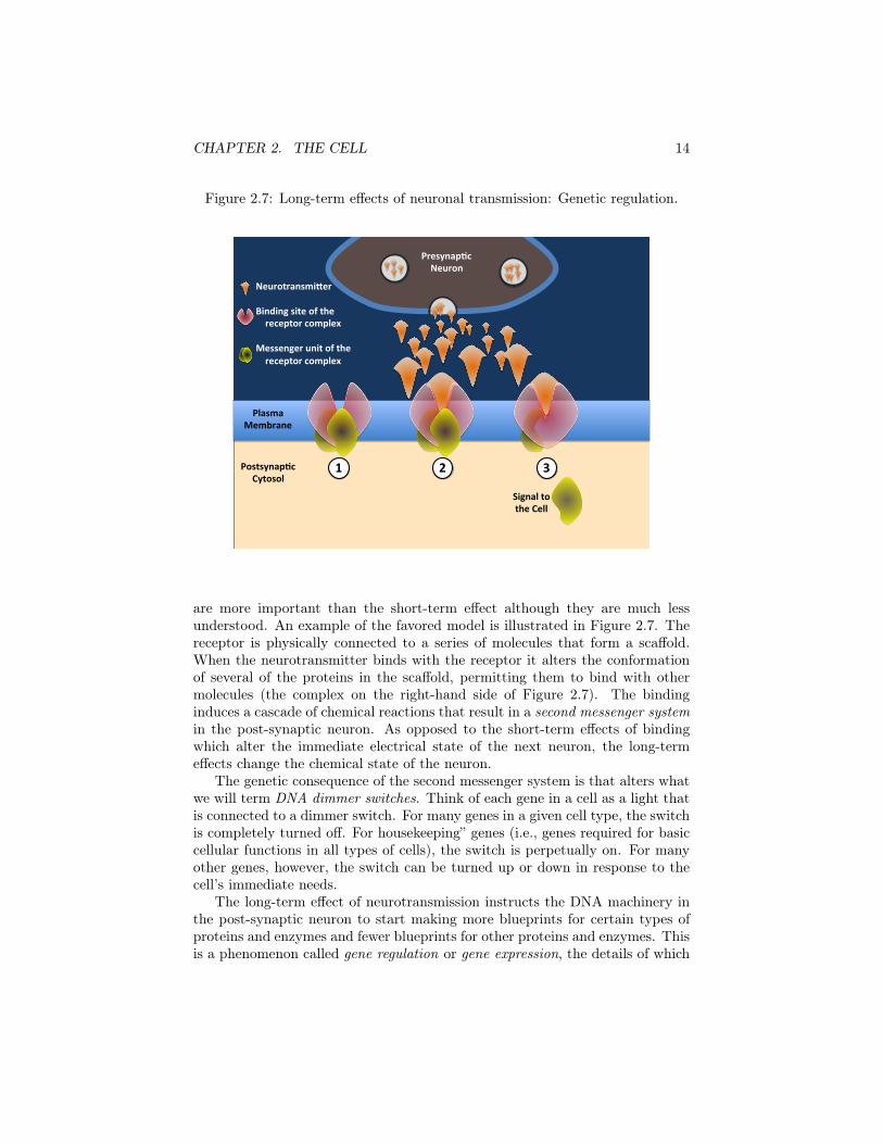
CHAPTER 2. THE CELL 14
Figure 2.7: Long-term effects of neuronal transmission: Genetic regulation.
Plasma&Membrane&
Postsynap0c&Cytosol&
Presynap0c&Neuron&
Signal&to&the&Cell&
Neurotransmi9er&&Binding&site&of&the&&&&&receptor&complex&&Messenger&unit&of&the&&&&&receptor&complex&
1& 2& 3&
are more important than the short-term effect although they are much lessunderstood. An example of the favored model is illustrated in Figure 2.7. Thereceptor is physically connected to a series of molecules that form a scaffold.When the neurotransmitter binds with the receptor it alters the conformationof several of the proteins in the scaffold, permitting them to bind with othermolecules (the complex on the right-hand side of Figure 2.7). The bindinginduces a cascade of chemical reactions that result in a second messenger systemin the post-synaptic neuron. As opposed to the short-term effects of bindingwhich alter the immediate electrical state of the next neuron, the long-termeffects change the chemical state of the neuron.
The genetic consequence of the second messenger system is that alters whatwe will term DNA dimmer switches. Think of each gene in a cell as a light thatis connected to a dimmer switch. For many genes in a given cell type, the switchis completely turned off. For housekeeping” genes (i.e., genes required for basiccellular functions in all types of cells), the switch is perpetually on. For manyother genes, however, the switch can be turned up or down in response to thecell’s immediate needs.
The long-term effect of neurotransmission instructs the DNA machinery inthe post-synaptic neuron to start making more blueprints for certain types ofproteins and enzymes and fewer blueprints for other proteins and enzymes. Thisis a phenomenon called gene regulation or gene expression, the details of which
CHAPTER 2. THE CELL 15
will be explicated in Chapter X.X. The important lesson is to recognize thelemonade quality of neuronal cell talk. Environmental stimuli that impingeon, say, your visual system initiate a whole cascade of events in your neuronsthat effectively turn some genes on and shut other genes down in your braincells. The mere fact of looking at something influences the genes in your centralnervous system.
2.5.3 Neuron talk and genesHow do genes fit into neuronal transmission? There are several different ways.In the previous discussion of metabolic pathways, we have already seen howDNA contains the blueprint for the enzymes that synthesize neurotransmitters(review Figure 2.2). The receptors for neurotransmitters, both on the firingneuron and its recipient, do not appear from anywhere. DNA contains the codefor these receptor proteins and/or the enzymes that synthesize them. DNA alsoholds the information for making the
enzymes that metabolize the extra neurotransmitter that gets released andfor the transporter proteins that carry the neurotransmitter back into the presy-naptic neuron.
Many steps in the second messenger system are also influenced by proteinsand enzymes the blueprints for which are encoded in the DNA. Finally, thelong-term result of cell talk among neurons is to “turn on” certain genes in thepost-synaptic neuron and to “turn off” other genes. Not only does DNA havethe blueprint for these important proteins and enzymes, but it may also playa role in the numbers of protein or enzyme molecules that are synthesized andtheir distribution throughout the neuron. Scientific knowledge on this is skimpy,but it is likely that genes may influence such factors as the number and size ofvesicles containing neurotransmitters, the number of receptor molecules, andperhaps even the density at which these receptors cluster at various places onthe neuronal wall. Much research is needed to clarify the role of DNA in thehuman nervous system, but there is no doubt that without DNA in each andevery neuron, we humans would have no nervous system at all.
2.6 Three disclaimers about the nervous system
First, if it has not already been done, I hope that one day an historian of theEnglish language will trace the evolution of the word “nervous.” The Latin word“nervus,” from which the English word derives, means a sinew, and the word wasapparently taken up by anatomists to refer to the tendon-like structure of theaxons of some nerves. Somehow along the way, the word developed connotationsof worry and apprehension on the one hand and jitteriness and agitation on theother. This is very curious because our nervous system plays just as importanta role when we are calm and relaxed as it does when we are tense and anxious.
Second, although the nervous system plays a crucial role in behavior, oneshould not conclude that genes influencing behavior must do so by acting directly

CHAPTER 2. THE CELL 16
in the nervous system. All of us large, multicellular life forms are conglomer-ations of many different systems that talk to one another and can influencebehavior. Later on (Section X.X), we will see how a gene for an enzyme in theliver can reduce the risk of alcoholism.
Finally, a disclaimer is needed for the simplicity with which the nervoussystem has been described. From a scientific view, almost every statement madeabove requires qualifications because the nervous system is a very, very, verycomplicated place where virtually every rule has its exception. For example,a few neurons do communicate electrically and not chemically, and not everyneurotransmitter is synthesized from enzymes. We will soon see that with genesand their physiological effects, complexity and perplexity are the rule instead ofthe exception.