The Case FOR Human Gamma Retroviruses (XMRV/HGRVs) in …
43
Max Pfost Amanda MacKenzie Debbie Taylor Cramer Deborah Goetz Vincent Lombardi Svetlana Khaiboullina Frank Ruscetti Dan Bertolette Ying Huang Cari Petrow-Sadowski Laboratory of Experimental Immunology David Strayer The Case FOR Human Gamma Retroviruses (XMRV/HGRVs) in CFS/ME Special Thanks: All the patients who participated in BWG, WPI, Maldarelli and Lipkin studies. Without YOU there is NO Research in ME/CFS!
Transcript of The Case FOR Human Gamma Retroviruses (XMRV/HGRVs) in …
Max Pfost
Amanda MacKenzie
Debbie Taylor Cramer
Deborah Goetz
Vincent Lombardi
Svetlana Khaiboullina
Frank Ruscetti
Dan Bertolette
Ying Huang
Cari Petrow-Sadowski
Laboratory of Experimental Immunology
David Strayer
The Case FOR Human Gamma Retroviruses (XMRV/HGRVs) in CFS/ME
Special Thanks:All the patients who participated in BWG, WPI,
Maldarelli and Lipkin studies. Without YOU there is
NO Research in ME/CFS!

Direct Isolation of a Gammaretrovirus from the PBMCs of CFS patients
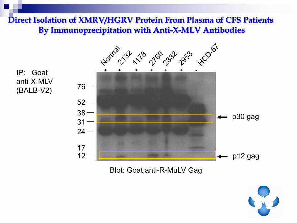
76
52
38
31
24
1712
Blot: Goat anti-R-MuLV Gag
IP: Goat
anti-X-MLV
(BALB-V2)
+ + + + + + -
Direct Isolation of XMRV/HGRV Protein From Plasma of CFS Patients By Immunoprecipitation with Anti-X-MLV Antibodies
p30 gag
p12 gag

Cell-Free Transmission of XMRV from CFS Patients’ PBMCs to the SupT1 Cell Line
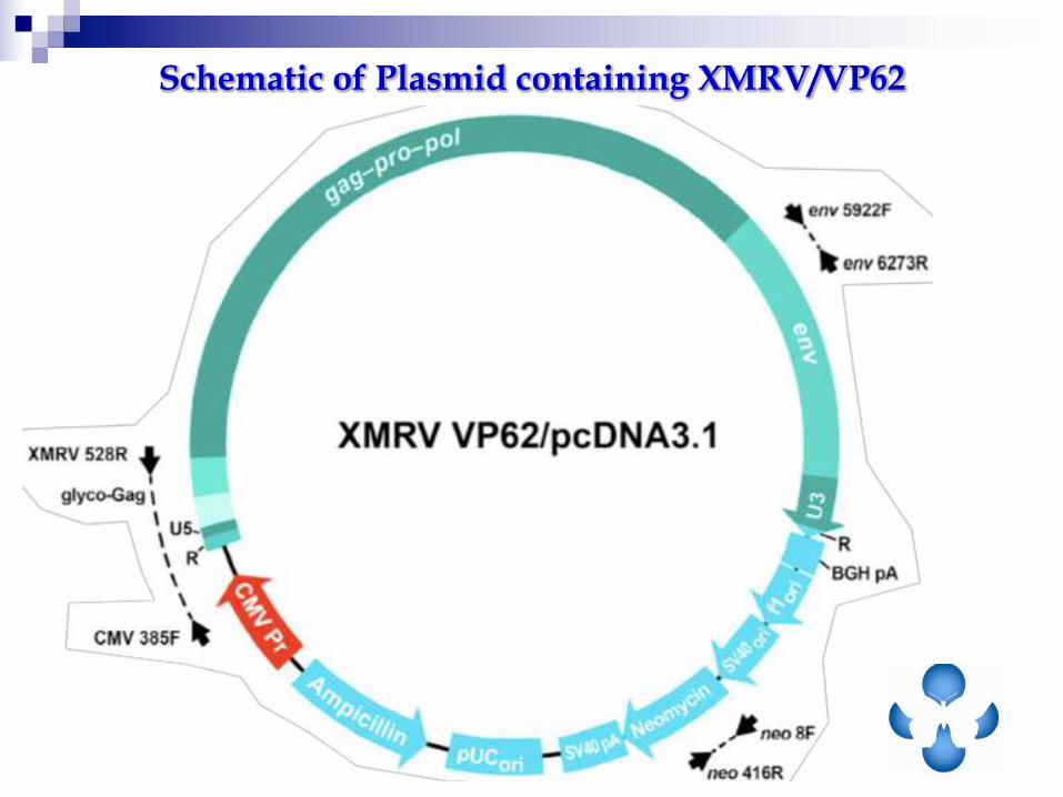
Schematic of Plasmid containing XMRV/VP62
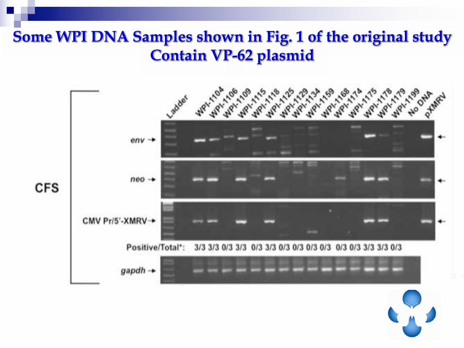
Some WPI DNA Samples shown in Fig. 1 of the original study Contain VP-62 plasmid
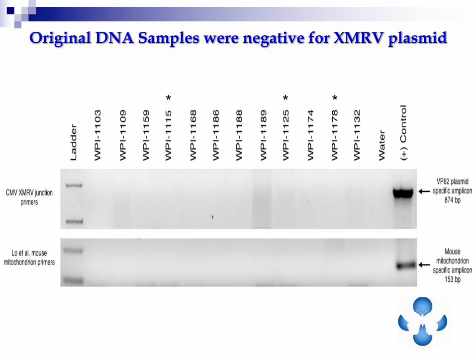
Original DNA Samples were negative for XMRV plasmid
* * *

PCRperformedwithUSBHotStart-IT
FideliTaqMasterMix
94°C2min
45cycles:
94°C30sec,54.8°C30sec,72°C,30
sec
72°C3min.
Non-specific(HumanDNA)
XMRVGag
AllthreearenegativeforIAPandnegative
forCMV385F/XMRV528RprimersforVP62
junctionfragment
Sequencingofbands:
Independent Reanalysis of samples used in Original Study Detected XMRV gag without plasmid or mouse contamination
H2O
H2O
H2O
WPI-1103
WPI-1109
WPI-1159
WPI-1115
WPI-1168
WPI-1186
WPI-1188
WPI-1189
WPI-1113
WPI-1134
WPI-1174
WPI-1178
WPI-1132

Cell-Free Transmission of XMRV/HGRV from PCR-negative CFS Patients’ Plasma to LNCaP cells

Horizontal Spread of Gammaretroviruses in Tissue Culture
Zhang et al., Cancer, Biol. Ther. 2011, 12:617

What does this mean?
1. Horizontal spread of these viruses is another potential source
viral contamination of cell lines
2. As presented in Ottawa, this could occur by spiking CR22V1
in LNCAP
3. It could occur by aerosolization, contaminated reagents, fau
technique
4. This is no evidence that such horizontal spread of gamma-
retroviruses occurs between individuals

XMRV
Pm-MLV
P-MLV
X-MLV
WPI-1104 P
WPI-1104 X
WPI-1104 P
WPI-1104 X
XMRV
Pm-MLV
P-MLV
X-MLV
A CFS Patient has a Strain of HGRV distinct from XRMV
DNA from several individuals carry both XMRV and PMRV
sequences, as confirmed by cloning and sequencing of PCR
products

Clones
Clones
vThe main XMRV/HGRV in this patient is unlikely to be VP-62
Clones of XMRV/HGRV Env SU Similar to Polytropic MLVs

n Exogenuous Infection (3x10⁶ particles
From VP62) in Rhesus Macaques
revealed:
n Viral and proviral signals disappeared in
the blood 1 month p.i. (acute vs chronic)
n Chronic infection present in tissues
n Latent infection and reactivation observed
Onlamoon et al., J Virol. 85: 4547, 2011
Why No XMRV/HGRV Detection in previously Positive Patients in BWG?
Animate or just say an a transition could these viruses also be latent in Man

slide HGRV can be latent
n Data here on two occasions acitvated
PBMC were positive for gag PCR
n One time the PBMC for both patient were
negative by gag PCR but positive after
5AZA, a demethylating agent, known to
activate latent viruses

Absence of XMRV Protein Expression
In Activated PBMC from Normal Donors
0/50 healthy donors were positive for XMRV proteins
CF
S
CF
S

Original Film for Science Fig 2C
Names changed to protect patient privacy
The 5 AZA treated samples were used as controls for negative activated normal PBMC
5 Aza treatment of these two samples was not identified
It was an error not to in methods..but nothing more
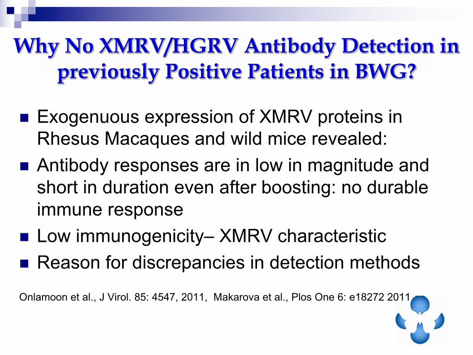
n Exogenuous expression of XMRV proteins in
Rhesus Macaques and wild mice revealed:
n Antibody responses are in low in magnitude and
short in duration even after boosting: no durable
immune response
n Low immunogenicity– XMRV characteristic
n Reason for discrepancies in detection methods
Onlamoon et al., J Virol. 85: 4547, 2011, Makarova et al., Plos One 6: e18272 2011
Why No XMRV/HGRV Antibody Detection in previously Positive Patients in BWG?

N-Terminus of SFFV ENV allows recognition of most potential HGRVs

Lombardi et al., 2009
Assay used to Detect Anti-XMRV/HGRV Antibodies
Plasma
FACS analysis of Murine Cell Line
+/- SFFV Env
- SFFV Env + SFFV Env
Plasma from CFS patients block binding of SFFV Env rat mAb to the B
cell line expressing SFFV Env, demonstrating specificity
Control mAb
α-SFFV env mAb
1:10 WPI1141 plasma
+ α-SFFV Env mAb
Control mAb
α-SFFV env mAb
1:100 WPI-1141 plasma
+ α-SFFV Env mAb

Plasma from XMRV/HGRV Infected CFS Patients are reactive to multiple XMRV proteins

Two Dimensional Western Blot Followed by Mass Spec Sequencing Confirms Human Antibody Specific Binding to
XMRV/HGRV Viral Proteins
Sypro-Ruby Stained 2-D Protein Gel Western Blot Probed with Human Serum
Both XMRV gp70 and p30 were recognized by CFS sera
vAbility to recognize XMRV proteins does not mean that XMRV was
the immunogen it could be any HGRV or a cross reactive protein
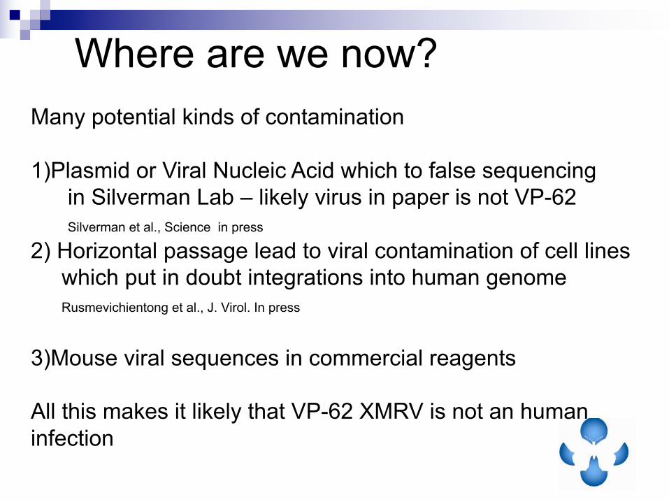
Where are we now?
Many potential kinds of contamination
1)Plasmid or Viral Nucleic Acid which to false sequencing
in Silverman Lab – likely virus in paper is not VP-62
Silverman et al., Science in press
2) Horizontal passage lead to viral contamination of cell lines
which put in doubt integrations into human genome
Rusmevichientong et al., J. Virol. In press
3)Mouse viral sequences in commercial reagents
All this makes it likely that VP-62 XMRV is not an human
infection
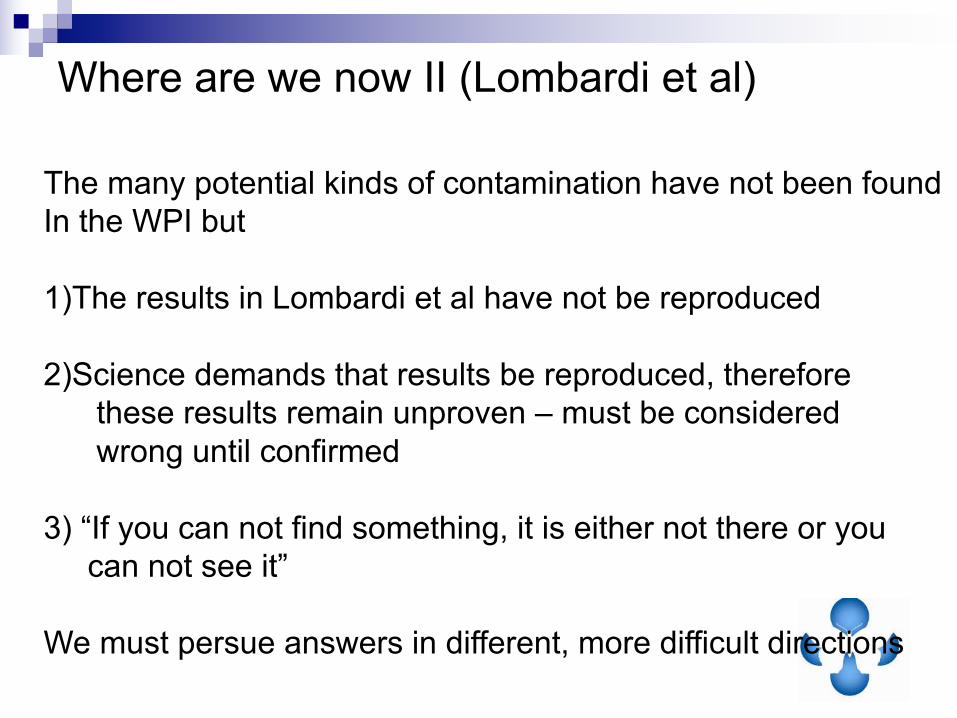
Where are we now II (Lombardi et al)
The many potential kinds of contamination have not been found
In the WPI but
1)The results in Lombardi et al have not be reproduced
2)Science demands that results be reproduced, therefore
these results remain unproven – must be considered
wrong until confirmed
3) “If you can not find something, it is either not there or you
can not see it”
We must persue answers in different, more difficult directions
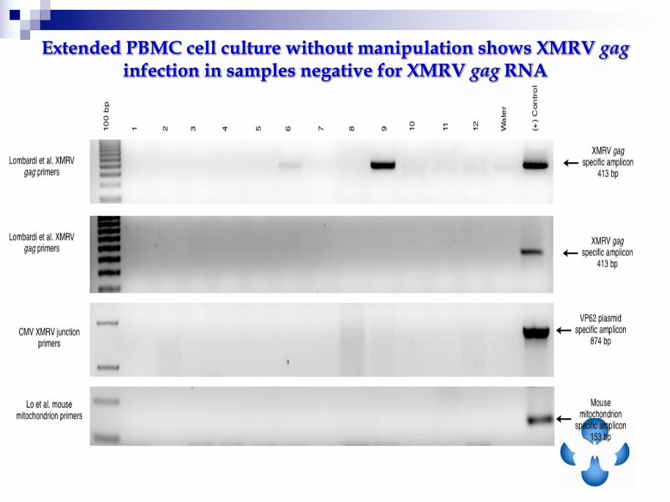
Extended PBMC cell culture without manipulation shows XMRV gag infection in samples negative for XMRV gag RNA

Serum samples processed for SOLiD4 sequencing:
•extraction of DNA (Std. silica method)
•random amplification (WGA – Genomeplex)
•Emulsion PCR -> Sequencing (50bp reads)
Post-sequencing Biometry:
•First alignment to human DB (Hg18)
•only reads that were not positive in first alignment used
•Second alignment to 6 XMRV DBs and 16 pMRV1 DBs (partial cds)
•Cleaned for low complexity and short hits
•Data mining for read significantly positive on Individual XMRV/pMRV DBs
•Stringency: Similarity ≥ 95% within ≥ 95% read length
Sera from Subjects•8 CFS patients (Source: Hemispherx Biopharma)
•17 Apparently Healthy Controls (Source: Vendor)
- 15 Russian samples- 2 Canadian samples
1 pMRV= polytrophic MLV-related virus
Next Generation Sequencing Study

NGS detected XMRV/HGRV in 7/8 CFS samples and 2/17 controls.
GenBank Accession # # Samples 8 Normals 17
+
Samples
#
Reads
+
Samples
SUM ALL XMRV 7 4 2
DQ241301.1Xenotropic MuLV-related virus
VP351 1 1
DQ241302.1Xenotropic MuLV-related virus
VP423 1 1
DQ399707.1Xenotropic MuLV-related virus
VP623 0 0
FN692043.2Xenotropic MuLV-related virus
22Rv1/CWR-R1 3 0 0
GQ497343.1
Xenotropic MuLV-related virus
clone WPI-1178 putative (gag-pro-pol)/(gag)/(env)
3 1 1
GQ497344.1
Xenotropic MuLV-related virus
clone WPI-1106 putative (gag-pro-pol)/(gag)/(env)
2 1 1
HQ601962.1, HQ601961.1, HQ601960.1, HQ601959..1, HQ601958.1|, HQ601957.1, HM630561.2, HM630557.2, HM630558.1,
HM630562.1, HM630560.1, HM630559.1, HQ157343.1, HQ157342.1, FJ907198.1, FJ907199.1
No hits on pMRV DBs:
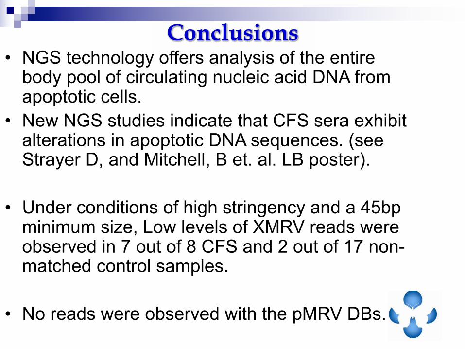
Conclusions• NGS technology offers analysis of the entire
body pool of circulating nucleic acid DNA from apoptotic cells.
• New NGS studies indicate that CFS sera exhibit alterations in apoptotic DNA sequences. (see Strayer D, and Mitchell, B et. al. LB poster).
• Under conditions of high stringency and a 45bp minimum size, Low levels of XMRV reads were observed in 7 out of 8 CFS and 2 out of 17 non-matched control samples.
• No reads were observed with the pMRV DBs.
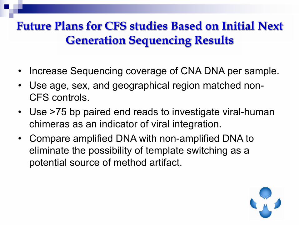
Future Plans for CFS studies Based on Initial Next Generation Sequencing Results
• Increase Sequencing coverage of CNA DNA per sample.
• Use age, sex, and geographical region matched non-
CFS controls.
• Use >75 bp paired end reads to investigate viral-human
chimeras as an indicator of viral integration.
• Compare amplified DNA with non-amplified DNA to
eliminate the possibility of template switching as a
potential source of method artifact.
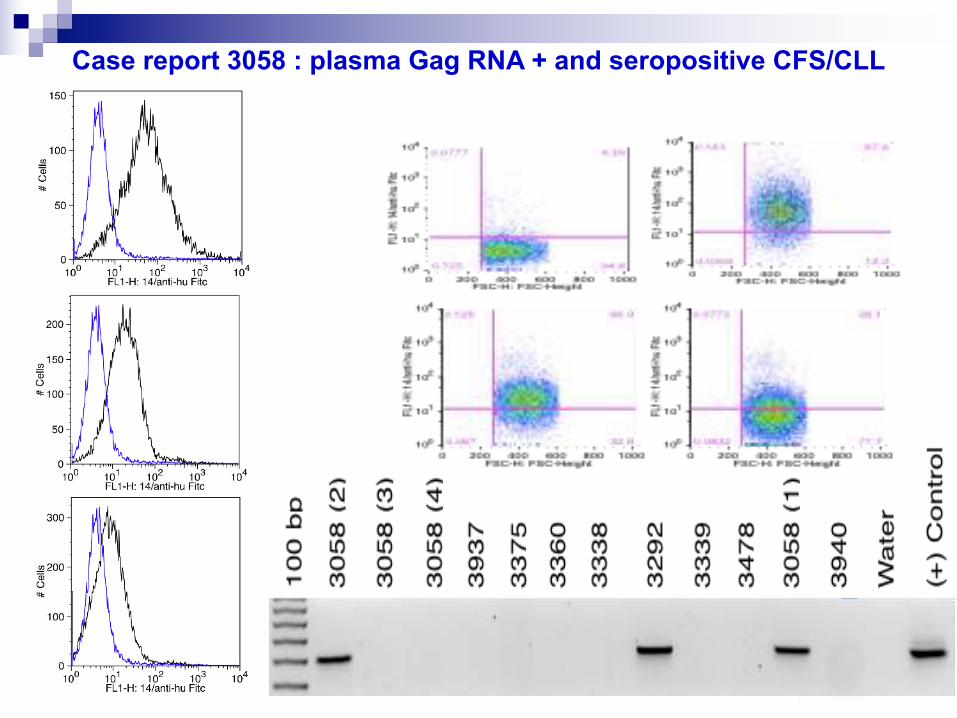
Case report 3058 : plasma Gag RNA + and seropositive CFS/CLL

Antiretroviral Therapy of CFS and CLL
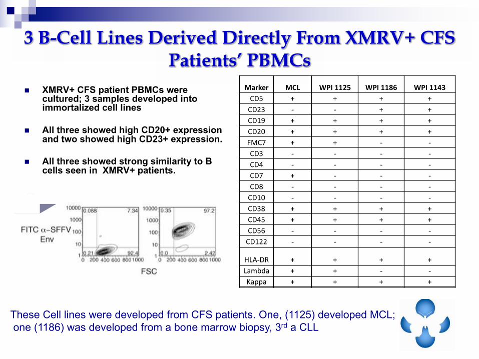
n XMRV+ CFS patient PBMCs were cultured; 3 samples developed into immortalized cell lines
n All three showed high CD20+ expression and two showed high CD23+ expression.
n All three showed strong similarity to B cells seen in XMRV+ patients.
Marker MCL WPI1125 WPI1186 WPI1143
CD5 + + + +
CD23 - - + +
CD19 + + + +
CD20 + + + +
FMC7 + + - -
CD3 - - - -
CD4 - - - -
CD7 + - - -
CD8 - - - -
CD10 - - - -
CD38 + + + +
CD45 + + + +
CD56 - - - -
CD122 - - - -
HLA-DR + + + +
Lambda + + - -
Kappa + + + +
These Cell lines were developed from CFS patients. One, (1125) developed MCL;
one (1186) was developed from a bone marrow biopsy, 3rd a CLL
3 B-Cell Lines Derived Directly From XMRV+ CFS Patients’ PBMCs

Pathogenesis:n Asymptomatic in majority of individuals
n 5% lifetime risk of developing either type of disease:- Adult T cell leukemia
- Clonal malignancy of CD4+ T cells.- Long latency; Immune deficiency - Tax and HBZ needed for transformation
- Inflammatory syndromesHTLV-I associated myelopathy/Tropical spastic paraparesis- Uveitis- Arthropathy
One reason for remarkable genetic stability is proviral load increases
By clonal expansion of infected T cells (Wattel et al 69:2863, 1995)
HTLV-I: Sequence Conservation and Pathogenesis
As opposed to HIV, HTLV has remarkable genetic stability:
fixation of base substitutions – 1% in 1000 years
complete sequence conservation in ENV and Tax

Summary
n WehaveshownthatwecandetectHGRV/XMRVfootprintsinthebloodby
serologyandnucleicacidanalysiswithoutanyevidenceofcontamination
n Some CFS plasma contains HGRV/XMRV proteins and
antibodies that recognize XMRV/HGRV viral antigens.
n XMRV producing Hematopoietic Cell Lines (B and NK-like)
were developed from CFS patients.
n XMRV/HGRV-infectedindividualsexhibitcytokineprofilescharacteristicof
inflammatoryprocesses
n SequencedataindicatetherearedifferentstrainsofXMRV/HGRVsthatcan
infecthumans.
34
n Conclusions
n ThepathogenicpotentialofHGRVsinME/CFSdeserves
furtherexploration

Future Plans
n Obtain full-length sequences of human HGRVs in CFS/ME
n Determine Human Integration Sites for HGRV infection in CFS/ME
n Identification of Tissue Reservoirs for human HGRV in CFS/ME
n Identify mechanisms of HGRV latency

76
52
38
31
24
WB: Anti-SFFV Env
Spiked 22Rv1 Virus in BWG Plasma Spreads to Uninfected Cells

War and Peace between Microbes: HIV-1 Interactions with Coinfecting Viruses:
Cell Host & Microbe 6, November 19, 2009 A. Lisco, C Vanpouille, & L Margolis
In Chronic Diseases Viruses Seldom Come Alone

CYTOKINES/
CHEMOKINES
Patient
N = 156
Control
N=140
P value FUNCTION IN INFLAMMATION
IL-8 1067 11.1 <0.0001 RNase L and CMV activated
IL-13 28 86 <0.0001 Inhibits inflammatory cytokine production
MIP-1b 1840 157 <0.0001 Elevated in Neurodegenerative disease
TNF-a 109 12.8 <0.0001 Stimulates chronic inflammation
MCP-1 468 421 0.003 Elevated in chronic inflammatory diseases
IL-7 21.1 82 <0.0001 Stimulates proliferation of B and T
lymphocytes and NK cells
IFN-a 35 60 <0.0001 Stimulates macrophages and NK cells to
elicit an anti-viral response
IL-6 271 29 <0.0001 Stimulates chronic inflammation
MIP-1a 673 91 0.0062 Elevated in Neurodegenerative disease
GM-CSF 108 166 <0.0001 Stimulates proliferation of B and T
lymphocytes and NK cells
Dysregulated Cytokine/Chemokine Production Detected
in Plasma from ME/CFS patients: Inflammatory Signature
of XMRV/MRV infection
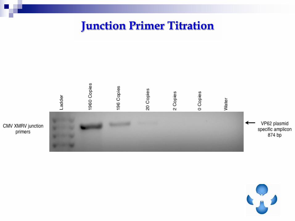
Junction Primer Titration

Science Healthy Controls
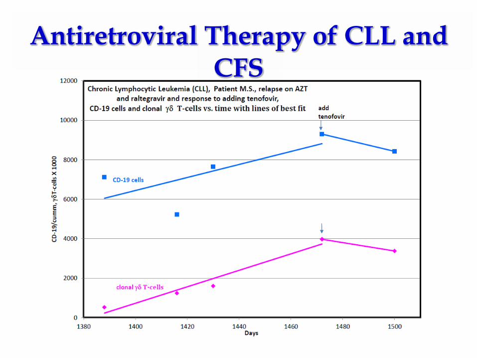
Antiretroviral Therapy of CLL and CFS
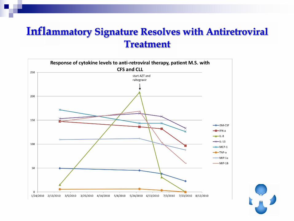
Inflammatory Signature Resolves with Antiretroviral
Treatment
XMRV (P)
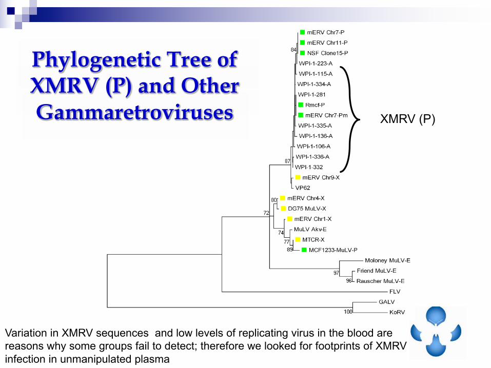
Variation in XMRV sequences and low levels of replicating virus in the blood are
reasons why some groups fail to detect; therefore we looked for footprints of XMRV
infection in unmanipulated plasma
Phylogenetic Tree of XMRV (P) and Other Gammaretroviruses