THE BRITISH JOURNAL OPHTHALMOLOGY130 THE BRITISH JOURNAL OF OPHTHALMOLOGY only may rod vision be...
20
THE BRITISH JOURNAL OF OPHTHALMOLOGY MARCH, 1935 COMMUNICATIONS THE VISUAL CELLS OF LAMPREYS* BY G. L. WALLS IOWA CITY PAGE INTRODUCTION - - 129 OBSERVATIONS - - - - - 134 CONCLUSIONS - - - - - - 146 BIBLIOGRAPHY - - - - - - 146 Introduction The question of the nature of the visual cells of these most primitive of living vertebrates has been controversial for decades. It is especially in need of a final answer because of its bearing upon the problem of the priority of origin of the rod and cone. The accepted theory in this latter connection, holding that the rod is the primitive visual cell and that the cone is a more complex derivative thereof, was put forward by Mlax Schultze (1866, 1867) and has served as the basis of Mrs. Ladd-Franklin's "genetic" theory of colour-vision and of Parsons' (1927) analysis of the visual sense into "dyscritic" and "epicritic" components. Examined critically, Schultze's evidence is seen to be no evidence at all; for he mistalkenly believed that not only the lampreys and elasmobranchs, but also the ganoids and primitive teleostsl have pure-rod retinae. Again, it is scarcely safe to assume with Schultze that the rod is physiologically the simpler element (and "therefore" the older), in spite of the apparent complexity of colour-vision; for not * A contribution from the Departments of Zoology of the University of Michigan and the State University of lowa. 1 Here Schultze was following Haeckel's belief in the eels as the most primitive teleosts; but the eel has enormtious cones (Garten 1907). copyright. on March 9, 2020 by guest. Protected by http://bjo.bmj.com/ Br J Ophthalmol: first published as 10.1136/bjo.19.3.129 on 1 March 1935. Downloaded from
Transcript of THE BRITISH JOURNAL OPHTHALMOLOGY130 THE BRITISH JOURNAL OF OPHTHALMOLOGY only may rod vision be...

THE BRITISH JOURNALOF
OPHTHALMOLOGY
MARCH, 1935
COMMUNICATIONS
THE VISUAL CELLS OF LAMPREYS*BY
G. L. WALLSIOWA CITY
PAGE
INTRODUCTION - - 129
OBSERVATIONS - - - - - 134
CONCLUSIONS - - - - - - 146
BIBLIOGRAPHY - - - - - - 146
IntroductionThe question of the nature of the visual cells of these most primitive of living
vertebrates has been controversial for decades. It is especially in need of a finalanswer because of its bearing upon the problem of the priority of origin of the rodand cone. The accepted theory in this latter connection, holding that the rod is theprimitive visual cell and that the cone is a more complex derivative thereof, wasput forward by Mlax Schultze (1866, 1867) and has served as the basis of Mrs.Ladd-Franklin's "genetic" theory of colour-vision and of Parsons' (1927) analysisof the visual sense into "dyscritic" and "epicritic" components.Examined critically, Schultze's evidence is seen to be no evidence at all; for he
mistalkenly believed that not only the lampreys and elasmobranchs, but also theganoids and primitive teleostsl have pure-rod retinae. Again, it is scarcely safeto assume with Schultze that the rod is physiologically the simpler element (and"therefore" the older), in spite of the apparent complexity of colour-vision; for not
* A contribution from the Departments of Zoology of the University of Michigan and the StateUniversity of lowa.
1 Here Schultze was following Haeckel's belief in the eels as the most primitive teleosts; but theeel has enormtious cones (Garten 1907).
copyright. on M
arch 9, 2020 by guest. Protected by
http://bjo.bmj.com
/B
r J Ophthalm
ol: first published as 10.1136/bjo.19.3.129 on 1 March 1935. D
ownloaded from

130 THE BRITISH JOURNAL OF OPHTHALMOLOGY
only may rod vision be quite as complex, but colour-vision may well have beeninvented long after the advent of the cone as a distinct, high-threshold, isolated-conduction element.2.
Heinrich Muller (1856) first noted that in the European River Lamprey, Lampetrafluviatilis, the visual cells are of two types, long and short, in equal numbers (seeFig. 1). He termed them both cones, but later examined Petromyzon marinus inwhich he found the short elements predominating, and in a footnote to a paper onquite another subject (1862) he suggested that the short elements might be rods.Schultze (1866) first tentatively, later (1867) with more assurance pronounced theL. fluviatilis retina pure-rod. W. Krause (1868) insisted tllat both rods and coneswere present, but failed to make clear which was which. Schultze now (1871 a)about-faced, and termed L. fluviatilis pure-cone; but in the same year (1871b) oncemore returned to the pure-rod concept in a contribution to a reference work, practi-cally the last of his many writings. Langerhans (1873, 1876) described the EuropeanBrook Lamprey, L. planeri, as having rods and cones, figuring tlle rod as the longcell and giving it a cylindrical outer segment.Wilhelm Muller (1874) now gave descriptions of L. fluviatilis, L. planeri, and P.
marinus, with figures of the two latter species. His drawing of "P. marinus" wascertainly based upon an exceptionally large L. fluviatilis, however, for the two cell-types are shown in equal numbers as they are in Lampetra. Muller saw only conicalouter segments and compared the long-cells with the rods of higher forms.
In Krause's later papers (1872, 1876a, 1876b) the duplex condition is again affirmed,and he definitely sides with Langerhans in identifying the long-cell as the rod.Kuhne (1878a, 1878b) reported rhodopsin in L. fluviatilis, and considered the small
amount he found indicative either of a low concentration in all cells or of the presenceof many rhodopsin-free elements (cones). From this point on it would seem thatnone could deny the presence of rods, but Kohl (1892a, 1892b), who made a maximumof errors in his histological observations, found only cones in L. planeri, and thoughtthat the Muller fibres had been mistaken for rods by others. Only Kohl has claimedto find " vacuoles " (oil-droplets) in lamprey visual cells-which, if true, would,of course, be the best of evidence for considering them cones.
Greeff (1900) was impressed with the ambiguity of the genus Lampetra, but followedLangerhans and Krause in his identifications. His drawing, though labelled L.planeri, must have been made from a small L. fluviatilis, as it shows the single outernuclear layer of the latter species. Putter (1912) added nothing original, but didcall attention to the obscure and neglected mention of P. marinus by H. Muller, whoseidentifications, it will be recalled, were the inverse of those of later workers. It isquite clear that W. Muller's erroneous figure of "P. marinus" has kept all subse-quent workers from discovering that in this form the situation is not so ambiguousas in Lamnpetra, for they would naturally be inclined to accept W. Muller's drawingover H. Muller's statement that the short-cells were much the more numerous.Mozejko (1912, 1913) made use of Kohl's pure-cone concept in arguing for the"primitiveness " of the lamprey eye.
Tretjakoff (1916) made a very great advance bv applying neurological methods toL. fluviatilis, and while he identified the long-cell as a rod and the short one as acone purely upon the basis of size, he found the long-cell to have a dendritic foot-piece and that of the short-cell to be a smooth knob, which by analogy with othercases of such differentiation would tend rather to support Heinrich Muller.
R. Krause (1923) followed Langerhans, W. Krause, and Tretjakoff in his designa-tions, but claimed that in L. fluviatilis the short-cells out-number the long to a " notinappreciable " extent. Plate (1924) now pronounced the elements " undifferentiated "-neither rods nor cones-and was followed by Ducker (1924), whose material hesupplied.The most recent writer on the subject, Franz (1932), has put forward a new form
of Schultze's pure-rod idea (foreshadowed by the views of Vogt and Yung, 1894),for he considers that in L. fluviatilis there is actually but a single type of elementpresent, the bacillary layer being passively pseudo-stratified because of the bulky
1 Greef (1900) has advanced the only other bit of evidence for rod-primitiveness, in his sugges-tion that the dendritic foot-piece of the cone is an advance over the smooth, compact rod end.knob.But in view of the ependymal origin of the visual cells (Walls, 1934b) the dendritic terminus is seento be the more primitive one, the compact rod ending being adaptive to the multiple connection ofrods to a bipolar neuron.
copyright. on M
arch 9, 2020 by guest. Protected by
http://bjo.bmj.com
/B
r J Ophthalm
ol: first published as 10.1136/bjo.19.3.129 on 1 March 1935. D
ownloaded from

VISUAL CELLS OF LAMPREYS 131
FIG. 1.
Retina of European River Lamprey, Lambetra fluviatilis.Kolmer's fluid; X 600.P.E-pigment epithelium; L.C.-long visual cells (cones);S.C.-short visual cells (rods); E.L.M.-external limitingmembrane; O.N.L.-outer nuclear layer; B.C.-bipolar cell;H.C.-horizontal cells; G.A.-ganglion and amacrine cells;N.F.-bundle of optic nerve fibres; M.F.-Muller fibre;I. L.M .-internal limiting membrane.
ellipsoids which interfere with close congregation.3. He bases his case upon thecylindrical form of all outer segments as seen in his formalin and Bouin fixations-both most unsuitable for visual cells-upon the presence of myeloidal spirals in allouter segments, a rod-characteristic in his present opinion, though he himself admitsthat they have often been seen in cones (cf.. Franz, 1913); and upon an allegednocturnality of lampreys, of which more later.
s Franz was, of course, unaware that there are species in which one type of cell so greatly out.numbers the other as to render this view untenable.
copyright. on M
arch 9, 2020 by guest. Protected by
http://bjo.bmj.com
/B
r J Ophthalm
ol: first published as 10.1136/bjo.19.3.129 on 1 March 1935. D
ownloaded from

1IHE BRITISH JOURNAL OF OPHTHALMOLOGY
From the above brief review it will be seen that all possibleviews have been held; that both cell-types are rods; that bothare cones; that the long cell is a rod, the short a cone; that thelong cell is a cone, the short a rod; that the cells are neither rodsnor cones, but "undifferentiated." Clearly, there is need for aclose examination of the criteria employed by the various investi-gators and for a careful evaluation of these and other possiblemeans of distinguishing rods from cones in ambiguous cases.The criteria upon which others have chiefly relied have been
the length of the cells and the form of their outer segments. Whilerods in general are longer than cones in general thereareexceptionssuch as certain snakes (Dasypeltis, Tarbophis, Leptodira) andmost teleost fishes. The form of the outer segment is so prone toartificial alterationi that the mere fact that both conical andcylindrical forms are claimed for each lamprey cell-type, coupledwith the fact that such fixatives as alcohol, formalin, and Bouin'sfluid have been principally employed, makes it necessary to with-hold conclusions upon this important point so far as the literatureto date is concerned.One thing is, however, certain: at least one of the cells is a
rod, for rhodopsin is present. If lampreys were known to bestrictly nocturnal, it would be almost safe to conclude that bothcells are rods in spite of Kiihne's tentative conclusion to thecontrary. But lampreys have no special adaptations fornocturnality, such as a tapetum or an exceptionally large lensor/and pupil. They do have at least one special adaptation forbright-light vision in the form of a physiological yellow coloura-tion of the lens (Walls and Judd, 1933). WVe may tentativelyconclude that both rods and cones are present, and have thento decide between the views first advanced by H. Muller andLangerhans respectively.
In re-examining the species of the genus Lampetra and insurveying other genera as yet untouched or but superficiallvstudied by others, we may look for the following features withsome hope of finding a meaningful differentiation of the two tvpesof cells.Form and size of the outer segment.-Herein lies the most
characteristic visible difference between rods and cones. In agiven retina, the rod ordinarily has the more massive outersegment and this is almost invariably perfectly cylindrical. Thecone outer segment is smaller in sympathy with its higherthreshold, and thus is conical unless exceotionally slender (seeRochon-Duvigneaud, 1917; Walls, 19734a). There are cases.however, where the rod and cone outer segments are about alikein size and shape, e.g., certain urodeles, where they are bluntlvconical, and the Macaque, where they are cylindrical and of equal
132
copyright. on M
arch 9, 2020 by guest. Protected by
http://bjo.bmj.com
/B
r J Ophthalm
ol: first published as 10.1136/bjo.19.3.129 on 1 March 1935. D
ownloaded from

VISUAL CELLS OF LAMPREYS
length (Garten, 1907). The absence of a differentiation is of nofundamental significance, however.
Pres.ence or absence of rhodopsin in certain cells.-Visual cellswhich contain rhodopsin when dark-adapted are unquestionablyrods, and very few cases are known of functional rods lacking thissensitizing pigment-such rods are among those which haveoriginated secondarily from cones (certain nocturnal snakes, thenight lizards, Sphenodon, etc.; Walls, 1934a). Cones nevercontain macroscopic amounts of rhodopsin and probably do notcontain even a trace.
Relative extent of summation of the cell-types in bipolar andganglion cells.-This criterion has been used by Woollard (1927)and by the writer (Walls, 1934a) as an index of sensitivity; forrods are always synapsed in multiple to bipolars while cones havemore isolated conduction to the brain. If Cell " A " out-numbersCell "B" more greatly in species "X" than in species "Y,"and species "X" has much the higher visual cell-ganglion cellratio, then Cell "A" is the rod and Cell "B" the cone.
Relative numbers of the cell-tvpes in relation to pelagic vsbenthic, and diurnal vs nocturnal habits.-This is a direct corollaryof the Duplicity Theory: if Cell "A" out-numbers Cell "B"more greatly in species "X" than in species "Y," and species"X" is the more strongly benthic or/and nocturnal, then Cell"A" is the rod and Cell "B" the cone.
Relative numbers of the cell-types in the ftundus as comparedwith the periphery.-In duplex retinae, cones are always concen-trated in the fundus and more sparsely set in the periphery. IfCell " A " out-numbers Cell " B " much more greatly in theperiphery than in the fundus, then Cell " A " is the rod and Cell"B" the cone.
Direction of migration in light and darkness.--This criterionwas made use of by Laurens and Detwiler (1921) on the alligator,and depends upon the rule that if the visual cells of a given retinamigrate at all, they move in opposite directions. If Cell "A"elongates in light and contracts in darkness while Cell " B "elongates in darkness and contracts in light, then Cell "A" isthe rod and Cell " B" the cone.
Differentiation of the nuclei.-Cone nuclei tend to be larger,more ovoid, and nearer the limitans; rod nuclei smaller, morespherical, and in contact with the limitans only between conenuclei. Cone nuclei tend to have small chromatin granules andlinin; rod nuclei, large masses of chromatin and no linin (Menner,.1929). In amphibians, however, the nuclei are all of the "cone"type as here described, and the rod myoids being much the heavier(a unique situation) the result is that the rod nuclei are the nearerto tthe limitans.
133
copyright. on M
arch 9, 2020 by guest. Protected by
http://bjo.bmj.com
/B
r J Ophthalm
ol: first published as 10.1136/bjo.19.3.129 on 1 March 1935. D
ownloaded from
1THE BRITISH JOURNAL OF OPHTHALMOLOGY
Differentiation of the foot-pieces.-Cone foot-pieces are alwaysheavy and dendritic, while rod fibres are often slender and termi-nate in a smooth knob. This difference holds only for forms whoserods very greatly out-number their cones. In amphibians, allfoot-pieces are of the "cone" type.
We may now proceed to apply each of these criteria in turn tothe eight species of lampreys studied by the writer: Icthyomyzonconcolor, I. unicolor, Petromyzon marinus unicolor, Entosphenustridentatus, E. appendix, Lampetra fluviatilis, L. planeri, and L.lamottenii.
I wish, first, to express my gratitude to the many persons who,with advice and. labour, furthered the solution of the problem dis-cussed in this paper. The accuinulation of material of severalof the species, preserved by the special methods necessary for thestudy of the retina, was made possible only by enlisting theco-operation of a number of conveniently located biologists. Mythanks are due especially to Dr. C. U. Ariens Kappers of Amster-dam, Mr. Leonard P. Schultz of the University of Washington,Dr. David H. Thompson of the Illinois State Natural HistorySurvey, and Dr. Albert M. Reese of the University of WestVirginia. Facilities and supplies were kindly furnished by theDepartment of Zoology of the University of Michigan and theFaculty Research Fund of that institution.
Special mention must be made of the valuable advice andassistance received from Dr. Carl L. Hubbs of the Museum ofZoology of the University of Michigan and Dr. Simon H. Gageof Cornell University. The latter placed his Ithaca laboratory atthe disposal of the writer during the breeding season of P. m.unicolor in two successive years.The research was supervised by Dr. John F. Shepard of the
Department of Psychology of the University of Michigan, and thewriter is heavily indebted to him for excellent advice and keenicriticism. He is also grateful to Miss Gladys Larsen for her carefulwork on the drawings.
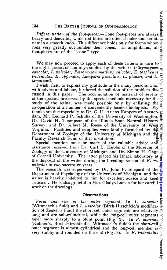
ObservationsForm and size of the outer -segment.-In I. concolor
(Wittmaack's fluid) and I. unicolor (Birch-Hirschfeld's modifica-tion of Zenker's fluid) the short-cell outer segments are relativelylong and are sub-cylindrical, while the long-cell outer segmentstaper more sharplv to a blunt point (Fig. 2). In P. marinus(Kolmer's, Birch-Hirschfeld's, Wittmaack's fluids) the short-cellouter segment is almost cylindrical and the long-cell member isvery stubby and rounded on the end (Fig. 3). In E. tridentatus
134
copyright. on M
arch 9, 2020 by guest. Protected by
http://bjo.bmj.com
/B
r J Ophthalm
ol: first published as 10.1136/bjo.19.3.129 on 1 March 1935. D
ownloaded from

VISUAL CELLS OF LAMPREYS
(Birch-Hirschfeld) and E. appendix (Birch-Hirschfeld, Kolmer)the short-cell outer segment is a long, perfect cylinder, but thatof the long-cell is a small cone (Fig. 4; cf. Fig. 2 in Walls, 1928a),Myeloidal spirals are especially evident in the short cells of E.tridenttatus. In L. Jluviatilis (Birch-Hirschfeld, Kolmer), L. planeri(Birch-IHirschfeld) and L. lamnottenii (Zenker) the short-cellmember is cylindrical, the long-cell outer segment smaller and
.0
E __
IhE L
E'
FIG. 2. FIG. 3.
Visual cell types of Icthyomlyzont Visual cell types of landlockedunicolor. Birch - Hirschfeld 's Atlantic Lamprey, Petromyzonfluid; X 1000. I.-long cell (cone); tmarinus unicolor. Wittmaack'sII.-short cell (rod); 0.-outer fluid; X1000. I.-long cell (cone);segment; E.-ellipsoid; L.- II.-short cell (rod); O.-outerlimitans; N.-nucleus. segment; E.-ellipsoid; L.-
limitans; N.-nucleus.
bluntly conical (Fig. 5). In the huge elements of L. fluviatilis,spiral threads are visible in both cases.Thus wherever there is a clear-cut structural differentiation in
this important feature, it is the short-cell outer segment which islarge and cylindrical, whereas the long-cell member is smallerand more conical, though never sharply so as in many vertebrates.A staining differentiation is also usual, the short-cell outer segmenttaking the Orange G and the long-cell organelle the Aniline Bluein Mallory's triple stain, while in haematoxylin preparations thelong-cell structure shows the greater affinity for the dye.The differentiation of the outer segment is perhaps most sharp
in E. tridentatus, but in all except the two species of the primitive
135
copyright. on M
arch 9, 2020 by guest. Protected by
http://bjo.bmj.com
/B
r J Ophthalm
ol: first published as 10.1136/bjo.19.3.129 on 1 March 1935. D
ownloaded from

3I HE BRITISH JOURNAL OF OPHTHALMOLOGY
genus Icthyornyzon the evidence clearly supports HeinrichMuller's view.
Presence or absence of rhodopsin in certain cells.-The presenceof rhodopsin in lampreys was confirmed on P. mnarinus by placingthe excised retinae of five specimens, which had been in darknessovernight, in a vial of normnal saline solution by ruby light.Removed to diffuse daylight and exposed before witnesses, the
E 11 E
AwAlI II IlD
FIG. 4. FIG. 5.
Visual cell types of Pacific Lam- Visual cell types of Lampetraprey, Entosphenus tridentatus. fluviatilis. Kolmer'sfluid; X1000.B3irch-Hirschfeld's fluid; XIOOO. I.-long cell (cone); II.-shortL.-long cell (cone); II.-short cell (rod); O.-outer segment;cell (rod) O.-outer segment; E.-ellipsoid; L.-limitans; N.-E.-ellipsoid; L.-limitans; N.- nucleus.nucleus.
mass of retinae was seen to be truly purple (rhodopsin is usuallyred in colour), the colour fading in two seconds when placed inthe light from a south window.Attempts to determine which type of cell contained the rhodop-
sin, by the use of Stern's (1905) platinic chloride technique, wereall failures. The method was tried on I. concolor, I. unicolor, P.marinus, and E. appendix. In all of these a yellow colourationwas seen in sections of both dark- and light-adapted retinae, thecolour not restricted to the outer segments, but being deepest inthe ellipsoids. A proper Stern's test should show the outer
136
copyright. on M
arch 9, 2020 by guest. Protected by
http://bjo.bmj.com
/B
r J Ophthalm
ol: first published as 10.1136/bjo.19.3.129 on 1 March 1935. D
ownloaded from
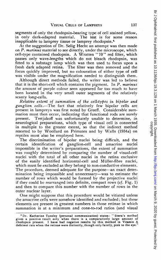
VISUAL CELLS OF LAMPREYS
segments of only the rhodopsin-bearing type of cell stained yellow,in only dark-adapted material. The test is for some reasoninapplicable to lamprey tissue or lamprey rhodopsin.4At the suggestion of Dr. Selig Hecht an attempt was then made
on P. marinus material to see directly, under the microscope, whichcell-type contained rhodopsin. A Wratten " 70 " red filter, whichpasses, only wave-lengths which do not bleach rhodopsin, wasfitted to a substage lamp which was then used to focus upon afresh dark adapted retina. The filter was then removed and thefocus quickly improved, but no colouration of either type of cellwas visible under the magnification needed to distinguish them.Although direct methods failed, the writer was led to believe
that it is the short-cell which contains the pigment. In P. marinusthe amount of purple colour seen appeared far too much to havebeen located in the very small outer segments of the relativelyscanty long-cells.
Relative extent of summation of the cell-types in bipolar andganglion cells.-The fact that relatively few bipolar cells arepresent in lampreys was first noted by Greeff. Considerable sum-mation must then occur, indicating that functional rods are surelypresent. Tretjakoff was unfortunately unable to determine, inneurological preparations, which type of visual cell is connectedin multiple to the greater extent, so that the indirect methodresorted to by Woollard on Primates and by Walls (1934a) onreptiles must also be employed here.
IThe discrimination of bipolar nuclei being difficult, and thecertain identification of ganglion-cell and amacrine nucleiimpossible in the writer's preparations, the extent of summationwas roughly determined by comparing the number of visual-cellnuclei with the total of all other nuclei in the retina exclusiveof the easily identified horizontal-cell and Muller-fibre nuclei,which must be excluded as they belong to non-conductive elements.The procedure, deemed adequate for the purpose-an exact deter-mination being impossible and unnecessary-was to estimate thenumber of rows which would be formed by the projective nucleiif they could be rearranged into definite, compact rows (cf. Fig. 1)and then to compare this number with the number of rows in theouter nuclear layer.One might suppose that this procedure would be vitiated unless
the amacrine cells were somehow identified and excluded; but theseelements are present in greatest numbers in those retinae in whichsummation is at a minimum and cone-to-rod ratios (and visual
'Dr. Katherine Tansley (personal communication) states: "Stern's methodgives a positive result only when there is a comparatively large amount ofrhodopsin present. I have had negative results by this method in Vitamin Adeficient rats when the retinae were distinctly, though only faintly, pink to the eye."
137
copyright. on M
arch 9, 2020 by guest. Protected by
http://bjo.bmj.com
/B
r J Ophthalm
ol: first published as 10.1136/bjo.19.3.129 on 1 March 1935. D
ownloaded from
THE BRITISH JOURNAL OF OPHTHALMOLOGY
acuity) at a maximum (e.g., birds), so that inasmuch as they arerelatively abundant when bipolars, etc., are numerous, and scantywhen much summation exists, their inclusion in the present deter-mination cannot result in a masking of the true situation. Thereason for these "comings and goings" of amacrines along with thestraightforward elements of the visual pathway is, of course,wholly mysterious, as is the functional significance of the amacrineelement in the first place.The situation in the various species, determined as above, is as
follows:-
I. concolorI. unicolorP. marinusE. tridentatusE. appendixL. fluviatilisL. planeriL. lamtottenii
Rows of visualcell nuclei:
... ... 1
... ... I I
... ... 2
... ... 2
... ... 2
... ... 3
... ... 1
1- Rows of projective(and amacrine) nuclei:
...... 2... ... 24... . ... 2... ... 2...... 4
... ... 23
... ... 4
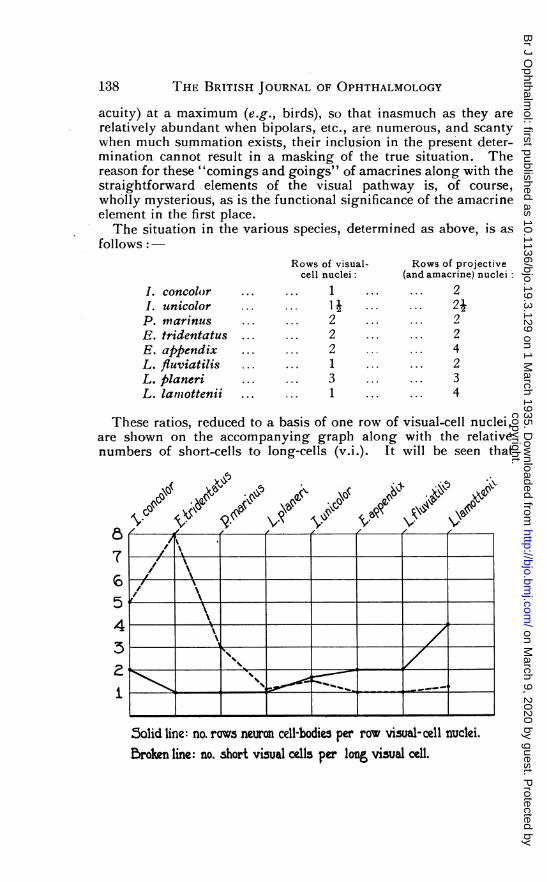
These ratios, reduced to a basis of one row of visual-cell nuclei,are shown on the accompanying graph along with the relativenumbers of short-cells to long-cells (v.i.). It will be seen that
,, '
5/t4 -4- ___
i ___._._____
5olid line: no. rows neuron cell-bodies per row visual-cell nuclei.Broken line: no. short visual cells per long, visual cell.
138
copyright. on M
arch 9, 2020 by guest. Protected by
http://bjo.bmj.com
/B
r J Ophthalm
ol: first published as 10.1136/bjo.19.3.129 on 1 March 1935. D
ownloaded from

VISUAL CELLS OF LAMPREYS 139
there is a decided tendency for high numbers of short visual cellsto go with low numbers of projective cells-that is, the specieswith the most short-cells have the most summation, and those withthe most long-cells have the most isolated conduction.This can only mean that the long-cell is the cone, the short-cell
the rod.Relative numbers of the cell-types in relation to pelagic vs
benthic, and diurnal vs nocturnal habits.-The relative numbers ofthe cell-types were determined by counting the cells in a micro-scope field 290A in diameter when the bacillary layer lay alongthe diameter of the field. The ratio in the fundus was found tobe as follows for each species:
I. concolor ... ... 5 short to I long.I. unicolor ... ... 3 ,, 2P. marinus ... ... 3 ,. 1E. tridentatus ... .. 8 ,, 1E. abpendix ... ... 1 ,, 1L. fuviatilis ... 1 ., 1L. blaneri... ..... 8 , 7L. lamottenii ... ... 5 ,, 4
Thu-s, in the species which live in the shallowest water, L.fluviatilis and the four brook lampreys I. unicolor, E. append&x,L. planeri antd L. lamottenii, the ratio of short-cells to long islowest- : 1, or nearly so. I. concolor, until the recent invasionby the land-locked marine lamprey, the characteristic lamprey ofthe Great Lakes, and the two marine forms P. marinus and E.tridentatus have at least the greatest depths available in which toswim, though it is not known what depths any of these speciesprefer.5- In these three species the short-cells greatly predominate.so that if there is indeed a relation of cell numbers to depth ofhabitat, the short-cell is inevitably indicated as the rod.Of previous investigators, only Franz (1932) has sought to
apply the duplicity theory as a criterion. He states that L. fluviatilisbecomes more active at nightfall,, and that only at this hour doesit come forth from places of concealment. Franz does not saywhether this observation was made in the laboratory or in the field,on vegetating specimens or on those excited by the breedingseason. Gage has recorded in several places that P. marinus movesonly at night on its way upstream to breeding grounds. This is,however, in the same category with the nocturnal migrational
6A. U.S. National Museum specimen designated as the type of the genusBathymyzon, which Creaser and Hubbs (1922) have pronounced a P. marinus, wastaken at a depth of 547 fathoms. The chorioids of the two marine lampreys areremarkably thick, that of E. tridentatus particularly. This is presumably adaptiveto maintaining the circulation under the pressure of considerable depths.
copyright. on M
arch 9, 2020 by guest. Protected by
http://bjo.bmj.com
/B
r J Ophthalm
ol: first published as 10.1136/bjo.19.3.129 on 1 March 1935. D
ownloaded from
140 'IHE BRITISH JOURNAL OF OPHTHALMOLOGY
flights of otherwise strictly diurnal birds; and photographs haveoften been made, in daylight, showing E. tridentatus leapingriffles on its way to spawn, much as does the salmon. All lampreyscarry on their breeding activities in bright light, and no onehas ever found them on the nests at night or even early in themorning. Professor T. L. Hankinson has told the writer thatthe easiest way he found to collect P. marinus in Oneida Lake inNew York State was to go about for awhile in a white-bottomedmotorboat and scoop the lampreys from the bottom of the boatwith a dipnet as soon as the boat was stopped. Curiously, thefollowing, summer a red-bottomed boat was used and though thelampreys were as numerous as ever they were no longer attractedto the moving boat. Lampreys have given much inconvenienceto swimmers during " marathon " swims (in daylight, of course)in Lake Ontario (see also Dymond et al., 1929; Creaser, 1932).
Captive, breeding lampreys may be photophobic, as the obEerve-tions of Reighard and Cummins (1916) and Lubosch (1902) attest,but the undisturbed lamprey is apparently diurnal in its breedingand at least not nocturnal in its feeding activities. Moreover,there is the yellow lens, a positive adaptation to diurnality (knownelsewhere only in diurnal snakes, diurnal squirrels, the diurnaltree-shrew Tupaia and the diurnal gecko Lygodactylus) to accountfor.
Franz regards the sparse retinal pigment as an indication ofnocturnality, but this-to anticipate-has the same significanceas the similarly scanty pigmentation in the human; the pigmentdoes not migrate, and there is, therefore, no need for much of it.Franz also makes the surprising statement that the "moderatesize of the eye" predisposes to nocturnality, for the eye is " muchlarger in indubitably diurnal fishes, e.g., Esox." Apart fromthe impropriety of conmparing such diverse forms as Lampetra andEsox, it is common knowledge that large eyes are characteristicof nocturnal and crepuscular vertebrates; and this is no betterseen anywhere than among the fishes.
There are no direct or indirect reasons for considering thelampreys nocturnal and hence pure-rod. The fairest possibleestimate we can make of them at present is to say that they maybe indifferent to night and day-for which behaviour only theduplex retina lays a basis. The writer would suggest that non-breeding E. tridentatus may well be quite nocturnal and that thisspecies may prove to have a very pale yellow or even colourlesslens.The evidence from the habits of lampreys supports the conclusion
that they have both rods and cones, but is too incomplete forparticular. species of diverse long-to-short-cell ratios to shed lightupon the identity of the two cell-types.
copyright. on M
arch 9, 2020 by guest. Protected by
http://bjo.bmj.com
/B
r J Ophthalm
ol: first published as 10.1136/bjo.19.3.129 on 1 March 1935. D
ownloaded from

VISUAL CELLS OF LAMPREYS
Relative numbers of the cell-types in the fundus as comparedwith the periphery.-Cell counts were made in the periphery asdescribed above for the fundus.6" In the small eyes of I. concolor,I. unicolor, and E. appendix and in the case also of L. fiuviatilisno difference was found, the same ratio obtaining throughout theretina. In the remaining four species the situation in the tworetinal regions was found to differ as follows:-
Fundus: Periphery:
P. marinus ... 3 short to 1 long. 4 short to 1 long.E. tridentatus ... 8 ,, 1 ,, 15 ,, 1L. blaneri ... 8 ,, 7 ,, 1 , 1L. lamottenii ... 5 ,, 4 ,, 4 ,, 3
It will be seen that there is a concentration of long-cells in thefundus, marked in P. marinus and especially so in E. tridentatus,insignificant in the degenerate L. lamottenii; and that only in L.planeri is there a reversed situation, the long-cells being slightlymore frequent in the periphery.Such evidence as there is here-and it is again excellent for
the marine species-supports Heinrich Muller's view that theshort-cell is the rod, the long-cell the cone.
Direction of migration in light and darkness.-There has beenno certain demonstration by previous investigators of a migrationof either type of visual cell. Kohl and Franz have thought thatphotomechanical chaniges might occur in lampreys, and Tretjakoffdescribes an extensive migration of the retinal pigment in L.fluviatilis7 accompanied by a 5 per cent. shortening of the short-cellmyoid in light. This latter observation, if verified, would supportLangerhans'and Krause; but Tretjakoff fails to mention durationof exposures, water temperatures, number of animals, number ofcells measured, etc.The writer's material of the various species, light- and dark-
adapted by himself or others, received varying treatment withrespect to light strength, water temperature, etc., to be mentionedin connection with the respective species.
All material received similar treatment in preparation and study,however; the excised eyes of the larger species and the entire headsof the brook types were embedded by the hot celloidin method(Walls, 1932) and the eyes sectioned at 10 in their median verticalplanes. Mallory's triple stain was found especially valuable for
'These ratios as seen in sagittal sections are, of course, not the true ratioswhich would be seen in tangential views of the mosaic, except in the case of the1:1 proportion.
'On the other hand Gage (1911-unpublished) found no pigment migrationin P. marinus.
141
copyright. on M
arch 9, 2020 by guest. Protected by
http://bjo.bmj.com
/B
r J Ophthalm
ol: first published as 10.1136/bjo.19.3.129 on 1 March 1935. D
ownloaded from

THE BRITISH JOURNAL OF OPHTHALMOLOGY
differentiating clearly the parts to be measured, and some sectionswere mounted unstained to afford a clear view of the retinalpigment. A portion of the fundus was chosen in which the retinashowed no distortion and the full length of the ocular-micrometerline was placed at a level representing the average position ofmany pigment-process tips, ellipsoids, etc. In this way the averagelength of a 100 or more short- or long-cell myoids, pigmentstreamers, etc., was quickly obtained as accurately as could havebeen done by any other method. The following measurementswere made on each eye :-
(a) The distance from the limitans to the short-cell ellipsoids.(b) The distance from the limitans to the long-cell ellipsoids.(c) The thickness of the mass of retinal pigment.(d) The thickness of the retina. This last was taken to make
possible an expression of the extent of migration in per cent.of retinal thickness, so as to average more fairly the individuals ofa species; but as will be seen, there was no need to resort to thisrefinement.
All dark-adapted material was, of course, killed under ruby lightsand the material allowed to fix in complete darkness. Light-adapted specimens were killed and fixed in the light. No adaptedmaterial of I. concolor or of E. tridentatus was obtainable. Thedata for the experimented species follow:-
I. unicolorTwelve specimens were placed in diffuse daylight for tlhree hours,
then in a jar with a white background and substrate, surroundedclosely by six 60-watt lamps. After three hours, six animals werekilled. The room was darkened for three hours, when the remain-ing six were beheaded. The water temperature was kept between180C. and 220C.The long-cell myoids averaged 1.2,u longer, the short-cell myoids
0.35,A longer, in the light group. Here also the pigment bandaveraged 0.8,u thicker. These figures are of no consequence, andit is obvious that there are no photomechanical changes in thisspecies.
P. marinusThe water temperature was constant at 170C. in both light and
darkness, and the animals stayed overnight at this temperaturebefore adaptations were begun. Light exposures were made withthe animals swimming in a white sink, in daylight supplementedby a 300-watt lamp in a mirror reflector.The six animals in each gro'up were exposed for one hour (one
142
copyright. on M
arch 9, 2020 by guest. Protected by
http://bjo.bmj.com
/B
r J Ophthalm
ol: first published as 10.1136/bjo.19.3.129 on 1 March 1935. D
ownloaded from

VISUAL CELLS OF LAMPREYS
specimen), two hours (one specimen), and four hours (four speci-mens). The dark group received a preliminary light-exposure offour hours.The long-cell myoids averaged 1.47k longer, the short-cell
myoids 0-17kA shorter, in the light group. The pigment bandsaveraged 1-74,u thicker in the light group, but the retinae of thesesix animals averaged 11ju thicker. Such small differences weredeemed negligible.
E. appendixIn an earlier paper (Walls, 1928a), the writer reported the
absence of photomechanical changes in this species. Laterexperiments were made at a constant temperature of 150C. Lightadaptations were made in a white-lined box seven inches square,with a 60-watt lamp in a parabolic reflector pulled down over afour-inch hole in the lid, which could be covered for dark-adapta-tions. A light-tight air inlet insured sufficient oxygen.Twelve specimens were placed in light for three hours; six
were killed, and the remaining six dark-adapted for three hours.The long-cell myoids averaged 142pu longer, the short-cell myoids103,u longer, in light. The pigment band was 143Mu thicker in thelight group.
In a second series, six animals were light-adapted for 24 hours;three were sacrificed and the other three beheaded after 24 hours indarkness. The long-cell myoids were 0-97/A longer, the short-cellmyoids 0.5,u shorter, in light. The pigment band was 2-1, thickerin the dark group. Obviously, there are no migrations in thisform.
L. fluviatilisThe water temperature was not recorded. Ten animals were
left in a dark-room overnight. TIhey were then flooded with brightartificial light and killed in twos and threes at intervals of one-half,one, two, and three hours. Nine other specimens were left inthe dark-room, flooded with light, overnight. The lights werethen turned off and fixations made after one-half, one, two, andthree hours as before. The purpose of these various lengths ofexposure was, of course, to determine the minimum times of theadaptations, if any.
rhe long-cell myoids averaged 0*88,u shorter, the short-cellmyoids 0.68,u longer, in light. The pigment band was 3.64k- widerin the dark group. The insignificance of these differences isemphasized by the fact that they seem to show the short-cellelongating by about the same amount which Tretjakoff claimed itto shorten, while the pigment "migration" is in the wrongdirection ! There are no photomechanical changes in L. fluviatilis.
143
copyright. on M
arch 9, 2020 by guest. Protected by
http://bjo.bmj.com
/B
r J Ophthalm
ol: first published as 10.1136/bjo.19.3.129 on 1 March 1935. D
ownloaded from

4 HE BRITISH JOURNAL OF OPHTHALMOLOGY
L. planeriWater temperature was maintained practically constant at 200C.
Twelve animals were used in two equal groups, the exposures toeach situation varying from one to twelve hours; all were givena preliminary 12 hours' exposure to darkness. The adaptationswere made in a dark-room, three 100-watt lamps furnishing thelight.The long-cell myoids averaged 0.391A longer, the short-cell
myoids 0-8ju shorter, in the light group. The pigment bandaveraged 0.59M thicker in the light-adapted specimens. Here, again,the figures are meaningless.
L. lamottenjiiWater temperature was not kept constant. Exposures were made
in a dark-room, the animals being in a jar which rested on whitepaper and was surrounded by four 60-watt la'mps at six inchesdistance. Eight animals were placed in darkness and pairs of themkilled each hour for three hours. 'The remaining pair wasilluminated for one hour and sacrificed.The long-cell myoids averaged 07,u longer, the short-cell
myoids 0.9M shorter, in the light animals. The pigment bandaveraged 5-3,p thicker in the light specimens. 'Once more, thereis no evidence of photo-mechanical changes.
The total absence, in lampreys, of these phenomena seemssurprising in view of the fact that they are more and more con-spicuous as one goes down the vertebrate scale toward the teleostfishes, while the pupil reaction (of similar dazzle-preventivefunctio'n) shows the opposite trend, being absent in the teleostsand reaching its peak in the mammals (Walls, 1928b).
Photomnechanical 'changes are secondarily lacking in duplexophidian retinae, some of which, as in Tarbophis, are strikinglylike lamp'rey retinae in visual-cell pattern and are similarly"statically dark-adapted." They are lacking also in mammals;hut i'n all of these cases the great mobility of the pupil is sufficientexplanation.One can only concluide that the lampreys, whose pupils are
notionless, are simpl'y too primitive to have evolved a pattern ofphotomechanical' migrations.
Differentiation of the nuclei.-This matter was omitted entirelvfrom the review of literature above, because of the many con-tradictions resulting from the crude technical methods of the earlierworkers, which so shrank and distorted the tissue as to shift thenuclei from their normal locations a.nd elongate them abnormally.
144
copyright. on M
arch 9, 2020 by guest. Protected by
http://bjo.bmj.com
/B
r J Ophthalm
ol: first published as 10.1136/bjo.19.3.129 on 1 March 1935. D
ownloaded from

VISUAL CELLS OF LAMPREYS
In I. concolor the outer nuclear layer is irregular, but funda-mentally single. Either type of cell may have its nucleus againstthe limitans. or slightly distant from it. This is true also of I.unicolor, whose outer nuclear layer is more nearly double. Theouter nuclear layer of P. marinus is definitely double and thelong-cell nuclei always touch the membrane, while those of theshort-cells may lie at any level in the layer. In E. tridentatus,on the other hand, it is the long-cell nuclei which are mostirregularly distributed, though none of the nuclei of either typeare normally in contact with the limitans. The outer nuclear layerof E. app.endix is quite precisely organized, the long-cell nucleiinvariably lying against the limitans or even protruding throughit. The short-cell nuclei form a definite second layer.The visual-cell nuclei of L. fluviatilis form a compact, single
layer; for in this form the cells are not very closely congregated.In L. planteri the layer is triple8 and precisely formed with thelong-cell nucleus always against the limitans. The L. lamotteniimaterial was not sufficiently well preserved (Zenker's fluid) topermit of rigid conclusions.
It is clear that wherever there is a well-marked differentiation ofposition, the long-cell nuclei always occupy the place of the conenuclei of higher vertebrates. There is, lhowever, no regulardifferentiation of shape or size of nuclei in any species, and theorganization of the chromatin in all visual-cell nuclei of all speciesis that described by Menner for cone nuclei in general.9There is thus little or no evidence from the nuclei pointing to
a definite conclusion-but such as it is, it tends to indicate thatthe long-cell is a cone and the short-cell a rod. The basis of thenuclear differentiation in higher vertebrates, it should be noted,is entirely unknown and in any case is assuredly not fundamentalto the physiological differences between rods and cones.
Differentiation of the foot-pieces.-Here we must rely solelyupon Tretjakoff's demonstration of a dendritic terminus in the caseof the long-cell and a smooth knob ending in the short elementof L. fluviatilis. If this difference means anything whatever, itis that the long-cell is the cone, the short-cell the rod; for as Putter
8 Because the first specimens supplied by Mr. Schultz were small compared withthe average of European material, it was thought that this might be a juvenilecharacteristic, and that the layer might thin out as the eye grew. Very largeindividuals showed the same situation, however. The triple condition has notbeen seen by any European observer. Perhaps a subspecific difference of theWest Coast L. Planeri population may lie here.
9 There is, of course, no more reason for considering the lamprey retina pure-cone, on this basis, than for similarly terming the visual cells of amphibians allcones. The usefulness of Menner's criterion is decidedly limited. Mennerunfortunately failed to include a Cyclostome in his extensive survey of outer-nuclear layers.
145
copyright. on M
arch 9, 2020 by guest. Protected by
http://bjo.bmj.com
/B
r J Ophthalm
ol: first published as 10.1136/bjo.19.3.129 on 1 March 1935. D
ownloaded from
THE BRITISH JOURNAL OF OPHTHALMOLOGY
(1912) has emphasized, all vertebrates which exhibit any foot-piecedifferences have dendritic cone-feet and compact rod end-knobs.
Conclusions
From the above descriptions, it is obvious that in the lampreyswe are not dealing with "undifferentiated" cells. Nor can thelamprey retina possibly be considered either pure-rod or pure-cone.It is certain that both rods and cones are present, and it is equallycertain that Heinrich Muller alone has hitherto held the correctview-that the long cells are cones and the short cells rods. Thoughthe differentiation is " perfect" only in E. tridentatus and(histologically) "poor" in the genus Icthyomyzon, there is surelyno reason to suppose that these identifications do not hold for allHolarctic lampreys. The situation in Geotria and related generais entirely unknown, though Plate (1924) has given observationson a macrophthalmia of G. chilensis which he himself says was"schlecht konserviertes."The presence of both rods and cones in these, the most primitive
vertebrates, makes it impossible to rely, as Schultze did, uponcomparative adult histology to solve the problem of the order inwhich the rod and cone originally evolved. The writer hopeseventually to present an embryological attack upon this problem.for the comparative histogenesis of vertebrate retinae, structuraland physiological, appears now to be our sole possible source ofevidence.
BIBLIOGRAPHY
CREASER, C. W. (1932).-The lamprey Petromyzon marinus in Michigan. Copeia,1932, p. 157.
and HUJBBS, C. L. (1922).-A revision of the Holarctic lampreys. Occ.Papers Mus. Zool., Univ., Mich., No. 120.
DUiCKER, M. (1924).-Ueber die Augen der Zyklostomen. Jena. Zeits. f. Natur-wiss., Bd. LX, pp. 471-530.
DYMOND, J. R. et al. (1929).-The fishes of the Canadian waters of Lake Ontario.Univ. Toronto Studies; Pub. Ont. Fish. Res. Lab., No. 37.
FRANZ, V. (1913).-Sehorgan. Oppel's Lehrbuch der vergleichenden mikroskop-ischen Anatomie der Wirbeltiere, von Teil VII. Jena: Fischer.
(1932).-Auge und Akkomodation von Petromyzon (Lampetra) fluviatilis L.Zool. Jahrb., Abt. f. allg. Zool. u. Physiol. d. Tiere, Bd. LII, pp.118-178.
GARTEN, S. (1907).-Die Veranderungen der Netzhaut durch Licht. Graefe-Saemisch Handb. ges. Augenheilk., 2te Aufl., Bd. III, Kap. 12, Anhang.Leipzig: Engelmann.
GREEFF, R. (1900).-Die mikroskopische Anatomie des Sehnerven und der Netzhaut.Graefe-Saemisch Handb. d. ges. Augenheilk., 2te Aufl., Bd. I, Kap. 5.Leipzig: Engelmann.
KOHL, C. (1892a).-Das Auge von Petromyzon Planeri und von Myxine glutinosa.Leipzig-Reudnitz: Schmidt.
(1892b)-Rudimentare Wirbelthieraugen. Teil I. Bibliotheca zoologica;Original-Abhandlungen aus dem Gesammtgebiete der Zoologie, Bd.IV, Heft 13.
146
copyright. on M
arch 9, 2020 by guest. Protected by
http://bjo.bmj.com
/B
r J Ophthalm
ol: first published as 10.1136/bjo.19.3.129 on 1 March 1935. D
ownloaded from

VISUAL CELLS OF LAMPREYS 147
KRAUSE, R. (1923).-Mikroskopische Anatomie der Wirbeltiere. IV. Teleostier,Plagiostomen, Zykiostomen und Leptokardier. Berlin und Leipzig:De Gruyter.
KRAUSE, W. (1868).-Ueber Stabchen und Zapfen der Retina. Nachrichten v. d.k. Ges. d. Wiss. u. d. G. A. Univ. zu Gottingen, 1868, No. 22. p. 484.
(1872).-Waldeyer's Jahresber. d. Histol. f., 1872, and Vierteljahrsschriftf. d. prakt. Heilk. (Prague), Bd. CXVI.
(1876a).-Die Nerven-Endigungen in der Retina. Arch. f. tnik. Anat.,Bd. XII, pp. 742-790.
(1876b).-Allgemeine und microscopische Anatomie. C. Krause's Handb.d. menschlichen Anat., Bd. I. Hannover: Hahn.
KUHNE, W. (1878a).-Ueber den Sehpurpur. Unters. a. d. physiol. Inst. d. Univ.Heidelberg, Bd. I, pp. 15-104.
(1878b).-Nachtrage zu den Abhandlungen uber Sehpurpur. Unters. a. d.fhysiol. Inst. d. Univ. Heidelberg, Bd. I, pp. 455-469.
LANGEIHANS, P. (1873).-In Section fur Anat. u. Physiol., 19 Sept. (p. 69). (Con-tributions on the eye of the lamprey.) Tagebl. d. 46. Vers. deutsch.Naturf. u. Aerzte (Wiesbaden). Wiesbaden: Feller und Gecks.
(1876).-Untersuchungen iuber Petromyzon Planeri. Ber. uber die Verh.d. naturf. Ges. zu Freiburg I. B., Bd. VI, Heft 3, pp. 1-114. (Andpublished privately under same title, Freiburg, 1873 )
LAURENS, H., and DETWILER, S. R. (1921).-Studies on the retina. The structureof the retina of Alligator mississippiensis and its photomechanical changes.Journ. Exp. Zool., Vol. XXXII, pp. 207-234.
LUBOSCH, W. (1902).-Einige Mitteilungen uber Vorkommen, Fang und Zuchtder Neunaugen. Zeits. f. Fischerei, Bd. IX.
MENNER, E. (1929).-Untersuchungen uber die Retina mit besonderer Beruck-sichtigung der ausseren Kornerschicht. Ein Beitrag zur Duplizitats-theorie. Zeits.f. vergl. Physiol., Bd. VIII, pp. 761-826.
MOZEJKO, B. (1912).-Ist das Cyclostomenauge primitiv oder degeneriert ? Anat.Anz., Bd. XLII, pp 612-620.
(I1913)-.Cyclostomi: Sehorgan. Bronn's Klassen und Ordnungen desTier-Reichs, Bd. VI, Abt. 1, pp. 545-563.
MULLER, H. (1856).-Anatomisch-physiologische Untersuchungen uber die Retinabei Menschen und Wirbelthieren. Zeits. f. wiss. Zool., Bd. VIII(1857), pp. 1-122.
(1862).-Ueber das Auge des Chamaleon mit vergleichenden Bemerkungen.Wur'burger itaturwiss. Zeits., Bd. III, pp. 10-42.
MOLLER, W. (1874).-Ueber die Stammesentwicklung des Sehorgans der Wirbel-thiere. Beitrage z. Anat. u. Physiol., als Festgabe Carl Ludwiggewidmet. Leipzig: Vogel.
PARSONS, J. H. (1927).-Introduction to the theory of perception. Cambridge:University Press.
PLATE, L. (1924).-Allgemeine Zoologie und Abstammungslehre. 2te Teil: DieSinnesorgane der Tiere. Jena: Fischer.
POTTER, A. (1912) -Organologie des Auges Graefe-Samisch Handb. d. gesAugenheilk., 2te Aufl., Bd. II, Abt. 1, Kap. 10. Leipzig: Engelmann.
REIGHARD, J., and CUMMINS, H. (1916). Description of a new species of thegenus Icthyomyzon. Occ. Papers Mus. Zool., Univ. Mich., No. 31.
ROCHON-DUVIGNEAUD, A. (1917).-Les fonctions des c6nes et des bAtonnets.Indications fournies par la physiologie comparee. Ann. d'Ocul., Vol.CLIV, pp. 633-648.
SCHULTZE, M. (1866).-Zur Anatomie und Physiologie der Retina. Arch. f. mik.Anat., Bd. II, pp. 175-286.
(1867).-Ueber Stabchen und Zapfen der Retina. Arch. f. mik. Anat.,Bd. III, pp. 215-247.
(1871a).-In Allgemeine Sitzung, 6 Nov. (pp. 133-134). (On the structureof the retina in Lampetra fluviatilis.) Sitzungsber. d. neiderrhein.Ges. f. Natur- und Heilkunde in Bontn. (Bound with Bd. XXVIII ofVerh. d. naturhist. Ver. d. preussischen Rheini. u. Westphal.)
(1871b).-Die Retina. Stricker's Handb. d. Lehre v. d. Geweben d.Menschen u. d. Thiere, Kap. 26. Leipzig: Engelmann.
copyright. on M
arch 9, 2020 by guest. Protected by
http://bjo.bmj.com
/B
r J Ophthalm
ol: first published as 10.1136/bjo.19.3.129 on 1 March 1935. D
ownloaded from
148 THE BRITISH JOURNAL OF OPHTHALMOLOGY
TRETJAKOFF, D. K. (1916).-The sense organs of Lampetra fluviatilis (in Russian).Bull. Physico-Math. Dept. Irnp. Novoross. Univ., Odessa.
VoGTr, C., und YUNG, E. (1894).-Lehrbuch der praktischen vergleichendenAnatomie. Bd. II. Braunschweig: Vieweg.
WALLS, G. L. (1928a).-An experimental study of the retina of the brook lamprey,Entosphenus appendix (De Kay). Journ. Comp. Neurol., Vol. XLVI,pp. 465-473.
(1928b).-The photo-mechanical changes in the retina of mammals. Science.Vol. LXVII, pp. 655-656.
(1932).-The hot celloidin technic for animal tissues. Stain Technol.,Vol. VII, pp. 135-145.
(1934a). The reptilian retina. I. A new concept of visual-cell evolution.Amer. Journ. Ophthal., Vol. XVII, pp. 892-915.
(1934b).-Human rods and cones. The state of knowledge. Arch. ofO,hthal., Vol. XII, pp. 914-930.
and JUDD, H. D. (i933).-The intra-ocular colour-filters of vertebrates.Brit. Journ. of Obhthal., Vol. XVII, pp. 641-675 and 705-725.
WOOLLARD, H. H. (1927). The differentiation of the retina in the Primates.Proc. Zool Soc., London, 1927, pp. 1-17.
SOME NOTES ON THE TREATMENTOF STRABISMUS*
BY
SANFORD R. GIFFORD, M.D.FROM THE DEPARTMENT OF OPHTHALMOLOGY, NORTHWESTERN
UNIVERSITY MEDICAL SCHOOL, CHICAGO, ILLINOIS
IT is the object of these remarks to outline a routine of treat-ment which has proved, in my hands and those of the groupworking in our clinic, very satisfactory. Many will differ withme on the details of operative procedure, for there are a numberof operations for strabismus, each of which will give good resultsin the hands of those familiar with it. On the other phases oftreatment, however, it seems that we should agree fairly well.After our two years' experience with a special clinic for orthoptictraining I think we may set down certain definite facts as to thepossibilities and limitations of such training, and give it itsdefinite place in the treatment of strabismus.The first step in any case is, of course, a complete examination,
including refraction under atropine in children under 10 yearsof age. In older children homatropine is usually as effective. Atleast our retinoscopy under homatropine will tell us whethercycloplegia is complete and in a few cases will indicate the needfor atropine refraction.
In concomitant convergent squint with hyperopia or hyperopicastigmatism as much of the full correction as will be tolerated is
* Read at the Pacific Coast Oto-Ophthalmologic Society, Butte, Montana, July,1934.
copyright. on M
arch 9, 2020 by guest. Protected by
http://bjo.bmj.com
/B
r J Ophthalm
ol: first published as 10.1136/bjo.19.3.129 on 1 March 1935. D
ownloaded from