The branchial arches and HGF are ... - Home |...
16
INTRODUCTION During neural development, axons navigate with precision to their targets. This process depends on the exact deployment in space and time of molecules that bind to axonal surface receptors. Axon guidance cues may act in a contact-mediated fashion, or via diffusion, and may display positive or negative interactions (reviewed by Tessier-Lavigne and Goodman, 1996). The current balance of evidence favours the idea that directional information is imparted to growth cones by diffusible molecules which are chemoattractants, luring growth cones towards their targets, or chemorepellents, deflecting them from inappropriate territory. Examples of molecules mediating these effects are members of the Netrin and Semaphorin families of guidance molecules; individual molecules within these families may possess dual function (reviewed by Varela-Echavarría and Guthrie, 1997; Bagnard et al., 1998). In addition, membrane-associated molecules such as the Ephrins and their Eph receptors play extensive roles in axon guidance (reviewed by O’Leary and Wilkinson, 1999). We have investigated the possible influence of diffusible molecules on axon pathfinding of subpopulations of cranial motor neurons in the rat embryo. Within the midbrain and hindbrain, motor neurons form a ventral column in the basal plate on either side of the floor plate. Cranial motor neurons may be categorised as somatic motor (SM), branchiomotor (BM) or visceral motor (VM), based on the position of their cell bodies within the dorsoventral and mediolateral axes, their axonal trajectories, and eventual synaptic targets. More than one of these neuronal classes may co-exist within a single nucleus or rhombomere. Cranial motor nuclei lie in the midbrain (oculomotor nucleus, III – SM) and in the hindbrain where they occupy single segments (rhombomeres) or pairs of rhombomeres (Fig.1A; Lumsden and Keynes, 1989; Gilland and Baker, 1993). Within the hindbrain, the SM nuclei of the trochlear (IV), abducens (VI) and hypoglossal (XII) lie within rhombomere 1 (r1), r5 and r8 respectively. Of the BM and VM nuclei the trigeminal (V – BM) lies in r2/3, the facial (VII – BM/VM) in r4/r5, the glossopharyngeal (IX – BM/VM) in r6 and the vagus (X – BM/VM) and cranial accessory (XI – BM) in r7/r8. 1751 Development 127, 1751-1760 (2000) Printed in Great Britain © The Company of Biologists Limited 2000 DEV9622 During development, cranial motor neurons extend their axons along distinct pathways into the periphery. For example, branchiomotor axons extend dorsally to leave the hindbrain via large dorsal exit points. They then grow in association with sensory ganglia, to their targets, the muscles of the branchial arches. We have investigated the possibility that pathway tissues might secrete diffusible chemorepellents or chemoattractants that guide cranial motor axons, using co-cultures in collagen gels. We found that explants of dorsal neural tube or hindbrain roof plate chemorepelled cranial motor axons, while explants of cranial sensory ganglia were weakly chemoattractive. Explants of branchial arch mesenchyme were strongly growth-promoting and chemoattractive for cranial motor axons. Enhanced and oriented axon outgrowth was also elicited by beads loaded with Hepatocyte Growth Factor (HGF); antibodies to this protein largely blocked the outgrowth and orientation effects of the branchial arch on motor axons. HGF was expressed in the branchial arches, whilst Met, which encodes an HGF receptor, was expressed by subpopulations of cranial motor neurons. Mice with targetted disruptions of HGF or Met showed defects in the navigation of hypoglossal motor axons into the branchial region. Branchial arch tissue may thus act as a target- derived factor that guides motor axons during development. This influence is likely to be mediated partly by Hepatocyte Growth Factor, although a component of branchial arch-mediated growth promotion and chemoattraction was not blocked by anti-HGF antibodies. Key words: Cranial motor axons, Branchial arches, Chemoattraction, Hepatocyte Growth Factor, Rat embryo SUMMARY The branchial arches and HGF are growth-promoting and chemoattractant for cranial motor axons Adele Caton 1, *, Adam Hacker 1, *, Arifa Naeem 1 , Jean Livet 2 , Flavio Maina 3 , Friedhelm Bladt 4 , Rüdiger Klein 3 , Carmen Birchmeier 4 and Sarah Guthrie 1,‡ 1 Centre for Developmental Neurobiology, 4 th Floor New Hunt’s House, King’s College, Guy’s Campus, London SE1 9RT, UK 2 INSERM Unité 382, IBDM (CNRS-INSERM-Université de la Méditerranée), Campus de Luminy, 13288 Marseille Cedex 09, France 3 Cell Regulation Programme, European Molecular Biology Laboratory, 69117, Heidelberg, Germany 4 Max-Delbruck-Centrum für Molekulare Medizin, Robert-Rossle-Strasse 10, 13122 Berlin, Germany *These authors contributed equally to the work ‡ Author for correspondence (e-mail: [email protected]) Accepted 26 January; published on WWW 21 March 2000
-
Upload
vuongthuan -
Category
Documents
-
view
217 -
download
0
Transcript of The branchial arches and HGF are ... - Home |...

INTRODUCTION
During neural development, axons navigate with precision totheir targets. This process depends on the exact deployment inspace and time of molecules that bind to axonal surfacereceptors. Axon guidance cues may act in a contact-mediatedfashion, or via diffusion, and may display positive or negativeinteractions (reviewed by Tessier-Lavigne and Goodman,1996). The current balance of evidence favours the idea thatdirectional information is imparted to growth cones bydiffusible molecules which are chemoattractants, luring growthcones towards their targets, or chemorepellents, deflectingthem from inappropriate territory. Examples of moleculesmediating these effects are members of the Netrin andSemaphorin families of guidance molecules; individualmolecules within these families may possess dual function(reviewed by Varela-Echavarría and Guthrie, 1997; Bagnard etal., 1998). In addition, membrane-associated molecules such asthe Ephrins and their Eph receptors play extensive roles in axonguidance (reviewed by O’Leary and Wilkinson, 1999).
We have investigated the possible influence of diffusiblemolecules on axon pathfinding of subpopulations of cranialmotor neurons in the rat embryo. Within the midbrain andhindbrain, motor neurons form a ventral column in the basal plateon either side of the floor plate. Cranial motor neurons may becategorised as somatic motor (SM), branchiomotor (BM) orvisceral motor (VM), based on the position of their cell bodieswithin the dorsoventral and mediolateral axes, their axonaltrajectories, and eventual synaptic targets. More than one of theseneuronal classes may co-exist within a single nucleus orrhombomere. Cranial motor nuclei lie in the midbrain(oculomotor nucleus, III – SM) and in the hindbrain where theyoccupy single segments (rhombomeres) or pairs of rhombomeres(Fig.1A; Lumsden and Keynes, 1989; Gilland and Baker, 1993).Within the hindbrain, the SM nuclei of the trochlear (IV),abducens (VI) and hypoglossal (XII) lie within rhombomere 1(r1), r5 and r8 respectively. Of the BM and VM nuclei thetrigeminal (V – BM) lies in r2/3, the facial (VII – BM/VM) inr4/r5, the glossopharyngeal (IX – BM/VM) in r6 and the vagus(X – BM/VM) and cranial accessory (XI – BM) in r7/r8.
1751Development 127, 1751-1760 (2000)Printed in Great Britain © The Company of Biologists Limited 2000DEV9622
During development, cranial motor neurons extend theiraxons along distinct pathways into the periphery. Forexample, branchiomotor axons extend dorsally to leave thehindbrain via large dorsal exit points. They then grow inassociation with sensory ganglia, to their targets, themuscles of the branchial arches. We have investigated thepossibility that pathway tissues might secrete diffusiblechemorepellents or chemoattractants that guide cranialmotor axons, using co-cultures in collagen gels. We foundthat explants of dorsal neural tube or hindbrain roofplate chemorepelled cranial motor axons, while explantsof cranial sensory ganglia were weakly chemoattractive.Explants of branchial arch mesenchyme were stronglygrowth-promoting and chemoattractive for cranial motoraxons. Enhanced and oriented axon outgrowth was alsoelicited by beads loaded with Hepatocyte Growth Factor
(HGF); antibodies to this protein largely blocked theoutgrowth and orientation effects of the branchial arch onmotor axons. HGF was expressed in the branchial arches,whilst Met, which encodes an HGF receptor, was expressedby subpopulations of cranial motor neurons. Mice withtargetted disruptions of HGF or Met showed defects in thenavigation of hypoglossal motor axons into the branchialregion. Branchial arch tissue may thus act as a target-derived factor that guides motor axons duringdevelopment. This influence is likely to be mediated partlyby Hepatocyte Growth Factor, although a componentof branchial arch-mediated growth promotion andchemoattraction was not blocked by anti-HGF antibodies.
Key words: Cranial motor axons, Branchial arches, Chemoattraction,Hepatocyte Growth Factor, Rat embryo
SUMMARY
The branchial arches and HGF are growth-promoting and chemoattractant for
cranial motor axons
Adele Caton1,*, Adam Hacker1,*, Arifa Naeem1, Jean Livet2, Flavio Maina3, Friedhelm Bladt4, Rüdiger Klein3,Carmen Birchmeier4 and Sarah Guthrie1,‡
1Centre for Developmental Neurobiology, 4th Floor New Hunt’s House, King’s College, Guy’s Campus, London SE1 9RT, UK2INSERM Unité 382, IBDM (CNRS-INSERM-Université de la Méditerranée), Campus de Luminy, 13288 Marseille Cedex 09,France3Cell Regulation Programme, European Molecular Biology Laboratory, 69117, Heidelberg, Germany4Max-Delbruck-Centrum für Molekulare Medizin, Robert-Rossle-Strasse 10, 13122 Berlin, Germany*These authors contributed equally to the work‡Author for correspondence (e-mail: [email protected])
Accepted 26 January; published on WWW 21 March 2000
1752
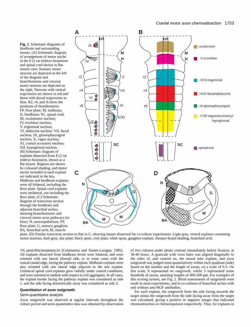
Initially, motor axons grow away from the midline floorplate, before segregating along either ventral or dorsalpathways. SM axons exit the neural tube ventrally in smallgroups, with the exception of the trochlear nerve, which exitsdorsally (Fig. 1A). BM and VM axons project to large singledorsal exit points within rhombomeres 2, 4, 6 and 7 (Fig. 1A).Motor axons from two adjacent rhombomeres converge on asingle exit point; for example the trigeminal nucleus occupiesr2 and r3, but all axons exit in r2. Once in the periphery, BMand VM axons grow in association with the cranial sensoryganglia. Then BM axons navigate towards the muscle plates ofthe branchial arches (Fig. 1C), whilst VM axons grow rostrallytowards the parasympathetic ganglia (VM). SM axons projecttowards extra-ocular or tongue muscles, which are derivedfrom paraxial or prechordal plate mesoderm (extra-ocularmuscles) or the occipital somites (tongue muscles).
Among the pathway tissues implicated in axon guidance, thefloor plate is known to produce diffusible chemorepellents thatexclude motor axons from the midline (Guthrie and Pini, 1995;Tucker et al., 1996). The expression patterns of thechemorepellents Netrin 1 and Semaphorin 3A, together with thechemosensitivity of motor neuron subpopulations to thesemolecules make them promising candidates to mediate thiseffect (Kennedy et al., 1994; Colamarino and Tessier-Lavigne,1995; Püschel et al., 1995; Varela-Echavarría et al., 1997). Theexit point also seems to play a role in motor axon guidance sincefollowing reversal of rostrocaudal polarity of rhombomere 3 or5, the majority of axons originating in the reversed rhombomerestill grew towards the appropriate exit point (Guthrie andLumsden, 1992). This suggests that signals from the exit pointspecific to appropriate motor neuron subsets may predominateover intrinsic polarity cues within the segment.
Candidate tissues producing chemoattractant cues thusinclude the dorsal neural tube (alar plate) that contains the exitpoint, and the sensory ganglia (Figure 1C). Possible sourcesof exit point cues include a late-emigrating population ofneural crest cells, which form the interface between theneuroepithelium and the sensory ganglion (Niederländer andLumsden, 1996). Moreover, the roof plate might bechemoattractive, or might limit motor axons’ dorsal trajectoriesby repulsion, since in the spinal cord BMPs present in the roofplate repel commissural axons (Augsburger et al., 1999). Lastly,the peripheral targets of cranial motor axons such as the musclesof the branchial arches might be the origin of chemoattractantguidance cues. This possibility is supported by the finding thatin the trunk, spinal motor axons are attracted by their targets,the somitic sclerotome and the limb buds (Ebens et al., 1996).In this study, limb bud-mediated chemoattraction was attributedto Hepatocyte Growth Factor (HGF), a protein originallyidentified as a mitogen for hepatocytes and a motilityenhancing-factor for epithelial cells (Nakamura et al., 1989;Stoker et al., 1987). HGF influences the growth of a number ofneuronal types (reviewed by Maina and Klein, 1999). It is asurvival and outgrowth-promoting factor for spinal motorneurons (Ebens et al., 1996; Yamamoto et al., 1997), as well asenhancing the survival and outgrowth of sensory neurons(Maina et al., 1997) and the outgrowth of sympathetic neurons(Maina et al., 1998). These properties make HGF a promisingcandidate to influence cranial motor axon pathfinding.
We have explored the role of diffusible guidance moleculesby culturing tissue explants containing subsets of cranial motor
neurons together with pathway tissues in collagen gels.Explants containing motor neurons were isolated from theventral third of the neural tubes of E12 rat embryos at midbrain,hindbrain or spinal cord levels (Fig. 1B). Tissues selected forco-culture were the dorsal neural tube and the roof plate of thehindbrain, the cranial sensory ganglia, and the branchial arches(Fig. 1D). We tested the possibilities that these tissues mightprovide diffusible signals that guide cranial motor axons.
MATERIALS AND METHODS
Dissection of embryonic tissues for co-cultureSprague-Dawley rat embryos were obtained at E12 and E13. Motorneuron-containing explants were dissected using Dispase(Boehringer) and tungsten needles, as described previously (Guthrieand Pini, 1995; Varela-Echavarría et al., 1997). Tissues were washedin Hank’s Balanced Salt Solution (HBSS; Gibco) and kept on iceuntil needed. Midbrain and hindbrain explants were bilateral,encompassing the ventral third of the neuroepithelium on either sideof and including the floor plate. Unilateral explants containing SMspinal motor neurons were isolated from the cervical spinal cord (Fig.1B). Oculomotor explants consisted of the caudal part of the midbrainwhilst trochlear explants consisted of the rostral part of r1. Otherhindbrain explants contained r2/r3, r4/r5, r6 or r7/r8. For cultures ofabducens or hypoglossal neurons labelled from their ventral exitpoints, r5 or r7/8 explants respectively were used.
Pathway tissues were dissected as shown in Fig. 1D. Dorsal neuraltube explants were two rhombomeres long consisting of the dorsalthird of the neural tube including the exit points. The explants usedwere taken from r2/r3 or r4/r5 axial levels. Roof plate explantsconsisted of the entire roof of the fourth ventricle from a singleembryo. Trigeminal ganglia were isolated by making transverse bodysections of E12 embryos at trigeminal level and then dissecting theganglion free of its adjacent tissues. First or second branchial archeswere dissected into pieces one third to one half of an arch in size, andthe ensheathing ectoderm was removed.
Retrograde labelling of cranial nerves before cultureE12 rat embryos were pinned ventral side up in Sylgard dishes, andthe dorsal or ventral nerve roots (Fig. 1A) were transected.Fluorescein dextran crystals (Molecular Probes, Oregon) were dilutedin PBS and allowed to dry to a viscous consistency before beingapplied to the cut ends of the nerves using fine forceps. Embryos werethen incubated in Earle’s Balanced Salt Solution (Gibco) in a 95% O2,5% CO2 atmosphere for 3 hours to allow retrograde transport of tracerbefore dissection of tissues.
HGF beads and anti-HGF neutralising antibodiesHeparin-acrylic beads were incubated in a solution of humanrecombinant HGF (200 µg/ml; R & D Systems) protein or in HBSSalone for 3 hours at room temperature. Beads were then washedseveral times in HBSS before use as clusters of 5-10 beads as a focalsource of HGF in co-cultures. Neutralising antibodies against humanHGF (R & D Systems), at 20 µg/ml, were used in selected cultureswith HGF-loaded beads. Neutralising antibodies against murine HGF(IW66, kind gift from E. Gherardi) were made up at 20-30 µg/ml andadded to the medium in selected cultures at the beginning of theculture period.
Collagen gels and orientation of explantsRat tail collagen was prepared and made into gels as describedpreviously (Guthrie and Lumsden, 1994). Tissue pieces and/or beadswere placed in the gels in various combinations, with 100-500 µmseparation between the tissues. Tissue pieces were cultured in mediumconsisting of 75% OptiMEM with GLUTAMAX (Gibco) and 25% F12(Gibco) supplemented with 5% foetal calf serum, 40 mM glucose and
A. Caton and others
1753Cranial motor axon chemoattraction
1% penicillin/streptomycin (Colamarino and Tessier-Lavigne, 1995).All explants dissected from hindbrain levels were bilateral, and wereoriented with one lateral (dorsal) side, or in some cases with therostral/caudal edge, facing the pathway explant. Midbrain explants werealso oriented with one lateral edge adjacent to the test explant.Unilateral spinal cord explants grew radially under control conditions,and were oriented at random with respect to cell aggregates. In all cases,the explant border facing the pathway explant was considered as side1, and the side facing diametrically away was considered as side 2.
Quantitation of axon outgrowth Semi-quantitative analysisAxon outgrowth was observed at regular intervals throughout theculture period and semi-quantitative data was obtained by observation
of live cultures under phase contrast immediately before fixation, at36-40 hours. A graticule with cross hairs was aligned diagonally tothe sides of, and centred on, the neural tube explant, and axonoutgrowth was judged semi-quantitatively within each quadrant (side)based on the number and the length of axons, on a scale of 0-5. Onthis scale, 0 represented no outgrowth, whilst 5 represented somehundreds of axons, attaining lengths of 400-500 µm. For examples ofthis scoring system, see Fig. 2. Blind assessments of outgrowth weremade in most experiments, and in co-cultures of branchial arches withand without anti-HGF antibodies.
For each explant, the outgrowth from the side facing towards thetarget minus the outgrowth from the side facing away from the targetwas calculated, giving a positive or negative integer that indicatedchemoattraction or chemorepulsion respectively. Thus, for explants in
Fig. 1. Schematic diagrams ofhindbrain and surroundingtissues. (A) Schematic diagramof arrangement of motor nucleiin the E12 rat embryo brainstemand spinal cord shown in flat-mount view. Somatic motorneurons are depicted on the leftof the diagram andbranchiomotor and visceralmotor neurons are depicted onthe right. Neurons with ventraltrajectories are shown in red andthose with dorsal trajectories inblue. R2, r4, and r6 show thepositions of rhombomeres.FP, floor plate; M, midbrain;H, hindbrain; SC, spinal cord;III, oculomotor nucleus;IV, trochlear nucleus;V, trigeminal nucleus;VI, abducens nucleus; VII, facialnucleus; IX, glossopharyngealnucleus; X, vagus nucleus;XI, cranial accessory nucleus;XII, hypoglossal nucleus.(B) Schematic diagram ofexplants dissected from E12 ratembryo brainstem, shown as aflat mount. Regions are shownby coloured shading, and motornuclei included in each explantare indicated in the key.Midbrain and hindbrain explantswere all bilateral, including thefloor plate. Spinal cord explantswere unilateral, not including thefloor plate. (C) Schematicdiagram of transverse sectionthrough the hindbrain andadjacent branchial arches,showing branchiomotor andvisceral motor axon pathways (inblue). N, neuroepithelium; FP,floor plate; G, sensory ganglion;BA, branchial arch; M, muscleplate. (D) Similar transverse section to that in C, showing tissues dissected for co-culture experiments. Light gray, ventral explants containingmotor neurons; dark gray, alar plate; black spots, roof plate; white spots, ganglion explant; chequer-board shading, branchial arch.
H
M
FP
V
VII
IX
X/XI
III
VI
XII
r2
r4
r6
IVH
M
FP
SC
r2
r4
r6
oculomotor
r1-trochlear
r2/r3-trigeminal
r4/r5-facial/abducens
r6-glossopharyngeal
r7/r8-vagus/accessory/
hypoglossal
spinalcordSC
N
G
M
FP
BAM
FPG
MBA
N
A B
C D

1754
lateral orientation, growth from the lateral sides was counted, whilstfor explants co-cultured in rostral/caudal orientation, growth fromrostral and caudal sides was counted. For each category of co-culture,the percentage of explants in a particular category on the −5 to +5scale was represented in a bar chart (see Figs 4, 5). If the majority ofexplants gave a value of 0 this indicates symmetrical outgrowth. If themajority of explants show positive or negative values this indicateschemoattraction or chemorepulsion respectively. These relative valuesfor each explant were pooled and presented as bar charts for eachtissue combination. For each explant category, the values obtainedwere compared with symmetrical outgrowth, using the Wilcoxon testor Mann-Whitney U-test (see Tables 1 and 2). In addition, pairwisecomparisons of the distribution of values for explant categories wasmade where relevant, for example, hindbrain and arch cultures withand without anti-HGF antibodies.
Quantitative analysisSome gels containing a hindbrain explant cultured alongside abranchial arch, or a hindbrain explant placed with its rostral/caudalborder adjacent to an arch explant were analysed by computerisedmethods. Immunostained explants were photographed and imageswere scanned into Photoshop. The explant tissue was deleted from theimage so that only pixels representing axon outgrowth remained.Cross hairs were placed on the image to denote quadrants containingexplant borders facing arch explants, and those facing away.Quadrants were pasted into ImageTool and converted to black andwhite. For each explant, black pixels were counted in towards andaway-facing quadrants and the data recorded in Excel. Subtraction ofaway-facing values from towards facing values for each explant gavea point of comparison with the semi-quantitative analysis, using theMann-Whitney U-Test. To present these results graphically, wecalculated a ratio for axon outgrowth towards and away from the targetexplant in each co-culture and then derived a mean for each category.Where this value is 1 it denotes symmetrical outgrowth, whilst a valueexceeding 1 shows chemoattraction (see Fig. 6E).
Immunostaining of collagen gels Some gels were fixed for immunostaining as described previously(Guthrie and Lumsden, 1992; Varela- Echavarría et al., 1997) usingmonoclonal antibody 2H3 (Developmental Studies Hybridoma Bank)which recognises the 165 kDa neurofilament protein (Dodd et al.,1988). Gels were mounted under propped coverslips in 90%glycerol/10% PBS and photographed using Nomarski optics. Forexplants in which motor neurons were prelabelled with fluoresceindextran, gels were observed using blue epifluorescence or imagedusing a laser-scanning confocal microscope. Counts of fluorescentlylabelled axons were made from explant quadrants facing towards andaway from target explants, to give an indication of chemoattraction orchemorepulsion.
In situ hybridisation of embryos for HGF, Islet 1 and MetexpressionWhole-mount in situ hybridisation was performed on E11-12 ratembryos or E10-13 mouse embryos, or on E4 chick embryos. TheIslet 1 probe was a 1.5 kb rat fragment kindly provided by T. Jessell.The Met probe was transcribed from two mouse Met fragments(nucleotides 301-1576 and 1673-2730; GenBank Y00671) subclonedfrom a Met clone provided by E. Audero and C. Ponzetto. The chickand rat HGF probes were obtained from C. Stern and T. Braunrespectively.
Embryos or dissected brainstems were fixed overnight in 4%paraformaldehyde, 0.1% Tween 20, followed by permeabilisationwith ethanol or methanol and proteinase K. Preparations were thenpostfixed (20 minutes in 4% paraformaldehyde, 0.1% glutaraldehyde,0.1% Tween 20) and prehybridised for 1 hour at 70°C in 1.3× SSC,50% formamide, 2% Tween 20, 0.5% Chaps, 5 mM EDTA and 50µg/ml yeast RNA. Hybridisation was performed overnight with DIG-
or fluorescein-labelled riboprobes in the same buffer. For the mouseMet-specific probe, post-hybridisation washes were followed byRNase A treatment (10 µg/ml in 0.5 M NaCl, 10 mM Tris (pH 7.5)and 0.1% Tween 20, 1 hour at 37°C), and other washes withhybridisation buffer. Embryos were then blocked in maleate buffercontaining 20% sheep serum or in PBT (PBS, 0.1% Tween 20) and10% sheep serum, and incubated overnight at 4°C with AP-conjugatedantibody. After extensive washes, the colour reaction was performedusing NBT and BCIP (blue) or IBT/BCIP (red). Double in situhybridisations were performed sequentially using Islet 1 (red reactionproduct) and Met (blue reaction product).
Analysis of HGF and MetD/D mutant miceThe generation of mice carrying targetted disruptions of HGF and Methas been described previously (Schmidt et al., 1995; Maina et al.,1996). In HGF−/− embryos, exon 2 of the HGF gene encoding part ofthe protein binding domain essential for receptor binding has beenreplaced with the neomycin resistance gene (Schmidt et al., 1995).MetD/D embryos carry mutations in two tyrosine residues in the Metreceptor necessary for downstream signal transduction, and manifestdefects identical to that in Met null mutants (Maina et al., 1996).HGF−/− and MetD/D E10.5 mouse embryos were fixed overnight at4°C in Dent’s fixative (1:4 DMSO/methanol) and immunostainedusing anti-NF160 antibody (N-5264; Sigma) as described previously(Maina et al., 1997).
RESULTS
Analysis of cranial motor axon outgrowth in co-cultures with pathway tissuesFor hindbrain explants grown alone, motor axon outgrowthoccurred from the lateral and from the rostral/caudal bordersof the neuroepithelium (Fig. 3A). Based on previous retrogradelabelling experiments, it is likely that motor neurons constitutemost of the population of differentiated neurons in this regionof the neurepithelium at E12, and extend axons from lateralexplant borders as they do in vivo (Guthrie and Pini, 1995;Varela-Echavarría et al., 1997). Straighter, more fasciculatedaxons which extend from rostral and caudal edges of explants
A. Caton and others
3
Controls
Co-cultures
A B C
DF
1
1
22
1
2
3 3
2
2
4
E
3
3
3
5
2
3 3
4
0
0
1 1
BA BA
BA
Fig. 2. Diagrammatic representation of the system used for scoringcranial motor axon outgrowth. In each case a graticule with cross-hairs was superimposed on a diagonal relative to the explant (notshown) and the outgrowth from each quadrant was recorded.Examples of different patterns of outgrowth are illustrated for controlexplants (A-C) and co-cultures with branchial arch explants (D-F) inlateral orientation (D,E) and rostrocaudal orientation (F).
1755Cranial motor axon chemoattraction
probably represent axons that form the medial longitudinaltracts in vivo. There was some variability in the axon outgrowthobserved in control explants (compare Fig. 3A with Fig. 5D).
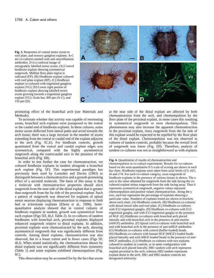
Most co-culture combinations involved placing pathwaytissues laterally, in the region where motor axons wouldnormally emerge. In some cultures, pathway tissues wereplaced rostral/caudal of the hindbrain explant, since thisallowed for the possibility of axons growing directly towardsor away from a putative source of guidance cues. The field ofaxon guidance is fraught with semantic difficulties. Many, butnot all axon guidance molecules have been shown both topromote growth and chemoattract, or to inhibit andchemorepel. In our experiments we did see effects of tissuesand molecules on the amount of axon growth as well as thedirection of guidance. Thus, in experiments with explantsplaced in lateral orientation, the responses we saw were likelyto be a combination of these two aspects; for example branchialarch explants elicited both promotion of growth andchemoattraction. In experiments with explants placed inrostral/caudal orientation, we could analyse directional effectsseparately. For the purposes of representing these resultsgraphically, however, we have considered that irrespective ofexplant orientation, greater axon outgrowth towards thepathway tissue than away reflects ‘chemoattraction’, whereasgreater outgrowth away from the pathway tissue than towardsreflects ‘chemorepulsion’ (see Materials and Methods). Pleasenote that these terms are used largely for convenience.Symmetrical outgrowth therefore gives a score of 0 and canreadily be compared with asymmetric outgrowth indicative ofchemorepulsion (negative values) or chemoattraction (positivevalues). For control explants, 85.8% grew symmetrically, with7.1% showing attraction and 7.1% showing repulsion; thisdistribution is not significantly different from symmetry (Fig.4A).
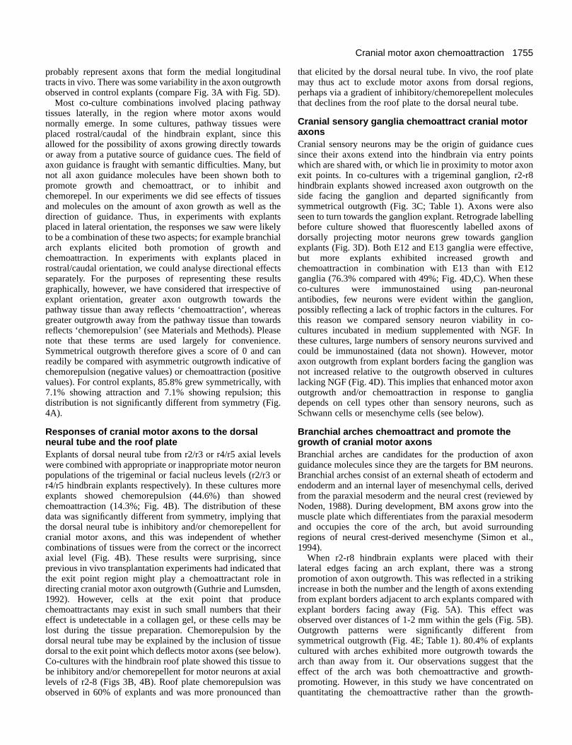
Responses of cranial motor axons to the dorsalneural tube and the roof plate Explants of dorsal neural tube from r2/r3 or r4/r5 axial levelswere combined with appropriate or inappropriate motor neuronpopulations of the trigeminal or facial nucleus levels (r2/r3 orr4/r5 hindbrain explants respectively). In these cultures moreexplants showed chemorepulsion (44.6%) than showedchemoattraction (14.3%; Fig. 4B). The distribution of thesedata was significantly different from symmetry, implying thatthe dorsal neural tube is inhibitory and/or chemorepellent forcranial motor axons, and this was independent of whethercombinations of tissues were from the correct or the incorrectaxial level (Fig. 4B). These results were surprising, sinceprevious in vivo transplantation experiments had indicated thatthe exit point region might play a chemoattractant role indirecting cranial motor axon outgrowth (Guthrie and Lumsden,1992). However, cells at the exit point that producechemoattractants may exist in such small numbers that theireffect is undetectable in a collagen gel, or these cells may belost during the tissue preparation. Chemorepulsion by thedorsal neural tube may be explained by the inclusion of tissuedorsal to the exit point which deflects motor axons (see below).Co-cultures with the hindbrain roof plate showed this tissue tobe inhibitory and/or chemorepellent for motor neurons at axiallevels of r2-8 (Figs 3B, 4B). Roof plate chemorepulsion wasobserved in 60% of explants and was more pronounced than
that elicited by the dorsal neural tube. In vivo, the roof platemay thus act to exclude motor axons from dorsal regions,perhaps via a gradient of inhibitory/chemorepellent moleculesthat declines from the roof plate to the dorsal neural tube.
Cranial sensory ganglia chemoattract cranial motoraxons Cranial sensory neurons may be the origin of guidance cuessince their axons extend into the hindbrain via entry pointswhich are shared with, or which lie in proximity to motor axonexit points. In co-cultures with a trigeminal ganglion, r2-r8hindbrain explants showed increased axon outgrowth on theside facing the ganglion and departed significantly fromsymmetrical outgrowth (Fig. 3C; Table 1). Axons were alsoseen to turn towards the ganglion explant. Retrograde labellingbefore culture showed that fluorescently labelled axons ofdorsally projecting motor neurons grew towards ganglionexplants (Fig. 3D). Both E12 and E13 ganglia were effective,but more explants exhibited increased growth andchemoattraction in combination with E13 than with E12ganglia (76.3% compared with 49%; Fig. 4D,C). When theseco-cultures were immunostained using pan-neuronalantibodies, few neurons were evident within the ganglion,possibly reflecting a lack of trophic factors in the cultures. Forthis reason we compared sensory neuron viability in co-cultures incubated in medium supplemented with NGF. Inthese cultures, large numbers of sensory neurons survived andcould be immunostained (data not shown). However, motoraxon outgrowth from explant borders facing the ganglion wasnot increased relative to the outgrowth observed in cultureslacking NGF (Fig. 4D). This implies that enhanced motor axonoutgrowth and/or chemoattraction in response to gangliadepends on cell types other than sensory neurons, such asSchwann cells or mesenchyme cells (see below).
Branchial arches chemoattract and promote thegrowth of cranial motor axonsBranchial arches are candidates for the production of axonguidance molecules since they are the targets for BM neurons.Branchial arches consist of an external sheath of ectoderm andendoderm and an internal layer of mesenchymal cells, derivedfrom the paraxial mesoderm and the neural crest (reviewed byNoden, 1988). During development, BM axons grow into themuscle plate which differentiates from the paraxial mesodermand occupies the core of the arch, but avoid surroundingregions of neural crest-derived mesenchyme (Simon et al.,1994).
When r2-r8 hindbrain explants were placed with theirlateral edges facing an arch explant, there was a strongpromotion of axon outgrowth. This was reflected in a strikingincrease in both the number and the length of axons extendingfrom explant borders adjacent to arch explants compared withexplant borders facing away (Fig. 5A). This effect wasobserved over distances of 1-2 mm within the gels (Fig. 5B).Outgrowth patterns were significantly different fromsymmetrical outgrowth (Fig. 4E; Table 1). 80.4% of explantscultured with arches exhibited more outgrowth towards thearch than away from it. Our observations suggest that theeffect of the arch was both chemoattractive and growth-promoting. However, in this study we have concentrated onquantitating the chemoattractive rather than the growth-
1756
promoting effect of the branchial arch (see Materials andMethods).
To ascertain whether this activity was capable of reorientingaxons, branchial arch explants were juxtaposed to the rostralor the caudal end of hindbrain explants. In these cultures, somemotor axons deflected from lateral paths and arced towards thearch tissue; there was a large increase in the number of axonsextending from the rostral or caudal end of the explant adjacentto the arch (Fig. 5C,E). For hindbrain controls, growthquantitated from the rostral and caudal explant edges wassymmetrical, compared with the highly asymmetricaloutgrowth along the rostrocaudal axis in the presence of thebranchial arch (Fig. 6B).
In order to test further the case for chemoattraction, wecultured hindbrain explants in tandem alongside a branchialarch explant (Fig. 5F). This experimental paradigm haspreviously been used by Lumsden and Davies (1983) todistinguish between a chemoattractive and a growth-promotingeffect of a secreted molecule. The basis of this assay is thata molecule with chemoattractive properties should elicitoutgrowth from the near side of the distal explant that is greaterthan outgrowth from the far side of the proximal explant. Sucha pattern of outgrowth was observed for explants of spinalmotor neurons displaying chemoattraction in response to limbbud or sclerotome explants (Ebens et al., 1996). Semi-quantitative analysis showed that in control cultures ofhindbrain explants in tandem, growth was symmetrical fromeach explant (Figs 5D, 4I,J; Table 2). In co-cultures of tandemhindbrains with branchial arch, proximal explants displayedenhanced outgrowth relative to distal explants. In addition,proximal explants were chemoattracted by the arch, showingasymmetrical outgrowth that was significantly different fromcontrols. Among distal explants, chemoattraction was alsoobserved, but to a lesser extent and in fewer cases (Figs 5F,4I,J). When tested statistically, the chemoattraction shown bydistal explants was not significantly different from symmetry(Table 1) and some explants exhibited chemorepulsion (Fig.6C).
This observation may be accounted for by the fact that axons
at the near side of the distal explant are affected by bothchemoattraction from the arch, and chemorepulsion by thefloor plate of the proximal explant, in some cases this resultingin symmetrical outgrowth or even chemorepulsion. Thisphenomenon may also increase the apparent chemoattractionin the proximal explant, since outgrowth from the far side ofthis explant would be expected to be repelled by the floor plateof the distal explant. Chemorepulsion was not observed incultures of tandem controls, probably because the overall levelof outgrowth was lower (Fig. 5D). Therefore, analysis oftandem co-cultures was not as straightforward as with explants
A. Caton and others
Fig. 4. Quantitation of results of chemoattraction andchemorepulsion in co-culture experiments. Results for co-culturesbased on the semi-quantitative 0-5 scale of scoring are shown in eachbar chart. Hindbrain explants were taken from axial levels r2/3, r4/5,r6 and r7/8. For each co-culture category, axon outgrowth ofhindbrain explants in the presence of various tissues is shown. The xaxis is the value obtained for outgrowth from the side facing the co-cultured explant minus outgrowth from the side facing away. Thus 0represents symmetrical outgrowth, negative values representchemorepulsion and positive values chemoattraction. Along the yaxis, each bar represents the percentage of explants showing aparticular value. Numbers of explants tested are shown in bracketsabove each chart. (A) Hindbrain controls. (B) Hindbrain co-cultureswith dorsal neural tube and roof plate. (C) Hindbrain co-cultureswith E12 trigeminal ganglia. (D) Hindbrain co-cultures with E13trigeminal ganglia, and with E13 trigeminal ganglia in the presenceof NGF. (E) Hindbrain co-cultures with branchial arch placedlaterally and with branchial arch in the presence of anti-murine(mHGF) antibodies. (F) Spinal cord co-cultures with branchial archand with branchial arch in the presence of anti-mHGF antibodies.(G) Hindbrain co-cultures with control (buffer-loaded) beads.(H) Hindbrain co-cultures with human HGF (hHGF)-loaded beadsplaced laterally and with hHGF-loaded beads in the presence of anti-hHGF antibodies. (I,J) Hindbrain co-cultures with two explantscultured in tandem as controls, or in same configuration withbranchial arch placed laterally. HB1 tandem with arch representsexplant proximal to the arch, whereas HB2 tandem representsexplant distal to the arch. HB1 and HB2 tandem controls aredesignated arbitrarily.
Fig. 3. Responses of cranial motor axons toroof plate, and sensory ganglion explants. A-Care co-cultures stained with anti-neurofilamentantibodies. D is a confocal image ofretrogradely labelled motor axons. (A) Controlhindbrain explant showing symmetricaloutgrowth. Midline floor plate region isindicated (FP). (B) Hindbrain explant culturedwith roof plate explant (RP). (C) Hindbrainexplant co-cultured with trigeminal ganglionexplant (VG). (D) Lower right portion ofhindbrain explant showing labelled motoraxons growing towards a trigeminal ganglionexplant (VG). Scale bar, 300 µm (A-C), and150 µm (D).
1757Cranial motor axon chemoattraction
of trigeminal ganglia or spinal motor neurons which growradially in vitro (Lumsden and Davies, 1983; Ebens et al.,1996). In these cultures, axons from the far sides of explantreoriented in response to chemoattraction, by growing acrossthe tissue explant, whereas in our hindbrain explants axontracing shows that motor axons did not cross the floor plate
(e.g. Fig. 5G,H). We attempted to overcome these problems byremoving the floor plate tissue from ventral explants anddissecting each explant into a number of small fragments,which were then co-cultured with the branchial arch. However,in these cultures motor axons still grew out in a highlypolarised fashion, to some extent irrespective of the position of
.
0102030405060708090
100
-4 -3 -2 -1 0 1 2 3 4Chemorepulsion Chemoattraction
Per
cent
age
of e
xpla
nts
HB Controls (n=85)
A
0102030405060708090
100
-4 -3 -2 -1 0 1 2 3 4
Chemorepulsion Chemoattraction
Per
cent
age
of e
xpla
nts
HB + dorsal neural tube (n=56) HB + roof plate (n=80)
+
B
0
10
20
30
40
50
60
70
-4 -3 -2 -1 0 1 2 3 4
Chemorepulsion Chemoattraction
Per
cent
age
of e
xpla
nts
HB + E12 ganglia (n=49)
0
10
20
30
40
50
60
70
-4 -3 -2 -1 0 1 2 3 4
Chemorepulsion Chemoattraction
Per
cent
age
of e
xpla
nts
HB + E13 ganglia (n=42) HB + E13 ganglia with NGF (n=86)
D
0
10
20
30
40
50
60
70
-4 -3 -2 -1 0 1 2 3 4Chemorepulsion Chemoattraction
Per
cent
age
of e
xpla
nts
HB + arches (n=219) HB + arches with HGF ab (n=61)
0
10
20
30
40
50
60
70
-4 -3 -2 -1 0 1 2 3 4Chemorepulsion Chemoattraction
Per
cent
age
of e
xpla
nts
Sc + arches (n=42) Sc + arches with HGF ab (n=24)
0
10
20
30
40
50
60
70
-4 -3 -2 -1 0 1 2 3 4Chemorepulsion Chemoattraction
Per
cent
age
of e
xpla
nts
HB +control beads (n=16)
0
10
20
30
40
50
60
70
-4 -3 -2 -1 0 1 2 3 4
Chemorepulsion Chemoattraction
Per
cent
age
of e
xpla
nts
HB + HGF beads (n=53) HB + HGF beads with HGF ab (n=23)H
+
01020304050607080
-4 -3 -2 -1 0 1 2 3 4
Chemorepulsion Chemoattraction
Per
cent
age
of e
xpla
nts
HB 1 tandem control (n=10) HB 1 tandem + arch (n=39)
01020304050607080
-4 -3 -2 -1 0 1 2 3 4
Chemorepulsion Chemoattraction
Per
cent
age
of e
xpla
nts
HB 2 tandem control (n=10) HB 2 tandem + arch (n=39)
J
–
+
+
G
E
+
F
+
+– +
+
+
I
+
C
–
––
–
–
–
–
–
1758
the branchial arch explant, as if responding to directional cueswithin the neuroepithelium (data not shown). We thereforeconclude that arch-derived chemoattractant molecules areincapable of predominating over cues intrinsic to theneuroepithelium, and can only reorient cranial motor axonswithin the collagen gel (Fig. 5C,E).
Branchial arch-mediated chemoattraction can bedemonstrated using semi-quantitative andquantitative methodsTo provide a comparison with our semi-quantitative analysisof cultures, images of immunostained gels were scanned intothe computer and axon outgrowth was measured by counting
pixels (see Materials and Methods). Mean numbers of pixelsin quadrants facing towards and away from co-cultured tissueswas derived (Fig. 6E), and differences between outgrowth fromtowards and away facing quadrants were tested statistically(Table 2). For hindbrain controls, outgrowth was symmetricalwhen counted from either lateral or rostral/caudal borders.Growth from the lateral or rostral/caudal sides of hindbrainsjuxtaposed to branchial arches was significantly greater thanthat from the away-facing sides (Fig. 6E; Table 2).
Several cranial motor axon subpopulations arechemoattracted by the branchial archTrigeminal or facial motor neuron explants co-cultured eitherwith their appropriate branchial arch targets (arch 1 or 2respectively) or with the inappropriate target showed noapparent preference for outgrowth towards the correct target(data not shown). Since only BM axons (trigeminal, facial,glossopharyngeal, vagus and cranial accessory) innervate thebranchial arches, this raises the question of whether only theseneurons respond to the branchial arch chemoattractant.Responses of dorsally directed axons (BM and VM classes) wasconfirmed by retrograde labelling from their exit points, whichyielded larger numbers of fluorescently labelled axons growingfrom the explant side facing the arch tissue than from the away-facing side (Fig. 5G,H). The trigeminal nucleus (r2/r3 explants)contains only BM neurons, showing that neurons of this classrespond to the arch influence, but the common pathway of BMand VM axons and the lack of any markers that distinguish theseneuronal types means that we are unable to confirmunequivocally whether VM axons respond to the arch influence.However, we were able to test the responses to branchial archexplants of SM neurons by retrograde axonal labelling beforeculture. We found that abducens and hypoglossal neurons in r5
A. Caton and others
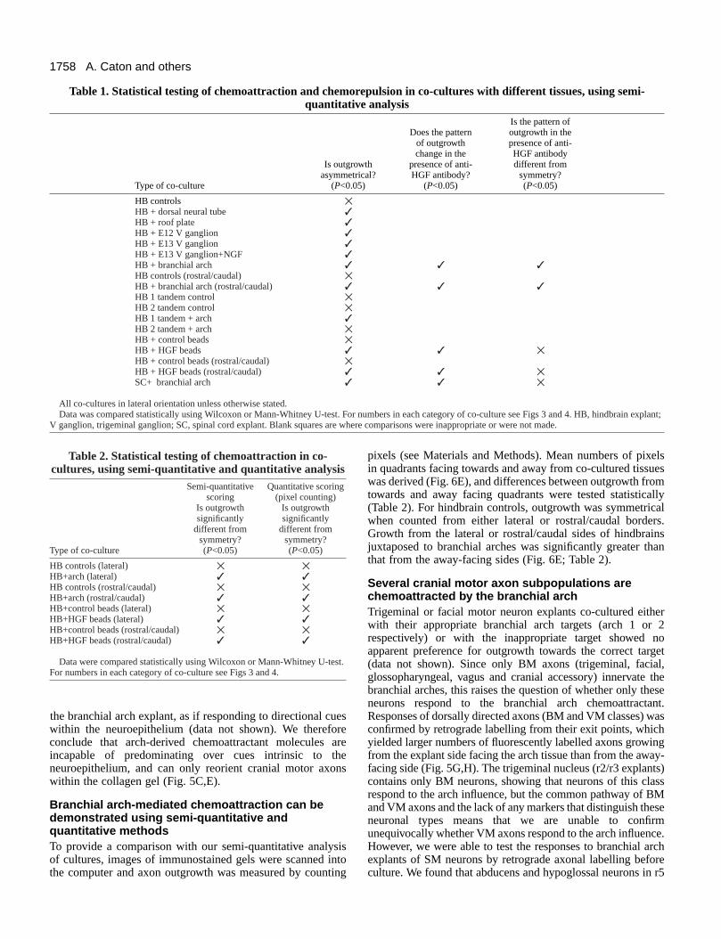
Table 1. Statistical testing of chemoattraction and chemorepulsion in co-cultures with different tissues, using semi-quantitative analysis
Is the pattern of Does the pattern outgrowth in the
of outgrowth presence of anti- change in the HGF antibody
Is outgrowth presence of anti- different from asymmetrical? HGF antibody? symmetry?
Type of co-culture (P<0.05) (P<0.05) (P<0.05)
HB controls ✕HB + dorsal neural tube ✓HB + roof plate ✓HB + E12 V ganglion ✓HB + E13 V ganglion ✓HB + E13 V ganglion+NGF ✓HB + branchial arch ✓ ✓ ✓HB controls (rostral/caudal) ✕HB + branchial arch (rostral/caudal) ✓ ✓ ✓HB 1 tandem control ✕HB 2 tandem control ✕HB 1 tandem + arch ✓HB 2 tandem + arch ✕HB + control beads ✕HB + HGF beads ✓ ✓ ✕HB + control beads (rostral/caudal) ✕HB + HGF beads (rostral/caudal) ✓ ✓ ✕SC+ branchial arch ✓ ✓ ✕
All co-cultures in lateral orientation unless otherwise stated.Data was compared statistically using Wilcoxon or Mann-Whitney U-test. For numbers in each category of co-culture see Figs 3 and 4. HB, hindbrain explant;
V ganglion, trigeminal ganglion; SC, spinal cord explant. Blank squares are where comparisons were inappropriate or were not made.
Table 2. Statistical testing of chemoattraction in co-cultures, using semi-quantitative and quantitative analysis
Semi-quantitative Quantitative scoring scoring (pixel counting)
Is outgrowth Is outgrowth significantly significantly
different from different fromsymmetry? symmetry?
Type of co-culture (P<0.05) (P<0.05)
HB controls (lateral) ✕ ✕HB+arch (lateral) ✓ ✓HB controls (rostral/caudal) ✕ ✕HB+arch (rostral/caudal) ✓ ✓HB+control beads (lateral) ✕ ✕HB+HGF beads (lateral) ✓ ✓HB+control beads (rostral/caudal) ✕ ✕HB+HGF beads (rostral/caudal) ✓ ✓
Data were compared statistically using Wilcoxon or Mann-Whitney U-test.For numbers in each category of co-culture see Figs 3 and 4.

1759Cranial motor axon chemoattraction
and r7/8 explants respectively displayed chemoattraction to thearch, and reorientation of hypoglossal axons was seen in r7/8explants placed with rostral/caudal edges facing the arch (Fig.5I). In addition, trochlear and oculomotor neurons bothresponded to the branchial arch influence (data not shown).Taken together, these data suggest that outgrowth of a numberof groups of cranial motor neurons is affected by a factorproduced in the branchial arches, and possibly in other targettissues of the head.
Cranial and spinal motor axons show reciprocalinteractions with the branchial arches and the limbbudsWe next asked whether this chemoattractant effect was specific
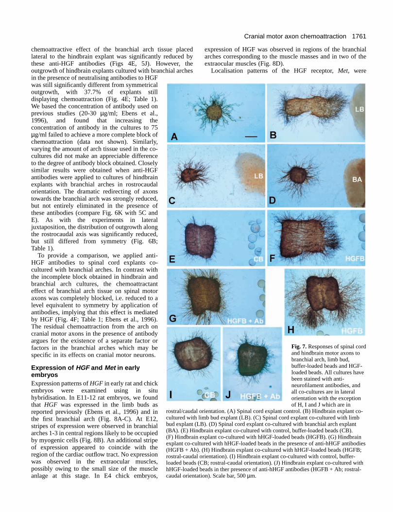
to the branchial arches. Spinal motor neurons have been shownto respond to a chemoattractant secreted by limb bud tissues,raising the possibility that the same chemoattractant could bepresent in the head and affect cranial motor axons (Ebens etal., 1996). We repeated experiments involving spinal cordexplants and limb bud explants and found that whereas spinalcord explants cultured alone showed radial outgrowth (Fig.7A), in co-cultures with limb bud tissue many explants showedchemoattraction (Fig. 7C). Furthermore, spinal motor axonswere chemoattracted by the branchial arches (Figs 7D, 4F). Inreciprocal experiments, limb bud tissue was capable of exertinga chemoattractive influence on hindbrain explants (Fig. 7B).These results demonstrate that cranial and spinal motor axonsare capable of responding to the chemoattractive influence of
Fig. 5. Responses of cranial motoraxons to branchial arch explants.(A-F) Co-cultures immunostainedusing anti-neurofilament antibodies.(G-I) Co-cultures in which motor axonswere retrogradely labelled before co-culture and confocal images obtainedafter culture. (A,B) Hindbrain explantscultured with branchial arch explantplaced laterally. (C,E) Hindbrainexplant cultured with branchial archexplant placed rostrally or caudally ofthe hindbrain explant. Branchial archexplant is below. (D) Tandem controlhindbrain explants. (F) Tandemhindbrain explants cultured withbranchial arch explant.(G,H) Hindbrain explant in whichdorsally projecting motor axons havebeen retrogradely labelled beforeculture. (G) Facial motor neurons in anr4/5 explant and (H) glossopharyngealmotor neurons in an r6 explant.(I) Hindbrain explant in which ventrallyprojecting hypoglossal motor axonshave been retrogradely labelled beforeculture of an r8 explant, positioned inrostral/caudal orientation. (J) Hindbrainexplant co-cultured with branchial archexplant placed laterally, in the presenceof anti-mHGF antibodies.(K) Hindbrain explant cultured in thepresence of branchial arch explantplaced rostral/caudal, in the presence ofanti-mHGF antibodies. BA, branchialarch; FP, floor plate. Scale bar, 500 µm(A-H), 300 µm (I).
1760
the other’s target, and imply that limb bud may contain thesame chemoattractant (s) as the branchial arch.
Is Hepatocyte Growth Factor the branchial archchemoattractant?The chemoattractant effect of limb bud tissues on spinal motorneurons could be blocked by neutralising antibodies to HGF(Ebens et al., 1996), suggesting that HGF is produced in theperiphery. Given that cranial and spinal motor neurons can bothrespond to limb and arch-secreted factors, it is possible that thearch-secreted factor is also HGF. To test this possibility, humanHGF protein was loaded on to heparin-acrylic beads, whichwere co-cultured lateral to hindbrain explants. Cranial motoraxons showed increased outgrowth in the presence of HGF-loaded beads when compared with responses to control beadsincubated in buffer (Figs 4G,H, 7E,F). There was a strikingreorientation of axons towards HGF-loaded beads when thelatter were cultured in rostro-caudal orientation relative to
hindbrain explants (Fig. 7H). This chemoattractant effect wassignificant, compared with no effect on direction of axonoutgrowth by control beads placed in rostrocaudal orientation(Figs 6D, 7H,I). These effects of HGF-loaded beads in bothorientations were also significant when compared usingquantitative methods (Fig. 6E; Table 2). When an antibody tohuman HGF was applied to cultures with HGF-loaded beadsplaced either in lateral or in rostrocaudal orientation, HGF-mediated outgrowth and chemoattraction was completelyblocked since axon outgrowth was not significantly differentfrom controls (Figs 4G,H, 6C,D, 7G,J; Table 1).
To test the idea that the branchial arch chemoattractantis HGF, we investigated whether the arch-mediatedchemoattraction could be blocked using a neutralising antibodyagainst murine HGF (kind gift from E. Gherardi) which alsorecognises rat HGF (Ebens et al., 1996). Co-cultures ofhindbrain and branchial arch explants with and withoutantibody were scored blind. The results showed that the
A. Caton and others
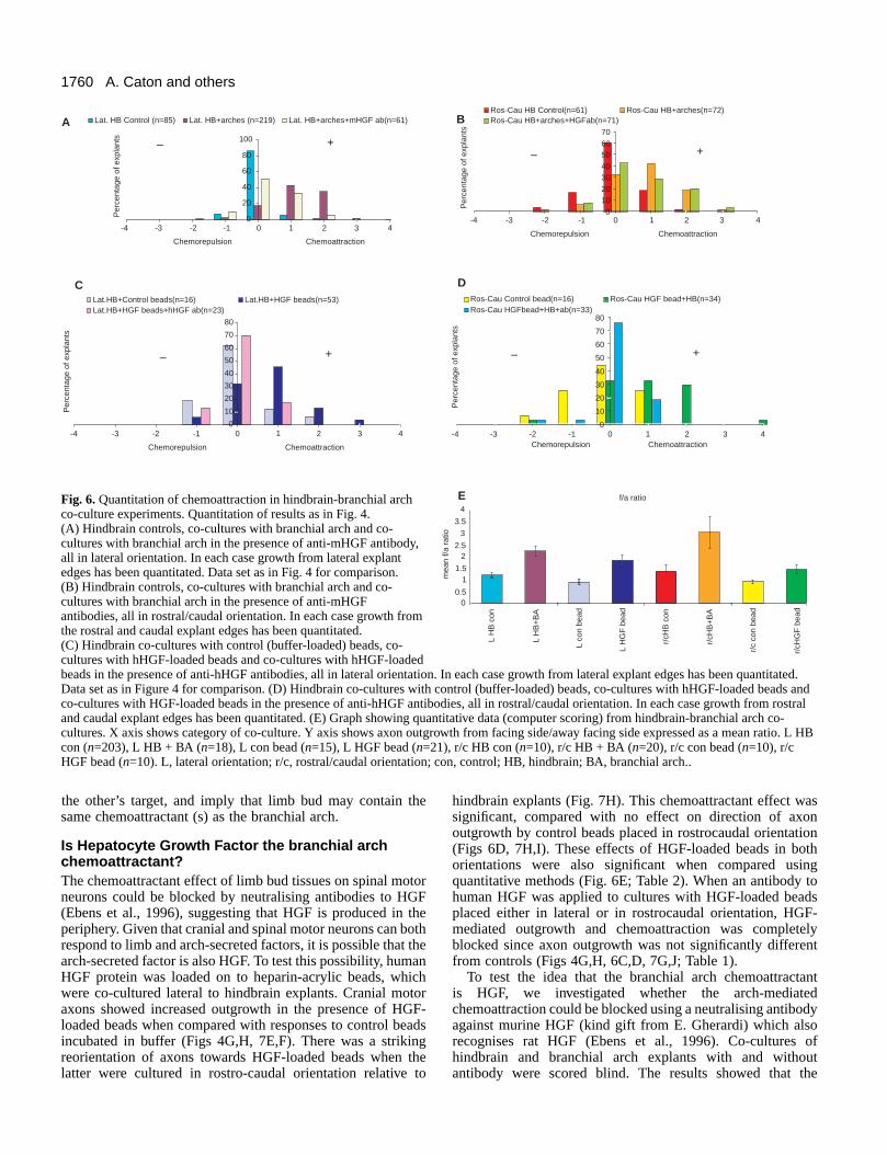
Fig. 6. Quantitation of chemoattraction in hindbrain-branchial archco-culture experiments. Quantitation of results as in Fig. 4.(A) Hindbrain controls, co-cultures with branchial arch and co-cultures with branchial arch in the presence of anti-mHGF antibody,all in lateral orientation. In each case growth from lateral explantedges has been quantitated. Data set as in Fig. 4 for comparison.(B) Hindbrain controls, co-cultures with branchial arch and co-cultures with branchial arch in the presence of anti-mHGFantibodies, all in rostral/caudal orientation. In each case growth fromthe rostral and caudal explant edges has been quantitated.(C) Hindbrain co-cultures with control (buffer-loaded) beads, co-cultures with hHGF-loaded beads and co-cultures with hHGF-loadedbeads in the presence of anti-hHGF antibodies, all in lateral orientation. In each case growth from lateral explant edges has been quantitated.Data set as in Figure 4 for comparison. (D) Hindbrain co-cultures with control (buffer-loaded) beads, co-cultures with hHGF-loaded beads andco-cultures with HGF-loaded beads in the presence of anti-hHGF antibodies, all in rostral/caudal orientation. In each case growth from rostraland caudal explant edges has been quantitated. (E) Graph showing quantitative data (computer scoring) from hindbrain-branchial arch co-cultures. X axis shows category of co-culture. Y axis shows axon outgrowth from facing side/away facing side expressed as a mean ratio. L HBcon (n=203), L HB + BA (n=18), L con bead (n=15), L HGF bead (n=21), r/c HB con (n=10), r/c HB + BA (n=20), r/c con bead (n=10), r/cHGF bead (n=10). L, lateral orientation; r/c, rostral/caudal orientation; con, control; HB, hindbrain; BA, branchial arch..
0
20
40
60
80
100
Chemorepulsion Chemoattraction
Per
cent
age
of e
xpla
nts
Lat. HB Control (n=85) Lat. HB+arches (n=219) Lat. HB+arches+mHGF ab(n=61)A
0
10
20
30
40
50
60
70
80
Chemorepulsion Chemoattraction
Per
cent
age
of e
xpla
nts
Lat.HB+Control beads(n=16) Lat.HB+HGF beads(n=53)Lat.HB+HGF beads+hHGF ab(n=23)
010203040506070
Chemorepulsion Chemoattraction
Per
cent
age
of e
xpla
nts
Ros-Cau HB Control(n=61) Ros-Cau HB+arches(n=72)Ros-Cau HB+arches+HGFab(n=71)
0
10
20
30
40
50
60
70
80
Chemorepulsion ChemoattractionP
erce
ntag
e of
exp
lant
s
Ros-Cau Control bead(n=16) Ros-Cau HGF bead+HB(n=34)Ros-Cau HGFbead+HB+ab(n=33)
++
B
C
+
D
+
f/a ratio
00.5
11.5
22.5
3
3.5
4
L H
B c
on
L H
B+
BA
L co
n be
ad
L H
GF
bea
d
r/cH
B c
on
r/cH
B+
BA
r/c
con
bead
r/cH
GF
bea
d
mea
n f/a
rat
io
E
–
––
-4 0-3 -2 -1 1 2 3 4 -4 0-3 -2 -1 1 2 3 4
-4 0-3 -2 -1 1 2 3 4-4 0-3 -2 -1 1 2 3 4
–
1761Cranial motor axon chemoattraction
chemoattractive effect of the branchial arch tissue placedlateral to the hindbrain explant was significantly reduced bythese anti-HGF antibodies (Figs 4E, 5J). However, theoutgrowth of hindbrain explants cultured with branchial archesin the presence of neutralising antibodies to HGFwas still significantly different from symmetricaloutgrowth, with 37.7% of explants stilldisplaying chemoattraction (Fig. 4E; Table 1).We based the concentration of antibody used onprevious studies (20-30 µg/ml; Ebens et al.,1996), and found that increasing theconcentration of antibody in the cultures to 75µg/ml failed to achieve a more complete block ofchemoattraction (data not shown). Similarly,varying the amount of arch tissue used in the co-cultures did not make an appreciable differenceto the degree of antibody block obtained. Closelysimilar results were obtained when anti-HGFantibodies were applied to cultures of hindbrainexplants with branchial arches in rostrocaudalorientation. The dramatic redirecting of axonstowards the branchial arch was strongly reduced,but not entirely eliminated in the presence ofthese antibodies (compare Fig. 6K with 5C andE). As with the experiments in lateraljuxtaposition, the distribution of outgrowth alongthe rostrocaudal axis was significantly reduced,but still differed from symmetry (Fig. 6B;Table 1).
To provide a comparison, we applied anti-HGF antibodies to spinal cord explants co-cultured with branchial arches. In contrast withthe incomplete block obtained in hindbrain andbranchial arch cultures, the chemoattractanteffect of branchial arch tissue on spinal motoraxons was completely blocked, i.e. reduced to alevel equivalent to symmetry by application ofantibodies, implying that this effect is mediatedby HGF (Fig. 4F; Table 1; Ebens et al., 1996).The residual chemoattraction from the arch oncranial motor axons in the presence of antibodyargues for the existence of a separate factor orfactors in the branchial arches which may bespecific in its effects on cranial motor neurons.
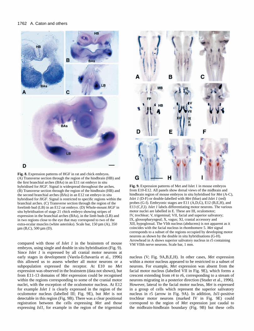
Expression of HGF and Met in earlyembryosExpression patterns of HGF in early rat and chickembryos were examined using in situhybridisation. In E11-12 rat embryos, we foundthat HGF was expressed in the limb buds asreported previously (Ebens et al., 1996) and inthe first branchial arch (Fig. 8A-C). At E12,stripes of expression were observed in branchialarches 1-3 in central regions likely to be occupiedby myogenic cells (Fig. 8B). An additional stripeof expression appeared to coincide with theregion of the cardiac outflow tract. No expressionwas observed in the extraocular muscles,possibly owing to the small size of the muscleanlage at this stage. In E4 chick embryos,
expression of HGF was observed in regions of the branchialarches corresponding to the muscle masses and in two of theextraocular muscles (Fig. 8D).
Localisation patterns of the HGF receptor, Met, were
Fig. 7. Responses of spinal cordand hindbrain motor axons tobranchial arch, limb bud,buffer-loaded beads and HGF-loaded beads. All cultures havebeen stained with anti-neurofilament antibodies, andall co-cultures are in lateralorientation with the exceptionof H, I and J which are in
rostral/caudal orientation. (A) Spinal cord explant control. (B) Hindbrain explant co-cultured with limb bud explant (LB). (C) Spinal cord explant co-cultured with limbbud explant (LB). (D) Spinal cord explant co-cultured with branchial arch explant(BA). (E) Hindbrain explant co-cultured with control, buffer-loaded beads (CB).(F) Hindbrain explant co-cultured with hHGF-loaded beads (HGFB). (G) Hindbrainexplant co-cultured with hHGF-loaded beads in the presence of anti-hHGF antibodies(HGFB + Ab). (H) Hindbrain explant co-cultured with hHGF-loaded beads (HGFB;rostral-caudal orientation). (I) Hindbrain explant co-cultured with control, buffer-loaded beads (CB; rostral-caudal orientation). (J) Hindbrain explant co-cultured withhHGF-loaded beads in ther presence of anti-hHGF antibodies (HGFB + Ab; rostral-caudal orientation). Scale bar, 500 µm.
1762
compared with those of Islet 1 in the brainstem of mouseembryos, using single and double in situ hybridisation (Fig. 9).Since Islet 1 is expressed by all cranial motor neurons atearly stages in development (Varela-Echavarría et al., 1996)this allowed us to assess whether all motor neurons or asubpopulation expressed the receptor. At E10 no Metexpression was observed in the brainstem (data not shown), butfrom E11-13 domains of Met expression could be recognisedwithin the regions corresponding to some of the cranial motornuclei, with the exception of the oculomotor nucleus. At E12for example Islet 1 is clearly expressed in the region of theoculomotor nucleus (labelled III; Fig. 9E), but Met is notdetectable in this region (Fig. 9B). There was a clear positionalregistration between the cells expressing Met and thoseexpressing Isl1, for example in the region of the trigeminal
nucleus (V; Fig. 9A,B,E,H). In other cases, Met expressionwithin a motor nucleus appeared to be restricted to a subset ofneurons. For example, Met expression was absent from thefacial motor nucleus (labelled VII in Fig. 9E), which forms acrescent extending from r4 to r6, corresponding to a stream ofneurons migrating in a posterior direction (Studer et al., 1996).However, lateral to the facial motor nucleus, Met is expressedin a group of cells which represent the superior salivatorynucleus in r5 (arrow in Fig. 9A). In addition, Isl1-positivetrochlear motor neurons (marked IV in Fig. 9E) couldcorrespond to the region of Met expression just caudal tothe midbrain-hindbrain boundary (Fig. 9B) but these cells
A. Caton and others
Fig. 8. Expression patterns of HGF in rat and chick embryos.(A) Transverse section through the region of the hindbrain (HB) andthe first branchial arches (BAs) in an E11 rat embryo in situhybridised for HGF. Signal is widespread throughout the arches.(B) Transverse section through the region of the hindbrain (HB) andthe second branchial arches (BAs) in an E12 rat embryo in situhybridised for HGF. Signal is restricted to specific regions within thebranchial arches. (C) Transverse section through the region of theforelimb bud (LB) in an E12 rat embryo. (D) Whole-mount HGF insitu hybridisation of stage 21 chick embryo showing stripes ofexpression in the branchial arches (BAs), in the limb buds (LB) andin two regions close to the eye that may correspond to two of theextra-ocular muscles (white asterisks). Scale bar, 150 µm (A), 350µm (B,C), 500 µm (D).
Fig. 9. Expression patterns of Met and Islet 1 in mouse embryosfrom E10-E12. All panels show dorsal views of the midbrain andhindbrain region of mouse embryos in situ hybridised for Met (A-C),Islet 1 (D-F) or double-labelled with Met (blue) and Islet 1 (red)probes (G-I). Embryonic stages are E11 (A,D,G), E12 (B,E,H), andE13 (C,F,I). Islet 1 labels differentiating motor neurons. The variousmotor nuclei are labelled in E. These are III, oculomotor;IV, trochlear; V, trigeminal; VII, facial and superior salivatory;IX, glossopharyngeal; X, vagus; XI, cranial accessory andXII, hypoglossal. The VIth nucleus (abducens) is not apparent as itcoincides with the facial nucleus in rhombomere 5. Met signalcorresponds to a subset of the regions occupied by developing motorneurons as shown by the double in situ hybridisations (G-H).Arrowhead in A shows superior salivatory nucleus in r5 containingVM VIIth nerve neurons. Scale bar, 1 mm.

1763Cranial motor axon chemoattraction
appeared to form a more diffuse cluster than those defined byIsl1 staining. The more caudal groups of motor neurons, theglossopharyngeal, vagus and cranial accessory (IX, X and XI)and hypoglossal nuclei (XII) also express Met (Fig. 9B,E,H).The pattern of Met expression observed at E13 (Fig. 9C) wasclosely similar to that at E12. Staining was also observed indorsal regions in rhombomere 1, which may correspond to thedeveloping cerebellum (Fig. 9A,B).
BM and VM neurons undergo a medial to lateral migrationwithin the brainstem during their maturation (Simon et al.,1994). The localisation of Met in lateral regions therefore
suggests that this receptor is expressed in older motor neuronsthat have already extended axons into the periphery. If thiscohort of neurons represents only a subpopulation within eachnucleus, then it may be the earliest cohort to differentiate,representing the pioneer neurons that first extend axonsperipherally. Axon tracing coupled with expression studieswould be needed to clarify this as well as to confirm expressionby particular neuronal subpopulations. This far it appears thatat least a subpopulation of each motor nucleus, with theexception of the oculomotor and the facial motor nucleus,express Met.
Cranial nerve morphology in mice with targetteddisruptions of HGF and MetIn order to further investigate a possible role for HGFsignalling in cranial motor axon pathfinding we examined micewith targetted disruptions of the genes encoding HGF (Schmidtet al., 1995) or Met (the MetD/D mutant; Maina et al., 1996).Anti-neurofilament staining of E10.5 embryos revealed theaxonal pathways in the cranial region of wild-type embryos(Fig. 10A,C). In HGF−/− embryos these nerve pathways lookedsuperficially similar, except for the hypoglossal nerve, whichwas truncated (arrow in Fig. 10B). This reflects a failure of thisnerve to grow along its normal course rostrally through thebranchial arches (Fig. 10C,E,G). A closely similar phenotypewas observed in MetD/D embryos (Fig. 10D,F,H). Since HGF-Met signalling is required for correct migration of a subset oftongue myoblasts (Bladt et al., 1995), this defect might beinterpretable in terms of a loss of the synaptic targets ofhypoglossal axons. However, in the MetGrb2/Grb2 mutant thesemuscles develop correctly, and yet a defect in the hypoglossalnerve is still observed (F. M. and R. K., unpublished data). Inaddition, HGF expression may not be restricted to myogenicprecursors, since it is present along the pathway of thehypoglossal nerve in Met mutants which lack these migratingcells (Dietrich et al., 1999). This suggests that aberranthypoglossal nerve outgrowth is due to a direct effect of loss ofHGF-Met signalling on axon guidance. Observation of HGF−/− embryos at later stages of development, however, indicatethat the delay in hypoglossal outgrowth is compensated at laterstages of development, perhaps by other guidance mechanisms.Other cranial nerve pathways appeared normal in the mutantembryos, suggesting that additional guidance mechanismsoperate, perhaps including branchial arch chemoattractants thatare distinct from HGF.
DISCUSSION
In this study we have explored the possible influences ofdiffusible guidance molecules produced by pathway tissues oncranial motor axon navigation. Our major conclusion is that thebranchial arches exert a growth-promoting and chemoattractantinfluence on motor axons as they extend towards theirperipheral targets. Since anti-HGF antibodies block thisinteraction in vitro, this effect is at least partially mediated byHGF. HGF is expressed in regions corresponding to developingmuscles, including those of the branchial arches, and is capableof causing increased outgrowth and orientation of motor axonsof all classes, suggesting that it is a general cue for axonoutgrowth in the head, rather than a signal that governs specific
Fig. 10. Neurofilament staining of mouse embryos with targetteddisruptions of HGF or Met. All views of embryos are lateral withrostral to the right. (A-D) E10.5 embryos, (E-H) E11.5 embryos.Arrowheads in A-F show position of the hypoglossal nerve.(A) HGF+/+ control embryo showing morphology of the cranialnerves. V, trigeminal; VII, facial; IX, glossopharyngeal; X/XI, vagus;cranial accessory; XII, hypoglossal. (B) HGF mutant embryo. Allcranial nerves are normal with the exception of the hypoglossal nervewhich is truncated, and has failed to grow rostrally through thebranchial arches. (C) Met+/+ control embryo. Cranial nerves labelledas in (A). (D) MetD/D mutant embryo. All cranial nerves are normalwith the exception of the hypoglossal, which has a morphologyclosely similar to that in the HGF mutant. (E) Higher magnificationof Met+/+ control embryo. (F) Higher magnification of Met−/−
embryo. (G) Camera lucida drawing of normal hypoglossal pathwayin Met+/+ embryo. (H) Camera lucida drawing of truncatedhypoglossal nerve in MetD/D embryo. Scale bar, 500 µm (A,B); 330µm in (C,D) and 250 µm (E,F).

1764
wiring of cranial motor axons to their targets. The HGFreceptor Met is present in only a subset of cranial motorneurons, and disruption of HGF or Met gene function producesonly minor defects in cranial motor axon pathfinding. Thus,additional arch-derived chemoattractants or other guidancemolecules are implicated in pathfinding to the branchial arches.
The neuroepithelium of the dorsal neural tube, whichcontains the exit point, fails to chemoattract motor axons, andso cannot account for the navigation of axons towards thisregion. Both the dorsal neural tube and the roof plate areinhibitory and/or chemorepellent, and may provide a stopsignal that limits the dorsal growth of cranial motor axons. Thegrowth promoting and/or chemoattractant effect of the cranialsensory ganglia on cranial motor axons may depend uponmesenchymal cells isolated together with the ganglia.
HGF is a guidance cue for cranial motor axonsWe have shown that cranial motor axons exhibit increasedgrowth and chemoattraction in response to branchial archtissues, and that anti-HGF antibodies partially block arch-mediated chemoattraction. Coupled with observations ofhypoglossal nerve defects in HGF−/− and MetD/D mutantembryos, this implies that HGF is an important factor producedby the arches which participates in motor axon guidance. Insupport of this idea were our findings that HGF presented onbeads can chemoattract cranial motor axons. These data areconsistent with previous studies showing that chemoattractionof spinal motor axons by the limb bud depends on HGF (Ebenset al., 1996), and that mouse embryos mutant for HGF or Metdisplay defects in motor nerve branching within the limbs, aswell as in sensory innervation of the limbs and thorax (Ebenset al., 1996; Maina et al., 1997). Our findings extend therepertoire of known roles for HGF, which in addition toinfluencing the growth of motor neurons, promotes theoutgrowth and survival of DRG sensory neurons (Maina et al.,1997) and the outgrowth of sympathetic neurons (Maina et al.,1998).
The precise localisation of HGF in the tissues of the headrequires further investigation, including double labellingstudies using muscle markers. We found HGF to be expressedby branchial arch tissues in both rat and chick embryos (seealso Théry et al., 1995), and in the latter, this region overlappedwith the muscle plate containing immigrant myogenicprecursors. In the limb, HGF is not expressed by muscle cells,but is confined to the lateral plate mesoderm (Ebens et al.,1996). Recent studies also showed the expression of HGFalong the pathway of tongue muscle precursors in the absenceof these cells, pointing to additional mesenchymal cellpopulations as a source of HGF (Dietrich et al., 1999).Certainly, a wider axon guidance role of HGF than is reflectedsimply by its production by the branchial arches is also impliedby the expression of this factor by the extra-ocular muscles(Fig. 8D). Possibly HGF is involved in pathfinding of manymotor axon populations, but close spatiotemporal regulationprevents axons growing aberrantly. Consistent with this werefindings that branchial arches chemoattracted both BM axons(their normal innervation) and SM axons. Diffusion of HGFmight be limited by tissues such as the presumptive cartilage,which acts as a barrier to motor axon outgrowth (Tosney,1991), or by binding to components of the extracellular matrix.
Insights into the role of HGF will also come from studying,
in more detail, the regulation of Met expression during motorneuron development, since we observed restricted Metexpression by subpopulations of cranial motor neurons at alater phase of their pathfinding into the periphery. In the spinalcord Met is also expressed in a restricted pattern, being presentat high levels within brachial level motor neurons destined toinnervate the limbs, and at much lower levels in spinal motorneurons at other axial levels (Ebens et al., 1996; Yamamoto etal., 1997).
Is there an additional chemoattractant activity in thehead?A number of hindbrain explants cultured with branchial archesand anti-HGF antibodies exhibited a significant degree ofchemoattraction. We believe that this effect is unlikely to bedue to degradation of the antibody in the culture system, sincein tests we found that the degree of chemoattraction manifestwas equivalent at 24 hours and at 36-40 hours after thebeginning of culture, and increasing the antibody concentrationdid not inhibit the chemoattraction more completely. Theobservation that chemoattraction of cranial motor axons byHGF-loaded beads was completely blocked by antibodies alsoshows that technical constraints are unlikely to account for theabsence of block observed in branchial arch co-cultures. Thesedata therefore lead to the conclusion that an additionalchemoattractive factor may be generated by the branchialarches.
Two other lines of evidence support the idea of additionalchemoattractants. First, only a subset of cranial motor neuronsexpressed the HGF receptor Met, despite the fact that all motoraxon subpopulations tested were chemoattracted by the arch.It seems unlikely that HGF acts on cranial motor axons bybinding to a receptor other than Met; there is a close correlationbetween the phenotypes of animals mutant for Met and forHGF (Bladt et al., 1995; Schmidt et al., 1995; Uehara et al.,1995). Secondly, mice carrying targetted disruptions of HGFand Met showed modest defects in the pathfinding of thehypoglossal nerve, indicating a role for HGF-Met signalling inhypoglossal nerve outgrowth. However, the integrity of othercranial nerve pathways was preserved, pointing to the existenceof additional axon guidance factors. We will aim to test thispossibility by co-culturing cranial motor axons with branchialarches derived from HGF−/− mice, and investigating whetherchemoattraction is still present.
What is the role of the exit point, roof plate andcranial sensory ganglia? The roles of tissues other than the branchial arches in cranialmotor axon guidance deserve to be investigated further. Thedorsal neural tube, which may contain elements of the exitpoint, proved to be inhibitory and/or chemorepellent, despiteprevious evidence of its chemoattractive role (Guthrie andLumsden, 1992). This may not be surprising, given that exitpoint-forming cells might be too few in number to exertchemoattraction in collagen gels. Exit point signals might arisefrom a late-emigrating population of hindbrain neural crestcells, which in the chick form the dorsal exit points(Niederländer and Lumsden, 1996). These cells, or adjacentmesenchymal cells, might be lost during preparation of dorsalneural tube explants. Studies on the dorsal root entry zone ofthe spinal nerves in the chick have shown that similar clusters
A. Caton and others

1765Cranial motor axon chemoattraction
of ‘boundary cap’ cells are likely to be involved in the guidanceof incoming dorsal root afferents and the establishment of anon-permissive region for later axon ingrowth (Goldingand Cohen, 1997). If they do not act exclusively bychemoattraction, boundary cap cells might guide outgrowingmotor axons by contact-mediated interactions. In the case ofchemoattraction by the cranial ganglia, we believe thatthis is not a phenomenon distinct from branchial archchemoattraction, but instead is most likely to be due tocontaminating mesenchyme cells which are difficult to removeand also proliferate extensively in collagen gel cultures.Among these cells may be myogenic precursors or othermesenchymal cells which express HGF. Possible candidatesto mediate dorsal neural tube/roof-plate dependentchemorepulsion may be Semaphorin 3D (previously collapsin2), based on its localisation on either side of the roof plate inthe spinal cord (Luo et al., 1995), or BMPs, based on theirexpression in the roof plate and ability to repel commissuralaxons (Liem et al., 1995; Augsburger et al., 1999).
Sequential steps in cranial motor axon guidanceEarly cranial motor axon pathway choices reflect the BM/VMor SM phenotypes of neurons that differentiate in response tograded activity of Sonic Hedgehog (SHH) protein (reviewedby Tanabe and Jessell, 1996). An early step in SHH inductionis the expression of the transcription factor MNR2, which isrequired for the differentiation of SM but not BM/VM neurons(Tanabe et al., 1998). In addition, Pax6 is expressed in adorsoventral gradient in response to SHH signalling, and whilstBM/VM neurons differentiate within a ventral, Pax6-negativedomain of the neuroepithelium, SM neurons differentiatewithin a more dorsal domain of low Pax6 expression (Ericsonet al., 1997). In mice and rats lacking Pax6, ventrally projectinghindbrain SM neurons fail to differentiate and may assume thefates of BM/VM neurons (Ericson et al., 1997; Osumi et al.,1997). However, Pax6 appears to act indirectly, by controllingthe expression of Nkx2.2, since in mice mutant for this gene,spinal ventral interneurons assume the fates of more dorsalmotor neurons (Briscoe et al., 1999). In the hindbrain, BM/VMneurons normally derive from the Nkx2.2-positive domain, butwere unaffected in the mutant, and the authors speculate thatanother transcription factor (perhaps Nkx2.9) assumes thefunction of assigning motor neuron fates in this region (Briscoeet al., 1999).
Downstream of these early events, dorsal or ventral motoraxon pathway choices appear to be under the control of LIMhomeodomain transcription factors, which are expressed in acombinatorial manner among motor neuron subpopulations inthe chick embryo (Tsuchida et al., 1994; Varela-Echavarría etal., 1996). In mouse embryos deficient for two of thesefactors, Lhx3 and Lhx4, ventrally projecting motor neuronschange their identity and form dorsal axon projections,whilst misexpression of Lhx3 in dorsally projectingneurons causes them to reorient their projections ventrally(Sharma et al., 1998). Formation of an exit point thereforeappears cell autonomous in the case of ventrally projectingneurons, even if additional cells are recruited to the exit siteonce formed.
An early hallmark of differentiation for ventrally or dorsallyprojecting neurons is presumably expression of receptors forchemorepellents expressed at the midline, since motor axons
are repelled by the floor plate (Guthrie and Pini, 1995).Molecules involved in midline repulsion are likely to be Netrin1 Semaphorin 3A and Slit, (Colamarino and Tessier-Lavigne,1995; Varela-Echavarría et al., 1997; Brose et al., 1999). Spinalmotor neurons express the Slit receptors, Robo 1 and 2 (Broseet al., 1999), but so far there has not been a detailed analysisof the localisation of Robo or of receptors for Netrins andSemaphorins among cranial motor neurons. Since dorsallyprojecting cranial motor neurons respond to both Netrin 1 andSemaphorin 3A, whereas ventrally projecting cranial motorneurons respond only to the latter molecule (Varela-Echavarríaet al., 1997), the expression of receptors for the repulsiveeffects of Netrin 1 might be more restricted.
For somatic motor neurons of the abducens and hypoglossalnuclei, other cues are required to account for the longitudinalpathways taken. Possibly, HGF is produced by the targetmuscles of these neurons, the lateral rectus and the tonguemuscles respectively. In the case of abducens guidance cuesinclude early contact with the muscle anlage of lateral rectusand coordinate growth/migration of nerve and muscle to theirdestination adjacent to the eye (Wahl et al., 1994). BM and VMneurons may respond to as yet unidentified polarity cues in theneuroepithelium which guides their course dorsally, androstrally in the case of axons orginating in odd-numberedrhombomeres. Their dorsal trajectory is limited by repulsionby the roof plate and dorsal neural tube. Dorsally projectingaxons grow out via the exit points, which already formpathways for incoming sensory axons. Chemoattraction fromsome component of the exit point and/or contact guidance bysensory neurons is likely to be required for their egress fromthe neural tube. Once in the periphery, BM axons grow towardsthe muscle plates of the branchial arches under the influenceof HGF and other factors.
Although HGF is one important guidance cue for cranialmotor axons, other axon guidance cues are required in thisscheme to account for the selectivity and shaping ofprojections. Notably, Semaphorin 3A appears to be involved inchannelling motor axons into the cores of the branchial arches,since in mice mutant for Semaphorin 3A or its receptorNeuropilin 1, widespread defasciculation of nerves intoinappropriate arch regions is observed (Taniguchi et al., 1997;Kitsukawa et al., 1997). Furthermore, little is known of themechanisms that segregate nerve branches to individualmuscles later in development. Understanding how HGF andother guidance cues act in vivo will involve a detailedconsideration of how these molecules are organisedspatiotemporally and interact with each other, in relation to thepathways of different cranial motor nerves.
We thank C. Henderson for valuable discussions about themanuscript and for sharing data with us. We also thank E. Audero, H.Funakoshi, T. Jessell, T. Nakamura, C. Ponzetto and C. Stern forproviding HGF and Met probes, and E. Gherardi for the anti-HGFantibody. This work was supported by a grant from the Wellcome Trust.
REFERENCES
Augsburger, A., Schuchardt, A., Hoskins, S., Dodd, J. and Butler, S.(1999). BMPs as mediators of roof plate repulsion of commissural neurons.Neuron 24, 127-141.
Bagnard, D., Lohrum, M., Uziel, D., Püschel, A. and Bolz, J. (1998).

1766
Semaphorins act as attractive and repulsive guidance during thedevelopment of cortical projections. Development 125, 5043-5053.
Bladt, F., Riethmacher, D., Isenmann, S., Aguzzi, A., and Birchmeier, C.(1995). Essential role for the c-met receptor in the migration of myogenicprecursor cells into the limb bud. Nature 376, 768-771.
Briscoe, J., Sussel, L., Serup, P., Hartigan O’Connor, Jessell, T. M.,Rubenstein, J. L. R. and Ericson, J. (1999). Homeobox gene Nkx2.2 andspecification of neuronal identity by graded Sonic hedgehog signalling.Nature 398, 622-627.
Brose, K., Bland, K. S., Wang, K. H., Arnott, D., Henzel, W., Goodman,C. S., Tessier-Lavigne, M. and Kidd, T. (1999). Slit proteins bind Roboreceptors and have an evolutionarily conserved role in repulsive axonguidance. Cell 96, 795-806.
Colamarino, S. A. and Tessier-Lavigne, M. (1995). The axonalchemoattractant netrin-1 is also a chemorepellent for trochlear motorneurons. Cell 81, 621-629.
Dietrich, S., Abou-Rebyeh, F., Brohmann, H., Bladt, F., Sonnenberg-Riethmacher, E., Yamaai, T., Lumsden, A., Brand-Saberi, B. andBirchmeier C. (1999). The role of SF/HGF and c-Met in the developmentof skeletal muscle. Development 126, 1621-1629.
Dodd, J., Morton, S. B., Karagogeos, D., Yamamoto, M. and Jessell, T. M.(1988). Spatial regulation of axonal glycoprotein expression on subsets ofembryonic spinal neurons. Neuron 1, 105-116.
Ebens, A., Brose, K., Leonardo, E. D., Gartz Hanson Jnr., M., Bladt, F.,Birchmeier, C., Barns, B. A. and Tessier-Lavigne, M. (1996). HepatocyteGrowth Factor/Scatter Factor is an axonal chemoattractant and neurotrophicfactor for spinal motor neurons. Neuron 17, 1157-1172.
Ericson, J., Rashbass, P., Schedl, A., Brenner-Morton, S., Kawakami, A.,van Heyningen, V., Jessell, T. M. and Briscoe, J. (1997). Pax6 controlsprogenitor cell identity and neuronal fate in response to graded Shhsignalling. Cell 90, 169-180.
Gilland, E. and Baker, R. (1993). Conservation of neuroepithelial andmesodermal segments in the embryonic vertebrate head. Acta Anat. 148,110-123.
Golding, J. P. and Cohen, J. (1997). Border controls at the mammalian spinalcord: late-surviving neural crest boundary cap cells at dorsal root entry sitesmay regulate sensory afferent ingrowth and entry zone morphogenesis. Mol.Cell Neurosci. 9, 381-396.
Guthrie, S. and Lumsden, A. (1992). Motor neuron pathfinding followingrhombomere reversals in the chick embryo hindbrain. Development 114,663-673.
Guthrie, S. and Lumsden, A. (1994). Collagen gel coculture of neural tissue.Neuroprotocols 4, 116- 120.
Guthrie, S. and Pini, A. (1995). Chemorepulsion of developing motor axonsby the floor plate. Neuron 14, 117-1130.
Kennedy, T. E., Serafini, T., de la Torre, J. R. and Tessier-Lavigne, M.(1994). Netrins are diffusible chemotropic factors for commissural axons inthe embryonic spinal cord. Cell 83, 161-169.
Kitsukawa, T., Shimizu, M., Sanbo, M., Hirata, T., Taniguchi, M., Bekku,Y., Yagi, T. and Fujisawa, H. (1997). Neuropilin-semaphorin D-mediatedchemorepulsive signals play a crucial role in peripheral nerve projection inmice. Neuron 19, 995-1005.
Liem, K. F. Jr., Tremml, G., Roelink, H. and Jessell, T. M. (1995). Dorsaldifferentiation of floor plate cells induced by BMP-mediated signals fromepidermal ectoderm. Cell 82, 969-979.
Lumsden, A. G. and Davies, A. M. (1983). Earliest sensory nerve fibres areguided to peripheral targets by attractants other than nerve growth factor.Nature 306, 786-788.
Lumsden, A. and Keynes, R. (1989). Segmental patterns of neuronaldevelopment in the chick hindbrain. Nature 337, 424-428.
Luo, Y., Raible, D. and Raper, J. A. (1995). A family of molecules relatedto collapsin in the embryonic chick nervous system. Neuron 14, 1131-1140.
Maina, F., Casagranda, F., Audero, E., Simeone, A., Comoglio, P.,Klein, R. and Ponzetto, C. (1996). Uncoupling of Grb2 from the Metreceptor in vivo reveals complex roles in muscle development. Cell 87,531-542.
Maina, F. and Klein, R. (1999). Hepatocyte growth factor, a versatile signalfor developing neurons. Nature Neurosci. 2, 213-217.
Maina, F., Hilton, M. C., Andres, R., Wyatt, S., Klein, R. and Davies, A.M. (1998). Multiple roles for hepatocyte growth factor in sympatheticneuron development. Neuron 20, 835-846.
Maina, F., Hilton, M. C., Ponzetto, C., Davies, A. M., and Klein, R. (1997).Met receptor signalling is required for sensory nerve development and HGF
promotes axonal growth and survival of senory neurons. Genes Dev. 11,3341-3350.
Nakamura, T., Nishizawa, T., Hagiya, M., Seki, T., Shimonishi, M.,Sugimura, A., Tashiro, K. and Shimizu, S. (1989). Molecular cloning andexpression of human hepatocyte growth factor. Nature 342, 440-443.
Niederländer, C. and Lumsden, A. (1996). Late emigrating neural crest cellsmigrate specifically to the exit points of cranial branchiomotor nerves.Development 122, 2367-2374.
Noden, D. M. (1988). Interactions and fates of avian craniofacial mesenchyme.Development 103 Suppl., 121-140.
O’Leary, D. D. M. and Wilkinson, D. G. (1999). Eph receptors and ephrinsin neural development. Curr. Opin. Neurobiol. 9, 65-73.
Osumi, N., Hirota, A., Ohuchi, H., Nakafuku, M., Limura, T., Kuratani,S., Fujiwara, M., Noji, S. and Eto, K. (1997). Pax-6 is involved inspecification of the hindbrain motor neuron subtype. Development 124,2691-2972.
Püschel, A. W., Adams, R. H. and Betz, H. (1995). Murine semaphorin D isa member of a diverse gene family and creates domains inhibitory for axonalextension. Neuron 14, 941-948.
Schmidt, C., Bladt, F., Goedecke, S., Brinkmann, V., Zschiesche, W.,Sharpe, M., Gherardi, E. and Birchmeier, C. (1995). Scatterfactor/hepatocyte growth factor is essential for liver development. Nature373, 699-702.
Sharma, K., Sheng H. Z., Lettieri, K., Li, H., Karavanov, A., Potter, S.,Westphal, H. and Pfaff, S. L. (1998). LIM homeodomain factors Lhx3 andLhx4 assign subtype identities for motor neurons. Cell 95, 817-828.
Simon, H., Guthrie, S. and Lumsden, A. (1994). Regulation of SC1/DM-GRASP during the migration of motor neurons in the chick embryobrainstem. J. Neurobiol. 25 1129-1143.
Stoker, M., Gherardi, E., Perryman, M. and Gray, J. (1987). Scatter factoris a fibroblast-derived modulator of epithelial cell mobility. Nature 327, 239-242.
Studer, M. Lumsden, A. ArizaMcNaughton, L. Bradley, A. and Krumlauf,R. (1996). Altered segmental identity and abnormal migration of motorneurons in mice lacking Hoxb-1. Nature 384, 630- 634.
Tanabe, Y. and Jessell, T. M. (1996). Diversity and pattern in the developingspinal cord. Science 274, 1115-1123.
Tanabe, Y., William, C. and Jessell, T. M. (1998). Specification of motorneuron identity by the MNR2 homeodomain protein. Cell 95, 67-80.
Taniguchi, M., Yuasa, S., Fujisawa, H., Naruse, I., Saga, S., Mishina, M.and Yagi, T. (1997). Disruption of semaphorin III/D gene causes severeabnormality in peripheral nerve projection. Neuron 19, 519-530.
Tessier-Lavigne, M. and C. Goodman. (1996). The molecular biology ofaxon guidance. Science 274, 1123-1133.
Théry, C., Sharpe, M. J., Batley, S. J., Stern, C. J. and Gherardi, E. (1995).Expression of HGF/SF, HGF1/MSP and c-met suggests new functionsduring early chick development. Dev. Genet. 17, 90-101.
Tosney, K. W. (1991). Cells and cell-interactions that guide motor axons inthe developing chick embryo. BioEssays 13, 17-23.
Tsuchida, T., Ensini, M., Morton., S. B., Baldassare, M., Edlund, T.,Jessell, T. M. and Pfaff, S. L. (1994). Topographic organization ofembryonic motor neurons defined by expression of LIM homeobox genes.Cell 79, 957-970.
Tucker, A., Lumsden, A. and Guthrie, S. (1996). Cranial motor axonsrespond differentially to the floor plate and cranial sensory ganglia incollagen gel co-cultures. Eur. J. Neurosci. 8, 906-916.
Uehara, Y., Minowa, O., Mori, C., Shiota, K., Kuno, J., Noda, T. andKitamura, N. (1995). Placental defect and embryonic lethality in micelacking hepatocyte growth factor/scatter factor. Nature 373, 702-705.
Varela-Echavarria, A. Pfaff, S. L. and Guthrie, S. (1996). Differentialexpression of LIM homeobox genes among motor neuron subpopulations inthe developing chick brainstem. Mol. Cell Neurosci. 8, 242-257.
Varela-Echavarría, A. and Guthrie, S. (1997). Molecules making waves inaxon guidance. Genes Dev. 11, 545-557.
Varela-Echavarría, A., Tucker, A., Püschel, A. W. and Guthrie, S. (1997).Motor axon subpopulations respond differentially to the chemorepellentsnetrin-1 and semaphorin D. Neuron 18, 193-207.
Wahl, C. M., Noden, D. M. and Baker, R. (1994). Developmental relationsbetween sixth nerve motor neurons and their targets in the chick embryo.Dev. Dyn. 201, 191-202.
Yamamoto, Y., Livet, J., Pollock, R. A., Garces, A., Arce, V., deLaypeyrière, O. and Henderson, C. E. (1997). Hepatocyte Growth Factor(HGF/SF) is a muscle-derived survival factor for a subpopulation ofembryonic motoneurons. Development 124, 2903-2913.
A. Caton and others


![kGw|f+} of]hgf](https://static.fdocuments.in/doc/165x107/617a3827366f29437c4237e9/kgwf-ofhgf.jpg)