THE BONES OF THE SKELETON Chapter 7. Skeletal System Composed of bones, cartilages, joints,...
46
The Bones of the skeleton Chapter 7
-
Upload
sophie-pitts -
Category
Documents
-
view
218 -
download
0
Transcript of THE BONES OF THE SKELETON Chapter 7. Skeletal System Composed of bones, cartilages, joints,...

The Bones of the skeletonChapter 7

Skeletal System
• Composed of bones, cartilages, joints, ligaments
• 20% of body mass• Two major parts
– Axial– Appendicular
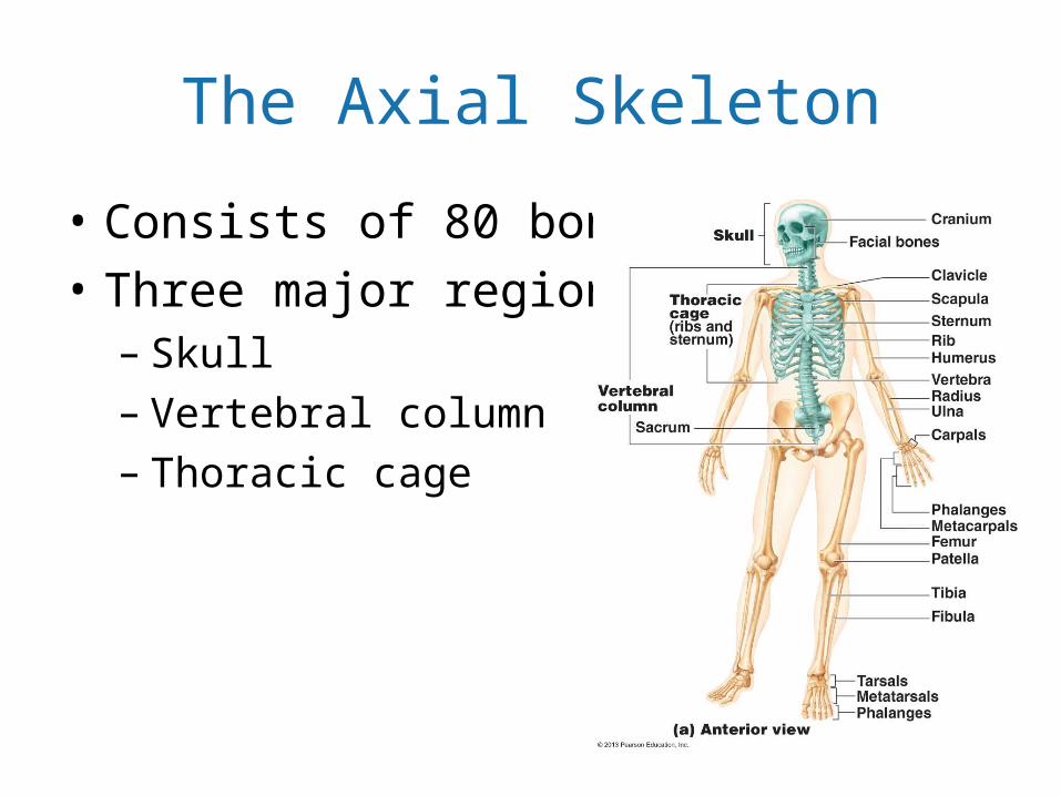
The Axial Skeleton
• Consists of 80 bones• Three major regions
– Skull– Vertebral column– Thoracic cage

The Skull• Formed by two sets of bones
1. Cranial bones (cranium)• Enclose the brain in the cranial cavity• Provide sites of attachment for head and neck
muscles
2. Facial bones• Framework of face• Cavities for special sense organs • Openings for air and food passage• Sites of attachment for teeth and muscles of
facial expression

© 2013 Pearson Education, Inc.
Figure 7.2a The skull: Cranial and facial divisions and fossae.
Bones of cranium
Coronal suture
Squamous suture
Lambdoidsuture
Facialbones
Cranial and facial divisions of the skull

Skull Geography
• Cranial cavity• Middle and internal
ear cavities• Nasal cavity• Orbits• 85 named openings
– Foramina, canals, fissures

Eight Cranial Bones
• Frontal bone• Parietal bones (2)• Occipital bone• Temporal bones (2)
– House middle ear ossicles
• Sphenoid bone– Keystone bone: articulates w/ all other cranial bones
• Ethmoid bone

Parietal Bones and Major Associated Sutures
1. Coronal suture—between parietal bones and frontal bone
2. Sagittal suture—between right and left parietal bones
3. Lambdoid suture—between parietal bones and occipital bone
4. Squamous (squamosal) sutures—between parietal and temporal bones on each side of skull
Four sutures: articulations of parietal bones w/frontal, occipital, and temporal bones:

Fourteen Facial Bones
• Mandible• Maxillary bones (maxillae)
(2)• Zygomatic bones (2)• Nasal bones (2)• Lacrimal bones (2)• Palatine bones (2)• Vomer• Inferior nasal conchae (2)

Orbits
• Cavities that encase eyes and lacrimal glands• Sites of attachment for eye muscles• Formed by parts of seven bones
– Frontal, sphenoid, zygomatic, maxilla, palatine, lacrimal, and ethmoid

Nasal Cavity• Roof, lateral walls, and floor
formed by parts of four bones– Ethmoid– Palatine bones– Maxillary bones– Inferior nasal conchae
• Nasal septum of bone and hyaline cartilage– Perpendicular plate of ethmoid – Vomer – Anterior septal cartilage

Paranasal Sinuses
• Mucosa-lined, air-filled spaces • Lighten skull • Enhance resonance of voice• Warm and humidify air• Found in frontal, sphenoid,
ethmoid, and maxillary bones

Hyoid Bone
• NOT a skull bone! • Does not directly articulate with another bone• Movable base for tongue• Site of attachment for muscles of swallowing
and speech

Vertebral Column
• Transmits weight of trunk to lower limbs• Surrounds and protects spinal cord• Flexible curved structure containing 26 irregular
bones (vertebrae) in five major regions– Cervical vertebrae (7)—vertebrae of neck– Thoracic vertebrae (12)—vertebrae of thoracic cage– Lumbar vertebrae (5)—vertebrae of lower back– Sacrum—bone inferior to lumbar vertebrae – Coccyx—terminus of vertebral column

Curvatures of Vertebral Column• Increase resilience and flexibility
of spine– Cervical and lumbar curvatures
• Concave posteriorly
– Thoracic and sacral curvatures• Convex posteriorly
• Abnormal spine curvatures– Scoliosis - abnormal lateral curve– Kyphosis (hunchback) –
exaggerated thoracic curvature– Lordosis (swayback) – accentuated
lumbar curvature

Ligaments• Anterior and
posterior longitudinal ligaments– From neck to sacrum
• Ligamentum flavum– Connects adjacent
vertebrae• Short ligaments
– Connect each vertebra to those above and below

Intervertebral Discs
• Cushionlike pad composed of two parts– Nucleus pulposus
• Inner gelatinous nucleus • Gives disc its elasticity and
compressibility
– Anulus fibrosus• Outer collar composed of collagen
and fibrocartilage

General Structure of Vertebrae
• Body or centrum– Anterior weight-bearing region
• Vertebral arch– Composed of pedicles and laminae that, along with
centrum, enclose vertebral foramen• Vertebral foramina
– Together make up vertebral canal for spinal cord • Intervertebral foramina
– Lateral openings between adjacent vertebrae for spinal nerves

General Structure of Vertebrae
• Seven processes per vertebra:– Spinous process
• projects posteriorly
– Transverse processes (2)• project laterally
– Superior articular processes (2)• protrude superiorly
– Inferior articular processes (2)• protrude inferiorly

Cervical Vertebrae• C1 to C7: smallest, lightest
vertebrae• C1 (atlas) and C2 (axis) have unique
features • C3 to C7 share following features
– Oval body– Spinous processes are bifid (except
C7)– Large, triangular vertebral foramen– Transverse foramen in each
transverse process– C7 is vertebra prominens

Cervical Vertebrae• Atlas (C1)
– No body or spinous process– Consists of anterior and posterior arches, and two lateral
masses– Superior surfaces of lateral masses articulate with occipital
condyles– Movement for "Yes"

Cervical Vertebrae
• Axis (C2)– Dens projects superiorly into anterior arch of atlas– Dens is a pivot for rotation of atlas – Movement for "No"

Thoracic Vertebrae
• T1 to T12
• All articulate with ribs • Long, spinous process that
points inferiorly• Circular vertebral foramen• Location of articular facets
allows rotation of this area of spine

Lumbar Vertebrae• L1 to L5
• Short, thick pedicles and laminae• Flat hatchet-shaped spinous
processes point posteriorly • Vertebral foramen triangular• Orientation of articular facets
locks lumbar vertebrae together to prevent rotation
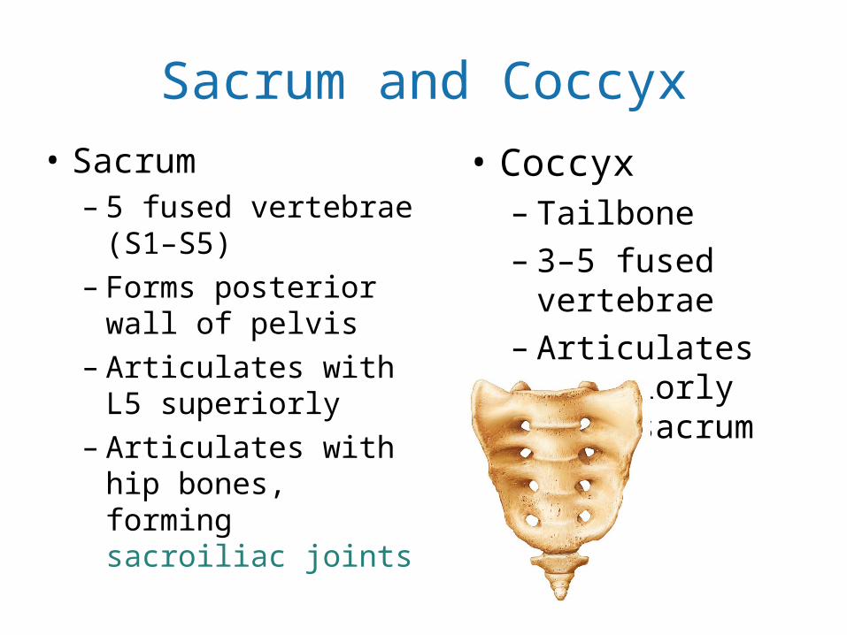
Sacrum and Coccyx• Sacrum
– 5 fused vertebrae (S1–S5)
– Forms posterior wall of pelvis
– Articulates with L5 superiorly
– Articulates with hip bones, forming sacroiliac joints
• Coccyx– Tailbone– 3–5 fused vertebrae– Articulates superiorly
with sacrum

Thoracic Cage• Composed of
– Thoracic vertebrae posteriorly – Sternum and costal cartilages anteriorly– Ribs laterally
• Functions– Protects vital organs of thoracic cavity– Supports shoulder girdles and upper limbs– Provides attachment sites for muscles of neck,
back, chest, and shoulders

Sternum (Breastbone)
• Three fused bones– Manubrium – Superior portion
• Articulates with clavicles and ribs 1 and 2
– Body (midportion)• Articulates with costal cartilages
of ribs 2 through 7
– Xiphoid process – Inferior end• Site of muscle attachment• Not ossified until ~age 40

Ribs and Their Attachments
• 12 pairs• Pairs 1 through 7
– True ribs (vertebrosternal)– Attach directly to sternum by individual costal cartilages
• Pairs 8 through12– False ribs– Pairs 8–10 (vertebrochondral ribs)
• Attach indirectly to sternum by joining costal cartilage of rib above – Pairs 11–12 (vertebral (floating) ribs)
• No attachment to sternum

Appendicular Skeleton
• Bones of limbs and their girdles– Pectoral girdle
• Attaches upper limbs to body trunk
– Pelvic girdle • Attaches lower limbs to body trunk

Pectoral Girdle (Shoulder Girdle)
• Clavicles and scapulae– Attach upper limbs to axial skeleton – Provide attachment sites for muscles that move
upper limbs

The Upper Limb
• 30 bones form skeletal framework of each upper limb– Arm
• Humerus– Forearm
• Radius and ulna– Hand
• 8 carpal bones in the wrist• 5 metacarpal bones in the palm• 14 phalanges in the fingers

Hand: Carpus, Metacarpus, and Phalanges
• Carpus (Wrist)– Eight bones in two rows
• Proximal row—lateral to medial– Scaphoid, lunate, triquetrum, and pisiform
• Distal row—lateral to medial– Trapezium, trapezoid, capitate, and hamate
• Metacarpus (Palm)– Five metacarpal bones (#1 to #5 from thumb to little
finger) form the palm

Hand Continued• Phalanges (Fingers)
– Fingers numbered 1–5 starting at thumb (pollex)– Digit #1 (Pollex) has 2 bones - no middle phalanx– Digits #2 – 5 have 3 bones—distal, middle, and proximal
phalanx

Pelvic (Hip) Girdle• Two hip bones (coxal bones or os coxae) and sacrum
– Attach lower limbs to axial skeleton with strong ligaments– Transmit weight of upper body to lower limbs– Support pelvic organs
• Less mobility but more stable than shoulder joint• Three fused bones form coxal bone
– Ilium, ischium, and pubis
• Bony pelvis formed by coxal bones, sacrum, and coccyx

Figure 7.30 Pelvis.
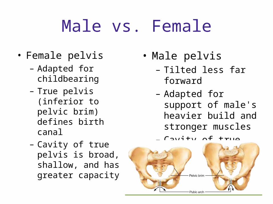
Male vs. Female
• Female pelvis– Adapted for childbearing– True pelvis (inferior to
pelvic brim) defines birth canal
– Cavity of true pelvis is broad, shallow, and has greater capacity
• Male pelvis– Tilted less far forward– Adapted for support of
male's heavier build and stronger muscles
– Cavity of true pelvis is narrow and deep

The Lower Limb
• Carries entire weight of erect body• Subjected to exceptional forces• Three segments of lower limb
– Thigh– Leg– Foot

Bones Of The Thigh
• Femur– Largest and strongest bone in the body– Length ~ ¼ of person's height– Articulates proximally with acetabulum of
hip and distally with tibia and patella
• Patella– Sesamoid bone in quadriceps tendon

Bones Of The Leg
• Tibia– Medial leg bone– Receives weight of body from femur;
transmits to foot
• Fibula– Not weight bearing; no articulation with
femur– Articulates proximally and distally with tibia
• Tibia and fibula connected by interosseous membrane

Foot: Tarsus, Metatarsus, Phalanges• Tarsus
– Seven tarsal bones form posterior half of foot– Body weight carried primarily by talus and
calcaneus– Other tarsal bones: cuboid, navicular, and medial,
intermediate, and lateral cuneiform bones• Metatarsals:
– Five metatarsal bones (#1 to #5 from hallux to little toe) – Enlarged head of metatarsal 1 forms "ball of the foot"

Foot Continued
• Phalanges– 14 bones of toes– Digit #1 (Hallux) has 2 bones
- no middle phalanx– Digits #2–5 have 3 bones—
distal, middle, and proximal phalanx

Arches Of The Foot
• Maintained by interlocking foot bones, ligaments, and tendons
• Allow foot to bear weight• Three arches
– Lateral longitudinal – Medial longitudinal – Transverse

Developmental Aspects: Fetal Skull
• Infant skull has more bones than adult skull– Skull bones such as mandible and
frontal bones are unfused – Skull bones connected by
fontanelles• Unossified remnants of fibrous
membranes• Ease birth and allow brain growth

Developmental Aspects: Growth Rates
• At birth, cranium huge relative to face• At 9 months, cranium is ½ adult size• Mandible and maxilla are shortened but lengthen
with age• Arms and legs grow at faster rate than head and
trunk, leading to adult proportions

Developmental Aspects: Spinal Curvature
• Primary thoracic and sacral curvatures obvious at birth– Give spine a C shape– Convex posteriorly
• Secondary curvatures– Cervical and lumbar—convex anteriorly– Appear as child develops

Developmental Aspects: Old Age
• Intervertebral discs thin, less hydrated, and less elastic
• Several centimeter height loss common by 55 • Costal cartilages ossify
– Rigid thorax causes shallow breathing and less efficient gas exchange
• All bones lose mass, so fracture risk increases