The Biology of Behavior Chapter 3 The Biology of Behavior Copyright 2007 Horizon Textbook Publishing...
71
The Biology of Behavior Chapter 3 The Biology of Behavior Copyright 2007 Horizon Textbook Publishing This multimedia product and its contents are protected under copyright law. The following are prohibited by law: •Any public performance or display, including transmission of any image over a network; •Preparation of any derivative work, including the extraction, in whole or in part, of any images; •Any rental, lease, or lending of the program Slide authors: Larry D. Thomas Landon O. Thomas Book authors: E.H. Ettinger
-
Upload
karlie-needler -
Category
Documents
-
view
218 -
download
0
Transcript of The Biology of Behavior Chapter 3 The Biology of Behavior Copyright 2007 Horizon Textbook Publishing...
The Biology of Behavior
Chapter 3
The Biology of Behavior
Copyright 2007 Horizon Textbook Publishing
This multimedia product and its contents are protected under copyright law. The following are prohibited by law:
•Any public performance or display, including transmission of any image over a network;
•Preparation of any derivative work, including the extraction, in whole or in part, of any images;
•Any rental, lease, or lending of the program
Slide authors:
Larry D. Thomas
Landon O. Thomas
Book authors:
E.H. Ettinger

Copyright © 2007 Horizon Textbook Publishing All Rights Reserved
Neurons and the Neurotransmitters
Neuron– A specialized cell that conducts impulses through
the nervous system and contains three major parts-a cell body, dendrites, and an axon
– Afferent neurons relay messages from the sense organs and receptors—eyes, ears, nose, mouth, and skin—to the brain or spinal cord
– Efferent neurons convey signals from the central nervous system to the glands and the muscles, enabling the body to move
– Interneurons carry information between neurons in the brain and between neurons in the spinal cord

Copyright © 2007 Horizon Textbook Publishing All Rights Reserved
Neurons and the Neurotransmitters
Anatomy of a Neuron– Cell body
The part of the neuron that contains the nucleus and carries out the neuron’s metabolic functions
– Dendrites The branchlike extensions of a neuron that receive signals
from other neurons– Axon
The slender, tail-like extension of the neuron that transmits signals to the dendrites or cell body of the other neurons or to muscles or glands
– Synapse The junction where the axon of a sending neuron
communicates with a receiving neuron across the synaptic cleft

Copyright © 2007 Horizon Textbook Publishing All Rights Reserved
Neurons and the Neurotransmitters
Anatomy of a Neuron– Glia Cells
Cells that help to make the brain more efficient by holding neurons together, removing waste products such as dead neurons, making the myelin coating for the axons, and performing other manufacturing, nourishing, and cleanup tasks
– Mylin Sheath Insulating covers around some axons that increases
neuron’s ability to conduct electrical impulses. Loss of mylin is Multiple Sclerosis
– Nodes of Ranvier Small gap or exposed portion of an axon between mylin
covers.

Copyright © 2007 Horizon Textbook Publishing All Rights Reserved
Neurons and the Neurotransmitters

Copyright © 2007 Horizon Textbook Publishing All Rights Reserved
Neurons and the Neurotransmitters
Sodium-Potassium Pump– The penetration and removal of sodium (Na+) and potassium
(K+) through the cell membrane allows for the transmission of the action potential down the axon of the neuron without electrical energy loss.
Permeability– The capability of being penetrated or passed through
Resting potential– The membrane potential of a neuron at rest, about -55 millivolts
Action potential– The sudden reversal of the resting potential, which initiates the
firing of a neuron– Complete process takes about 1 millisecond (1/1,000) of a
second

Copyright © 2007 Horizon Textbook Publishing All Rights Reserved
Neurons and the Neurotransmitters
Na+Na+ & K+
Cl- & K+

Copyright © 2007 Horizon Textbook Publishing All Rights Reserved
Neurons and the Neurotransmitters
Action of Neurotransmitters
– Excitatory Postsynaptic Potentials EPSPs Influencing surrounding neurons
to fire– Inhibitory Postsynaptic Potentials
IPSPs Influencing surrounding neurons
not to fire
Copyright © 2007 Horizon Textbook Publishing All Rights Reserved
Neurons and the Neurotransmitters
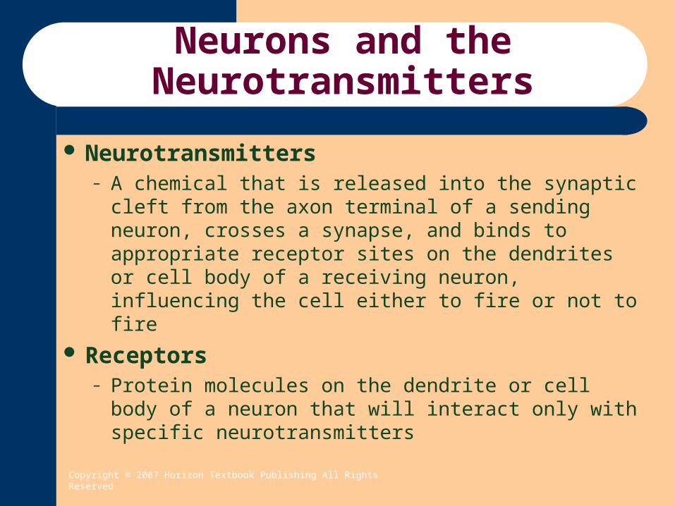
Neurotransmitters– A chemical that is released into the synaptic cleft
from the axon terminal of a sending neuron, crosses a synapse, and binds to appropriate receptor sites on the dendrites or cell body of a receiving neuron, influencing the cell either to fire or not to fire
Receptors– Protein molecules on the dendrite or cell body of a
neuron that will interact only with specific neurotransmitters
Copyright © 2007 Horizon Textbook Publishing All Rights Reserved
Neurons and the Neurotransmitters
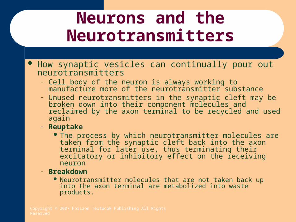
How synaptic vesicles can continually pour out neurotransmitters
– Cell body of the neuron is always working to manufacture more of the neurotransmitter substance
– Unused neurotransmitters in the synaptic cleft may be broken down into their component molecules and reclaimed by the axon terminal to be recycled and used again
– Reuptake The process by which neurotransmitter molecules are
taken from the synaptic cleft back into the axon terminal for later use, thus terminating their excitatory or inhibitory effect on the receiving neuron
– Breakdown Neurotransmitter molecules that are not taken back up into the
axon terminal are metabolized into waste products.

Copyright © 2007 Horizon Textbook Publishing All Rights Reserved
Neurons and the Neurotransmitters
Neurotransmitters called monoamines– Acetylcholine
Excitatory neurotransmitter in the brain but may be excitatory or inhibitory in organs of PNS. Involved in learning, movement and memory.
– Norepinephrine Excitatory neurotransmitter of reticular system and
involved in eating, emotional behavior, learning & memory
– Dopamine Inhibitory neurotransmitter involved in movement,
emotional behavior, attention, learning, memory and reward.
Copyright © 2007 Horizon Textbook Publishing All Rights Reserved
Neurons and the Neurotransmitters
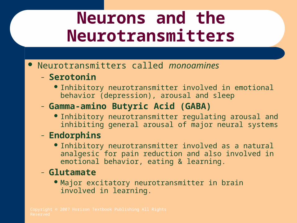
Neurotransmitters called monoamines– Serotonin
Inhibitory neurotransmitter involved in emotional behavior (depression), arousal and sleep
– Gamma-amino Butyric Acid (GABA) Inhibitory neurotransmitter regulating arousal and inhibiting
general arousal of major neural systems
– Endorphins Inhibitory neurotransmitter involved as a natural analgesic for
pain reduction and also involved in emotional behavior, eating & learning.
– Glutamate Major excitatory neurotransmitter in brain involved in learning.
Copyright © 2007 Horizon Textbook Publishing All Rights Reserved
Central Nervous System

Copyright © 2007 Horizon Textbook Publishing All Rights Reserved
Central Nervous System
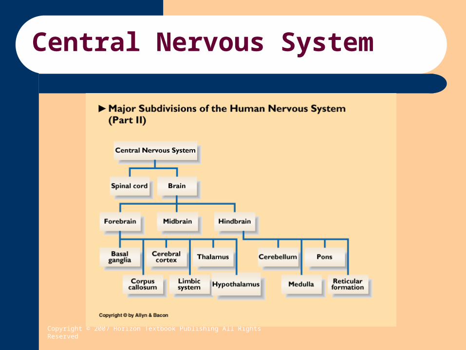
Two parts of a nervous system– Central Nervous System (CNS)
The brain and the spinal cord
– Peripheral Nervous System (PNS) Connects the central nervous system to all other parts of
the body
Copyright © 2007 Horizon Textbook Publishing All Rights Reserved
Central Nervous System
Spinal cord– An extension of the brain,reaching form the base of
the brain through the neck and spinal column, that transmits messages between the brain and the peripheral nervous system
– Protected by bone and spinal fluid– Spinal nerves are 31 matched pairs with one on the
right side of the spinal cord and it’s counter part on the left side of the spinal cord
– Basic reflexes (such as the quick withdrawal of the hand from a hot surface) is controlled by the spinal cord.

Copyright © 2007 Horizon Textbook Publishing All Rights Reserved
Spinal Cord Reflex
Copyright © 2007 Horizon Textbook Publishing All Rights Reserved
Central Nervous System
Brainstem– The structure that begins at the point where the
spinal cord enlarges as it enters the brain – Medulla
The part of the brainstem that controls heartbeat, blood pressure, breathing, coughing, sneezing and swallowing
– Cerebellum Coordinates and regulates motor movements and muscle
control necessary for posture– Pons
Brainstem structure just above the medulla does fine-tuning of motor messages, programming species-typical behaviors, processing sensory information and controlling respiration

Copyright © 2007 Horizon Textbook Publishing All Rights Reserved
Central Nervous System
Brainstem (continued)– Reticular Formation or Reticular Activating
System (RAS) Neural circuits extending from lower brain to thalamus that
play a critical role in arousal and alertness. Research suggest that Attention-Deficit Hyperactivity
Disorder (ADHD) results from insufficient arousal of the noradrenergic system.
– Stimulant medications as amphetamines and Ritalin facilitate norepinephrine and increase alertness helping ADHD individuals to stay more alert and thus pay attention.
Copyright © 2007 Horizon Textbook Publishing All Rights Reserved
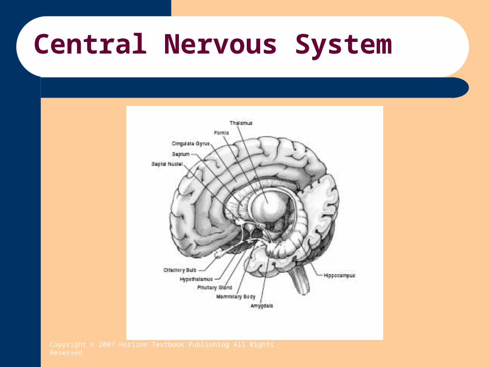
Central Nervous System
Limbic System– Amygdala
Expression of anger, rage, fear and aggressive behavior (intense uncomfortable emotions)
– Hippocampus Plays an important role in memory
– Septum Plays an important role in the experiencing of pleasure (intense
comfortable emotions)– Hypothalamus
Helps maintain the bodies homeostasis (sleep, hunger, thirst, body temperature, & sex drive), controls the pituitary gland which in turn controls the endocrine (hormone) system, and emotional expression
– Thalamus Relay station for routing sensory information to the cerebral cortex
and regulating sleep cycles (circadian rhythm)
Copyright © 2007 Horizon Textbook Publishing All Rights Reserved
Central Nervous System
Copyright © 2007 Horizon Textbook Publishing All Rights Reserved
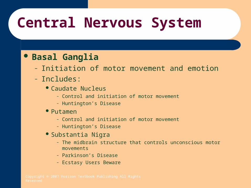
Central Nervous System
Basal Ganglia– Initiation of motor movement and emotion– Includes:
Caudate Nucleus– Control and initiation of motor movement– Huntington’s Disease
Putamen– Control and initiation of motor movement– Huntington’s Disease
Substantia Nigra– The midbrain structure that controls unconscious motor movements– Parkinson’s Disease– Ecstasy Users Beware

Copyright © 2007 Horizon Textbook Publishing All Rights Reserved
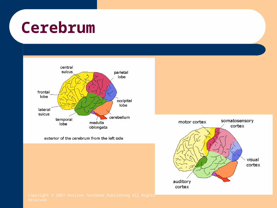
Cerebrum
Cerebrum– Largest structure of the human brain comprised of billions of
neurons– Consisting of the two cerebral hemispheres– Connected by the Corpus Callosum – Covered by the Cerebral Cortex– Gray in color because neurons are not covered with myelin on
the surface.– Inner core is white because neurons are not myelinated and
occur in three types: Commissural fibers - pass from one hemisphere to another Projection fibers – convey impulses to and from cortex Association fibers – connects various parts within a
hemisphere
Copyright © 2007 Horizon Textbook Publishing All Rights Reserved
Cerebrum
Copyright © 2007 Horizon Textbook Publishing All Rights Reserved
Cerebrum
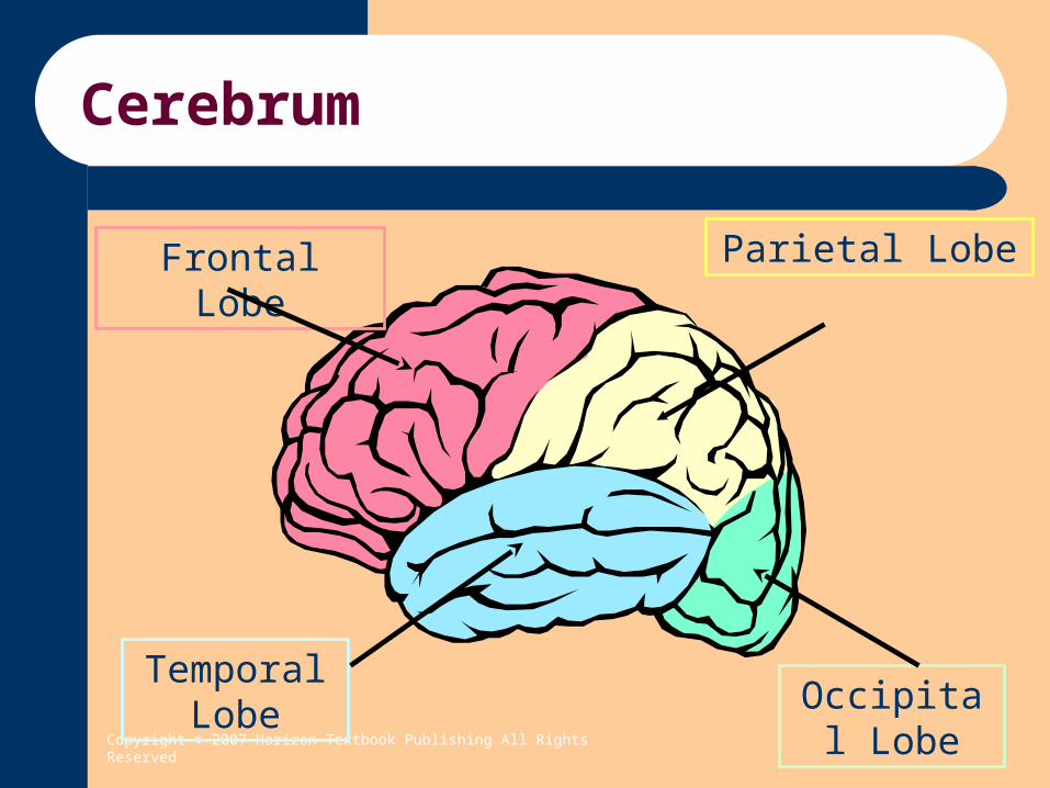
Frontal Lobe Parietal Lobe
Temporal Lobe Occipital
Lobe

Copyright © 2007 Horizon Textbook Publishing All Rights Reserved
Cerebrum
Motor Cortex Somatosensory Cortex
AuditionVisual
Copyright © 2007 Horizon Textbook Publishing All Rights Reserved
Somatosensory Man

Copyright © 2007 Horizon Textbook Publishing All Rights Reserved
Cerebrum
Sensory Cortex– Involved in receiving sensory messages– Primarily Parietal Lobes
Motor Cortex– Involved in transmitting messages to muscles for intentional
movement of body– Primarily Frontal Lobes
Association Cortex– Largest portion of brain (75%)– Integrating sensory and motor messages– Higher functions such as thinking, interpreting & remembering
Copyright © 2007 Horizon Textbook Publishing All Rights Reserved
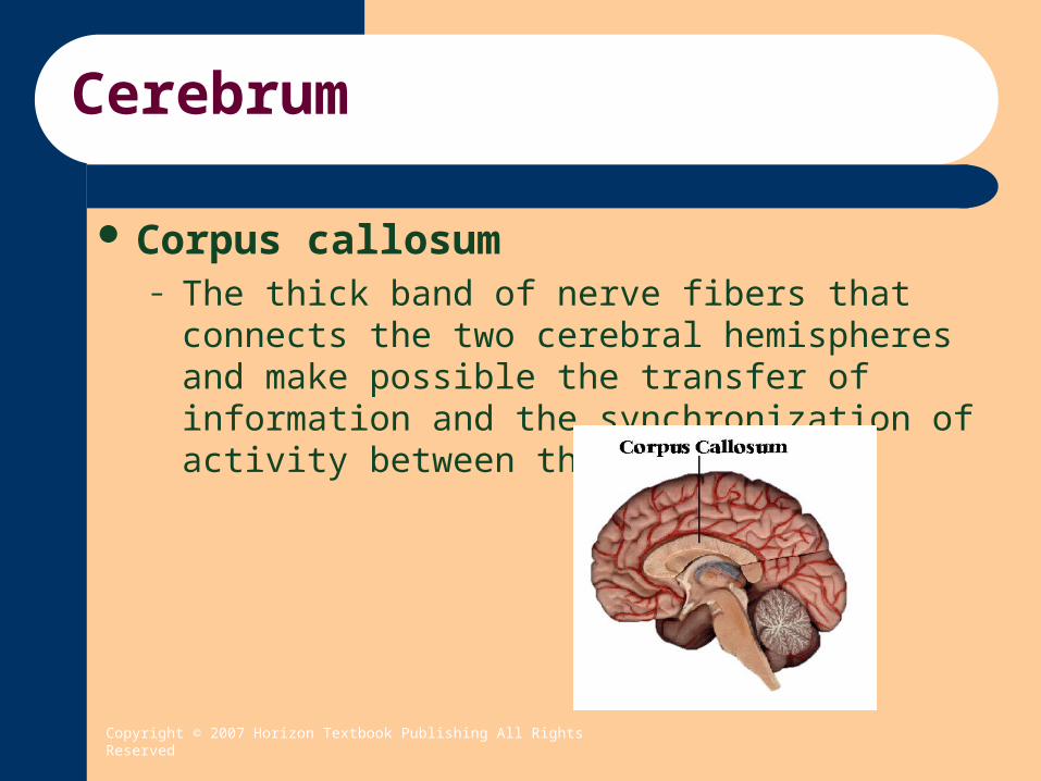
Cerebrum
Corpus callosum– The thick band of nerve fibers that connects the two
cerebral hemispheres and make possible the transfer of information and the synchronization of activity between them

Copyright © 2007 Horizon Textbook Publishing All Rights Reserved
Cerebrum
Frontal lobes– The lobes that control voluntary body movements,
speech production, and such functions as thinking, motivation, planning for the future, impulse control, and emotional responses
Motor cortex Broca’s area Frontal association areas
Copyright © 2007 Horizon Textbook Publishing All Rights Reserved
Cerebrum
Motor Cortex– The strip of tissue at the rear of the frontal lobes that
controls voluntary body movement– Discovered by Fritsch and Hitzig– Wilder Penfield
Applied electrical stimulation to the motor cortex of conscious human patients undergoing neurosurgery
Mapped the primary motor cortex in humans
– Plasticity is maintained throughout life The capacity of the brain to adapt to changes such as
brain damage
Copyright © 2007 Horizon Textbook Publishing All Rights Reserved
Cerebrum
Broca’s area– The area in the frontal lobe, usually in the left
hemisphere, that controls the production of speech sounds
– Paul Broca Among the first scientists to demonstrate the existence of
localized functions in the cerebral cortex and concluded that the site of damage was the part of the brain responsible for speech production
– Broca’s aphasia An impairment in the physical ability to produce speech
sounds, or in extreme cases an inability to speak at all
Copyright © 2007 Horizon Textbook Publishing All Rights Reserved
Cerebrum
Bronca’s area (continued)– Aphasia
A loss or impairment of the ability to understand or communicate through the written or spoken word, which results from damage to the brain
Frontal Association Areas– Consists of association areas involved in thinking,
motivation, planning for the future, impulse control, and emotional responses
Copyright © 2007 Horizon Textbook Publishing All Rights Reserved
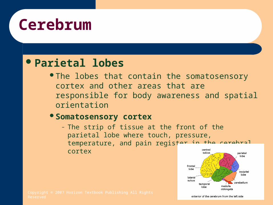
Cerebrum
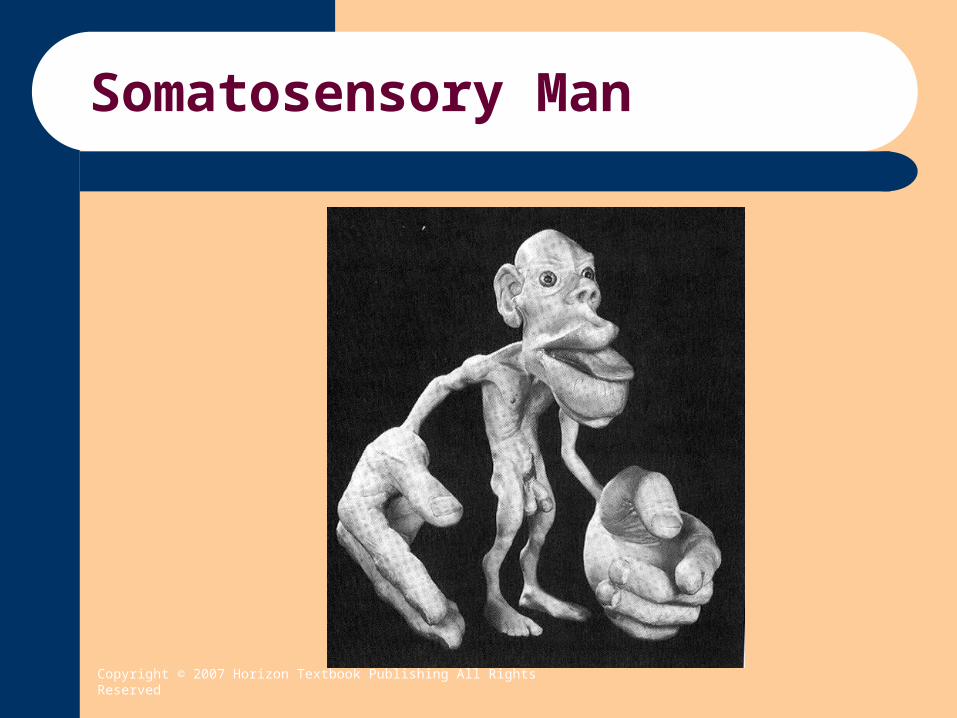
Parietal lobesThe lobes that contain the somatosensory cortex
and other areas that are responsible for body awareness and spatial orientation
Somatosensory cortex– The strip of tissue at the front of the parietal lobe where
touch, pressure, temperature, and pain register in the cerebral cortex

Copyright © 2007 Horizon Textbook Publishing All Rights Reserved
Cerebrum
Occipital lobes– The lobes that contain the primary visual cortex and
association areas involved in the interpretation of visual information
– Primary visual cortex The area at the rear of the occipital lobes where vision
registers in the cerebral cortex Each eye is connected the the primary visual cortex in both
right and left occipital lobes

Copyright © 2007 Horizon Textbook Publishing All Rights Reserved
Cerebrum
Temporal lobes– The lobes that contain the primary auditory cortex,
Wernicke’s area, and association areas for interpreting auditory information
– Primary auditory cortex The part of the temporal lobes where hearing registers in
the cerebral cortex
Copyright © 2007 Horizon Textbook Publishing All Rights Reserved
Cerebrum
Wernicke’s area– The language area in the temporal lobe involved in
comprehension of the spoken work and in formulation of coherent speech and written language
– Wernicke’s aphasia Aphasia that results from damage to Wernicke’s area and
in which the person’s spoken language is fluent, but the content is either vague or incomprehensible to the listener
– Another kind of aphasia is auditory aphasia Word deafness

Copyright © 2007 Horizon Textbook Publishing All Rights Reserved
Cerebrum
Temporal association areas– House memories and are involved in the
interpretation of auditory stimuli– There is a special association area where familiar
melodies are stored

Copyright © 2007 Horizon Textbook Publishing All Rights Reserved
Cerebral Hemispheres
Lateralization– The specialization of one of the cerebral hemispheres to
handle a particular function
Left hemisphere– The hemisphere that controls the right side of the body,
coordinates complex movements, and in 95% of right-handers and 62% of left-handers, controls most functions of speech and written language
Right hemisphere– The hemisphere that controls the left side of the body and that,
in most people, is specialized for visual-spatial perception and for interpreting nonverbal behavior

Copyright © 2007 Horizon Textbook Publishing All Rights Reserved
Cerebral Hemispheres

Copyright © 2007 Horizon Textbook Publishing All Rights Reserved
Cerebral Hemispheres
Right hemisphere’s role in emotion– The right hemisphere is involved in our expression
of emotion through tone of voice and facial expressions
– Controls the left side of the face and usually conveys stronger emotion than the right side of the face
– Lawrence Miller Describes the facial expressions and the voice inflection of
people with right hemisphere damage as “often strangely blank – almost robotic”

Copyright © 2007 Horizon Textbook Publishing All Rights Reserved
Cerebral Hemispheres
Right hemisphere’s roles

Copyright © 2007 Horizon Textbook Publishing All Rights Reserved
Cerebral Hemispheres
Left hemisphere’s roles
Copyright © 2007 Horizon Textbook Publishing All Rights Reserved
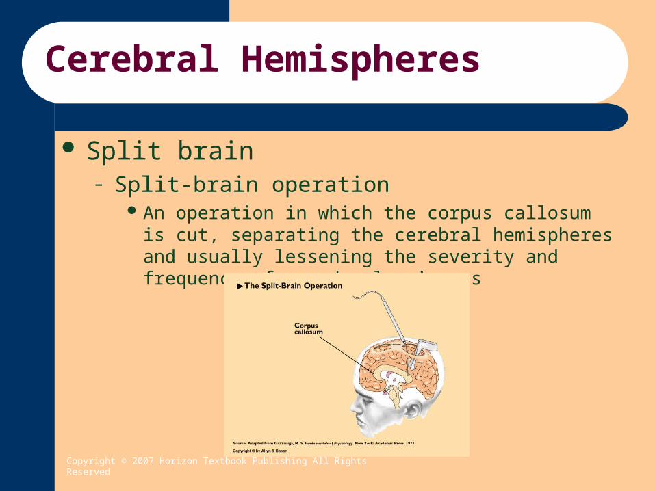
Cerebral Hemispheres
Split brain– Split-brain operation
An operation in which the corpus callosum is cut, separating the cerebral hemispheres and usually lessening the severity and frequency of grand mal seizures

Copyright © 2007 Horizon Textbook Publishing All Rights Reserved
Cerebral Hemispheres
Split brain (continued)– Joseph Bogen and Philip Vogel
Found that patients with severe epilepsy could be helped by surgery that severed their corpus callosum rendering communication between the two hemispheres impossible
– Roger Sperry and Michael Gazzaniga and Jerre Levy
Their research with split-brain patients has expanded knowledge of the unique capabilities of the individual hemispheres
– Roger Sperry Found that when the brain was surgically separated, each
hemisphere continued to have individual and private experiences, sensations, thoughts, and perceptions

Copyright © 2007 Horizon Textbook Publishing All Rights Reserved
Cerebral Hemispheres
Roger Sperry 1981 Nobel Prize research– The right hemisphere knows and remembers what it
sees just as well as the left, but unlike the left hemisphere, the right cannot name what it has seen

Copyright © 2007 Horizon Textbook Publishing All Rights Reserved
How the Brain is Studied
Hans Berger– Invented the electroencephalograph, a machine that
amplifies a million times the electrical activity occurring in the brain
Electroencephalogram– A record of brain-wave activity made by the
electroencephalograph
Copyright © 2007 Horizon Textbook Publishing All Rights Reserved
How the Brain is Studied
Beta wave– The brain wave associated with mental or physical
activity
Alpha wave– The brain wave associated with deep relaxation
Delta wave– The brain wave associated with slow-wave (deep)
sleep

Copyright © 2007 Horizon Textbook Publishing All Rights Reserved
How the Brain is Studied
Microelectrode– An electrical wire– So small that it can be inserted near or into a single
neuron without damaging it– Can be used to monitor the electrical activity of a
single neuron or to stimulate activity within it– Used to discover the exact functions of single cells
within the primary visual cortex and the primary auditory cortex

Copyright © 2007 Horizon Textbook Publishing All Rights Reserved
How the Brain is Studied
CAT scan (computerized axial tomography)– A brain-scanning technique involving a rotating X-
ray scanner and a high-speed computer analysis that produces slice-by-slice, cross-sectional images of the structure of the brain
MRI (magnetic resonance imagery)– A diagnostic scanning technique that produces high-
resolution images of the structures of the brain

Copyright © 2007 Horizon Textbook Publishing All Rights Reserved
How the Brain is Studied
PET scan (positron-emission tomography)– A diagnostic scanning technique that produces high-
resolution images of the structures of the brain
Functional MRI (fMRI)– A brain-imaging technique that reveals both brain
structure and brain activity

Copyright © 2007 Horizon Textbook Publishing All Rights Reserved
How the Brain is Studied
SQUID (superconducting quantum interference device)– Images brain activity by measuring magnetic
changes produced by the electric current neurons discharge when they fire
MEG (magnetoencephalography)– Measures magnetic changes produced by the
electrical activity from firing neurons and can also image neural activity within the brain as rapidly as it occurs, much faster than PET or fMRI

Copyright © 2007 Horizon Textbook Publishing All Rights Reserved
Brain Across the Lifespan
Synaptogenesis– Process where synapses develop as a result of
growth of both dendrites and axons– Pruning
The process through which the developing brain eliminates unnecessary or redundant synapses
Myelination– The development of myelin sheaths around axons,
begins prior to birth but continues well into adulthood
Copyright © 2007 Horizon Textbook Publishing All Rights Reserved
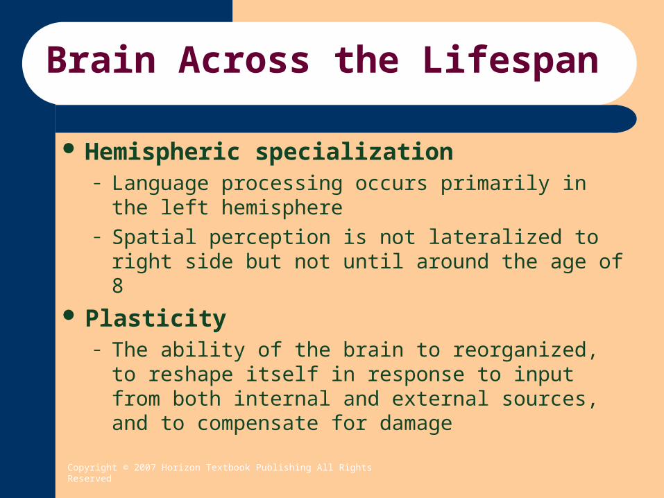
Brain Across the Lifespan
Hemispheric specialization– Language processing occurs primarily in the left
hemisphere– Spatial perception is not lateralized to right side but
not until around the age of 8
Plasticity– The ability of the brain to reorganized, to reshape
itself in response to input from both internal and external sources, and to compensate for damage

Copyright © 2007 Horizon Textbook Publishing All Rights Reserved
Brain Across the Lifespan
Stroke– The most common cause of damage to adult brains,
arising when blockage of an artery cuts off the blood supply to a particular area of the brain or when a blood vessel bursts
– A high percentage of stroke survivors suffer from depression
– Patients who receive TPA (a blood clot-dissolving drug) within 3 hours of the onset of a stroke are 30% more likely to have minimal or no disability

Copyright © 2007 Horizon Textbook Publishing All Rights Reserved
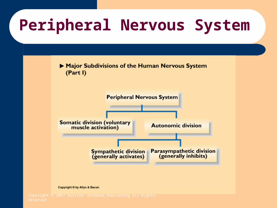
Peripheral Nervous System
Peripheral Nervous System (PNS)– The nerves connecting the central nervous system
to the rest of the body– Contains two subdivisions
Somatic Division Autonomic Division – contains two divisions
– Sympathetic Division (Activates)– Parasympathetic Division (Inhibits)
Copyright © 2007 Horizon Textbook Publishing All Rights Reserved
Peripheral Nervous System

Copyright © 2007 Horizon Textbook Publishing All Rights Reserved
Peripheral Nervous System
Somatic Division– Consists of all the sensory nerves, which transmit
information from the sense receptors-eyes, ears, nose, tongue, and skin-to the central nervous system
– Consists of all the motor nerves, which relay messages from the central nervous system to all the skeletal muscles of the body

Copyright © 2007 Horizon Textbook Publishing All Rights Reserved
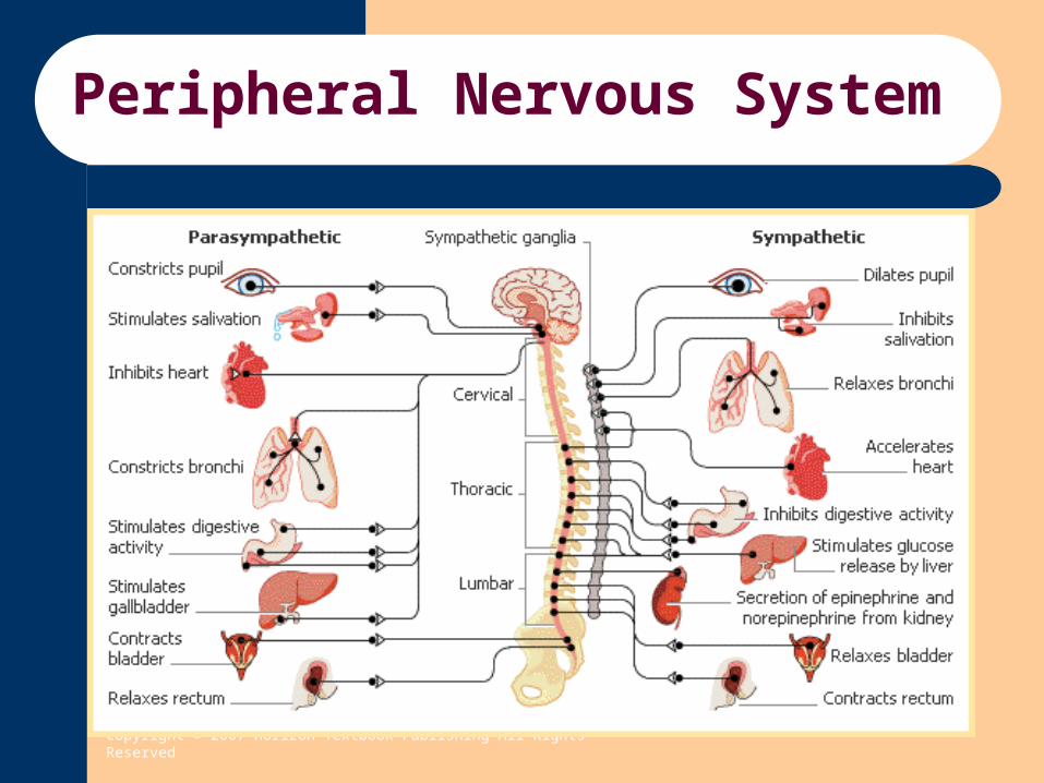
Peripheral Nervous System
Autonomic Division– Operates without any conscious control or
awareness on your part– Transmits messages between the central nervous
system and the glands, the cardiac muscle, and the smooth muscles

Copyright © 2007 Horizon Textbook Publishing All Rights Reserved
Peripheral Nervous System
Autonomic Division (continued)– Divided into two parts
Sympathetic nervous system– Mobilizes the body’s resources during stress,
emergencies, or heavy exertion, preparing the body for action
– Named the fight-or-flight response by Walter Cannon Parasympathetic nervous system
– Associated with relaxation and the conservation of energy. The division that brings the heightened bodily responses back to normal following an emergency.
Copyright © 2007 Horizon Textbook Publishing All Rights Reserved
Peripheral Nervous System

Copyright © 2007 Horizon Textbook Publishing All Rights Reserved
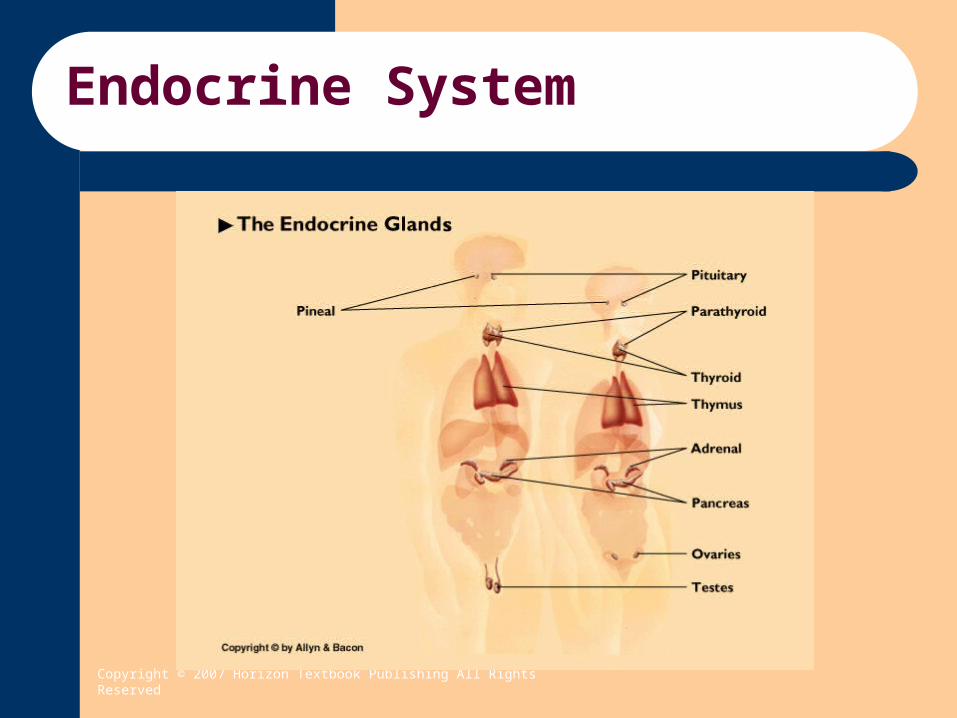
Endocrine System
Endocrine system– A system of ductless glands in various parts of the
body that manufacture and secrete hormones into the bloodstream or lymph fluids, thus affecting cells in other parts of the body
– Hormones A substance manufactured and released in one part of the
body that affects other parts of the body Chemical messengers of the endocrine system
Copyright © 2007 Horizon Textbook Publishing All Rights Reserved
Endocrine System

Copyright © 2007 Horizon Textbook Publishing All Rights Reserved
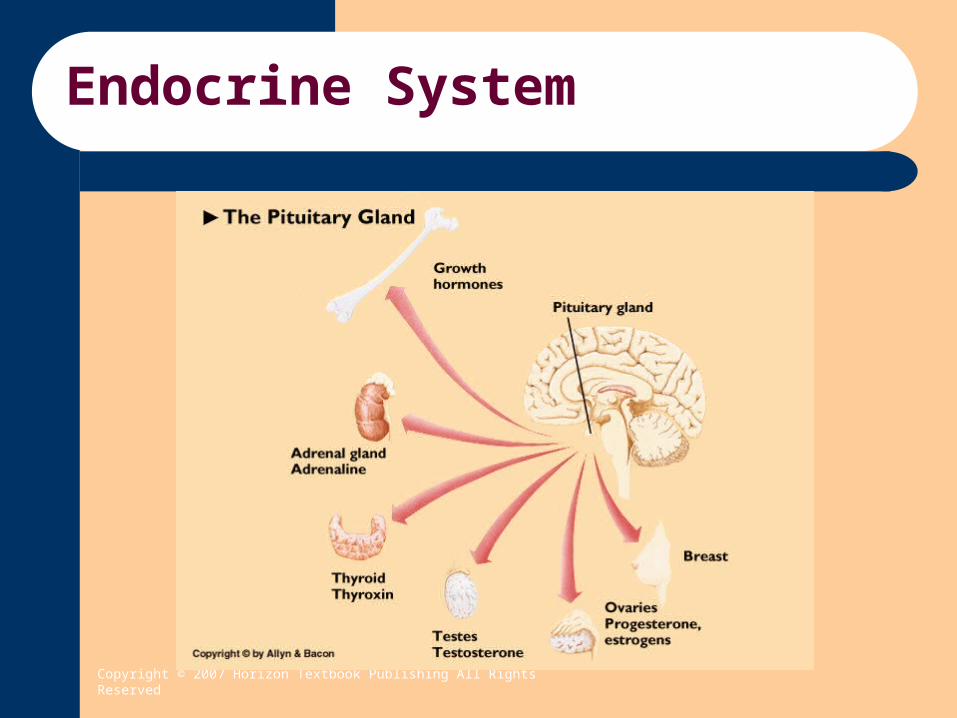
Endocrine System
Pituitary gland– The endocrine gland located in the brain and often
called the “master gland”, which releases hormones that control other endocrine glands and also releases a growth hormone
– Releases the hormones that activate the other glands in the endocrine system
– Brain controls activity of the pituitary gland through the production of a group of chemicals known as the hypothalamic-releasing factors
– Hormones exert primary influence over a single organ or specific cells which are referred to as target organs.
Copyright © 2007 Horizon Textbook Publishing All Rights Reserved
Endocrine System

Copyright © 2007 Horizon Textbook Publishing All Rights Reserved
Endocrine System
Thyroid gland– Produces the important hormone thyroxine, which
regulates the rate at which food is metabolized Pancreas
– Regulates the body’s blood sugar levels by releasing the hormones insulin and glucagon into the bloodstream
Adrenal glands– A pair of endocrine glands that release hormones
that prepare the body for emergencies and stressful situations and also release small amounts of the sex hormones
Copyright © 2007 Horizon Textbook Publishing All Rights Reserved
Endocrine System
Gonads– Sex glands –the ovaries in females and the testes in
males– Release the sex hormones that make reproduction
possible and that are responsible for the secondary sex characteristics
Copyright © 2007 Horizon Textbook Publishing All Rights Reserved
Depressants & Behavior
Sedatives– Class includes tranquilizers, barbituates & non-barbituates– Induce relaxation, calmness and sleep
Opiates (Narcotics)– Includes opium, morphine, codeine & heroine– Widely used as pain killers
Alcohol– Dampens impulse control (less inhibited)– Withdrawal (nausea, vomiting, fever, shakes [delirium
tremens] & bizzare hallucinations– 0.10% blood alcohol level is 5 times more likely to have a car
accident– Loss of memory, Fetal Alcohol Syndrome

Copyright © 2007 Horizon Textbook Publishing All Rights Reserved
Stimulants & Behavior
Caffeine– Most widely used stimulant– Chocolate, tea, coffee– Heart and respiration rates and blood pressure increase– Blocks adenosine receptors
Adenosine is an inhibitory neurotransmitter that produces neural sedation and regulates the dilation of blood vessels (Julian, 2001)
Nicotine– Second most widely used stimulant– Tobacco products– Increases heart rate, blood pressure and stomach activity while
constricting blood vessels– Withdrawal causes craving for tobacco, increased appetite, stomach
cramps, headaches, restlessness, irritability, insomnia, anxiety and depression
– Smoking causes higher incidents of miscarriages, still births, low birth-weight babies and babies who die from Sudden Infant Death Syndrome (SIDS) [Zotti, 2003]

Copyright © 2007 Horizon Textbook Publishing All Rights Reserved
Stimulants & Behavior
Amphetamines– Benzedrine, Dexedrine, Methedrine, Methamphetamine (street drug),– Dramatically increase alertness and activity and counteract fatigue
and promote feelings of euphoria and well-being– Likely works on norepinephrine and dopamine by increasing the
amount released from the nerve terminal and blocking reuptake Cocaine
– Made from the leaves of coca shrub and was active ingredient in Coca Cola until 1903
– Increases heart rate & respiration rates, constricts blood vessels and dilates the pupils
– High only last 20-30 minutes so reuse must occur quickly– Perhaps most addictive drug we know– Heart and lung damage are common as well as anemia, damage to
the nasal tissue, immune system impairment and rare cases of sudden death.

Copyright © 2007 Horizon Textbook Publishing All Rights Reserved
Hallucinogens & Behavior
LSD (Lysergic acid diethylamide)– Derived from a fungus that grows on rye grass– Profound distortions of sensations, feelings, time & thought– Suppresses the activity of Serotonin thus dream activity becomes activated
without restraint Psilocybin (Mushrooms)
– Suppresses the activity of Serotonin Mescaline (Peyote Cactus)
– Effects the way the brain responds to Norepinephrine & Acetylcholine Ecstasy (MDMA)
– Body and visual distortions– Depersonalization– Releases large amounts of Serotonin and interferes with synthesis of Serotonin– Hyperthermia, rapid heart rate, high blood pressure, muscle rigidity and
convulsions– Appears to irreversibly destroy serotonin-containing neurons– Parkinson’s Disorder Syndrome
Copyright © 2007 Horizon Textbook Publishing All Rights Reserved
Hallucinogens & Behavior
Marijuana– Derived from hemp plant Cannibis Sativa– THC (delta9-tetrahydrocannabinol)– Hallucinogen at high levels but usually a stimulant and depressant
effect– Increased heart rate & enhanced appetite– Small doses create euphoria and enhance some sensory experiences– Impairs reaction time and ability to concentrate– Some people become confused, agitated or extremely anxious– Has proved effective in epilepsy and glaucoma and reduces nausea
during chemotherapy treatment of cancer patients Anadamide
– Devine et al., 1992 Brains natural THC Regulates mood, pain, movement and appetite