The Biological Basis of Behavior - fratstock.eu · The Biological Basis of Behavior CHAPTER OUTLINE...
26
Full file at https://FratStock.eu Chapter 2 The Biological Basis of Behavior CHAPTER OUTLINE I. Enduring Issues in the Biological Basis of Behavior II. Neurons: The Messengers A The Neural Impulse B. The Synapse C. Neural Plasticity and Neurogenesis III. The Central Nervous System A. The Organization of the Nervous System B. The Brain C. Hemispheric Specialization D. Tools for Studying the Brain E. The Spinal Cord IV. The Peripheral Nervous System V. The Endocrine System VI. Genes, Evolution, and Behavior A. Genetics B. Behavior Genetics C. Social Implications D. Evolutionary Psychology
Transcript of The Biological Basis of Behavior - fratstock.eu · The Biological Basis of Behavior CHAPTER OUTLINE...

Full file at https://FratStock.eu
Chapter 2
The Biological Basis of Behavior
CHAPTER OUTLINE
I. Enduring Issues in the Biological Basis of Behavior
II. Neurons: The Messengers
A The Neural Impulse
B. The Synapse
C. Neural Plasticity and Neurogenesis
III. The Central Nervous System
A. The Organization of the Nervous System
B. The Brain
C. Hemispheric Specialization
D. Tools for Studying the Brain
E. The Spinal Cord
IV. The Peripheral Nervous System
V. The Endocrine System
VI. Genes, Evolution, and Behavior
A. Genetics
B. Behavior Genetics
C. Social Implications
D. Evolutionary Psychology

Full file at https://FratStock.eu27 Chapter 2 The Biological Basis of Behavior
LEARNING OBJECTIVES
After reading this chapter, students should be able to:
1. Define the terms neuroscience and psychobiology.
2. Draw and describe all the components of a typical myelinated neuron. Trace the path of a neural
impulse and explain what happens when the impulse reaches the terminal button.
3. Discuss the importance of glial cells.
4. Demonstrate and discuss both the resting membrane potential and the action potential.
5. Illustrate and describe the synapse. Identify the roles of various neurotransmitters and receptors,
and explain how drugs can alter synapse function.
6. Discuss the importance of neural plasticity.
7. Diagram, label, and state the functions of the various parts of the inner right hemisphere.
8. Diagram, label, and state the functions of each component of the cerebral cortex.
9. Locate and state the various components of the limbic system.
10. Devise a split-brain experiment.
11. Compare and contrast the various tools available for studying the nervous system.
12. Trace a spinal cord reflex.
13. Compare and contrast the sympathetic and parasympathetic nervous systems.
14. Describe the functions of the endocrine system. Explain how hormones released by the endocrine
system affect metabolism, blood-sugar level, sex characteristics, and the body’s reaction of stress.
15. Explain the relationship between chromosomes, genes, and DNA.
16. Distinguish between genotype and phenotype.
17. Outline the methods used by human behavioral geneticists.
18. Explain the concepts of dominant and recessive genes.
19. Discuss the social implications of behavior genetics.
20. Report how natural selection may influence human social behavior.
LECTURE SUGGESTIONS AND DISCUSSION TOPICS
Brain Metaphors
Metaphors are powerful tools in psychology, because they help us to understand systems that aren’t directly
observable through reference to things that are more familiar and perhaps better understood (Weiner,
1991). Our understanding of the human brain and its activity has been helped through a reliance on
metaphor. The metaphors used, however, have changed over time.
• Hydraulic models. Thinkers such as Galen and Descartes described the brain as a
pneumatic/hydraulic system, relying on the “new-fangled” plumbing systems dominant during
their lifetimes. Galen, for example, believed that the liver generated “spirits” or gases that flowed
to the brain, where they then formed “animal spirits” that flowed throughout the nervous system.
Descartes expanded on this view, adding that the pineal gland (the supposed seat of the soul) acted
on the animal spirits to direct reasoning and other behaviors. In short, the brain was a septic tank,
storing, mixing, and directing the flow of spirit gases throughout the body for the purposes of
behavior and action.
• Mechanical and telephone models. With the advent of new technology came new metaphors for
the brain. During the Industrial Revolution machine metaphors dominated, and in particular the
brain was conceived as a complex mechanical apparatus involving (metaphorical) levers, gears,
trip hammers, and pulleys. During the 1920s, the brain developed into a slightly more
sophisticated machine resembling a switchboard; the new technology of the telephone provided a
new metaphor. Inputs, patch cords, outputs, and busy signals (though no “call waiting”) dominated
explanations of brain activity. This metaphor, however, faltered by viewing the brain as a system

Full file at https://FratStock.euChapter 2 The Biological Basis of Behavior 28
that shut down periodically, as when no one was dialing a number. We now know, of course, that
the brain is continually active.
• Computer models. Current metaphors for the brain rely on computer technology. Input, output,
memory, storage, information processing, and circuitry are all terms that seem equally suited to
talking about computer chips or neurons. Although perhaps a better metaphor than plumbing or
telephones, the computer model still has its shortcomings. As a descriptive device, however, this
metaphor can at least suggest limits in our understanding and point the way to profitable areas of
research.
McGuigan, F. J. (1994). Biological psychology: A cybernetic science. Englewood Cliffs, NJ: Prentice
Hall.
Weiner, B. (1991). Metaphors in motivation and attribution. American Psychologist, 46, 921–930.
The Cranial Nerves
The textbook discusses various divisions of the nervous system. You may want to add a description of the
cranial nerves to your outline of the nervous system. Although the function of the cranial nerves is not
different from that of the sensory and motor nerves in the spinal cord, they do not enter and leave the brain
through the spinal cord. There are twelve cranial nerves, numbered 1 to 12 and ordered from the front to the
back of the brain, that primarily transmit sensory information and control motor movements of the face and
head. The twelve cranial nerves are:
1. Olfactory. A sensory nerve that transmits odor information from the olfactory receptors to the
brain.
2. Optic. A sensory nerve that transmits information from the retina to the brain.
3. Oculomotor. A motor nerve that controls eye movements, the iris (and therefore pupil size), lens
accommodation, and tear production.
4. Trochlear. A motor nerve that is also involved in controlling eye movements.
5. Trigeminal. A sensory and motor nerve that conveys somatosensory information from receptors in
the face and head and controls muscles involved in chewing.
6. Abducens. Another motor nerve involved in controlling eye movements.
7. Facial. Conveys sensory information and controls motor and parasympathetic functions associated
with facial muscles, taste, and the salivary glands.
8. Auditory-vestibular. A sensory nerve with two branches, one of which transmits information from
the auditory receptors in the cochlea and the other conveys information concerning balance from
the vestibular receptors in the inner ear.
9. Glossopharyngeal. This nerve conveys sensory information and controls motor and
parasympathetic functions associated with the taste receptors, throat muscles, and salivary glands.
10. Vagus. Primarily transmits sensory information and controls autonomic functions of the internal
organs in the thoracic and abdominal cavities.
11. Spinal accessory. A motor nerve that controls head and neck muscles.
12. Hypoglossal. A motor nerve that controls tongue and neck muscles.
Carlson, N. R. (1994). Physiology of behavior (5th ed.). Boston: Allyn and Bacon.
Thompson, R. F. (1993). The brain: A neuroscience primer (2nd ed.). New York: W. H. Freeman.
Reprinted from Hill, W. G. (1995). Instructor’s resource manual for Psychology by S. F. Davis and J. J.
Palladino. Englewood Cliffs, NJ: Prentice Hall.

Full file at https://FratStock.eu29 Chapter 2 The Biological Basis of Behavior
Would You Like Fries with That Peptide?
Toast and juice for breakfast. Pasta salad for lunch. An orange, rather than a bagel, for an afternoon snack.
These sound like reasonable dietary choices, involving some amount of deliberation and free will.
However, our craving for certain foods at certain times of the day may be more a product of the brain than
of the mind.
Sarah F. Leibowitz, Rockefeller University, has been studying food preferences for more than a decade.
What she has learned is that a stew of neurochemicals in the paraventricular nucleus, housed in the
hypothalamus, plays a crucial role in helping to determine what we eat and when. Two in particular—
Neuropeptide Y and galanin—help guide the brain’s craving for carbohydrates and for fat.
Here’s how they work. Neuropeptide Y (NPY) is responsible for turning on and off our desire for
carbohydrates. Animal studies have shown a striking correlation between NPY and carbohydrate intake; the
more NPY produced, the more carbohydrates eaten, both in terms of meal size and duration. Earlier in the
sequence, the stress hormone cortisol seems responsible, along with other factors, for upping the production
of Neuropeptide Y. This stress —> cortisol —> Neuropeptide Y —> carbohydrate craving sequence may
help explain overweight due to high carbohydrate intake. But weight, and craving, rely on fat intake as
well. Leibowitz has found that the neuropeptide galanin plays a critical role in this case. Galanin is the
on/off switch for fat craving, correlating positively with fat intake; the more galanin produced, the heavier
an animal will become. Galanin also triggers other hormones to process the fat consumed into stored fat.
Galanin itself is triggered by metabolic cues resulting not only from burning fat as energy, but also from
another source: estrogen.
Neuropeptide Y triggers a craving for carbohydrate, and galanin triggers a craving for fat, but the two
march to different drummers throughout a day’s cycle. Neuropeptide Y has its greatest effects in the
morning (at the start of the feeding cycle), after food deprivation (such as dieting), and during periods of
stress. Galanin, by contrast, tends to increase after lunch and peaks toward the end of our daily feeding
cycle.
The implications of this research are many. For example, the findings suggest that America’s obsession
with dieting is a losing proposition (but not around the waistline). Skipping meals, gulping appetite
suppressers, or experiencing the stress of dieting will trigger Neuropeptide Y to encourage carbohydrate
consumption, which in turn can foster overeating. Paradoxically, then, by trying to fight nature we may
stimulate it even more. As another example, the onset and maintenance of anorexia may be tied to the
chemical cravings in the hypothalamus. Anorexia tends to develop during puberty, a time when estrogen is
helping to trigger galanin’s craving for fat consumption. Some women (due to societal demands, obsessive-
compulsive tendencies, or other pressures) react to this fat trigger by trying to accomplish just the opposite:
subsisting on very small, frequent, carbohydrate-rich meals. The problem is that the stress and starvation
produced by this diet cause Neuropeptide Y to be released, confining dietary interest to carbohydrates, but
also affecting the sex centers nearby in the hypothalamus. Specifically, Neuropeptide Y may act to shut
down production of gonadal hormones.
Marano, H. E. (1993, January/February). Chemistry and craving. Psychology Today, pp. 30–36, 74.
Women, Men, and PETs
The 1990s were dubbed “the decade of the brain,” and it is true that remarkable advances were made by the
neurosciences in discovering how the brain operates. Several studies suggest that the operation of men’s
and women’s brains may differ in significant ways.
For example, Ruben Gur and his colleagues at the University of Pennsylvania recorded PET scans of men
and women who were asked to think of nothing in particular. That is, the research participants were
instructed to relax and let their brains idle as they exerted as little mental effort as possible. The researchers
found that for most participants the task was difficult to complete: PET scans revealed that these idle minds

Full file at https://FratStock.euChapter 2 The Biological Basis of Behavior 30
nonetheless hummed with activity. The locus of that activity, however, differed across the sexes. Men’s
brains often showed activity in the limbic system, whereas women’s often showed activity in the posterior
cyngulate gyrus. The meaning of these differences is difficult to interpret; the difficulty is compounded by
the 13 men and 4 women who showed patterns of activity characteristic of their opposite-sex peers. As an
early peek into the brain, however, they hint that the centers of activity for “blank” brains differ for women
and men.
In a separate study, researchers at the University of California, Irvine, asked 22 men and 22 women to solve
SAT math problems while undergoing a PET scan. Half of each group had SAT math scores above 700,
whereas the other half had scores below 540. The temporal lobes of the 700+ men showed heightened
activity during the math task, although this was not true for the women: the 700+ women’s temporal lobes
were no more intensively used than those of the 540-group women. Richard Haier, who helped lead the
study, speculates that women in the top group might be using their brains more efficiently than women in
the average-scoring group. More generally, although both men and women did well at the task, their brains
were operating differently to accomplish it.
Ruben and Raquel Gur also studied men’s and women’s brains in response to emotional expressions.
Shown pictures of either happy or sad faces, both men and women were quite adept at spotting happiness.
Women, however, could identify sadness about 90% of the time, regardless of whether it was on the face of
a man or a woman. By comparison, men were accurate in spotting sadness 90% of the time on a man’s
face, but only 70% of the time if the expression was posed by a woman. Once again, PET scans revealed
that women’s brains didn’t have to work as hard at this task as did men’s; in fact, women’s limbic systems
were less active than the limbic systems of the poor-scoring men.
There are a number of other differences between women’s and men’s brains. Women tend to have a larger
corpus callosum than men, for example. Women may also have a higher concentration of neurons in their
cortexes than men. But the meaning behind these differences is a matter far from decided.
Begley, S. (1995, March 27). Gray matters. Newsweek, pp. 48–54.
“Would You Like a Smoking or Non-Smoking Brain?...”
Most people who have tried to quit smoking report that it is at best a hit-or-miss proposition: a few days off
the coffin nails, and the body dearly longs for that wispy blue smoke. Despite claims made by the tobacco
industry, the culprit seems to be nicotine, an addictive substance that produces craving and withdrawal.
Evidence from the Brookhaven National Laboratory suggests that nicotine alone may not be responsible for
addiction to smoking. Rather, the action of certain enzymes in the brain may also contribute to the
pleasures of cigarettes.
All addictive substances—cocaine, heroin, cigarettes—cause an increase in dopamine (a pleasure-
enhancing neurotransmitter). Dr. Joanna Fowler led a research team that studied live images of smokers’
and nonsmokers’ brains. They found that an enzyme called monoamine oxidase B, or MAO B for short,
was 40% less active in smokers. MAO B is responsible for breaking down dopamine. Therefore, when
MAO B is inhibited, dopamine levels continue to remain high, which in turn allows smoking to remain
pleasurable. The trick now is to find the ingredient in tobacco smoke that inhibits MAO B.
Oddly enough, these same findings may account for the fact that smokers are about half as likely as
nonsmokers to develop Parkinson’s disease, which is characterized by decreased dopamine levels. If
smokers’ dopamine remains high it may contribute to staving off the onset of Parkinson’s. Although this
observation is hardly a reason to start smoking, it at least suggests that drugs that are effective in treating
Parkinson’s disease might be adapted for use in treating smoking addiction.
Leary, W. E. (1996, February 22). Brain enzyme linked to smoking addiction. Austin American-
Statesman, A5.

Full file at https://FratStock.eu31 Chapter 2 The Biological Basis of Behavior
Understanding Hemisphere Function
It seems clear that parts of the brain are specialized to perform different functions. To some extent this is an
old idea, dating back to the time of phrenology. Phrenologists believed that specific functions were
localized to specific parts of the brain, and they developed elaborate maps showing the location of
functions. Most importantly, they believed that well-developed functions were indicated by bumps in the
skull, which could be palpated by the skilled phrenologist.
After phrenology was discredited, there was a great deal of doubt as to whether functions were localized in
the brain. Much of the research in the early part of the 19th century seemed to suggest that the brain was
diffusely organized and that specific functions were not localized. In fact, Paul Broca’s (1861) description
of a speech center localized in the frontal lobe on the left side of the brain is one of the first pieces of
evidence for localization of a specific function in the brain. Broca based his localization of the speech area
on his observations of a single individual who had lost his ability to speak many years earlier, while
retaining the ability to understand what was said to him. At autopsy, Broca noted the area of most severe
and enduring brain damage, and concluded that this part of the brain was responsible for the ability to
speak.
At about the same time, two Prussian surgeons, Fritsch and Hitzig, were able to demonstrate that there is a
specific area of the brain that controls movement (now called the motor cortex). Whereas Broca used the
lesion method to arrive at his localization of what we now call a motor speech area, Fritsch and Hitzig used
a primitive version of electrical brain stimulation to delineate areas of “excitable cortex,” which meant
cortex that triggered movements when stimulated. Fritsch and Hitzig used “galvanic stimulation” to
delineate the extent of the motor cortex in dogs, and even did some electrical brain stimulation patients on
dying soldiers with open head wounds during one of the Prussian Wars. They demonstrated that the motor
cortex is confined to a specific strip of cortex in the back part of the frontal lobes in humans.
Many studies since then have confirmed that functions are localized to specific parts of the brain. Although
present-day methods such as PET scan or fMRI have helped to refine our understanding of functional
localization in the brain, much of our understanding of functional localization was arrived at through
studies in the early twentieth century, mainly using the lesion method. The two “great wars” produced a
large number of young men with brain injuries (mainly from gunshots). Researchers who looked at the
relationships between the location of the injuries and the functions that were lost were able to map the
localization of most of the major functions in the brain, including all of the major sensory and motor areas.
Thus, although neuroscientists continue to refine our understanding of the ways that the brain carries out
and integrates its multiple functions, the basic map of functions in the brain has been available for more
than half a century.
Broca, P. (1861). Perte de la parole, ramollissement chronique et destruction partielle du lobe antérieur
gauche du cerveau. Bulletin de la Société Anthropologique, 2, 235–238. (This paper has been translated
into English by Christopher Green and is published in its entirety on Green’s Classics in the History of
Psychology Web site at http://psychclassics.yorku.ca/Broca/perte-e.htm)
Fritsch, G., & Hitzig, E. (1960). On the electrical excitability of the cerebrum. In G. Von Bonin (ed.),
Some papers on the cerebral cortex. Springfield: Charles C. Thomas.
Psychophysiological Measurement
The text discusses various strategies for measuring activity in the brain, focusing especially on more
recently developed techniques such as PET, SPECT, or MRI. There are, of course, other bodily systems
and other techniques for measuring them, many of which rely on the electrophysical activity of the body.
• EMG—Electromyography. An electromyogram records the action potential given off by
contracting muscle fibers. A common example is the recording of facial EMG, in which either
inserted electrodes or surface electrodes record the activity of muscles as they pose various
expressions.

Full file at https://FratStock.euChapter 2 The Biological Basis of Behavior 32
• EGG—Electrogastrography. Electrogastrograms provide a record of smooth muscle activity in the
abdomen. The contractions of the stomach or intestines, for example, can be measured by
comparing the readings from a surface electrode attached to the abdomen with those of an
electrode attached to the forearm. In the special case of measuring contractions in the esophagus,
surface electrodes are attached to a balloon, which is “swallowed” by the person being measured.
EGG may be used successfully to gain information about fear, anxiety, or other emotional states.
• EOG—Electrooculography. Readings from electrodes placed around the posterior of the eyes are
the basis for EOG. Electrical signals result from both small saccadic eye movements as well as
more gross movements that can be directly observed. EOG can be used for measuring rapid eye
movements during sleep.
• EKG—Electrocardiography. EKG records changes in electrical potential associated with the
heartbeat. Electrodes are placed at various locations on the body, and their recordings yield five
waves that can be analyzed: P-waves, Q-waves, R-waves, S-waves, and T-waves. EKG may be
used by psychologists to supplement observations relevant to stress, heart disease, or Type A
behavior patterns.
• EDA—Electrodermal Activity. Formerly called galvanic skin response, skin resistance, and skin
conductance, EDA refers to the electrical activity of the skin. As activity in the sympathetic
nervous system increases, it causes the eccrine glands to produce sweat. This activity of the
eccrine glands can be measured by EDA, regardless of whether or not sweat actually rises to the
skin surface. The folklore of “sweaty palms” associated with a liar might be measured using this
technique.
• EEG—Electroencephalography. As discussed in the text, EEG provides information about the
electrical activity of the brain, as recorded by surface electrodes attached to the scalp. EEG has
been used in a variety of ways to gather information about brain activity under a wide range of
circumstances.
• Pneumography. Pneumographs measure the frequency and amplitude of breathing, and are
obtained through a relatively straightforward procedure. A rubber tube placed around the chest
expands and contracts in response to the person’s inhalations and exhalations. These changes can
then be recorded with either an ink pen or electrical signal.
McGuigan, F. J. (1994). Biological psychology: A cybernetic science. Englewood Cliffs, NJ: Prentice
Hall.
En Garde! Dualism versus Monism
Rene Descartes certainly didn’t lack for credentials. As the “Father of Rationalism,” “Father of Modern
Philosophy,” and originator of Cartesian geometry, he had more than enough interests to fill his spare time.
But his role as “Father of Skepticism” helped popularize a major change in thinking about the nature of
human experience. Dualism, or the doctrine that mind and body are of two distinct natures, is one of the
key philosophical problems inherited by psychology. In both philosophy and psychology there have been
several attempts to reconcile the mind and body.
On the dualism side of the argument, psychophysical parallelism and psychophysical interactionism have
been advanced as explanations for the workings of mind and body. Parallelism has it that mental and
physical events are independent of one another but occur simultaneously. Philosophers such as Leibnitz, for
example, held that the activities of the mind and body were predetermined, and that both simply ran their
course in a carefully orchestrated, synchronized, yet independent fashion. Interactionists, on the other hand,
hold that mental and physical events are related in a causal way, such that the mind can influence the body
and vice-versa. Descartes championed this idea with his notion that humans are “pilots in a ship”: mental
beings who guide physical bodies through the world. Both psychophysical parallelism and psychophysical
Full file at https://FratStock.eu33 Chapter 2 The Biological Basis of Behavior
interactionism agree that the mind and body are of two different natures, and disagree over how closely
those natures may interact.
Monists, by comparison, argue that there is one nature to things, although they disagree about whether it is
primarily mental or primarily physical. Subjective idealism (or “mentalism,” as it is often called) argues
that there is only the mental world, and that the reality of the physical world is suspect. George Berkeley,
for example, provided numerous arguments as to why the essence of existence is to be perceived; when not
in direct perception, the physical world cannot support the claim of its existence. (Berkeley, by the way,
apparently hated walks in the forest, for fear of all those falling trees that he may or may not have heard.) In
contrast, materialistic monism takes the position that there is only physical “stuff” to the world, such that
ideas, thoughts, and images are actually physical events in the body. Many modern biological scientists
would agree with this form of monism, arguing that the brain is primary while the “mind” is either illusory
or epiphenomenal.
Add to this mix a handful of specialty doctrines and you’ve got quite an argument. But why all the fuss? As
research on mind and behavior grows to embrace evidence gathered in the neurosciences, what once was a
stuffy philosophical issue takes on a new importance. Many thinkers, especially those in the materialist
camp, would claim that we are closer and closer to identifying the neural connections and chemical actions
that produce our experience of “an idea.” Fueled by the boom in neural network modeling, the notion that
the circuitry of the brain can be mapped to identify where thoughts, images, memories, creativity, and
similar “mental activities” originate seems more like science and less like science fiction. Whether dualism
will be completely resolved to everyone’s satisfaction seems doubtful, but biopsychologists and
neuropsychologists continue to contribute data to address this philosophical puzzle.
Dennett, D. C. (1991). Consciousness explained. Boston: Little, Brown, and Company.
McGuigan, F. J. (1994). Biological psychology: A cybernetic science. Englewood Cliffs, NJ: Prentice
Hall.
Ryle, G. (1949). The concept of mind. London: Hutchinson.
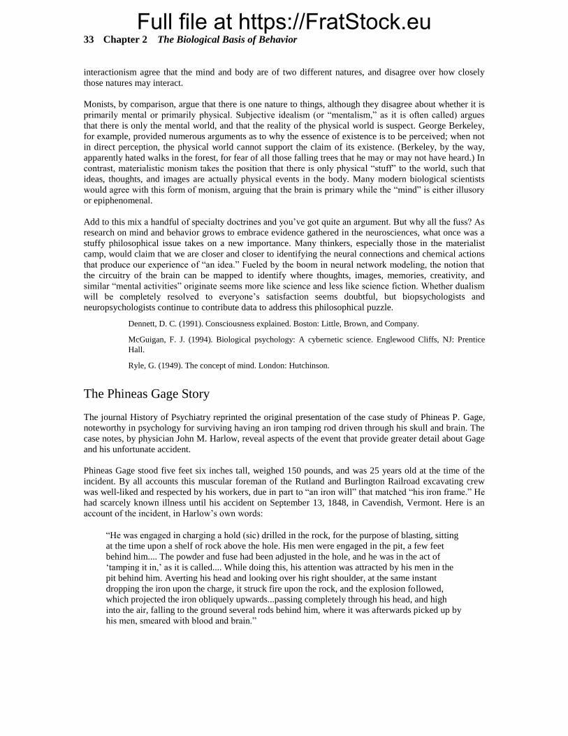
The Phineas Gage Story
The journal History of Psychiatry reprinted the original presentation of the case study of Phineas P. Gage,
noteworthy in psychology for surviving having an iron tamping rod driven through his skull and brain. The
case notes, by physician John M. Harlow, reveal aspects of the event that provide greater detail about Gage
and his unfortunate accident.
Phineas Gage stood five feet six inches tall, weighed 150 pounds, and was 25 years old at the time of the
incident. By all accounts this muscular foreman of the Rutland and Burlington Railroad excavating crew
was well-liked and respected by his workers, due in part to “an iron will” that matched “his iron frame.” He
had scarcely known illness until his accident on September 13, 1848, in Cavendish, Vermont. Here is an
account of the incident, in Harlow’s own words:
“He was engaged in charging a hold (sic) drilled in the rock, for the purpose of blasting, sitting
at the time upon a shelf of rock above the hole. His men were engaged in the pit, a few feet
behind him.... The powder and fuse had been adjusted in the hole, and he was in the act of
‘tamping it in,’ as it is called.... While doing this, his attention was attracted by his men in the
pit behind him. Averting his head and looking over his right shoulder, at the same instant
dropping the iron upon the charge, it struck fire upon the rock, and the explosion followed,
which projected the iron obliquely upwards...passing completely through his head, and high
into the air, falling to the ground several rods behind him, where it was afterwards picked up by
his men, smeared with blood and brain.”

Full file at https://FratStock.euChapter 2 The Biological Basis of Behavior 34
The tamping rod itself was three feet seven inches in length, with a diameter of 11/4 inches at its base and a
weight of 131/4 pounds. The bar was round and smooth from continued use, and it tapered to a point 12
inches from the end; the point itself was approximately 1/4 inch in diameter.
The accounts of Gage’s frontal lobe damage and personality change are well-known, and are corroborated
by Harlow’s presentation. Details of Gage’s subsequent life (he lived 12 years after the accident) are less
known. Gage apparently tried to regain his job as a railroad foreman, but his erratic behavior and altered
personality made it impossible for him to do so. He took to traveling, visiting Boston and most major New
England cities, and New York, where he did a brief stint at Barnum’s sideshow. He eventually returned to
work in a livery stable in New Hampshire, but in August 1852 he turned his back on New England forever.
Gage lived in Chile until June of 1860, then left to join his mother and sister in San Francisco. In February
1861, he suffered a series of epileptic seizures, leading to a rather severe convulsion at 5 a.m. on February
20. The family physician unfortunately chose bloodletting as the course of treatment. At 10 p.m., May 21,
1861, Gage eventually died, having suffered several more seizures. Although an autopsy was not
performed, Gage’s relatives agreed to donate his skull and the iron rod (which Gage carried with him
almost daily after the accident) to the Museum of the Medical Department of Harvard University.
Miller (1993) also briefly notes that John Martyn Harlow himself had a rather pedestrian career, save for
his association with the Gage case. Born in 1819, qualifying for medical practice in 1844, and dying in
1907, he practiced medicine in Vermont and later in Woburn, Massachusetts, where he engaged in civic
affairs and apparently amassed a respectable fortune as an investor. Like Gage himself, Harlow was an
unremarkable person brought into the annals of psychology by one remarkable event.
Harlow, J. M. (1848). Passage of an iron rod through the head. Boston Medical and Surgical Journal, 39,
389–393.
Harlow, J. M. (1868). Recovery from the passage of an iron bar through the head. Paper read before the
Massachusetts Medical Society.
Miller, E. (1993). Recovery from the passage of an iron bar through the head. History of Psychiatry, 4,
271–281.
Neural Effects of a Concussion
During the fall term, when college football is in season, it is especially appropriate to stress the discussion
of the neuronal and behavioral effects of concussion. Chances are good that in any given class, you will
have several students who will report having had a concussion in the past, usually as a result of
participation in football or other sports activities, or as a result of an automobile accident. You can ask the
students to discuss their experiences with the class, asking what kind of physiological and cognitive effects
occurred. The most common effects include loss of vision (“black out”), blurred vision, ringing in the ears,
nausea/vomiting, and not being able to think clearly. However, the physiological and cognitive effects vary
between individuals; some may not have experienced nausea at all, whereas others only experienced
blurred vision. It is important to point out the variability between individuals, because it can be inferred that
concussions vary greatly in terms of the severity of brain damage and the brain areas affected.
The brain sits in the cranium surrounded by cerebral fluid. When a severe blow to the head occurs, the
brain may collide with the cranium, then “bounce back” and collide with the opposite side of the cranium.
For example, if a football player falls and hits the back of his or her head, the brain may hit the back of the
cranium, then the front. At this point, you might ask students what brain areas would be affected in this
example (“occipital and frontal lobes” are a pretty decent answer). Therefore, both vision and some
cognitive functioning may be affected. At the neuronal level, a concussive blow to the head results in a
twisting or stretching of the axons, which in turn creates swelling. Eventually, the swelling may subside
and the neuron may return to its normal functioning. However, if the swelling of the axon is severe enough,
the axon may disintegrate. A more severe blow to the head may even sever axons, rendering those neurons
permanently damaged. Either way, neuronal signaling is disrupted, either temporarily or permanently.
Depending on the brain areas where the damaged axons are located, different physiological symptoms may
occur.

Full file at https://FratStock.eu35 Chapter 2 The Biological Basis of Behavior
Handedness, Eyedness, Footedness, Facedness
Although the title sounds like a Dr. Seuss rhyme, it actually denotes something sensible to neuroscientists.
Most people are familiar with the concept of handedness. The human population is distributed across many
people who are adept at using their right hands for most tasks, some who have greater skill using the left
hand, and a smaller proportion of those who are equally skilled using either hand (or who alternate hands
for certain tasks). The concepts of footedness, leggedness, eyedness, and facedness may be less familiar to
the layperson, although they stem from the same principle as handedness.
The basis of these distinctions lies in the concept of laterality. Just as the cerebral hemispheres show
specialization (e.g., left hemisphere language functions, right hemisphere visual-spatial functions), so too
are there preferences or asymmetries in other body regions. The concept of eyedness, then, refers to the
preference for using one eye over another, such as when squinting to site down the crosshairs of a rifle or to
thread a needle. Footedness and leggedness similarly refer to a preference for one limb over the other;
drummers and soccer players will attest to the importance of being equally adept at using either foot, and to
the difficulty in achieving that. Finally, facedness refers to the strength with which information is conveyed
by the right or left side of the face. It has been suggested that verbal information shows a right-face bias
whereas emotional expressions are more strongly shown on the left side of the face, although these
conclusions remain somewhat controversial.
Why are these distinctions useful? They play their largest role in the areas of sensation and perception,
engineering psychology, and neuropsychology. Studies of reaction time, human-machine interaction,
ergonomic design, and so on, take into account the preferences and dominances of some body systems over
others. In the case of facedness and emotional expression, researchers are working to illuminate the link
between facial expressions and cerebral laterality. Given the right hemisphere’s greater role in emotional
activities, the contralateral control between the right hemisphere and the left hemiface becomes an
important proving ground for investigating both brain functions and the qualities of expression.
Borod, J. C., Caron, H. S., & Koff, E. (1981). Asymmetry of facial expression related to handedness,
footedness, and eyedness: A quantitative study. Cortex, 17, 381–390.
Ekman, P., Hagar, C. J., & Friesen, W. V. (1981). The symmetry of emotional and deliberate facial
actions. Psychophysiology, 18, 101–106.
Friedlander, W. J. (1971). Some aspects of eyedness. Cortex, 7, 357–371.
McGuigan, F. J. (1994). Biological psychology: A cybernetic science. Englewood Cliffs, NJ: Prentice
Hall.
Sackheim, H. A., Gur, R. C., & Saucy, M. C. (1978). Emotions are expressed more intensely on the left
side of the face. Science, 202, 434–436.
Hey, Simpleton!
“The culmination of evolution.” “Master of the universe.” “Erect-walking swell young fops.” There’s no
shortage of appellations humans heap upon themselves in recognition of their arguably advanced position
among the species. Some genetic research suggests, however, that the praise might be premature.
Francis Collins and Eric Lander, researchers working with data from the National Human Genome
Research Institute, led a team that discovered that humans have about 20,000 to 25,000 genes, fewer than
the 30,000 to 40,000 they had previously estimated in 2001. Big deal? Yeah, sort of. A flowering
houseplant has about 20,000 to 25,000 genes. As does a small worm. So, worm-boy, there’s your genetic
company: We’re about as genetically complex as other not-so-complex living things.
So why isn’t a worm writing this piece? (Hey, watch it!) Because clearly it’s not just the parts, but rather
how those parts are assembled, that makes a difference. Granted, these are only the genes that tell cells how

Full file at https://FratStock.euChapter 2 The Biological Basis of Behavior 36
to make proteins, but some of those genes make multiple proteins, and some make more complex proteins.
Some of our biological complexity, then, comes from combinations of proteins rather than individual
proteins, so it’s somewhat a case where “less is more.”
By the way, in case you’re curious: Rice has about 40,000 genes; corn, about 50,000. But at least we’ve got
the fruit fly’s 13,600 genes beat ... by a little.
Ritter, M. (October 21, 2004). Human complexity built with fewer genetic bricks. Austin American-
Statesman, A1, A13.
DISCUSSION TOPICS
• How might the structure and function of the central nervous system be different if neural impulses
could travel both directions in a neuron?
• We have seen that each cerebral hemisphere is responsible for somewhat different functions,
although typically the two hemispheres work together. How might our knowledge of hemispheric
specialization be put to some sort of practical use?
• How might the human race (or human behavior) be different if we had evolved with a dominant
right hemisphere?
• Should people with genetic abnormalities associated with psychological disorders be prohibited
from reproducing? If so, which disorders? What if they are just carriers of a recessive gene?
Should people routinely be screened for genetic problems before having children?
• After reading Chapter 2, you are aware that the frontal lobes and portions of the limbic system are
important in the control of emotions. Surgery that severs the connections to and from these
portions of the brain or surgery that removes them (partially or entirely) can have a profound
effect on behavior. Similarly, electrically stimulating these portions of the brain can have a
significant effect on behavior. Under what circumstances (if any) do you think psychosurgery or
electrical stimulation of people’s brains might be warranted? Should those with a chronic history
of criminal offenses be treated with psychosurgery or electrical brain stimulation? What do you
think of psychosurgery or brain stimulation as an alternative to the death penalty?
• Chapter 2 alludes to an interesting phenomenon: In almost all higher organisms, sensory
information from the left side of the body goes to the right half of the brain and vice versa.
Moreover, motor control of the left side of the body occurs in the right half of the brain, and vice
versa. Two prominent physiologists once said, “No one has the remotest idea why there should be
this amazing tendency for nervous system pathways to cross.”
DEMONSTRATIONS AND ACTIVITIES
Using Dominoes to Understand the Action Potential
Walter Wagor suggests using real dominoes to demonstrate the so-called “domino effect” of the action
potential as it travels along the axon. For this demonstration, you’ll need a smooth table-top surface (at
least 5 feet long) and one or two sets of dominoes. Set up the dominoes beforehand, on their ends and about
an inch apart, so that you can push the first one over and cause the rest to fall in sequence. Proceed to
knock down the first domino in the row and students should clearly see how the “action potential” is passed
along the entire length of the axon. You can then point out the concept of refractory period by showing that,
no matter how hard you push on the first domino, you will not be able to repeat the domino effect until you
take the time to set the dominoes back up (i.e., the resetting time for the dominoes is analogous to the

Full file at https://FratStock.eu37 Chapter 2 The Biological Basis of Behavior
refractory period for neurons). You can then demonstrate the all-or-none characteristic of the axon by
resetting the dominoes and by pushing so lightly on the first domino that it does not fall. Just as the force
on the first domino has to be strong enough to knock it down before the rest of the dominoes will fall, the
action potential must be there in order to perpetuate itself along the entire axon. Finally, you can
demonstrate the advantage of the myelin sheath in axonal transmission. For this demonstration, you’ll need
to set up two rows of dominoes (approximately 3 or 4 feet long) next to each other. The second row of
dominoes should have foot-long sticks placed end-to-end in sequence on top of the dominoes. By placing
the all-domino row and the stick-domino row parallel to each other and pushing the first domino in each,
you can demonstrate how much faster the action potential can travel if it can jump from node to node rather
than having to be passed on sequentially, single domino by single domino. Ask your students to discuss
how this effect relates to myelinization.
Wagor, W. F. (1990). Using dominoes to help explain the action potential. In V. P. Makosky, C. C. Sileo,
L. G. Whittemore, C. P. Landry, & M. L. Skutley (Eds.), Activities handbook for the teaching of
psychology: Vol. 3 (pp. 72–73). Washington, DC: American Psychological Association.
Hemispheric Communication and the Split Brain
Even after reading the textbook and listening to your lecture, many students may have difficulty
conceptualizing the effects of a split-brain operation on an individual’s behavior. Morris (1991) described
five activities designed to simulate the behavior of split-brain patients. All of the activities have the same
basic setup. You will need to solicit two right-handed volunteers and seat them next to each other at a table,
preferably in the same chair. The volunteer on the left represents the left hemisphere, and the other student
is the right hemisphere. The students are instructed to place their outer hands behind their backs and their
inner hands on the table with their hands crossed, representing the right and left hands of the split-brain
patient. Finally, the student representing the right hemisphere is instructed to remain silent for the
remainder of the activity. In one of the activities described by Morris, both students are blindfolded and a
familiar object (Morris suggested a retractable ball-point pen) is placed in the left hand of the “split-brain
patient” (the hand associated with the right hemisphere). Then ask the “right hemisphere” student if he or
she can identify the object, reminding him or her that they must do so nonverbally. Next, ask the “right
hemisphere” to try to communicate, without using language, what the object is to the “left hemisphere.”
Your more creative volunteers may engage in behaviors that attempt to communicate what the object is
through sound or touch. If your “right hemisphere” has difficulty in figuring out how to communicate, ask
the class for suggestions. This demonstration can be used to elicit discussion about why only the “left
hemisphere” student can talk, the laterality of the different senses, and how split-brain patients are able to
adjust their behavior to accommodate. You should refer to Morris’s original article for descriptions of the
other activities.
Morris, E. J. (1991). Classroom demonstration of behavioral effects of the split-brain operation. Teaching
of Psychology, 18, 226–228.
Reprinted from Hill, W. G. (1995). Instructor’s resource manual for Psychology by S. F. Davis and J. J.
Palladino. Englewood Cliffs, NJ: Prentice Hall.
Hemispheric Lateralization
Kemble (1987) described three demonstrations designed to illustrate cerebral lateralization. One of these
demonstrations explores the cerebral lateralization of language. This demonstration requires a wooden
dowel stick (about 1.25 cm in diameter and 92 cm long), a stopwatch, and several difficult verbal problems
(for example, reciting the alphabet backwards or spelling a long word backwards). After identifying a
student volunteer, he or she is given between 5 and 10 minutes to practice balancing the dowel on the index
finger of both hands. Approximately the same amount of time should be spent practicing with each hand.
Following the practice period, the student is then asked to balance the dowel eight times, four per hand in a
variable order, and the amount of time that he or she balances the dowel is recorded. However, during half
of the trials for each hand, the student is asked to perform one of the verbal problems out loud, while the

Full file at https://FratStock.euChapter 2 The Biological Basis of Behavior 38
other trials are conducted in silence. Since the verbal task requires a high degree of activity in the left
hemisphere, and the left hemisphere controls the right hand, the competition between the verbal and motor
activity will consistently result in a decrease in balancing time for the right hand compared to that of the
left hand when performing the verbal task. Kemble also suggested comparing the performance of male and
female subjects since gender differences are often observed in this demonstration. This can provide an
opportunity to discuss how and why these gender differences occur. The other demonstrations described by
Kemble illustrate the effects of cerebral lateralization on pattern recognition.
Kemble, E. D. (1987). Cerebral lateralization. In V. P. Makosky, L. G. Whittemore, & A. M. Rogers
(Eds.), Activities handbook for the teaching of psychology, Vol. 2 (pp. 33–36). Washington, DC:
American Psychological Association.
Reprinted from Hill, W. G. (1995). Instructor’s resource manual for Psychology by S. F. Davis and J. J.
Palladino. Englewood Cliffs, NJ: Prentice Hall.
Demonstrating Neural Conduction: The Class as a Neural Network
In this engaging exercise (suggested by Paul Rozin and John Jonides), students in the class simulate a
neural network and get a valuable lesson in the speed of neural transmission. Depending on your class size,
arrange 15 to 40 students so that each person can place his or her right hand on the right shoulder of the
person in front of him or her. Note that students in every other row will have to face backwards in order to
form a snaking chain so that all students (playing the role of individual neurons) are connected to each
other. Explain to students that their task as a neural network is to send a neural impulse from one end of the
room to the other. The first student in the chain will squeeze the shoulder of the next person, who, upon
receiving this “message,” will deliver (i.e., “fire”) a squeeze to the next person’s shoulder and so on, until
the last person receives the message. Before starting the neural impulse, ask students (as “neurons”) to label
their parts; they typically have no trouble stating that their arms are axons, their fingers are axon terminals,
and their shoulders are dendrites.
To start the conduction, the instructor should start the timer on a stopwatch while simultaneously squeezing
the shoulder of the first student. The instructor should then keep time as the neural impulse travels around
the room, stopping the timer when the last student/neuron yells out “stop.” This process should be repeated
once or twice until the time required to send the message stabilizes (i.e., students will be much slower the
first time around as they adjust to the task). Next, explain to students that you want them to again send a
neural impulse, but this time you want them to use their ankles as dendrites. That is, each student will “fire”
by squeezing the ankle of the person in front of them. While students are busy shifting themselves into
position for this exercise, ask them if they expect transmission by ankle-squeezing to be faster or slower
than transmission by shoulder-squeezing. Most students will immediately recognize that the ankle-
squeezing will take longer because of the greater distance the message (from the ankle as opposed to the
shoulder) has to travel to reach the brain. Repeat this transmission once or twice and verify that it indeed
takes longer than the shoulder squeeze.
This exercise—a student favorite—is highly recommended because it is a great ice-breaker during the first
few weeks of the semester and it also makes the somewhat dry subject of neural processing come alive.
Rozin, P., & Jonides, J. (1977). Mass reaction time measurement of the speed of the nerve impulse and the
duration of mental processes in class. Teaching of Psychology, 4, 91–94.
The Dollar Bill Drop
After engaging in the neural network exercise, consider following it up with the “dollar bill drop” (Fisher,
1979), which not only delights students but also clearly illustrates the speed of neural transmission. Ask
students to divide themselves into pairs and to come up with one crisp, flat, one-dollar bill (or something
bigger, if they trust their fellow classmates!) between them. First, each member of the pair should take
turns trying to catch the dollar bill with their nondominant (for most people, the left) hand as they drop it
from their dominant (typically right) hand. To do this, they should hold the bill vertically so that the top,

Full file at https://FratStock.eu39 Chapter 2 The Biological Basis of Behavior
center of the bill is held by the thumb and middle finger of their dominant hand. Next, they should place the
thumb and middle finger of their nondominant hand around the dead center of the bill, as close as they can
get without touching it. When students drop the note from one hand, they should be able to easily catch it
with the other before it falls to the ground.
Now that students are thoroughly unimpressed, ask them to replicate the drop, only this time one person
should try to catch the bill (i.e., with the thumb and middle finger of the nondominant hand) while the other
person drops it (i.e., from the top center of the bill). Student “droppers” are instructed to release the bill
without warning, and “catchers” are warned not to grab before the bill is dropped. (Students should take
turns playing dropper and catcher.) There will be stunned looks all around as dollar bills whiz to the
ground. Ask students to explain why it is so much harder to catch it from someone other than themselves.
Most will quickly understand that when catching from ourselves, the brain can simultaneously signal us to
release and catch the bill, but when trying to catch it from someone else, the signal to catch the bill can’t be
sent until the eyes (which see the drop) signal the brain to do so, which is unfortunately a little too late.
Fisher, J. (1979). Body Magic. Briarcliff Manor, NY: Stein and Day.
Reaction Time and Neural Processing
Yet another exercise that illustrates the speed of neural processing is suggested by Harcum (1988). The
point made by this simple but effective exercise is that reaction times increase as more response choices
become available (i.e., because more difficult choices in responses involve more neuronal paths and more
synapses, both of which slow neural transmission). Depending on your class size, recruit two equal groups
of students (10 to 20 per group is ideal) and have each group stand together at the front of the room. First,
explain that all subjects are to respond as quickly as possible to the name of a U.S. President. Then, give
written instructions to each group so that neither group knows the instructions given to the other. One
group should be instructed to raise their right hands if the president served before Abraham Lincoln and to
raise their left hands if the president served after Lincoln. The other group should be instructed simply to
raise their left hands when they a hear a president’s name. Ask participants and audience members to note
which group reacts more quickly. When all students are poised and ready to go (i.e., hands level with
shoulders and ready to raise), say “ready?” and then “Ford.” The group with the simpler reaction time task
will be obviously faster than the group whose task requires a choice.
Harcum, E. R. (1988). Reaction time as a behavioral demonstration of neural mechanisms for a large
introductory psychology class. Teaching of Psychology, 4, 208–209.
The Importance of a Wrinkled Cortex
At the beginning of your lecture on the structure and function of the brain, ask students to explain why the
cerebral cortex is wrinkled. There are always a few students who correctly answer that the wrinkled
appearance of the cerebral cortex allows it to have a greater surface area while fitting in a relatively small
space (i.e., the head). To demonstrate this point to your class, hold a plain, white sheet of paper in your
hand and then crumple it into a small, wrinkled ball. Note that the paper retains the same surface area, yet is
now much smaller and is able to fit into a much smaller space, such as your hand. You can then mention
that the brain’s actual surface area, if flattened out, would be roughly the size of a newspaper page (Myers,
1995). Laughs usually erupt when the class imagines what our heads would look like if we had to
accommodate an unwrinkled, newspaper-sized cerebral cortex!
Myers, D. G. (1995). Psychology (4th ed.). New York: Worth.

Full file at https://FratStock.euChapter 2 The Biological Basis of Behavior 40
DEBATE
Are Genetic Explanations for ADHD Faulty?
Attention Deficit Hyperactivity Disorder (ADHD), like Attention Deficit Disorder (ADD), has been
increasingly diagnosed among schoolchildren, adolescents, and even adults. The origins of ADHD remain a
matter of some debate. Although there is evidence that genetics plays a substantial role in the development
of ADHD, not all researchers agree on the extent of its influence. Some writers, in fact, prefer to devote
more attention to exploring social and psychological explanations for the disorder. Given that many
disorders (e.g., schizophrenia, depression) have revealed their genetic underpinnings (and that substantial
research money and time have been allocated to pursuing these explanations), the origins of ADHD offer a
case for further discussion. Ask your students to become conversant with the arguments on both sides of
this issue.
Slife, B. (2003). Taking sides: Clashing views on controversial psychological issues (13th ed.). Guilford,
CT: Dushkin Publishing Group.
STUDENT ASSIGNMENTS
The Brain Diagram
Students often have trouble encoding the location and function of the different parts of the brain, both
because (a) they glance too quickly over the colorful textbook illustrations and (b) their eyes tend to glaze
over during class discussion of the brain’s structure and function. As an easy remedy to this problem, try
asking students to draw their own colorful rendition of the human brain, an active learning strategy that
ensures that they encode and think about the parts of the brain rather than passively glossing over them in
the text. Prior to the class period in which you will be discussing the brain, ask students to read Chapter 2
and to hand-draw a diagram of the brain (in a cross-section, much like Figure 2-6 in the text) on a clean
white sheet of unlined paper. For each of the following sections of the brain, students should color (using
map pencils) and label the appropriate structure, and also list at least one or two of its major functions: (a)
the cerebral cortex, including the four lobes, (b) the thalamus, (c) the hypothalamus, (d) the hippocampus,
(e) the amygdala, (f) the cerebellum, (g) the pons, and (h) medulla. Added benefits of this assignment are
that it is easy to grade, students enjoy doing it (and it is an easy and fun way for them to get points), and it
can be used by students as a study aid for the exam.
Reunited Twins
Although twin studies (particularly studies of identical twins reared apart) seem to confirm genetic links to
intelligence, psychological disorder, and some complex personality traits, critics become skeptical when the
same research reveals eerie (and ostensibly genetically-based) similarities between twins on such things as
aftershave brand, selection of hobbies, attraction to tattoos, and even child name preferences. Although
amazing behavioral similarities do indeed turn up between identical twins raised apart (see Rosen, 1987),
Wyatt and his colleagues (1994) suggest that, rather than being genetically based, these similarities are
merely selected examples of coincidences that are inevitable given the hundreds or even thousands of
questions typically asked of reunited twins by eager researchers. In other words, it is likely that similar
“amazing” coincidences would be found if genetically unrelated people were asked a large number of
questions about their behavior.
To illustrate this point, Lester Sdorow (1994) designed the “Identical Twins Reunited Questionnaire” and
accompanying exercise. For this assignment, students should first read the articles by Rosen (1987) and
Wyatt et. al (1994); you can put these on reserve in the library. Next, you’ll need to distribute one copy of
the ITRQ to each student. Handout 2-1 contains the questionnaire (which, as you can see, asks students
about their behaviors, relationships, and characteristics) along with instructions for the assignment. After

Full file at https://FratStock.eu41 Chapter 2 The Biological Basis of Behavior
students have completed their surveys, you should collect them and identify pairs of students who are the
most similar. [Note: You may want to take the surveys home with you and present the results during the
next class period.] Once you have described for your class the “reunited twins” among them, instruct them
to write a 2–3 page paper discussing how the results from the class study bear on the rationale for reunited
twin studies. Ask students to incorporate into their papers insights from the Wyatt et al. study and an
additional reference of their choosing from Psychological Abstracts. [Note: This assignment can also be
used in Chapter 10, which covers personality.]
Rosen, C. M. (1987, September). The eerie world of reunited twins. Discover, pp. 36–46.
Sdorow, L. (1994). The Frankenstein course: Teaching assistants, laboratory exercises, and papers in
introductory psychology. Paper presented at the Southwest Regional Conference for Teachers of
Psychology, Fort Worth. Used by permission of the author.
Wyatt, W. J., Posey, A., Welker, W., & Seamonds, C. (1984). Natural levels of similarities between
identical twins and between unrelated people. The Skeptical Inquirer, 9, 62–66.
Psychology in Literature: The Man Who Mistook His Wife for a Hat
Oliver Sacks’s (1985) national bestseller chronicles over 20 case histories of patients with a variety of
neurological disorders. His compassionate retelling of bizarre and fascinating tales include patients plagued
with memory loss, useless limbs, violent tics and jerky mannerisms, the inability to recognize people or
objects, and unique artistic or mathematical talents despite severe mental deficits. A reading of this
absorbing book will surely increase your students’ understanding of the connection between the brain and
the mind, and will also give them invaluable insights into the lives of disordered individuals. Ask your
students to write a book report focusing on a few of the cases that most interest them, and to apply
principles from the text and lecture to the stories. As a more elaborate project, you might consider assigning
this book at the end of the semester, as many of the cases are ripe with psychological principles that may be
encountered later in the course (e.g., perception, memory, mental retardation).
Sacks, O. (1985). The man who mistook his wife for a hat. New York: HarperCollins.
VIDEO
The Autonomic Nervous System (28 min, FHS). Describes the role of the autonomic nervous system in
controlling the glands and organs. It also addresses how meditation, autosuggestion, and hypnosis can be
used to control autonomic functions.
Biology and Behavior (1980, 22 min, PENN). Examines the biological roots of behavior, including close-
ups on classic issues such as nature vs. nurture and basic studies in taste aversion, imprinting, and
instinctive drift.
Biology of Behavior (1990, 30 min, IM). This video gives an overview of the nervous system, including
material on neurotransmitters, neurons, and the fight or flight response.
The Birth of a Brain (1983, 33 min, CRM). Hereditary and environmental influences on brain development
are emphasized. A live birth may make this film unsuitable for less mature audiences.
The Brain, Part 1: The Enlightened Machine (1984, 60 min, ANN/CPB). Gives an overview of the study of
the brain from Franz Gall to the present and reviews neurotransmitters functioning at the synapse.
The Brain, Part 6: The Two Brains (1984, 60 min, ANN/CPB). Explores split-brain studies and what they
indicate about hemispheric functioning, the relationship between thought and language, and the issues of
sex differences in the brain.
The Brain, Part 8: States of Mind (1984, 60 min, ANN/CPB). The limits of our knowledge about the brain
are the focus of this program. Research in genetics, artificial intelligence, and medicine are discussed to
shed light on this mysterious gray lump.

Full file at https://FratStock.euChapter 2 The Biological Basis of Behavior 42
Teaching Modules from The Brain (30 segments from The Brain series, ANN/CPB). These self-contained
modules offer brief glimpses into the lives of split-brain patients, the story of Phineas Gage, the brain basis
of schizophrenia, REM, speech, and aspects of stress and health.
The Brain (50 min, IM). This BBC program uses animation and models of the brain to tour the structures
and functions of the brain.
The Brain (23 min, FHS). This “Brain” begins with an exploration of dreams, follows with a detailed look
at the nervous system and how it works, and ends with a discussion of EEG and NMRI as techniques for
peering into the brain.
The Brain (28 min, FHS). And this “Brain” takes a cellular approach. The relationship between cell
assemblies and complex processes such as hearing, vision, and language form the focus of this video.
Brain Sex (1993, 3 volumes, 150 min total, IM). Differences between the sexes in areas such as learning,
appetite, expectations, and behavior are examined. Parts of this 3-volume set can be profitably used in a
variety of contexts.
Brain Story (50 min each, FHS). This 6-part series, produced by the BBC, takes a fascinating look at
current and recent brain science. Videos include The Biochemistry of Feelings, How the Brain Develops,
How the Brain Sees the World, and What Is Consciousness?
Chemistry of Love (1985, 30 min, IM). Love, lust, and fidelity may have as much to do with evolution as
they do with societal norms. This video explains why.
Discovering Psychology, Part 3: The Behaving Brain (2000, 30 min, ANN/CPB). Provides an overview of
brain structure and function through a description of the biochemical reactions involved in thoughts,
feelings, and actions.
Discovering Psychology, Part 4: The Responsive Brain (2000, 30 min, ANN/CPB). Explores the interaction
between the development of brain structures and function and the environment.
Discovering Psychology, Part 14: The Mind Hidden and Divided (2000, 30 min, ANN/CPB). Examines the
influence of the subconscious mind on thought and behavior. The segment on the split-brain phenomenon
is relevant to this chapter.
Discovering Psychology, Part 25: A Union of Opposites (2000, 30 min, ANN/CPB). A unique approach to
a seldom addressed topic, this program presents “a yin-yang model of complementary opposites” to
facilitate an understanding of the basic principles thought to govern human nature and animal behavior. A
useful means of addressing the nature-nurture debate.
Endocrine Control: Systems in Balance (1997, 30 min, IM). This video focuses on the endocrine system’s
role in regulating and controlling other bodily systems.
Epilepsy: Breaking the Barrier (28 min, FHS). Footage of grand mal seizures in progress bring home the
often debilitating affliction of epilepsy. Various treatments, including drug therapy and surgical
interventions, are described.
Hormones (28 min, FHS). Illustrates the role of hormones in controlling a variety of bodily functions.
The Human Brain (1997, 25 min, IM). The focus of this video is that brain functioning can be enhanced by
the proper environment. Recovery from brain injury, brain surgery techniques, and silicon retinas are used
as examples.
Inside Information: The Brain and How It Works (58 min, FHS). This award-winning film contains
interviews with leading researchers, who discuss how and why the brain works, and works as it does.
Pattern recognition, computer analogs, and individual brain structures are also discussed.
Is Your Brain Really Necessary? (1988, 50 min, FHS). This must-see video follows the lives of three
people who have undergone drastic brain surgery (e.g., hemispherectomies). Their subsequent levels of
functioning should prove illuminating to your students.

Full file at https://FratStock.eu43 Chapter 2 The Biological Basis of Behavior
Journey to the Centers of the Brain (5 Parts, 58 min each, FHS). This series explores different topics related
to the brain. For example, The Electric Ape looks at brain structure and function, whereas Bubble Bubble
Toil and Trouble examines neural circuitry, and Through a Glass Darkly looks at brain imaging techniques.
Left Brain, Right Brain (1979, 56 min, FLI). Norman Geschwind narrates this intriguing look at
hemispheric specialization. Wada tests and split-brain operations illustrate the semi-independent
functioning of the hemispheres.
Living with Tourette’s (24 min, PENN). Combines case studies of individuals with Tourette’s syndrome
with an examination of chemical imbalances in the brain associated with the disorder.
The Mind: Development (60 min, PBS). Focusing on particular brain cells, this program (part of a 9-part
series) examines brain development from conception to age six.
Mind Games (Brain Functions) (1999, 30 min, IM). The focus here is on the effects of trauma, disease, and
mental illness on the brain. Scanning techniques and methods of “repairing” the brain are highlighted.
Nerves (1992, 24 min, IM). As the title suggests, this video is a bundle of nerves. Action potentials,
synapses, agonists and antagonists, brain slices, and neurons all take a bow.
Nervous System (1993, 14 min, IM). Using animation, this program demonstrates the action potential and
neurotransmitter activity. It also discusses disorders of the nervous system, including Alzheimer’s disease,
Parkinson’s disease, depression, and anxiety.
The Neural Connection (1997, 30 min, IM). Receptors galore: Thermoreceptors, chemoreceptors,
mechanoreceptors, nocireceptors, and photoreceptors display their charms to teach about neural
connections and neural impulses.
Neuron and Neural Transmission (2001, 30 min, IM). The structure of neurons and their ability to
communicate with one another are the focus of this video. Of special interest is a discussion of how
neurological disorders disrupt neural communication.
Neuropsychology (23 min, FHS). How a tangled collection of cells produces memory, emotion, language,
and thought is the subject of this film. Particular attention is given to recognizing facial expressions and
intellectual functioning.
The Secret Life of the Brain (2002, 60 min, IM). This 5-part series examines how the brain develops from
birth through old age.
Sex Hormones and Sexual Destiny (26 min, FHS). Examines the role of hormones on “masculine” and
“feminine” behavior, sex differences in brain structure, and environmental influences on male and female
behavior.
Split-Brain and Conscious Experience (18 min, PENN). Discusses the pioneering work conducted by
Michael Gazzaniga on the split-brain phenomenon using epileptic patients with severed corpus callosums.
Two Brains (1984, 55 min, ANN/CPB). Hemispheric specialization and split-brain surgery is highlighted.
A woman with a severed corpus callosum is featured.
Who Are You? (60 min, FHS). Genetic influences on behavior are reviewed through an examination of
several twin studies. Also included are case studies of individuals, such as the sober son of an alcoholic
who fears taking a drink.
Full file at https://FratStock.euChapter 2 The Biological Basis of Behavior 44
MULTIMEDIA
Video Classics
Probing the Cerebral Cortex
SYNOPSIS: This clip contains commentary by Wilder Penfield, a pioneer in mapping the areas of the
cerebral cortex. Penfield discusses the work that led to electrode-stimulation of the cortex. He also
interviews a brain surgery patient about her experiences during surgery: Stimulation of various areas of her
cortex produced memories of past events and the perception of music playing.
Form a Hypothesis
Q What happens when Penfield stimulates a small area of the temporal lobe, called the auditory cortex?
A The patient “hears” sounds.
Test Your Understanding
Q What are the four lobes of the cerebral cortex?
A The four lobes of cerebral cortex are occipital, parietal, temporal, and frontal.
Q What are the functions of the somatosensory cortex, motor cortex, and association cortex areas?
A Somatosensory cortex interprets sensations and coordinates the motor behavior of skeletal muscles.
Association areas, located on all four cortical lobes, are involved in the integration of various brain
functions, such as sensation, thought, memory, planning, etc.
Q What two areas of the association cortex specialize in language?
A Wernicke’s area, located toward the back of the temporal lobe, is important in understanding the speech
of others. Broca’s area is essential to sequencing and producing language.
Thinking Critically
Q What four types of research methods are commonly used in the study of behavioral neuroscience?
A Microelectrode techniques are used to study the functions of individual neurons. Macroelectrode
techniques, such as an EEG, record activities of brain areas. Structural imaging, such as computerized axial
tomography or CAT scans, is useful for mapping brain structures. Functional imaging, in which specific
brain activity can be recorded in response to tasks or stimulation, offers the potential to identify specific
brain areas and functions.
Web Links
Amazing Case of Phineas Gage
http://www.epub.org.br/cm/n02/historia/phineas.htm
Account by Renato M. E. Sabbatini, Ph.D., published in the online journal Brain & Mind.
Autonomic Nervous System
http://faculty.washington.edu/chudler/auto.html
Succinct summary of information about the structure and function of the autonomic nervous system,
prepared by Eric Chudler.
Basic Neural Processes Tutorials
http://psych.hanover.edu/Krantz/neurotut.html
A good site to help your students learn about basic brain functioning.

Full file at https://FratStock.eu45 Chapter 2 The Biological Basis of Behavior
Biological and Physiological Resources
http://psych.athabascau.ca/html/aupr/biological.shtml
Links to several sites and interesting topical articles relevant to biological and physiological psychology. A
good starting point for a number of assignments, such as writing short papers or assembling study guide
terms. Maintained by the Centre for Psychology Resources at Athabasca University, Alberta, Canada.
Biological Psychology
http://www.csuchico.edu/psy/BioPsych/definition.html
Information about the field from the biological psychologists at California State University, Chico.
Brain and Behavior
http://serendip.brynmawr.edu/bb/
This mega-site contains lots of links to information about the brain, behavior, and the bond between the
two. Students can complete several interactive exercises to learn more about brain functions.
Brain & Mind—Electronic Magazine on Neuroscience
http://www.epub.org.br/cm/
Includes a wealth of short articles devoted to the brain.
Brain Briefings—Society for Neuroscience
http://www.sfn.org/content/Publications/BrainBriefings/index.html
A series of 2-page reports that describes clinical applications of basic neuroscience research. Includes
reports in the following areas: brain injury, brain mechanisms, development, drugs, eating, emotions,
exercise, gender, memory, nervous system disorders and diseases, nervous system repair, pain, the senses,
sleep, and technology.
Brain Connection: The Brain and Learning
http://www.brainconnection.com/
A newspaper-style Web page that contains interesting articles, news reports, activities, and commentary on
brain-related issues.
Brain Function and Pathology
http://www.waiting.com/brainfunction.html
Concise table of diagrams of brain structures, descriptions of brain functions, and descriptions of signs and
symptoms associated with brain structures and functions.
Brain Model Tutorial
http://pegasus.cc.ucf.edu/~Brainmd1/brain.html
This tutorial teaches students about the various parts of the human brain and allows them to test their
knowledge of brain structures.
Brain Reorganization
http://www.sfn.org/content/Publications/BrainBriefings/brain_reorg.html
Brief information on how the brain changes with experience, prepared by the Society for Neuroscience.
Brain: Right Down the Middle
http://faculty.washington.edu/chudler/sagittal.html
Useful drawing and succinct information about the location and functions of brain structures that can be
seen on the midsagittal plane, presented by Eric Chudler.
Central Nervous System—CliniWeb International
http://www.ohsu.edu/cliniweb/A8/A8.186.html
Lots of links to information about the central nervous system. See MRI images, link to research labs, and
learn about the brain and spinal cord.

Full file at https://FratStock.euChapter 2 The Biological Basis of Behavior 46
Comprehensive Behavioral and Cognitive Sciences
http://mentalhelp.net/guide/pro02.htm
Includes theory and therapy. This site includes Web links with descriptions and ratings of each source.
Useful for spicing up your lectures or for more detailed study by your students.
Cross-sections of the Human Brain
http://www.neuropat.dote.hu/caud.gif
A cross-sectional image of the human brain. Good to have on hand if you need one. Show your students
and help them identify the various structures.
Dogma Overturned
http://www.sciam.com/1998/1198issue/1198infocus.html
Upending a long-held theory, a study finds that humans can grow new brain neurons throughout life—even
into old age. Research summary published in Scientific American.
Drugs, Brains, and Behavior
http://www.rci.rutgers.edu/~lwh/drugs/
An online textbook detailing the effects of various substances on the brain, authored by C. Robin Timmons
& Leonard W. Hamilton.
fMRI Reveals the Brain Areas Involved in Working Memory
http://www.nimh.nih.gov/events/prfmri.htm
An NIH press release (April 9, 1997) describing how functional magnetic resonance imaging (fMRI) has
been used to delineate brain areas involved in working memory.
Functional Divisions of the Cerebral Cortex
http://faculty.washington.edu/chudler/functional.html
Succinct information about the location and functions of the major functional subdivisions of the cerebral
cortex, presented by Eric Chudler. Link to self-test available at the end (not interactive; print it out):
http://faculty.washington.edu/chudler/pdf/revcort.pdf
(You must have Adobe Acrobat to view and print the quiz.)
Harvard Brain
http://www.hcs.harvard.edu/~husn/BRAIN/index.html
The brains behind Harvard University? No, just a journal published by the Harvard Undergraduate Society
for Neuroscience.
History of Phrenology
http://pages.britishlibrary.net/phrenology/
Follow the bumpy road to discovering phrenology’s past from a professor of history at the University of
Cambridge.
How Do Nerve Cells Communicate?
http://www.sfn.org/content/Publications/BrainBackgrounders/communication.htm
Information prepared by the Society for Neuroscience.
The Human Brain: A Learning Tool
http://uta.maymt.edu/~psychol/brain.html
These close-up pictures of the brain’s lobes can be added to your classroom presentations. Link to this site,
turn on your classroom’s media projector, and let the action begin.
Human Corpus Callosum
http://www.indiana.edu/~pietsch/callosum.html
Information and links about the corpus callosum and “split-brain surgery” by Paul Pietsch.
Full file at https://FratStock.eu47 Chapter 2 The Biological Basis of Behavior
Lobes of the Brain
http://faculty.washington.edu/chudler/lobe.html
Succinct information about the location and functions of the four lobes of the cerebrum, presented by Eric
Chudler. Includes link to “Lobes of the Brain Review,” a very brief quiz on functions associated with major
lobes of the brain. Answers provided online:
http://faculty.washington.edu/chudler/revlobe.html
Localization of Function Exercise
http://www.gpc.peachnet.edu/~bbrown/psyc1501/brain/locfunct.htm
Allows students to simulate the effects of stimulating the brain, recording electrical activity from the brain,
or creating lesions in the brain, then to try to figure out the functions of various parts of the brain based on
the data they have collected. Developed by Dr. Barbara Brown of Georgia Perimeter College.
Making Connections—The Synapse
http://faculty.washington.edu/chudler/synapse.html
Clear, comprehensible explanation of how synapses work, with nice illustrations, prepared by Eric Chudler.
Mapping the Brain
http://www.epub.org.br/cm/n03/tecnologia/eeg.htm
Article on the use of various methods of recording brain activity to map the location of functional areas of
the brain, by Renato Sabbatini, Ph.D. Published in the online journal Brain & Mind.
Neural Processes Tutorial
http://psych.hanover.edu/Krantz/neurotut.html
An excellent interactive animated tutorial.
Neuroguide.com—Neurosciences on the Internet
http://www.neuroguide.com/
A resource for all things related to neuroscience: databases, diseases, research centers, software, biology,
psychology, journals, tutorials, and so much more.
Neuropsychology Central
http://www.neuropsychologycentral.com/
Links to resources related to neuropsychology, including brain images and extensive, well-organized links
to other sites.
NPAC/OLDA Visible Human Viewer
http://www.dhpc.adelaide.edu.au/projects/vishuman2/VisibleHuman.html
A little tricky to use, but by following the instructions on this page you can view images of the brain in one
of several planes. Currently, only photos are available, but they are quite nice. MRI and CT scans in the
same planes are planned for the future.
One Brain—or Two?
http://faculty.washington.edu/chudler/split.html
Information on lateralization of function and how the functions of the hemispheres may be studied,
presented by Eric Chudler.
PET Scan: A New Window into the Brain
http://www.epub.org.br/cm/n01/pet/pet.htm
Article on uses of the PET scan to study brain function, by Renato Sabbatini, Ph.D. Published in the online
journal Brain & Mind.

Full file at https://FratStock.euChapter 2 The Biological Basis of Behavior 48
Phineas Gage Information Page
http://www.deakin.edu.au/hbs/GAGEPAGE
Everything you ever wanted to know about Phineas Gage is on this page prepared by Malcolm Macmillan
at Deakin University, Victoria, Australia.
Practice Quiz
http://www.gpc.peachnet.edu/~bbrown/psyc1501/brain/quiz.htm
Covers material from the “Neurons, Hormones, and the Brain” chapter of the Wade and Tavris text.
Prepared by Barbara Brown of Georgia Perimeter College.
Self-Quiz for Chapter on the Human Nervous System
http://www.psychwww.com/selfquiz/ch02mcq.htm
Self-quiz prepared by Russ Dewey at Georgia Southern University. Covers material typically found in an
introductory psychology textbook chapter with a title like “Brain and Behavior” or “Neuropsychology.”
She Brains/He Brains
http://faculty.washington.edu/chudler/heshe.html
Nice summary of evidence for sex-related differences in brain structure, prepared by Eric Chudler.
Split Brain Consciousness
http://www.macalester.edu/~psych/whathap/UBNRP/Split_Brain/Split_Brain_Consciousness.html
Nice summary of information on the effects of cutting the corpus callosum, with links to further
information on split brain experiments and hemispheric specialization.
Synapses
http://www.gpc.peachnet.edu/~bbrown/psyc1501/brain/synapses.htm
Contains basic information about synapses and an animation of neurotransmitter release and binding to
receptors at a synapse.
Views of the Brain
http://rpiwww.mdacc.tmc.edu:80/se/anatomy/brain/
Gross anatomical photographs of left, right, anterior, superior, and inferior views of the brain.
What Does Handedness Have to Do with Brain Lateralization (and Who Cares?)
http://www.indiana.edu/~primate/brain.html
Very nice page on lateralization of function in the brain.
What Happened to Phineas?
http://www2.mc.maricopa.edu/anthro/origins/phineas.html
The story of Phineas Gage, as told by James Shreve.
What Is Mind?
http://www.epub.org.br/cm/n04/editori4_i.htm
Article about the relationship between mind and brain, by Silvia Helena Cardoso, Ph.D. Published in the
online journal Brain & Mind.
What Is the Cerebellum?
http://www.sfn.org/content/Publications/BrainBackgrounders/cerebellum.htm
Information about the structure and function of the cerebellum, prepared by the Society for Neuroscience.
Whole Brain Atlas
http://www.med.harvard.edu:80/AANLIB/home.html
Prepared by Keith Johnson, M.D. and J. Alex Becker at Harvard University. Site includes brain images,
information about imaging techniques, and information about specific brain disorders.

Full file at https://FratStock.eu49 Chapter 2 The Biological Basis of Behavior
TRANSPARENCIES
T8 Divisions of the Nervous System
T9 Major Endocrine Glands
T10 Structure of the Neuron
T11 How Neurons Communicate
T12 Effects, Locations, and Functions of Neurotransmitters
T13 Divisions of the Brain
T14 The Cerebral Cortex
T15 A Typical Split-Brain Experiment
T110 DNA Molecule
T111 Genetic Recombination
T112 The Heritability of Intelligence

Full file at https://FratStock.euChapter 2 The Biological Basis of Behavior 50
HANDOUT 2-1: Reunited Twins Assignment
Research on identical twins who have been reunited later in life after having been separated in infancy has
revealed some amazing similarities. But researchers typically ask the reunited twins hundreds or even
thousands of questions. Perhaps their amazing similarities are coincidences and not attributable to their
identical heredity. This questionnaire has been designed to assess whether unrelated persons will also be
similar in unusual ways or in several not-so-unusual ways. The results of this questionnaire will be
announced in class. For this assignment, you will write a brief paper discussing the findings. Did students
show surprising similarities? If so, what are some of the possible explanations? What does this say about
research on identical twins who have been reunited?
IDENTICAL TWINS REUNITED QUESTIONNAIRE
Instructions: Give your response to the following questions; if you prefer not to answer a particular
question, feel free to leave it blank.
1. Academic major: ____________________________
2. Favorite musical group/performer: ____________________________
3. Mother’s name: ____________________________
4. Favorite dessert: ____________________________
5. Boyfriend’s/girlfriend’s first name: ____________________________
6. Favorite television show: ____________________________
7. Political affiliation: (Dem/Rep/Ind/Other): ___________________________
8. Favorite food: ____________________________
9. Favorite actor: ____________________________
10. Favorite actress: ____________________________
11. Favorite movie: ____________________________
12. Favorite hobby: ____________________________
13. Favorite sport to watch: ____________________________
14. Favorite sport to play: ____________________________
15. Favorite professional sports team: ____________________________
16. Favorite author: ____________________________
17. Father’s name: ____________________________
18. Most distinctive habit: ____________________________
19. Favorite politician: ____________________________

Full file at https://FratStock.eu51 Chapter 2 The Biological Basis of Behavior
20. Favorite professional athlete: ____________________________
21. Most disliked food: ____________________________
22. Favorite automobile: ____________________________
23. Favorite kind of pet animal: ____________________________
24. Professional goal: ____________________________
25. Most recent non-course book read: ____________________________
Sdorow, L. (1994). The Frankenstein course: Teaching assistants, laboratory exercises, and papers in
introductory psychology. Paper presented at the Southwest Regional Conference for Teachers of
Psychology, Fort Worth. Reprinted by permission of the author.