The Autonomic Nervous System and Visceral Sensory Neurons
29
The Autonomic Nervous System and Visceral Sensory Neurons
Transcript of The Autonomic Nervous System and Visceral Sensory Neurons
The Autonomic Nervous System and
Visceral Sensory Neurons
The Autonomic Nervous System and Visceral Sensory Neurons
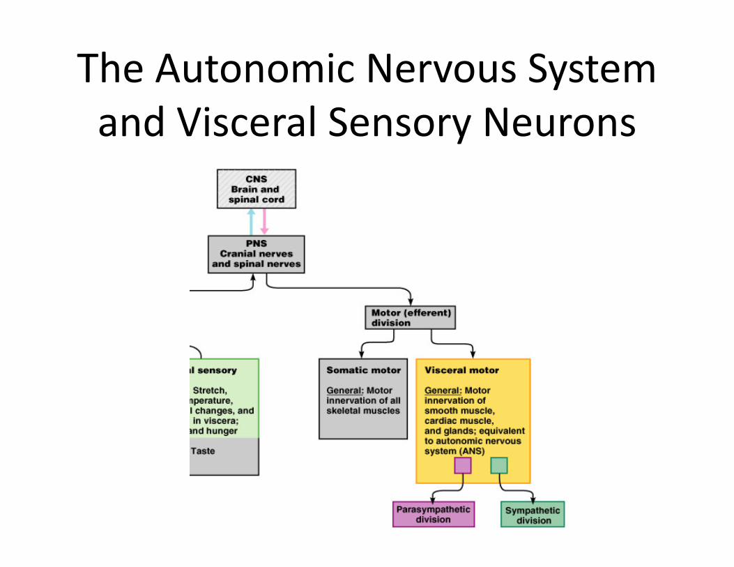
• The ANS – a system of motor neurons– The general visceral motor division of the PNS– Innervates smooth muscle, cardiac muscle, and glands
– Regulates visceral functions• Heart rate, blood pressure, digestion, urination . . .
The Autonomic Nervous System and Visceral Sensory Neurons
Comparison of Autonomic and Somatic Motor Systems
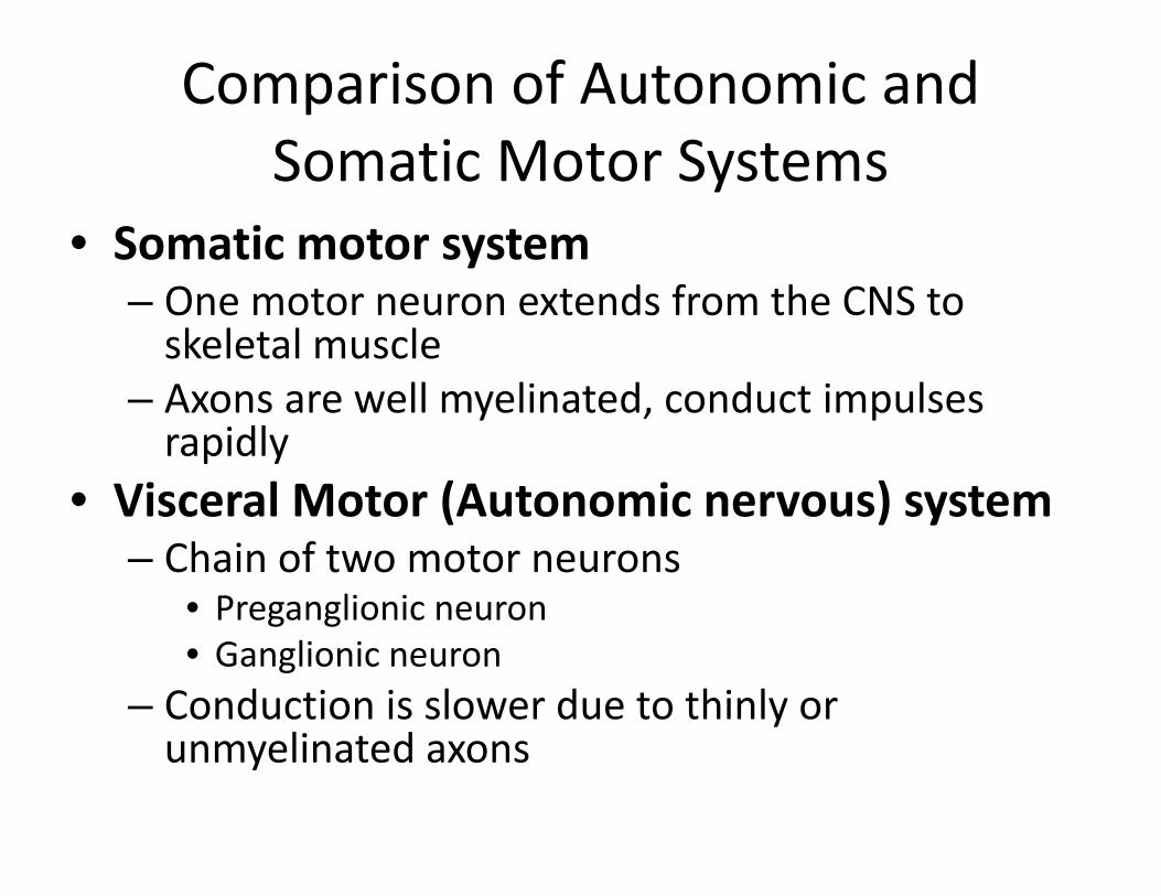
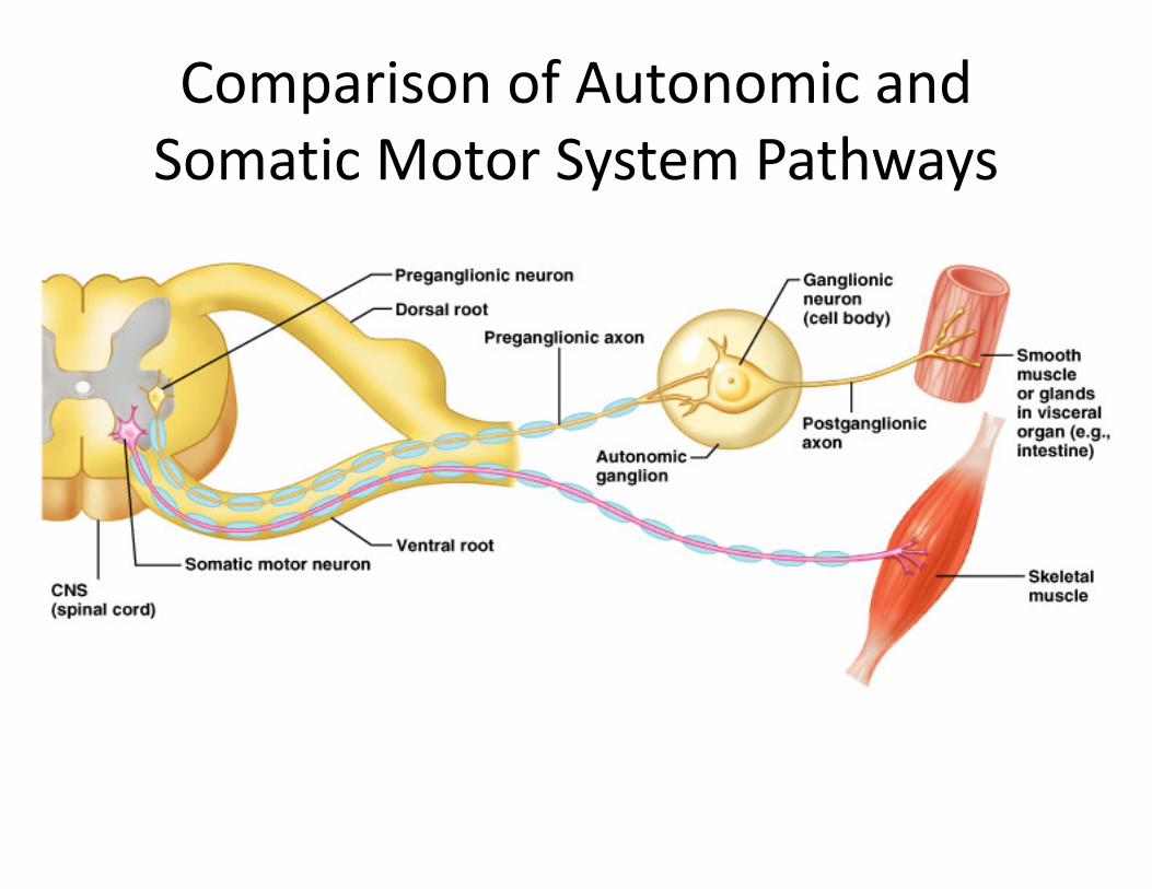
• Somatic motor system– One motor neuron extends from the CNS to skeletal muscle
– Axons are well myelinated, conduct impulses rapidly
• Visceral Motor (Autonomic nervous) system– Chain of two motor neurons
• Preganglionic neuron• Ganglionic neuron
– Conduction is slower due to thinly or unmyelinated axons
Comparison of Autonomic and Somatic Motor System Pathways
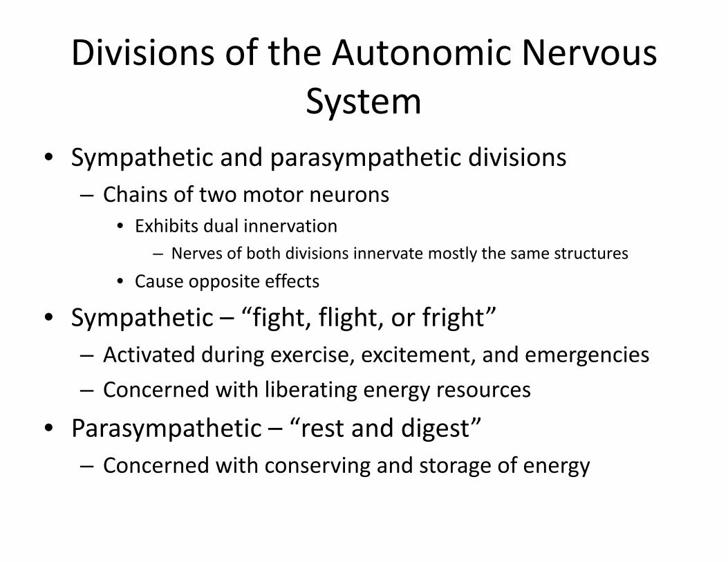
Divisions of the Autonomic Nervous System
• Sympathetic and parasympathetic divisions– Chains of two motor neurons
• Exhibits dual innervation– Nerves of both divisions innervate mostly the same structures
• Cause opposite effects
• Sympathetic – “fight, flight, or fright”– Activated during exercise, excitement, and emergencies– Concerned with liberating energy resources
• Parasympathetic – “rest and digest”– Concerned with conserving and storage of energy
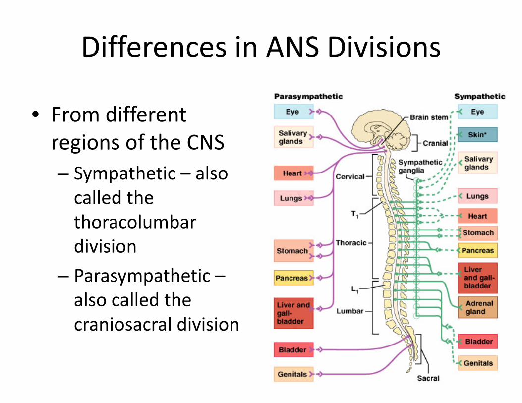
Differences in ANS Divisions
• From different regions of the CNS– Sympathetic – also called the thoracolumbar division
– Parasympathetic –also called the craniosacral division
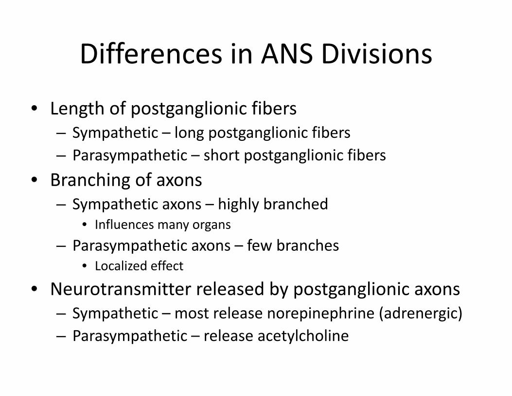
Differences in ANS Divisions
• Length of postganglionic fibers– Sympathetic – long postganglionic fibers– Parasympathetic – short postganglionic fibers
• Branching of axons– Sympathetic axons – highly branched
• Influences many organs– Parasympathetic axons – few branches
• Localized effect
• Neurotransmitter released by postganglionic axons– Sympathetic – most release norepinephrine (adrenergic)– Parasympathetic – release acetylcholine
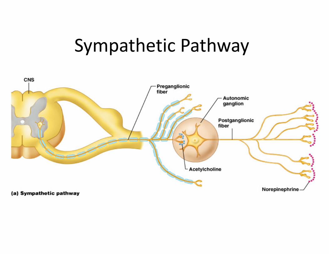
Sympathetic Pathway

Parasympathetic Pathway
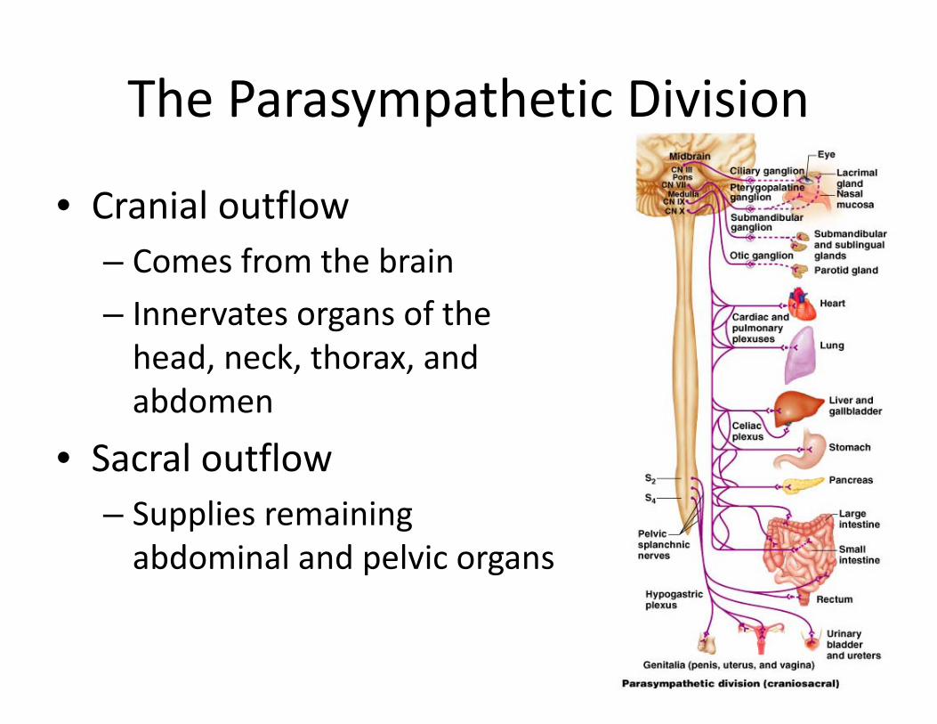
The Parasympathetic Division
• Cranial outflow – Comes from the brain– Innervates organs of the head, neck, thorax, and abdomen
• Sacral outflow – Supplies remaining abdominal and pelvic organs
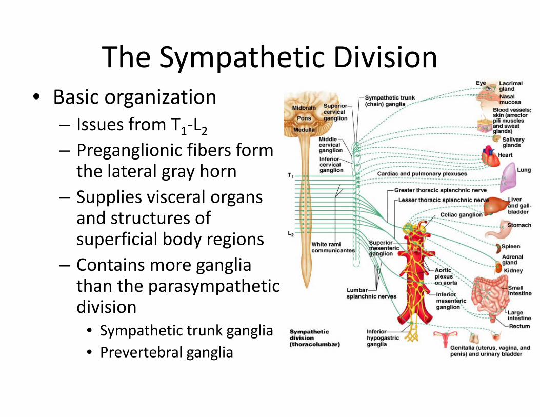
The Sympathetic Division• Basic organization
– Issues from T1‐L2– Preganglionic fibers form the lateral gray horn
– Supplies visceral organs and structures of superficial body regions
– Contains more ganglia than the parasympathetic division
• Sympathetic trunk ganglia• Prevertebral ganglia
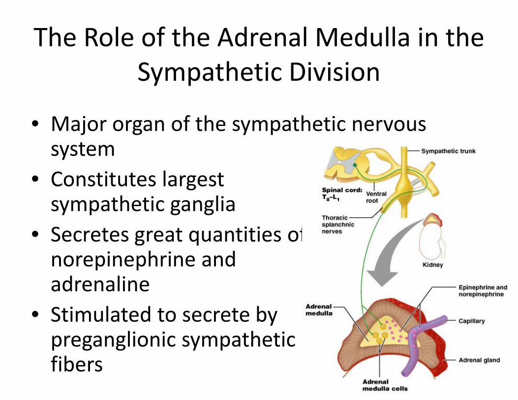
The Role of the Adrenal Medulla in the Sympathetic Division
• Major organ of the sympathetic nervous system
• Constitutes largest sympathetic ganglia
• Secretes great quantities of norepinephrine and adrenaline
• Stimulated to secrete by preganglionic sympathetic fibers
Visceral Sensory Neurons
• General visceral sensory neurons monitor:– Stretch, temperature, chemical changes, and irritation
• Cell bodies are located in the dorsal root ganglia
• Visceral pain – perceived to be somatic in origin– Referred pain
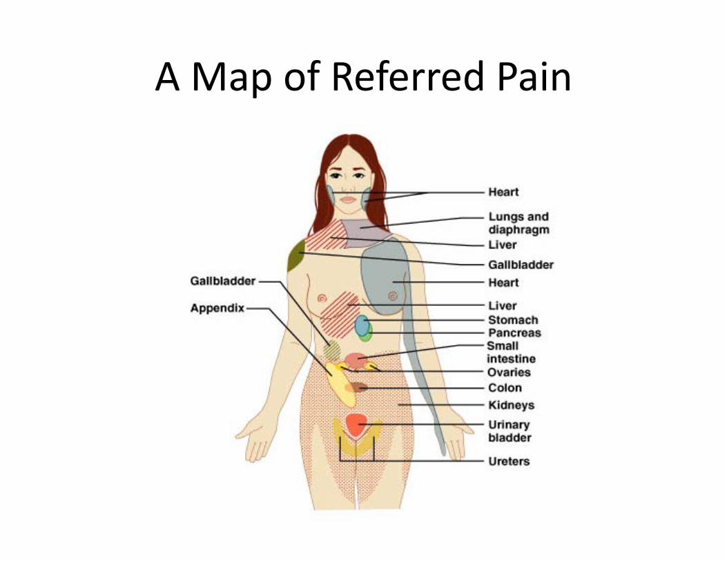
A Map of Referred Pain
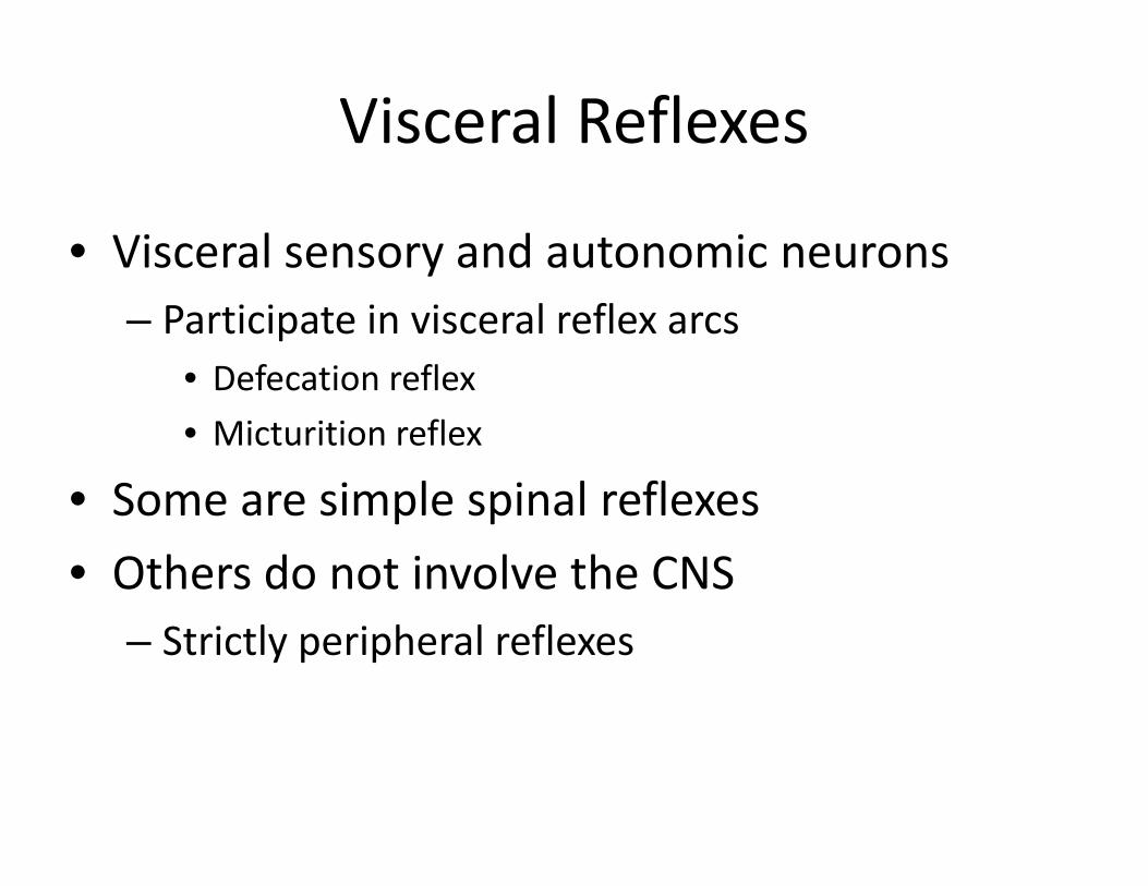
Visceral Reflexes
• Visceral sensory and autonomic neurons– Participate in visceral reflex arcs
• Defecation reflex• Micturition reflex
• Some are simple spinal reflexes• Others do not involve the CNS
– Strictly peripheral reflexes
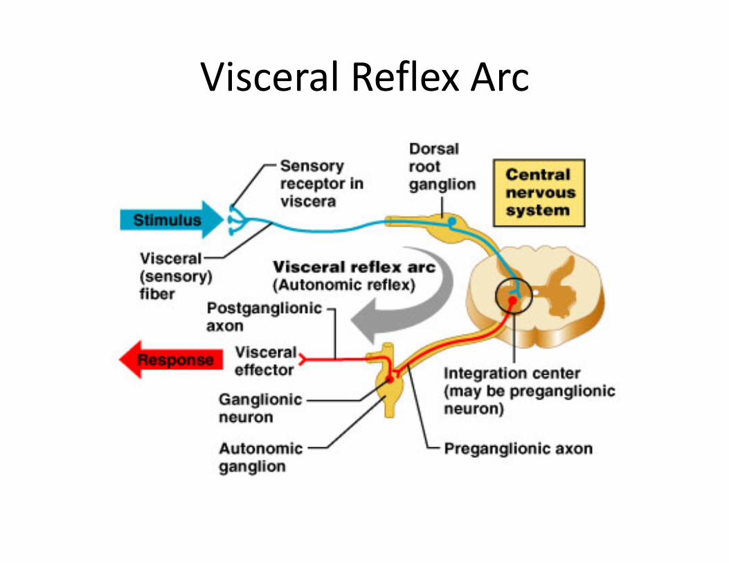
Visceral Reflex Arc
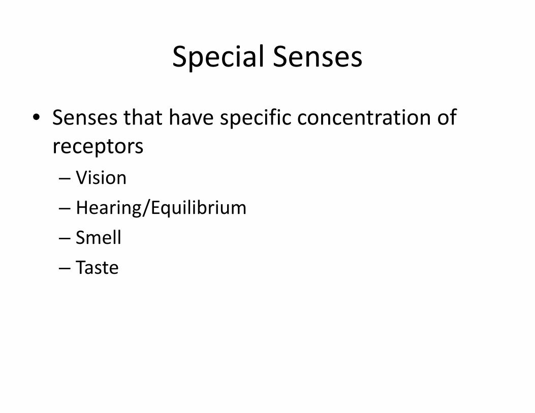
Special Senses
• Senses that have specific concentration of receptors– Vision– Hearing/Equilibrium– Smell– Taste
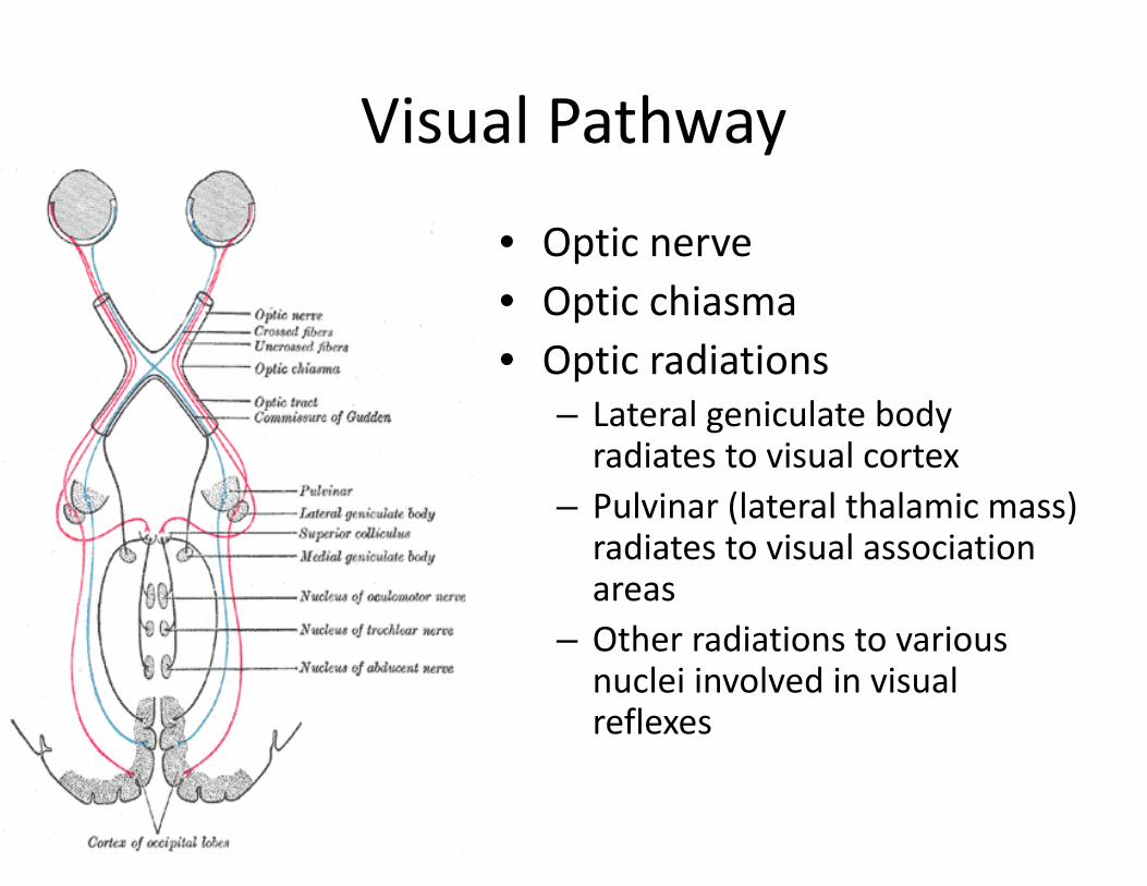
Visual Pathway
• Optic nerve• Optic chiasma• Optic radiations
– Lateral geniculate body radiates to visual cortex
– Pulvinar (lateral thalamic mass) radiates to visual association areas
– Other radiations to various nuclei involved in visual reflexes
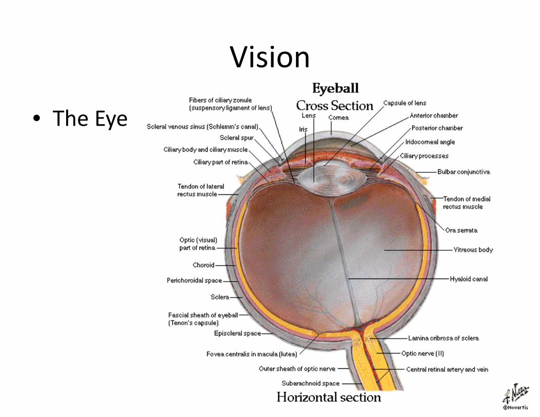
Vision
• The Eye
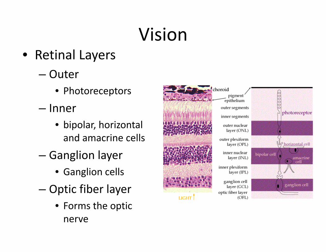
Vision• Retinal Layers
– Outer• Photoreceptors
– Inner• bipolar, horizontal and amacrine cells
– Ganglion layer• Ganglion cells
– Optic fiber layer• Forms the optic nerve

The Ear – Hearing & Equilibrium
1
2 3
1. Sound waves enter
2. Sound waves modified
3. Sound waves parsed & transduced
4. Action potentials sent
4
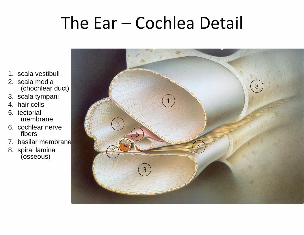
The Ear – Cochlea Detail
1. scala vestibuli2. scala media
(chochlear duct)3. scala tympani4. hair cells5. tectorial
membrane6. cochlear nerve
fibers7. basilar membrane8. spiral lamina
(osseous)
1
45
6
3
2
7
8

Basilar Membrane Resonance Frequencies
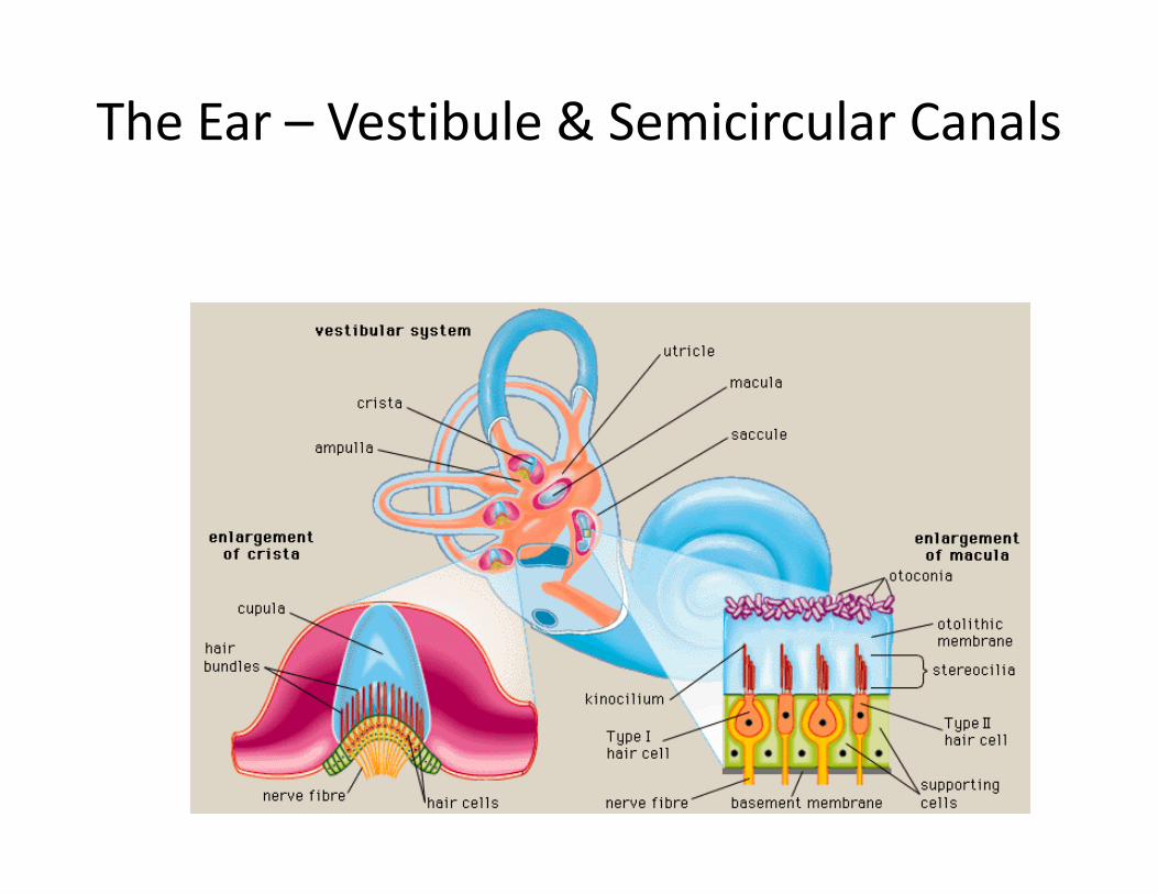
The Ear – Vestibule & Semicircular Canals
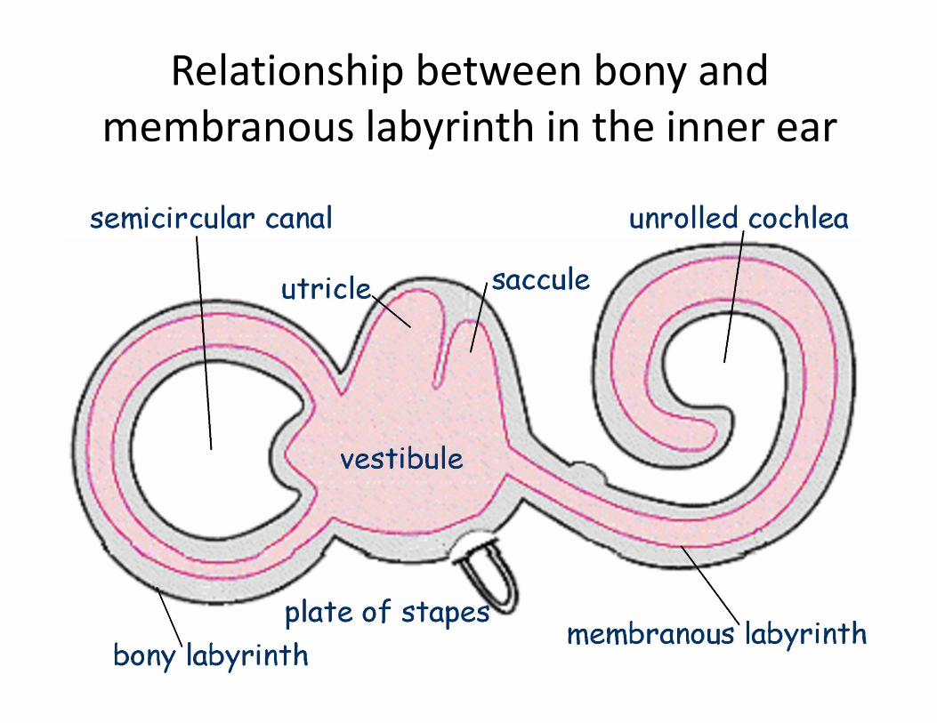
Relationship between bony and membranous labyrinth in the inner ear
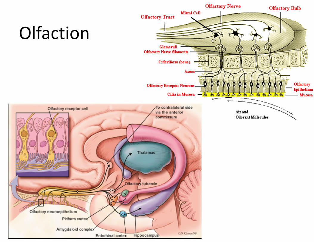
Olfaction
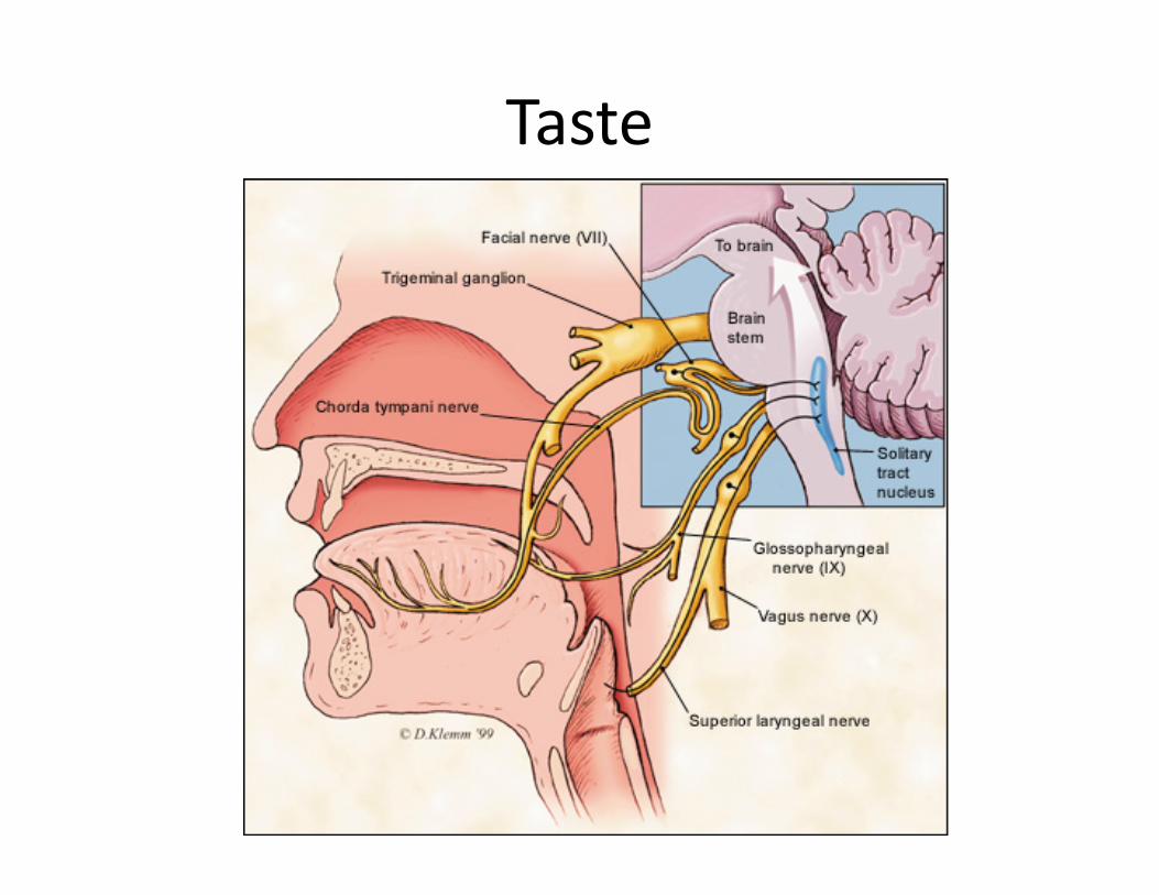
Taste
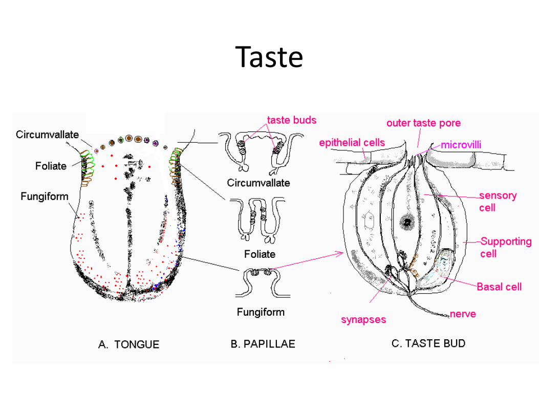
Taste