The Anticoagulant Activity Lysosomal Cationic...
10
Journal of Clinical Investigation Vol. 46, No. 4, 1967 The Anticoagulant Activity of Lysosomal Cationic Proteins from Polymorphonuclear Leukocytes * HUSSAIN I. SABA,t HAROLD R. ROBERTS, AND JOHN C. HERION t (From the Departments of Medicine and Pathology, University of North Carolina School of Medicine, Chapel Hill, N. C.) Summary. A cationic protein fraction from rabbit polymorphonuclear leu- kocyte lysosomes has been shown to exert a potent anticoagulant effect on human blood in vitro. The anticoagulant activity is detectable in the whole blood clotting time, the recalcification time of platelet-rich plasma, the pro- thrombin time, the partial thromboplastin time, and the thromboplastin gen- eration test. The lysosomal cationic proteins do not inhibit any of the known specific procoagulants. They appear to inhibit clotting by blocking the for- mation of intrinsic thromboplastin possibly by interfering with the role of phospholipids in the reaction involving Factors V and X and calcium. Introduction For many years there have been suggestions that leukocytes may play a role in blood coagula- tion (1, 2). It was not until 1951, however, that Martin and Roka (3) demonstrated prolongation of the clotting time of plasma in various clotting tests by lysates of leukocytes. Subsequently, Graham, Ebert, Ratnoff, and Moses (4) observed anticoagulant activity in a saline extract of granu- locytes. The specific substance or substances in the leukocyte responsible for this anticoagulant activity have not yet been identified. Recently the specific granules of polymorpho- nuclear leukocytes, established by Cohn and Hirsch to be lysosomes (5), have been found to contain a number of biologically active substances. In 1963 Zeya and Spitznagel isolated from poly- morphonuclear leukocyte lysosomes a group of apparently nonezymatic, highly cationic proteins that possess potent antimicrobial activity (6). Subsequent studies have demonstrated these lyso- somal cationic proteins (LCP) to be pyrogenic * Submitted for publication July 7, 1966; accepted December 22, 1966. Supported in part by U. S. Public Health Service grants AI 04925, HE 06350, and TI AM 5345. tU. S. Public Health Service fellow in hematology. t Address requests for reprints to Dr. John C. Herion, Dept. of Medicine, University of North Carolina School of Medicine, Chapel Hill, N. C. 27514. (7) and to produce inflammation and tissue injury (8-10). Since strongly cationic proteins like pro- tamine (11, 12) and synthetic basic polypeptides (13, 14) have been shown to inhibit coagulation, it seemed reasonable that cationic proteins of poly- morphonuclear leukocyte lysosomes might exhibit similar activity. This study demonstrates that lysosomal cationic proteins from polymorpho- nuclear leukocytes are very potent inhibitors of blood coagulation, and that they appear to inhibit the formation of intrinsic thromboplastin (intrin- sic prothrombin activator) by interfering with the role of phospholipid (possibly platelet mem- branes) in the last stages of blood coagulation. Methods Glassware. All glassware used for isolating cells and cell fractions was rendered endotoxin-free by either bak- ing at 180° C for 3 hours or soaking overnight in butanol, followed by repeated rinsing with pyrogen-free saline (7). Polymorphonuclear (PMN) leukocytes. Sterile peri- toneal exudates were induced in normal rabbits, 12 to 14 hours before harvesting, by ip injection of 200 ml of 0.25% glycogen in saline containing 68,000 U penicillin and 94 mg streptomycin per L. Exudates were pooled in an ice-chilled flask and the cells isolated by centrifugation as previously described (7). PMN leukocyte lysosomes and lysosomaal cationic pro- tein. The lysosomal fraction was obtained by homogeni- zation and differential centrifugation of granulocytes in 0.34 M sucrose according to the method of Cohn and Hirsch (5). 580
-
Upload
hoangquynh -
Category
Documents
-
view
221 -
download
0
Transcript of The Anticoagulant Activity Lysosomal Cationic...

Journal of Clinical InvestigationVol. 46, No. 4, 1967
The Anticoagulant Activity of Lysosomal Cationic Proteinsfrom Polymorphonuclear Leukocytes *
HUSSAIN I. SABA,t HAROLDR. ROBERTS,ANDJOHNC. HERION t(From the Departments of Medicine and Pathology, University of North Carolina School of
Medicine, Chapel Hill, N. C.)
Summary. A cationic protein fraction from rabbit polymorphonuclear leu-kocyte lysosomes has been shown to exert a potent anticoagulant effect onhuman blood in vitro. The anticoagulant activity is detectable in the wholeblood clotting time, the recalcification time of platelet-rich plasma, the pro-thrombin time, the partial thromboplastin time, and the thromboplastin gen-eration test. The lysosomal cationic proteins do not inhibit any of the knownspecific procoagulants. They appear to inhibit clotting by blocking the for-mation of intrinsic thromboplastin possibly by interfering with the role ofphospholipids in the reaction involving Factors V and X and calcium.
Introduction
For many years there have been suggestionsthat leukocytes may play a role in blood coagula-tion (1, 2). It was not until 1951, however, thatMartin and Roka (3) demonstrated prolongationof the clotting time of plasma in various clottingtests by lysates of leukocytes. Subsequently,Graham, Ebert, Ratnoff, and Moses (4) observedanticoagulant activity in a saline extract of granu-locytes. The specific substance or substances inthe leukocyte responsible for this anticoagulantactivity have not yet been identified.
Recently the specific granules of polymorpho-nuclear leukocytes, established by Cohn andHirsch to be lysosomes (5), have been found tocontain a number of biologically active substances.In 1963 Zeya and Spitznagel isolated from poly-morphonuclear leukocyte lysosomes a group ofapparently nonezymatic, highly cationic proteinsthat possess potent antimicrobial activity (6).Subsequent studies have demonstrated these lyso-somal cationic proteins (LCP) to be pyrogenic
* Submitted for publication July 7, 1966; acceptedDecember 22, 1966.
Supported in part by U. S. Public Health Servicegrants AI 04925, HE 06350, and TI AM 5345.
tU. S. Public Health Service fellow in hematology.t Address requests for reprints to Dr. John C. Herion,
Dept. of Medicine, University of North Carolina Schoolof Medicine, Chapel Hill, N. C. 27514.
(7) and to produce inflammation and tissue injury(8-10). Since strongly cationic proteins like pro-tamine (11, 12) and synthetic basic polypeptides(13, 14) have been shown to inhibit coagulation,it seemed reasonable that cationic proteins of poly-morphonuclear leukocyte lysosomes might exhibitsimilar activity. This study demonstrates thatlysosomal cationic proteins from polymorpho-nuclear leukocytes are very potent inhibitors ofblood coagulation, and that they appear to inhibitthe formation of intrinsic thromboplastin (intrin-sic prothrombin activator) by interfering with therole of phospholipid (possibly platelet mem-branes) in the last stages of blood coagulation.
MethodsGlassware. All glassware used for isolating cells and
cell fractions was rendered endotoxin-free by either bak-ing at 180° C for 3 hours or soaking overnight in butanol,followed by repeated rinsing with pyrogen-free saline(7).
Polymorphonuclear (PMN) leukocytes. Sterile peri-toneal exudates were induced in normal rabbits, 12 to14 hours before harvesting, by ip injection of 200 ml of0.25% glycogen in saline containing 68,000 U penicillinand 94 mg streptomycin per L. Exudates were pooled inan ice-chilled flask and the cells isolated by centrifugationas previously described (7).
PMNleukocyte lysosomes and lysosomaal cationic pro-tein. The lysosomal fraction was obtained by homogeni-zation and differential centrifugation of granulocytes in0.34 M sucrose according to the method of Cohn andHirsch (5).
580

ANTICOAGULANTACTIVITY OF LYSOSOMALCATIONIC PROTEINS
The lysosomal cationic protein fraction was preparedby acid extraction (0.2 N H2S04) of lysosomes, followedby precipitation with cold ethanol, 20%o vol/vol, as de-scribed by Zeya and Spitznagel (6). The precipitate wasdissolved in 0.01 N HCl, dialyzed against 0.01 N HC1overnight, then against distilled water for 6 hours, andlyophilized. After removal of the LCP, a second frac-tion was obtained from the 20% ethanol supernatant byprecipitation with cold ethanol (45% vol/vol). Thisfraction was also dissolved in 0.01 N HCl, dialyzed againstdistilled water, and lyophilized. Both the 20% ethanolprecipitate (LCP) and the 45%o ethanol precipitate weretested for the presence of the following enzymes: ly-sozyme (15), ribonuclease and deoxyribonuclease (16),#l-glucuronidase (17), acid and alkaline phosphatases(18), and cathepsin (19). All batches of LCP testedpossessed potent anticoagulant activity, but when com-pared in a standard partial thromboplastin time test,there was slight variation in potency among differentbatches probably due to loss of activity during lyophiliza-tion or from qualitative differences in the protein ob-tained from different batches of cells. Thus, in the ex-periments to be described, the amount of LCP necessaryfor a desired effect was not always the same. The LCPused in clotting studies were dissolved just before use inbarbital buffer, pH 7.35, ionic strength 0.154 (20). Al-though LCP were more readily soluble in weak acid, theresulting low pH was deemed undesirable for the clottingtests.
Lysozyme,' ribonuclease,2 deoxyribonuclease,2 p8-glucu-ronidase,2 and acid and alkaline phosphatases 2 were ob-tained commercially and tested for anticoagulant activityin the partial thromboplastin time and thromboplastingeneration tests in the same concentrations as those usedfor testing LCP.
Human citrated plasma was prepared by collecting ve-nous blood from five normal donors into 3.2% sodiumcitrate (1 part to 8 parts blood) and centrifuging at 40 Cfor 30 minutes at 2,000 g. The pooled plasma was storedat - 200 C and thawed at 370 C just before use. Plate-let-rich plasma was prepared by centrifuging citrated ve-nous blood at 40 C for 5 minutes at 400 g.
Plasmas deficient in Factors V (proaccelerin), VII[SPCA (serum prothrombin conversion accelerator),proconvertin], VIII (AHF, antihemophilic factor), IX(PTC, plasma thromboplastin component), X (Stuartfactor), or XII (Hageman factor) were prepared as de-scribed above but from patients with known congenitaldeficiency of the specific factor; these patients had notbeen transfused within 2 weeks before giving blood forthis study. Plasma deficient in Factor XI (PTA, plasmathromboplastin antecedent) was prepared from normalplasma by the method of Horowitz, Wilcox, and Fuji-moto (21).
Cephalin was either prepared from human brains asdescribed previously (22) or obtained commercially asThrombofax.3
1 Nutritional Biochemicals Corp., Cleveland, Ohio.2 Sigma Chemical Co., St. Louis, Mo.-3 Ortho Pharmaceutical Corp., Raritan, N. J.
Inosithin, 0.05%, used as the source of lipid in some ofthe thromboplastin generation tests (23), was obtainedcommercially as the dry powder.4
Tissue thromboplastin was obtained commercially asSimplastin.5
Thrombin 6 (topical, bovine) was diluted to the desiredconcentration with saline in siliconized glass tubes justbefore use.
Russell's viper venom (RVV) was the commercialpreparation, Stypven.7
Fibrinogen was prepared from canine plasma by(NH4) 2S04 precipitation as described previously (24).Human fibrinogen8 was dissolved in barbital buffer anddialyzed overnight against barbital buffer at 50 C.
Coagulation studies. The following tests were per-formed at least in duplicate with and without LCP: re-calcification time (25) ; one stage prothrombin time (PT)(26); partial thromboplastin time (PTT) (27); throm-bin clotting time (28); and specific assays for prothrom-bin (24) and Factors VIII (27), IX (29), X (30), XI(21), and XII (31). Factor V assay was performed bythe technique of the Factor VIII assay except that thesubstrate plasma was obtained from a patient congeni-tally deficient in Factor V (32). The effect of LCP onFactor VII was determined by adding LCP to normalplasma, diluting the resulting mixture to 10% with buf-fer, and comparing its corrective effect on Factor VII-deficient plasma with a simultaneous control containingbuffer without LCP. In all assays for specific factors theconcentration of LCP was adjusted so as to add anamount that prolonged the PTT of undiluted normalplasma to 150 seconds or greater (control 60 to 80seconds).
Except for the substitution of inosithin for plateletsin some studies, thromboplastin generation tests (TGT)were performed by the method of Biggs and Douglas(33). LCP were added to the incubation mixture eitherat the beginning of incubation (zero time) or after thegeneration of maximal thromboplastin activity as deter-mined by the shortest clotting time of the substrate plasma.In other experiments using the TGT, LCP were added tothe substrate plasma.
The effects of LCP were also tested in a multistagesequential clotting system. All reagents for this test,except the phospholipid, were prepared according tomethods previously described (34). In this system,crude Factor X, prepared from serum obtained from aFactor IX-deficient patient, was activated by Russell'sviper venom, crude Factor V was prepared from hemo-philic plasma, and crude prothrombin was prepared fromFactor X-deficient plasma. When all reagents weresupplied in the stages and sequence as shown in Figure7, the clotting time varied from about 27 to 36 seconds.When any clotting factor was omitted, the clotting time
4Associated Concentrates, Long Island, N. Y.5 Warner-Chilcott Laboratories, Morris Plains, N. J.6 Parke, Davis, Detroit, Mich.7 Burroughs Wellcome, Tuckahoe, N. Y.8 Merck Sharp & Dobme, West Point, Pa.
581

582 H. I. SABA, H. R. ROBERTS, AND J. C. HERION
*by others (8-10, 35-37). Most of these iyso-somal enzymes, however, were present in the frac-tion precipitated with ethanol, 45% vol/vol.
The lysosomal cationic protein fraction wasfound to be heterogenous when subjected to elec-trophoresis on cellulose acetate strips in acetatebuffer, pH 4.0 for 1 hour (200 v, 0.002 amp) asreported by others (9, 10, 35-37).
Commercial preparations of lysozyme, ribo-nuclease, deoxyribonuclease, /3-glucuronidase, andacid and alkaline phosphatases, added to clottingsystems in approximately the same concentrationas LCP, had no significant anticoagulant activity.All the cell fractions obtained during the process-ing of polymorphonuclear leukocytes for isolationof lysosomes were tested. None of these, exceptthe lysosomal fraction, exhibited anticoagulant ac-tivity. Although most of the anticoagulant activ-ity was found in the LCP, similar activity was
0., 0 200 3004,0,0,detected in the fraction containing lysosomal en-
O 100 200 300 400 500 zymes. This was attributed to contaminatingLCP in ug LCP and was supported by finding, upon electro-
*FECT OF LYSOSOMAL CATIONIC PROTEINS phoresis of this fraction with enzymes, bands cor-
IE RECALCIFICATION TIME OF NORMALPLASMA. responding to LCP. The lysosomal membranestelet-rich plasma, 0.1 ml, plus 0.1 ml of (washed sediment of acid-lysed granules), like allor buffer containing LCP was clotted by the other nonlysosomal leukocyte fractions, pos-of 0.02 M CaCl2. The abscissa shows mi- sessed no anticoagulant activity.
LCP in final clotting mixtures.Effect of LCP on whole blood and plasma.
bondss except that when Factor X was omitted LCP significantly prolong the clotting of whole
time was 114 seconds, presumably due to blood in proportion to the amount added. Foritamination of the prothrombin reagent. example, 600 tkg of LCP added to 1 ml of wholeof LCP on the activation of Factor X was blood resulted in a clotting time of 45 minutesing to the method described by Breckenridge (control 17 minutes) ; the clotting time was 58(34), but in some experiments 0.2 ml addi- minutes when 900jug LCP was added to 1 mlolipid was added to the incubation mixture whole blood. Similar results were obtained whenition of LCP. whole blood wasdeter minede controls for all clotting studies were per- the clotting time of whole blood was determinedsubstituting barbital buffer alone for buffer with the Lee-White technique. The recalcifica--P. tion time of platelet-rich plasma was also pro-
longed by LCP in proportion to the amount ofResults LCP added (Figure 1). The anticoagulant activ-
somal cationic protein fraction. The ity of LCP was demonstrable in both the pro-raction obtained by precipitation with thrombin and partial thromboplastin time as
1, 20%o vol/vol, was found to be solu- shown in Figure 2. Here, too, the prolongationled water and in solutions of acid pH was proportional to the amount of LCP added;ability decreased
in alkaline solutions. buffer alone in the controls produced no detectable
n was found to be free from the known prolongation in either the PT or PTT.enzymes lysozyme, ribonuclease, deoxy- Effect of LCP on specific procoagulants. To
~, 83-glucuronidase, acid and alkaline determine whether the prolonged clotting time of
s, and cathepsins as previously shown plasma and whole blood were due to inhibition of,J
P-
45.
40-
235-
4
z
2 30-z
n25 -
0iU. 20-0w
I" 15-
z
I-- 100-J
5-
FIG. 1. EF(LCP) ON TICitrated, plaineither bufferadding 0.1 mlcrograms of:
was > 300 secthe clottingFactor X cor
The effecttested accordiand Ratnofftional phosphafter the addi
Appropriat'formed by s1containing L(
The lyso.lysosomal fcold ethanolble in distillbut its soluThis fractiolysosomal etribonucleasephosphatase
582

ANTICOAGULANTACTIVITY OF LYSOSOMALCATIONIC PROTEINS
one or more of the procoagulants, specific assaysfor Factors V, VIII, IX, X, XI, and XII were
performed after adding LCP to normal plasmain a final concentration of 1.5 mg per ml. Theresults, shown in Figure 3, demonstrated no spe-
cific inhibition of any of the procoagulants studiedeven though assays were carried out on portionsof plasma containing sufficient LCP to prolongthe PTT of the undiluted plasma to more than180 seconds. Although not shown in the Figure,similar experiments demonstrated that LCP didnot inhibit Factor VII; a diluted sample of a nor-
mal plasma-LCP mixture had the same correctiveeffect on Factor VII-deficient plasma as did a
diluted sample of a normal plasma-buffer mixture.In normal plasma containing 1.5 mg of LCP per
ml, the concentration of prothrombin, determinedby specific assay, was 300 U per ml, a value simi-lar to that of the control plasma containing onlybuffer.
Effect of LCP on thromboplastin generation.When LCP were added to the thromboplastingeneration mixture at the beginning of incubation,no detectable thromboplastin activity appearedeven after incubation for as long as 30 minutes(Figure 4A). The same amount of LCP, addedafter 8 minutes of thromboplastin generation, alsoinhibited intrinsic thromboplastin activity, but theinhibitory effect was less marked than when LCPwas added at zero time. The apparent progres-
sive inhibitory effect of LCP when added after
280 FACTOR 1: FAC70R FACTOR3A
260 TEST o CONTROL * TEST o CONTROL * TEST o CONTROL
V) 240a
o 220
W200
E180
160z
I- 1400
20
100
280
260
240en
Z 2200
U 200z
w 180
I.140
0
'0 120
100
80
*FACTOR x
* TEST 0 CONTROL
:0<I toL
FACTOR 3TEST
0 CONTROL
FACTOR 3T* TEST 0 CONTROL
N"Nl
125 2.5 5 10 1.25 2.5 10 1.25 2.5 5 10
CONCENTRATIONOF PLASMASIN PERCENT
FIG. 3. EFFECT OF LCP ON FACTORSV, VIII, IX, X, XI,
AND XII. One ml of either buffer or buffer containing 3mg of LCP was incubated for 2 to 3 minutes at room
temperature with 1.0-ml samples of pooled normal plasma.(Partial thromboplastin time on plasma with LCP, 180seconds; on control, 69.2 seconds.) Test and controlplasmas were assayed for specific procoagulants as de-scribed under Methods.
25
en
0 20us
zISw
'o,0 10
5
A.
400 -
350 -
300-
250-
200-
150-
100-75
8.
0 10 20 30 40 50 60 70 80 90100 0 16 20 30 40 50 60 70 80 90 100
LCP in g
FIG. 2. EFFECT OF LCP ONA) THE PROTHROMBINTIME
ANDB) THE PARTIAL THROMBOPLASTINTIME. The abscissashows micrograms of LCP in final clotting mixtures. A)Pooled normal plasma, 0.1 ml, plus 0.1 ml of either bufferalone or buffer containing LCP was clotted by adding 0.2ml thromboplastin-CaCl2 mixture. B) Pooled normalplasma, 0.1 ml, plus 0.1 ml of either buffer or buffer con-
taining LCP plus 0.1 ml of cephalin was clotted by adding0.1 ml of 0.02 M CaC12.
generation of maximal thromboplastin activitysuggested that formation of thromboplastin didnot continue in the presence of LCP. To furthertest this possibility we added LCP to the substrateplasma, rather than to the incubation mixture.Illustrative results of such experiments are shownin Figure 4B. The inhibitory effect in this cir-cumstance is just barely detectable even thoughthe final concentration of LCP in the substrateplasma was comparable to that of the experimentsshown in Figure 4A.
Lysosomal cationic proteins not only inhibitedthromboplastin activity, but the degree of inhibi-tion was increased by greater amounts of protein.This is apparent on comparison of curves A, B,and C of Figure 5. This inhibitory effect, how-ever, was not reversed by dilution since, if thisoccurred, the curves shown in Figure 5 would not
583

H. I. SABA, H. R. ROBERTS, AND J. C. HERION
AB
ADDEDAT ZERO TIME* BUFFER OLCP
ADDEDAT 8 MINUTES* BUFFER OLCP
100
80
60
40
20
o
*- NORMALCONTROL*. SUBSTRATEPLASMA BUFFERD---o SUBSTRATEPLASMA+LCP
.*o
TIME OF INCUBATION IN MINUTES
FIG. 4. EFFECT OF LCP ON THROMBOPLASTINGENERATION. The thromboplastin generation mixtures, incubated at
370 C, contained 0.5 ml of each of the following: Al (OH) 3-adsorbed plasma, diluted 1:5; normal human serum, di-luted 1: 10; 0.05% inosithin in buffer; and 0.025 M CaCl2. A) At zero time and after 8 minutes of incubation, 0.5 ml
of either buffer alone or buffer containing LCP (3 mg per ml) was added to the thromboplastin generation mixture.At intervals after addition of buffer or LCP, 0.1 ml of the incubation mixture was added to 0.2 ml of normal plasmaand the mixture clotted by adding 0.1 ml 0.025 M CaCl2. B) At intervals beginning with zero time, 0.1 ml of thethromboplastin generation mixture was added to 0.2 ml normal plasma containing 0.1 ml of either buffer alone or buf-fer containing LCP (600 ,ug per ml) and the mixture clotted by adding 0.1 ml of 0.025 M CaCl2.
be parallel, but rather would converge toward thecontrol curve.
Effect of LCP on various stages of thrombo-plastin formation. Since the TGT probably re-
flects a composite effect of several reactions, LCPwere tested in the individual reactions thought to
be necessary for thromboplastin generation. The
activation of Factor X is likely to be one of these
(38). From the results shown in Figure 6, it is
apparent that after incubation for 60 to 90 sec-
onds, Factor X was maximally activated by RVV
even in the presence of LCP. The prolongedclotting time observed in the test system seemed
to be related to an effect of LCP on somethingother than the activation of Factor X; this effect
was reversed by adding more cephalin to the incu-
bation mixture containing LCP.The effect of LCP on other reactions necessary
for the production of prothrombin activator was
measured in the five stage test illustrated in Fig-ure 7. In this system the addition of LCP before
Factor V markedly inhibited the formation of in-trinsic thromboplastin as indicated by the pro-
longed clotting time (test A). The same amount
of LCP, added after Factor V, did not inhibit theformation of thromboplastin; the clotting time ap-
proximated that of the control (test B). In re-
peated tests in which LCP were added after Fac-tor V, the clotting time varied slightly with theconcentration of phospholipid and LCP, but inall these tests the clotting time was nearly thatof the control. It is also evident that, under theconditions of the five stage test, LCP had no de-tectable effect on the thrombin-fibrinogen re-
action; fibrinogen clotted as promptly as in thecontrol (test C).
The findings in the five stage test suggestedthat LCP inhibited the generation of intrinsicthromboplastin by interfering with one or more
of the reactions involving Factor V, Factor X,phospholipid, and calcium. The formation of a
fine precipitate upon mixing 300 ,ug of LCP with
Coen
z0
U
z
z
I~-0-iC,
584

ANTICOAGULANTACTIVITY OF LYSOSOMALCATIONIC PROTEINS
0.1 ml of a 1:25 dilution of cephalin suggested thepossibility that LCP interacted and thus interferedwith the action of phospholipid in the intrinsicthromboplastin generation. The phospholipid mi-celles carry an electronegative surface charge (39)opposite to that of electropositive LCP. The pos-sibility of specific electrostatic interaction betweenLCP and phospholipid was examined further inthe TGT (Figure 8). After generation of maxi-mal thromboplastin activity, LCP added to theincubation mixture prolonged the clotting timeof substrate plasma. This effect of LCP waspromptly reversed when more cephalin was added
350 -
C
300
Cn 250 4.4.
0 4
Z A.Q 4.~~~~~~~~~~~/410 -.
100 4.
50-
o 4.0
0I
100 5 . 12.5 6.25THROMBOPLASTINACTIVITY IN PERCENT
FIG. 5. EFFECT OF DILUTION ON THROMBOPLASTIN-LCPMIXTURE. Standard thromboplastin generation mixtures,prepared as described, were incubated at 37° C. After 10minutes, when maximal thromboplastin activity was foundto be present, the following reagents were added to thethromboplastin generation mixtures: test A: 0.5 ml buffer;test B: 0.5 ml buffer containing LCP (1.5 mg per ml) ;test C: 0.5 ml buffer containing LCP (3 mg per ml).After these additions the mixtures were placed in a melt-ing ice bath and serial dilutions of each made with barbitalbuffer. After warming again to 37° C, 0.1 ml of eachdilution was added to 0.2 ml of normal plasma and themixture clotted by adding 0.1 ml of 0.025 M CaClp, The100%o values on the abscissa represent the thrombo-plastin activity of 0.1 ml of the undiluted incubation mix-ture, the 5017o values a 1: 2 dilution, and so on.
* BUFFER ONLYA BUFFER + CEPHALINo LCP + CEPHALINo LCP + SALINEA LCP ONLY
C,)az0
wC,)
z
w
0
z
-i
-J
20-
40.
60-
80-
0 30 60 90 120 150 180
TIME OF INCUBATION IN SECONDS
FIG. 6. EFFECT OF LCP ON THE ACTIVATION OF FACTORx. The activation mixtures, incubated at 370 C, con-tained 0.1 ml of each of the following: crude Factor X,Russell's viper venom (diluted 1: 200,000), cephalin,0.025 MCaCl2, and either buffer alone or buffer containingLCP (2 mg per ml). To separate activation mixtures,with and without LCP, we added 0.2 ml of either cepha-lin or saline in an attempt to shorten the clotting time ofthe system containing LCP. For all five test systemsdesignated in the legend, a separate activation mixturewas incubated for each period of time shown on theabscissa. When incubation of the mixtures was com-pleted, 0.1 ml of Factor X-deficient plasma was addedto each and the clotting time recorded.
to the incubation mixture. The inhibitory effectof LCP on intrinsic thromboplastin generationalso occurred when platelets, rather than plateletsubstitute, were used in the TGT, and it was re-versible by the addition of more platelets. Com-plete reversal of the inhibitory effect of LCP onintrinsic thromboplastin formation was not ob-served when increased amounts of Factor V, Fac-tor X, or calcium were added to the incubationmixture used for the TGT (Figure 9).
Antithrombin activity. Antithrombin activitywas assayed by adding LCP to bovine thrombinand comparing the subsequent conversion of fi-brinogen to fibrin by this thrombin. In the con-trol, LCP were replaced with buffer.
Varying amounts of LCP did not prolong thethrombin clotting time of canine fibrinogen (TableI). Actually with increased amounts of LCP thethrombin-fibrinogen clotting time was consistentlyshorter than the control. Similar results were
585

H. I. SABA, H. R. ROBERTS, AND J. C. HERION
STAGE 1 STAGE 2 STAGE 3 STAGE 4 STAGE 5
(ACTIVATION OF FACTOR X) (ADDITION OF PHOSPHOLIPID) (ACTIVATION OF FACTOR V) (GENERATION OF THROMBIN) (CLOTTING OF FIBRINOGEN)
0.1 ml OF EACH OF THE 0.1 ml CEPHALIN 1:25 DIL. 0.1 ml OF CRUDE 0.1 ml OF CRUDE 0.2 ml OF PURIFIED
FOLLOWING REAGENTS: FACTOR V PROTHROMBIN HUMANFIBRINOGEN
1. CRUDE FACTOR X (INCUBATED 30 SECS.) (INCUBATED 90 SECS.) (CLOTTING TIME NOTED)
37'C 37'C 37'C 37'C2. RUSSELL'S VIPER VENOM
1:200,000 DIL. WITH
BUFFER
3. .05 M CaC12
(INCUBATED 60 SECS.)
370C
CLOTTING TIME IN SECONDS
TEST (LCP) CONTROL(BUFFER)
A. STAGE 1 - STAGE 2 - |150m LCP OR BUFFER I STAGE 3 - OSTAGE4 -- STAGE 5
B. STAGE 1 - STAGE 2 ---STAGE 3 | 150pg LCP OR BUFFER -* STAGE 4 - STAGE 5
C. STAGE 1 -4 STAGE 2 -4 STAGE 3 -4 STAGE 4 |150 pg LCP OR BUFFER - O-STAGE 5
218.8'
31.7
CLOTTEDIMMEDIATELY
29.0
27.2
CLOTTEDIMMEDIATELY
FIG. 7. EFFECT OF LCP AT VARIOUS STAGESOF A SEQUENTIAL CLOTTING SYSTEM. Factors and reagents were pre-
pared as described under Methods. In control tests, omission of any of the factors or reagents listed under the variousstages produced a clotting time greater than 300 seconds. Test mixtures were transferred to an ice bath before theaddition of 0.1 ml of either buffer alone or buffer containing LCP, and afterwards returned to the water bath at 370 Cfor 60 seconds before completing subsequent stages.
obtained when purified human fibrinogen orpooled normal citrated plasma was substituted forcanine fibrinogen.
Discussion
These studies indicate that the lysosomal cat-ionic proteins of polymorphonuclear leukocytes
TABLE I
Effect of LCP on thrombin-fibrinogenclotting system
Final concen-tration of
LCP in test Clottingmixture* timet
jIg seconds0 39.9
10 33.318.7 28.637.5 32.975 31.6
150 25.6300 25.0
* Test mixture contained 0.1 ml thrombin solution (2.5U per ml), 0.1 ml of buffer or buffer containing LCP, and0.3 ml of calcium-imidazole-saline-acacia mixture.
t Measured after adding to the test mixture 0.1 mlfibrinogen solution (300 mg per 100 ml).
possess a potent anticoagulant effect. This effectis manifested in many of the conventional tests ofcoagulation including whole blood clotting time,recalcification time, prothrombin time, and partialthromboplastin time. These cationic proteins,however, do not appear to act by inhibiting orinactivating any of the known plasma procoagu-lants, since in the assay for these factors normalplasma previously incubated with LCP is no dif-ferent from control plasma. Although the possi-bility exists that one or more of the specific pro-coagulants in plasma is inhibited by LCP, and theinhibition reversed by the dilution necessary forassay, this explanation seems unlikely, since dilu-tion does not reverse the inhibitory effect of LCPin the TGT. Rather, the results of studies usingthe TGT suggest that LCP interfere with the for-mation of intrinsic thromboplastin, thromboplastinitself, or both. The normal results obtained whenLCP were added to the substrate plasma, ratherthan to the thromboplastin incubation mixture,make it more likely that LCP act primarily byinterfering with the formation of thromboplastin.This interpretation is supported by the results ob-
ALWITIUN UF lLbl ANU LUNIKUL bUbblANULb Al VARIUUb .)TAGL!)
586
ANTICOAGULANTACTIVITY OF LYSOSOMALCATIONIC PROTEINS
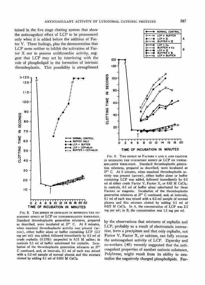
tained in the five stage clotting system that showthe anticoagulant effect of LCP to be pronouncedonly when it is added before the addition of Fac-tor V. These findings, plus the demonstration thatLCP seem neither to inhibit the activation of Fac-tor X nor to possess antithrombin activity, sug-gest that LCP may act by interfering with therole of phospholipid in the formation of intrinsicthromboplastin. This possibility is strengthened
>120,
120'
110
100 -
90e)
z80-
w
en
Z 70-
w
_ 60
0
Z 50-
I.-
0-j 40Q
30-
20
10
ii
i
i
i
NORMALCONTROL*-. BUFFER ONLY*#-* LCP + BUFFERA LCP + CEPHALIN.......,BUFFER+ CEPHALIN
I
I I I I I I I I I r I I I I I I I I I I I I
0 2 4 6 8 10 12 14 16 18 20 22
TIME OF INCUBATION IN MINUTES
FIG. 8. THEEFFECT OF CEPHALIN IN REVERSINGTHE IN-
HIBITORY EFFECT OF LCP ON THROMBOPLASTINFORMATION.
Standard thromboplastin generation mixtures, preparedas described, were incubated at 370 C. At 8 minutes,when maximal thromboplastin activity was present (ar-row), either buffer alone or buffer containing LCP (2.5mg per ml) was added, followed immediately by 0.5 ml ofcrude cephalin (0.15%) suspended in 0.15 M saline; incontrols 0.5 ml of buffer substituted for cephalin. Incu-bation of the thromboplastin generation mixtures at 370C continued, and, at intervals, 0.1 ml of each was mixedwith a 0.2-ml sample of normal plasma and this mixtureclotted by adding 0.1 ml of 0.025 MCaCl2.
ZS*NORMALCONTROL0--*-O LCP + BUFFER
*.......^ LCP + v ....ABUFFER+ v
'- *LCP + Co.CBUFFER+Co__ LCP+x B
*- _ BUFFER+ x0-0 LCP + BUFFER
120
120
0~~~~~~~~~~~1C 100 I,-P0
o~~~~~~~
80 2 1 4 1
dr
W6 if'I
60-
o 40-I-
~~~~ ~ ~ ~ ~ ~ ~ ~ ~ ~ .
20-
0 2 4 6 8 10 12' 14 16 18 20
TIME OF INCUBATION IN MINUTES
FIG. 9. THE EFFECT OF FACTORSV AND X AND CALCIUMIN REVERSINGTHE INHIBITORY EFFECT OF LCP ON THROM-BOPLASTIN FORMATION. Standard thromboplastin genera-tion mixtures, prepared as described, were incubated at370 C. At 6 minutes, when maximal thromboplastin ac-
tivity was present (arrow), either buffer alone or buffercontaining LCP was added, followed immediately by 0.5ml of either crude Factor V, Factor X, or 0.05 MCaCl2;in controls, 0.5 ml of buffer alone substituted for theseFactors or reagents. Incubation of the thromboplastingeneration mixtures at 370 C continued, and, at intervals,0.1 ml of each was mixed with a 0.2-ml sample of normalplasma and this mixture clotted by adding 0.1 ml of0.025 M CaCl2. In A, the concentration of LCP was 2.5mg per ml; in B, the concentration was 1.5 mg per ml.
by the observations that mixtures of cephalin andLCP, probably as a result of electrostatic interac-tion, form a precipitate and that only cephalin, not
Factor V, Factor X, or calcium, can fully reverse
the anticoagulant activity of LCP. Zipursky andco-workers (40) recently suggested that the anti-coagulant properties of another cationic substance,Polybrene, might result from its ability to neu-
tralize the negatively charged phospholipids. Fur-
587

H. I. SABA, H. R. ROBERTS, AND J. C. HERION
thermore, the report that platelets bear a negativesurface charge (41), and the suggestion by Marcus(42, 43) that the activity of platelets in clottingmay depend upon the platelet membrane, are com-patible with this hypothesis.
Although these studies do not clarify the exactnature of intrinsic thromboplastin, they do pointout the essential role of phospholipid in its for-mation and confirm that Factor X, Factor V, andcalcium, in addition to phospholipid, are requiredfor the development of full intrinsic thromboplas-tin activity (34, 44). It is possible that under-standing the mechanism of the LCP activity foundin this study will provide additional informationabout the nature and process of intrinsic thrombo-plastin formation.
The lysosomes of eosinophils and basophils havenot been specifically investigated; neither have ex-tensive studies of granulocytes of different speciesbeen carried out. Eosinophils from horses, how-ever, have been reported to inhibit the generationof thromboplastin in vitro (45), and in prelimi-nary studies we have detected anticoagulant ac-tivity in a fraction of human granulocyte lyso-somes containing nonenzymatic cationic proteins.
The role of LCP as an endogenous anticoagu-lant has yet to be investigated in vivo. However,considering the huge population of polymorpho-nuclear leukocytes in the body, and their rapidturnover, it is possible that the lysosomal cationicproteins from these cells might be involved in theregulation of normal hemostasis as well as in someof the poorly understood hemorrhagic disorders.It is noteworthy that lysosomal cationic proteinsinterfere with the same stages of intrinsic throm-boplastin formation as the circulating anticoagu-lant reported in some cases of disseminated lupuserythematosus (34).
AcknowledgmentsWe are indebted to Dr. H. I. Zeya, Department of
Microbiology, for invaluable suggestions and help. Thetechnical assistance of Mrs. Marva Dowdy, Mrs. DorisSparrow, and Mr. Bruce H. Beveridge is also gratefullyacknowledged.
References1. Bunting, C. H. The leukocytes. Physiol. Rev. 1922,
2, 505.2. Fowler, W. M. The leukocytes in Hematology, rev.
2nd ed. New York, Paul B. Hoeber, 1949, p. 28.
3. Martin, H., and L. Roka. Beeinflussung der Blut-gerinnung durch Leukocyten. Klin. Wschr. 1951,29, 510.
4. Graham, R. C., Jr., R. H. Ebert, 0. D. Ratnoff, andJ. M. Moses. Pathogensis of inflammation. II.Inz zivo observations of the inflammatory effectsof activated Hageman factor and bradykinin. J.exp. Med. 1965, 121, 807.
5. Cohn, Z. A., and J. G. Hirsch. The isolation andproperties of the specific cytoplasmic granules ofrabbit polymorphonuclear leukocytes. J. exp. Med.1960, 112, 983.
6. Zeya, H. I., and J. K. Spitznagel. Antibacterial andenzymic basic proteins from leukocyte lysosomes:separation and identification. Science 1963, 142,1085.
7. Herion, J. C., J. K. Spitznagel, R. I. Walker, andH. I. Zeya. Pyrogenicity of granulocyte lyso-somes. Amer. J. Physiol. 1966, 211, 693.
8. Golub, E. S., and J. K. Spitznagel. The role oflysosomes in hypersensitivity reactions: tissue dam-age by polymorphonuclear neutrophil lysosomes.J. Immunol. 1966, 95, 1060.
9. Janoff, A., and B. W. Zweifach. Adhesion and emi-gration of leukocytes produced by cationic proteinsof lysosomes. Science 1964, 144, 1456.
10. Janoff, A., and B. W. Zweifach. Production of in-flammatory changes in the microcirculation bycationic proteins extracted from lysosomes. J.exp. Med. 1964, 120, 747.
11. Chargaff, E. Studies on the chemistry of blood co-agulation. VII. Protamines and blood clotting. J.biol. Chem. 1938, 125, 671.
12. Portmann, A. F., and W. D. Holden. Protamine(salmine) sulphate, heparin, and blood coagula-tion. J. clin. Invest. 1949, 28, 1451.
13. De Vries, A., A. Schwager, and E. Katchalski. Theaction of some water-soluble poly-a-amino-acidson blood clotting. Biochem. J. 1951, 49, 10.
14. Rubini, J. R., R. R. Becker, and M. A. Stahmann.Effect of synthetic lysine polypeptides on rabbitblood coagulation. Proc. Soc. exp. Biol. (N. Y.)1953, 82, 231.
15. Shugar, D. The measurement of lysozyme activityand the ultra-violet inactivation of lysozyme. Bio-chim. biophys. Acta (Amst.) 1952, 8, 302.
16. Schneider, W. C., and G. H. Hogeboom. Intracellu-lar distribution of enzymes. X. Deoxyribonucleaseand ribonuclease. J. biol. Chem. 1952, 198, 155.
17. Fishman, W. H. 8-Glucuronidase in Methods ofEnzymatic Analysis, H.-U. Bergmeyer, Ed. NewYork, Academic Press, 1963, p. 869.
18. Andersch, M. A., and A. J. Szczypinski. Use ofp-nitrophenylphosphate as the substrate in deter-mination of serum acid phosphatase. Amer. J.clin. Path. 1947, 17, 571.
19. Lapresle, C., and T. Webb. The purification andproperties of a proteolytic enzyme, rabbit cathepsin
588

ANTICOAGULANTACTIVITY OF LYSOSOMALCATIONIC PROTEINS
E, and further studies on rabbit cathepsin D.Biochem. J. 1962, 84, 455.
20. Biggs, R., and R. G. Macfarlane. The preparationof reagents and coagulation factors in HumanBlood Coagulation and Its Disorders, 3rd ed.Philadelphia, F. A. Davis, 1962, p. 370.
21. Horowitz, H. I., W. P. Wilcox, and M. M. Fujimoto.Assay of plasma thromboplastin antecedent (PTA)with artificially depleted normal plasma. Blood1963, 22, 35.
22. Rodman, N. F., Jr., E. M. Barrow, and J. B. Graham.Diagnosis and control of the hemophilioid stateswith the partial thromboplastin time (P.T.T.) test.Amer. J. clin. Path. 1958, 29, 525.
23. Hyun, B. H., E. A. Dawson, J. Butcher, and R. P.Custer. Studies on soybean phosphatide (ino-sithin) as a platelet substitute. Stability and ef-fective concentration in thromboplastin generationtest. Amer. J. clin. Path. 1960, 33, 209.
24. Wagner, R. H., J. B. Graham, G. D. Penick, andK. M. Brinkhous. Estimation of prothrombin bythe two-stage method in Blood Coagulation, Hem-orrhage and Thrombosis, L. M. Tocantins andL. A. Kazal, Eds. New York, Grune & Stratton,1964, p. 159.
25. Biggs, R., and R. G. Macfarlane. Blood coagulationtheory and tests of clotting function in HumanBlood Coagulation and Its Disorders, 3rd ed.Philadelphia, F. A. Davis, 1962, p. 146.
26. Quick, A. J. The clinical application of the hippuricacid and the prothrombin tests. Amer. J. clin.Path. 1940, 10, 222.
27. Langdell, R. D., R. H. Wagner, and K. M. Brink-hous. Effect of antihemophilic factor on one-stageclotting tests. A presumptive test for hemophiliaand a simple one-stage antihemophilic factor assayprocedure. J. Lab. clin. Med. 1953, 41, 637.
28. Seegers, W. H. Preparation, purification and assayof thrombin in Blood Coagulation, Hemorrhageand Thrombosis, L. M. Tocantins and L. A. Kazal,Eds. New York, Grune & Stratton, 1964, p. 181.
29. Barrow, E. M., W. R. Bullock, and J. B. Graham.A study of the carrier state for plasma thrombo-plastin component (PTC, Christmas factor) defi-ciency, utilizing a new assay procedure. J. Lab.clin. Med. 1960, 55, 936.
30. Hougie, C., E. M. Barrow, and J. B. Graham. Stuartclotting defect. I. Segregation of an hereditaryhemorrhagic state from the heterogeneous groupheretofore called "stable factor" (SPCA, procon-vertin, Factor VII) deficiency. J. clin. Invest.1957, 36, 485.
31. Roberts, H. R., M. S. Scales, J. T. Madison, W. P.Webster, and G. D. Penick. A clinical and experi-mental study of acquired inhibitors to Factor VIII.Blood 1965, 26, 805.
32. Webster, W. P., H. R. Roberts, and G. D. Penick.Hemostasis in Factor V deficiency. Amer. J. med.Sci. 1964, 248, 194.
33. Biggs, R., and A. S. Douglas. The thromboplastingeneration test. J. clin. Path. 1953, 6, 23.
34. Breckenridge, R. T., and 0. D. Ratnoff. Studies onthe site of action of a circulating anticoagulant indisseminated lupus erythematosus. Evidence thatthis anticoagulant inhibits the reaction between ac-tivated Stuart factor (Factor X) and proaccelerin(Factor V). Amer. J. Med. 1963, 35, 813.
35. Zeya, H. I., J. K. Spitznagel, and J. H. Schwab.Antibacterial action of PMN lysosomal cationicproteins resolved by density gradient electrophore-sis. Proc. Soc. exp. Biol (N. Y.) 1966, 121, 250.
36. Zeya, H. I., and J. K. Spitznagel. Cationic proteinsof polymorphonuclear leukocyte lysosomes. I.Resolution of antibacterial and enzymatic activities.J. Bact. 1966, 91, 750.
37. Zeya, H. I., and J. K. Spitznagel. Cationic proteinsof polymorphonuclear leukocyte lyosomes. II.Composition, properties, and mechanism of anti-bacterial action. J. Bact. 1966, 91, 755.
38. Macfarlane, R. G. The coagulant action of Russell'sviper venom; the use of antivenom in defining itsreaction with a serum factor. Brit. J. Haemat.1961, 7, 496.
39. Papahadjopoulos, D., C. Hougie, and D. J. Hanahan.Influence of surface charge of phospholipids ontheir clot-promoting activity. Proc. Soc. exp. Biol.(N. Y.) 1962, 111, 412.
40. Zipursky, A., A. M. Wodzicki, J. I. Russet, E. D.Israels, and L. G. Israels. The anticoagulant ac-tion of Polybrene. Canad. J. Physiol. Pharmacol.1965, 43, 289.
41. Bangham, A. D., B. A. Pathica, and G. V. F. Seaman.The charge groups at the interface of some bloodcells. Biochem. J. 1958, 69, 12.
42. Marcus, A. J. Some biological properties of humanplatelet granules and membranes in Genetics andthe Interaction of Blood Clotting Factors, F. Kol-ler, Ed. Stuttgart, F. K. Schattauer, 1965, p. 85.
43. Marcus, A. J., D. Zucker-Franklin, L. B. Safier, andH. L. Ullman. Studies on human platelet granulesand membranes. J. clin. Invest. 1966, 45, 14.
44. Breckenridge, R. T., and 0. D. Ratnoff. The role ofproaccelerin in human blood coagulation. Evidencethat proaccelerin is converted to a prothrombin-converting principle by activated Stuart factor,with notes on the anticoagulant action of soybeantrypsin inhibitor, protamine sulfate, and hexa-dimethrine bromide. J. clin. Invest. 1965, 44, 302.
45. Archer, R. K. Studies with eosinophil leukocytesisolated from the blood of the horse. Brit. J.Haemat. 1960, 6, 229.
589